Embed Size (px)
Citation preview
Lower GIT Bleeding
Presented by
Students no (31-50)
Round 1
Definition
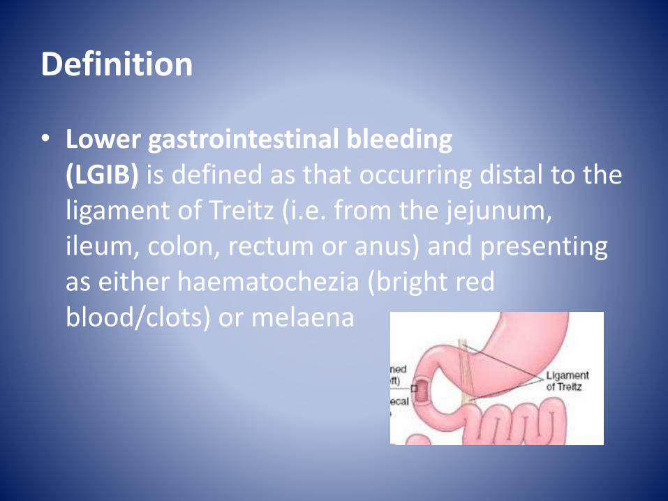
bull Lower gastrointestinal bleeding (LGIB) is defined as that occurring distal to the ligament of Treitz (ie from the jejunum ileum colon rectum or anus) and presenting as either haematochezia (bright red bloodclots) or melaena
Epidemiology
bull The incidence of LGIB is only one-fifth that of the upper gastrointestinal tract and is estimated to be ~24 per 100 000 adults per year
bull Male and older patients tend to suffer from more severe LGIB
Risk factors
bull medications (eg NSAID warfarin)
bull recent colonoscopy with polypectomy(postpolypectomy bleeding)
bull prior abdominalpelvic radiation (radiation proctitiscolitis)
bull prior operation
bull history of alcoholism or chronic liver disease
bull history of abdominal aortic anuerysm with or without surgical repair (aortoenteric fistula)
Causes
bull Diverticular diseasebull enterocolitis
ndash infectivendash Crohnrsquos diseasendash Ulcerative colitis ndash Ischemic colitis
bull vascular malformationndash vascular ectasiandash Angiodysplasiandash arteriovenous malformation (AVM)
bull polypbull tumourbull Vasculitidesbull Portal hypertensive enteropathy or colopathybull Meckel diverticulumbull ulcerbull Aorto-enteric fistula bull Anal fissure bull Haemorrhoidsbull Perianal fistula
Aetiology
bull Although LGIB can occur at any age specific disease processes are distinctive for different age groups and familiarity with this can help tailor the diagnostic workup
-adolescents and young adults inflammatory bowel diseases polyps Meckelrsquos diverticulum
-up to 60 years diverticula inflammatory bowel diseases malignancy
-older than 60 years arteriovenousmalformations diverticula malignancy
Clinical presentation
bull Acute bleeding is defined as bleeding of lt3 days duration resulting in instability of vital signs anaemia andor the need for blood transfusion
bull Chronic bleeding is defined as slow blood loss over a period of several days or longer presenting with symptoms of occult faecal blood intermittent melaena or scant hematochezia
bull LGIB usually is chronic and the bleeding ceases spontaneously (80)
Diverticular disease
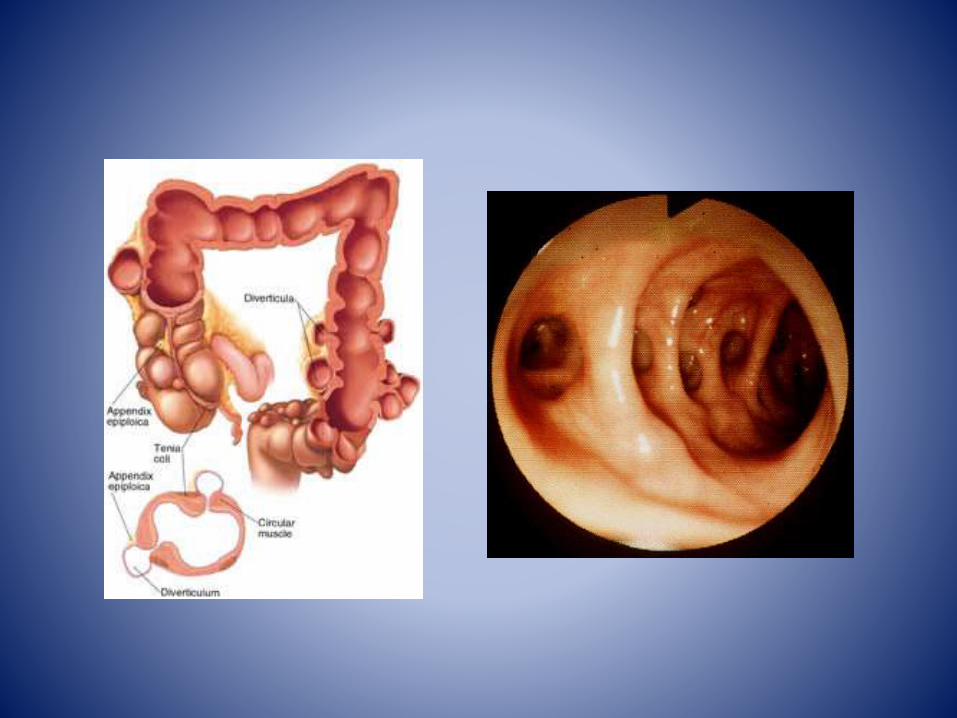
bull Out-pouchings of bowel result in blind-ended diverticulae in communication with the lumen of the bowel
bull They most commonly occur within the sigmoid colon although they may be present throughout the bowel
Clinical presentation
bull The vast majority of people with diverticulosisare asymptomatic
bull Patients complain of intermittent left sided abdominal pain and frequent constipation Symptomatic presenting features of diverticular disease (ie presentation of complicated diverticulosis) includes
-diverticulitis
-GIT hemorrhage
Crohnrsquos disease
bull idiopathic inflammatory bowel disease characterised by widespread GIT involvement typically with skip lesions thereby its synonym regional enteritis and frequently systemic involvement
Clinical presentation
bull Clinical presentation is typically with chronic diarrhoea and recurrent abdominal pain
bull Alternatively patients may present with one of the many complications or extraintestinalmanifestations
-skin hellip
-joints hellip
-eyes hellip
-liver and biliary system hellip
Ulcerative colitis
bull inflammatory bowel disease which predominantly affects the colon but also has extraintestinal manifestations
bull Clinically patients have chronic diarrhoea(sometimes bloody) associated with tenesmus pain and fever
Ischaemic colitis
bull inflammation of the colon secondary to vascular insufficiency and ischaemia
bull It sometimes considered under the same spectrum of intestinal ischeamia
bull The severity and consequences of the disease are highly variable
Clinical presentation
bull Presenting symptoms include abdominal pain and bloody
bull Tenderness may be present particularly of the left side of the abdomen
bull In severe cases where necrosis and perforation have occurred the signs and symptoms are those of peritonitis
Vasculitis
bull generalised inflammation of vessels Vasculidities carry a broad range of clinical presentations and as a whole can involve almost any organ system
Pathology
bull Some vasculitides are due to direct vessel injury from an infectious agent However a large proportion show evidence of immune complex related vessel wall injury
Classification
-Takayasu arteritis
-Wegener granulomatosis
-Henoch-Schoumlnlein purpura
-Churg-Strauss syndrome
Primary vasculitides -infection related
vasculitis
- SLE rheumatoid arthritis
-malignancy related vasculitis
Secondary vasculitides
Portal hypertensive gastropathy enteropathy colopathy
bull In portal hypertension chronic portal venous congestion leads to dilatation and ectasia of the submucosal vessels in the stomach (portal hypertensive gastropathy) small bowel (portal hypertensive enteropathy) and or large bowel (portal hypertensive colopathy)
bull This may result in upper or lower gastrointestinal bleeding even in the absence of varices
bull The bleeding may be acute or chronic but is most commonly chronic low-grade GI blood loss associated with an iron-deficiency anaemia
Fluoroscopy
bull Barium studies may show thickening of the mucosal folds and nodular filling defects
CT
there may be bowel wall
thickening and hyperaemia
which can mimic
enterocolitis
Meckel diverticulum
bull a type of congenital intestinal diverticulumthat occurs around the distal ileum
bull It is considered the most common structural congenital anomaly of the gastrointestinal tract
Clinical presentation
bull A large proportion of individuals remain asymptomatic although up to a third of them may experience clinical symptoms
bull Clinical presentation includes-pain-malaenahaematochezia-Small bowel obstruction-Intussuscption-volvulus-perforation-Littre hernia
Angiodysplasia
bull most common vascular lesion of the gastrointestinal tract after diverticulosis and this condition may be asymptomatic or it may cause gastrointestinal (GI) bleedingThe vessel walls are thin with little or no smooth muscle and the vessels are ectatic and thin
bull 77of angiodysplasias are located in the cecum and ascending colon
bull 15 are located in the jejunum and ileum
bull 8 is distributed throughout the alimentary tract
Clinical presentation
bull maroon-colored stool melena or hematochezia bull Bleeding is usually low grade but it can be
massive in approximately 15 of patients bull In 20-25 of bleeding episodes only tarry stools
are passedbull Iron deficiency anemia and stools that are
intermittently positive for occult blood can be the only manifestations of angiodysplasia in 10-15 of patients
bull Bleeding stops spontaneously in greater than 90 of cases but is often recurrent
Cancer colon
bull Most cases of colon cancer begin as small noncancerous (benign) clumps of cells called adenomatous polyps Over time some of these polyps become colon cancers
Clinical presentation
bull Bleeding per rectum
bull Alternating bowel habits
bull Discharge
bull Tenesmus
bull Intestinal obstruction
bull Mass
bull Systemic manifestations
polyps
bull Inflammatory
bull Hamartomatous
bull Neoplastic
bull Hyperplastic
Neoplastic polyps
Tubular
bull gt 85
bull Male
bull Multiple
bull Pedunculated
bull Malignancy 5
Villous
bull 15
bull Female
bull Single
bull Sessile
bull Malignancy 30
Clinical presentation
bull Age
bull Bleeding
bull Discharge
bull Colics
Reference
bull httpradiopaediaorgarticleslower-gastrointestinal-bleeding1 Ghassemi KA Jensen DM Lower GI bleeding epidemiology and management Curr
Gastroenterol Rep 201315 (7) 333 doi101007s11894-013-0333-5 - Free text at pubmed - Pubmed citation
2 Raphaeli T Menon R Current treatment of lower gastrointestinal hemorrhage ClinColon Rectal Surg 201225 (04) 219-27 doi101055s-0032-1329393 - Free text at pubmed - Pubmed citation
3 Jang BI Lower Gastrointestinal Bleeding Is Urgent Colonoscopy Necessary for All Hematochezia Clin Endosc 201346 (5) 476-479doi105946ce2013465476 -Free text at pubmed - Pubmed citation
4 Mariani G Pauwels EK AlSharif A et-al Radionuclide evaluation of the lower gastrointestinal tract J Nucl Med 200849 (5) 776-87doi102967jnumed107040113 - Pubmed citation
5 Geffroy Y Rodallec MH Boulay-Coletta I et-al Multidetector CT angiography in acute gastrointestinal bleeding why when and how Radiographics 201131 (3) E35-46 doi101148rg313105206 - Pubmed citation
Definition
bull Lower gastrointestinal bleeding (LGIB) is defined as that occurring distal to the ligament of Treitz (ie from the jejunum ileum colon rectum or anus) and presenting as either haematochezia (bright red bloodclots) or melaena
Epidemiology
bull The incidence of LGIB is only one-fifth that of the upper gastrointestinal tract and is estimated to be ~24 per 100 000 adults per year
bull Male and older patients tend to suffer from more severe LGIB
Risk factors
bull medications (eg NSAID warfarin)
bull recent colonoscopy with polypectomy(postpolypectomy bleeding)
bull prior abdominalpelvic radiation (radiation proctitiscolitis)
bull prior operation
bull history of alcoholism or chronic liver disease
bull history of abdominal aortic anuerysm with or without surgical repair (aortoenteric fistula)
Causes
bull Diverticular diseasebull enterocolitis
ndash infectivendash Crohnrsquos diseasendash Ulcerative colitis ndash Ischemic colitis
bull vascular malformationndash vascular ectasiandash Angiodysplasiandash arteriovenous malformation (AVM)
bull polypbull tumourbull Vasculitidesbull Portal hypertensive enteropathy or colopathybull Meckel diverticulumbull ulcerbull Aorto-enteric fistula bull Anal fissure bull Haemorrhoidsbull Perianal fistula
Aetiology
bull Although LGIB can occur at any age specific disease processes are distinctive for different age groups and familiarity with this can help tailor the diagnostic workup
-adolescents and young adults inflammatory bowel diseases polyps Meckelrsquos diverticulum
-up to 60 years diverticula inflammatory bowel diseases malignancy
-older than 60 years arteriovenousmalformations diverticula malignancy
Clinical presentation
bull Acute bleeding is defined as bleeding of lt3 days duration resulting in instability of vital signs anaemia andor the need for blood transfusion
bull Chronic bleeding is defined as slow blood loss over a period of several days or longer presenting with symptoms of occult faecal blood intermittent melaena or scant hematochezia
bull LGIB usually is chronic and the bleeding ceases spontaneously (80)
Diverticular disease
bull Out-pouchings of bowel result in blind-ended diverticulae in communication with the lumen of the bowel
bull They most commonly occur within the sigmoid colon although they may be present throughout the bowel
Clinical presentation
bull The vast majority of people with diverticulosisare asymptomatic
bull Patients complain of intermittent left sided abdominal pain and frequent constipation Symptomatic presenting features of diverticular disease (ie presentation of complicated diverticulosis) includes
-diverticulitis
-GIT hemorrhage
Crohnrsquos disease
bull idiopathic inflammatory bowel disease characterised by widespread GIT involvement typically with skip lesions thereby its synonym regional enteritis and frequently systemic involvement
Clinical presentation
bull Clinical presentation is typically with chronic diarrhoea and recurrent abdominal pain
bull Alternatively patients may present with one of the many complications or extraintestinalmanifestations
-skin hellip
-joints hellip
-eyes hellip
-liver and biliary system hellip
Ulcerative colitis
bull inflammatory bowel disease which predominantly affects the colon but also has extraintestinal manifestations
bull Clinically patients have chronic diarrhoea(sometimes bloody) associated with tenesmus pain and fever
Ischaemic colitis
bull inflammation of the colon secondary to vascular insufficiency and ischaemia
bull It sometimes considered under the same spectrum of intestinal ischeamia
bull The severity and consequences of the disease are highly variable
Clinical presentation
bull Presenting symptoms include abdominal pain and bloody
bull Tenderness may be present particularly of the left side of the abdomen
bull In severe cases where necrosis and perforation have occurred the signs and symptoms are those of peritonitis
Vasculitis
bull generalised inflammation of vessels Vasculidities carry a broad range of clinical presentations and as a whole can involve almost any organ system
Pathology
bull Some vasculitides are due to direct vessel injury from an infectious agent However a large proportion show evidence of immune complex related vessel wall injury
Classification
-Takayasu arteritis
-Wegener granulomatosis
-Henoch-Schoumlnlein purpura
-Churg-Strauss syndrome
Primary vasculitides -infection related
vasculitis
- SLE rheumatoid arthritis
-malignancy related vasculitis
Secondary vasculitides
Portal hypertensive gastropathy enteropathy colopathy
bull In portal hypertension chronic portal venous congestion leads to dilatation and ectasia of the submucosal vessels in the stomach (portal hypertensive gastropathy) small bowel (portal hypertensive enteropathy) and or large bowel (portal hypertensive colopathy)
bull This may result in upper or lower gastrointestinal bleeding even in the absence of varices
bull The bleeding may be acute or chronic but is most commonly chronic low-grade GI blood loss associated with an iron-deficiency anaemia
Fluoroscopy
bull Barium studies may show thickening of the mucosal folds and nodular filling defects
CT
there may be bowel wall
thickening and hyperaemia
which can mimic
enterocolitis
Meckel diverticulum
bull a type of congenital intestinal diverticulumthat occurs around the distal ileum
bull It is considered the most common structural congenital anomaly of the gastrointestinal tract
Clinical presentation
bull A large proportion of individuals remain asymptomatic although up to a third of them may experience clinical symptoms
bull Clinical presentation includes-pain-malaenahaematochezia-Small bowel obstruction-Intussuscption-volvulus-perforation-Littre hernia
Angiodysplasia
bull most common vascular lesion of the gastrointestinal tract after diverticulosis and this condition may be asymptomatic or it may cause gastrointestinal (GI) bleedingThe vessel walls are thin with little or no smooth muscle and the vessels are ectatic and thin
bull 77of angiodysplasias are located in the cecum and ascending colon
bull 15 are located in the jejunum and ileum
bull 8 is distributed throughout the alimentary tract
Clinical presentation
bull maroon-colored stool melena or hematochezia bull Bleeding is usually low grade but it can be
massive in approximately 15 of patients bull In 20-25 of bleeding episodes only tarry stools
are passedbull Iron deficiency anemia and stools that are
intermittently positive for occult blood can be the only manifestations of angiodysplasia in 10-15 of patients
bull Bleeding stops spontaneously in greater than 90 of cases but is often recurrent
Cancer colon
bull Most cases of colon cancer begin as small noncancerous (benign) clumps of cells called adenomatous polyps Over time some of these polyps become colon cancers
Clinical presentation
bull Bleeding per rectum
bull Alternating bowel habits
bull Discharge
bull Tenesmus
bull Intestinal obstruction
bull Mass
bull Systemic manifestations
polyps
bull Inflammatory
bull Hamartomatous
bull Neoplastic
bull Hyperplastic
Neoplastic polyps
Tubular
bull gt 85
bull Male
bull Multiple
bull Pedunculated
bull Malignancy 5
Villous
bull 15
bull Female
bull Single
bull Sessile
bull Malignancy 30
Clinical presentation
bull Age
bull Bleeding
bull Discharge
bull Colics
Reference
bull httpradiopaediaorgarticleslower-gastrointestinal-bleeding1 Ghassemi KA Jensen DM Lower GI bleeding epidemiology and management Curr
Gastroenterol Rep 201315 (7) 333 doi101007s11894-013-0333-5 - Free text at pubmed - Pubmed citation
2 Raphaeli T Menon R Current treatment of lower gastrointestinal hemorrhage ClinColon Rectal Surg 201225 (04) 219-27 doi101055s-0032-1329393 - Free text at pubmed - Pubmed citation
3 Jang BI Lower Gastrointestinal Bleeding Is Urgent Colonoscopy Necessary for All Hematochezia Clin Endosc 201346 (5) 476-479doi105946ce2013465476 -Free text at pubmed - Pubmed citation
4 Mariani G Pauwels EK AlSharif A et-al Radionuclide evaluation of the lower gastrointestinal tract J Nucl Med 200849 (5) 776-87doi102967jnumed107040113 - Pubmed citation
5 Geffroy Y Rodallec MH Boulay-Coletta I et-al Multidetector CT angiography in acute gastrointestinal bleeding why when and how Radiographics 201131 (3) E35-46 doi101148rg313105206 - Pubmed citation
Clinical presentation
bull Acute bleeding is defined as bleeding of lt3 days duration resulting in instability of vital signs anaemia andor the need for blood transfusion
bull Chronic bleeding is defined as slow blood loss over a period of several days or longer presenting with symptoms of occult faecal blood intermittent melaena or scant hematochezia
bull LGIB usually is chronic and the bleeding ceases spontaneously (80)
Diverticular disease
bull Out-pouchings of bowel result in blind-ended diverticulae in communication with the lumen of the bowel
bull They most commonly occur within the sigmoid colon although they may be present throughout the bowel
Clinical presentation
bull The vast majority of people with diverticulosisare asymptomatic
bull Patients complain of intermittent left sided abdominal pain and frequent constipation Symptomatic presenting features of diverticular disease (ie presentation of complicated diverticulosis) includes
-diverticulitis
-GIT hemorrhage
Crohnrsquos disease
bull idiopathic inflammatory bowel disease characterised by widespread GIT involvement typically with skip lesions thereby its synonym regional enteritis and frequently systemic involvement
Clinical presentation
bull Clinical presentation is typically with chronic diarrhoea and recurrent abdominal pain
bull Alternatively patients may present with one of the many complications or extraintestinalmanifestations
-skin hellip
-joints hellip
-eyes hellip
-liver and biliary system hellip
Ulcerative colitis
bull inflammatory bowel disease which predominantly affects the colon but also has extraintestinal manifestations
bull Clinically patients have chronic diarrhoea(sometimes bloody) associated with tenesmus pain and fever
Ischaemic colitis
bull inflammation of the colon secondary to vascular insufficiency and ischaemia
bull It sometimes considered under the same spectrum of intestinal ischeamia
bull The severity and consequences of the disease are highly variable
Clinical presentation
bull Presenting symptoms include abdominal pain and bloody
bull Tenderness may be present particularly of the left side of the abdomen
bull In severe cases where necrosis and perforation have occurred the signs and symptoms are those of peritonitis
Vasculitis
bull generalised inflammation of vessels Vasculidities carry a broad range of clinical presentations and as a whole can involve almost any organ system
Pathology
bull Some vasculitides are due to direct vessel injury from an infectious agent However a large proportion show evidence of immune complex related vessel wall injury
Classification
-Takayasu arteritis
-Wegener granulomatosis
-Henoch-Schoumlnlein purpura
-Churg-Strauss syndrome
Primary vasculitides -infection related
vasculitis
- SLE rheumatoid arthritis
-malignancy related vasculitis
Secondary vasculitides
Portal hypertensive gastropathy enteropathy colopathy
bull In portal hypertension chronic portal venous congestion leads to dilatation and ectasia of the submucosal vessels in the stomach (portal hypertensive gastropathy) small bowel (portal hypertensive enteropathy) and or large bowel (portal hypertensive colopathy)
bull This may result in upper or lower gastrointestinal bleeding even in the absence of varices
bull The bleeding may be acute or chronic but is most commonly chronic low-grade GI blood loss associated with an iron-deficiency anaemia
Fluoroscopy
bull Barium studies may show thickening of the mucosal folds and nodular filling defects
CT
there may be bowel wall
thickening and hyperaemia
which can mimic
enterocolitis
Meckel diverticulum
bull a type of congenital intestinal diverticulumthat occurs around the distal ileum
bull It is considered the most common structural congenital anomaly of the gastrointestinal tract
Clinical presentation
bull A large proportion of individuals remain asymptomatic although up to a third of them may experience clinical symptoms
bull Clinical presentation includes-pain-malaenahaematochezia-Small bowel obstruction-Intussuscption-volvulus-perforation-Littre hernia
Angiodysplasia
bull most common vascular lesion of the gastrointestinal tract after diverticulosis and this condition may be asymptomatic or it may cause gastrointestinal (GI) bleedingThe vessel walls are thin with little or no smooth muscle and the vessels are ectatic and thin
bull 77of angiodysplasias are located in the cecum and ascending colon
bull 15 are located in the jejunum and ileum
bull 8 is distributed throughout the alimentary tract
Clinical presentation
bull maroon-colored stool melena or hematochezia bull Bleeding is usually low grade but it can be
massive in approximately 15 of patients bull In 20-25 of bleeding episodes only tarry stools
are passedbull Iron deficiency anemia and stools that are
intermittently positive for occult blood can be the only manifestations of angiodysplasia in 10-15 of patients
bull Bleeding stops spontaneously in greater than 90 of cases but is often recurrent
Cancer colon
bull Most cases of colon cancer begin as small noncancerous (benign) clumps of cells called adenomatous polyps Over time some of these polyps become colon cancers
Clinical presentation
bull Bleeding per rectum
bull Alternating bowel habits
bull Discharge
bull Tenesmus
bull Intestinal obstruction
bull Mass
bull Systemic manifestations
polyps
bull Inflammatory
bull Hamartomatous
bull Neoplastic
bull Hyperplastic
Neoplastic polyps
Tubular
bull gt 85
bull Male
bull Multiple
bull Pedunculated
bull Malignancy 5
Villous
bull 15
bull Female
bull Single
bull Sessile
bull Malignancy 30
Clinical presentation
bull Age
bull Bleeding
bull Discharge
bull Colics
Reference
bull httpradiopaediaorgarticleslower-gastrointestinal-bleeding1 Ghassemi KA Jensen DM Lower GI bleeding epidemiology and management Curr
Gastroenterol Rep 201315 (7) 333 doi101007s11894-013-0333-5 - Free text at pubmed - Pubmed citation
2 Raphaeli T Menon R Current treatment of lower gastrointestinal hemorrhage ClinColon Rectal Surg 201225 (04) 219-27 doi101055s-0032-1329393 - Free text at pubmed - Pubmed citation
3 Jang BI Lower Gastrointestinal Bleeding Is Urgent Colonoscopy Necessary for All Hematochezia Clin Endosc 201346 (5) 476-479doi105946ce2013465476 -Free text at pubmed - Pubmed citation
4 Mariani G Pauwels EK AlSharif A et-al Radionuclide evaluation of the lower gastrointestinal tract J Nucl Med 200849 (5) 776-87doi102967jnumed107040113 - Pubmed citation
5 Geffroy Y Rodallec MH Boulay-Coletta I et-al Multidetector CT angiography in acute gastrointestinal bleeding why when and how Radiographics 201131 (3) E35-46 doi101148rg313105206 - Pubmed citation
Clinical presentation
bull The vast majority of people with diverticulosisare asymptomatic
bull Patients complain of intermittent left sided abdominal pain and frequent constipation Symptomatic presenting features of diverticular disease (ie presentation of complicated diverticulosis) includes
-diverticulitis
-GIT hemorrhage
Crohnrsquos disease
bull idiopathic inflammatory bowel disease characterised by widespread GIT involvement typically with skip lesions thereby its synonym regional enteritis and frequently systemic involvement
Clinical presentation
bull Clinical presentation is typically with chronic diarrhoea and recurrent abdominal pain
bull Alternatively patients may present with one of the many complications or extraintestinalmanifestations
-skin hellip
-joints hellip
-eyes hellip
-liver and biliary system hellip
Ulcerative colitis
bull inflammatory bowel disease which predominantly affects the colon but also has extraintestinal manifestations
bull Clinically patients have chronic diarrhoea(sometimes bloody) associated with tenesmus pain and fever
Ischaemic colitis
bull inflammation of the colon secondary to vascular insufficiency and ischaemia
bull It sometimes considered under the same spectrum of intestinal ischeamia
bull The severity and consequences of the disease are highly variable
Clinical presentation
bull Presenting symptoms include abdominal pain and bloody
bull Tenderness may be present particularly of the left side of the abdomen
bull In severe cases where necrosis and perforation have occurred the signs and symptoms are those of peritonitis
Vasculitis
bull generalised inflammation of vessels Vasculidities carry a broad range of clinical presentations and as a whole can involve almost any organ system
Pathology
bull Some vasculitides are due to direct vessel injury from an infectious agent However a large proportion show evidence of immune complex related vessel wall injury
Classification
-Takayasu arteritis
-Wegener granulomatosis
-Henoch-Schoumlnlein purpura
-Churg-Strauss syndrome
Primary vasculitides -infection related
vasculitis
- SLE rheumatoid arthritis
-malignancy related vasculitis
Secondary vasculitides
Portal hypertensive gastropathy enteropathy colopathy
bull In portal hypertension chronic portal venous congestion leads to dilatation and ectasia of the submucosal vessels in the stomach (portal hypertensive gastropathy) small bowel (portal hypertensive enteropathy) and or large bowel (portal hypertensive colopathy)
bull This may result in upper or lower gastrointestinal bleeding even in the absence of varices
bull The bleeding may be acute or chronic but is most commonly chronic low-grade GI blood loss associated with an iron-deficiency anaemia
Fluoroscopy
bull Barium studies may show thickening of the mucosal folds and nodular filling defects
CT
there may be bowel wall
thickening and hyperaemia
which can mimic
enterocolitis
Meckel diverticulum
bull a type of congenital intestinal diverticulumthat occurs around the distal ileum
bull It is considered the most common structural congenital anomaly of the gastrointestinal tract
Clinical presentation
bull A large proportion of individuals remain asymptomatic although up to a third of them may experience clinical symptoms
bull Clinical presentation includes-pain-malaenahaematochezia-Small bowel obstruction-Intussuscption-volvulus-perforation-Littre hernia
Angiodysplasia
bull most common vascular lesion of the gastrointestinal tract after diverticulosis and this condition may be asymptomatic or it may cause gastrointestinal (GI) bleedingThe vessel walls are thin with little or no smooth muscle and the vessels are ectatic and thin
bull 77of angiodysplasias are located in the cecum and ascending colon
bull 15 are located in the jejunum and ileum
bull 8 is distributed throughout the alimentary tract
Clinical presentation
bull maroon-colored stool melena or hematochezia bull Bleeding is usually low grade but it can be
massive in approximately 15 of patients bull In 20-25 of bleeding episodes only tarry stools
are passedbull Iron deficiency anemia and stools that are
intermittently positive for occult blood can be the only manifestations of angiodysplasia in 10-15 of patients
bull Bleeding stops spontaneously in greater than 90 of cases but is often recurrent
Cancer colon
bull Most cases of colon cancer begin as small noncancerous (benign) clumps of cells called adenomatous polyps Over time some of these polyps become colon cancers
Clinical presentation
bull Bleeding per rectum
bull Alternating bowel habits
bull Discharge
bull Tenesmus
bull Intestinal obstruction
bull Mass
bull Systemic manifestations
polyps
bull Inflammatory
bull Hamartomatous
bull Neoplastic
bull Hyperplastic
Neoplastic polyps
Tubular
bull gt 85
bull Male
bull Multiple
bull Pedunculated
bull Malignancy 5
Villous
bull 15
bull Female
bull Single
bull Sessile
bull Malignancy 30
Clinical presentation
bull Age
bull Bleeding
bull Discharge
bull Colics
Reference
bull httpradiopaediaorgarticleslower-gastrointestinal-bleeding1 Ghassemi KA Jensen DM Lower GI bleeding epidemiology and management Curr
Gastroenterol Rep 201315 (7) 333 doi101007s11894-013-0333-5 - Free text at pubmed - Pubmed citation
2 Raphaeli T Menon R Current treatment of lower gastrointestinal hemorrhage ClinColon Rectal Surg 201225 (04) 219-27 doi101055s-0032-1329393 - Free text at pubmed - Pubmed citation
3 Jang BI Lower Gastrointestinal Bleeding Is Urgent Colonoscopy Necessary for All Hematochezia Clin Endosc 201346 (5) 476-479doi105946ce2013465476 -Free text at pubmed - Pubmed citation
4 Mariani G Pauwels EK AlSharif A et-al Radionuclide evaluation of the lower gastrointestinal tract J Nucl Med 200849 (5) 776-87doi102967jnumed107040113 - Pubmed citation
5 Geffroy Y Rodallec MH Boulay-Coletta I et-al Multidetector CT angiography in acute gastrointestinal bleeding why when and how Radiographics 201131 (3) E35-46 doi101148rg313105206 - Pubmed citation
Clinical presentation
bull Clinical presentation is typically with chronic diarrhoea and recurrent abdominal pain
bull Alternatively patients may present with one of the many complications or extraintestinalmanifestations
-skin hellip
-joints hellip
-eyes hellip
-liver and biliary system hellip
Ulcerative colitis
bull inflammatory bowel disease which predominantly affects the colon but also has extraintestinal manifestations
bull Clinically patients have chronic diarrhoea(sometimes bloody) associated with tenesmus pain and fever
Ischaemic colitis
bull inflammation of the colon secondary to vascular insufficiency and ischaemia
bull It sometimes considered under the same spectrum of intestinal ischeamia
bull The severity and consequences of the disease are highly variable
Clinical presentation
bull Presenting symptoms include abdominal pain and bloody
bull Tenderness may be present particularly of the left side of the abdomen
bull In severe cases where necrosis and perforation have occurred the signs and symptoms are those of peritonitis
Vasculitis
bull generalised inflammation of vessels Vasculidities carry a broad range of clinical presentations and as a whole can involve almost any organ system
Pathology
bull Some vasculitides are due to direct vessel injury from an infectious agent However a large proportion show evidence of immune complex related vessel wall injury
Classification
-Takayasu arteritis
-Wegener granulomatosis
-Henoch-Schoumlnlein purpura
-Churg-Strauss syndrome
Primary vasculitides -infection related
vasculitis
- SLE rheumatoid arthritis
-malignancy related vasculitis
Secondary vasculitides
Portal hypertensive gastropathy enteropathy colopathy
bull In portal hypertension chronic portal venous congestion leads to dilatation and ectasia of the submucosal vessels in the stomach (portal hypertensive gastropathy) small bowel (portal hypertensive enteropathy) and or large bowel (portal hypertensive colopathy)
bull This may result in upper or lower gastrointestinal bleeding even in the absence of varices
bull The bleeding may be acute or chronic but is most commonly chronic low-grade GI blood loss associated with an iron-deficiency anaemia
Fluoroscopy
bull Barium studies may show thickening of the mucosal folds and nodular filling defects
CT
there may be bowel wall
thickening and hyperaemia
which can mimic
enterocolitis
Meckel diverticulum
bull a type of congenital intestinal diverticulumthat occurs around the distal ileum
bull It is considered the most common structural congenital anomaly of the gastrointestinal tract
Clinical presentation
bull A large proportion of individuals remain asymptomatic although up to a third of them may experience clinical symptoms
bull Clinical presentation includes-pain-malaenahaematochezia-Small bowel obstruction-Intussuscption-volvulus-perforation-Littre hernia
Angiodysplasia
bull most common vascular lesion of the gastrointestinal tract after diverticulosis and this condition may be asymptomatic or it may cause gastrointestinal (GI) bleedingThe vessel walls are thin with little or no smooth muscle and the vessels are ectatic and thin
bull 77of angiodysplasias are located in the cecum and ascending colon
bull 15 are located in the jejunum and ileum
bull 8 is distributed throughout the alimentary tract
Clinical presentation
bull maroon-colored stool melena or hematochezia bull Bleeding is usually low grade but it can be
massive in approximately 15 of patients bull In 20-25 of bleeding episodes only tarry stools
are passedbull Iron deficiency anemia and stools that are
intermittently positive for occult blood can be the only manifestations of angiodysplasia in 10-15 of patients
bull Bleeding stops spontaneously in greater than 90 of cases but is often recurrent
Cancer colon
bull Most cases of colon cancer begin as small noncancerous (benign) clumps of cells called adenomatous polyps Over time some of these polyps become colon cancers
Clinical presentation
bull Bleeding per rectum
bull Alternating bowel habits
bull Discharge
bull Tenesmus
bull Intestinal obstruction
bull Mass
bull Systemic manifestations
polyps
bull Inflammatory
bull Hamartomatous
bull Neoplastic
bull Hyperplastic
Neoplastic polyps
Tubular
bull gt 85
bull Male
bull Multiple
bull Pedunculated
bull Malignancy 5
Villous
bull 15
bull Female
bull Single
bull Sessile
bull Malignancy 30
Clinical presentation
bull Age
bull Bleeding
bull Discharge
bull Colics
Reference
bull httpradiopaediaorgarticleslower-gastrointestinal-bleeding1 Ghassemi KA Jensen DM Lower GI bleeding epidemiology and management Curr
Gastroenterol Rep 201315 (7) 333 doi101007s11894-013-0333-5 - Free text at pubmed - Pubmed citation
2 Raphaeli T Menon R Current treatment of lower gastrointestinal hemorrhage ClinColon Rectal Surg 201225 (04) 219-27 doi101055s-0032-1329393 - Free text at pubmed - Pubmed citation
3 Jang BI Lower Gastrointestinal Bleeding Is Urgent Colonoscopy Necessary for All Hematochezia Clin Endosc 201346 (5) 476-479doi105946ce2013465476 -Free text at pubmed - Pubmed citation
4 Mariani G Pauwels EK AlSharif A et-al Radionuclide evaluation of the lower gastrointestinal tract J Nucl Med 200849 (5) 776-87doi102967jnumed107040113 - Pubmed citation
5 Geffroy Y Rodallec MH Boulay-Coletta I et-al Multidetector CT angiography in acute gastrointestinal bleeding why when and how Radiographics 201131 (3) E35-46 doi101148rg313105206 - Pubmed citation
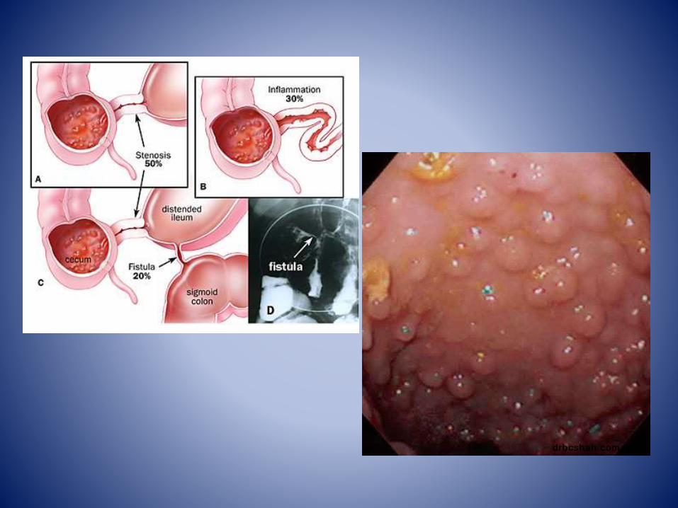
Clinical presentation
bull Presenting symptoms include abdominal pain and bloody
bull Tenderness may be present particularly of the left side of the abdomen
bull In severe cases where necrosis and perforation have occurred the signs and symptoms are those of peritonitis
Vasculitis
bull generalised inflammation of vessels Vasculidities carry a broad range of clinical presentations and as a whole can involve almost any organ system
Pathology
bull Some vasculitides are due to direct vessel injury from an infectious agent However a large proportion show evidence of immune complex related vessel wall injury
Classification
-Takayasu arteritis
-Wegener granulomatosis
-Henoch-Schoumlnlein purpura
-Churg-Strauss syndrome
Primary vasculitides -infection related
vasculitis
- SLE rheumatoid arthritis
-malignancy related vasculitis
Secondary vasculitides
Portal hypertensive gastropathy enteropathy colopathy
bull In portal hypertension chronic portal venous congestion leads to dilatation and ectasia of the submucosal vessels in the stomach (portal hypertensive gastropathy) small bowel (portal hypertensive enteropathy) and or large bowel (portal hypertensive colopathy)
bull This may result in upper or lower gastrointestinal bleeding even in the absence of varices
bull The bleeding may be acute or chronic but is most commonly chronic low-grade GI blood loss associated with an iron-deficiency anaemia
Fluoroscopy
bull Barium studies may show thickening of the mucosal folds and nodular filling defects
CT
there may be bowel wall
thickening and hyperaemia
which can mimic
enterocolitis
Meckel diverticulum
bull a type of congenital intestinal diverticulumthat occurs around the distal ileum
bull It is considered the most common structural congenital anomaly of the gastrointestinal tract
Clinical presentation
bull A large proportion of individuals remain asymptomatic although up to a third of them may experience clinical symptoms
bull Clinical presentation includes-pain-malaenahaematochezia-Small bowel obstruction-Intussuscption-volvulus-perforation-Littre hernia
Angiodysplasia
bull most common vascular lesion of the gastrointestinal tract after diverticulosis and this condition may be asymptomatic or it may cause gastrointestinal (GI) bleedingThe vessel walls are thin with little or no smooth muscle and the vessels are ectatic and thin
bull 77of angiodysplasias are located in the cecum and ascending colon
bull 15 are located in the jejunum and ileum
bull 8 is distributed throughout the alimentary tract
Clinical presentation
bull maroon-colored stool melena or hematochezia bull Bleeding is usually low grade but it can be
massive in approximately 15 of patients bull In 20-25 of bleeding episodes only tarry stools
are passedbull Iron deficiency anemia and stools that are
intermittently positive for occult blood can be the only manifestations of angiodysplasia in 10-15 of patients
bull Bleeding stops spontaneously in greater than 90 of cases but is often recurrent
Cancer colon
bull Most cases of colon cancer begin as small noncancerous (benign) clumps of cells called adenomatous polyps Over time some of these polyps become colon cancers
Clinical presentation
bull Bleeding per rectum
bull Alternating bowel habits
bull Discharge
bull Tenesmus
bull Intestinal obstruction
bull Mass
bull Systemic manifestations
polyps
bull Inflammatory
bull Hamartomatous
bull Neoplastic
bull Hyperplastic
Neoplastic polyps
Tubular
bull gt 85
bull Male
bull Multiple
bull Pedunculated
bull Malignancy 5
Villous
bull 15
bull Female
bull Single
bull Sessile
bull Malignancy 30
Clinical presentation
bull Age
bull Bleeding
bull Discharge
bull Colics
Reference
bull httpradiopaediaorgarticleslower-gastrointestinal-bleeding1 Ghassemi KA Jensen DM Lower GI bleeding epidemiology and management Curr
Gastroenterol Rep 201315 (7) 333 doi101007s11894-013-0333-5 - Free text at pubmed - Pubmed citation
2 Raphaeli T Menon R Current treatment of lower gastrointestinal hemorrhage ClinColon Rectal Surg 201225 (04) 219-27 doi101055s-0032-1329393 - Free text at pubmed - Pubmed citation
3 Jang BI Lower Gastrointestinal Bleeding Is Urgent Colonoscopy Necessary for All Hematochezia Clin Endosc 201346 (5) 476-479doi105946ce2013465476 -Free text at pubmed - Pubmed citation
4 Mariani G Pauwels EK AlSharif A et-al Radionuclide evaluation of the lower gastrointestinal tract J Nucl Med 200849 (5) 776-87doi102967jnumed107040113 - Pubmed citation
5 Geffroy Y Rodallec MH Boulay-Coletta I et-al Multidetector CT angiography in acute gastrointestinal bleeding why when and how Radiographics 201131 (3) E35-46 doi101148rg313105206 - Pubmed citation
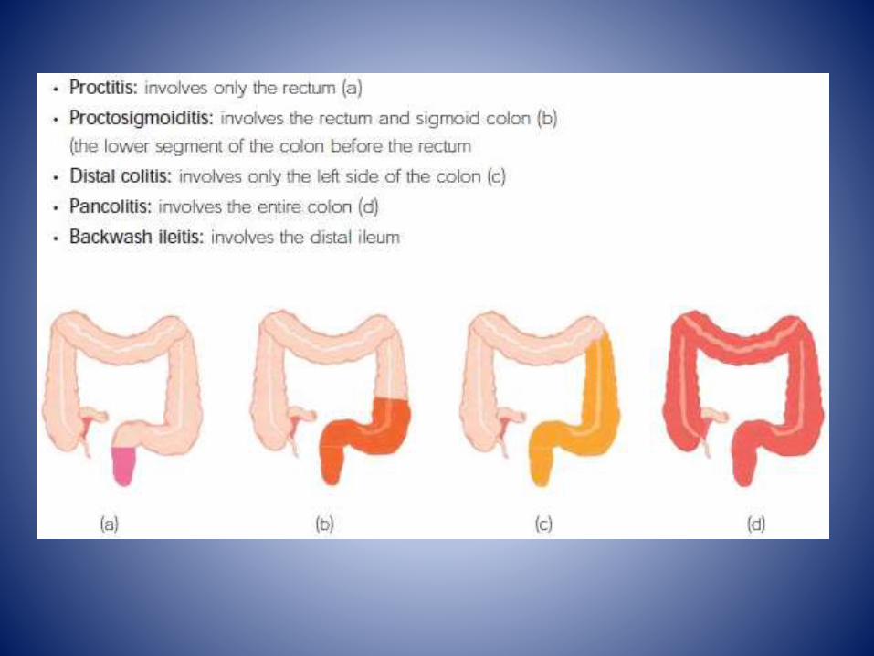
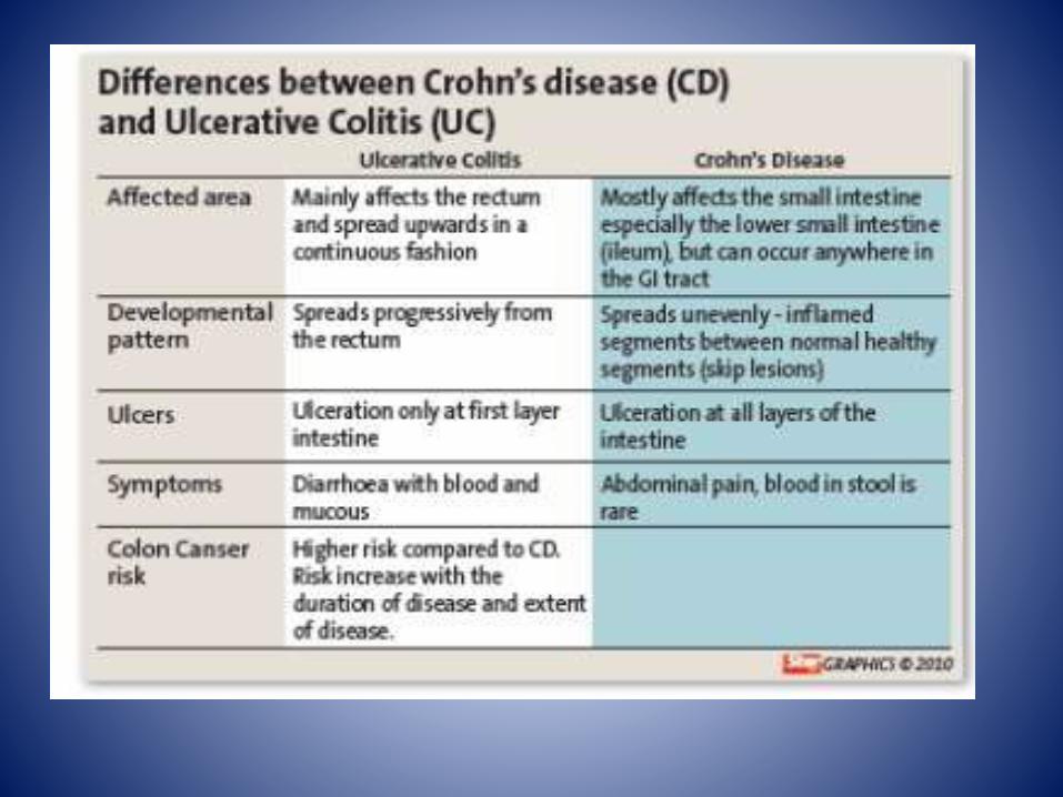
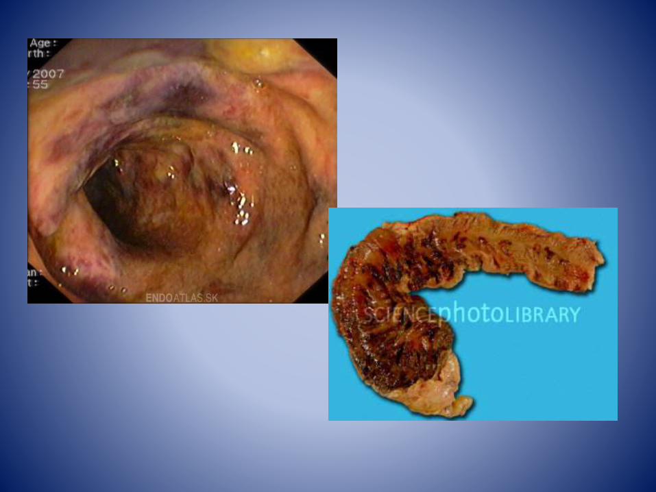
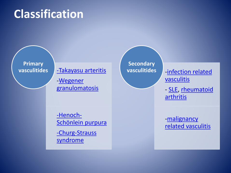
Clinical presentation
bull A large proportion of individuals remain asymptomatic although up to a third of them may experience clinical symptoms
bull Clinical presentation includes-pain-malaenahaematochezia-Small bowel obstruction-Intussuscption-volvulus-perforation-Littre hernia
Angiodysplasia
bull most common vascular lesion of the gastrointestinal tract after diverticulosis and this condition may be asymptomatic or it may cause gastrointestinal (GI) bleedingThe vessel walls are thin with little or no smooth muscle and the vessels are ectatic and thin
bull 77of angiodysplasias are located in the cecum and ascending colon
bull 15 are located in the jejunum and ileum
bull 8 is distributed throughout the alimentary tract
Clinical presentation
bull maroon-colored stool melena or hematochezia bull Bleeding is usually low grade but it can be
massive in approximately 15 of patients bull In 20-25 of bleeding episodes only tarry stools
are passedbull Iron deficiency anemia and stools that are
intermittently positive for occult blood can be the only manifestations of angiodysplasia in 10-15 of patients
bull Bleeding stops spontaneously in greater than 90 of cases but is often recurrent
Cancer colon
bull Most cases of colon cancer begin as small noncancerous (benign) clumps of cells called adenomatous polyps Over time some of these polyps become colon cancers
Clinical presentation
bull Bleeding per rectum
bull Alternating bowel habits
bull Discharge
bull Tenesmus
bull Intestinal obstruction
bull Mass
bull Systemic manifestations
polyps
bull Inflammatory
bull Hamartomatous
bull Neoplastic
bull Hyperplastic
Neoplastic polyps
Tubular
bull gt 85
bull Male
bull Multiple
bull Pedunculated
bull Malignancy 5
Villous
bull 15
bull Female
bull Single
bull Sessile
bull Malignancy 30
Clinical presentation
bull Age
bull Bleeding
bull Discharge
bull Colics
Reference
bull httpradiopaediaorgarticleslower-gastrointestinal-bleeding1 Ghassemi KA Jensen DM Lower GI bleeding epidemiology and management Curr
Gastroenterol Rep 201315 (7) 333 doi101007s11894-013-0333-5 - Free text at pubmed - Pubmed citation
2 Raphaeli T Menon R Current treatment of lower gastrointestinal hemorrhage ClinColon Rectal Surg 201225 (04) 219-27 doi101055s-0032-1329393 - Free text at pubmed - Pubmed citation
3 Jang BI Lower Gastrointestinal Bleeding Is Urgent Colonoscopy Necessary for All Hematochezia Clin Endosc 201346 (5) 476-479doi105946ce2013465476 -Free text at pubmed - Pubmed citation
4 Mariani G Pauwels EK AlSharif A et-al Radionuclide evaluation of the lower gastrointestinal tract J Nucl Med 200849 (5) 776-87doi102967jnumed107040113 - Pubmed citation
5 Geffroy Y Rodallec MH Boulay-Coletta I et-al Multidetector CT angiography in acute gastrointestinal bleeding why when and how Radiographics 201131 (3) E35-46 doi101148rg313105206 - Pubmed citation
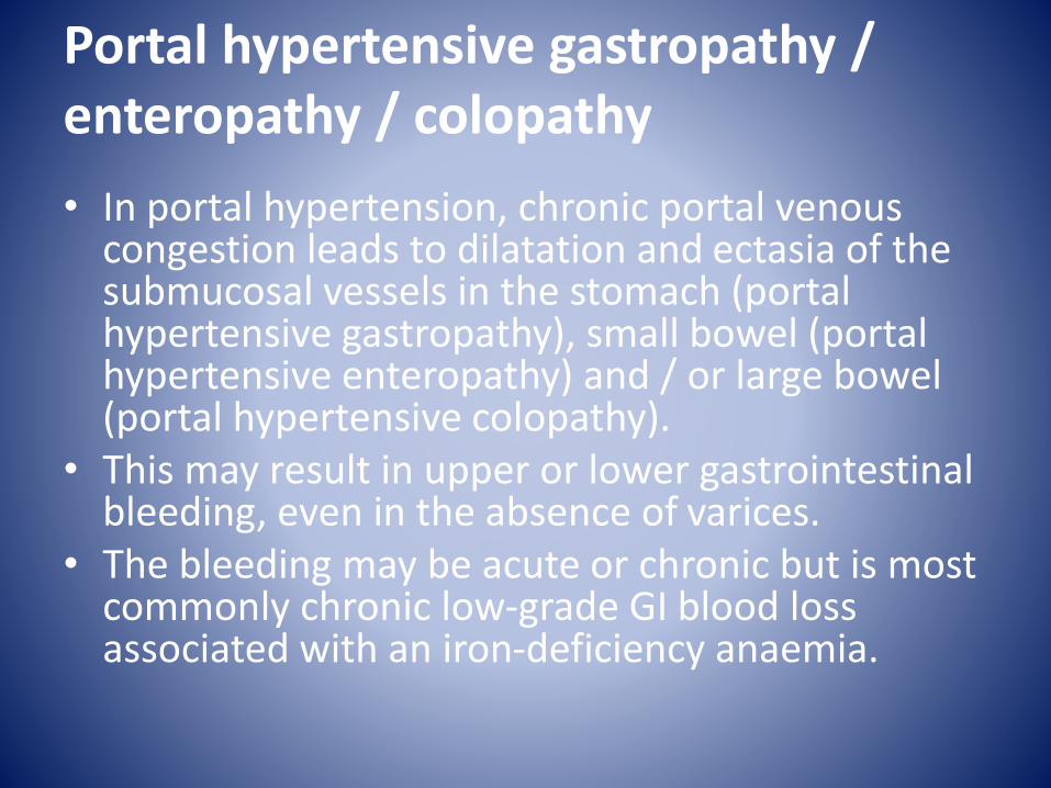
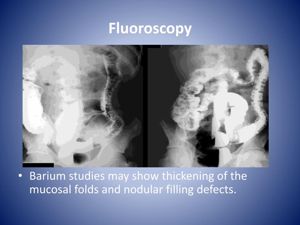
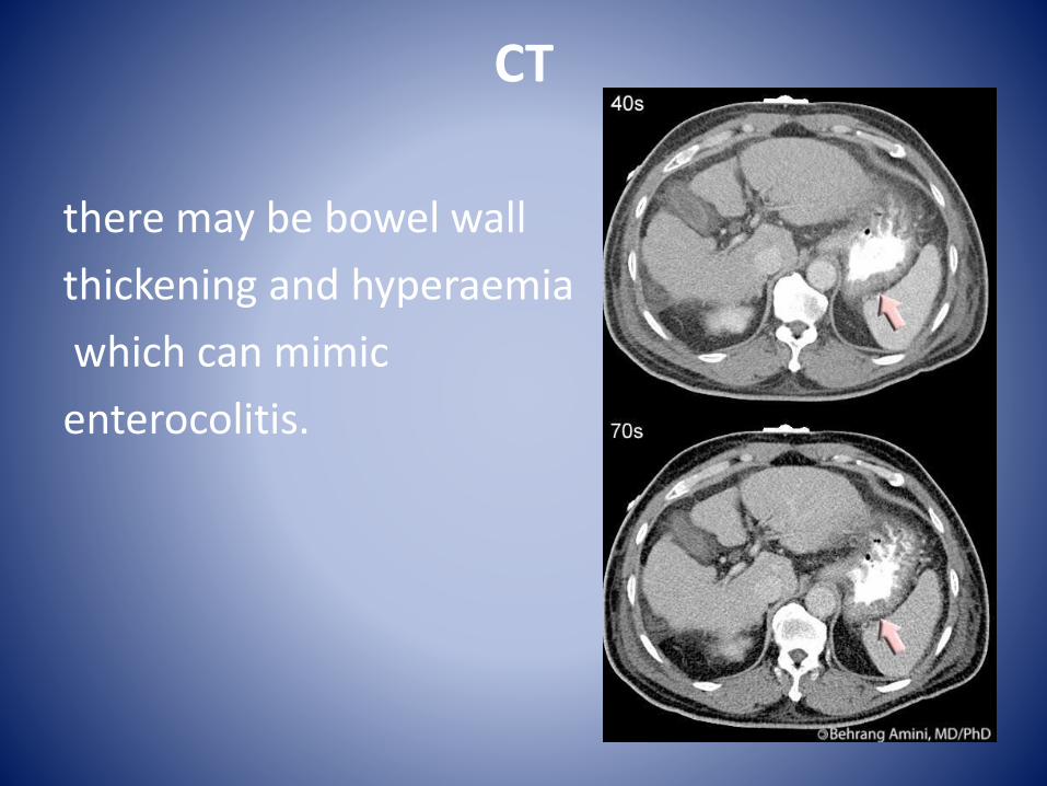
Clinical presentation
bull maroon-colored stool melena or hematochezia bull Bleeding is usually low grade but it can be
massive in approximately 15 of patients bull In 20-25 of bleeding episodes only tarry stools
are passedbull Iron deficiency anemia and stools that are
intermittently positive for occult blood can be the only manifestations of angiodysplasia in 10-15 of patients
bull Bleeding stops spontaneously in greater than 90 of cases but is often recurrent
Cancer colon
bull Most cases of colon cancer begin as small noncancerous (benign) clumps of cells called adenomatous polyps Over time some of these polyps become colon cancers
Clinical presentation
bull Bleeding per rectum
bull Alternating bowel habits
bull Discharge
bull Tenesmus
bull Intestinal obstruction
bull Mass
bull Systemic manifestations
polyps
bull Inflammatory
bull Hamartomatous
bull Neoplastic
bull Hyperplastic
Neoplastic polyps
Tubular
bull gt 85
bull Male
bull Multiple
bull Pedunculated
bull Malignancy 5
Villous
bull 15
bull Female
bull Single
bull Sessile
bull Malignancy 30
Clinical presentation
bull Age
bull Bleeding
bull Discharge
bull Colics
Reference
bull httpradiopaediaorgarticleslower-gastrointestinal-bleeding1 Ghassemi KA Jensen DM Lower GI bleeding epidemiology and management Curr
Gastroenterol Rep 201315 (7) 333 doi101007s11894-013-0333-5 - Free text at pubmed - Pubmed citation
2 Raphaeli T Menon R Current treatment of lower gastrointestinal hemorrhage ClinColon Rectal Surg 201225 (04) 219-27 doi101055s-0032-1329393 - Free text at pubmed - Pubmed citation
3 Jang BI Lower Gastrointestinal Bleeding Is Urgent Colonoscopy Necessary for All Hematochezia Clin Endosc 201346 (5) 476-479doi105946ce2013465476 -Free text at pubmed - Pubmed citation
4 Mariani G Pauwels EK AlSharif A et-al Radionuclide evaluation of the lower gastrointestinal tract J Nucl Med 200849 (5) 776-87doi102967jnumed107040113 - Pubmed citation
5 Geffroy Y Rodallec MH Boulay-Coletta I et-al Multidetector CT angiography in acute gastrointestinal bleeding why when and how Radiographics 201131 (3) E35-46 doi101148rg313105206 - Pubmed citation
Clinical presentation
bull Bleeding per rectum
bull Alternating bowel habits
bull Discharge
bull Tenesmus
bull Intestinal obstruction
bull Mass
bull Systemic manifestations
polyps
bull Inflammatory
bull Hamartomatous
bull Neoplastic
bull Hyperplastic
Neoplastic polyps
Tubular
bull gt 85
bull Male
bull Multiple
bull Pedunculated
bull Malignancy 5
Villous
bull 15
bull Female
bull Single
bull Sessile
bull Malignancy 30
Clinical presentation
bull Age
bull Bleeding
bull Discharge
bull Colics
Reference
bull httpradiopaediaorgarticleslower-gastrointestinal-bleeding1 Ghassemi KA Jensen DM Lower GI bleeding epidemiology and management Curr
Gastroenterol Rep 201315 (7) 333 doi101007s11894-013-0333-5 - Free text at pubmed - Pubmed citation
2 Raphaeli T Menon R Current treatment of lower gastrointestinal hemorrhage ClinColon Rectal Surg 201225 (04) 219-27 doi101055s-0032-1329393 - Free text at pubmed - Pubmed citation
3 Jang BI Lower Gastrointestinal Bleeding Is Urgent Colonoscopy Necessary for All Hematochezia Clin Endosc 201346 (5) 476-479doi105946ce2013465476 -Free text at pubmed - Pubmed citation
4 Mariani G Pauwels EK AlSharif A et-al Radionuclide evaluation of the lower gastrointestinal tract J Nucl Med 200849 (5) 776-87doi102967jnumed107040113 - Pubmed citation
5 Geffroy Y Rodallec MH Boulay-Coletta I et-al Multidetector CT angiography in acute gastrointestinal bleeding why when and how Radiographics 201131 (3) E35-46 doi101148rg313105206 - Pubmed citation
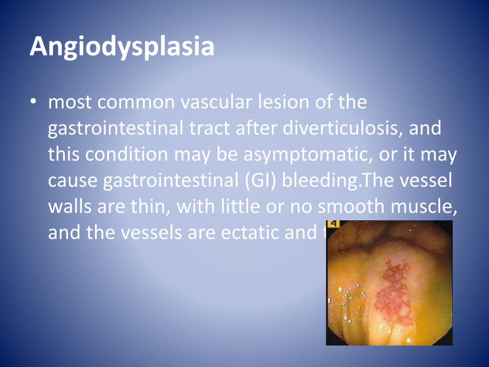