Embed Size (px)
Citation preview
The Mastication Apparatus

Mastication Apparatus
• The Mastication apparatus consist of a centre core or a framework consisting of the bones, the temporomandibular joint and the muscles of mastication.
Maxilla
• It is the second largest bone of the face• It forms the upper jaw with the fellow of the
opposite side• It also contributes to the formation of1. Floor of the nose and the orbit2. Roof of the mouth3. Lateral wall of the nose4. Pterigopalatine and infratemporal fossae5. Pterigomaxillary and infraorbital fissures

Anatomy of the maxilla
• The anatomy of the maxilla has two main parts:1. Body(pyramidal shape)
– Anterior surface– Posterior surface– Orbital surface– Nasal surface
2. Processes– Zygomatic– Frontal– Alveolar– Palatine


Anterior Surface:• Incisive Fossa:– Depressor septi nasi– Orbicularis oris
• Canine fossa:– Levator anguli oris
• Infraorbital foramen (above canine fossa)– Infraorbital nerves and vessels
• Above sharp border between anterior and orbital surface:– Levator labi superioris
• Nasal notch: Dilator Naris• Ant Nasal Spine

Posterior Surface• It is directed backwards and laterally• It forms anterior wall of the infratemporal fossa• Anterior and posterior surfaces are seperated by ridge which
leads to the socket of 1st molar tooth• Near the centre of posterior surface 2 to 3 openings of dental
canal for posterior superior alveolar vessels and nerves• At the lower end there is a raised maxillary tubrosity which is
rough in the upper part of its medial end for tubercle of the palatine bone which has the attachment of superficial fibres of themedial pterigoid muscles
• Above this smooth surface which forms the boundry of the ptrigopalatine fossa is grooved for the maxillary nerve, this groove is contineous with the infra orbital groove
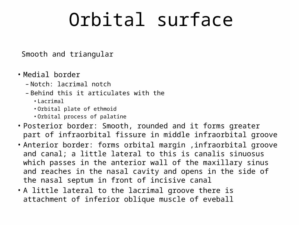
Orbital surface
Smooth and triangular
• Medial border– Notch: lacrimal notch– Behind this it articulates with the
• Lacrimal• Orbital plate of ethmoid• Orbital process of palatine
• Posterior border: Smooth, rounded and it forms greater part of infraorbital fissure in middle infraorbital groove
• Anterior border: forms orbital margin ,infraorbital groove and canal; a little lateral to this is canalis sinuosus which passes in the anterior wall of the maxillary sinus and reaches in the nasal cavity and opens in the side of the nasal septum in front of incisive canal
• A little lateral to the lacrimal groove there is attachment of inferior oblique muscle of eveball

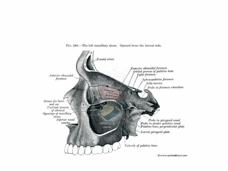
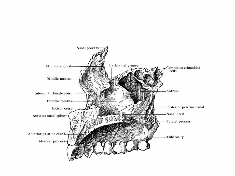
Nasal Surface
• In its upper posterior part there is a large maxillary hiatus which leads into the maxillary sinus
In articulated skull this hiatus is completed by ethmoid and lacrimal bones
• Behind this there is a rough impression for the perpendicular plate of palatine bone
• Infront of maxillary hiatus there is a lacrimal groove• More anteriorly concal crest for articulation with
inferior nasal concha
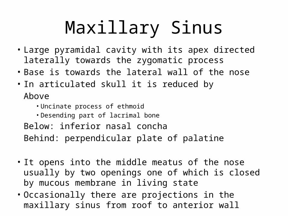
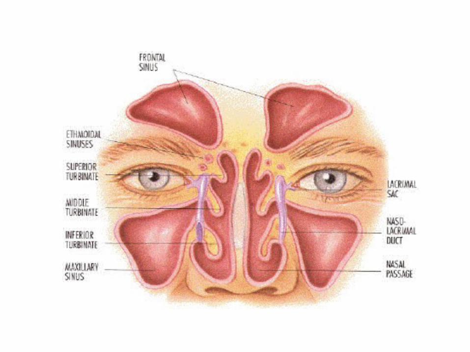
Maxillary Sinus• Large pyramidal cavity with its apex directed laterally towards the
zygomatic process• Base is towards the lateral wall of the nose• In articulated skull it is reduced by
Above• Uncinate process of ethmoid• Desending part of lacrimal bone
Below: inferior nasal conchaBehind: perpendicular plate of palatine
• It opens into the middle meatus of the nose usually by two openings one of which is closed by mucous membrane in living state
• Occasionally there are projections in the maxillary sinus from roof to anterior wall

Processes
• Zygomatic: it is rough and pyramidal– Front:it is contineous with the anterior surface of
body– Behind(concave):in continuity of the posterior
surface– Above: articulates with zygomatic bone– Below(arched border) which anterior and
posterior surface of the body

• Frontal Process:– Lateral Surface: • Vertical ridge (Lacrimal crest)• Groove for the lacrimal sac
– Medial surface: It is rough and uneven and articulates with the ethmoid and also closes the anterior ethmoidal sinus below ethmoidal crest• Upper end: Articulates with the frontal bone• Anterior border with the nasal bone• Posterior border with the lacrimal bone

• Alveolar processes: It has thick arched border behind and contains sockets to receive roots of teeth which vary in size and depth– Canine deepest– Molar widest and subdivided into 3 minor sockets
by septae – Incisors and premolars single– Occasionally incisors are divided into 2 sockes

• Palatine Process: Thick strong horizontal – Inferior surface is concave and presents numerous
foramina for passage of nutrient vessels and contains depressions for lodgement of glands
– Groove for grater palatine Vessels and nerves– Incisive fossa leads into the incisive canal– Sometimes anterior and posterior incisive foramen for
long sphenopalatine nerve which communicates with the greater palatine nerve
– Upper surface: forms the floor of the nasal cavity– Lateral Border fuses with rest of the bone – Posterior border fuses with the horizontal plate of the
palatine
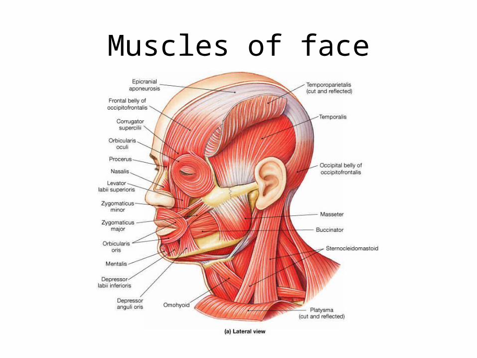
Muscles of face

Arterial supply
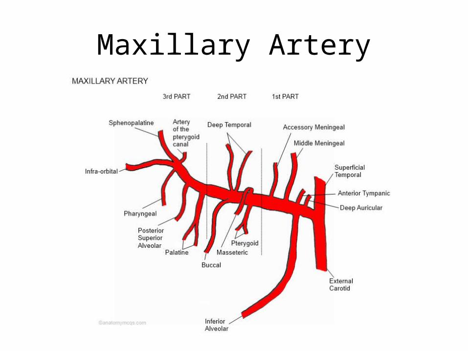
Maxillary Artery
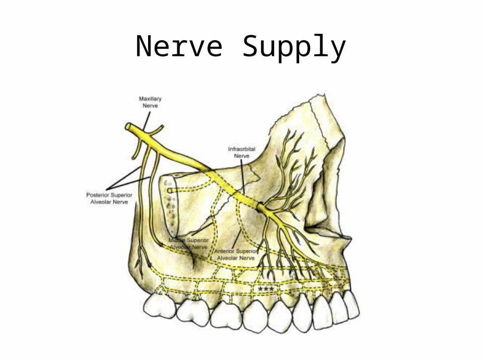
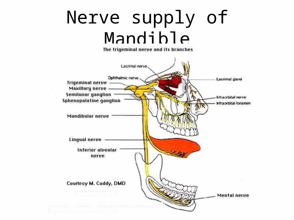
Nerve Supply

Mandible
• Largest and strongest bone of the face• Curved horizontal body; convex forwards• It has two rami which project upward from
posterior end of the body• The body is horse shoe shaped

External Surface
• Faint ridge: symphisis menti• Mental protuberance in the triangular area
below sympisis menti • Mental tubercle on each side of mental
protruberance• Mental foramen between premolar teeth• Oblique line
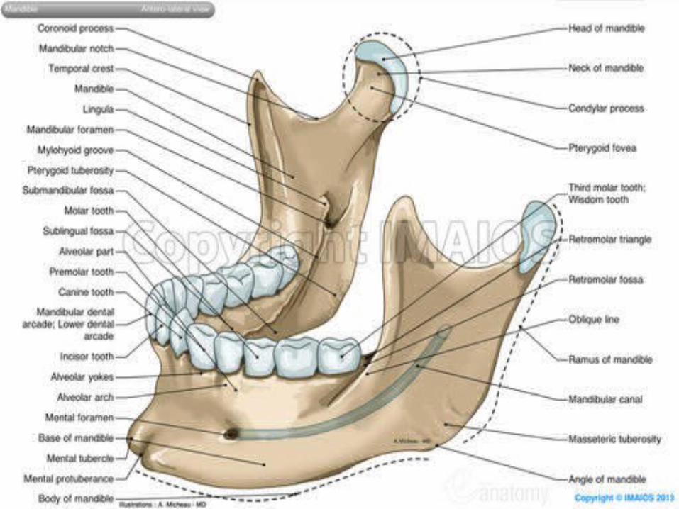
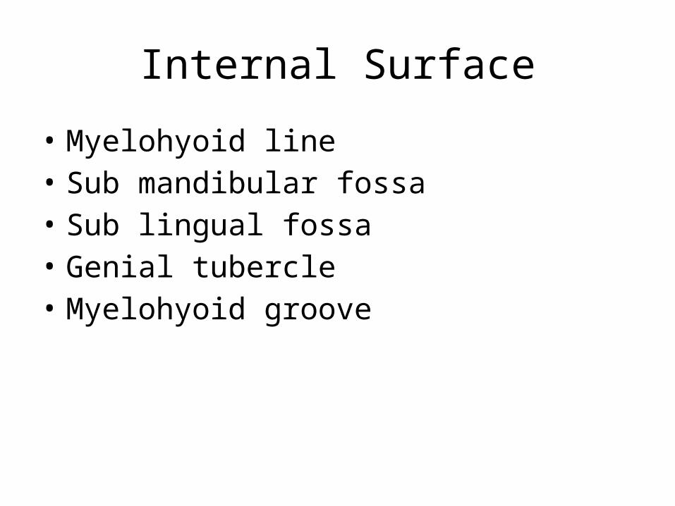
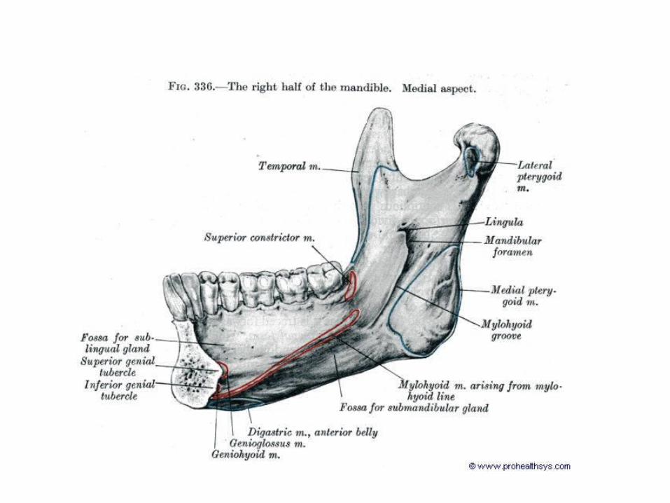
Internal Surface
• Myelohyoid line• Sub mandibular fossa• Sub lingual fossa• Genial tubercle• Myelohyoid groove

Borders• Upper boder:– Sockets for the mandibular teeth are present
• Lower border(Base) presents a digastric fossa• Ramus– Lateral Surface– Medial Surface
• Mandibular foramen canal• Lingula- mylohyoid groove
• Inferior border is continuous with the angle of mandible
• Upper Border: Mandibular Notch

Arterial Supply of Maxilla and Mandible


• Processes:– Condylar– Coronoid
• Mandibular canal

Age changes in mandible
Applied AnatomyMuscle injuries: Its cause and effects
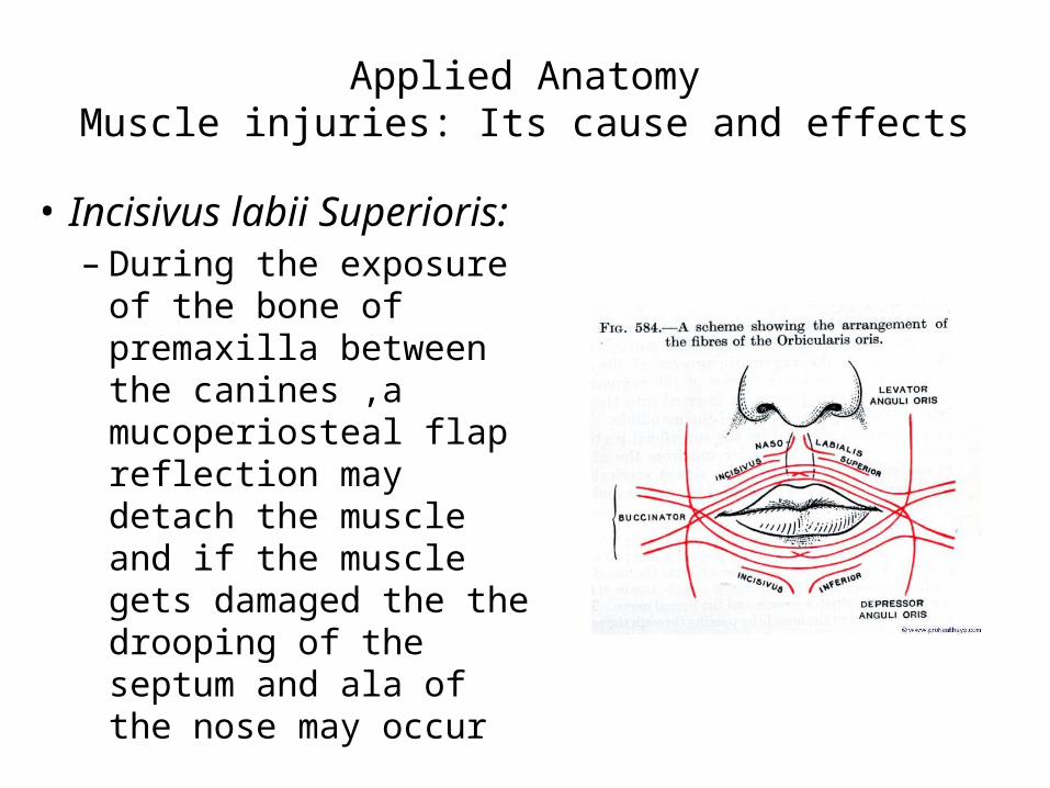
• Incisivus labii Superioris:– During the exposure of
the bone of premaxilla between the canines ,a mucoperiosteal flap reflection may detach the muscle and if the muscle gets damaged the the drooping of the septum and ala of the nose may occur
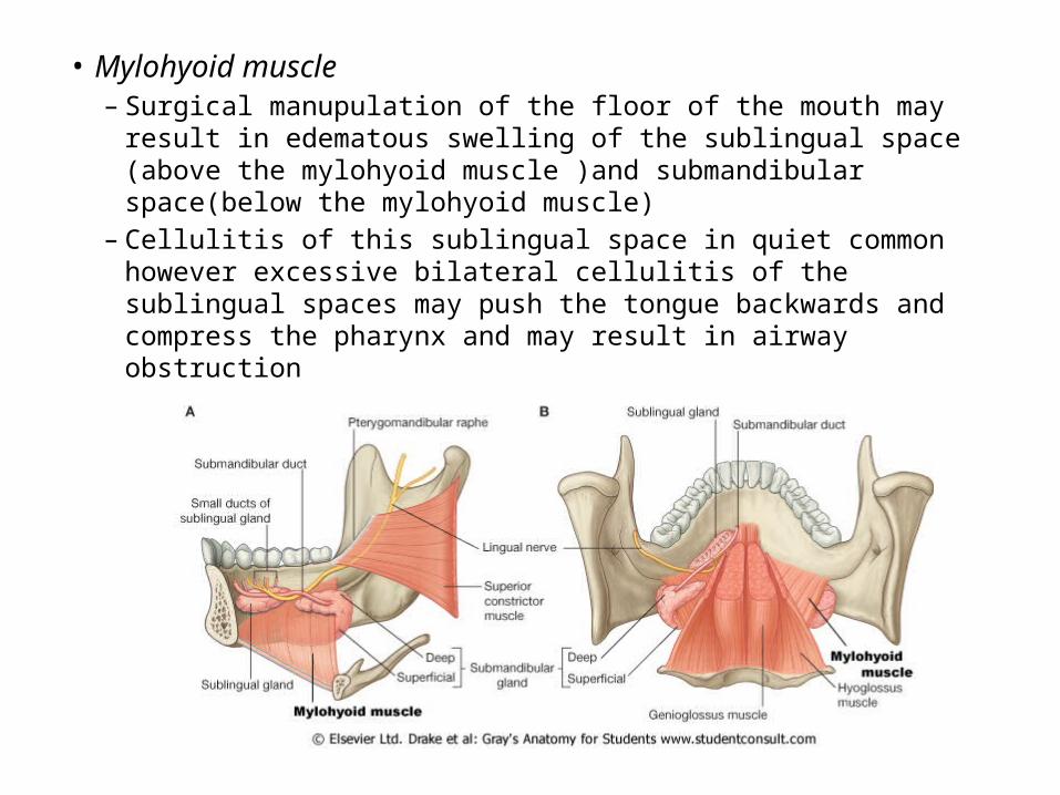
• Mylohyoid muscle– Surgical manupulation of the floor of the mouth may result in
edematous swelling of the sublingual space (above the mylohyoid muscle )and submandibular space(below the mylohyoid muscle)
– Cellulitis of this sublingual space in quiet common however excessive bilateral cellulitis of the sublingual spaces may push the tongue backwards and compress the pharynx and may result in airway obstruction

• Genoiglossus muscle– During the elevationof
the lingual mucosa before making an impression for a subperiosteal implant a portion of the muscle may be reflected from te genial tubercle, however if the muscle is completly detached from the tubercle it may lead to retrusion of the tongue and airway obstruction

• Medial pterigoid – The medial pterigoid muscle
binds the pterigomandibular space medially ,during surgical procedures involving the area of pterigomandibular space infection may occour and may be dangerous due to its closed proximity to the pharyngeal space
– Surgical exposure of the tissue posterior to the maxillary tubrosity may also involve the medial pterigoid muscle as a part of the muscle originates from the maxillary tubrosity

• Lateral pterigoid muscle– The lateral pterigoid muscle fibres are placed in an
angulated manner and because of this there may be pain in patients with a full arched subperiosteal implant or prosthetic splint

• Mentalis muscle:– Complete reflection of the
mentalis muscle for the purpose of extension of a subperiosteal implant may result in a condition known as witch’s chin
There is failure of the mentalis muscle reattachment following the implantation. An external bandage is applied for four days to help in the reattachment of the muscle

• Buccinator muscle:– Myositis of the detached buccinator muscle in
patients with subperiosteal implants may cause swelling and pain at the site of origin of the muscle
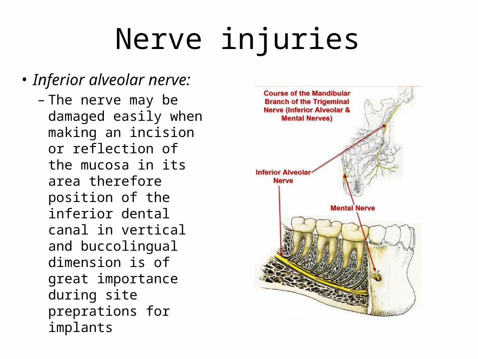
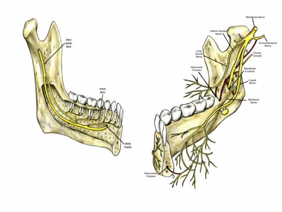
Nerve injuries• Inferior alveolar nerve:
– The nerve may be damaged easily when making an incision or reflection of the mucosa in its area therefore position of the inferior dental canal in vertical and buccolingual dimension is of great importance during site preprations for implants
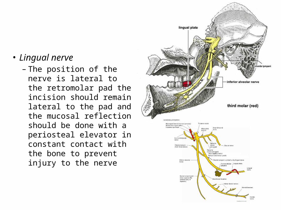
• Lingual nerve– The position of the nerve
is lateral to the retromolar pad the incision should remain lateral to the pad and the mucosal reflection should be done with a periosteal elevator in constant contact with the bone to prevent injury to the nerve
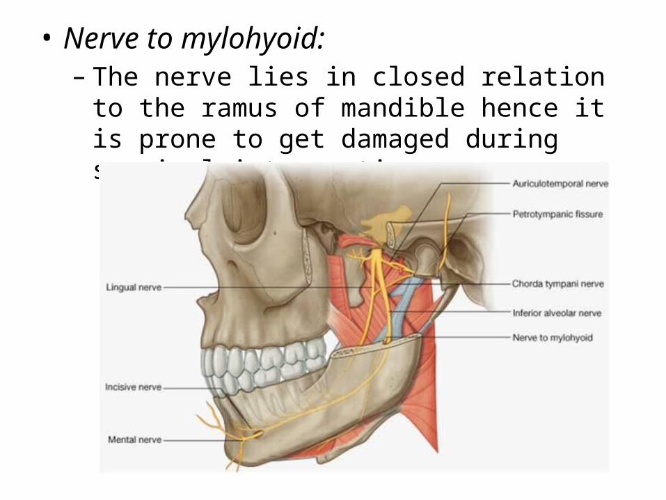
• Nerve to mylohyoid:– The nerve lies in closed relation to the ramus of
mandible hence it is prone to get damaged during surgical intervention
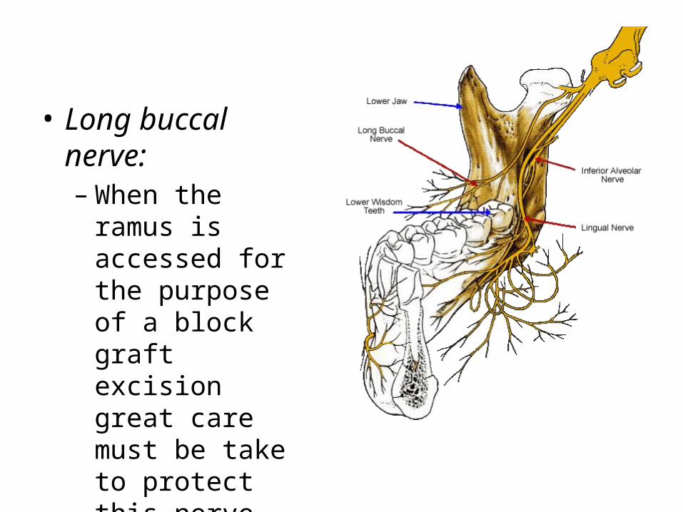
• Long buccal nerve:– When the ramus is
accessed for the purpose of a block graft excision great care must be take to protect this nerve from injury
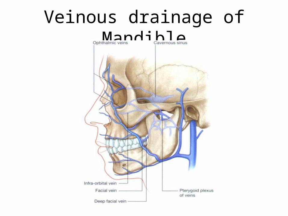
Injury to vessels
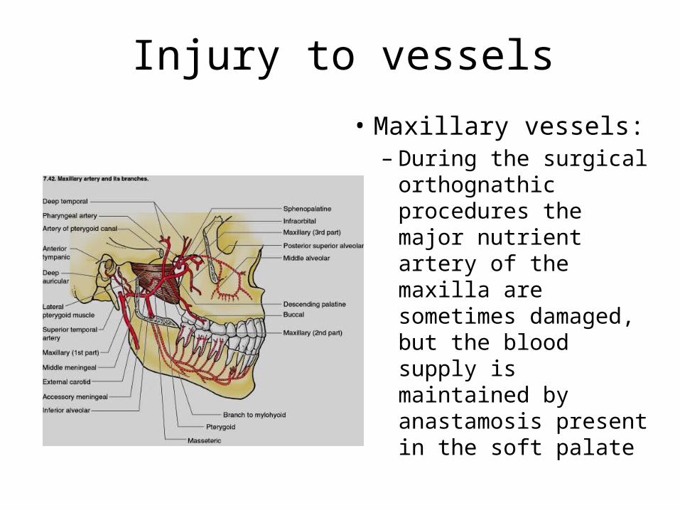
• Maxillary vessels:– During the surgical
orthognathic procedures the major nutrient artery of the maxilla are sometimes damaged, but the blood supply is maintained by anastamosis present in the soft palate
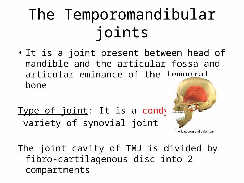
The Temporomandibular joints
• It is a joint present between head of mandible and the articular fossa and articular eminance of the temporal bone
Type of joint: It is a condylar variety of synovial joint
The joint cavity of TMJ is divided by fibro-cartilagenous disc into 2 compartments

Bony framework of joint
• Proximal : Articular fossa and the articular eminance of the mandible
• Distal side: Head of mandible
Articular surfaces: • The anterior eminance is formed by the root of zygoma
and the articular surface is formed by the smooth area of tht mandibular fossa of the temporal bone
• The distal articular surface is also a smootharea forned by the head of the mandible.

The ligaments of TMJ
• True ligament– Capsular ligament– Temporomandibular ligament
• Accessory ligament– Stylomandibular ligament– Sphenomandibular ligament
True ligaments
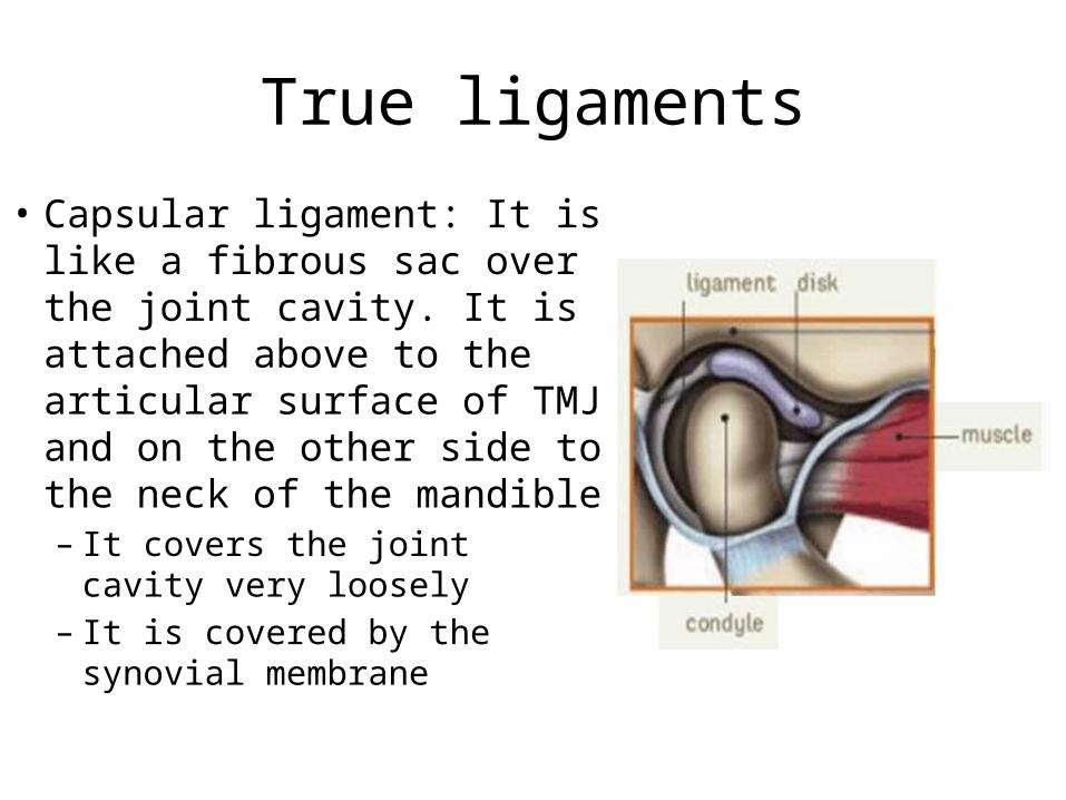
• Capsular ligament: It is like a fibrous sac over the joint cavity. It is attached above to the articular surface of TMJ and on the other side to the neck of the mandible– It covers the joint cavity very
loosely– It is covered by the synovial
membrane
• Temporomandibular ligament:– this ligament is the thickening of the lateral part
of the capsular ligament – It runs posterio-inferiorly and is attached above to
the articular surface and below to the ramus of mandible and the posterio-lateral aspect of the mandible

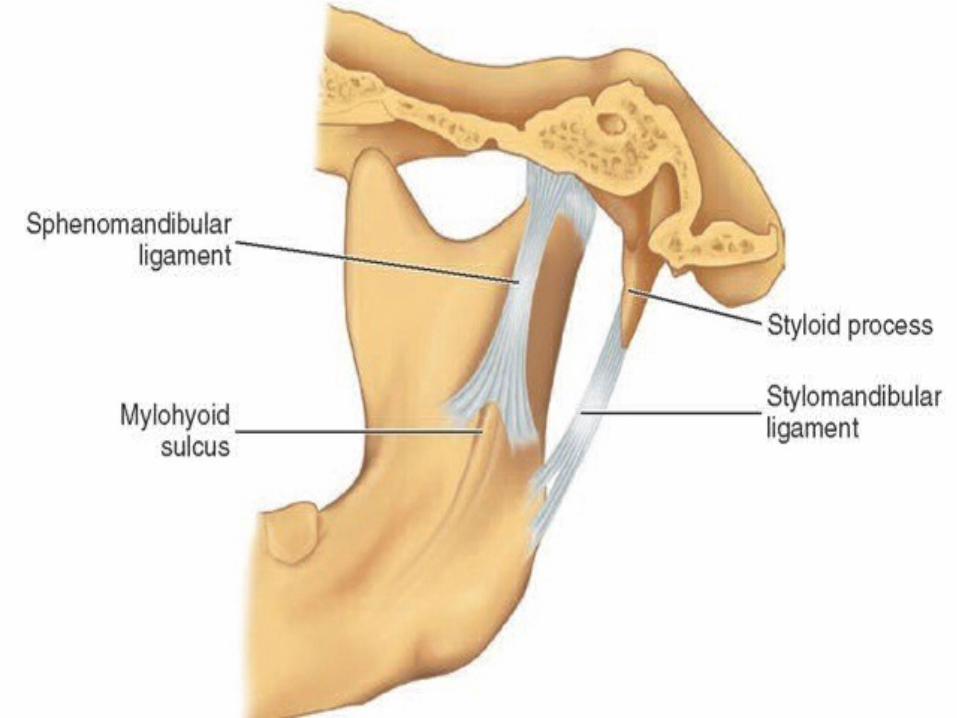
Accessory ligament
It provides additional support to the TMJ• Stylomandibular ligament: This ligament is
formed by the thickening of the deep cervical fascia.– It is attached above to the styloid process and
below to the angle of mandible and lateral border of ramus of mandible
– This ligament helps to seperate the parotid gland and the submandibular gland
• Spheno-mandibular ligament; This ligament is attached above to the spine of the sphenoid bone and below to the lingula on the mandible.– This ligament is innervated by various nerves and
vessels – Embryologically it is the un-ossified intermediate
part of the Meckel’s cartilage of the 1st branchial arch
Interior of the joint
• The bones forming the joint cavity are covered by fibrous cartilage rather than the hyaline cartilage
• The absence of hyaline cartilage makes it an atypical type of synovial joint
The articular disc
• The articular disc divides the joint into two compartments, the upper and the lower compartment
• Articular disc is a fibro-cartilagenous disc, it is thickened in the periphery and thin in the center
• The vascularity is also more in the periphery
Relations of the joint
• Anteriorly: fibres of the muscles of lateral pterigoid
• Posteriorly: part of the parotid gland• Medially: spheno mandibular ligament• Laterally: parotid gland

Development of TMJ• Temporomandibular ligament developes from the
mesenchyme situated between the developing temporal bone above and the mandibular condyl below
• During 12 weeks of development two clefts appear and remain separated from each other by an intervening mesenchymal plate – These clefts become the upper and the lower joint
cavities – The mesenchymal plate becomes the articular disc– Condensation of the mesenchyme around the
developing joint becomes the capsule of the joint
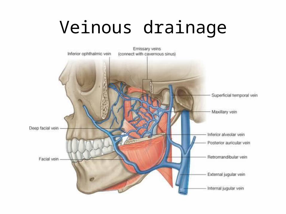
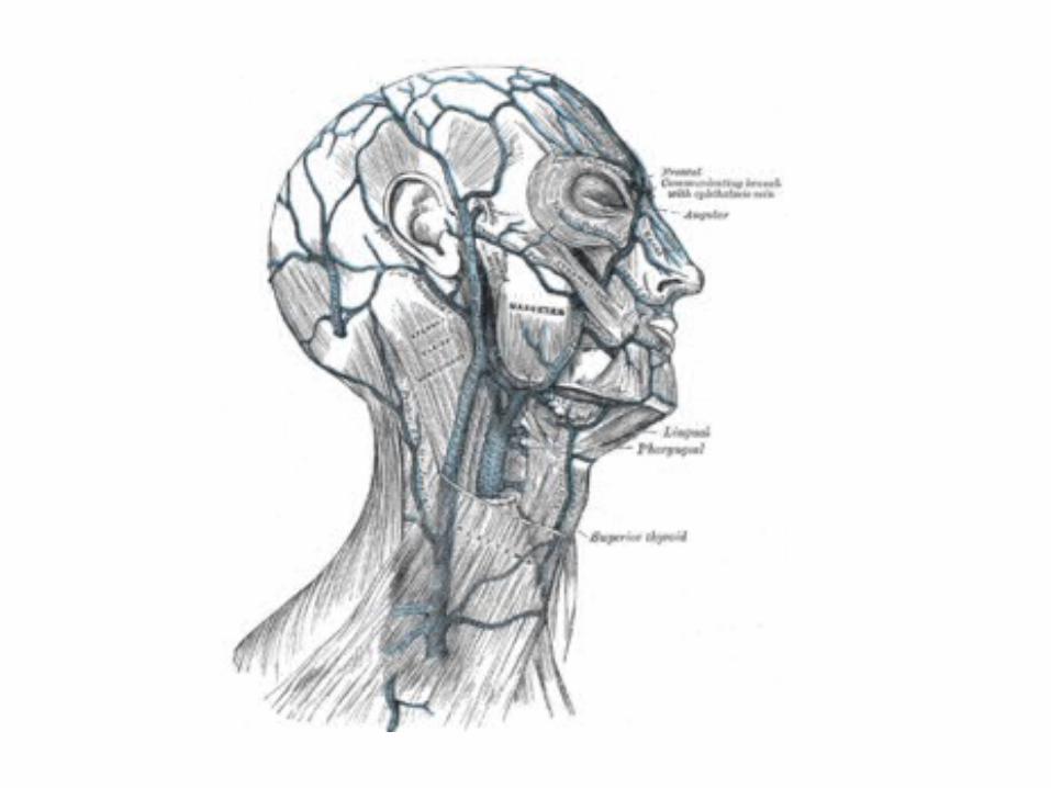
The supply of the gland
• Arterial supply• Superficial temporal artery• Maxillary artery
• Veinous drainage• Superficial temporal vein• Retromandibular vein
• Lymphatics: It drains into the superficial and deep parotid lymph nodes and deep cervical nodes.

Movements of the TMJ
• The principle movements of the joint are:» Elevation» Depression» Protraction» Retraction» Side to side movements
Various muscles are involved in these movements, collectively known as muscles of mastication.
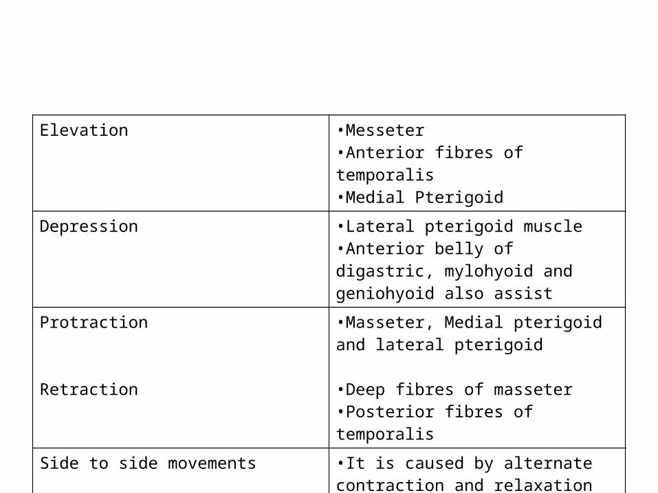
Elevation •Messeter•Anterior fibres of temporalis•Medial Pterigoid
Depression •Lateral pterigoid muscle•Anterior belly of digastric, mylohyoid and geniohyoid also assist
Protraction
Retraction
•Masseter, Medial pterigoid and lateral pterigoid
•Deep fibres of masseter •Posterior fibres of temporalis
Side to side movements •It is caused by alternate contraction and relaxation of Medial and lateral pterigoid

Muscles of mastication
• There are 4 muslces involved in mastication– Masseter– Temporalis – Medial pterigoid – Lateral pterigoid

Masseter
• Origin: it arises from 2 heads• Superficial hear arises from the lower border of the anterior 2/3rd of the
zygomatic arch• Deep head arises from the whol length of the zygomatic arch and lower
border of th posterior 1/3rd of the zygomatic arch.
• Insertion: The two heads unite and gets inserted into the whole length of the outer surface of the ramus and angle of mandible
• N. Supply: Massetric nerve and a branch of anterior division of the Mandibular nerve
• Actions: • Elevation of mandible• Protrusion of the mandible
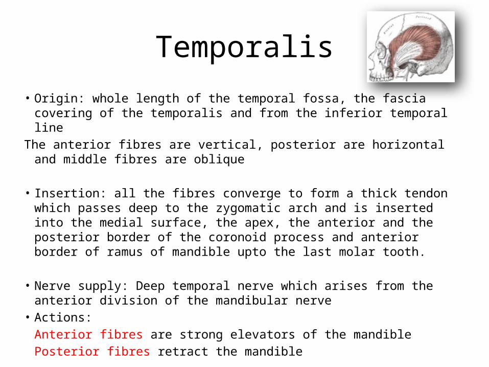
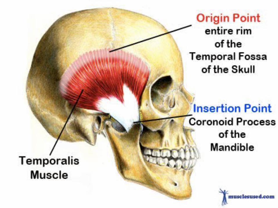
Temporalis
• Origin: whole length of the temporal fossa, the fascia covering of the temporalis and from the inferior temporal line
The anterior fibres are vertical, posterior are horizontal and middle fibres are oblique
• Insertion: all the fibres converge to form a thick tendon which passes deep to the zygomatic arch and is inserted into the medial surface, the apex, the anterior and the posterior border of the coronoid process and anterior border of ramus of mandible upto the last molar tooth.
• Nerve supply: Deep temporal nerve which arises from the anterior division of the mandibular nerve
• Actions:Anterior fibres are strong elevators of the mandiblePosterior fibres retract the mandible

Lateral Pterigoid• Origin: It also arises from two heads
– The upper/superior head originates on the infratemporal surface and infratemporal crest of the greater wing of the sphenoid bone,
– lower/inferior head on the lateral surface of the lateral pterigoid plate
• Insertion: Inferior head inserts onto the neck of condyloid process of the mandible; upper/superior head inserts onto the articular disc and fibrous capsule of the temporomandibular joint
• N. Supply: The mandibular branch of the fifth cranial nerve, the trigeminal nerve, specifically the lateral pterigoid nerve, innervates the lateral pterygoid muscle.
• Actions: Unlike the other three muscles of mastication, the lateral pterygoid is the only muscle of mastication that assists in depressing the mandible (opening the jaw).
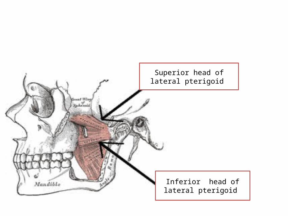
Superior head of lateral pterigoid
Inferior head of lateral pterigoid

Medial pterigoid• Origin:
– The bulk of the muscle arises as a deep head from just above the medial surface of the lateral pterigoid plate.
– The smaller, superficial head originates from the maxillary tuberosity and the pyramidal process of the palatine bone.
• Insertion: Its fibers pass downward, lateral, and posterior, and are inserted, by a strong tendinous lamina, into the lower and back part of the medial surface of the ramus and angle of the mandible
• N. Supply: Like all other muscles of mastication the medial pterygoid is innervated by the anterior root (motor root) of the mandibular branch of the trigeminal nerve (V).
• Actions: It contributes to following functions:– Elevation of the mandible (closes the jaw)– Minor contribution to protrusion of the mandible– Assistance in mastication– Excursion of the mandible; contralateral excursion occurs with unilateral contraction

Medial pterigoid
insertion
origin