Embed Size (px)
Citation preview
LECTURE NOTE
ON
RESPIRATORY PHYSIOLOGY
BY
MJ ADENIYI MSC
([email protected], +234 08066796517)
DEPARTMENT OF PHYSIOLOGY
UNIVERSITY OF BENIN, BENIN CITY, NIGERIA
HUMAN RESPIRATORY SYSTEM
INTRODUCTION
We have established the fact that oxygen is essential for human survival in the previous notes. We want to look into the system that is responsible for consumption of this essential substance and expulsion of other respiratory gases. Let us begin by differentiating external from internal respiration. External respiration is the process by which exchange of gases occurs between the alveoli and blood capillaries. Internal respiration or cellular respiration deals with the utilization of oxygen by body cells for the production of energy (check your previous note). We shall also the components of the system, the structural and functional adaptations of the structures that form the control, the mechanism of obtaining oxygen and expelling carbon dioxide, ventilation and gas exchange. The neural and non-neural cells that control ventilation and gas exchange, how respiratory gases are transported and oxygen carrying capacity and related issues.
Figure 1: The human respiratory system
The human respiratory system consists of:
Lungs encaged by thorax
System of tubes that act as air conduit to the lungs
Muscles that affect the size of thoracic cage
Respiratory centres (medulla & pons)
Nerve tract linking the respiratory centres with the muscles
LUNG
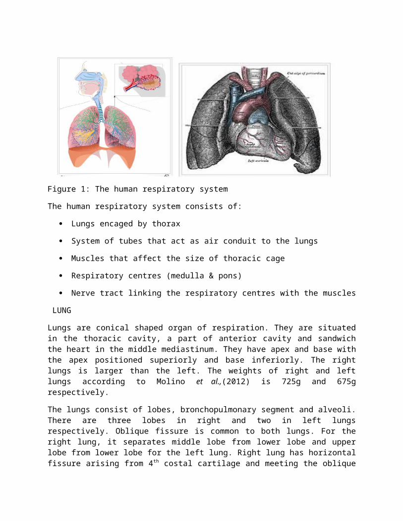
Lungs are conical shaped organ of respiration. They are situated in the thoracic cavity, a part of anterior cavity and sandwich the heart in the middle mediastinum. They have apex and base with the apex positioned superiorly and base inferiorly. The right lungs is larger than the left. The weights of right and left lungs according to Molino et al.,(2012) is 725g and 675g respectively.
The lungs consist of lobes, bronchopulmonary segment and alveoli. There are three lobes in right and two in left lungs respectively. Oblique fissure is common to both lungs. For the right lung, it separates middle lobe from lower lobe and upper lobe from lower lobe for the left lung. Right lung has horizontal fissure arising from 4th costal cartilage and meeting the oblique fissure at the mid-axillary line. Horizontal fissure separates upper lobe from middle lobe.
Furthermore, the upper lobe of the right lung has three bronchopulmonary segments and they are apical, anterior and posterior. The middle lobe has two; medial and lateral bronchopulmonary segments. The lower lobe has five; superior, posterior, anterior, medial and lateral bronchopulmonary segments. For the left lung, there are 8 bronchopulmonary segments and they are apicoposterior and anterior bronchopulmonary segments in the upper lobe, superior, anteriormedial, posterior and lateral bronchopulmonary segments. Left lung has a tongue like lobe called lingual. Lingula has superior and inferior segments. The bronchopulmonary segments consist of many alveoli. There are over 3 million alveoli in the lung.
When we stand, the apex of the lung is situated above the heart while the base is below (check figure 1). This means that perfusion at the apex will be lower than in the lower. This means that lesser gaseous exchange will take place in the apex than base. This also means that pulmonary edema will have little lesser effect on the apex than the base in erect posture.
It is important to also mention that the lungs are housed by thorax and suspended in its cavity. In anatomy class, you were taught the significant of pleura. Pluera space is the space between the two pleura and it is filled with a serous fluid called intrapleural fluid. The fluid reduces the friction between the two pleura. The negative pressure plays role in respiration. It is -6mmHg during inspiration and slighty positive at expiration (2.5mmHg). The pressure also helps in preventing oedema. Medical condition in which there is accumulation of fluid in the pleura is termed pleural effusion. The fluid may be air (pneumothorax) or pus (pyothorax) or blood (hemothorax) or water (hydrothorax).
RESPIRATORY TRACT
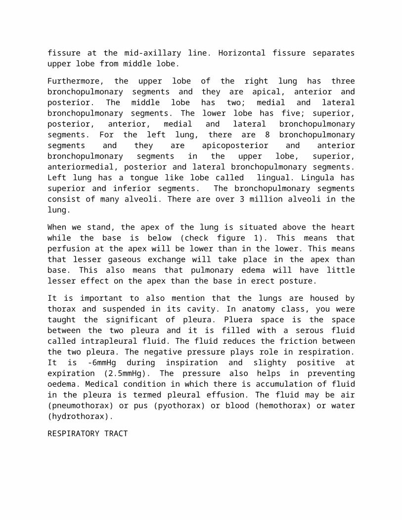
Figure 2: The bronchi and bronchial tree
The respiratory tract is a system of branched tubes that supply air to the lung. The length and width of the tract decreases as we approach the alveoli but the total cross sectional area increases. The cross sectional area of the trachea is 2.5cm2 but that of alveolar ducts is around 11800cm2.
Structurally, the tract can be classified into two:
-Upper respiratory tract leading from nose and mouth to the trachea and
-Lower respiratory from trachea to the alveolar duct
The trachea divides into two primary bronchia at the carina. The eparterial and hyparterial bronchus in the right and left primary bronchus. The right primary bronchus divides into three secondary bronchi (or lobar bronchi) with each secondary bronchi supplying air to each lobe of the lobe. The left primary bronchus divides into two lobar brochi with each left lobar bronchi supplying each lobe. The right lobar bronchi divides into ten tertiary or segmental bronchi with each right segmental bronchi supplying air to each right bronchopulmonary segments. The left lobar bronchi divides into 8 segmental bronchi with each left segmental bronchi supplying each left bronchopulmonary segments. The right and left bronchopulmonary segments further branch into terminal bronchioles when diameter is less or equal to 1mm. Terminal bronchioles divide and form respiratory bronchioles when the length is less or equal to 0.5mm. The respiratory bronchioles and alveolar ducts supply alveoli with air. There are over 300 million alveoli in the lung. Alveoli are the functional units of gaseous exchange.
The following are impor tant characteristics of airway
-Epithelial cells: The epithelial cells of the respiratory tract are predominantly pseudostratified squamous epithelial cells. They are also characterized by projections called cilia. Mucociliary action is an important innate and acquired immune defense in the body. It reproofs impurities (eg dust) that may be contained in air. It is also an integral process of allergy.

In the alveoli, 95% of epithelial cells are squamous and 5% are cuboidal. The cuboidal cells produce a surface acting substance called surfactant. Surfactant reduces surface tension and prevents the lungs from collapsing during expiration.
-Smooth muscle: Monounit smooth muscles are present in the airways. Constriction (brochioconstriction) occurs in response to impurities (eg dust) or parasympathetic stimulation. The density of smooth muscle increases from pharynx to respiratory bronchioles. Therefore the greatest airway resistance occurs in the bronchioles. Bronchoconstriction reduces amount of air that flows through the tube and bronchodilation will increase airflow. In Asthma, sabutamol, a beta 2 agonist is administered to dilate the air tube (bronchodilation) and improve airflow.
Serous producing glands; are present in the tract up to terminal bronchioles. These make the tract fluidy. In cystic fibrosis, the mechanism involved in production of serous fluid is impaired due to deficiency of chloride ion channel. This results in dryness of the airway. Mucus glands and serous gland assist in mucociliary clearance.
Cartilage: maintain patency (openness) of the airways
RESPIRATORY MUSCLES
There are two groups of muscles: inspiratory muscles and expiratory muscles.
Inspiratory muscles; assist in inspiration and the term primary inspiratory muscle means muscles whose contractions result in inflow of oxygen rich air (inspiration) at rest. These muscles are thoracic diaphragm and external intercoastal muscle. The former is responsible for 75% of inspiratory movement. Even at rest inspiration is active. Accessory respiratory are involved in forced inspiratory. They are pectoralis major and minor, seratus anterior, seratus posteriorsuperior, anterior, middle and posterior scalene, levator scapulae, levatores costarum, sternocleidomastoid muscle among others.
Expiratory muscles: assist in expiration, the outflow of carbon dioxide rich air from the lung. Expiration is passive (contraction isn’t involved). Forced expiration requires contraction of internal intercoastal muscle, innermost intercoastal muscle, transversus thoracis, seratus posteriorinferior, abdominal muscles among others.
Abnormalities of respiratory muscles may lead to restrictive pulmonary disease (if inspiration is impaired) and obstructive pulmonary disease (if expiration is compromised). Myasthenia gravis, flail chest, pulmonary fibrosis are examples of restrictive pulmonary disease. Emphysema, pleural effusion, asthma among others are associated are examples of obstructive respiratory disease. Although, some scholars consider emphysema as example of both restrictive and obstructive diseases
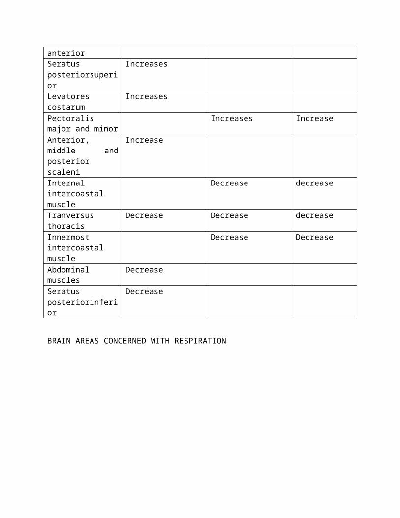
Table 1 showing the effects of respiratory muscles on the diameters of thoracic cavity
Respiratory muscles Vertical (superiorinferior) diameter
Anteriorposterior diameter
Transverse diameter
Thoracic diaphragm Increases moreExternal intercoastal Increases more Increase
muscleSternocleidomastoid muscle
Increases
Seratus anterior IncreasesSeratus posteriorsuperior
Increases
Levatores costarum IncreasesPectoralis major and minor
Increases Increase
Anterior, middle and posterior scaleni
Increase
Internal intercoastal muscle
Decrease decrease
Tranversus thoracis Decrease Decrease decreaseInnermost intercoastal muscle
Decrease Decrease
Abdominal muscles DecreaseSeratus posteriorinferior
Decrease
BRAIN AREAS CONCERNED WITH RESPIRATION
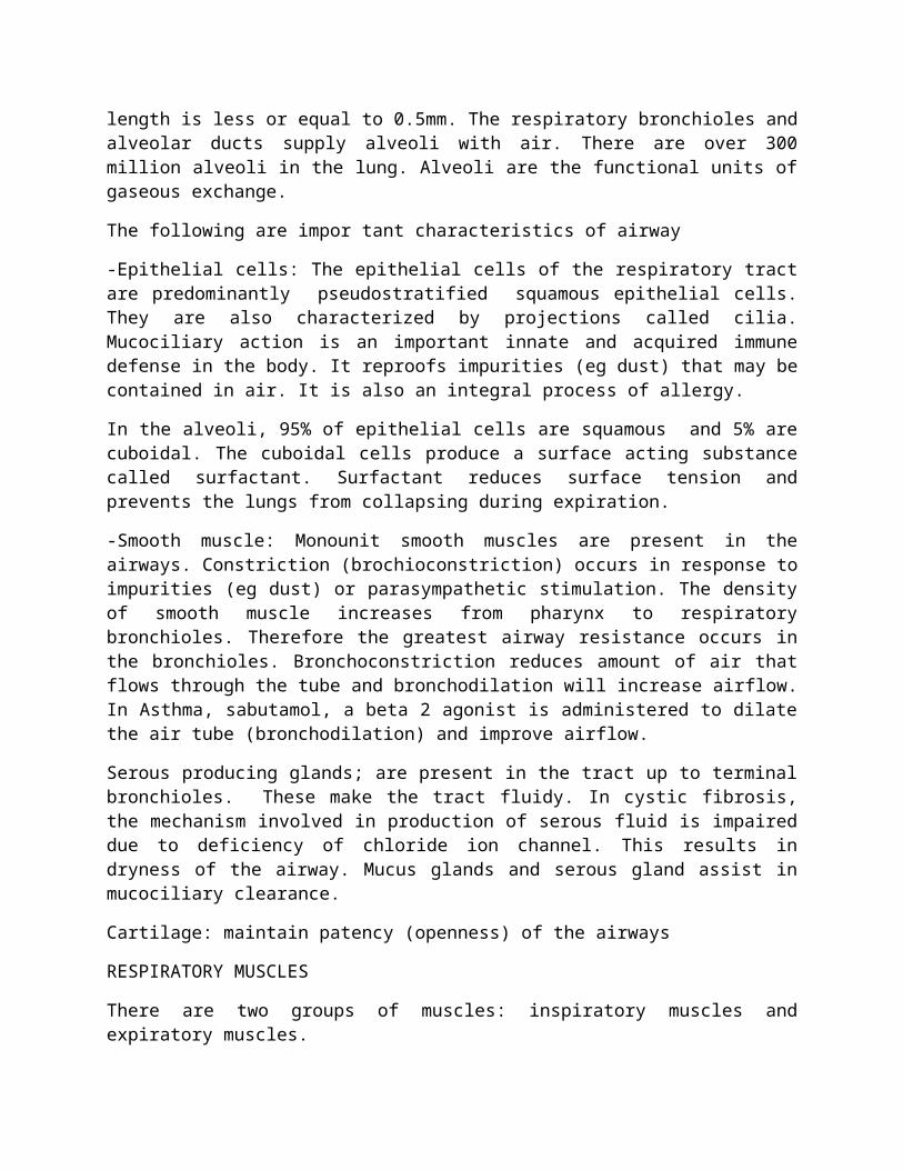
Figure 3: Respiratory centers
The respiratory centers are groups of pacemaker cells situated in the brainstem.
INSPIRATORY CENTER; is located in the solitary tract nucleus forming dorsal group of nuclei. Stimulation increases depth of inspiration as shown in figure 3.
EXPIRATORY CENTER: is situated in nucleus ambiguous forming ventral group of nuclei. Stimulation causes expiration. It must be noted that inhibitory (GABAergic) from solitary tract nucleus end on the doral motor nucleus and nucleus ambiguous. Because nucleus ambiguous and dorsal motor nucleus are cardiac center. Therefore, stimulation of these nuclei will result in both expiration and decrease in heart rate . During inspiration, heart rate will also increase. This respiration induced changes in heart rate is called sinus arrhythmia.
PNEUMOTACTIC CENTER: is located in upper Pons. Stimulation decreases depth of inspiration.
APNEUSTIC CENTER: is situated in lower Pons. Stimulation increases depth of respiration.
HIGHER BRAIN AREAS: such as cerebral cortex and limbic cortex are responsible for voluntary respiration
NERVE TRACT CONNECTING RESPIRATORY CENTERS WITH EFFECTORS
Affarent somatic nerve fibres connect stretch receptors in the lungs and respiratory structures with the brainstem and brain. While efferent somatic nerve fibres connect the respiratory centers with effectors. The phrenic nerves and intercoastal nerves are important efferent nerves that link muscles and respiratory structures with the brainstem.
MECHANISM OF RESPIRATION
Understanding how human beings respire will be easier since we have learnt the functions of each structure that makes up the respiratory system and most importantly the effects of respiratory muscles on diameters of thoracic cavity.
As we have explained earlier, respiration is composed of inspiration (inflow of oxygen rich air) and expiration (outflow of carbondioxide rich air).
What then triggers inspiration?
As cells undergo metabolism to obtain energy, carbon dioxide is one of the products of this process. In the last semester, we identified that carbon dioxide must be excreted otherwise it is going to affect body cells. If you remember, we said there is a positive correlation between carbon dioxide load and hydrogen ions concentration. This is so as carbon dioxide combines with water to produce carbonic acid, an unstable substance which in the presence of carbonic anhydrase will be converted to hydrogen ion. Hydrogen ions formed in the inspiratory center stimulates chemoreceptors in this area thereby generating a neural drive for inspiration.
The efferent nerve fiber conduct signals to the inspiratory muscle resulting in the contraction of inspiratory muscles.
-Contraction of inspiratory muscles brings about increases in vertical, anteriorposterior and transverse diameters of the thoracic cage.
-Increase in the diameters of thoracic cage increases intrathoracic volume.
-Increase in intrathoracic volume correlates with decrease in intrathoracic pressure (boyle’s law)
-Decrease in pressure within the thorax implies decreases in intrapleural pressure (from -2.5 to -6mmHg) and intralveolar pressure (from +4 to -4mmHg). Transpulmonary pressure, the difference between intrapleural pressure and intralveolar pressure also decreases. -6mmHg means that intrapleural pressure is lesser than atmospheric pressure by 6mmHg. -4mmHg means intralveolar pressure is lesser than atmospheric pressure by 4mmHg.
-The difference between atmospheric pressure and the intrathoracic pressures creates diffusion gradient for air to flow into the lung.
- During forced inspiration as occurs during exercise, contraction of accessory inspiratory muscles occurs such that the intrathoracic pressure may decrease up to -30mmHg. This great pressure helps in drawing oxygen rich air into the lung
Expiration follows inspiration
-Filling and distension of lungs stimulates stretch receptors of the lungs resulting in inhibition of inspiratory center and stimulation of expiratory centre.
-Inhibition of inspiratory centre results in inhibition of inspiratory muscle and relaxation of these muscles.
-At rest, inhibition of inspiratory centre brings about relaxation of diaphragm and external intercoastal muscles and outflow of carbondioxide rich air.
- Forced expiration involves contraction of primary and accessory expiratory muscles in addition to relaxation of primary and accessory inspiratory muscles.
Other stimulants of respiration include:
-Stimulation of chemoreceptors located in the aortic arc and carotid sinus
-Stimulation of stretch receptors in lungs
-Stimulation of irritant receptors in respiratory tract and lungs
-Stimulation of thermoreceptors
-Stimulation of pain receptors
-Stimulation of baroreceptors
-Stimulation of proprioreceptors. Proprioreceptors are stretch receptors located in joint and tendon
-Intentional respiration: is coordinated by higher brain parts especially the cerebral cortex

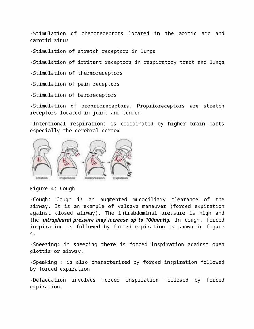
Figure 4: Cough
-Cough: Cough is an augmented mucociliary clearance of the airway. It is an example of valsava maneuver (forced expiration against closed airway). The intrabdominal pressure is high and the intrapleural pressure may increase up to 100mmHg. In cough, forced inspiration is followed by forced expiration as shown in figure 4.
-Sneezing: in sneezing there is forced inspiration against open glottis or airway.
-Speaking : is also characterized by forced inspiration followed by forced expiration
-Defaecation involves forced inspiration followed by forced expiration.
-Yawning involves forced inspiration followed by forced expiration.
-Crying involves deep inspiration, forced expiration and apnea (absence of breath).
The respiratory rate is 12 to 20cycles per minutes in adult at rest. 500mL of air enters and leaves the respiratory tract per breath at rest. About 250mL of oxygen enters the lung and 200mL of carbon dioxide leaves the lung per minute.
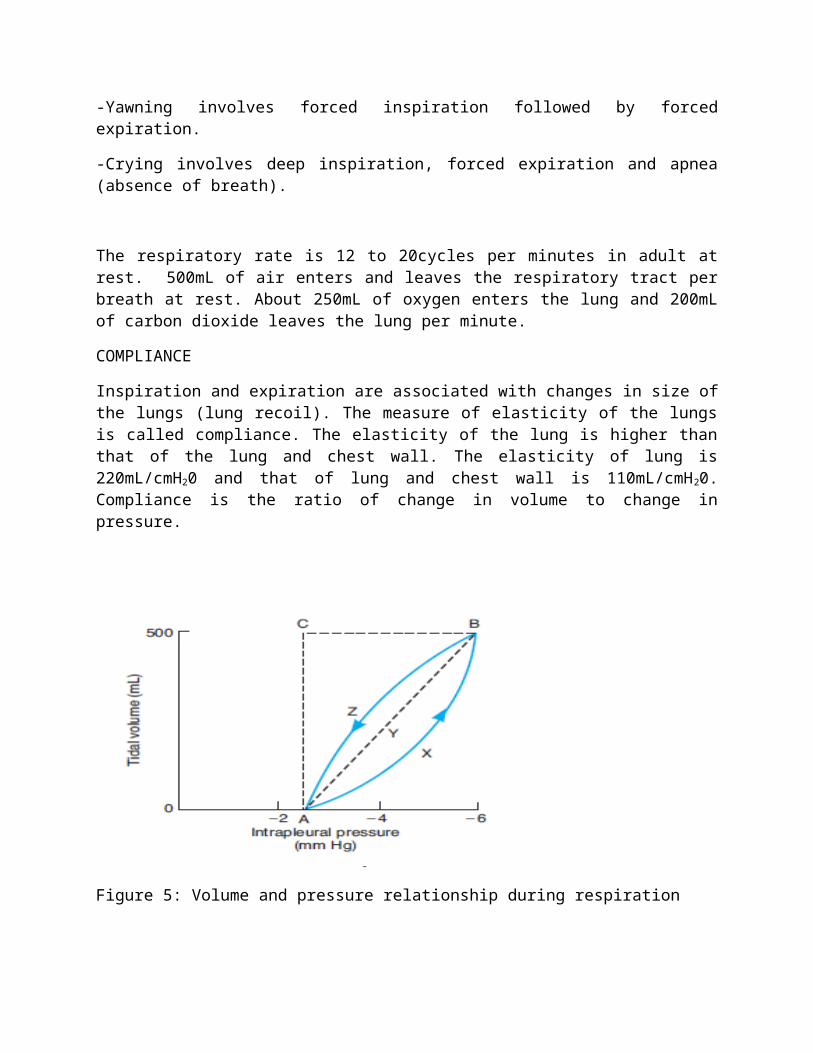
COMPLIANCE
Inspiration and expiration are associated with changes in size of the lungs (lung recoil). The measure of elasticity of the lungs is called compliance. The elasticity of the lung is higher than that of the lung and chest wall. The elasticity of lung is 220mL/cmH20 and that of lung and chest wall is 110mL/cmH20. Compliance is the ratio of change in volume to change in pressure.
Figure 5: Volume and pressure relationship during respiration
Compliance decreases in reclining position, hydrothorax, kyphosis, obesity and increases in emphysema.
SURFACTANT
In inspiration, the lung increases in size and in expiration, the size of the lung decreases. Despite the decrease in lung volume in expiration, the lungs do not collapse. What prevents the lungs from collapsing is surfactant. Surfactant is a surface acting substance produced by type II alveoli epithelial cells (cuboidal epithelial cells). It is made up of lipids and proteins.
In Physics, we learnt that the cohesive force of water molecules is greater than its adhesive force. This means that water molecules in the pleural space will tend to pile together and limit the size of the lung. The presence of a lipid rich surfactant prevents water from piling together. You will recall that lipid is immiscible with water.
Surfactants reduce surface tension of water and prevent the lungs from collapsing. During intrauterine life, the fetal lungs are non functioning. At birth, the first respiratory movements occur (cry). Surfactants play important role in preventing neonatal lungs from collapsing. Deficiency of surfactants at this stage result in hyaline membrane disease (Infant Respiratory Distress).
Proteins and cortisol play important role in maturation of surfactant.
LUNG VOLUME
Figure 6: Lung volumes
TABLE 2 SHOWING LUNG VOLUME AND THE VALUES
FLUCTUATION IN RESPIRATION
-Normal respiratory rate is 12-20cycle/minutes in adult at rest. This is termed eupnea.
-Increase in respiratory rate is called tachypnea. Tachypnea is a respiratory mechanism designed to draw oxygen rich air into the lungs. Usually when the frequency of respiration is very high, the duration of inspiration and expiration is greatly reduced. This form of rapid shallow occur during intense exercise in an untrained individuals. Kussmaul breathing seen in diabetic mellitus subjects is another form of rapid shallow breathing.
-Decrease in respiratory rate is termed bradypnea.
-Change in respiration due to posture is called orthopnea
-Temporary arrest of breathing is called apnea. Contraction of adductor muscles of the larynx and the flapping over of epiglottis close the airway during swallowing. This is called deglutition apnea. This prevents aspiration. Apnea may also be intentional.
-Hypnea means increase in air flow that meets up metabolic demand.
-Ventilation means inflow of oxygen rich air. Hyperventilation means increase in ventilation above metabolic demand. It is due overstimulation of inspiratory centre. The effects of
hyperventilation are respiratory alkalosis and hypocalcemia tetany. Hypoventilation means decrease in ventilation. Depression of inspiratory center may cause hypoventilation. The effect of hypoventilation is respiratory acidosis.
-Respiratory Minute Volume is the volume of air that passes through the respiratory tract into the lung per minute. It is a product of respiratory rate and tidal volume. Assume that respiratory rate is 15cycle/min; Respiratory Minute Volume is given as:
15 x 500mL = 7500mL/min
-Alveolar ventilation is the volume of oxygen involved in gaseous exchange. It is also known as depth of respiration. About 150ml of air does not reach the lung. Therefore, alveolar ventilation is given as:
15 x 350mL = 5250mL/min
Periodic breathing: is characterized by cycle of hypnea and apnea. Example of this is cheyne stokes breathing which occurs in Random Eye Movement sleep in normal adult, it is also seen in babies, pregnant women and so on.
Hypoxia: Inadequate tissue oxygenation is called hypoxia. Hypoxia can be classified into:
-Hypoxic hypoxia: It is a form of hypoxia caused by either reduction in atmospheric oxygen as occurs when one moves high the altitude, impairment of inspiratory muscles, some diseases of the lung. In hypoxic hypoxia, the ability of hemoglobin to carry oxygen isn’t affected but the reduction in partial pressure of oxygen in blood is due to decrease in volume of oxygen in alveoli.
-Stagnant hypoxia is a form of hypoxia that is due to decrease blood flow to tissues. Decreased blood flow to tissues decreases tissue oxygenation. It may be due to polycythemia vera and shock. Decrease in blood flow to tissue also occurs due to vascular related diseases such as atherosclerosis, stroke, hypertension, phaechromocytoma among others
-Anemic hypoxia is due to presence of abnormal hemoglobin in the blood. Abnormal hemoglobin has poor ability to carry oxygen. The ability of hemoglobin to carry oxygen is called oxygen carrying capacity of the hemoglobin. Anemic hypoxia occurs in sickle cell anemia.
-Histotoxic hypoxia is due to problem with utililization of oxygen by tissues for production of ATP. Chemical substances such as cyanides, mercury and methanol have been reported to inhibit cytochrome oxidase, a mitochondrial enzyme that is essential for ATP production.
HYPERCAPNEA: is an increase in partial pressure of carbon dioxide
ASPHYXIA: is combination of hypoxia and hypercapnea.
TRANSPORT OF RESPIRATORY GASES
Transport of Oxygen
Oxygen is carried majorly by hemoglobin in form of oxyhemoglobin. It is also carried by plasma proteins and plasma. The ability of hemoglobin to carry oxygen is called oxygen carrying capacity of blood. Hemoglobin is not completely saturated with oxygen (it is only 95% saturated with oxygen). 1mg of hemoglobin carries 1.34mL of oxgen and there is 15mg of hemoglobin in 100dL of blood. Therefore the oxygen carrying capacity of blood is 19.8mg/ml. In sickle cell anemia, this capacity is reduced.
Transport of carbon dioxide
Carbon dioxide is carried majorly as bicarbonate. It can also be carried in combination with hemoglobin as carbhemoglobin. It can also be transported by plasma protein and plasma fluid.
![· [MJ [M] (M] (M] (M] [MJ (MJ (M J (M] [M) [MJ [MJ [Mj [a 3rd ~pecker) [ Mj .. 2/0.t::JT.01-58 I om turning toward [point] 135. Yes, I om over [point J 136 now. {B% Roger). Roger,](https://img.pdfslide.net/doc/110x75/5c7742dc09d3f2322f8be721/-mj-m-m-m-m-mj-mj-m-j-m-m-mj-mj-mj-a-3rd-pecker-mj-.jpg)