Embed Size (px)
Citation preview

Dr. PRAVEEN MEHAR. J

Goals
• Review anatomy of acromioclavicular joint
• Mechanism of injury
• Classification of acromioclavicular injuries
• To Define treatment based on grade
• Review of clinical outcomes and biomechanical literature regarding AC Injury

Anatomy
• Diarthrodial joint
• Has fibrocartilaginous disk
• Clavicle rotates with external rot and abduction
• AC ligaments stabilize in AP direction, insert on clavicle 1.5 cm from joint –superior and posterior fibers most robust
• CC ligaments – predominant restraint to vertical translation

Anatomy
• Coraco-clavicular Interval: average 1.1 - 1.3 cm
• Trapezoid: attaches anterior and lateral on clavicle– Average distance distal clavicle to center 2.54 cm males/ 2.29 cm females
• Conoid: attaches posterior and medial on clavicle– Larger of the 2 CC ligmaments
– Next ligament to fail after AC ligament disruption
– Ave distance distal clavicle to medial aspect of conoid tuberosity 4.72 cm males/ 4.28 cm females
OKU 4 Sports Medicine

Dynamic stabilizers
• Muscles that cross joint important to stability
• Anterior deltoid helps to suspend arm from clavicle attachment
• Trapezius has confluent fascial attachment over dorsum of acromion
• Importance noted in the higher grades of injury, i.e.. Type V

MECHANISM : Direct vs. Indirect• Direct by far most common
• Direct force to acromion with the shoulder adducted, usually result of fall
• Acromion moves inferiorly and medially while clavicle is stabilized by the SC joint ligaments
• Force results in systematic failure of stabilizing structures as it propagates– AC ligaments/capsule– CC ligaments– deltotrapezial fascia
• Indirect is more rare– results from fall onto outstretched
hand/arm with superiorly directed force– typically affect AC ligaments only

Diagnosis
• Examn should be in sitting or standing w/o support for the injured arm
• Check for tenderness to palpation at the AC joint and the CC interspace
• If patient can tolerate check joint for stability
• Check to see if reducible
• Examine SC joint as well
• Neurologic exam to r/o brachial plexus injury

Radiographs
• AP, axillary, lateral and Zanca views can be taken to best assess the joint
• Should be taken with the patient upright and no support of injured arm
• Stress views ??
Stress Radiographs ?
ASES Survey
81% Not in ER91% No change in
Treatment
Stress views are costly, painful, and don’t often provide new info, so aren’t routinely used anymore

Radiographs
• Zanca View– Underpenetrated
view
– 10-15 degree cephalic tilt
• Axillary View– Assess horizontal
displacement

Biplanar Instability/Displacement
Vertical translation Horizontal

Classification
• Originally described by Tossy and Allman in the 1960’s
• Included types I,II, and III
• In 1984, classification modified by Rockwood
• Now types IV, V, and VI added, better predictor of prognosis, need for surgical intervention

Type I
• No visible deformity
• Swelling/pain over AC joint
• No pain over CC interspace
• Radiographs appear normal

Type II
• AC ligaments disrupted
• Horizontally unstable
• Absent or very minimal vertical instability
• Tenderness over CC interspace
• Abnormal radiographs

Type III
• Horizontal and vertical instability
• Radiographically AC joint is dislocated
• Pain in CC interspace
• Typically pain is greater with Type III and higher injury
• Historically more debate with choice of treatment

Type IV
• AC joint dislocation
• Clavicle displaced posteriorly
• AC joint irreducible on exam
• Occasionally associated with SC dislocation

Type IV

Type V
• All stabilizing ligaments disrupted
• Deltoid and trapezius muscles and fascia at least partially detached from clavicle
• AC joint irreducible
• May develop symptoms due to brachial plexus traction

Type V
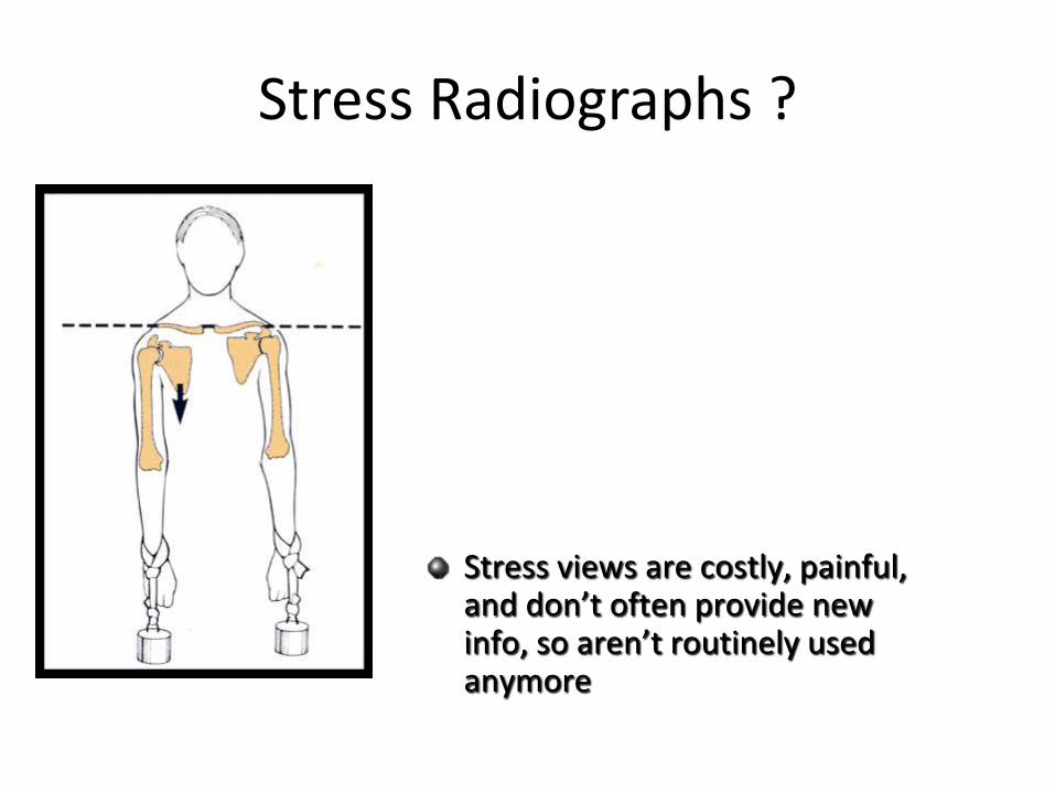
Type VI
• High energy variant
• Result of hyperabduction and external rotation
• Distal clavicle comes to rest in subcoracoid position

Summary

Treatment I and II• Nonsurgical management is uniformly recommended for type I
and II injuries
• A period of immobilization in a sling for comfort until pain subsides– Usually 7-10 days for type I, up to 2 weeks for type II
• Possible anesthetic injection for return to
high level play and labour.
• Once acute pain has subsided rehabilitation
program is instituted

DISADVANTAGES OF NON OPERATIVE TREATMENT
• SKIN PRESSURE AND ULCERATION
• RECURRENCE OF DEFORMITY
• WEARING A BRACE FOR LONG TIME(8 WEEKS)
• POOR PATIENT COOPERATION
• INTERFERENCE WITH DAILY ACTIVITIES
• LOSS OF SHOULDER AND ELBOW MOTION
• SOFT TISSUE CALCIFICATIONS
• LATE ACROMIOCLAVICULAR ARTHRITIS
• LATE MUSCULAR ATROPHY,FATIGUE AND WEAKNESS.

• Mouhsine et al JSES 2003– 33 patients Grade I and II injuries
treated conservatively
– 27% required surgery within 36 months (6 distal clavicle excision, 3 Weaver-Dunn)
– 24 pts remaining assessed 6 yrs post injury both clinically and radiographically
– Only 16% patients with no radiographic degenerative changes or osteolysis evident

• Mikek AJSM 2008
– 23 patients with Type I and II AC Disruption with 10 year Follow-up
– 52% reported occasional symptoms
– Constant score 70.5 injured vs 86.8 (P < .001)
– UCLA score 24.1 vs 29.2 (P < .001)
– Simple Shoulder Test 9.7 vs 10.9 (P < .002)

Rehabilitation
• Early focus is on passive and active ROM
• Once symmetric and painless ROM achieved then progress to isometric shoulder strengthening
• Isotonic strengthening is next with gradual escalation of strength and endurance with return to sport in mind
• Return to sport is not allowed until painless/full ROM is achieved and strength has returned.
• This may take longer for type II injuries, and some recommend contact sports/heavy lifting should be avoided for 2-3 months

Type III injuries
• In 1974 Powers and Bach reported that 92% of 116 type III injuries were treated operatively
• Of 163 ortho residency program chairmen surveyed at that time 91.5% advocated surgical treatment
• In 1992 Cox surveyed 231 chairmen and 62 orthopedists participating in care of athletes– 72% of chairmen favored non-op management– 86% of team orthopedists favored no-op management
• trend toward non-surgical management is well supported in the literature

Natural History of Type III
• Schlegel et al AJSM 2001
– Prospective study non-operative treatment of 20 patients with Type III AC injuries assessed strength, ROM, subjective questionnaire
– Ave Sling use: 8 days (2-25)
– Ave return to work: 9 days (1-24)
• 7 professionals, 8 laborers, 2 students, 3 unemployed/retired
– Analgesic discontinued: 1 wk (15); 2 wk (5)

Natural History of Type III
• Schlegel et al 1 year results
• All had full, pain free, symmetric ROM
• No statistical difference in dynamometer strength
• A statistically significant 17% decrease in bench press strength on injured side was noted
• 80% favorable subjective results
• 20% unfavorable – 3 of 4 secondary gain bias
– Only 1 of 4 elected to undergo surgical intervention

Non-op management
• Galpin et al 1984, retrospective review comparing outcome in type III injuries, 21 treated non-op, 16 with surgery (Bosworth screw and ligament repair) avg 3 yr f/u– Showed overall chances of late pain or altered function were not statistically different
– Surgical patients took longer to become pain free, and longer to return to work• 2.8 vs. 4.5 months, and 2.6 vs. 6.8 weeks, respectively
– Numbers were limited to correlate treatment with patient demands
• Glick et al 1977, retrospective review of 35 AC dislocations treated non-operatively.– 29/35 had no pain, none had disabling pain, 31/35 had no weakness, none had disabling weakness
– None of the patients who had supervised rehabilitation complained
– 8/10 throwers were not affected while throwing, two were professional quarterbacks and one a collegiate javelin thrower
– Concluded that complete reduction not necessary for satisfactory function

Randomized prospective trial
• Bannister et al 1989 – 60 patients with acute AC dislocations random number drawn to allocate operative vs. no-op treatment– Faster return to work for manual and clerical workers treated non-operatively
– After 4 years of f/u no real difference between the two groups in terms of pain/function with one exception
– In the 12 dislocations with more severe dislocation, i.e.. Type V, surgery gave better results
• Concluded that younger patients with severe displacement are more likely to achieve an excellent result if stabilized early
• Felt that surgical treatment created greater morbidity in the lesser grades of injury

Slight strength loss?
• Wojtys et al, 1989, retrospective review of 22 patients with type III treated non-operatively– Showed that laborers and athletes can recover strength and endurance, return
to pre-injury level of activity without surgery
– Strength testing showed some statistically insignificant strength loss, indicating that the strength and endurance advantage one might expect of the dominant arm may be lessened or lost
• May be a factor to consider for those requiring high levels of shoulder strength for work/athletics, or those involved in highly repetitious endurance activities such as swimming/pitching
• Rarity of type III AC separation precludes study of significant numbers with controls to determine treatment that is best for athletes who rely on their elite throwing ability

McFarland MLB Survey Study 1997
American Journal of
Orthopedics, Nov 1997

Phillips et. al CORR 1998 Meta-analysis Type III AC Injury
OP vs NON-OP
• Pain absent or minimal 93% 95%
• ROM normal/near normal 86% 95%
• Strength normal /near normal 87% 91%
• Subsequent surgery 59% 6%

Operative results and Timing
• Weinstein et al AJSM 1995– 44 patients Type III Injury
• 27 acute, 17 late repairs
• CC nonabsorbable suture repair/reconstruction
• 15/27 and 17/17 CA transfer
– 89% satisfactory results, 93% return to sports
– Timing Acute (<3 wk) vs Late (> 3wk)• Satisfactory results 96% vs 77%
• Loss of reduction 15% vs 29%

Types IV, V, IV
• All require operative intervention
• All stabilizers, static and dynamic are injured

Treatment Old
• Not well tolerated, Dermal Complications
Surgical management
• Fixation across AC joint
• Fixation between coracoid and clavicle
• Ligament reconstruction
• Distal clavicle excision

ANY SURGICAL PROCEDURE FOR AC JOINT DISLOCATION SHOULD FULFILL THREE REQUIREMENTS
• AC JOINT MUST BE EXPOSED AND DEBRIDED
• CC AND AC LIGAMENTS MUST BE REPAIRED OR RECONSTRUCTED
• STABLE REDUCTION OF THE AC JOINT MUST BE OBTAINED
Achieving these three goals , no matter how the joint is fixed , should give acceptable results.

DISADVANTAGES OF SURGICAL MANAGEMENT
• INFECTION
• HEMATOMA FORMATION
• ANAESTHETIC RISK
• SCAR FORMATION
• RECURRENCE OF DEFORMITY
• METAL BREAKAGE, LOOSENING,MIGRATION
• SECOND SURGERY FOR REMOVAL
• BREAKAGE OR LOOSENING OF SUTURES
• EROSION OR FRACTURE OF DISTAL CLAVICLE
• LATE ARTHRITIS AND LOSS OF JOINT MOTION.

Acromioclavicular Fixation
• Pin fixation
• Has been abandoned since reports of rare pin migration
– Heart, Lung, Great vessels

Acromioclavicular fixation
• Hook Plate
• Only used for acute injury
• Requires subsequent surgery for removal
Fixation between coracoid and clavicle
• Bosworth popularized the use of a screw for fixation of the clavicle to the coracoid
• This technique initially did not include recommendation for repair or reconstruction of the CC ligaments
• Today the use of screws and suture loops has been described alone and in combo with ligament reconstruction
• Placement of synthetic loops between the coracoid and clavicle can be done arthroscopically, main advantage: doesn’t require staged screw removal


Ligament reconstruction
• Weaver and Dunn were the 1st to describe transfer for the native CA ligament to reestablish AC joint stability
• Their technique described excision of the distal clavicle with this ligament transfer
• Construct can be augmented with a suture loop for protection until the transferred ligament heals

Ligament Reconstruction
Open and Arthroscopic techniques
Restore Anatomy
orthoillustrated.com

Anatomic Ligament Reconstruction
• Alternative technique is use of semitendinosus autograft for reconstruction– Loop around or fix into coracoid, then fix through two separate clavicle bone
tunnels to approximate normal anatomic location of CC ligaments
• Recent biomechanical studies have demonstrated the superiority of this construct

Biomechanical studies • Lee et al., 2003 – 11 cadaveric shoulders tensile tested to failure
comparing suture loop, CA transfer, and free tendon reconstruction.– Reconstructions found to have failure strengths as strong as those of native CC
ligaments
– CA transfer was the weakest construct, and shows that greatest elongation at failure
• Concluded tendon graft reconstruction to be an alternative to CA ligtransfer possibly providing a permanent biologic reconstruction
• Given its biomechanical properties similar to native CC ligaments, reconstruction with tendon graft may allow for shorter post op immobilization and accelerated rehab program

Biomechanical studies
• Mazzocca et al, 2006, studied 42 cadaveric specimens comparing stability of 3 AC joint reconstruction techniques– Anatomic CC reconstruction with tendon graft provided ant, post, and
superior stability similar to intact state
– Modified Weaver-Dunn had significantly greater laxity compared to anatomic CC recon and arthroscopic reconstruction
• Concluded that anatomic reconstruction with free tendon graft may provide stronger, more permanent biologic solution for AC joint dislocation

Newer proposed techniques• Grutter and Petersen 2005 AJSM
(Cadaveric study)
• Anatomic Reconstruction using FCR graft to reconstruct CC and superior AC ligament
• Similar strength as native AC joint in coronal plane

Newer Proposed Techniques
• Freedman et al AJSM 2010 (Cadaveric study)
• Intramedullary AC reconstruction with 5 cm semitendinosus graft and fibertape
• Did not reproduce similar stiffness, load to failure, and energy absorption as intact AC complex

Newer proposed techniques
Improved horizontal stability with
addition of intramedullary AC
ligament reconstruction.
-50% less AP translation
No difference in:
- Superior/inferior translation
- Load to failure
AJSM 2010

SummaryCHILDREN LESS THAN 15 YEARS OF AGE :
TYPE I,II,III : NON OPERATIVE TREATMENT
TYPE IV,V,VI : a) CLOSED REDUCTION OF CLAVICLE b) OPEN REDUCTION AND REPAIR

Summary
AGE BETWEEN 15-45 YEARS :• Type I, II
– restricted activity initially– Analgesic, Injection– Rehabilitation
• Type III – Controversial- trends toward initial non-operative management– Late reconstruction if symptomatic– Optimal strategy has changed and alternated over time
• Type IV,V, VI– Repair/reconstruction

Summary
AGE MORE THAN 45 YEARS :
TYPE I,II,III : NON OPERATIVE
TYPE IV,V,VI : Surgery(Distal clavicle excision)

Complications• Non operative
– Pain
– Post traumatic arthritis/osteolysis
– Possible neurologic injury
– Possible strength deficit
• Surgical – Same as above plus…
– Clavicle or coracoid fracture
– Loss of reduction
– Pneumothorax
– Neurovascular injury

Case

• HPI: 21 yo R HD male college student presents to clinic after fell off bike over the handle bars onto his R shoulder
• Exam:
– Ecchymosis, Gross deformity Right AC joint
– Tenderness distal clavicle and CC interspace
– Distally No Neurovascular Injury

Radiographs

Post op
• Acute CC repair with Fiberwire Suture Loops

![Presentation1.ppt [โหมดความเข้ากันได้] · Title: Microsoft PowerPoint - Presentation1.ppt [โหมดความเข้ากันได้]](https://img.pdfslide.net/doc/110x75/5ec776d210d7bd5f6f00774b/aaaaaaaaaaaaaaaaaa-title-microsoft-powerpoint.jpg)