Embed Size (px)
Citation preview

HUDSON DE CARVALHO NAKAMURA, MD, CBO
FUNDAÇÃO BANCO DE OLHOS DE GOIÁS
GOIÂNIA, BRAZIL
www.fubog.org
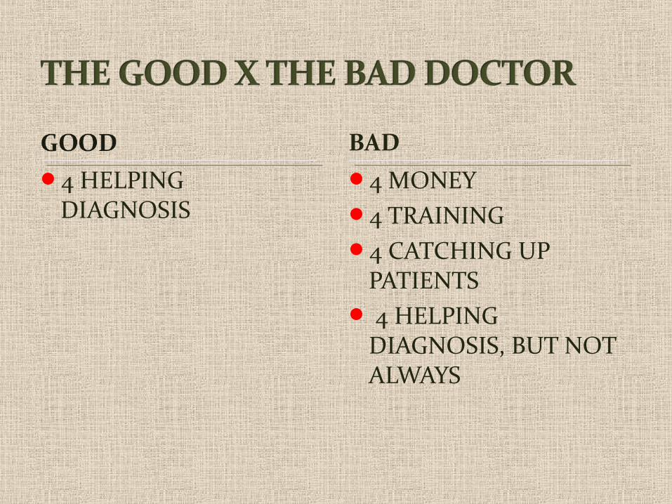
4 HELPING DIAGNOSIS
4 MONEY4 TRAINING4 CATCHING UP
PATIENTS 4 HELPING
DIAGNOSIS, BUT NOT ALWAYS
BAD
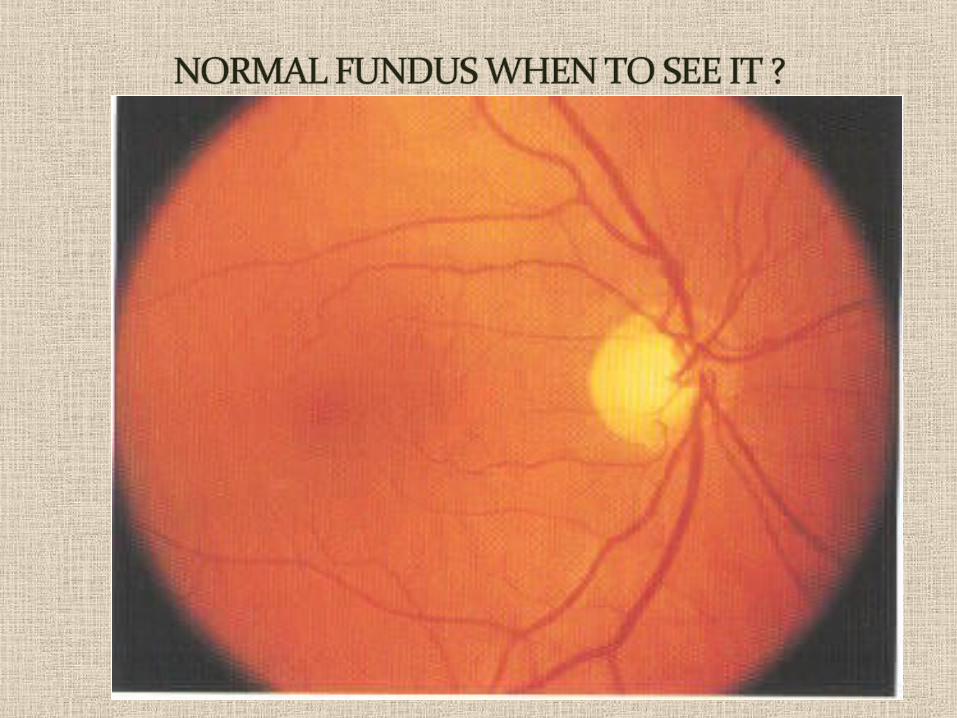
APPERANCE : Uniform red Punctate stippling-periphery Varies-color of individualNormal choroidal vessels,invisible
PARTS : DISC VESSELS
MACULA
PERIPHERY
RED EYELOW VISIONOCULAR PAINFLOATERS & FLASHESMETAMORPHOPSIASSCOTOMASMICROPSIASIMAGE DISTORTIONDIPLOPIA – MONOCULAR AND BINOCULAROUTROS
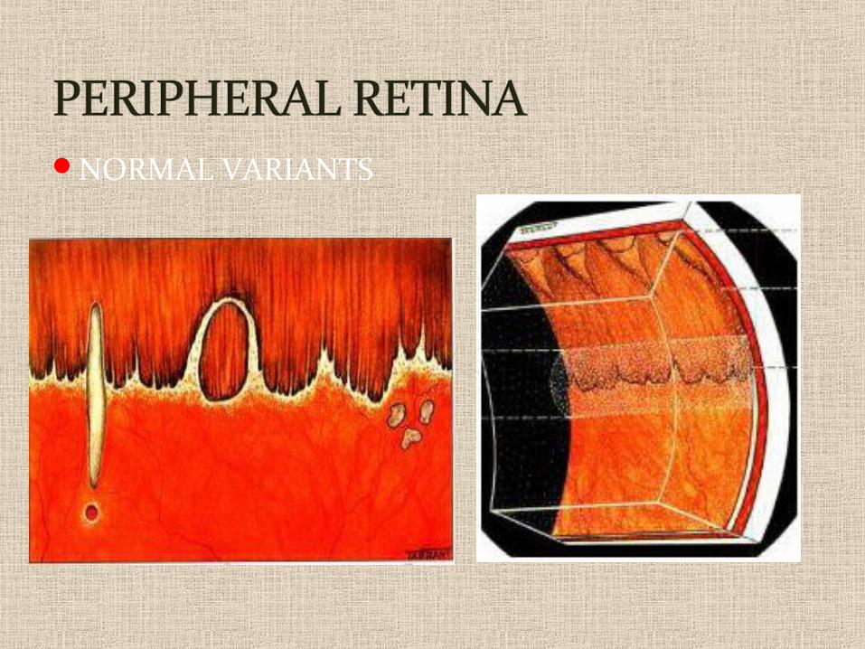
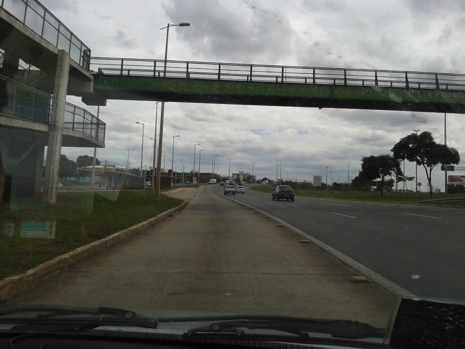
NORMAL VARIANTS
Why it is performed:
It can detect some signs & physiological effects of various circulatory, metabolic and neurological disorders.
Routinely used to assess and diagnose vitro-retinal diseases (such as Diabetic retinopathy, retinal tear and detachment, macular hole, retinal haemorrhage, retinal artery and vein occlusion, choroidal tumor, or macular edema), optic nerve defects, and hereditary diseases.

Fundus examination is used to:
Identify and locate vitro-retinal and optical nerve defects caused by eye diseases or trauma.
Examine the extent of the defects or abnormalities to plan a proper treatment.
Evaluate the success of treatment.
DIRECT OPHTHALMOSCOPYRED REFLEX
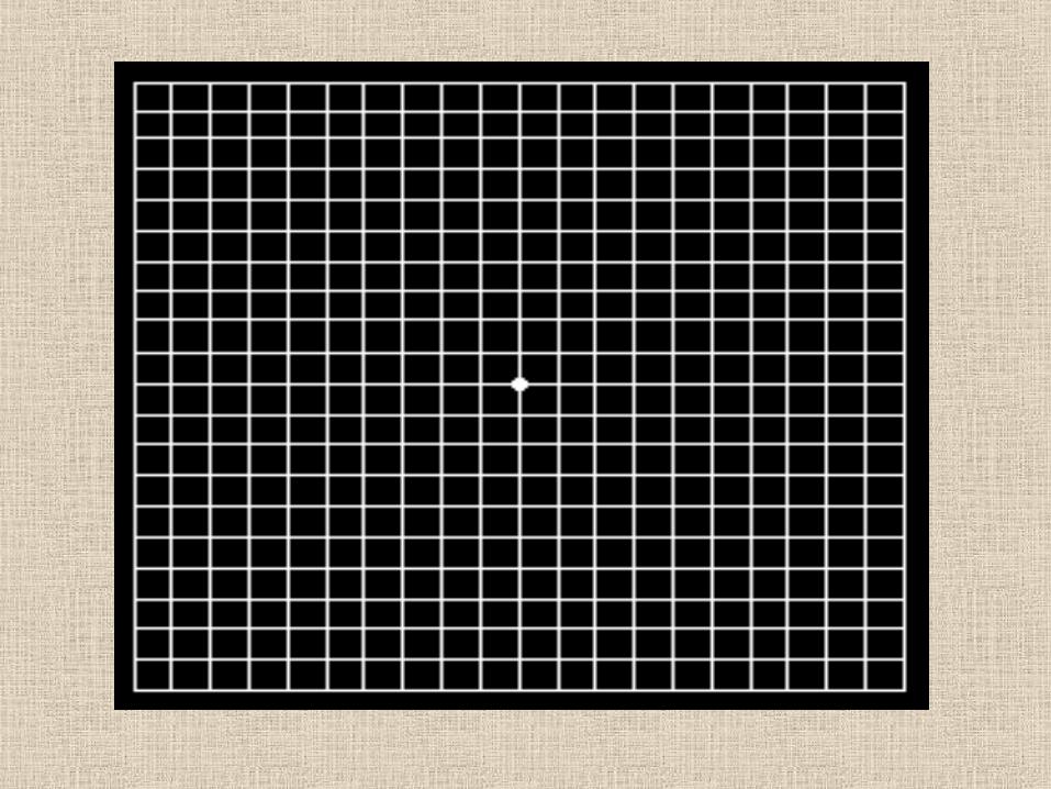
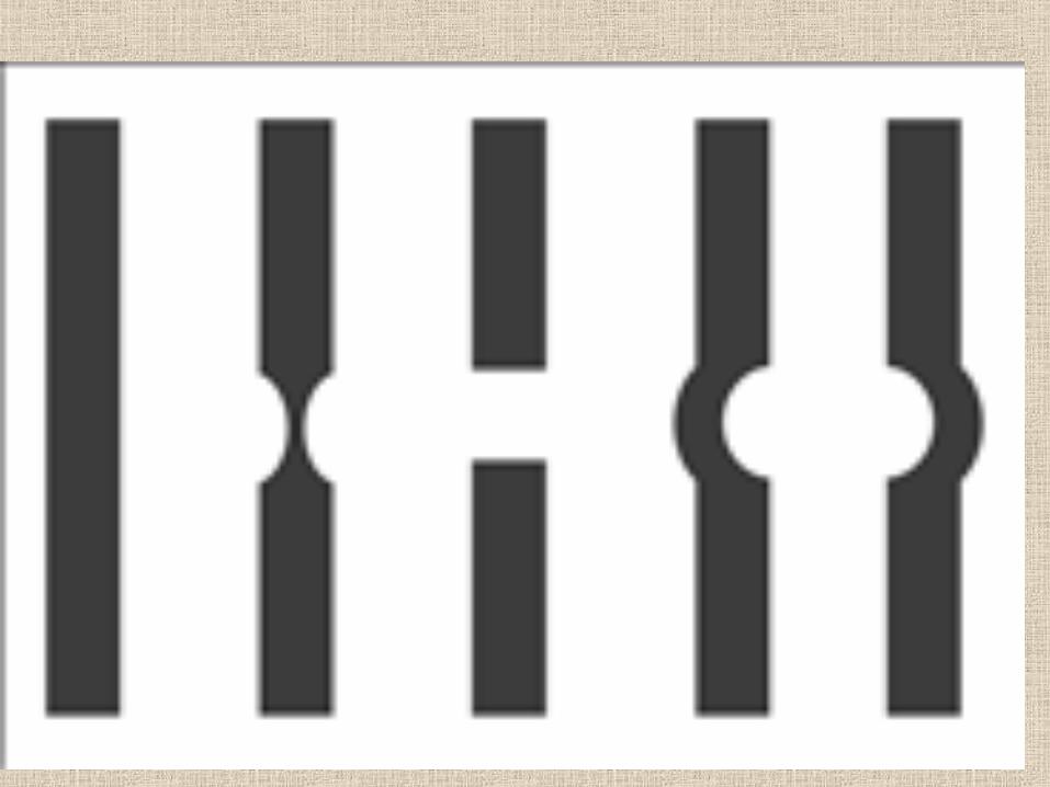
INDIRECT OPHTHALMOSCOPYAMSLER GRID TESTINGWATZKE ALLEN TEST
EYE TEST FOR ROP SCREENING
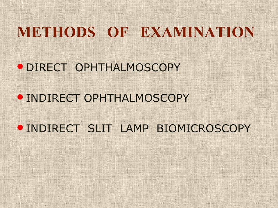
METHODS OF EXAMINATION
DIRECT OPHTHALMOSCOPY
INDIRECT OPHTHALMOSCOPY
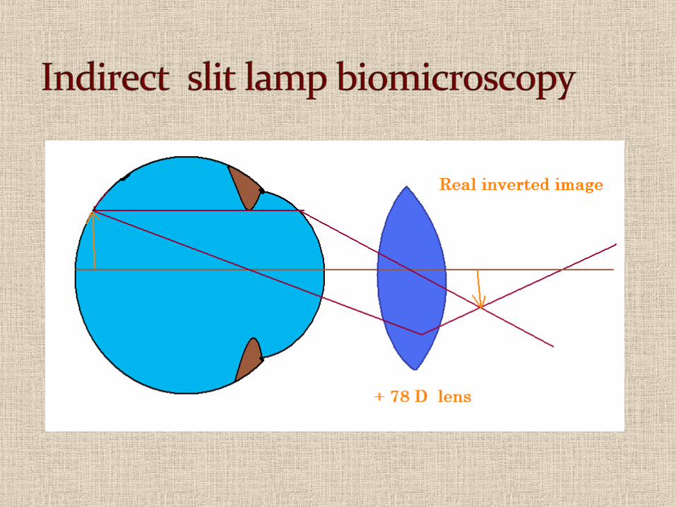
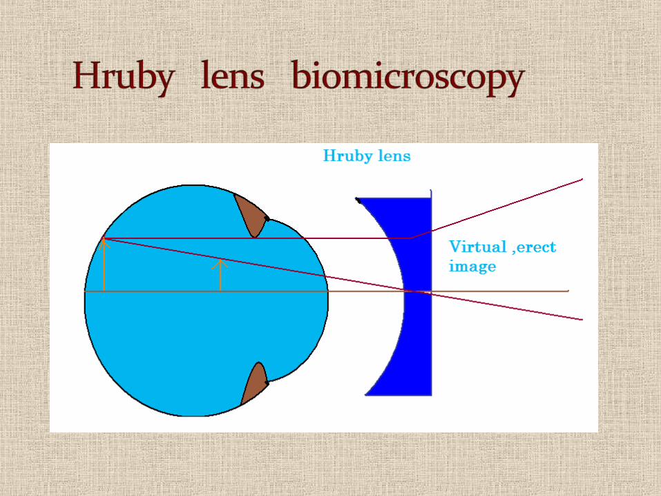
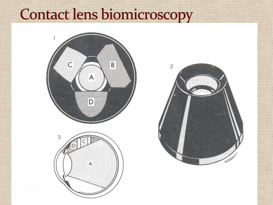
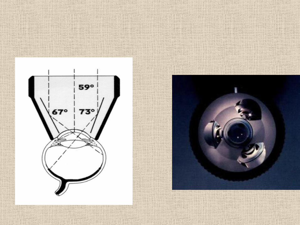
INDIRECT SLIT LAMP BIOMICROSCOPY
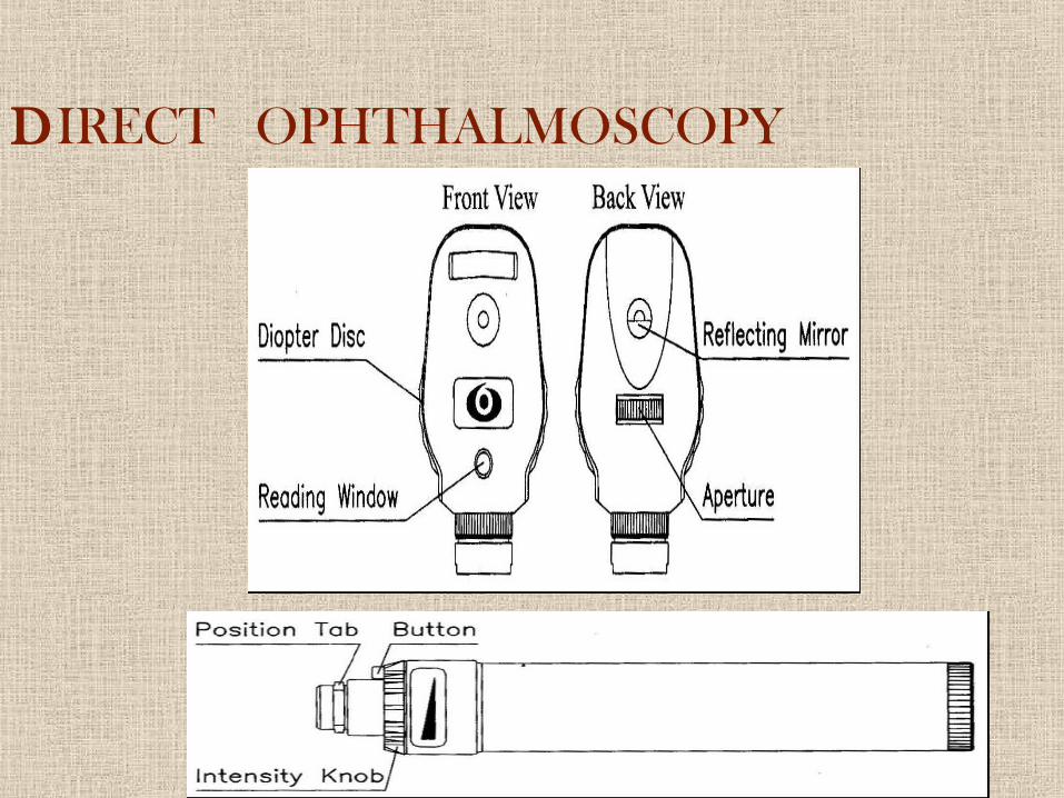
DIRECT OPHTHALMOSCOPY
DIRECT OPTHALMOSCOPE
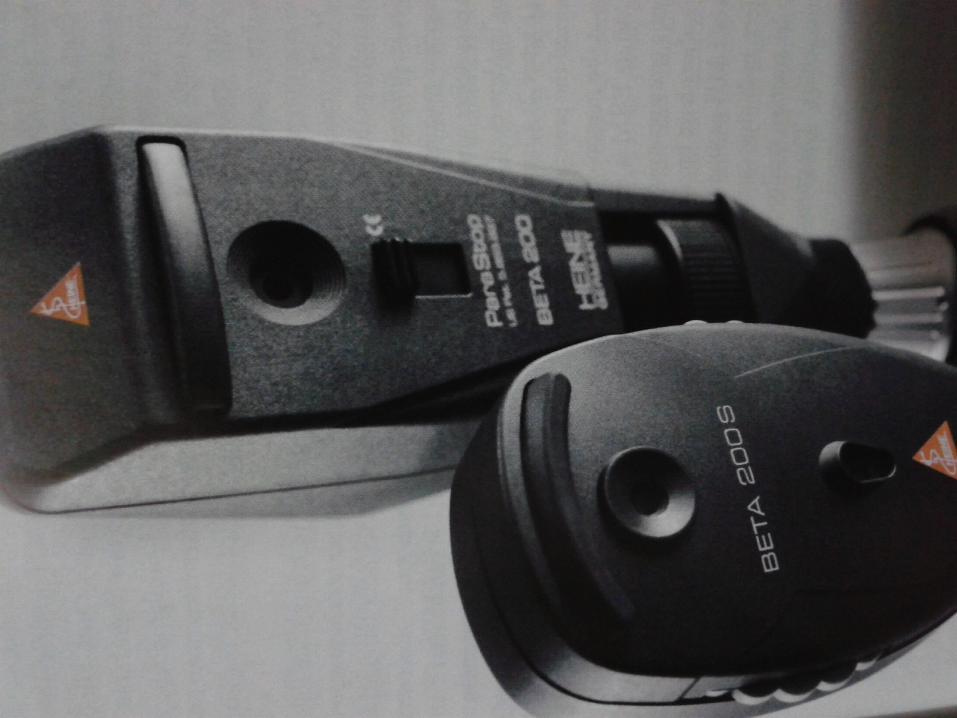
INDIRECT OPHTHALMOSCOPE
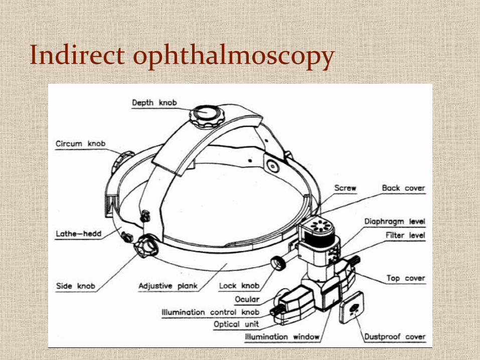
Indirect ophthalmoscopy

LOOK FOR DISTORTIONS, CROOKED LINES, SCOTOMAS, OTHER IRREGULARITIES
SPECIALLY 4 MAC HOLES
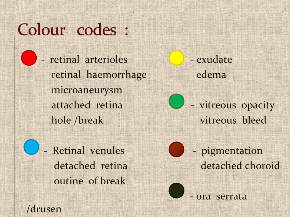
- retinal arterioles - exudate retinal haemorrhage edema microaneurysm attached retina - vitreous opacity hole /break vitreous bleed
- Retinal venules - pigmentation detached retina detached choroid outine of break - ora serrata
/drusen hyperpigmentation
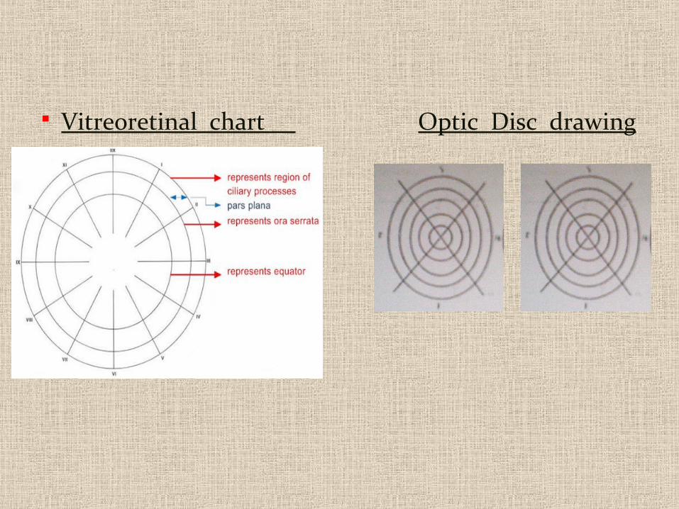
Vitreoretinal chart Optic Disc drawing
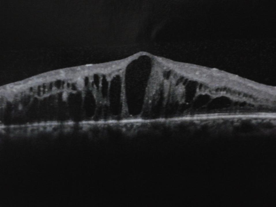
ERM – CME – CNM – ME – CSME – POST-UVEITIS – POST INJECTIONS – MAC HOLES – PSEUDO MAC HOLES
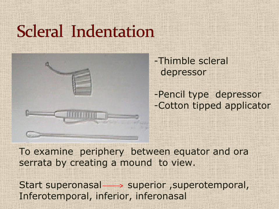
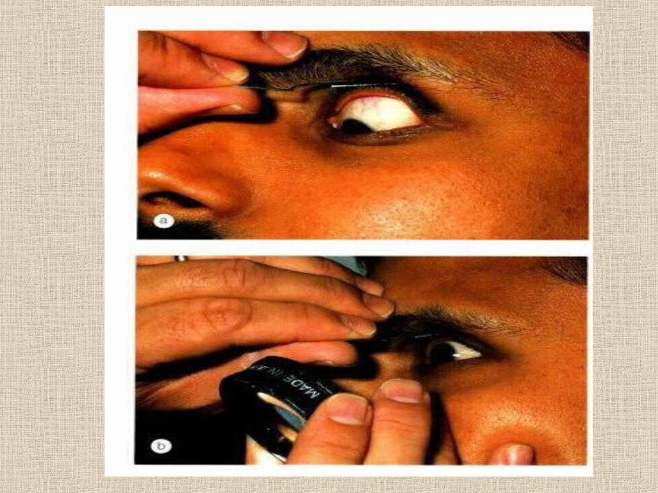
-Thimble scleral depressor
-Pencil type depressor-Cotton tipped applicator
To examine periphery between equator and ora serrata by creating a mound to view. Start superonasal superior ,superotemporal,Inferotemporal, inferior, inferonasal
ERM – CME – CNM – ME – CSME – POST-UVEITIS – POST INJECTIONS – MAC HOLES – PSEUDO MAC HOLES – PVD IN
THE X-LATERAL EYE 4 MAC HOLE PREVENTION
ERM – CME – CNM – ME – CSME – POST-UVEITIS – POST INJECTIONS – MAC HOLES – PSEUDO MAC HOLES
CMENON CME, DIABETIC
OTHERS
MEDIA OPACITY - PVD – UVEITIS – HYALOID ASTERITIS – IOFB – LENS & IOL DISLOCATIONS
B-scan ultrasound is most useful when direct visualization of intraocular structures is difficult or impossible. Situations that prevent normal examination include lid problems (eg, severe edema, partial or total tarsorrhaphy), keratoprosthesis, corneal opacities (eg, scars, severe edema), hyphema, hypopyon, miosis, pupillary membranes, dense cataracts, or vitreous opacities (eg, hemorrhage, inflammatory debris).

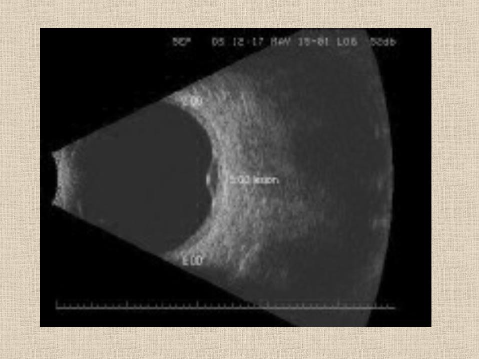
NO MEDIA OPACITY – WHAT FOR?
In such cases, diagnostic B-scan ultrasound can accurately image intraocular structures and give valuable information on the status of the lens, vitreous, retina, choroid, and sclera. However, in many instances, ultrasound is used for diagnostic purposes even though pathology is clinically visible. Such instances include differentiating iris or ciliary body lesions; ruling out ciliary body detachments; and differentiating intraocular tumors, serous versus hemorrhagic choroidal detachments, rhegmatogenous versus exudative retinal detachments, and disc drusen versus papilledema.
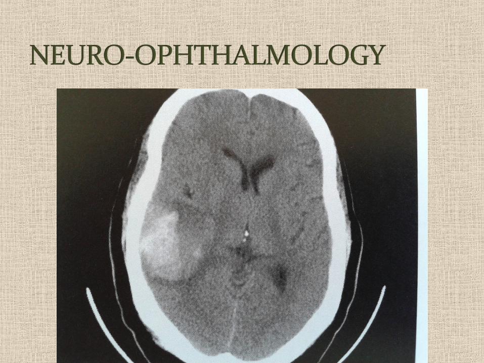
VISUAL EVOKED POTENTIAL - VEP
The visually evoked potential (VEP) measures the electrical response of the brain's primary visual cortex to a visual stimulus.
NEURO-OPHTH DISORDERSOPTIC PATHWAY DISORDERSEXCLUDING CRITERIAMEDICAL LEGALOTHERS
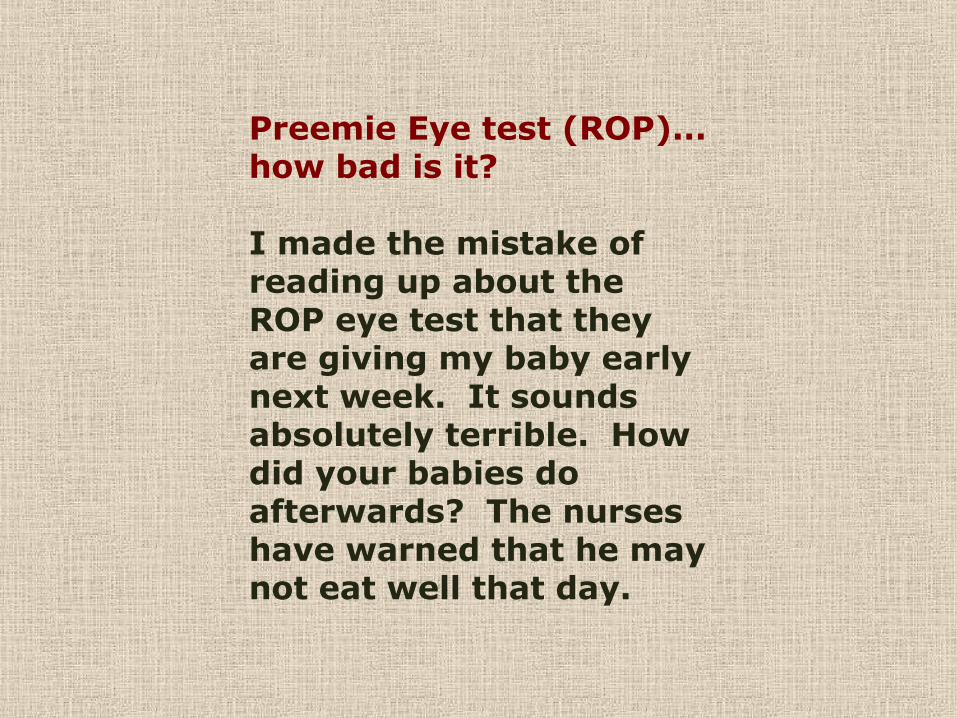
Preemie Eye test (ROP)... how bad is it?
I made the mistake of reading up about the ROP eye test that they are giving my baby early next week. It sounds absolutely terrible. How did your babies do afterwards? The nurses have warned that he may not eat well that day.
Your Baby’s Eye Test
Why your baby needs an eye test
All babies on the neonatal unit are screened for an
eye condition called retinopathy of prematurity
(ROP) if they are born before 32 weeks of
pregnancy or if they weighed 1500 grams or less
at birth.
ERM – CME – CNM – ME – CSME – POST-UVEITIS – POST INJECTIONS – MAC HOLES – PSEUDO MAC HOLES


NO DIABETIC RETINOPATHY WHAT FOR ???NO POSTERIOR POLE CHANGES WHAT FOR ???NO KNOWN ALLERGIES AND THE TWO ABOVE WHAT FOR ???NO CHANGES SO MEDICAL LEGAL ASPECT WHAT FOR ???
THINK ABOUT

CONCENTRATION AND QUANTITY
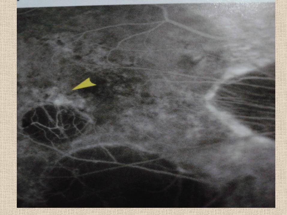
WAIT ENOUGH TIME OTHERWISE WAIST ALL THE TIME
EXAMPLE : MAC EDEMA DUE TO DIABETIC RETINOPATHY

THANK YOU


![II./2.3. Examination of cranial nervesII./2.3.2. Examination of vision (Optic nerve [2nd cranial nerve]) Anatomy: The visual pathway originates from the ganglion cells of the retina,](https://img.pdfslide.net/doc/110x75/5f61f204b901471ec658d72f/ii23-examination-of-cranial-nerves-ii232-examination-of-vision-optic-nerve.jpg)