Embed Size (px)
Citation preview

TUBERCULOSIS OF HIP JOINT
INTRODUCTIONNext to spine, hip joint is the most
common site for involvement by tuberculosis.
Mostly common first 3 decade of life like other osteo – articular disease.
It constitute 15 percent of osteoarticular tuberculosis.
PATHOLOGY Infection of hip is secondary to some primary focus
either in lungs or mediastinal node or iliocaecal region and spread to hip by blood stream.
Initial focus may start in acetabular roof > epiphysis ( head ) > Metaphysis or neck ( Babcock triangle ) > greater trochanter . Rarely the disease may start in synovial membrane and may remain as synovitis for months.
When initial focus is acetabular roof -- joint involvement is late and severity of symptom is mild – by the time pt. report to hospital extensive destruction already present .
TB of greater trochanter may involve the trochanteric bursa without involving the hip for long time .
As the upper end of femur is entirely intracapsuler the joint get involve rapidly and disease become osteoarticular
Cold abcess in joint – perforate inferior weaker part of capsule rarely acetabular roof – cold abcess can present anywhere around the hip ( femoral triangle , medial ,post and lateral side of thigh ,ischeo – rectal fossa , pelvis )
CLINICAL FEATURES Insidious in onset
Pain and swelling in the hip and limping are the usual presenting symptoms
Sometimes there is referred pain in the knee and is often misleading.
Pain is maximum at end of day. Child may wake up from sleep due to pain(night cry)
Constitutional symptom like loss of appetite, loss of weight, fever
Limp is the earliest and commonest symptom
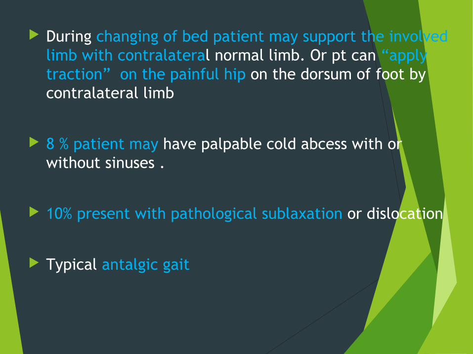
During changing of bed patient may support the involved limb with contralateral normal limb. Or pt can “apply traction” on the painful hip on the dorsum of foot by contralateral limb
8 % patient may have palpable cold abcess with or without sinuses .
10% present with pathological sublaxation or dislocation
Typical antalgic gait

STAGES OF T.B. HIP
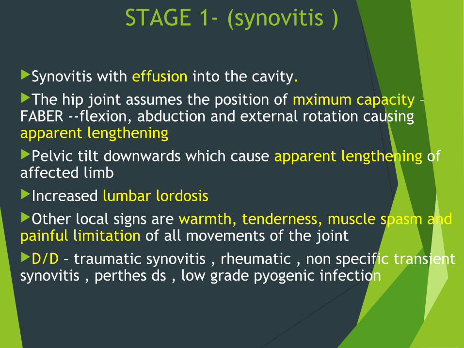
STAGE 1- (synovitis )
Synovitis with effusion into the cavity.The hip joint assumes the position of mximum capacity –FABER --flexion, abduction and external rotation causing apparent lengthening Pelvic tilt downwards which cause apparent lengthening of affected limb Increased lumbar lordosisOther local signs are warmth, tenderness, muscle spasm and painful limitation of all movements of the jointD/D – traumatic synovitis , rheumatic , non specific transient synovitis , perthes ds , low grade pyogenic infection
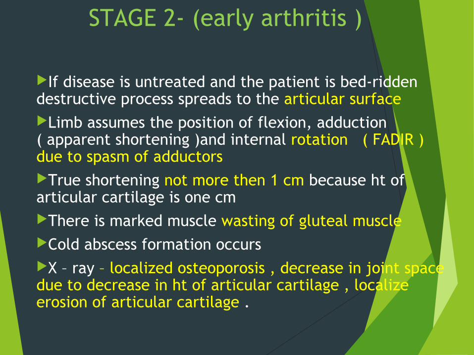
If disease is untreated and the patient is bed-ridden destructive process spreads to the articular surfaceLimb assumes the position of flexion, adduction ( apparent shortening )and internal rotation ( FADIR )due to spasm of adductorsTrue shortening not more then 1 cm because ht of articular cartilage is one cm There is marked muscle wasting of gluteal muscleCold abscess formation occurs X – ray – localized osteoporosis , decrease in joint space due to decrease in ht of articular cartilage , localize erosion of articular cartilage .
STAGE 2- (early arthritis )

Stage 3(advanced arthritis)
Clinical sign of stage 2 is exaggerated
Gross destruction of articular cartilage and femoral head and acetabulam
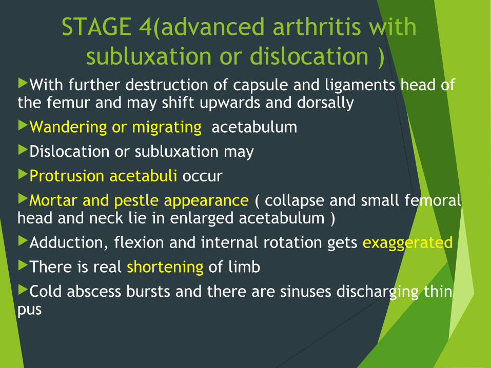
STAGE 4(advanced arthritis with subluxation or dislocation )
With further destruction of capsule and ligaments head of the femur and may shift upwards and dorsally Wandering or migrating acetabulumDislocation or subluxation may Protrusion acetabuli occur Mortar and pestle appearance ( collapse and small femoral head and neck lie in enlarged acetabulum )Adduction, flexion and internal rotation gets exaggeratedThere is real shortening of limbCold abscess bursts and there are sinuses discharging thin pus
Hip may not assume the posture of triple deformity of F- AD – IR instead hip may assume F – AB – ER . This may be due to continuous adoption of of lateral aspect of thigh of diseased hip resting on bed or due to destruction of ilio – femoal ligament
If limb has been plastered more than 12 month as in first half of twentieth century growth plate around the knee may get closed – frame knee
Coxa megna , coxa vulgus , coxa vara
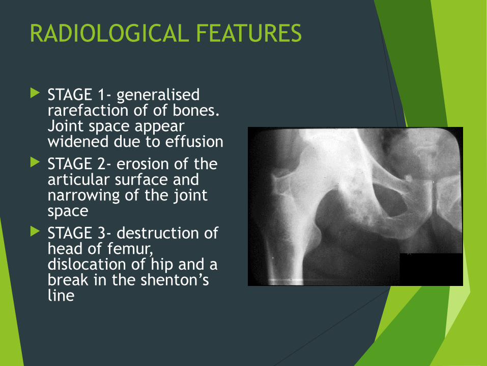
RADIOLOGICAL FEATURES
STAGE 1- generalised rarefaction of of bones. Joint space appear widened due to effusion
STAGE 2- erosion of the articular surface and narrowing of the joint space
STAGE 3- destruction of head of femur, dislocation of hip and a break in the shenton’s line
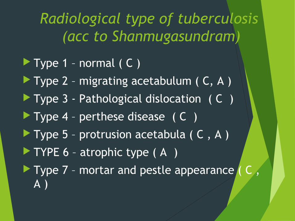
Radiological type of tuberculosis (acc to Shanmugasundram)
Type 1 – normal ( C ) Type 2 – migrating acetabulum ( C, A ) Type 3 - Pathological dislocation ( C ) Type 4 – perthese disease ( C ) Type 5 – protrusion acetabula ( C , A ) TYPE 6 – atrophic type ( A ) Type 7 – mortar and pestle appearance ( C ,
A )
Hyperamia – large head and neck – coxa megna Thromboembolic phenomina – perthese disease Coxa breva due to dec . In blood supply Restriction growth of capital femoral epiphyseal
plate and normal trochanteric physis – coxa vara
Normal growth of capital femoral epiphyseal plate and Restriction
trochanteric physis – coxa vulga
If joint space is reduced > 3mm – poor prognois
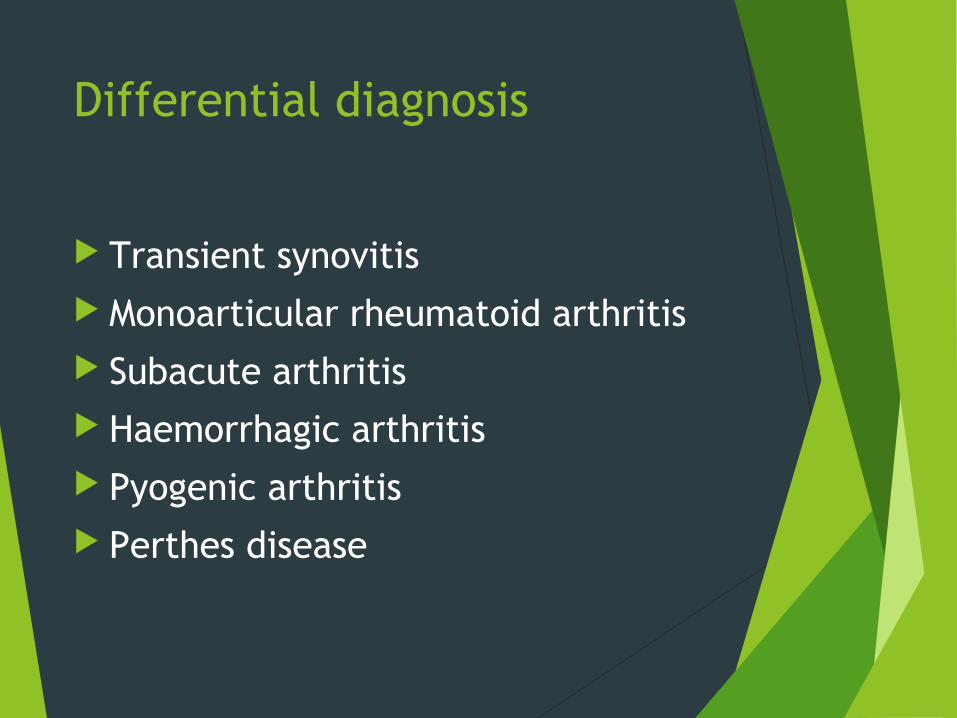
Differential diagnosis
Transient synovitis Monoarticular rheumatoid arthritis Subacute arthritis Haemorrhagic arthritis Pyogenic arthritis Perthes disease
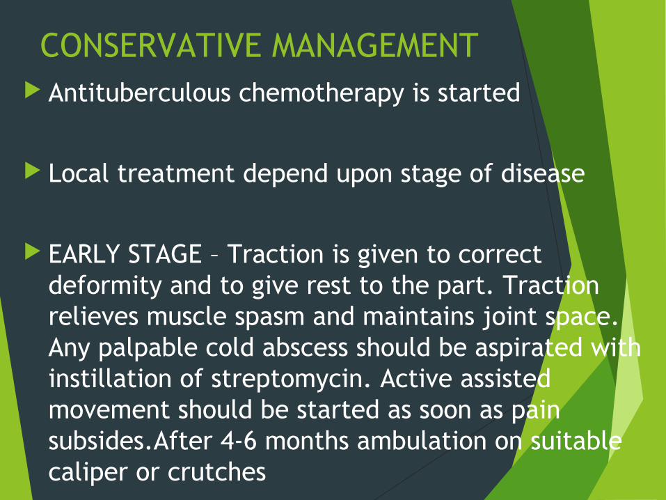
CONSERVATIVE MANAGEMENT Antituberculous chemotherapy is started
Local treatment depend upon stage of disease
EARLY STAGE – Traction is given to correct deformity and to give rest to the part. Traction relieves muscle spasm and maintains joint space. Any palpable cold abscess should be aspirated with instillation of streptomycin. Active assisted movement should be started as soon as pain subsides.After 4-6 months ambulation on suitable caliper or crutches
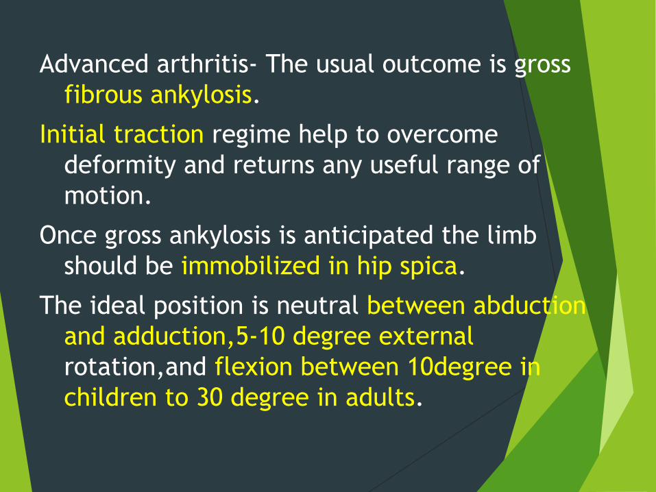
Advanced arthritis- The usual outcome is gross fibrous ankylosis.
Initial traction regime help to overcome deformity and returns any useful range of motion.
Once gross ankylosis is anticipated the limb should be immobilized in hip spica.
The ideal position is neutral between abduction and adduction,5-10 degree external rotation,and flexion between 10degree in children to 30 degree in adults.

PROGNOSIS
Early anti TB drugs – good prognosis Early disease ( synovitis and early arthritis ) –
good prognosis Advanced arthritis – fibrous ankylosis TB may interfere blood supply of head – same as
perthese disease – should be treated like perthes disease with antituberculer coverage

MANAGEMENT
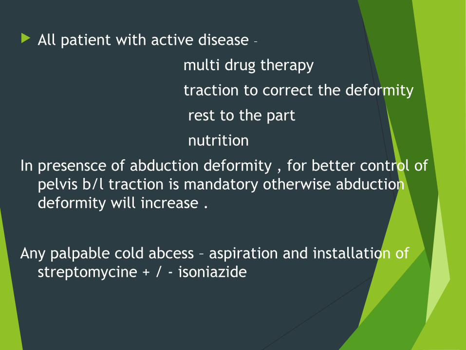
All patient with active disease –
multi drug therapy
traction to correct the deformity
rest to the part
nutrition
In presensce of abduction deformity , for better control of pelvis b/l traction is mandatory otherwise abduction deformity will increase .
Any palpable cold abcess – aspiration and installation of streptomycine + / - isoniazide
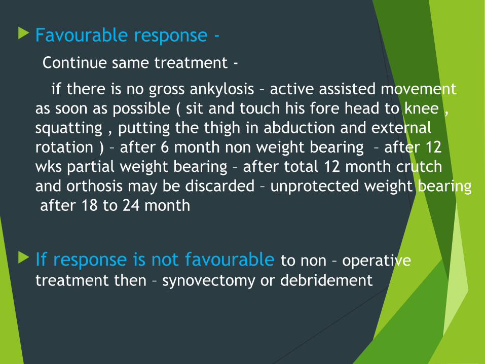
Favourable response -
Continue same treatment -
if there is no gross ankylosis – active assisted movement as soon as possible ( sit and touch his fore head to knee , squatting , putting the thigh in abduction and external rotation ) – after 6 month non weight bearing – after 12 wks partial weight bearing – after total 12 month crutch and orthosis may be discarded – unprotected weight bearing after 18 to 24 month
If response is not favourable to non – operative treatment then – synovectomy or debridement
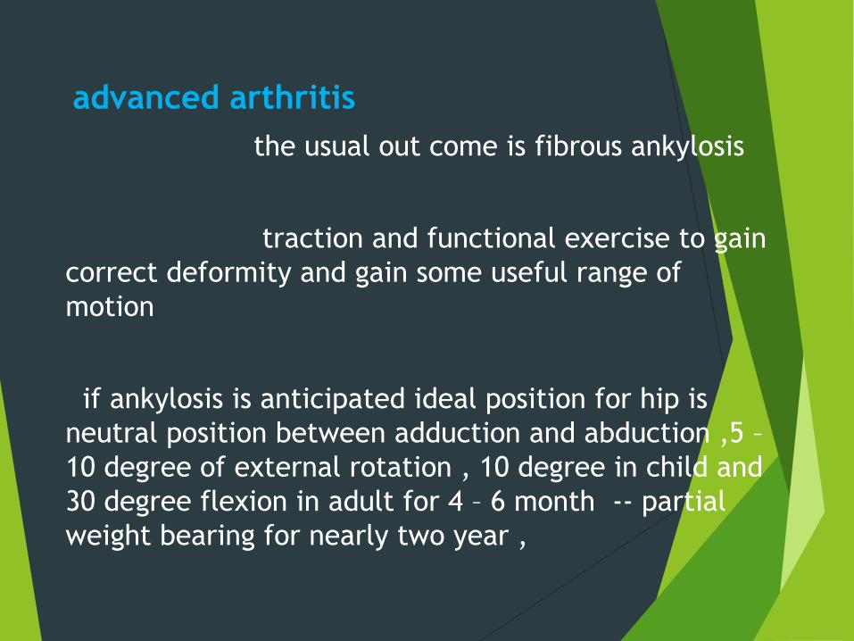
advanced arthritis the usual out come is fibrous ankylosis
traction and functional exercise to gain correct deformity and gain some useful range of motion
if ankylosis is anticipated ideal position for hip is neutral position between adduction and abduction ,5 – 10 degree of external rotation , 10 degree in child and 30 degree flexion in adult for 4 – 6 month -- partial weight bearing for nearly two year ,
Stage of disease and surgical procedure
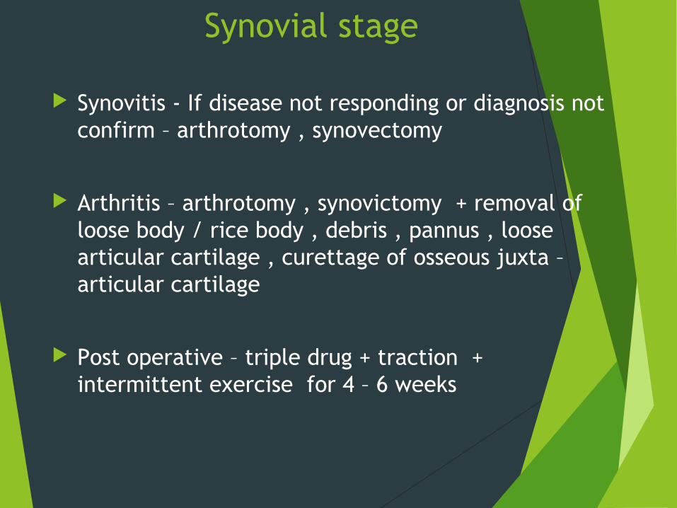
Synovial stage
Synovitis - If disease not responding or diagnosis not confirm – arthrotomy , synovectomy
Arthritis – arthrotomy , synovictomy + removal of loose body / rice body , debris , pannus , loose articular cartilage , curettage of osseous juxta – articular cartilage
Post operative – triple drug + traction + intermittent exercise for 4 – 6 weeks
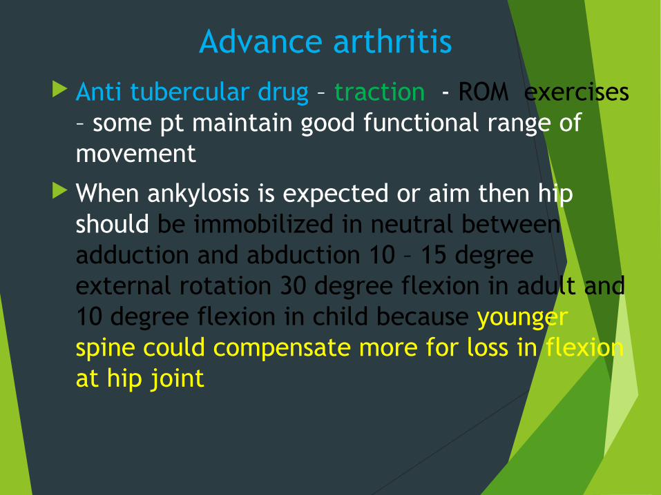
Advance arthritis Anti tubercular drug – traction - ROM exercises
– some pt maintain good functional range of movement
When ankylosis is expected or aim then hip should be immobilized in neutral between adduction and abduction 10 – 15 degree external rotation 30 degree flexion in adult and 10 degree flexion in child because younger spine could compensate more for loss in flexion at hip joint
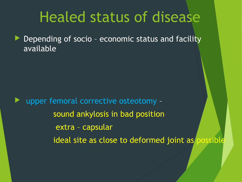
Healed status of disease Depending of socio – economic status and facility
available
upper femoral corrective osteotomy –
sound ankylosis in bad position
extra – capsular
ideal site as close to deformed joint as possible
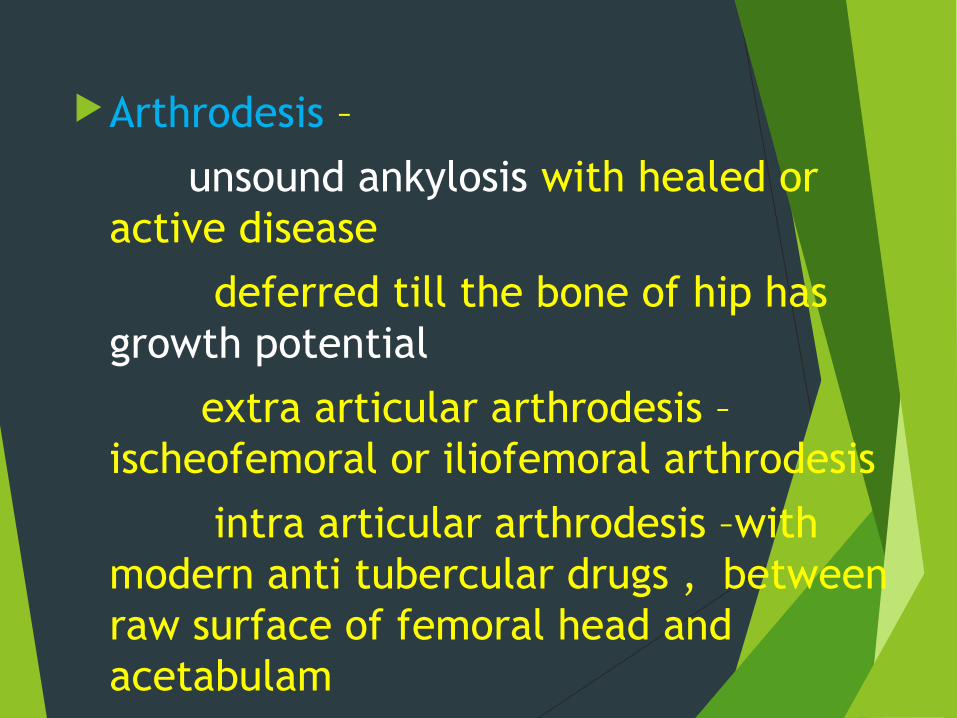
Arthrodesis –
unsound ankylosis with healed or active disease
deferred till the bone of hip has growth potential
extra articular arthrodesis – ischeofemoral or iliofemoral arthrodesis
intra articular arthrodesis –with modern anti tubercular drugs , between raw surface of femoral head and acetabulam
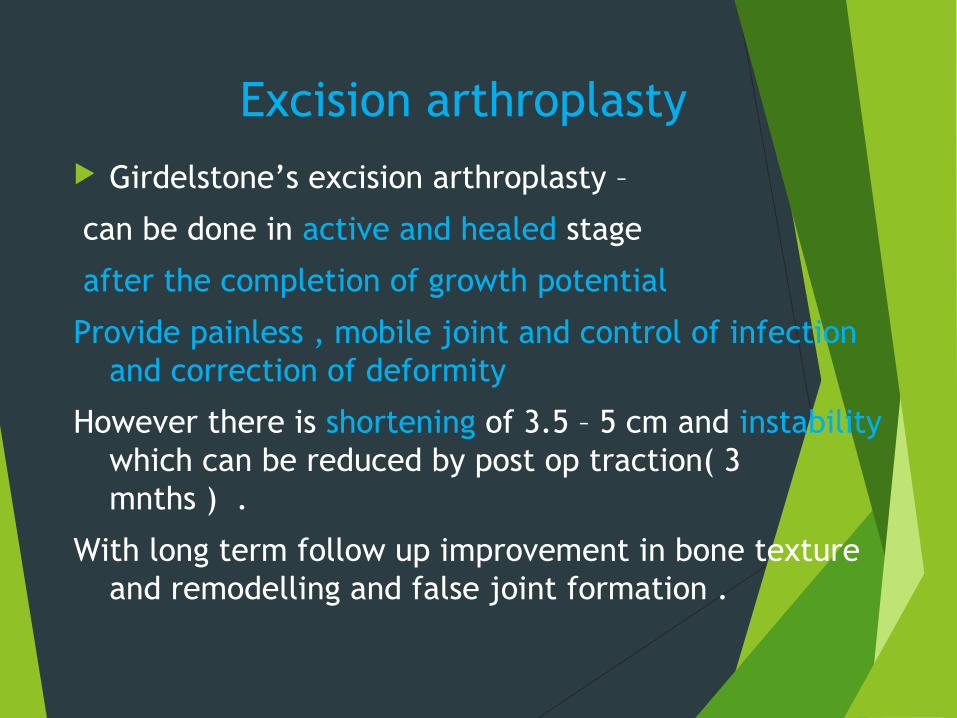
Excision arthroplasty Girdelstone’s excision arthroplasty –
can be done in active and healed stage
after the completion of growth potential
Provide painless , mobile joint and control of infection and correction of deformity
However there is shortening of 3.5 – 5 cm and instability which can be reduced by post op traction( 3 mnths ) .
With long term follow up improvement in bone texture and remodelling and false joint formation .

Joint replacement
After maintaining 5 yrs. of healed status
After replacement 5 months anti tubercular drugs
Still reactivation occure in 1/3 patients


![102 VI TA MIN ZEE (TB) 102 Bay Mare 1988 · Hip No Con signed by Mighty Acres Hip No. 102 VI TA MIN ZEE (TB) 102 Bay Mare 1988 Nearc tic Nearco [Ity] *Lady Angela Explodent Ven om](https://img.pdfslide.net/doc/110x75/5f10985b7e708231d449e153/102-vi-ta-min-zee-tb-102-bay-mare-1988-hip-no-con-signed-by-mighty-acres-hip-no.jpg)
![Hip, Hip, Hooray! - goodsamdayton.org1].pdf · right hip within the month, ... Hip, Hip, Hooray! ... to her new hip. H E A LT H TA L K| O RTHOPEDICS 6. Title: SHTK602-Sum06REVfin](https://img.pdfslide.net/doc/110x75/5ab989bf7f8b9ac1058dfdf4/hip-hip-hooray-1pdfright-hip-within-the-month-hip-hip-hooray-.jpg)


![Appendix 1 HIP Male and Female - University of East Anglia · App14.1!HIP!v3.2_02_05_2012!!!!!Health’Improvement’Profile[HIP]’ ’’’’’’’’’’’’’’’’’’’’’’’’’’’’(HIP)–’Male](https://img.pdfslide.net/doc/110x75/5f0af26b7e708231d42e1f1c/appendix-1-hip-male-and-female-university-of-east-anglia-app141hipv3202052012healthaimprovementaprofilehipa.jpg)