Embed Size (px)
Citation preview
Laboratory diagnosis in infections produced by the genera:
Vibrio, Campylobacter, Helicobacter, Pseudomonas
http://www.slideshare.net/DanaSinzianaBreharCi/vibrio-campylobacter-helicobacterpseudomonas
Genus Vibrio
• Family Vibrionaceae:
– Genera: • Vibrio – species clinically significant for human pathology:
– Vibrio cholerae– Vibrio parahaemolyticus– Vibrio vulnificus
• Aeromonas
• Plesiomonas
Vibrio cholerae
Classification (~structure of ”O” antigen):
2 serogroups: • O1 (epidemic cholera):
– 3 subtypes:• Ogawa• Inaba• Hikoshima
– 2 biotypes:• Classic• El Tor
• Non-O1
(non-epidemic cholera-like illness)
Clinical forms: - Diarrhoeic disease (favourable
evolution)- Severe forms with acute
dehydration (choleric enterotoxin)
Vibrio cholerae- Collection and transport of specimens -
• Faeces – as early as possible (within the first 24 hours after onset of symptoms)
• Vibrios - High sensitivity to dryness – inoculation onto transportation media (Cary Blair and alkaline peptone water – pH 8.6)
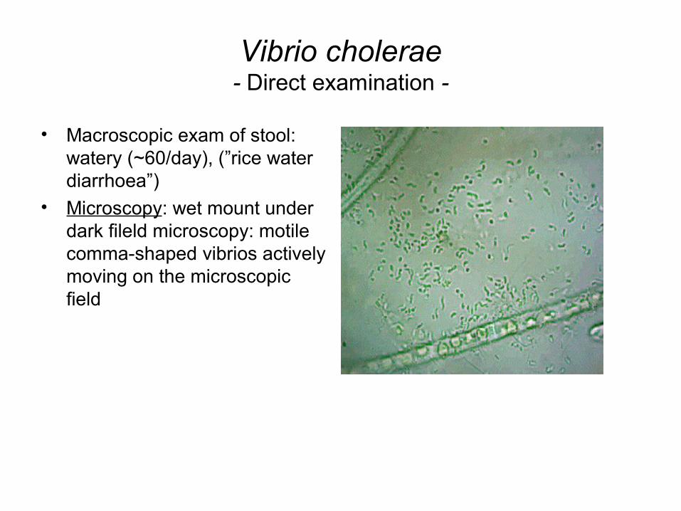
Vibrio cholerae- Direct examination -
• Macroscopic exam of stool: watery (~60/day), (”rice water diarrhoea”)
• Microscopy: wet mount under dark fileld microscopy: motile comma-shaped vibrios actively moving on the microscopic field
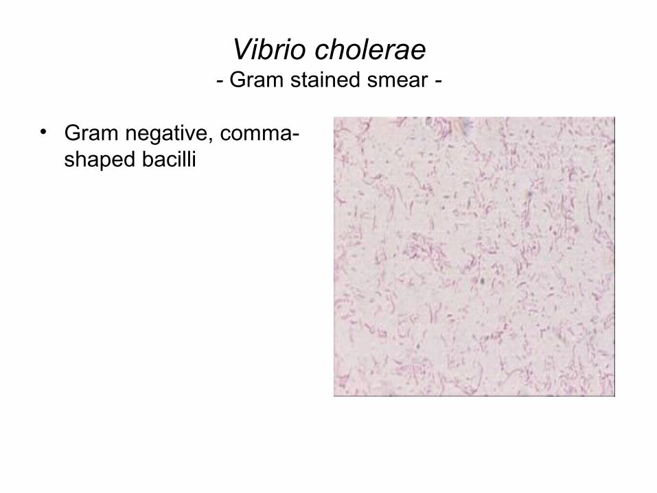
Vibrio cholerae- Gram stained smear -
• Gram negative, comma-shaped bacilli
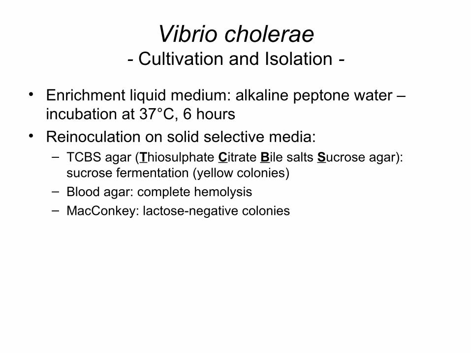
Vibrio cholerae- Cultivation and Isolation -
• Enrichment liquid medium: alkaline peptone water – incubation at 37°C, 6 hours
• Reinoculation on solid selective media:– TCBS agar (Thiosulphate Citrate Bile salts Sucrose agar):
sucrose fermentation (yellow colonies)– Blood agar: complete hemolysis– MacConkey: lactose-negative colonies
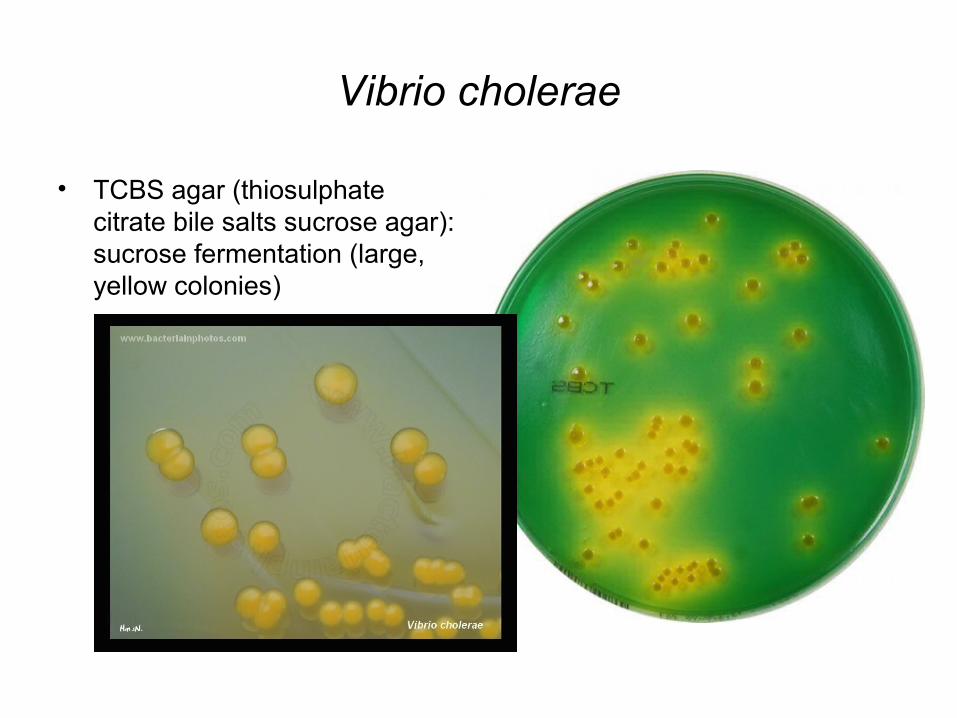
Vibrio cholerae
• TCBS agar (thiosulphate citrate bile salts sucrose agar): sucrose fermentation (large, yellow colonies)
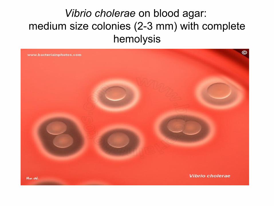
Vibrio cholerae on blood agar: medium size colonies (2-3 mm) with complete
hemolysis
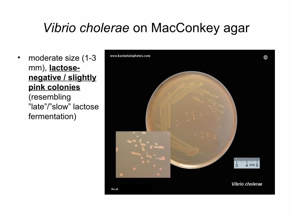
Vibrio cholerae on MacConkey agar
• moderate size (1-3 mm), lactose-negative / slightly pink colonies (resembling ”late”/”slow” lactose fermentation)
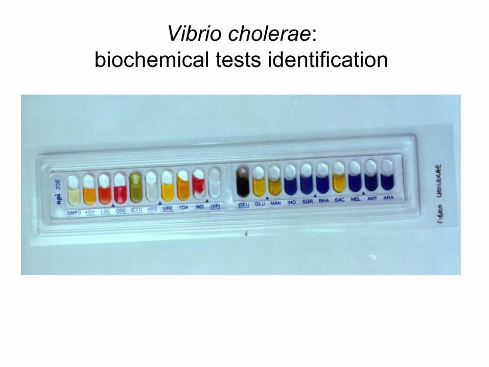
Vibrio cholerae: biochemical tests identification
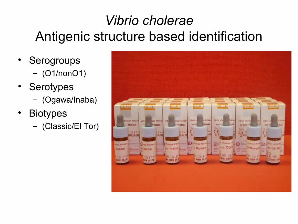
Vibrio choleraeAntigenic structure based identification
• Serogroups– (O1/nonO1)
• Serotypes– (Ogawa/Inaba)
• Biotypes– (Classic/El Tor)
Vibrio cholerae- Antibiotic sensitivity -
• High sensitivity to tetracyclines, nalidixic acid, norfloxacin• Antimicrobial sensitivity testing – required to monitor
acquired resistance of certain strains
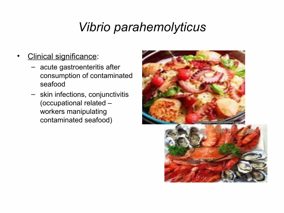
Vibrio parahemolyticus
• Clinical significance: – acute gastroenteritis after
consumption of contaminated seafood
– skin infections, conjunctivitis (occupational related – workers manipulating contaminated seafood)
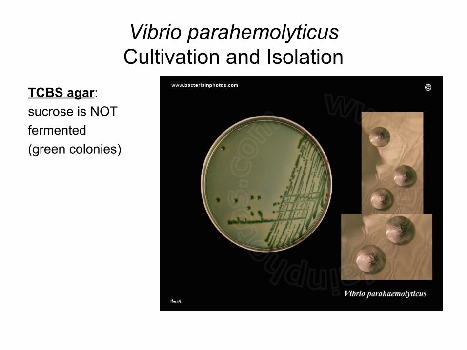
Vibrio parahemolyticusCultivation and Isolation
TCBS agar:
sucrose is NOT
fermented
(green colonies)
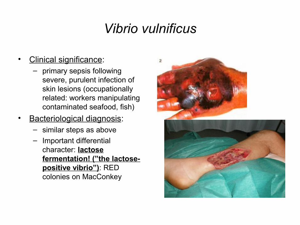
Vibrio vulnificus
• Clinical significance: – primary sepsis following
severe, purulent infection of skin lesions (occupationally related: workers manipulating contaminated seafood, fish)
• Bacteriological diagnosis: – similar steps as above– Important differential
character: lactose fermentation! (”the lactose-positive vibrio”): RED colonies on MacConkey
Genus Campylobacter
• Clinical significance: acute diarrhoeal disease; risk factors: long term hospitalization, long term antibiotic treatments (disbalance of intestinal microbiota)
• 11 species: – C.jejuni, C.coli – involved in human infections
• Microscopy: – Gram negative bacilli, encurved/S-shaped, motile (single polar
flagellus)
• Cultivation: – special media + microaerophilia (anaerobic jar/candle
jar/anaerobic kits)
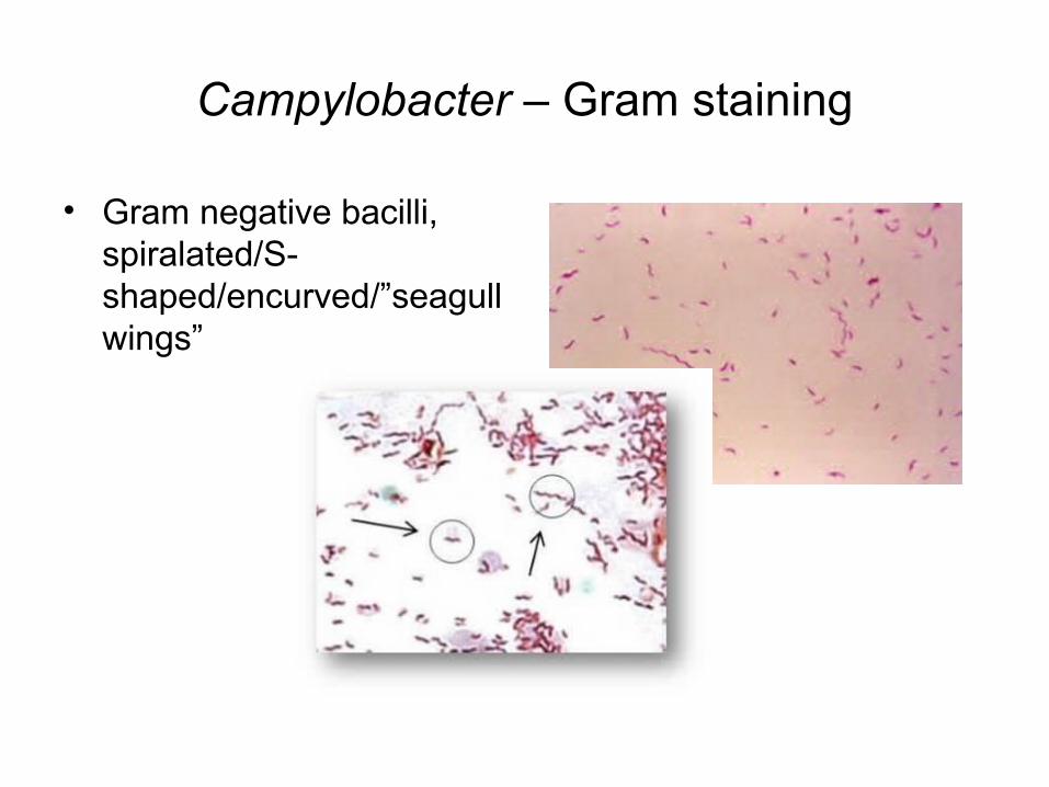
Campylobacter – Gram staining
• Gram negative bacilli, spiralated/S-shaped/encurved/”seagull wings”
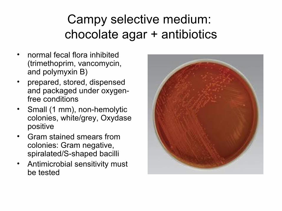
Campy selective medium: chocolate agar + antibiotics
• normal fecal flora inhibited (trimethoprim, vancomycin, and polymyxin B)
• prepared, stored, dispensed and packaged under oxygen-free conditions
• Small (1 mm), non-hemolytic colonies, white/grey, Oxydase positive
• Gram stained smears from colonies: Gram negative, spiralated/S-shaped bacilli
• Antimicrobial sensitivity must be tested
Genus Helicobacter
• Clinical significance: – gastro-duodenal ulcer, gastritis (Helicobacter pylori)
• Collection of specimens: – gastric mucosa fragments (endoscopy, gastric lavage)– transport media (e.g. ”Portagerm pylori”, BioMerieux), at +4°C
• Microscopy: – Gram stained smear: Gram negative bacilli, encurved/spiraled,
4-6 polar flagella
• Cultivation: – selective, blood containing media + antibiotics e.g. Helicobacter
pylori agar; at least 3 days incubation!!– Microaerophilic incubation* (see next slide)
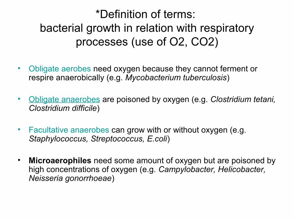
*Definition of terms: bacterial growth in relation with respiratory
processes (use of O2, CO2)
• Obligate aerobes need oxygen because they cannot ferment or respire anaerobically (e.g. Mycobacterium tuberculosis)
• Obligate anaerobes are poisoned by oxygen (e.g. Clostridium tetani, Clostridium difficile)
• Facultative anaerobes can grow with or without oxygen (e.g. Staphylococcus, Streptococcus, E.coli)
• Microaerophiles need some amount of oxygen but are poisoned by high concentrations of oxygen (e.g. Campylobacter, Helicobacter, Neisseria gonorrhoeae)
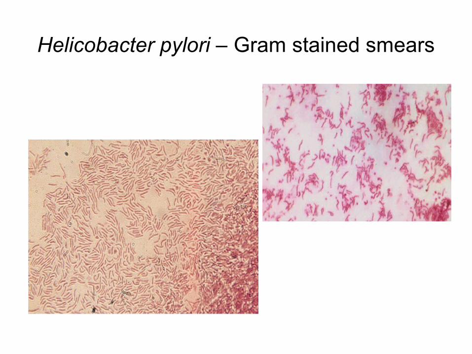
Helicobacter pylori – Gram stained smears
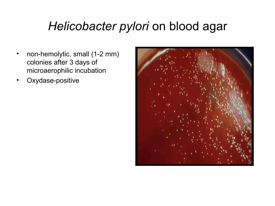
Helicobacter pylori on blood agar
• non-hemolytic, small (1-2 mm) colonies after 3 days of microaerophilic incubation
• Oxydase-positive
Genus Pseudomonas
• Gram negative bacilli, obligate aerobic, motile, nonsporulating, do not ferment glucose, oxydase positive
• Pseudomonas aeruginosa a.k.a. pyocyanic bacillus• Habitat:
– water, soil, air, human skin;
– humid conditions - biofilm e.g. in hospitals (toilets, humidifiers, respirators, plants, etc); human carriers
Pseudomonas aeruginosa
• Clinical significance:– Comensal, facultatively pathogen; colonizes mucous
membranes e.g. mere isolation in throat swab in the absence of relevant clinical context does not mean etiologic diagnosis but rather positive selection after antibiotic treatment
– opportunistic pathogen = infects impaired tissues & organs (tissues with lesions, patients with decreased immunity, etc); involved in hospital acquired infections e.g. eye infections, infection of burns/surgical wounds, UTI, lung infections, meningitis (by iatrogenic inoculation), sepsis
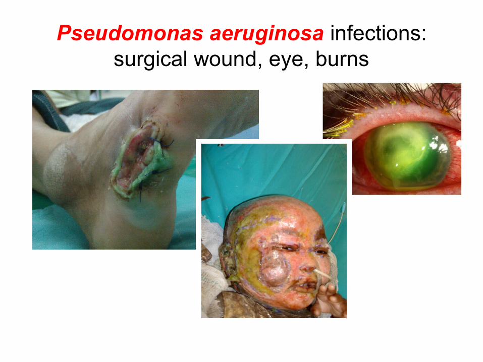
Pseudomonas aeruginosa infections: surgical wound, eye, burns
Pseudomonas aeruginosa- Bacteriological diagnosis -
• Collection of specimens: – depending on site of infection (pus, wound secretion, CSF, etc)
• Microscopy: – Gram stained smear: only relevant for naturally sterile
specimens (e.g. CSF): high number of PMNs + Gram negative bacilli
• Cultivation: – nonfastidious germ; grows on any medium– Naturally sterile specimens (CSF): blood agar– Faeces: selective media for enterobacteria
Pseudomonas aeruginosa
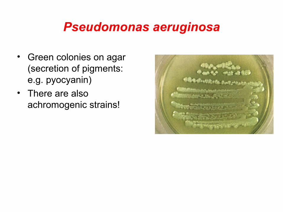
• Green colonies on agar (secretion of pigments: e.g. pyocyanin)
• There are also achromogenic strains!
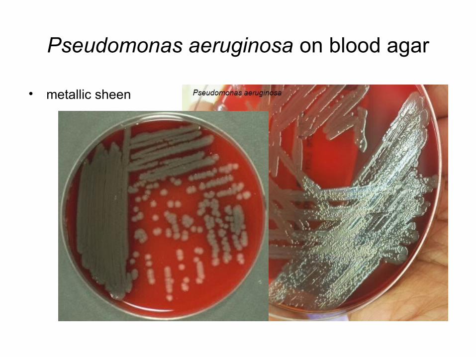
Pseudomonas aeruginosa on blood agar
• metallic sheen

Pseudomonas aeruginosa- Biochemical tests -
• API 20NE – identification system for non-fastidious, non-enteric, Gram negative bacilli
OR• API 20E
Pseudomonas aeruginosa- Antimicrobial susceptibility testing -
• Required due to rapid acquisition of antibiotic resistance (especially in case of ”hospital strains” – sometimes highly resistant)