
A high-sensitivity phospho-switchtriggered by Cdk1 governs chromosomemorphogenesis during cell division
Xavier Robellet,1,2,4 Yogitha Thattikota,1,2,4 Fang Wang,1,2 Tse-Luen Wee,3 Mirela Pascariu,1,2
Sahana Shankar,1,2 �Eric Bonneil,1 Claire M. Brown,3 and Damien D’Amours1,2
1Institute for Research in Immunology and Cancer (IRIC), 2D�epartement de Pathologie et Biologie Cellulaire, Universit�e deMontr�eal, Montr�eal, Quebec H3C 3J7, Canada; 3Advanced BioImaging Facility (ABIF), Department of Physiology, McGillUniversity, Montr�eal, Quebec H3G 0B1, Canada
The initiation of chromosome morphogenesis marks the beginning of mitosis in all eukaryotic cells. Althoughmany effectors of chromatin compaction have been reported, the nature and design of the essential trigger forglobal chromosome assembly remain unknown. Here we reveal the identity of the core mechanism responsible forchromosome morphogenesis in early mitosis. We show that the unique sensitivity of the chromosomecondensation machinery for the kinase activity of Cdk1 acts as a major driving force for the compaction ofchromatin at mitotic entry. This sensitivity is imparted by multisite phosphorylation of a conserved chromatin-binding sensor, the Smc4 protein. The multisite phosphorylation of this sensor integrates the activation state ofCdk1 with the dynamic binding of the condensation machinery to chromatin. Abrogation of this event leads tochromosome segregation defects and lethality, while moderate reduction reveals the existence of a novelchromatin transition state specific to mitosis, the intertwist configuration. Collectively, our results identify themechanistic basis governing chromosome morphogenesis in early mitosis and how distinct chromatin compactionstates can be established via specific thresholds of Cdk1 kinase activity.
[Keywords: chromosome; morphogenesis; Cdk1; Smc4; multisite phosphorylation]
Supplemental material is available for this article.
Received September 27, 2014; revised version accepted January 14, 2015.
In his seminal description of mitosis, Flemming (1882)recognized that the formation of visible chromosomes isone of the earliest cytological landmarks of the celldivision program. Since then, much effort has been de-voted to unraveling the structural and regulatory mech-anisms that underpin the formation of mitotic andmeiotic chromosomes (for review, see Maeshima andEltsov 2008; Baxter and Aragon 2012; Hirano 2012). Anumber of independent steps—such as chromosomereplication, condensation, and the establishment of sisterchromatid cohesion—are required for the formation ofmature and functional chromosomes during cell division.The morphological changes that associate with thesesteps are collectively referred to as the process of chro-mosome morphogenesis (van Heemst et al. 1999; Yu andKoshland 2005).The compaction of amorphous chromatin into visible
chromosomes is one of the earliest and most extensivechanges in the morphogenetic process (Flemming 1882).
Given the physical challenges associated with the assem-bly of micrometer-scale chromosomes in the crowdedcellular environment (Marko 2008), it is not surprisingthat many chromatin and cell cycle effectors have beensuggested as possible regulators of the process. Chiefamong those is the condensin complex, a pentamericATPase that binds to chromatin and alters its configura-tion and/or association status with distant chromatinregions (Bazile et al. 2010; Baxter and Aragon 2012;Hirano 2012). Other factors, such as cell cycle kinasesand histone-modifying enzymes, have also been proposedas possible regulators of chromosome condensation dur-ing mitosis (Morishita et al. 2001; St-Pierre et al. 2009;Abe et al. 2011; Neurohr et al. 2011; Wilkins et al. 2014).Although it is clear that these enzymes impact chroma-tin compaction at specific genomic locations and/orduring specific stages of mitosis, it is remarkable that
� 2015 Robellet et al. This article is distributed exclusively by ColdSpring Harbor Laboratory Press for the first six months after the full-issuepublication date (see http://genesdev.cshlp.org/site/misc/terms.xhtml).After six months, it is available under a Creative Commons License(Attribution-NonCommercial 4.0 International), as described at http://creativecommons.org/licenses/by-nc/4.0/.
4These authors contributed equally to this work.Corresponding author: [email protected] is online at http://www.genesdev.org/cgi/doi/10.1101/gad.253294.114.
426 GENES & DEVELOPMENT 29:426–439 Published by Cold Spring Harbor Laboratory Press; ISSN 0890-9369/15; www.genesdev.org
Cold Spring Harbor Laboratory Press on February 6, 2016 - Published by genesdev.cshlp.orgDownloaded from

the chromosome condensation process as a whole remainslargely operational when these enzymes are fully inhibitedin mammalian cells (Cimini et al. 2003; Ditchfield et al.2003; Hauf et al. 2003; Ono et al. 2004; Lenart et al. 2007;Abe et al. 2011). The fact that no specific mutations and/orinhibitory conditions prevent the formation of con-densed chromosomes during metaphase in mammaliancells suggests that the fundamental nature of the mech-anism responsible for global chromosome assembly isstill unknown.Another key issue about chromosome morphogenesis
relates to the timing of the process in relationship toother mitotic events. It was established several decadesago that mitosis is initiated by a sudden increase in Cdk1activity (Morgan 2007). The fact that the early assemblyof mitotic chromosomes correlates well with the earlyincrease in Cdk1 activity in prophase (Gavet and Pines2010) suggests that Cdk1 may regulate early chromo-some assembly directly. However, the dependency of thechromosome morphogenesis process on mitotic entry(Vassilev et al. 2006; Paulson 2007; Gong and Ferrell2010) makes it difficult to determine whether theimpact of Cdk1 on chromosome morphology reflectsa direct role in this process or, alternatively, a need toestablish a mitotic state prior to initiating chromosomeassembly. Moreover, given that a requirement for Cdk1activity is shared between many mitotic processes, it isunclear why the establishment of chromosome conden-sation should precede other mitotic landmarks if allmitotic processes respond to the same Cdk1 signal. Thetemporal primacy of condensation in the mitotic pro-gram could be due to heightened sensitivity to Cdk1phosphorylation, higher specificity for specific cyclin–Cdk1 complexes, or a yet-unknown Cdk1-independentmechanism. In this study, we tested those possibilitiesand showed how Cdk1 can initiate chromosome mor-phogenesis directly using quantitative multisite phos-phorylation of the Smc4 protein. Moreover, we identi-fied a novel two-step mechanism necessary for thefolding of chromatin and subsequent assembly of func-tional chromosome during mitosis.
Results
Regulation of chromosome morphology by Cdk1
To investigate the mechanistic basis for chromosomemorphogenesis, we first determined whether the processwas under direct Cdk1 control in Saccharomyces cerevi-siae. Yeast cells carrying a Cdk1 temperature-sensitive(ts) mutation, cdc28-4, were arrested in mitosis, and themorphology of the ribosomal DNA (rDNA) locus wasevaluated in these cells. The shape of the rDNA locus isdramatically reorganized during mitosis, which providesa sensitive assay to monitor chromosome morphogenesisin yeast (Guacci et al. 1994). Whereas wild-type cellsarrested in mid-mitosis showed the typical condensed‘‘loop’’ configuration of the rDNA locus at both 23°C and37°C, inactivation of Cdk1 resulted in the formation of anuncondensed ‘‘puff’’ rDNA signal at 37°C (Fig. 1A; Guacci
et al. 1994). Having established the Cdk1 dependency ofthe chromosome morphogenesis process in yeast, wenext asked whether chromosome condensation could bequantitatively modulated by down-regulation of Cdk1activity using conditional B-type cyclin mutations (i.e.,clb1 clb3 clb4 clb2-VI; clb-ts mutant henceforth). Al-though able to enter mitosis, clb-ts cells are incapable ofexecuting subsequent mitotic events (Amon et al. 1993).Analysis of chromosome morphology in this mutantrevealed the existence of a novel intertwined rDNAconfiguration distinct from the uncondensed ‘‘puff’’ signal(Fig. 1B) or the fully condensed loop signal (Fig. 1A).Specifically, under low Cdk1 activity, individual chromo-somal ‘‘threads’’ are clearly visible at the rDNA locus andappear to follow an elaborate intertwined path distinctfrom the nonoverlapping path of chromosome threads inthe loop configuration (Fig. 1A,B; Supplemental Fig. S1).We therefore refer to this novel stage in chromosomecondensation as the intertwist configuration. Interest-ingly, chromatin folding within the intertwist configura-tion is consistent in shape with the early condensationintermediates that were recently proposed to exist basedon polymer simulation models (Naumova et al. 2013).Cytological characterization of clb-ts mutants confirmedthat other mitotic events, such as bipolar spindle forma-tion and chromosome segregation, do not occur in thesecells (Supplemental Fig. S1; Amon et al. 1993).To exclude the possibility that rDNA intertwist forma-
tion is a consequence of a change in Cdk1 specificity in clb-tsmutants, we monitored chromosomemorphology in thecdc28-as1 mutant. This mutant, when treated with lowconcentrations of 1NM-PP1 inhibitor, experiences a cellcycle arrest at mitotic entry (i.e., after DNA replication butprior to mitotic spindle formation) (Bishop et al. 2000)similar to the point of arrest of clb-ts mutants. Examina-tion of chromosomemorphology in cdc28-as1 cells treatedwith the inhibitor revealed a striking enrichment in thenumber of cells carrying the intertwist configuration at therDNA,whereas untreated cells formedmostly loops at thislocus under identical conditions (Fig. 1C). Interestingly, theintertwist configuration appears to be stabilized at lowtemperature and could be readily observed in wild-typecells progressing synchronously into mitosis at 16°C (Sup-plemental Fig. S2). As previously observed with the fullycondensed loop configuration (Lavoie et al. 2004), forma-tion of the intertwist rDNA intermediate also requirescohesin activity, since inactivation of mcd1-1 preventedthe appearance of this rDNA configuration in mitosis(Supplemental Fig. S3). Taken together, our results indicatethat chromosome condensation is initiated at levels ofCdk1 activity that are too low to induce other mitoticevents. Moreover, conditions of lowCdk1 activity revealedthe existence of a hitherto unknown early chromatin-folding step in the formation of mitotic chromosomes.
The Smc4 subunit of condensin is a target for Cdk1in early mitosis
What is the target of Cdk1 in the induction of chromo-somemorphogenesis? A likely candidate is the condensin
Regulation of chromosome assembly in mitosis
GENES & DEVELOPMENT 427
Cold Spring Harbor Laboratory Press on February 6, 2016 - Published by genesdev.cshlp.orgDownloaded from

complex, a central effector of chromosome condensationin eukaryotes (for review, see Baxter and Aragon 2012;Hirano 2012). To test this possibility, we removed all ofthe core Cdk1 consensus sites (i.e., Ser/Thr–Pro) (Holtet al. 2009) from condensin subunits and determined theeffect of these mutations on cell proliferation (Fig. 2A,B).Only smc4-10A showed detectable growth defects in theabsence of Cdk1 phosphorylation (Fig. 2B). Combining allmutations in one yeast strain had only modest additiveeffects on cell proliferation relative to the smc4-10Asingle mutant (Fig. 2B). These results indicate that theSmc4 subunit of condensin is a likely target of Cdk1 invivo. To further substantiate this notion, we immuno-purified the condensin complex from metaphase-arrestedcells and subjected the immunoprecipitate to mass spec-trometry analysis to identify possible in vivo phosphor-ylation sites. This analysis revealed the existence of fivephosphorylation sites that conform to the Cdk1 consen-sus in Smc4 (Fig. 2C; Supplemental Fig. S4A) and none inthe other subunits of condensin. An additional Cdk1phospho-site, Ser117, was uncovered in Smc4 in pro-teome-wide analyses of mitotic cells (Holt et al. 2009;Kao et al. 2014). Interestingly, all of these Cdk1 phospho-sites were clustered in the N-terminal extension of
Smc4, a region of the protein that is conserved amongeukaryotic Smc4 family members but absent in theSmc1–3 families (Supplemental Fig. S4B,C). Deletion ofthe N-terminal extension of Smc4 results in a stable butinactive protein, thereby revealing the essential roleplayed by this part of Smc4 in condensin function (Fig.2D). Finally, we asked whether Cdk1 is directly respon-sible for condensin phosphorylation. To test this possi-bility, we purified condensin from yeast and exposed it topurified Cdk1–Clb2 in the presence of radiolabeled ATP.We observed that a single band corresponding to themolecular mass of Smc4 became phosphorylated follow-ing the kinase reaction (Fig. 2E; St-Pierre et al. 2009).Performing a similar experiment using only the N-termi-nal fragment of Smc4 (residues 1–163) resulted in a Cdk1phosphorylation-induced gel retardation of the substrateafter electrophoresis (Fig. 2F). Taken together, these exper-iments demonstrate that Cdk1 phosphorylates Smc4 invitro and in vivo.We next characterized the timing of Smc4 phosphory-
lation during the cell cycle. Since phosphorylation of theN-terminal part of Smc4 by Cdk1 causes a gel shift afterelectrophoresis (Fig. 2F), we used cells expressing theN-terminal extension of Smc4 fused to an epitope tag
Figure 1. Modulation of Cdk1 activity unveilsdistinct steps in the process of chromosomemorphogenesis. (A–C) The morphology of theyeast rDNA locus was revealed by fluorescencein situ hybridization (FISH). Representative mi-crographs of the most prominent rDNA morphol-ogy observed for each condition are shown at theleft. Propidium iodide (PI; red) and fluoresceinisothiocyanate (FITC; green) were used to labelthe nucleus and rDNA locus, respectively. Quan-tification of each rDNA species is shown at theright. At least 100 nuclei were counted percondition (n = 3 for all experiments). Error barsrepresent SD. (A) cdc28-4mutants exhibit classiccondensation defects. Cells were blocked inmetaphase at 23°C using nocodazole and shiftedfor 1 h to 37°C before processing of the samplesfor FISH analysis. To ensure that the quantifica-tion reflects loss of condensation in mitotic cellsrather than a return to interphase due to the lossof cdc28-4 activity (Sanchez-Diaz et al. 2012),we normalized the rDNA quantification accord-ing to the budding index of cells during thearrest. (B,C) Reducing Cdk1 activity uncoversa novel condensation intermediate at the rDNA.(B) clb-ts cells growing asynchronously at 23°Cwere arrested in G1 or early mitosis by incubationwith a factor or by shifting the culture to 37°Cfor 135 min, respectively. Cells were subsequentlyharvested, and their rDNA morphology was re-vealed by FISH. (C) cdc28-as1 cells growing asyn-chronously at 25°C were arrested in early mitosisby incubation with nocodazole with or withoutthe kinase inhibitor NM-PP1 for 150 min. Sampleswere then fixed, and the morphology of the rDNAlocus was monitored as above.
Robellet et al.
428 GENES & DEVELOPMENT
Cold Spring Harbor Laboratory Press on February 6, 2016 - Published by genesdev.cshlp.orgDownloaded from

(henceforth, Smc4-NT) tomonitor in vivo phosphorylation.As expected, the electrophoretic behavior of Smc4-NTchanges dramatically during the cell cycle, starting asa single band in G1 and acquiring at least two retardedspecies as cells progressed toward mitosis (Fig. 3A).Phosphatase treatment of Smc4-NT confirmed that theretarded species were due to phosphorylation (Fig. 3B).Importantly, Smc4-NT became phosphorylated simulta-neously with or slightly prior to Swe1, an early Cdk1substrate during mitosis (Harvey et al. 2005), and muchearlier than Ycg1, a condensin subunit phosphorylated inanaphase (Fig. 3A; St-Pierre et al. 2009). These resultsindicate that Smc4 phosphorylation occurs at or veryclose to mitotic entry. Consistent with this interpreta-tion, monitoring the phosphorylation of two of the Cdk1sites—Ser4 and Ser128—on Smc4 using phospho-specificantibodies confirms that these residues are also modifiedearly inmitosis (Fig. 3C,D; Supplemental Fig. S5A,B). Thein vivo kinetics of Smc4 phosphorylation revealed by theSmc4-NT construct and the phospho-specific Ser128antibody were essentially identical (Fig. 3D), thereby
validating Smc4-NT as an effective reporter to monitorSmc4 phosphorylation status. Importantly, removal ofthe seven Cdk1 sites in the Smc4-NT fragment com-pletely abrogated its phosphorylation-induced gel retar-dation in live cells (Fig. 3E; Supplemental Fig. S5C). Theseresults strongly suggest that Cdk1 is the kinase thattargets Smc4 for phosphorylation in early mitosis. Con-sistent with this prediction, cells defective in early (clb5and clb6) and late (clb1, clb3, clb4, and clb2-ts) cyclinsubunits showed marked reductions in the extent ofSmc4 phosphorylation in vivo (Fig. 3F; SupplementalFig. S5D,E). Moreover, removal of the Clb5 targetingRxL motifs (Loog and Morgan 2005; Koivomagi et al.2011) in the N terminus of Smc4 caused a substantialreduction in the extent of its phosphorylation (Fig. 3G;Supplemental Fig. S5F). Finally, we wanted to test thepossibility that Smc4 phosphorylationmight bemediatedby Cdc5, since this kinase is known to be activated byCdc28 in mitosis (Mortensen et al. 2005). However, thefact that Smc4-NT phosphorylation remained normal incdc5-99mutant cells entering mitosis (Supplemental Fig.
Figure 2. Smc4 is a key target of Cdk1 in theyeast condensin complex. (A) Schematic represen-tation of the number of putative CDK sites (i.e.,Ser/Thr-Pro) (Holt et al. 2009) in the subunits ofthe yeast condensin complex. (B) Growth pheno-type of condensin subunits lacking putative Cdk1sites. Fivefold serial dilutions of the wild type andphospho-mutants were spotted on solid mediumto evaluate growth at 23°C and 37°C. (C) Sche-matic representation of Smc4 domain organiza-tion. The positions of Cdk1 phosphorylation sitesare marked above the protein schematic. Sitesidentified by mass spectrometry are labeled witha white ‘‘P,’’ while other consensus Cdk1 siteswere marked with a black ‘‘P.’’ (D) Deletion ofthe N-terminal extension of Smc4 is lethal inS. cerevisiae. Dissection of a sporulated heterozy-gous diploid strain carrying an allele of Smc4 thathas lost its N-terminal extension is shown at theleft. The sequence of this particular allele starts atthe position of the blue arrow in the sequenceshown in Supplemental Figure S4C, which corre-sponds to the start site of other SMC familymembers. At the right, an immunoblot showsthat loss of the N-terminal extension of Smc4does not affect its protein abundance in hetero-zygous diploid cells. (E) In vitro kinase assayshowing that Smc4 is a direct substrate forCdk1. Purified condensin was incubated alone(first lane), with 32P-ATP (second lane), or in thepresence of purified Cdc28–Clb2 complex and32P-ATP (third lane). (Fourth lane) Purified his-tone H1 was used as positive control for Cdk1phosphorylation. After incubation, proteins wereseparated on a 4%–12% SDS–polyacrylamide geland revealed by Coomassie staining, as shown atthe left. An autoradiograph of the same gel is
shown at the right. Bullets indicate the different subunits of condensin. Asterisks correspond to the Cdk1–Clb2 complex, and theopen circle marks the position of histone H1. (F) The N-terminal extension of Smc4 is phosphorylated by Cdk1 in vitro. PurifiedSmc4-NT was incubated with ATP or purified Cdc28–Clb2 complex and ATP. After the kinase reaction, proteins were resolved bySDS-PAGE and stained with Coomassie.
Regulation of chromosome assembly in mitosis
GENES & DEVELOPMENT 429
Cold Spring Harbor Laboratory Press on February 6, 2016 - Published by genesdev.cshlp.orgDownloaded from
S5G) or in cells already arrested at this stage of the cellcycle (Supplemental Fig. S5H) argues against a role forthis kinase as the major or sole Smc4 kinase in earlymitosis. Taken together, our results show that effectivephosphorylation of Smc4 in early mitosis requires directtargeting of condensin by cyclin–Cdk1 complexes.
Smc4 phosphorylation activates chromosomemorphogenesis
What is the physiological significance of Smc4 phosphor-ylation by Cdk1? To answer this question, we introducedin the yeast genome mutations that either prevent ormimic the phosphorylation of the seven Cdk1 sites in theN-terminal extension of Smc4. Whereas removal of in-dividual Cdk1 sites resulted in little or no effect on growthproperties and on chromosome condensation (Supplemen-tal Fig. S6A,B), cells carrying a SMC4 allele that lacks all
N-terminal Cdk1 sites exhibited a strong growth defectat the nonpermissive temperature (i.e., smc4-7A) (Fig.4A). We noticed that the Smc4-7A protein was lessabundant than its wild-type counterpart in immunoblotanalysis (Fig. 4B, first two lanes). This reduction in Smc4-7A protein levels is not responsible for its growth defect,since down-regulation of wild-type Smc4 abundanceusing the auxin-inducible degron (i.e., Smc4-AID) didnot result in detectable growth defects at Smc4 proteinlevels comparable with or lower than those of the Smc4-7A mutant (Fig. 4B,C, cf. 40 mM auxin/IAA and smc4-7Alanes). Consistent with this, inactivation of the pathwayresponsible for the degradation of unstable nuclear pro-teins did not suppress the ts phenotype of smc4-7A cells(Supplemental Fig. S7A), and no defect in condensincomplex formation was detected in smc4-7A mutants(Fig. 4D). Collectively, these results indicate that theconditional lethality phenotype of the smc4-7A mutant
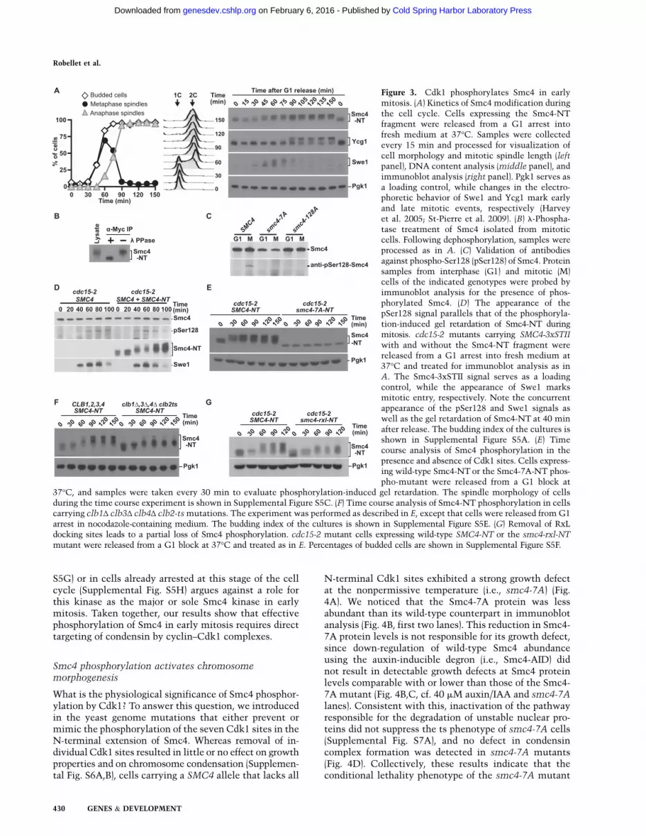
Figure 3. Cdk1 phosphorylates Smc4 in earlymitosis. (A) Kinetics of Smc4 modification duringthe cell cycle. Cells expressing the Smc4-NTfragment were released from a G1 arrest intofresh medium at 37°C. Samples were collectedevery 15 min and processed for visualization ofcell morphology and mitotic spindle length (leftpanel), DNA content analysis (middle panel), andimmunoblot analysis (right panel). Pgk1 serves asa loading control, while changes in the electro-phoretic behavior of Swe1 and Ycg1 mark earlyand late mitotic events, respectively (Harveyet al. 2005; St-Pierre et al. 2009). (B) l-Phospha-tase treatment of Smc4 isolated from mitoticcells. Following dephosphorylation, samples wereprocessed as in A. (C) Validation of antibodiesagainst phospho-Ser128 (pSer128) of Smc4. Proteinsamples from interphase (G1) and mitotic (M)cells of the indicated genotypes were probed byimmunoblot analysis for the presence of phos-phorylated Smc4. (D) The appearance of thepSer128 signal parallels that of the phosphoryla-tion-induced gel retardation of Smc4-NT duringmitosis. cdc15-2 mutants carrying SMC4-3xSTII
with and without the Smc4-NT fragment werereleased from a G1 arrest into fresh medium at37°C and treated for immunoblot analysis as inA. The Smc4-3xSTII signal serves as a loadingcontrol, while the appearance of Swe1 marksmitotic entry, respectively. Note the concurrentappearance of the pSer128 and Swe1 signals aswell as the gel retardation of Smc4-NT at 40 minafter release. The budding index of the cultures isshown in Supplemental Figure S5A. (E) Timecourse analysis of Smc4 phosphorylation in thepresence and absence of Cdk1 sites. Cells express-ing wild-type Smc4-NT or the Smc4-7A-NT phos-pho-mutant were released from a G1 block at
37°C, and samples were taken every 30 min to evaluate phosphorylation-induced gel retardation. The spindle morphology of cellsduring the time course experiment is shown in Supplemental Figure S5C. (F) Time course analysis of Smc4-NT phosphorylation in cellscarrying clb1Δ clb3Δ clb4Δ clb2-tsmutations. The experiment was performed as described in E, except that cells were released from G1arrest in nocodazole-containing medium. The budding index of the cultures is shown in Supplemental Figure S5E. (G) Removal of RxLdocking sites leads to a partial loss of Smc4 phosphorylation. cdc15-2 mutant cells expressing wild-type SMC4-NT or the smc4-rxl-NT
mutant were released from a G1 block at 37°C and treated as in E. Percentages of budded cells are shown in Supplemental Figure S5F.
Robellet et al.
430 GENES & DEVELOPMENT
Cold Spring Harbor Laboratory Press on February 6, 2016 - Published by genesdev.cshlp.orgDownloaded from

is specifically due to the loss of Cdk1-mediated phos-phorylation of condensin.Microscopic examination of smc4-7A mutants at the
nonpermissive temperature provided critical insightsinto the nature of the cellular defect responsible for thelethality of this mutant. We noticed that there wasa significant fraction of dividing cells in the mutantpopulation that contained unequal amounts of nuclearmaterial and/or connecting threads of chromosomalDNA between separating nuclei (Fig. 4E, see arrowheads).This phenotype was much less frequent in wild-typecells (Fig. 4F) and is indicative of severe chromosomesegregation defects in the absence of Smc4 phosphory-lation. The root cause for the segregation defect ofcondensin mutants has been ascribed to their inabilityto promote chromosome condensation (Hirano 2012).To test this notion, we examined the rDNA condensa-tion proficiency of the smc4-7A mutant and compared
it with that of SMC4 and smc4-82 cells (i.e., a strong tsmutant of condensin) (see below). Whereas wild-typecells were able to condense the rDNA effectively at37°C, both the smc4-7A and smc4-82 mutants werecompletely defective in rDNA loop formation at non-permissive temperatures (Fig. 4G,H). These results in-dicate that Cdk1 phosphorylation is absolutely necessaryto activate condensin and initiate chromosome conden-sation in vivo.A previous study using the fission yeast Schizosaccharo-
myces pombe suggested that Cdc2 might regulate con-densin by phosphorylation. Sutani et al. (1999) showedthat overexpression of the cut3-T19A phosphomutantcaused a dominant-negative chromosome segregation de-fect in this organism (however, no chromosome conden-sation defects were observed in this study). Since thephenotype of the cut3-T19A mutant expressed at itsnatural level was not reported by Sutani et al. (1999) and
Figure 4. Cdk1-mediated phosphorylation ofSmc4 is required for chromosome condensation.(A) Growth properties of phospho-mimetic andphospho-mutant forms of Smc4. Fivefold serialdilutions of smc4 phospho-mutants were spottedon solid medium to evaluate growth at 23°C and37°C. (B) Regulation of Smc4-AID protein abun-dance by auxin/IAA. Cells were grown asyn-chronously at 23°C until exponential phase andexposed to different concentrations of auxin for2 h at 30°C. Smc4-AID abundance was evaluatedby immunoblot analysis using the 3xStrepTagII epi-topes at the C terminus of the protein. (C) Effectsof reduced Smc4-AID abundance on yeast growthproperties. Strains carrying SMC4 or smc4-7A
were included as controls to show normal andts growth phenotypes in the presence of auxin.(D) The smc4-7A mutant is competent for con-densin complex formation. Smc4 was immu-noprecipitated from whole-cell extracts usinganti-STII antibodies, and its ability to associatewith Ycg1-3xFlag was determined by immuno-blotting. Whole-cell extracts (T) and beads only(�) were loaded as controls. Approximately equalamounts of immunoprecipitated (IP) Smc4 wereloaded in each lane to allow effective comparisonof the levels of Ycg1 interacting with Smc4. (E)smc4 phospho-mutants show cytological markersassociated with chromosome segregation defects.A high proportion of anaphase nuclei connectedwith lagging genetic material (arrowheads) andhaving an elongated morphology can be seen ina culture of smc4-7A mutants growing at thenonpermissive temperature. Chromatin and spin-dle pole bodies were visualized using HTA1 fusedto mCherry and Spc42 fused to GFP, respectively.(F) Quantification of anaphase bridges in the wildtype and smc4 phospho-mutant (n = 3). Error bars
indicate SD. (G) Chromosome condensation defects in the smc4 phospho-mutant. For each genotype, a representative micrograph of themost prominent rDNA species detected by FISH is shown. Cells were grown asynchronously at 23°C until exponential phase and shiftedto 37°C for 2.5 h. Nocodazole was used to block cells in metaphase. Nuclei and rDNA were stained with PI (red) and FITC (green),respectively. (H) Quantification of rDNA condensation in SMC4, smc4-7A, and smc4-82 mutants grown at the nonpermissivetemperature. At least 100 cells were counted for each genotype in three independent experiments. Error bars indicate SD.
Regulation of chromosome assembly in mitosis
GENES & DEVELOPMENT 431
Cold Spring Harbor Laboratory Press on February 6, 2016 - Published by genesdev.cshlp.orgDownloaded from

protein overexpression can often obscure the physiolog-ical relevance of natural regulatory processes (Robbins andCross 2010), we wondered what might be the phenotypeassociated with loss of Thr19 phosphorylation when Cut3is expressed at physiological levels. To address this, weintegrated the cut3-T19V phosphomutant at its naturallocus in diploid S. pombe. The valine substitution wasused in cut3 instead of alanine, since valine is structurallymore similar to threonine, thus precluding destabilizingeffects not related to loss of phosphorylation. Dissectionof a sporulated heterozygous diploid yeast carrying thecut3-T19V allele revealed that loss of Thr19 phosphory-lation is lethal in haploid cells when the mutant isexpressed from its physiological locus (SupplementalFig. S7B). Interestingly, the heterozygous diploid carry-ing cut3-T19V grew normally (Supplemental Fig. S7C),thereby suggesting that the dominant-lethal effects pre-viously observed with the cut3-T19A mutant might bespecific to the alanine substitution and/or the over-expression of the protein (Sutani et al. 1999). Takentogether, these results further strengthen the conclusionthat Cdk1 phosphorylation of the N-terminal extensionof Smc4 family members plays a critical role in theregulation of condensins in eukaryotes.
Cdk1 phosphorylation sites activate condensinby altering its charge
We next examined the phenotype of a SMC4 allelecarrying phospho-mimetic residues (i.e., negativelycharged glutamates) at the position of Cdk1 sites. Con-stitutive activation of condensin by such mutations isexpected to create a lethal form of the enzyme because itwould permanently alter chromatin structure in yeast.However, replacement of phosphorylated residues byglutamates in Smc4 did not result in an obvious activa-tion of condensin or a strong growth defect in the smc4-7Emutant (Fig. 4A). This is not completely unexpected, sincea single negatively charged residue does not accuratelymimic the structure or net charge of a phosphorylatedresidue, as previously observed (Lyons and Morgan 2011;Pearlman et al. 2011). Based on the fact that the N-terminal extension of Smc4 is predicted to be highlyunstructured (Supplemental Fig. S8A–C), we consideredthe possibility that the change in charge imparted byphosphorylation of this domain would be more relevantas a regulatory mechanism than a possible change instructure. We therefore created a smc4-7EE allele wherethe phosphorylated residues and adjacent prolines (+1position of the CDK motif) were mutated to doubleglutamates to more accurately mimic phosphate charges(Fig. 5A,B; Pearlman et al. 2011). It was recently shownthat this approach is more effective at mimicking an‘‘activated’’ state than single glutamate/aspartate substi-tutions in substrates targeted by proline-directed kinases(Strickfaden et al. 2007; Li et al. 2014). The charge-mimeticallele, together with additional control mutations, wasintegrated at the SMC4 locus in a diploid yeast strain, andthe resulting heterozygous mutants were sporulated anddissected to reveal the phenotypes of the mutations in
haploid spores. Remarkably, introducing dual charge-mi-metic mutations at the seven phospho-sites and adjacentpositions (i.e., Ser/Thr–Promotif) of Smc4 led to lethality inall spores inheriting those mutations, whereas simulta-neously altering the seven phospho-sites with singleglutamates at either position of the Ser/Thr–Pro motifshad no detectable effect on viability (i.e., four viable sporesper tetrad) (Fig. 5B). Moreover, a mutant carrying dualglutamines—an amino acid approximately isosteric withglutamate but lacking the negative charge (Fig. 5A)—didnot show any obvious viability defects (Fig. 5B). Theseresults demonstrate that the lethality of the smc4-7EEmutant is due to the presence of negative charges thateffectively mimic the phosphorylated state of the activeprotein. Immunoblot analysis of heterozygote diploidsconfirmed that the Smc4-7EE mutant is expressed atnormal levels in yeast (Supplemental Fig. S8D).To further strengthen our previous interpretation, we
asked whether an alternative means of imposing a consti-tutive state of phosphorylation on Smc4 would recapitu-late the phenotype of the smc4-7EE allele. To achievethis, we fused the B-type cyclin Clb2 to the N terminus ofSmc4 in order to target Cdk1 to Smc4 throughout the cellcycle, an experimental approach previously described(Lyons and Morgan 2011). Dissecting a sporulated hetero-zygous diploid strain carrying the CLB2-SMC4 fusiongene led to lethality or severe growth defects in most ofthe spores inheriting the fusion construct (Fig. 5B).Preventing phosphorylation of Smc4 by removal of itsphospho-sites or deletion of Clb2’s cyclin box (the part ofthe protein responsible for interaction with Cdk1) largelysuppressed the lethality associated with the cyclin-Smc4fusion protein (Fig. 5B; Supplemental Fig. S9A). Theseresults are consistent with the phenotype of the smc4-7EE allele and indicate that inducing a permanent state ofphosphorylation in Smc4 is incompatible with viabilityin yeast. Since the phenotype associated with mimickingphosphorylation is much stronger than that caused by lossof phosphorylation, we posit that the smc4-7EE andCLB2-SMC4 mutants behave as gain-of-function alleles in vivo.We envision two possible regulatory modes that can
account for the lethality of the smc4-7EE and CLB2-SMC4 mutants. First, constitutive phosphorylation ofcondensin may simply activate the enzyme and resultin a permanent state of chromosome condensation that isnot compatible with viability. Alternatively, preventingphosphate removal in phospho-mimetic mutants mayabrogate iterative phosphorylation/dephosphorylation cy-cles that are normally required to maintain full enzymeactivity during mitosis (De Wulf et al. 2009). Overexpres-sion of smc4-7EE can distinguish between these tworegulatory modes, since a version of condensin that is fullyactivated by static phosphorylation (i.e., no turnovermodel) would be expected to induce unscheduled con-densation following overexpression, whereas a version ofthis enzyme that is engaged into its activation cycleby phosphorylation but not allowed to complete it bydephosphorylation (i.e., dynamic phosphorylation model)would act as a dominant-negative inhibitor. To test thesehypotheses, we expressed smc4-7EE ectopically in wild-
Robellet et al.
432 GENES & DEVELOPMENT
Cold Spring Harbor Laboratory Press on February 6, 2016 - Published by genesdev.cshlp.orgDownloaded from

type and clb2-ts-arrested cells. Remarkably, ectopic ex-pression of smc4-7EEwas not able to induce unscheduledcondensation of the rDNA locus in G1 cells or full rDNAcondensation in G2/early mitotic cells (SupplementalFigs. S9B–E, S10A), which suggests a need for dynamicphosphorylation in the regulation of condensin in vivo.Consistent with this, overexpression of smc4-7EE led todominant-negative lethality in both the wild type andsmc4-82 mutants (Fig. 5C). This lethality is not due tounspecific loss of function in smc4-7EE or subunit im-balance in condensin, since overexpression of smc4-1(Freeman et al. 2000), smc4-7A, or SMC4 complementedthe lethality of the smc4-82 mutant at 37°C withoutcausing dominant-lethal effects in wild-type cells (Fig.5C). However, we note that overexpression of SMC4 inwild-type cells caused a weak proliferation delay at 23°C.It had been previously demonstrated that the effects of
mutations that confer a gain of activity on a protein canbe abrogated by fusion with known inactivating muta-
tions (Schott and Hoyt 1998). To formally test the gain-of-function nature of the charge-mimetic mutations inSMC4, we combined the 7EE substitutions with theinactivating mutations in the smc4-1 allele and moni-tored the ability of the chimeric mutant to behave ina dominant-negative manner. As expected, overexpres-sion of the smc4-7EE-1 chimeric mutant in conditionsunder which its charge-mimetic mutations are not coun-teracted by the smc4-1 mutations (i.e., 23°C, the permis-sive temperature for smc4-1) led to a dominant-negativelethality similar to that of the smc4-7EE single mutant(Fig. 5, D vs. C,E). In contrast, overexpression of thechimeric mutant at 37°C (i.e., the inactivating conditionfor smc4-1 and, by extension, smc4-7EE) suppressed thelethal gain of function associated with the charge-mi-metic substitutions (Fig. 5D). Since Smc4-7EE must beendowed with an activity to be able to lose it, weconclude from this experiment that the charge-mimeticmutations impart a new activity on Smc4 that requires
Figure 5. Constitutive phosphorylation creates adominant-negative form of Smc4. (A) Structuralbasis of phospho-mimicking mutations in SMC4.(Top) Ball-and-stick representation of different aminoacids used to substitute the phosphorylated residuesin Smc4. (Bottom) Positions of the mutagenic sub-stitutions used in this study relative to the Cdk1optimal consensus for phosphorylation. (B) Physio-logical effect of charge-mimetic mutations in SMC4.Diploid strains carrying smc4 phospho-mimetic,charge-mimetic, and phospho-constitutive muta-tions were induced to sporulate, and the viabilityof the resulting haploid spores was determined after3 d of growth on solid medium. Two typical tetradsof spores are shown per genotype. The genotype ofspores was ascertained using the HIS3 markerassociated with the mutant alleles of SMC4 (i.e., +sign means wild-type SMC4). Phospho-defectivesubstitutions are included as controls. (C) Over-expression of smc4-7EE leads to dominant lethalityin cells. Fivefold dilution series of the wild type andsmc4-82 mutant harboring galactose-inducible ver-sions of SMC4 integrated at the URA3 locus werespotted on solid medium to evaluate growth at 23°Cand 37°C. Unlike smc4-1 or smc4-7A alleles, expres-sion of smc4-7EE induced strong dominant-negativelethality irrespective of the genetic background ortemperature used for overexpression. (D) The smc4-7EE allele encodes a gain-of-function mutant. Five-fold dilution series of wild-type cells expressinga galactose-inducible smc4-7EE-1 chimeric mutant(or SMC4, smc4-1, or smc4-7EE controls) integratedat the URA3 locus were spotted on solid medium toevaluate growth as in C. (E) The charge-mimeticmutations can act in trans. Wild-type or smc4-7A
mutant cells expressing a galactose-inducible smc4-7EE-NT construct were spotted on solid medium, asin C.
Regulation of chromosome assembly in mitosis
GENES & DEVELOPMENT 433
Cold Spring Harbor Laboratory Press on February 6, 2016 - Published by genesdev.cshlp.orgDownloaded from

otherwise normal protein function to mediate its effect.Importantly, this new activity can be induced equallywell by fusion of SMC4 with CLB2 or by charge-mimick-ing mutations, which indicates that Cdk1 phosphoryla-tion activates condensin by altering its charge in livingcells and that preventing Smc4 dephosphorylation islethal in vivo. Consistent with this interpretation, in-activation of a major mitotic phosphatase and knowncondensin interactor, PP2A (encoded by the PPH3,PPH21, and PPH22 genes in budding yeast) (Yeong et al.2003; Takemoto et al. 2009; Peplowska et al. 2014),caused a large reduction in the ability of phosphatase-defective cells to maintain chromosome condensation inmetaphase (Supplemental Fig. S10B). Collectively, ourresults indicate that dynamic turnover of Cdk1 phos-phorylation events on condensin is important for mitoticchromosome condensation.
Hypersensitivity of Smc4 to Cdk1 phosphorylation
How might charge-driven activation of condensin pro-mote early mitotic appearance of condensed chromo-somes? One likely possibility would be that condensinis hypersensitive to low levels of Cdk1 activity becausephosphorylation of any individual Cdk1 site is function-ally equivalent for enzyme activation, and only a few ofthem need to be modified (in any combination) to fullyactivate the enzyme. The dominant effects of charge-mimetic mutations provide a unique means to test thishypothesis. Indeed, one would expect that progressiveremoval of the seven dual glutamates in Smc4 wouldeventually reverse the partial gain of function of thesmc4-7EE allele, thereby revealing the minimal number ofphospho-sites that is required to activate condensin in vivo.Prompted by this rationale, we created a series of
heterozygous SMC4/smc4-EE mutants carrying progres-sively smaller numbers of dual-glutamate mutations andtested their phenotypes after sporulation of diploid cells.Remarkably, diploid strains carrying as few as three tofour charge-mimetic mutations gave rise to tetrads witha very penetrant 2:2 lethality phenotype cosegregatingwith smc4mutations (Fig. 6, rows 5,6). This is identical tothe phenotype of the original smc4-7EE or smc4-10EEalleles (Fig. 6, rows 2,3) but is in contrast to the absence ofphenotype of mutations affecting the first three Cdk1sites at the N terminus of the protein (Fig. 6, row 4). Thisresult suggests that three to four phosphorylation eventsin the middle or C-terminal clusters of Cdk1 sites may besufficient to activate Smc4. To further refine this conclu-sion, we focused our analysis of charge-mimetic muta-tions on the middle cluster of phospho-sites (i.e., posi-tions 109, 113, 117, and 128). Removing a single dual-glutamate mutation from the four present in this clusterdid not fully suppress the lethality of the remainingcharge-mimetic mutations (Fig. 6, rows 7,8). However,loss of any additional site either reduced the penetranceof the lethality phenotype (Fig. 6, row 9) or allowedcomplete recovery of spore viability (Fig. 6, rows 10–14).Importantly, preventing phosphorylation of the Cdk1sites away from the middle cluster of dual glutamates
(i.e., the smc4-3A-4EE-3A mutant) did not suppress theeffect of the smc4-4EE mutant (i.e., Fig. 6, cf. rows 5 and17), thereby indicating that the lethality of this strain wasnot due to phosphorylation of the remaining Cdk1 sites inSmc4-4EE. Taken together, these results indicate thata threshold of two to three phospho-mimicking charges issufficient to generate dominant condensin activity invivo (Fig. 6, rows 7,16). Moreover, our data reveal a degreeof functional equivalency (or redundancy) in Cdk1 phos-pho-sites, thereby explaining how condensin activationin early mitosis can be hypersensitive to Cdk1 levels.
Cdk1 controls condensin binding to chromatin
Finally, we wished to identify the specific aspect ofcondensin behavior that was regulated by Cdk1 phos-phorylation. Multiple events have been previouslyshown to be required for full activation of condensin ineukaryotes, including nuclear import (Sutani et al. 1999),recruitment on chromatin (Freeman et al. 2000), andenzymatic activation (Kimura et al. 1998). The fact thatbudding yeast condensin and mammalian condensin IIare constitutively located in the nucleus (Freeman et al.2000; Ono et al. 2004; Gerlich et al. 2006) suggests thatthe Cdk1 regulation of these enzymes may not occur atthe level of nuclear import. Interestingly, previousstudies in human cells showed that condensin II bindingto chromatin is very dynamic prior to chromosomecondensation but then becomes much more stable ascells enter mitosis (Gerlich et al. 2006). The mechanisticbasis for this change in chromatin-binding dynamicsis unknown, but it seems likely that increasing theduration of condensin interactions with chromatinwould stimulate chromosome condensation. To evalu-ate whether condensin phosphorylation by Cdk1 couldaffect its chromatin-binding dynamics, we used pho-tobleaching confocal laser-scanning microscopy. A sub-domain in the nucleus of live yeast cells expressingGFP-fused Smc4 was photobleached using the line scanfeature of the microscope, and the kinetics of fluorescencedecay were determined over time at the region of interestin the cell (i.e., red rectangle in Fig. 7A). In this experiment,the Smc4-3xGFP contained within the unbleached areais expected to replenish the fluorescence in the photo-bleached area of the cell at a rate that is directly pro-portional to the exchange of condensin complexes betweenthe two regions within the nucleus. As expected, prevent-ing exchange by fixation of cells with paraformaldehyde ledto a very rapid loss of Smc4-3xGFP fluorescence signal inthe photobleached area while leaving the fluorescence inthe rest of the nucleus unaffected (Fig. 7A). In contrast,photobleaching a subdomain of the Smc4-3xGFP signal inlive cells resulted in a rapid decay of fluorescence signalthroughout the entire yeast cell (Fig. 7A,B). This rapid lossof fluorescence is consistent with the highly dynamicexchange of condensin complexes between the site ofphotobleaching and the rest of the nucleus, a behaviorconsistent with that of condensin I in higher eukaryotes(Gerlich et al. 2006). Remarkably, performing the sameexperiment in cells expressing Smc4-7A revealed that the
Robellet et al.
434 GENES & DEVELOPMENT
Cold Spring Harbor Laboratory Press on February 6, 2016 - Published by genesdev.cshlp.orgDownloaded from

fluorescence decay of the phospho-mutant is significantlyslower than that of thewild-type protein (Fig. 7B). This resultis consistent with Smc4-3xGFP residing in the bleachingvolume for longer periods of time while it is bound tochromatin, thereby causing it to bleach more quickly. Therapidly exchanging Smc4-7A still binds toDNAbut does notreside in the bleaching region as long, so it takes a longertime to photobleach. To quantify this difference, decaycurves were fit with a two-component exponential decaymodel with a short (d) and long (b) time decay constant (cf.Equation 1 in the Supplemental Material). Both decayconstants were significantly longer for the highly dynamicSmc4-7A or cytosolic GFP protein than that of wild-typeSmc4 (P # 0.001) (Fig. 7B,C; decay constant d is shown inSupplemental Fig. S11A). Taken together, these results
reveal that Cdk1 phosphorylation extends the duration ofcondensin’s interaction with chromatin, thereby providingan explanation of how this post-translational modificationregulates chromosome condensation during mitosis. Theserapid protein dynamics in the yeast nucleus are consistentwith other studies done using fluorescence correlationspectroscopy in this organism (Slaughter et al. 2007).
Discussion
Quantitative activation of chromosome condensationin early mitosis
The central prediction from the quantitativemodel of Cdk1action is that cellular events occurring early in the celldivision program must be more readily activated by Cdk1
Figure 6. Determination of the minimal num-ber of charge-mimetic mutations required tocreate a dominant form of condensin. A sche-matic representation of Smc4 domains depict-ing the positions of the phosphorylated residuesmodified in this analysis is shown at the top.The combinations of dual charge-mimetic sub-stitutions used in the various mutants areshown in the chart at the left. Phosphorylatedresidues are represented in the chart by smallrectangles that are color-coded according totheir mutagenic status (i.e., gray is wild-type,red corresponds to single alanine, and greenrepresents dual charge-mimetic). The viabilityand genotypes of two typical tetrads of sporesresulting from the dissection of diploid strainscarrying the various smc4 mutations are shownat the right of the chart.
Regulation of chromosome assembly in mitosis
GENES & DEVELOPMENT 435
Cold Spring Harbor Laboratory Press on February 6, 2016 - Published by genesdev.cshlp.orgDownloaded from

than later events of the cell cycle (Stern and Nurse 1996).Although differential susceptibility to Cdk1 activationexplains the directionality of S and M phases (Coudreuseand Nurse 2010), the notion that a similar process wouldalso explain the specific order of events in the mitoticprogram is currently unknown. Here, we show that initia-tion of chromosome condensation can be achieved at levelsof Cdk1 activity that are too low to activate other mitoticprocesses. This result demonstrates that the order ofcellular events in early mitosis is dictated by quantitativedifferences in substrate susceptibility to Cdk1 activation.Mechanistically, we show that the temporal kinetics ofchromosome condensation in mitosis can be explained byfunctional redundancy in the phosphorylation sites thatCdk1 uses to activate condensin in vivo. Multisite redun-dancy in phospho-regulatory events is known to enablequantitative activation of effectors at very low levels ofkinase activity in signaling cascades (Ferrell 1996).
Identification of a novel chromosome-folding statein early mitosis
The nature of the individual steps leading to the forma-tion of condensed mitotic chromosomes has been a topic
of much debate over the years. Previous attempts todefine the architecture of mitotic chromosomes havebeen frustrated by the absence of cytologically definedchromatin-folding intermediates in the process of conden-sation. More recently, a study has proposed a two-stagemechanism of chromosome condensation that involveslongitudinal compression of arrays of consecutive chroma-tin loops (Naumova et al. 2013). This model was generatedusing in silico polymer simulations and predicts that theformation of local loops of chromatin would represent theinitial stage of chromosome condensation. Our identifi-cation of rDNA condensation intermediates containingintertwined loops of thin chromatin threads—the inter-twist configuration—gives credence to this in silicomodel. Moreover, the resolution of the intertwist config-uration into a thicker thread of rDNA chromatin later inmitosis indicates that intertwined loops of rDNA areeventually resolved by compaction into a linear organiza-tion in yeast, as previously suggested for human chromo-somes (Naumova et al. 2013). Taken together, our resultsprovide the first direct observation of a two-step processinvolving distinct chromatin-folding states as intermedi-ates in the process of chromosome condensation.
Figure 7. Cdk1 phosphorylation regulates the dy-namics of condensin interaction with chromatin. (A)Decay of fluorescence after photobleaching of cellsexpressing Smc4-3xGFP. Images were collected, andphotobleaching was performed using a 633/1.4 NAplan-apochromat oil immersion objective lens. SingleDIC and fluorescence images of cells in metaphasewere collected before and after each photobleachingexperiment. Photobleaching was performed using20,000 line scans of 512 pixels across the center ofthe cell with a 488-nm laser at 20% power. Emissionfrom the sample was collected from 493 nm to 598 nm.Photobleaching was performed on fixed cells (left) orlive cells (right). The top panels show Smc4-3xGFPfluorescence prior to treatment, whereas the bottom
panels show remaining fluorescence after line scanphotobleaching. The region of interest in each cell islabeled with a red rectangle, whereas the cell outlineis labeled with a dashed line. DIC micrographs areshown for live cells. (B) Graphs representing thekinetics of GFP fluorescence decay over time afterline scan photobleaching of cells expressing SMC4-
3xGFP, the smc4-7A-3xGFP mutant, or cytosolicGFP. Details of the data analysis are in the Materialsand Methods section. The bottom graph shows datacorresponding to the first 5-sec interval of theexperiment. (C) Histogram showing the calculateddecay constant b of wild-type Smc4, the Smc4-7Amutant, and cytosolic GFP. Triple asterisks indicatea significant difference in decay constant for cellsexpressing different versions of Smc4-3xGFP ([***]P < 0.001). Error bars indicate SD over three independentexperiments (n$ 12 cells per experiment). (D) Model forthe role of Cdk1 in the regulation of chromosomemorphogenesis (see the text for details).
Robellet et al.
436 GENES & DEVELOPMENT
Cold Spring Harbor Laboratory Press on February 6, 2016 - Published by genesdev.cshlp.orgDownloaded from

Mechanistic basis for condensin activation in earlymitosis
Our study reveals that the unique N-terminal extensionof Smc4 family members acts as a modulator of conden-sin’s ability to bind on chromatin. Upon phosphorylationby Cdk1, condensin binding to the bulk of chromatinbecomes less dynamic relative to the unphosphorylatedenzyme and thus allows extended interactions withmitotic recruiters to promote condensation (Fig. 7D;Supplemental Fig. S11B). It is tempting to speculate thatCdk1 phospho-sites on Smc4 could enhance—by virtue oftheir negative charges—the stability of the interactionbetween condensin and the positively charged residues inthe N-terminal tails of histone H2A and H2A.Z onchromosomes (Tada et al. 2011). Such stabilization wouldnicely explain the observed Cdk1-dependent reduction inthe dynamic binding of condensin to chromatin in yeastand human cells (Gerlich et al. 2006). Importantly, thismodel explains how condensin can associate with chro-matin throughout interphase without actually inducingchromosome condensation before mitosis (Freeman et al.2000).From an evolutionary perspective, the mechanisms
that we unraveled in budding yeast are likely to beconserved in a wide range of eukaryotes. Notably, theCdk1 consensus sites within the N-terminal extension ofSmc4 homologs are modified by phosphorylation inseveral organisms, including fission yeast, budding yeast,and humans (Supplemental Fig. S4C; Bazile et al. 2010).We envision that these Smc4 phosphorylation eventscollaborate with other regulatory events, such as AuroraB, Polo kinase, and Cdk1 phosphorylation of non-SMCsubunits, to establish a chromosome architecture that issufficiently condensed and yet adaptable to the uniquecellular conditions experienced during mitosis (Bazileet al. 2010; Hirano 2012). In particular, it is conceivablethat the appearance of a second condensin complexduring evolution has resulted in a ‘‘division of labor’’and sharing of Cdk1 phospho-sites between Smc4 andCapD subunits (Supplemental Fig. S12) to allow a regu-lation that is highly specific to either condensin I or IIcomplexes. In organisms where condensin is monomor-phic, such as yeast, current data indicate that thisregulation would occur solely at the level of Smc4. Thissimplified regulation has enabled us to reveal how Cdk1can act as an essential and high-sensitivity trigger forthe initiation of chromosome morphogenesis duringmitosis.
Materials and methods
Yeast genetics and molecular biology
All S. cerevisiae strains used in this study are isogenic with K699and K700. Yeast growth conditions, medium composition, andprocedures for genetic analysis were as described previously(Guthrie and Fink 1991). The genotypes of yeast strains used inthis study are listed in Supplemental Table 1. Additional detailsrelating to mutant yeast construction and molecular biologyprocedures are also included in the Supplemental Material.
Microscopy
For fluorescence in situ hybridization (FISH) analyses, cells werefixed in 0.1 M KPO4 buffer (pH 6.4) containing 3.7% formalde-hyde for 2 h at 23°C. Probe preparation and hybridization wereperformed as described previously (Guacci et al. 1994; Lavoieet al. 2004). Images of rDNA morphology were acquired on aDeltaVision microscope equipped with the softWoRx software(Applied Precision). For photobleaching experiments, cells weregrown at 25°C, and images were acquired on a Zeiss 710 confocallaser-scanning microscope (Carl Zeiss) after photobleaching. Seethe Supplemental Material for a detailed description of specificconditions used for microscopy experiments.
Electrophoresis and immunoblotting
Eight percent Phos-tag acrylamide gels (Wako Chemicals USA)were used to resolve Smc4-NT phosphorylated species (Kinoshitaet al. 2006), whereas gels containing 7.5% Next gel acrylamide(Amresco) were used to monitor Smc4 abundance and Ycg1phosphorylation (St-Pierre et al. 2009). All gels were transferredusing the iBlot apparatus (Invitrogen). Antibodies and condi-tions used for immunoblotting are listed in the SupplementalMaterial.
Mass spectrometry analysis of Smc4 phosphorylation
To identify the Cdk1 phosphorylation sites on Smc4, conden-sin was immunopurified from protein extracts prepared fromcells arrested in metaphase using nocodazole. Liquid chroma-tography-tandemmass spectrometry (LC-MS/MS) analysis wasperformed on immunoprecipitated condensin, as describedpreviously (St-Pierre et al. 2009). An extended description ofall methods used in this study is provided in the SupplementalMaterial.
Acknowledgments
We thank Julie St-Pierre andmembers of theD’Amours laboratoryfor their comments on the manuscript; Adam Rudner, PascalBernard, Alain Verreault, Angelika Amon,Michael Stark, and FredCross for antibodies and/or yeast strains; Martin Audet for Figure5A design; and David Morgan for communicating unpublishedresults. Research in D.D.’s laboratory is supported by grants fromthe Canadian Institutes of Health Research (CIHR; MOP–136788,MOP–82912) and the Canadian Cancer Society Research Institute(CCSRI; 20304). X.R. is supported by a fellowship from the ColeFoundation. D.D. is recipient of a Canada Research Chair in CellCycle Regulation and Genomic Integrity. Photobleaching experi-ments were performed in the McGill Life Sciences ComplexAdvanced BioImaging Facility (ABIF).
References
Abe S, Nagasaka K, Hirayama Y, Kozuka-Hata H, Oyama M,Aoyagi Y, Obuse C, Hirota T. 2011. The initial phase ofchromosome condensation requires Cdk1-mediated phos-phorylation of the CAP-D3 subunit of condensin II. GenesDev 25: 863–874.
Amon A, Tyers M, Futcher B, Nasmyth K. 1993. Mechanismsthat help the yeast cell cycle clock tick: G2 cyclins tran-scriptionally activate G2 cyclins and repress G1 cyclins. Cell74: 993–1007.
Baxter J, Aragon L. 2012. A model for chromosome condensa-tion based on the interplay between condensin and topo-isomerase II. Trends Genet 28: 110–117.
Regulation of chromosome assembly in mitosis
GENES & DEVELOPMENT 437
Cold Spring Harbor Laboratory Press on February 6, 2016 - Published by genesdev.cshlp.orgDownloaded from

Bazile F, St-Pierre J, D’Amours D. 2010. Three-step model forcondensin activation during mitotic chromosome condensa-tion. Cell Cycle 9: 3243–3255.
Bishop AC, Ubersax JA, Petsch DT, Matheos DP, Gray NS,Blethrow J, Shimizu E, Tsien JZ, Schultz PG, Rose MD, et al.2000. A chemical switch for inhibitor-sensitive alleles of anyprotein kinase. Nature 407: 395–401.
Cimini D, Mattiuzzo M, Torosantucci L, Degrassi F. 2003.Histone hyperacetylation in mitosis prevents sister chroma-tid separation and produces chromosome segregation de-fects. Mol Biol Cell 14: 3821–3833.
Coudreuse D, Nurse P. 2010. Driving the cell cycle witha minimal CDK control network. Nature 468: 1074–1079.
De Wulf P, Montani F, Visintin R. 2009. Protein phosphatasestake the mitotic stage. Curr Opin Cell Biol 21: 806–815.
Ditchfield C, Johnson VL, Tighe A, Ellston R, Haworth C,Johnson T, Mortlock A, Keen N, Taylor SS. 2003. Aurora Bcouples chromosome alignment with anaphase by targetingBubR1, Mad2, and Cenp-E to kinetochores. J Cell Biol 161:267–280.
Ferrell JE Jr. 1996. Tripping the switch fantastic: how a proteinkinase cascade can convert graded inputs into switch-likeoutputs. Trends Biochem Sci 21: 460–466.
Flemming W. 1882. Zellsubstanz, Kern und Zelltheilung. F.C.W.Vogel, Leipzig, Germany.
Freeman L, Aragon-Alcaide L, Strunnikov A. 2000. The con-densin complex governs chromosome condensation andmitotic transmission of rDNA. J Cell Biol 149: 811–824.
Gavet O, Pines J. 2010. Progressive activation of CyclinB1–Cdk1coordinates entry to mitosis. Dev Cell 18: 533–543.
Gerlich D, Hirota T, Koch B, Peters JM, Ellenberg J. 2006.Condensin I stabilizes chromosomes mechanically througha dynamic interaction in live cells. Curr Biol 16: 333–344.
Gong D, Ferrell JE Jr. 2010. The roles of cyclin A2, B1, and B2 inearly and late mitotic events. Mol Biol Cell 21: 3149–3161.
Guacci V, Hogan E, Koshland D. 1994. Chromosome condensa-tion and sister chromatid pairing in budding yeast. J Cell Biol125: 517–530.
Guthrie C, Fink GR, ed. 1991. Guide to yeast genetics and
molecular biology. Academic Press, San Diego.Harvey SL, Charlet A, Haas W, Gygi SP, Kellogg DR. 2005.
Cdk1-dependent regulation of the mitotic inhibitor Wee1.Cell 122: 407–420.
Hauf S, Cole RW, LaTerra S, Zimmer C, Schnapp G, Walter R,Heckel A, van Meel J, Rieder CL, Peters JM. 2003. The smallmolecule Hesperadin reveals a role for Aurora B in correctingkinetochore-microtubule attachment and in maintaining thespindle assembly checkpoint. J Cell Biol 161: 281–294.
Hirano T. 2012. Condensins: universal organizers of chromo-somes with diverse functions. Genes Dev 26: 1659–1678.
Holt LJ, Tuch BB, Villen J, Johnson AD, Gygi SP, Morgan DO.2009. Global analysis of Cdk1 substrate phosphorylation sitesprovides insights into evolution. Science 325: 1682–1686.
Kao L, Wang YT, Chen YC, Tseng SF, Jhang JC, Chen YJ, TengSC. 2014. Global analysis of cdc14 dephosphorylation sitesreveals essential regulatory role in mitosis and cytokinesis.Mol Cell Proteomics 13: 594–605.
Kimura K, Hirano M, Kobayashi R, Hirano T. 1998. Phosphor-ylation and activation of 13S condensin by Cdc2 in vitro.Science 282: 487–490.
Kinoshita E, Kinoshita-Kikuta E, Takiyama K, Koike T. 2006.Phosphate-binding tag, a new tool to visualize phosphory-lated proteins. Mol Cell Proteomics 5: 749–757.
Koivomagi M, Valk E, Venta R, Iofik A, Lepiku M, Morgan DO,Loog M. 2011. Dynamics of Cdk1 substrate specificity duringthe cell cycle. Mol Cell 42: 610–623.
Lavoie BD, Hogan E, Koshland D. 2004. In vivo requirements for
rDNA chromosome condensation reveal two cell-cycle-reg-
ulated pathways for mitotic chromosome folding.Genes Dev
18: 76–87.Lenart P, Petronczki M, Steegmaier M, Di Fiore B, Lipp JJ,
Hoffmann M, Rettig WJ, Kraut N, Peters JM. 2007. The
small-molecule inhibitor BI 2536 reveals novel insights into
mitotic roles of polo-like kinase 1. Curr Biol 17: 304–315.Li Y, Cross FR, Chait BT. 2014. Method for identifying phos-
phorylated substrates of specific cyclin/cyclin-dependent
kinase complexes. Proc Natl Acad Sci 111: 11323–11328.Loog M, Morgan DO. 2005. Cyclin specificity in the phosphor-
ylation of cyclin-dependent kinase substrates. Nature 434:
104–108.Lyons NA, Morgan DO. 2011. Cdk1-dependent destruction of
Eco1 prevents cohesion establishment after S phase. Mol Cell
42: 378–389.Maeshima K, Eltsov M. 2008. Packaging the genome: the
structure of mitotic chromosomes. J Biochem 143: 145–153.Marko JF. 2008. Micromechanical studies of mitotic chromo-
somes. Chromosome Res 16: 469–497.Morgan DO. 2007. The cell cycle: principles of control. New
Science Press, London.Morishita J, Matsusaka T, Goshima G, Nakamura T, Tatebe H,
Yanagida M. 2001. Bir1/Cut17 moving from chromosome to
spindle upon the loss of cohesion is required for condensa-
tion, spindle elongation and repair. Genes Cells 6: 743–763.Mortensen EM, Haas W, Gygi M, Gygi SP, Kellogg DR. 2005.
Cdc28-dependent regulation of the Cdc5/Polo kinase. Curr
Biol 15: 2033–2037.Naumova N, Imakaev M, Fudenberg G, Zhan Y, Lajoie BR,
Mirny LA, Dekker J. 2013. Organization of the mitotic
chromosome. Science 342: 948–953.Neurohr G, Naegeli A, Titos I, Theler D, Greber B, Diez J,
Gabaldon T, Mendoza M, Barral Y. 2011. A midzone-based
ruler adjusts chromosome compaction to anaphase spindle
length. Science 332: 465–468.Ono T, Fang Y, Spector DL, Hirano T. 2004. Spatial and temporal
regulation of condensins I and II in mitotic chromosome
assembly in human cells. Mol Biol Cell 15: 3296–3308.Paulson JR. 2007. Inactivation of Cdk1/Cyclin B in metaphase-
arrested mouse FT210 cells induces exit from mitosis
without chromosome segregation or cytokinesis and allows
passage through another cell cycle. Chromosoma 116: 215–
225.Pearlman SM, Serber Z, Ferrell JE Jr. 2011. A mechanism for the
evolution of phosphorylation sites. Cell 147: 934–946.Peplowska K, Wallek AU, Storchova Z. 2014. Sgo1 regulates
both condensin and Ipl1/Aurora B to promote chromosome
biorientation. PLoS Genet 10: e1004411.Robbins JA, Cross FR. 2010. Requirements and reasons for effec-
tive inhibition of the anaphase promoting complex activator
CDH1. Mol Biol Cell 21: 914–925.Sanchez-Diaz A, Nkosi PJ, Murray S, Labib K. 2012. The mitotic
exit network and Cdc14 phosphatase initiate cytokinesis by
counteracting CDK phosphorylations and blocking polarised
growth. EMBO J 31: 3620–3634.Schott EJ, Hoyt MA. 1998. Dominant alleles of Saccharomyces
cerevisiae CDC20 reveal its role in promoting anaphase.
Genetics 148: 599–610.Slaughter BD, Schwartz JW, Li R. 2007. Mapping dynamic
protein interactions in MAP kinase signaling using live-cell
fluorescence fluctuation spectroscopy and imaging. Proc Natl
Acad Sci 104: 20320–20325.
Robellet et al.
438 GENES & DEVELOPMENT
Cold Spring Harbor Laboratory Press on February 6, 2016 - Published by genesdev.cshlp.orgDownloaded from

Stern B, Nurse P. 1996. A quantitative model for the cdc2control of S phase and mitosis in fission yeast. Trends Genet12: 345–350.
St-Pierre J, Douziech M, Bazile F, Pascariu M, Bonneil E, SauveV, Ratsima H, D’Amours D. 2009. Polo kinase regulatesmitotic chromosome condensation by hyperactivation ofcondensin DNA supercoiling activity. Mol Cell 34: 416–426.
Strickfaden SC, Winters MJ, Ben-Ari G, Lamson RE, Tyers M,Pryciak PM. 2007. A mechanism for cell-cycle regulation ofMAP kinase signaling in a yeast differentiation pathway.Cell 128: 519–531.
Sutani T, Yuasa T, Tomonaga T, Dohmae N, Takio K, YanagidaM. 1999. Fission yeast condensin complex: essential roles ofnon-SMC subunits for condensation and Cdc2 phosphoryla-tion of Cut3/SMC4. Genes Dev 13: 2271–2283.
Tada K, Susumu H, Sakuno T, Watanabe Y. 2011. Condensinassociation with histone H2A shapes mitotic chromosomes.Nature 474: 477–483.
Takemoto A, Maeshima K, Ikehara T, Yamaguchi K, MurayamaA, Imamura S, Imamoto N, Yokoyama S, Hirano T, WatanabeY, et al. 2009. The chromosomal association of condensin II isregulated by a noncatalytic function of PP2A. Nat Struct Mol
Biol 16: 1302–1308.van Heemst D, James F, Poggeler S, Berteaux-Lecellier V, Zickler
D. 1999. Spo76p is a conserved chromosome morphogenesisprotein that links the mitotic and meiotic programs. Cell 98:261–271.
Vassilev LT, Tovar C, Chen S, Knezevic D, Zhao X, Sun H,Heimbrook DC, Chen L. 2006. Selective small-moleculeinhibitor reveals critical mitotic functions of human CDK1.Proc Natl Acad Sci 103: 10660–10665.
Wilkins BJ, Rall NA, Ostwal Y, Kruitwagen T, Hiragami-HamadaK, Winkler M, Barral Y, Fischle W, Neumann H. 2014. Acascade of histone modifications induces chromatin conden-sation in mitosis. Science 343: 77–80.
Yeong FM, Hombauer H, Wendt KS, Hirota T, Mudrak I, MechtlerK, Loregger T, Marchler-Bauer A, Tanaka K, Peters JM, et al.2003. Identification of a subunit of a novel Kleisin-b/SMCcomplex as a potential substrate of protein phosphatase 2A.Curr Biol 13: 2058–2064.
Yu HG, Koshland D. 2005. Chromosome morphogenesis: con-densin-dependent cohesin removal during meiosis. Cell 123:397–407.
Regulation of chromosome assembly in mitosis
GENES & DEVELOPMENT 439
Cold Spring Harbor Laboratory Press on February 6, 2016 - Published by genesdev.cshlp.orgDownloaded from

10.1101/gad.253294.114Access the most recent version at doi: 2015 29: 426-439 Genes Dev.
Xavier Robellet, Yogitha Thattikota, Fang Wang, et al. chromosome morphogenesis during cell divisionA high-sensitivity phospho-switch triggered by Cdk1 governs
Material
Supplemental
http://genesdev.cshlp.org/content/suppl/2015/02/17/29.4.426.DC1.html
References
http://genesdev.cshlp.org/content/29/4/426.full.html#ref-list-1
This article cites 53 articles, 25 of which can be accessed free at:
License
Commons Creative
.http://creativecommons.org/licenses/by-nc/4.0/at Creative Commons License (Attribution-NonCommercial 4.0 International), as described
). After six months, it is available under ahttp://genesdev.cshlp.org/site/misc/terms.xhtmlsix months after the full-issue publication date (see This article is distributed exclusively by Cold Spring Harbor Laboratory Press for the first
ServiceEmail Alerting
click here.right corner of the article orReceive free email alerts when new articles cite this article - sign up in the box at the top
http://genesdev.cshlp.org/subscriptionsgo to: Genes & Development To subscribe to
© 2015 Robellet et al.; Published by Cold Spring Harbor Laboratory Press
Cold Spring Harbor Laboratory Press on February 6, 2016 - Published by genesdev.cshlp.orgDownloaded from
Recommended


