
Seediscussions,stats,andauthorprofilesforthispublicationat:https://www.researchgate.net/publication/14864306
AnovelsmallnucleolarRNA(U16)isencodedinsidearibosomalproteinintronandoriginatesbyprocessingofthepre-mRNA.EMBOJ
ARTICLEinTHEEMBOJOURNAL·AUGUST1993
ImpactFactor:10.43·Source:PubMed
CITATIONS
84
READS
27
5AUTHORS,INCLUDING:
PaolaFragapane
ItalianNationalResearchCouncil
26PUBLICATIONS756CITATIONS
SEEPROFILE
SilviaPrislei
CatholicUniversityoftheSacredHeart
25PUBLICATIONS935CITATIONS
SEEPROFILE
AlessandroMichienzi
UniversityofRomeTorVergata
20PUBLICATIONS824CITATIONS
SEEPROFILE
ElisaCaffarelli
ItalianNationalResearchCouncil
48PUBLICATIONS1,329CITATIONS
SEEPROFILE
Availablefrom:AlessandroMichienzi
Retrievedon:05February2016

The EMBO Journal vol.12 no.7 pp.2921 -2928, 1993
A novel small nucleolar RNA (U 16) is encoded inside a
ribosomal protein intron and originates by processing ofthe pre-mRNA
Paola Fragapane, Silvia Prislei1,Alessandro Michienzil, Elisa Caffarelliand Irene Bozzoni1l2
Centro Acidi Nucleici of CNR and lIstituto Pasteur Fondazione Cenci-Bolognetti, Dipartimento di Genetica e Biologia Molecolare, Universita'La Sapienza', Roma, Italy2Corresponding author
Communicated by F.Amaldi
We report that the third intron of the Li ribosomalprotein gene of Xenopus laevis encodes a previouslyuncharacterized small nucleolar RNA that we called U16.This snRNA is not independently transcribed; instead itoriginates by processing of the pre-mRNA in which it iscontained. Its sequence, localization and biosynthesis arephylogenetically conserved: in the corresponding intronof the human Li ribosomal protein gene a highlyhomologous region is found which can be released fromthe pre-mRNA by a mechanism similar to that describedfor the amphibian U16 RNA. The presence of a snoRNAinside an intron of the Li ribosomal protein gene andthe phylogenetic conservation of this gene arrangementsuggest an important regulatory/functional link betweenthese two components.Key words: intron/processing/r-proteins/snRNA/splicing
IntroductionRibosome biosynthesis is a complex process which requiresthe coordinated synthesis of many different components andtheir precise temporal and stoichiometrical assembly. Untila few years ago studies were mainly focused on theregulation of expression of rRNAs and r-proteins. Recentlythere has been a growing interest in small nucleolar RNAs(snoRNAs) which are found associated with the pre-ribosomein the nucleolus. Although the number of known snoRNAsis continuously increasing (Reddy et al., 1979; Trinh-Rohlikand Maxwell, 1988; Tyc and Steitz, 1989) very little isknown about their function; U3 is the only one for whicha specific role in rRNA processing has been demonstrated(Kass et al., 1990; Savino and Gerbi, 1990). Thus, the studyof snoRNAs and of their function with respect to thesynthesis of the structural components of the ribosome hasacquired a pivotal role for clarifying the molecular steps ofribosome biogenesis. In the present paper we deal with thisaspect of gene expression coordination as we found aphysical association between an r-protein gene and that ofa snoRNA.We previously reported (Caffarelli et al., 1987; Fragapane
et al., 1992) that the third intron of the LI ribosomal proteingene of Xenopus laevis has a peculiar behaviour uponinjection into oocyte nuclei: it splices very inefficiently andundergoes site specific endonucleolytic cleavages with the
accumulation of truncated molecules. The low splicingefficiency depends on the presence of suboptimal splice sites(Caffarelli et al., 1992; Fragapane et al., 1992) whilecleavages depend on an internal, 110 nucleotide (nt) long,intron region. Phylogenetic comparison showed that boththe splice site sequences and the internal intron region areconserved in the second copy of the X. laevis LI gene andin the single copy of X.tropicalis. In this paper we reportan extension of the phylogenetic analysis showing that boththe splice site sequences and the 110 nt intron region areconserved in the human species. In addition, we show thatthe internal intron region, when excised from the pre-mRNA,becomes a small RNA which is associated with fibrillarinand is localized inside the nucleolus. This previouslyuncharacterized species (U 16) originates not fromindependent transcription but from a processing event.
ResultsThe third intron of the X.laevis L 1 r-protein genecontains an internal region which is highly conservedin the corresponding intron of the L 1 human geneIn order to gain more information on the possible role ofthe conserved sequence of the LI third intron, we isolatedthe corresponding sequences from the human genome. Twooligonucleotides, spanning 19 nt in the 3' portion of the thirdexon and 20 nt in the 5' portion of the fourth exon, wereused as primers for PCR amplification on total genomichuman DNA [the sequence was derived from the human LIcDNA clone isolated by C.Bagni, F.Annesi and F.Amaldi(personal communication)]. The amplified fragments werecloned under the T7 promoter of the Bluescript plasmid andsequenced. Figure la shows the sequence deduced for theentire intron and part of the flanking exons. The nucleotidescorresponding to the conserved intron region of the Lla genecopy of X. laevis are indicated in bold characters and appearto be highly conserved with the exception of a fewsubstitutions, deletions and insertions which are indicatedabove. The rest of the intron differs completely in lengthand sequence from the Xenopus intron, with the onlyexception of the 5' splice junction which is identical(AC/GTATC). This is quite interesting because this sub-optimal splice site was previously shown (Fragapane et al.,1992) to be responsible for the low splicing efficiency ofthe X. laevis LI third intron. The evolutionary conservationof this suboptimal splice sequence, which confers lowsplicing efficiency also in the human species (see below),suggests that it must have a fundamental role for the correctexpression of this gene.We previously reported that the corresponding introns
isolated from the second copy of the LI gene in X.laevisand from the single copy of the X.tropicalis gene behavedsimilarly when microinjected in oocytes: namely, very littlesplicing occurred and specific endonucleolytic cleavagesconverted the majority of the input RNA into truncated
2921© Oxford University Press

P.Fragapane et al.
q4
rnrTC("rTTr'7"( AC,C(A(-AG'T"7C ~'(TCIGCGKNXTC AGA(GT
AATh-.- --ACITC~CGAAG.AAA... . .TY-.:.A,t, : ;:i
h) Xenopiasa' HI:.rr
P 4v :)P;11
40 H$. -,
0. EfuF1g iatjl-2 ,tf XiC!t]f)fen ips
.- .; ¢.(JFUDF
Fig. 1. (a) Nucleotide sequence of the third intron and part of theflanking exons (outlined nucleotides) of the Li human gene. Theconserved 5' splice site sequence is underlined; the sequencehomologous to the Xlaevis intron region is in bold characters. Thedifferences in the X.laevis sequence are indicated above: the insertionsare represented by shadowed characters and deletions by dashes.Arrows point to the 5' and 3' ends of the D- and C-type moleculesrespectively. (b) In vivo splicing analysis of the third intron containingprecursors of the X.Iaevis and human LI genes. The 32P-labelledtranscripts were injected into the germinal vesicles of Xlaevis oocytesand incubated for 0, 20 and 60 min; the nuclei were manuallydissected and RNA was extracted and loaded on to a 10% denaturingpolyacrylamide gel. C, cytoplasmic RNAs of the 60 min incubations;M, MspH-digested pBR322 plasmid. C, D and F cleavage products areindicated at the side and schematically represented below. (c) Reversetranscriptase elongations on gel purified D and F molecules of human(D' and F') and Xenopus (D and F) using the T-h and -y primerswhich are complementary to the 19 terminal nucleotides of the intron'sconserved regions. The products were run in parallel with thesequence (G, A, T and C) performed with the same oligos on 003hand 003a plasmid DNAs. The extended products comigrate with the G(indicated by the asterisk) corresponding to the C of the coding strandat positions +146 and +36 of the human and X. laevis intronsrespectively (for the 003a sequence see Prislei et al., 1992).
molecules (Prislei et al., 1992). We performed the sameexperiment with the human sequence by microinjecting 32p_labelled pre-mRNA containing the third intron (003h) intothe nuclei of X. laevis oocytes and analysing the nuclear RNAproducts after 20 and 60 min of incubation. Figure lb showsthat the human third intron behaves exactly like the
amphibian sequence: very little splicing occurs and sitespecific cleavages produce C- and D-type molecules withthe final release of a 106 nt molecule (band F) which is stablyaccumulated. All these different RNAs were eluted from thegel and analysed by RNase H digestion and reversetranscriptase elongation using the y-h and -y oligonucleotideswhich are complementary to the terminal 19 nt of the humanand Xenopus conserved intron regions. The RNase Hanalysis (not shown) demonstrated that in analogy with theamphibian intron (Fragapane et al., 1992), D-type moleculesextend from the beginning of the conserved region to the3' end of the precursor, C-type molecules extend from the5' end of the transcripts up to the end of the conserved regionand the F molecules originate from double C and D cleavageof the pre-mRNA (see schematic representation in Figurelb). The reverse transcriptase elongation analysis allowedthe precise identification of the 5' end of the D and Fmolecules. Panel c shows that the human and Xenopus Dand F molecules give extension products of the same size.The sequences run in parallel allow the 5' ends to be mappedat the Gs indicated by asterisks. These Gs correspond, onthe coding strands, to the C at the beginning of the conservedintron region: position + 146 of the human intron andposition +36 of the Xenopus one (Prislei et al., 1992). Thebands of the reverse transcriptase products are not as sharpas those of the sequences run alongside because they originatefrom gel purified material; independent experiments haveconfirmed the comigration with the indicated Gs.Reverse transcriptase analysis of the human D and F
molecules shows two minor extension products mapping 6and 30 nt upstream of the major site. As already describedfor the Xenopus sequence (Fragapane et al., 1992) and asrecently observed in vitro (manuscript in preparation) itappears that the primary cleavages that produce D-typemolecules occur upstream of the mature end. Similarly,C-type molecules also originate by downstream primarycleavage(s). Mature D and C termini will then derive fromrapid exo/endo trimming of the precursor molecules.
The highly conserved region hybridizes to a snoRNAthat is associated with fibrillarinThe peculiar conservation of both the sequence and theprocessing of the third intron internal region stimulated thesearch for corresponding sequences among endogenousoocyte transcripts. Figure 2 shows that when the conservedintron region is utilized as a probe (e--y DNA, referred toas U16, see Materials and methods) on Northern blots itindeed identifies an snRNA species (lane Nu). The size,calibrated with RNA markers and by comigration with U6RNA, is in the range 105-110 nt. Hybridization with RNAprobes corresponding to each of the two DNA strands wasalso performed, confirming that the identified snRNAcorresponds to the coding strand of L1 (not shown).
Since it is known that many snRNAs are associated withvery well characterized antigens, we screened specificsnRNP particles for the presence of RNA sequencescorresponding to the conserved intron region. Antibodiesagainst Sm, trimethylguanosine cap (m3G) and fibrillarinantigens were used to immunoprecipitate X.laevis nuclearRNPs and subsequent Northern analysis of the recoveredRNA. Figure 2 shows that the U16 probe gives specifichybridization with the anti-fibrillarin immunoprecipitationsample (Fibr). In order to control the specificity of the
2922
I
311
i
5
311

snoRNA biosynthesis
E;zr- L-
Z3*-.. .C. ;z
Q.-0 . ., 1- p-.. :..* iE. zz 01-
ftl." 1-ft z Z Li..... (1)
U16
U3
Ul
U6
U0
*#i i~.
Fig. 2. (Left): Northern analysis of RNAs from 10 X.laevis oocytes at
different stages (I-VI, according to Dumont, 1971), 10 nuclei (Nu),or 30 nuclei immunoprecipitated with non-immune rabbit serum
(Nonimm), anti-fibrillarin (Fibr), anti-Sm (Sm) and anti-trimethylguanosine cap (m3G) antibodies. (Right): Northern analysis ofRNAs from nucleolar (No) and nucleoplasmic (Super) preparationsfrom 50 nuclei. The same filters were utilized for subsequenthybridizations with the different snRNA probes indicated on the left.
immunoprecipitations the same filter was re-hybridized withthe U3, U1 and U6 RNA probes. The different panels showthat each probe identifies the corresponding sequence in theappropriate sample: the U3 RNA probe hybridizes tofibrillarin and m3G immunoprecipitates (Tyc and Steitz,1989) whereas U1 RNA is positive with Sm and m3Gantibodies. U6 RNA is negative for all these antibodiesbecause it does not possess the m3G cap and is notassociated with Sm antigens (for a review see Reddy andBush, 1988). No indirect reactivity of U6 RNPs with theseantibodies, due to the U4 association, is expected since inthe oocyte most of the U6 RNA is present in U6 snRNPsrather than in U4/U6 snRNP complexes as in somatic cells(Hamm and Mattaj, 1989).The analysis of expression during oogenesis shows that
the four different RNAs behave very similarly: expressionincreases from stage I to III (Dumont, 1972) and thereafterremains at a plateau with some decrease in mature oocytes.The next effort was directed towards identifying the
nuclear localization of this snRNA. 50 manually dissectednuclei were treated according to Peculis and Gall (1992) inorder to fractionate nucleoli from nucleoplasm. The RNAfrom the nucleolar pellet was extracted and analysed byNorthern blotting (lane No) in parallel with the supernatantfraction (lane Super). The right panel of Figure 2 shows thata specific hybridization with the U16 probe is detected onlyin the pellet fraction such as the control hybridization withthe U3 probe. On the other hand, control hybridizations withnon-nucleolar snRNAs, such as Ul and U6, show that theseRNAs are found predominantly in the nucleoplasmsupernatant.
The conserved third intron sequence is representedamong the endogenous oocyte RNAsIn order to verify the sequence correspondence between thethird intron conserved region and products endogenous to
the oocyte we proceeded to clone the small RNA identifiedby hybridization. RNA from a 4-8S sucrose gradientfraction was separated on a 6% polyacrylamide gel and theRNA corresponding to a size of 100-120 nt was eluted andutilized as substrate for poly(A) tailing with poly(A)polymerase and reverse transcription in the presence ofoligo(dT) primers. The cDNA was ligated to adaptor oligosand PCR amplified with primers complementary to thesynthetic tails (see schematic representation of Figure 3). Theamplified DNA was cloned into the EcoRI and XhoI sitesof the Bluescript vector and the clones were screened forhybridization to the intron conserved region (a--y probe).Three positives were isolated from more than 1000 clonesanalysed: all three have the same 3' end: it maps at one ofthe five A residues at the end of the intron conserved region.Due to the poly(A) tailing cloning procedure it is impossibleto determine which of the five As is the 3' terminus. Asfar as the 5' end is concerned the isolated clones appear tobe products of partial reverse transcriptase elongation, thelongest extending up to the G indicated by the asterisk inFigure 3. In order to verify the real 5' end of the endogenousU16 RNA, reverse transcriptase elongation was performedwith the 7y-primer which is complementary to the terminal19 nt of the conserved intron region (see schematicrepresentation and lower panel of Figure 3). The elongationproducts (lane U16) extend up to the C at the beginning ofthe conserved intron region (position +36 of the XenopusLla intron). The extension is the same as that obtained withthe same primer on the D and F molecules (see Figure 1,lanes D and F) indicating that the 5' ends of all these differentmolecules are the same. Thus our longest cDNA clone isonly three nucleotides shorter than the endogenous RNA.These results demonstrate that in the endogenous oocyteRNA population, sequences corresponding to the conservedregion of the LI third intron are present. We propose to callthis previously uncharacterized species U16 RNA. The threeU16 clones analysed contained sequences corresponding tothe LIa intron; in consideration of the limited number ofclones analysed it cannot be excluded that the Llb intronalso contributes to the U16 RNA pool (Ul6b). RT-PCRamplification performed with primers internal to the U16RNA sequence on the same RNA preparation utilized forthe cloning, showed the presence also of LIb sequences (notshown). It is then likely that the U16 RNA is composed oftwo subpopulations: the U16a and U16b RNAs.The U3, U8 and U13 nucleolar snRNAs contain two
highly conserved sequences, a 9 nt box C and a 6 nt boxD (Tyc and Steitz, 1989). The U16a and U16b sequencesshow that boxes C and D are present and that box D is alsosituated a few nucleotides from the 3' end (see Table I). Thesame boxes are also present in the putative U16 sequenceidentified in the LI human intron, although we do not havea formal proof that a corresponding RNA exists in thesnRNA population.
The only coding sequences for U16 are inside the L1geneA Southern blot analysis was performed on X laevis genomicDNA to check if, outside the LI gene, there was anysequence contributing to the accumulation of U16 RNA.X. laevis genomic DNA was digested with EcoRI and BamHIand hybridized with the Lla (003a) and Llb (003b) thirdintrons and with an LI cDNA probe. Figure 4 shows that
2923
M;=- --------i
II40 I

P.Fragapane et al.
z-) r n-, e
t_ A 003 DNA
- ~ k. .uiq .1..
'FrI - I a20i 1 leotidc n! n
C Vicy n s r e, s c
:.~~~~~~~~~~~~~~~~~~~~~~~~~~~~~~~~~~~~~~b
_0 --
a._m_ -
-
U-_
nfll
_ S _SU_
FrT
r
TrA
r
FC7"Ar
G 4A 7" C
Fig. 3. (Above) schematic representation of the procedure used to clone U16 RNA sequences. The underlined regions correspond to the C and Dboxes. (Centre) the nucleotide sequence of U16 RNA; the asterisk indicates the 5' end of the longest cDNA clone isolated, the three additionalnucleotides are deduced from the RT experiment. (Below) identification of the 5' end of U16 RNA. End-labelled -y primer was annealed with 7 jigof gel selected RNA and elongated with reverse transcriptase. The product (lane U16) was run in parallel with the sequence (G, A, T and C)performed with the same oligo on 003a plasmid DNA; it comigrates (indicated by the arrow) with the G corresponding to the C of the coding strandat position + 36 of the X. laevis third intron. On the right the deduced complementary nucleotides to the shown sequence are given.
the two intron probes identify two bands of 2 and 1.7 kb.They correspond to the EcoRI-BamHI fragments containingthe third intron of the two gene copies (these restrictionfragments extend from the middle of the first intron to themiddle of the fifth one). As a control, the same filter washybridized to the LI cDNA probe. The LIa and LIb genesare single copy genes (Bozzoni et al., 1981), so thehybridization with the cDNA probe, which had almost thesame specific activity as the other two probes, represents
a control for a single copy gene. The cDNA proberecognizes, in addition to these two bands, a 7 kb fragmentwhich contains the remaining part of the gene downstreamof the fifth intron (Loreni et al, 1985). The EcoRI-BamHIfragment containing the first exon is not visualized becausethe cDNA probe lacks this sequence. The asterisked band,which is recognized by all probes, is a specific product ofpartial digestion (the first exon-containing fragment plus thedownstream flanking segment). The fact that the cDNA
2924
WI

snoRNA biosynthesis
Table I. Comparison of C and D box sequences of U16 RNAs withknown examples of snoRNAs
snRNA Organism C box D box
U3 human UGAUGAUUG GUCUGAU3 Xenopus UGAUGAACG G-CUGAU8 human UGAUGAUCG AUCUGAU13 human UGAUGAUUG GUCUGAU16a Xenopus UGAUG-UCG UUCUGAU16b Xenopus UGAUG-UCG UUCUGAU16 human UGAUG-UCG UUCUGA
A
U3 is the only other snoRNA known in Xenopus (Jeppesen et al.,1988).
30' iO-
4_6 0._-pre.iiaRNA
-C
a**af --D
1 2 3 2 3
7Kh
2K1(- 1.7 k Xj
Fig. 5. Analysis of fibrillarin association with the products of 003RNA injection. 32P-labelled 003 RNA was injected into X.1evisnuclei, incubation was allowed to proceed for 30 or 60 min and nucleiwere manually purified. RNA was extracted from 15 nucleiimmunoprecipiteted with preimmune serum (lanes 1), 15 nucleiimmunoprecipitated with anti-fibrillarin antibodies (lanes 2) and 15untreated nuclei (lanes 3). The different C, D and F products areindicated on the right.
Fig. 4. Southern blot analysis of total genomic DNA digested withEcoRl and BamHI. The probes utilized are indicated above: 003a and003b contain the third intron plus a few nucleotides of the flankingexons of the Lla and Llb genes (see Materials and methods). The LIcDNA contains all the LI coding region except the first exon. Thesize of the hybridization bands are indicated on the right. Theasterisked band is a product of partial digestion.
probe recognizes all the bands identified with the intronprobes indicates that the only coding sequences for U16 RNAare inside the LI gene.
The U16 RNA originates by processing of the L1 pre-mRNA and not by autogenous transcriptionTo investigate further the mechanism responsible for thesynthesis of U16 RNA, and to test its correlation with theF molecule (see Figure 1), we analysed the fibrillarinassociation of the different RNA products obtained afteroocyte microinjection of 32P-labelled 003a RNA. Figure 5
shows that in comparison with samples immunoprecipitatedwith preimmune serum (lanes 1) and to control nonimmuno-precipitated samples (lanes 3), the samplesimmunoprecipitated with anti-fibrillarin antibodies (lanes 2)show specific reactivity that follows the time course ofproduction of F molecules: at 30 min the D band is almosttotally precipitated together with small amounts of Fmolecules; the latter become the major products ofimmunoprecipitation at 60 min when their accumulationreaches a maximum. It is more difficult to interpret thereactivity of the C-band because of its low level of
accumulation. We have previously shown that the productionof the C molecules varies considerably depending on thebatch of oocytes (Fragapane et al., 1992). Finally, the pre-mRNA shows reactivity similar to the background. Thus,fibrillarin appears to be associated with both the mature FRNA (U16) and its precursor D molecule.
In 003a RNA, which is a poor splicing substrate andproduces very low levels of the lariat form, F moleculesoriginate predominantly from cleavage of the pre-mRNA.In any case, the lariat form does not seem to be a substratefor cleavage and F molecule production as shown by theanalysis of mutant F4 (Fragapane et al., 1992), whichproduces high levels of the lariat form due to the conversionof the suboptimal 5' splice site into a canonical site. Thissubstrate shows no significant conversion of the lariat intoF molecules; equimolar amounts of lariat and mature RNAcan in fact be visualized (and quantified by densitometricscanning) at all times of incubation (Fragapane et al., 1992,Figure 3); if conversion of the lariat form into F moleculeswas occurring, one would have expected a loweraccumulation of lariat with respect to mature RNA. Inaddition, no reactivity of the lariat has been observed withanti-fibrillarin antibodies (not shown).These results demonstrate that the F molecules are present
in RNP complexes containing fibrillarin, such as theendogenous U16 sequences; it is reasonable to think that theendogenous U16 RNA originates from a processing eventanalogous to that leading to the production of F molecules.The results presented above also suggest that, since the U16RNA can be released only from the unspliced intron andnot from the excised lariat, each precursor molecule will giverise to either a molecule of U16 RNA or a molecule of LImRNA.
2925
IRI k
Mt CZ(1) "I uQ t V%t t "4
VI
I1
~ -u
RU1NRNA
F
C 1 2
U1RNA
5SRNA
F
a
0
b* 5S- RNA
F
C34M
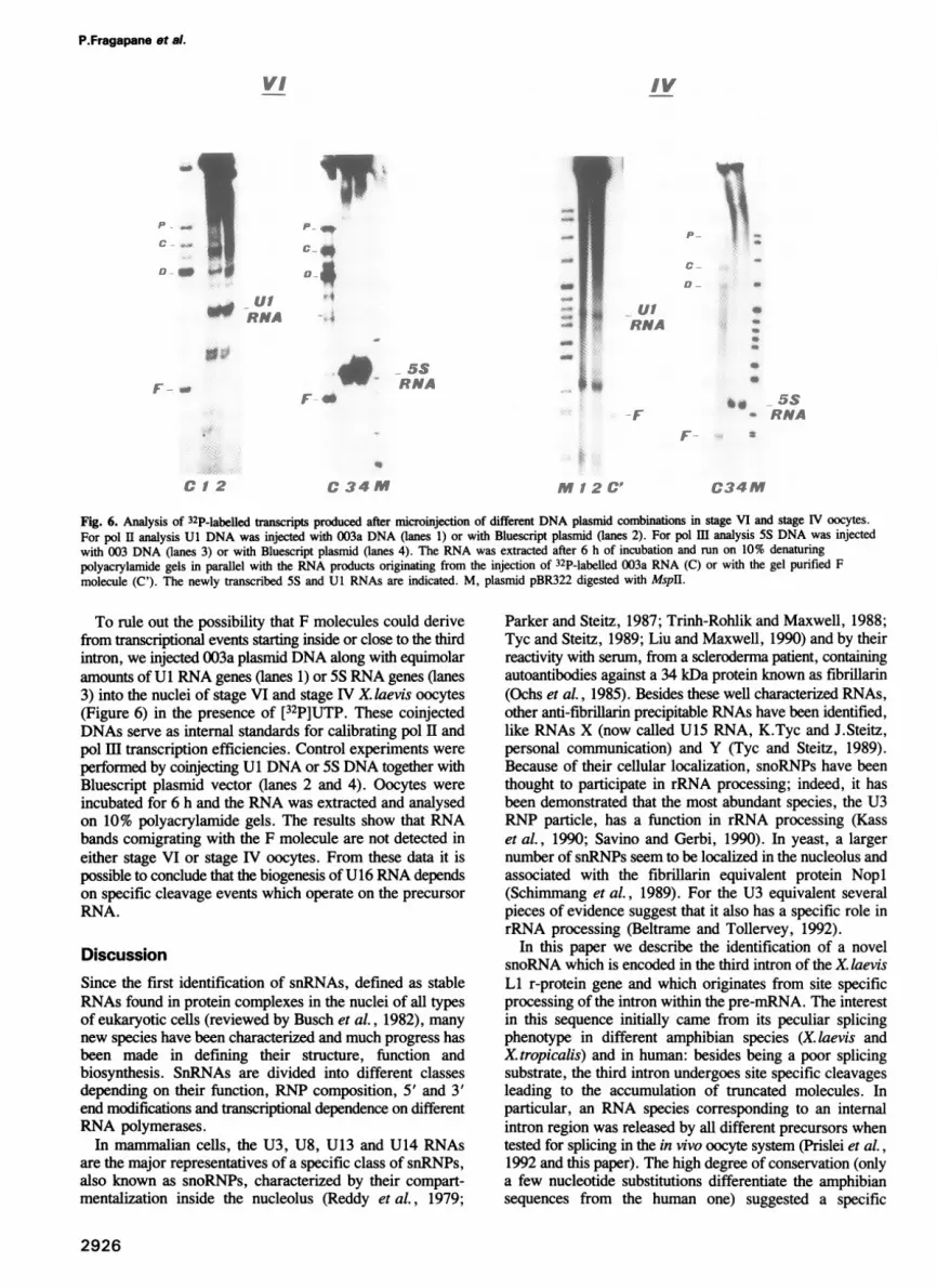
Fig. 6. Analysis of 32P-labelled transcripts produced after microinjection of different DNA plasmid combinations in stage VI and stage IV oocytes.For pol II analysis Ul DNA was injected with 003a DNA (lanes 1) or with Bluescript plasmid (lanes 2). For pol HI analysis 5S DNA was injectedwith 003 DNA (lanes 3) or with Bluescript plasmid (lanes 4). The RNA was extracted after 6 h of incubation and run on 10% denaturingpolyacrylamide gels in parallel with the RNA products originating from the injection of 32P-labelled 003a RNA (C) or with the gel purified Fmolecule (C'). The newly transcribed 5S and Ul RNAs are indicated. M, plasmid pBR322 digested with Mspll.
To rule out the possibility that F molecules could derivefrom transcriptional events starting inside or close to the thirdintron, we injected 003a plasmid DNA along with equimolaramounts of Ul RNA genes (lanes 1) or 5S RNA genes (lanes3) into the nuclei of stage VI and stage IV X. laevis oocytes(Figure 6) in the presence of [32P]UTP. These coinjectedDNAs serve as internal standards for calibrating pol II andpol HI transcription efficiencies. Control experiments were
performed by coinjecting Ul DNA or 5S DNA together withBluescript plasmid vector (lanes 2 and 4). Oocytes were
incubated for 6 h and the RNA was extracted and analysedon 10% polyacrylamide gels. The results show that RNAbands comigrating with the F molecule are not detected ineither stage VI or stage IV oocytes. From these data it ispossible to conclude that the biogenesis of U16 RNA dependson specific cleavage events which operate on the precursorRNA.
DiscussionSince the first identification of snRNAs, defined as stableRNAs found in protein complexes in the nuclei of all typesof eukaryotic cells (reviewed by Busch et al., 1982), many
new species have been characterized and much progress hasbeen made in defining their structure, function andbiosynthesis. SnRNAs are divided into different classesdepending on their function, RNP composition, 5' and 3'end modifications and transcriptional dependence on differentRNA polymerases.
In mammalian cells, the U3, U8, U13 and U14 RNAsare the major representatives of a specific class of snRNPs,also known as snoRNPs, characterized by their compart-mentalization inside the nucleolus (Reddy et al., 1979;
Parker and Steitz, 1987; Trinh-Rohlik and Maxwell, 1988;Tyc and Steitz, 1989; Liu and Maxwell, 1990) and by theirreactivity with serum, from a scleroderma patient, containingautoantibodies against a 34 kDa protein known as fibrillarin(Ochs et al., 1985). Besides these well characterized RNAs,other anti-fibrillarin precipitable RNAs have been identified,like RNAs X (now called U15 RNA, K.Tyc and J.Steitz,personal communication) and Y (Tyc and Steitz, 1989).Because of their cellular localization, snoRNPs have beenthought to participate in rRNA processing; indeed, it hasbeen demonstrated that the most abundant species, the U3RNP particle, has a function in rRNA processing (Kasset al., 1990; Savino and Gerbi, 1990). In yeast, a largernumber of snRNPs seem to be localized in the nucleolus andassociated with the fibrillarin equivalent protein Nopl(Schimmang et al., 1989). For the U3 equivalent severalpieces of evidence suggest that it also has a specific role inrRNA processing (Beltrame and Tollervey, 1992).
In this paper we describe the identification of a novelsnoRNA which is encoded in the third intron of the X. laevisLI r-protein gene and which originates from site specificprocessing of the intron within the pre-mRNA. The interestin this sequence initially came from its peculiar splicingphenotype in different amphibian species (X. laevis andX. tropicalis) and in human: besides being a poor splicingsubstrate, the third intron undergoes site specific cleavagesleading to the accumulation of truncated molecules. Inparticular, an RNA species corresponding to an internalintron region was released by all different precursors whentested for splicing in the in vivo oocyte system (Prislei et al.,1992 and this paper). The high degree of conservation (onlya few nucleotide substitutions differentiate the amphibiansequences from the human one) suggested a specific
2926
P.Fragapane et a!.
IV
P _
_Ia
OC-
C 34 M M 12 C'
........
A
.W*.'i
i.0

snoRNA biosynthesis
functional role of this region per se and stimulated the searchfor endogenous equivalents.
Probing of Northern blots with the LI third intronconserved region proved the existence of homologoussequences in the endogenous RNA population of the oocyte.This RNA species is associated with RNP complexescontaining the fibrillarin antigen, does not possess an m3Gcap and colocalizes with U3 RNA in nucleolar fractionations.By synthetic polyadenylation followed by reversetranscriptase it was possible to clone a cDNA version of thisRNA corresponding to the conserved sequence present inthe third intron of the LIa gene copy. In consideration ofthe limited number of clones isolated it cannot be excludedthat the Llb copy also contributes to the U16 RNA pool.Independent cloning performed with RT-PCR amplificationwith primers internal to the U16 sequence has in fact shownthe presence of transcripts deriving also from the Llb copy.We have called this previously uncharacterized RNA U16and it very probably comprises the U16a and U16bsubspecies. In addition, their 5' end is identical to one ofthe D- and F-type of molecules obtained upon injection ofthe RNA precursor containing the third intron.Immunoprecipitation with fibrillarin antibodies of nucleimicroinjected with 32P-labelled 003a RNA showed that theF molecules are associated with this antigen, as the U16RNA. These results, together with the evidence that no Fmolecules are produced when a plasmid DNA containingthe third intron plus flanking sequences is microinjected intooocyte nuclei, demonstrates that the U16 RNA originatesby processing of the LI pre-mRNA and not by independenttranscription. Several pieces of evidence suggest that the Fmolecule, and as a consequence U16 RNA, derives fromthe unspliced pre-mRNA and not from the excised lariat:(i) the F4 mutant, which has a high splicing efficiency, showsequimolar accumulation of lariat and ligated exons suggestingthat no conversion of lariat into F-type of molecules occurs(Fragapane et al., 1992); (ii) reinjection of gel purified lariatdoes not produce any molecule similar to F-RNA(unpublished).The production of U16 RNA represents quite a peculiar
example of snRNA biosynthesis; in fact, the snRNAscharacterized so far are produced by pol II or pol IIItranscription (for a review see Dahlberg and Lund, 1988).Two snRNAs originating not by independent transcriptionbut by RNA processing have been recently described: U14RNAs (Leverette et al., 1992) and U15 RNA (K.Tyc andJ.Steitz, personal communication). The common feature ofthese RNAs is that they are all localized in nucleoli. Theinteresting feature of U16 is that it is encoded inside aribosomal protein intron which undergoes specific regulation(Caffarelli et al., 1987; Pierandrei-Amaldi et al., 1987). Itwill obviously be important to identify the specific functionof U16 RNA in order to understand its functional andphysical association with the r-protein LI and to correlateit with rRNA synthesis. However, if the presence of snRNAsinside ribosomal protein genes is found to be a morewidespread phenomenon, some additional functions besiderRNA processing can be envisaged. An intriguing hypothesisis that they could be involved in the assembly of rRNA withribosomal proteins. This is quite a complex process whichrequires many structural rRNA rearrangements. In analogywith what is emerging from studies on spliceosome function,where productive structural rearrangements are achieved by
alternative snRNA interactions, it might be that snoRNAs,by specifically interacting with rRNA, regulate the assemblyof specific r-proteins (for instance, those coexpressed on thesame pre-mRNA). The molecular characterization of thesesequences and the availability of suitable in vivolin vitrosystems will hopefully allow in the near future a betterunderstanding of the unclarified aspects of ribosomebiosynthesis.
Materials and methodsCloning of third intron sequences from the human L 1 geneThe third intron of the human LI gene was isolated by PCR amplificationof genomic human DNA using primers derived from the sequence of thecorresponding cDNA (C.Bagni, F.Annesi and F.Amaldi, personalcommunication).The forward oligo (5'-GGGAATTCCCAGAGTTCGAGG-3') includes
19 nt of the 3' portion of the third exon (underlined) and an EcoRI site(bold characters), while the backward primer (5'-CCCTCGAGATGGC-GTATCGTTTTTGG-3') includes 20 nt of the 5' portion of the fourth exon(underlined) and an XhoI site (bold characters).PCR reactions were carried out in 50 Al of 50mM KCI, 10mM Tris-HCI
pH 8.3, 1.5 mM MgCl2, 250 AM dNTP, 0.1 mg/ml BSA, 1 AM of eachprimer, 4.5 units of Taq polymerase and 1 mg of total genomic DNA. Eachamplification cycle consisted of 1 min denaturation at 940C, 2 min annealingat 58°C and 3 min extension at 720C. Thirty cycles were performed.The EcoRI and XhoI sites of the primers allow the amplified DNA to
be cloned in the corresponding sites of the Bluescript vector (Stratagene).The recombinant plasmid (003h) was sequenced according to Sanger (1977).
In vitro RNA synthesis and oocyte microinjections003a RNA was transcribed from plasmid 003a Sacl, digested with HindIm(see Prislei et al., 1992); it contains 89 nt of the third exon, the entire thirdintron (241 bases), 59 nt of the fourth exon and 82 nt of vector polylinker.The template for 003h transcription was obtained byXhoI digestion of
the corresponding plasmid. The transcript includes the 307 nt intron of thehuman LI gene. The plasmid also contains 59 nt of exon 3 and 91 nt ofexon 4 plus 67 nt derived from the vector polylinker.The 32P-labelled T7 transcripts were synthesized invitro and gel purified;
transcripts were injected into the nuclei of stage VI oocytes and total RNAwas extracted frommanually purified nuclei as described by Caffarelli et al.(1987). Equal numbers of injected counts were loaded on 10% denaturingpolyacrylamide gels.DNAmicroinjections were performed as follows. 100 ng of 5S (Razvi
et al., 1983) and Ul (Zeller et al., 1984) plasmids were mixed with 100ng of 003a or Bluescript plasmids, precipitated and resuspended in 2 Alof injection buffer in the presence of 20 ltCi of [32P]UTP. The nuclei ofoocytes were injected with 20 nl of the DNA solution and the oocytes werethen incubated for 6 h. Nuclear RNA from stage VI and total RNA fromstage IV oocytes was extracted and run on 10% denaturing polyacrylamidegels.
Northern blot analysisRNAs were electrophoresed on 6% acrylamide-7 M urea gels,electrotransferred to Amersham's Hybond-N paper and UV crosslinked.All hybridizations were carried out in standard conditions but the one withthe U3 probe which is a 22mer synthetic oligonucleotide complementaryto nt 101-122 of X.laevis U3 snRNA sequences (Caizergues-Ferrer et al.,1991). The oligo was 5' 32P-end-labelled and hybridized according toCaizergues-Ferrer et al. (1991). The U16 analysis was performed with thea--y probe obtained by PCR amplification of the intron conserved region.
Southern blot analysisTotal genomic X.laevis DNA was digested with EcoRI and BamHI and 15Ag were loaded on single slots of a 0.8% agarose gel. The DNA wastransferred to nitrocellulose filters and hybridized with the different DNAprobes: 003a and 003b contain the third intron of the Lia and Llb genesrespectively (Prislei et al., 1992); The LI cDNA contains all the LI codingregion except for the first exon (plasmid p103, Loreni et al., 1985).
Cloning of oocyte endogenous U16 RNA and 5' end mappingTotal RNA extracted from ovaries was fractionated on 10-30% sucrosegradient. 45 Ag of the 4-8S fractions were run on a 6% acrylamide-ureagel and the RNA corresponding to the region of U16 was eluted. The RNA
2927

P.Fragapane et al.
was 3' elongated with poly(A) polymerase and reverse transcribed inthe presence of an oligo(dT)-tailed primer (5'-GCCTCG-AG 1-1-1 1 1-1-1-1----------1-1-3') containing an XhoI site. The cDNAwas ligated to adaptor oligos (A, 5'-GGAATTCGAAGGGGG-3'; B,5'-CCCCCTTCGAAITCCCCC-3') containing an EcoRI site. The mixturewas then PCR amplified utilizing the dT oligo as backward primer and theA adaptor oligo as forward primer. Each amplification cycle consisted of1 min denaturation at 94°C, 2 min annealing at 50°C and 3 min extensionat 72°C. Thirty cycles were performed. The EcoRI and XhoI sites of theprimers allow the cloning of the amplified DNA in the corresponding sitesof the Bluescript vector (Stratagene). The clones positive for colonyhybridization with the a--y probe (see above) were sequenced accordingto Sanger (1977).To characterize the 5' end of the U16 RNA, reverse transcriptase
elongation was performed on 7 Ag of gel selected RNA (see above) utilizingas primer the -y oligonucleotide (5'-CCAAGCTTTTTGCTCAGAACGC-GAT-3') which includes 19 nt of the 3' portion of the conserved intronregion (underlined nucleotides). The same oligo was utilized as primer forthe reverse transcription of gel purified D and F RNAs originating fromoocyte injection of 003a RNA. The reverse transcriptase elongation of Dand F RNAs, obtained after the injection of the human third intron, wasperformed with the -y-h oligonucleotide (5'-CAAGCTTTCTTGCTCAGTA-AGAAT-3') which includes 19 nt of the 3' portion of the human conservedintron region (underlined nucleotides). The RNAs and oligo mixtures wereheated at 950C for 5 min, annealed at 550C for 15 min and incubated onice for 10 min. 30 units of AMV reverse transcriptase (Biolabs), 20 unitsof RNasin (Boehringer) and dNTPs to a final concentration of 1 mM eachwere added to the reactions and incubations were continued at 42°C for60 min. The products of the reactions were run on 6% denaturingpolyacrylamide gels in parallel with a Sanger sequence performed with thesame primers on the corresponding plasmid DNAs.
AntibodiesClinical anti-Sm sera were obtained from P.Janteur (Universite des Scienceset Techniques du Languedoc, Montpellier), the anti-m3G cap antibodieswere obtained from R.Luhrmann (Philipps University, Marburg) and theanti-fibrillarin sera were provided by M.Caizergues-Ferrer (CNRS,Toulouse).
ImmunoprecipitationsThe antisera utilized (10 Al of anti-fibrillarin, 10 Al of anti-Sm, 2 Al of anti-m3G cap and 10 A1 of rabbit nonimmune serum) were preadsorbed to 201tl of protein A-Sepharose CL-4B (Pharmacia, 0.1 g/ml preswollen inIPP500) in 400 1l of IPP500 overnight at room temperature and then washedthree times with 1 ml of IPP500 (Hamm et al. 1989). Pools of 30 nucleiwere homogenized in 500 Id of 10 mM Tris pH 8.0, 50 mM NaCl and80 units of RNasin (Boehringer) and added to each antibody suspension.10 Al of Nonidet P40 was added and the samples were rolled for 2 h atroom temperature. The beads were washed three times for 3-5 min with1 ml of IPPSW, and RNA was released from the beads by digestion with400 11 of Homomedium (Hamm et al., 1989) The samples were phenolextracted and the RNA was precipitated.
Preparation of nucleoliGerminal vesicles were manually dissected from X. laevis oocytes and treatedaccording to Peculis and Gall (1992) with the only modification of increasingthe NaCl concentration to 100 mM. After sonication and centrifugation apellet fraction containing membranes, chromosomes and nucleoli wasseparated from a supernatant fraction containing soluble nucleoplasm.
Caffarelli,E., Fragapane,P., Gehring,C. and Bozzoni,I.(1987) EMBO J.,6, 3493-3498.
Caffarelli,E., Fragapane,P. and Bozzoni,I. (1992) Biochem. Biophys. Res.Commun., 183, 680-687.
Caizergues-Ferrer,M., Mathieu,C., Mariottini,P., Amalric,F. and Amaldi,F.(1991) Development, 112, 317-326.
Dahlberg,J.E. and Lund.,E. (1988) In Birnstiel,M.L. (ed.), Structure andFunction of Major and Minor Small Nuclear Ribonucleotide Particles.Springer-Verlag, Berlin, pp. 38-70.
Dumont,J.N. (1972) J. Morphol., 136, 153-179.Fragapane,P., Caffarelli, E., Lener,M., Prislei,S., Santoro,B. and Bozzoni,I.
(1992) Mol. Cell. Biol., 12, 1117-1125.Hamm,J. and Mattaj,I.W. (1989) EMBO J., 8, 4179-4187.Hamm,J., Dathan,N.A. and Mattaj,I.W. (1989) Cell, 59, 159-169.Kass,S., Tyc,K., Steitz,J.A. and Sollner-Webb,B. (1990) Cell, 60,
897-908.Jeppesen,C., Stebbins-Boaz,B. and Gerbi,S.A. (1988) Nucleic Acids Res.,
16, 2127-2148.Leverette,R.D., Andrews,M.T. and Maxwell, S.E. (1992) Cell, 71,
1215-1221.Liu,J. and Maxwell,E.S. (1990) Nucleic Acids Res., 18, 6565-6571.Loreni,F., Ruberti,I., Bozzoni,I., Pierandrei-Amaldi,P. and Amaldi,F.
(1985) EMBO J., 4, 3483-3488.Ochs,R.L., Lischwe,M.A., Spohn,W.H. and Bush,H. (1985) Biol. Cell,
54, 123-134.Parker,K.A. and Steitz,J.A. (1987) Mol. Cell. Biol., 7, 2899-2913.Peculis,B.A. and Gall,J.G. (1992) J. Cell Biol., 116, 1-14.Pierandrei-Amaldi,P., Amaldi,F., Bozzoni,I. and Fragapane,P. (1987) In
Firtel,R.A. and Davidson,E.H. (eds), Molecular Approaches toDevelopmental Biology. Alan R.Liss, Inc., New York, pp. 267-278.
Prislei,S., Sperandio,S., Fragapane,P., Caffarelli,E., Presutti,C. andBozzon,I. (1992) Nucleic Acids Res., 16, 4473-4479.
Razvi,F., Gargiulo,G. and Worcel,A. (1983) Gene, 23, 175-183.Reddy,R. and Busch,H. (1988) In Bimstiel,M.L. (ed.), Structure and
Function of Major and Minor Small Nuclear Ribonucleotide Particles.Springer-Verlag, Berlin, pp. 1-37.
Reddy,R., Henning,D. and Busch,H. (1979) J. Biol. Chem., 254,11097-11105.
Sanger,F., Nicklen,S., Coulson,A.R. (1977) Proc. Natl Acad. Sci. USA,74, 5463-5467.
Savino,R. and Gerbi,S. A. (1990) EMBO J., 9, 2299-2308.Schimmang,T., Tollervey,D., Kern,H., Frank,R. and Hurt,E.C. (1989)EMBO J., 8, 4015-4024.
Trinh-Rohlik,Q. and Maxwell,E.S. (1988) Nucleic Acids Res., 16,6041 -6055.
Tyc,K. and Steitz,J.A. (1989) EMBO J., 8, 3113-3119.Zeller,R., Carri,M.T., Mattaj,I.W. and De Robertis,E.M. (1984) EMBO
J., 3, 1075-1081.
Received on December 15, 1992; revised on April 16, 1993
Note added in proof
The sequence data for Llh have been deposited in the EMBL/GenBankdatabases under the accession number X72205.
AcknowledgementsWe thank Dr Michelle Caizergues-Ferrer for kindly providing the reagentsfor fibrillarin and U3 analysis; we also thank Mr Arceci, Mr Di Francescoand Mr Ricci for technical assistance and Dr Fabio Riccobono for oligofacilities. This work was partially supported by grants from the ProgettiFinalizzati 'Biotecnologie e Biostrumentazioni' and 'Ingegneria Genetica'of CNR and from the Istituto Pasteur Fondazione Cenci-Bolognetti.
ReferencesBeltrame,M. and Tollervey,D. (1992) EMBO J., 11, 1531-1542.Bozzoni,I., Beccari,E., Luo,Z.-X., Amaldi,F., Pierandrei-Amaldi,P. and
Campioni,N. (1981) Nucleic Acids Res., 9, 1069-1086.Busch,H., Reddy,R., Rothblum,L. and Choi,Y.C. (1982) Annu. Rev.
Biochem., 51, 617-654.
2928
Recommended


