
A Tick Gut Protein with Fibronectin III Domains AidsBorrelia burgdorferi Congregation to the Gut duringTransmissionSukanya Narasimhan1.*, Jeroen Coumou2., Tim J. Schuijt2, Eric Boder3, Joppe W. Hovius2"*,
Erol Fikrig1,4"
1 Department of Internal Medicine, Yale University School of Medicine, New Haven, Connecticut, United States of America, 2 Center for Experimental and Molecular
Medicine, Academic Medical Center, University of Amsterdam, Amsterdam, The Netherlands, 3 Department of Chemical and Biomolecular of Engineering, University of
Tennessee, Knoxville, Tennessee, United States of America, 4 Howard Hughes Medical Institute, Chevy Chase, Maryland, United States of America
Abstract
Borrelia burgdorferi transmission to the vertebrate host commences with growth of the spirochete in the tick gut andmigration from the gut to the salivary glands. This complex process, involving intimate interactions of the spirochete withthe gut epithelium, is pivotal to transmission. We utilized a yeast surface display library of tick gut proteins to perform aglobal screen for tick gut proteins that might interact with Borrelia membrane proteins. A putative fibronectin type IIIdomain-containing tick gut protein (Ixofin3D) was most frequently identified from this screen and prioritized for furtheranalysis. Immunization against Ixofin3D and RNA interference-mediated reduction in expression of Ixofin3D resulted indecreased spirochete burden in tick salivary glands and in the murine host. Microscopic examination showed decreasedaggregation of spirochetes on the gut epithelium concomitant with reduced expression of Ixofin3D. Our observationssuggest that the interaction between Borrelia and Ixofin3D facilitates spirochete congregation to the gut duringtransmission, and provides a ‘‘molecular exit’’ direction for spirochete egress from the gut.
Citation: Narasimhan S, Coumou J, Schuijt TJ, Boder E, Hovius JW, et al. (2014) A Tick Gut Protein with Fibronectin III Domains Aids Borrelia burgdorferiCongregation to the Gut during Transmission. PLoS Pathog 10(8): e1004278. doi:10.1371/journal.ppat.1004278
Editor: Jose de la Fuente, Oklahoma State University, United States of America
Received February 23, 2014; Accepted June 12, 2014; Published August 7, 2014
Copyright: � 2014 Narasimhan et al. This is an open-access article distributed under the terms of the Creative Commons Attribution License, which permitsunrestricted use, distribution, and reproduction in any medium, provided the original author and source are credited.
Funding: This work was supported by grant funding (AI076705) from NIAID/NIH to SN. JWH is a recipient of a VENI stipend (91611065) from the NetherlandsOrganization for Health Research and Development (ZonMw). EF is an investigator of the Howard Hughes Medical Institute. The funders had no role in studydesign, data collection and analysis, decision to publish, or preparation of the manuscript.
Competing Interests: The authors have declared that no competing interests exist.
* Email: [email protected] (SN); [email protected] (JWH)
. These authors contributed equally to this work.
" JWH and EF also contributed equally to this work.
Introduction
Ixodes scapularis is the predominant vector of several human
pathogens including Borrelia burgdorferi, the agent of Lyme
borreliosis in North America [1,2]. There is currently no
commercial vaccine to prevent Lyme borreliosis in humans [3],
although recent efforts have been made in that direction [4]. An
increased molecular understanding of how the tick acquires,
sustains and transmits Borrelia would be conducive to the
development of novel strategies, including anti-tick vaccines [5–
7], to control Lyme borreliosis. Borrelia resides in the unfed tick
gut anchored to a gut protein, TROSPA [8]. Transmission begins
with the growth of the spirochetes in the gut when the Borrelia-
infected tick begins to take a blood meal. Somewhere between 24
and 36 hours of tick feeding [9], coincident perhaps with an
optimal quorum of spirochetes or its gene expression profile
[10,11], the spirochete travels from the gut to the salivary glands
from where it exits the tick along with tick saliva into the host skin.
Growth and migration from the tick gut is therefore an essential
prelude to Borrelia transmission.
The spirochete proteome changes dramatically during tick
feeding to facilitate migration from the gut [10]. Rudenko et al
[12] showed that Borrelia infection alters the transcriptome of the
Ixodes ricinus gut during feeding, suggesting a dynamic interaction
between the tick gut and the growing spirochete. Consistent with
this, Dunham-Ems et al [13] showed by live imaging of spirochetes
in feeding I. scapularis guts that the spirochete engages intimately
with the epithelial cells of the tick gut during transmission, moving
away from the gut lumen towards the basal lamina of the gut. It is
likely that Borrelia-gut interactions provide molecular signals that
direct the movement of the spirochete from the luminal side of the
gut epithelium to the basal lamina of the gut to facilitate egress
from the gut. Further, Zhang et al [14] showed that interaction
between a secreted tick gut protein, TRE31, and a spirochete
outer surface protein BBE31 enables migration of the spirochete
through the hemolymph to the salivary glands by mechanisms that
remain to be understood. These observations highlight a thematic
strategy of the spirochete to interact with gut proteins during
growth and migration, which we are only just beginning to
understand. In order to delineate the complex molecular
interactions of spirochetes with the gut epithelium we screened
for Borrelia-interacting tick gut proteins by probing a tick gut yeast
surface display (YSD) library with Borrelia outer surface proteins.
The YSD approach has traditionally utilized specific proteins
PLOS Pathogens | www.plospathogens.org 1 August 2014 | Volume 10 | Issue 8 | e1004278
individually to probe libraries of single chain antibodies to identify
and characterize protein-protein interactions [15]. Work by Cho
and Shusta [16] demonstrated that biotinylated whole cell lysates
of mammalian cell lines or plasma membrane proteins can be used
to screen a YSD library expressing human single chain antibody
fragments and identify specific antigen-antibody interactions
without a priori knowledge of the candidate antigens [17].
Building on this work, we have, in this study, extended the utility
of YSD to examine tick gut-B. burgdorferi interactions without apriori knowledge of either interactants.
We screened 107 tick gut YSD clones with total Borreliamembrane extracts derived from in vitro-grown B. burgdorferiN40 and identified four potential Borrelia-interacting gut proteins
from the initial screen. One of the predominant clones encoded a
surface exposed tick gut protein with four putative fibronectin type
III domains. In this report, we present our observations that
suggest that the fibronectin type III domain-containing tick gut
protein helps congregation of spirochetes to the gut epithelium
during transmission. These observations invoke a functional role
for spirochete ‘‘clustering’’ in spirochete egress from the gut.
Results
Yeast surface display library screening identifies fourpotential Borrelia-interacting tick gut proteins
B. burgdorferi membrane protein extracts were prepared as
described [18] from in vitro grown B. burgdorferi (N40)
temperature-shifted to 37uC for 24 hours. A YSD expression
library of I. scapularis gut cDNAs [14] was probed with biotin-
labeled B. burgdorferi membrane protein extracts as described in
Materials and Methods. Four rounds of magnetic-activated cell
sorting (MACS) screens provided a 40-fold enrichment of YSD
clones expressing gut proteins that interacted with B. burgdorferimembrane proteins (Fig. 1A–B). Cells from the 4th sort were
plated and one hundred colonies were individually tested for their
ability to bind to B. burgdorferi membrane protein extracts by
fluorescence-activated cell sorting (FACS) analysis using Alexa488-
labeled B. burgdorferi membrane protein extracts. Recombinant
plasmids were isolated from colonies that showed greater than
15% binding (40 clones) (Fig. 1C) and insert sizes compared by
restriction digestion analysis. Clones with identical insert sizes were
grouped (four groups) and two representative clones from each
group were sequenced. Four unique clones encoding partial
fragments of tick gut proteins were identified and provided a
unique identifier based on their in silico predicted function
(Table 1).
Clone 1 (identified 15 times in this screen, ,29% binding)
contained an in-frame insert that showed 100% identity to
ISCW008121 (www.vectorbase.org) and encoded a protein that
contained four putative fibronectin type III-domains. PSORT
(http://psort.hgc.jp/) protein localization analysis suggested that it
was likely a surface exposed transmembrane protein (Table 1).
Clone 2 (identified 10 times in this screen, ,30% binding) showed
100% identity to ISCW015135. ISCW015135 encoded a putative
signal peptide indicative of a secreted protein as seen by Signal P
analysis (www.cbs.dtu.dk/services/SignalP). No known functions
or domains were identified, and no paralogs or orthologs of Clone
2 were observed by BLAST analysis. Clone 3 (identified 10 times
in this screen, ,37% binding) showed 100% identity to
ISCW015049 and encoded a protein with two dystroglycan-like
cadherin domains. The cellular location could not be predicted by
PSORT analysis. Orthologs of clone 3 were found in eight other
invertebrate vectors. Clone 4 (identified three times, 26% binding)
showed 100% identity to ISCW016197 and encoded a nuclear
membrane localization signal and a putative Guanylate-kinase
associated protein domain, and orthologs were identified in 13
other invertebrate vectors (Table 1). B. burgdorferi is an
extracellular pathogen, hence the physiological relevance of
interactions between B. burgdorferi extracellular proteins and a
tick nuclear protein (clone 4) is unclear and hence not prioritized
for further analysis.
The expression profiles of the genes contained in the three
prioritized clones were assessed by quantitative RT-PCR (qRT-
PCR) in the salivary glands and guts of I. scapularis nymphs
(Fig. 1D) during feeding. While ISCW008121 (Clone 1) was
expressed preferentially in the gut, ISCW015135 and
ISCW015049 (clones 2 and 3) were expressed both in the salivary
glands and guts. Furthermore, B. burgdorferi infection increased
the expressions of Clones 1 and 3 significantly (,2-fold) in the guts
after 72 hours of tick feeding (Fig. 1D).
Full-length ISCW008121 encodes a protein with fourputative fibronectin type III domains and atransmembrane domain
We addressed Clone 1 in further detail because it was identified
most frequently in this screen and because Clone 1 encoded a tick
protein with fibronectin type III domains. While B. burgdorferihas been shown to encode lipoproteins that facilitate Borreliaadhesion to fibronectin in the mammalian host to promote
infection [19], it is not known if similar interactions occur in the
tick. Further, fibronectin type III domains, originally identified in
the extracellular matrix protein fibronectin [20], have also been
identified in several receptor-like proteins and shown to play a
critical role in cell signaling [21]. Therefore, we reasoned that
understanding the physiological significance of Clone 1-Borreliainteraction might provide new insights into tick-Borrelia interac-
tions.
The protein encoded by YSD Clone 1 in this screen
encompasses amino acids 97 to 449 of the protein encoded by
ISCW008121 (Figure 2A). To confirm the start site of the
annotated ISCW008121 (www.vectorbase.org), we performed a
59end RLM RACE (RNA Ligase-Mediated Rapid Amplification
of cDNA Ends) using fed I. scapularis gut total RNA and gene
specific primers complementary to the first 411 bp of the
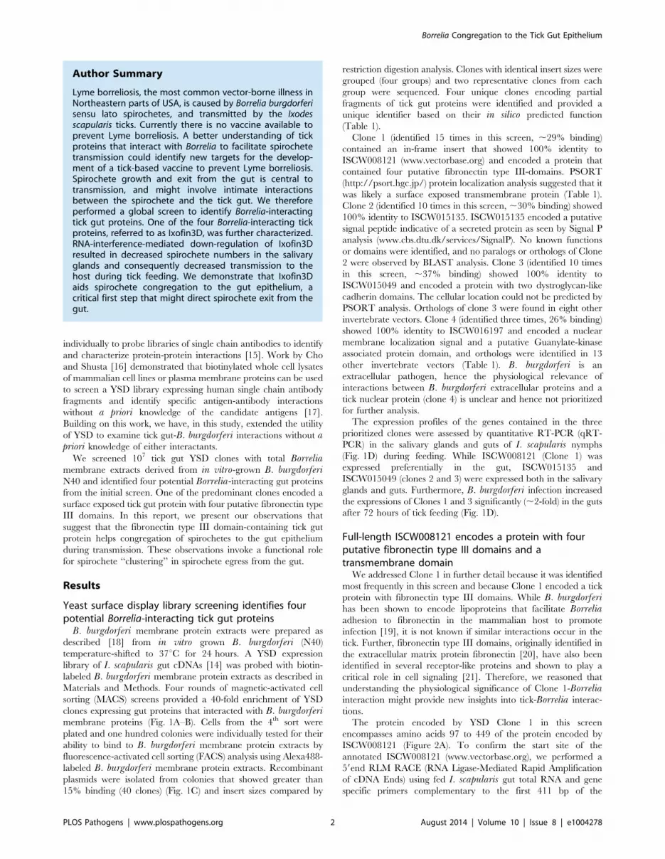
Author Summary
Lyme borreliosis, the most common vector-borne illness inNortheastern parts of USA, is caused by Borrelia burgdorferisensu lato spirochetes, and transmitted by the Ixodesscapularis ticks. Currently there is no vaccine available toprevent Lyme borreliosis. A better understanding of tickproteins that interact with Borrelia to facilitate spirochetetransmission could identify new targets for the develop-ment of a tick-based vaccine to prevent Lyme borreliosis.Spirochete growth and exit from the gut is central totransmission, and might involve intimate interactionsbetween the spirochete and the tick gut. We thereforeperformed a global screen to identify Borrelia-interactingtick gut proteins. One of the four Borrelia-interacting tickproteins, referred to as Ixofin3D, was further characterized.RNA-interference-mediated down-regulation of Ixofin3Dresulted in decreased spirochete numbers in the salivaryglands and consequently decreased transmission to thehost during tick feeding. We demonstrate that Ixofin3Daids spirochete congregation to the gut epithelium, acritical first step that might direct spirochete exit from thegut.
Borrelia Congregation to the Tick Gut Epithelium
PLOS Pathogens | www.plospathogens.org 2 August 2014 | Volume 10 | Issue 8 | e1004278

annotated ISCW008121 gene transcript. Further, YSD Clone 1
sequence and the annotated ISCW008121 transcript did not
contain a stop codon, suggesting that the annotated gene sequence
did not contain a full-length 39 sequence. Therefore, we performed
a 39end RLM RACE using fed I. scapularis gut total RNA and
gene-specific primers, and identified the transcript from bp 1350
to 1836, complete with stop codon. The full-length sequence was
shown to encode a ,66 kDa protein containing four fibronectin
type III domains and a transmembrane domain (Fig. 2B) and is
henceforth referred to as Ixodes scapularis Fibronectin 3Domain-containing gut protein (Ixofin3D) and is assigned the
GenBank accession number KF709698.
Ixofin3D-PF binds in vitro grown spirochetesWe were unable to express the full-length protein transcript of
Ixofin3D in the Drosophila expression system (DES), but
Figure 1. Yeast Surface Display (YSD) approach to identify tick gut proteins that interact with B. burgdorferi membrane proteins. A.EBY-100 yeast cells transformed with an Ixodes scapularis salivary gland cDNA library were induced overnight before magnetic sorting. The followingday, surface tick protein expression as fusion proteins with the yeast agglutinin protein Aga2p was confirmed with antibodies against the Xpressepitope using flow cytometry. B. After each magnetic sort, binding of Alexa488-labeled B. burgdorferi membrane protein extract to EBY-100 yeastcells was analysed using flow cytometry. C. Flow cytometric analysis of individual yeast displayed clones from Sort 4. Borrelia-binding clones 1, 2 and3 and one representative non-binding clones are shown. In panels B and C cell populations within the gate represent yeast-displayed clones boundto Alexa488-labeled B. burgdorferi membrane protein extract. D. Quantitative RT-PCR assessment of the mRNA expression profiles of the genesencoding clones 1, 2 and 3 in salivary glands (SG) and guts (MG) of B. burgdorferi-infected and uninfected nymphs at 24 and 72 hours post tickattachment. Error bars represent mean 6 SEM and mean values significantly different in a two-tailed non-parametric Mann-Whitney test (p,0.05)indicated by one asterisk (p,0.05) or two asterisks (p,0.01).doi:10.1371/journal.ppat.1004278.g001
Borrelia Congregation to the Tick Gut Epithelium
PLOS Pathogens | www.plospathogens.org 3 August 2014 | Volume 10 | Issue 8 | e1004278

succeeded in expressing a partial fragment of Ixofin3D protein
encompassing amino acids 104 to 319 (rIxofin3D-PF) that is also
contained in the protein fragment expressed in YSD Clone 1
(Fig. 2A). The 37 kDa rIxofin3D-PF generated in Drosophila S2
cells was glycosylated as seen by Periodic-Acid Schiff’s staining
(Fig. 3A). Polyclonal rabbit antibodies against rIxofin3D-PF
bound to uninfected and B. burgdorferi-infected fed guts (Fig. 3B)
as seen by confocal microscopy, suggesting that Ixofin3D is
expressed on the surface of the gut. Consistent with the qRT-PCR
analysis, quantification of pixel intensity in the TRITC channel
(representing anti-rIxofin3D-PF serum binding to native Ixofin3D)
using the ImageJ software showed significantly increased binding
of rIxofin3D-PF antibodies to the tick gut in 24 h and 72 h fed
ticks upon B. burgdorferi infection compared to that in 24 h and
72 h fed uninfected guts (Fig. 3C), and in B. burgdorferi-infected
72 h fed tick guts when compared to B. burgdorferi-infected 24 h
fed guts (Fig. 3C) suggesting that Ixofin3D expression was
increased in B. burgdorferi infected guts during feeding.
rIxofin3D-PF incubated with in vitro grown PFA-fixed non-
permeabilized B. burgdorferi N40 showed binding of rIxofin3D-
PF to spirochetes as seen by indirect immunofluorescence using
rabbit polyclonal antibodies against purified rIxofin3D-PF
(Fig. 4A) indicating a potential interaction between Ixofin3D
and an exposed B. burgdorferi protein ligand. Under similar
conditions, rIxophilin, a tick gut thrombin inhibitor protein [22],
not known to engage directly with spirochetes, did not show
binding to in vitro grown spirochetes (Fig. 4A). Furthermore, in an
ELISA assay using microplates coated with B. burgdorferimembrane protein extract, a dose-dependent increase in rIxo-
fin3D-PF binding to B. burgdorferi membrane protein extracts
was observed, whereas dose-dependent binding of rIxophilin was
not observed (Fig. 4B).
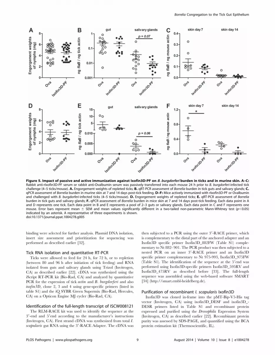
Active and passive immunity against Ixofin3D-PF impairsBorrelia transmission
To determine the role of Ixofin3D in spirochete transmission,
we passively transferred purified rabbit IgG against rIxofin3D-PF
into eight C3H/HeN mice and challenged these mice with
Borrelia-infected I. scapularis nymphs. Control mice received
purified rabbit IgG against ovalbumin (Ova). Ticks fed to
repletion and engorged comparably on both control and
experimental mice (Fig. 5A). Guts and salivary glands were
dissected from engorged nymphs and Borrelia burden assessed by
qRT-PCR. While the spirochete burden in the guts were
comparable in both groups, Borrelia burden in the salivary
glands was reduced in nymphs that fed on mice that received
anti-rIxofin3D-PF antibodies (Fig. 5B) when compared to that in
salivary glands of nymphs fed on mice that received anti-
Ovalbumin antibodies, however, the decrease was not statistically
significant. Borrelia burden in the skin of mice that received anti-
rIxofin3D-PF antibodies (Fig. 5C) was also reduced at 7 days post
tick feeding when compared to that in the skin of mice that
received anti-Ovalbumin antibodies, however, the decrease was
not statistically significant.
As seen with passive immunization, active immunization against
rIxofin3D-PF did not impact the engorgement weights of nymphal
ticks, and Borrelia burden in the nymphal guts (Fig. 5D–E). Active
immunization against rIxofin3D-PF decreased Borrelia burden in
the salivary glands of fed nymphs, although, the decrease was not
statistically significant (Fig. 5E). However, Borrelia burden in the
skin of mice at 7 days post tick feeding was significantly reduced
when compared to that in the skin of mice that were immunized
against ovalbumin (Fig. 5F).
Ta
ble
1.
I.sc
ap
ula
ris
gu
tye
ast
surf
ace
dis
pla
ylib
rary
scre
en
ing
ide
nti
fie
sfo
ur
po
ten
tial
Bo
rrel
ia-i
nte
ract
ing
tick
gu
tp
rote
ins.
Clo
ne
Nu
mb
er
of
tim
es
ide
nti
fie
din
the
scre
en
%ce
lls
bin
din
gto
Bb
me
mb
ran
ep
rote
ins
Ge
ne
ide
nti
tyI
Ma
tch
ing
reg
ion
on
OR
FI
Po
ssib
lym
issi
ng
ex
on
sII
Pu
tati
ve
fun
ctio
na
nd
do
ma
insII
IC
ell
ula
rlo
cati
on
IVS
ign
al
pe
pti
de
V
Tra
ns-
me
mb
ran
ed
om
ain
III
MW
VI
(kD
a)
Pa
ralo
gsI
Ort
ho
log
sI
11
52
9%
ISC
W0
08
12
12
94
–1
34
7Y
es
Ce
ll-ad
he
sio
n(f
ibro
ne
ctin
IIId
om
ain
s)T
ran
sme
mb
ran
eY
es
Ye
s6
5*
Ye
s(3
)N
on
e
21
03
0%
ISC
W0
15
13
51
–3
20
No
No
ne
Extr
ace
llula
rY
es
No
ne
13
No
ne
No
ne
31
03
7%
ISC
W0
15
04
91
–6
04
Ye
sC
ell-
adh
esi
on
(Dys
tro
ph
in-
asso
ciat
ed
gly
cop
rote
in-1
)U
nce
rtai
nN
on
eY
es
91
No
ne
Ye
s(8
)
43
26
%IS
CW
01
61
97
19
2–
76
2Y
es
Gu
anyl
ate
-kin
ase
asso
ciat
ed
pro
tein
(GK
AP
)N
ucl
ear
No
ne
No
ne
70
No
ne
Ye
s(1
3)
I dat
abas
eo
fw
ww
.ve
cto
rbas
e.o
rg,
IIas
stat
ed
on
Ge
nb
ank
ann
ota
tio
ns,
III SM
AR
Td
om
ain
pre
dic
tio
n(h
ttp
://s
mar
t.e
mb
l-h
eid
elb
erg
.de
),IV
PSO
RT
(htt
p:/
/pso
rt.h
gc.
jp/)
,V
Sig
nal
Pan
alys
is(w
ww
.cb
s.d
tu.d
k/se
rvic
es/
Sig
nal
P/)
,V
I Th
eo
reti
cal
mo
lecu
lar
we
igh
t(M
W)
inkD
au
sin
gEx
PA
Syp
rote
om
ics
serv
er
(htt
p:/
/we
b.e
xpas
y.o
rg/c
om
pu
te_
pi/
).*B
ase
do
nIx
ofi
n3
Dse
qu
en
ce,
see
Fig
ure
2.
OR
F:O
pe
nR
ead
ing
Fram
e.
do
i:10
.13
71
/jo
urn
al.p
pat
.10
04
27
8.t
00
1
Borrelia Congregation to the Tick Gut Epithelium
PLOS Pathogens | www.plospathogens.org 4 August 2014 | Volume 10 | Issue 8 | e1004278

RNA interference-mediated decrease in ixofin3Dexpression results in decreased Borrelia transmission
To circumvent the possibility that antibodies against partial
Ixofin3D might not efficiently abrogate Ixofin3D function in vivo,
and to clarify the role of Ixofin3D in Borrelia transmission, we
decreased the expression of Ixofin3D by RNA interference (RNAi)
as described earlier [23]. Four to five double stranded (ds)
ixofin3D RNA-injected nymphs or ds gfp RNA-injected were
allowed to engorge on each mouse (8 mice/group). Nymphs
injected with ds ixofin3D RNA engorged comparably to control
nymphs injected with ds gfp RNA (Fig. 6A) despite a significant
decrease in the expression of ixofin3D RNA in the guts as seen by
qRT-PCR (Fig. 6B). While Borrelia burden in the guts was
comparable in ds gfp and ds ixofin3D-injected nymphs (Fig. 6C),
Borrelia burden in the salivary glands of fed ds ixofin3D-injected
nymphs when compared to that in the salivary glands of ds gfpRNA-injected nymphs was significantly decreased (Fig. 6C).
Borrelia burden in the skin of mice fed upon by ds ixofin3DRNA-injected nymphs (experimental group) at 7 and 14 days post
tick feeding was significantly decreased when compared to that in
the skin of mice that were fed upon by ds gfp RNA-injected
nymphs (Fig. 6D).
RNAi-mediated knock-down of ixofin3D expressionresults in decreased aggregation of spirochetes on thetick gut
Borrelia replicates in the gut in preparation for transmission,
and adheres tightly to the gut epithelium in order to migrate
towards the basal lamina of the gut epithelium, moving away from
the lumen [13]. RNAi-mediated decrease in ixofin3D expression
did not demonstrate alteration in Borrelia burden in the tick gut as
seen by qRT-PCR (Fig. 5B). However, this assessment cannot
distinguish gut epithelium-bound Borrelia from those that are not
bound to the gut epithelium. Therefore, we assessed by confocal
microscopy, if Ixofin3D might enhance Borrelia adherence to the
gut epithelium. RNAi-mediated decrease in ixofin3D expression
resulted in significantly decreased Borrelia clustering to the gut
epithelium as seen by confocal microscopy (Fig. 7A) and
quantification of the pixel intensity in the FITC channel
(representing binding of anti-B. burgdorferi serum to spirochetes)
using the ImajeJ software (Fig. 7B). Consistent with the qRT-PCR
observations, the numbers of spirochetes was also significantly
reduced in the salivary glands of ds ixofin3D RNA-injected
nymphs (Fig. 7C–D). To further assess if Ixofin3D might facilitate
spirochete aggregation to the tick gut, fed tick guts were washed to
remove luminal blood-meal contents and spirochetes loosely
adhering to the gut epithelium. Borrelia burden assessed in the
washed gut epithelium by confocal microscopy and quantification
of the pixel intensity in the FITC channel using the ImageJ
software and by qRT-PCR showed decreased gut-bound Borreliaburden in ds ixofin3D RNA-injected nymphal guts when
compared to that in ds gfp RNA-injected nymphal guts
(Fig. 7E–G).
Discussion
Understanding the biology of B. burgdorferi and its pivotal
interactions with the host and the vector remains a key area of B.burgdorferi research [24]. In this study we focused on the tick gut,
a tissue central to spirochete growth and transmission [11,25,26].
Little is known of the molecular interactions between the tick gut
and the spirochete that facilitate exit from the gut for transmission
to occur. We utilized a yeast surface display (YSD) approach and
identified four tick gut proteins that might engage with the
spirochete during transmission (Table 1). Zhang et al [14] showed
that BBE31, a Borrelia outer surface protein binds to TRE31, a
secreted tick gut protein. Our initial screening of the YSD library
with in vitro-grown spirochetes did not identify TRE31, presum-
ably, due to the very low expression levels of BBE31 in in vitrogrown spirochetes [14].
Clone 1, the most frequently identified clone in this YSD screen,
encoded a partial fragment of the gene ISCW008121. Three
paralogs of ISCW008121 are represented in the Ixodes scapularisgenome. BLAST analysis did not reveal orthologs of ISCW008121
in other tick species. The full-length ,66 kDa protein referred to
as Ixofin3D contained four putative fibronectin type III domains.
The fibronectin type III domain was originally identified within
the protein fibronectin [27]. B. burgdorferi encodes at least two
proteins that bind to fibronectin, RevA [28] and BBK32 [29,30],
and this binding has been invoked in the infection of the murine
host, but not the tick. ELISA assessment did not demonstrate the
binding of RevA or BBK32 to Ixofin3D-PF (data not shown),
suggesting that Ixofin3D might not be a fibronectin-like protein,
and that the fibronectin III domains might provide a novel
function that remain to be elucidated.
Although active and passive immunizations against rIxofin3D-
PF showed a consistent trend towards decreased spirochete burden
in the salivary glands, the decrease was not statistically significant.
However, we observed a significant decrease in the skin seven days
post-tick challenge upon active immunization against Ixofin3D.
We expect that active immunization achieved higher levels of
circulating antibodies and likely provided more efficient impair-
ment of spirochete migration from the gut to the salivary glands
when compared to that observed upon passive immunization.
While a threshold of spirochete numbers critical for effective tick
transmission is not defined, when lesser numbers of spirochetes are
deposited in the skin they might be more vulnerable to the host
immune responses. Similarly, RNAi-mediated silencing of ixo-fin3D expression provided a significant decrease in spirochete
burden in salivary glands and consequently significantly decreased
spirochete burden in the murine host skin at 7 and 14 days post-
feeding. However, burden in the distal organs assessed 21 days
post feeding was not different upon active immunization or RNAi-
mediated knockdown of ixofin3D expression. This suggests that
spirochetes that escape the initial host immune response, replicate,
and disseminate successfully with time. Tick challenge experiments
described herein utilized 4–5 Borrelia-infected ticks, which
provides a combined inoculum of immunomodulatory tick
proteins and spirochetes from 4–5 ticks, and thus potentially
deflates the significance of Ixofin3D in spirochete transmission.
Challenge experiments using smaller numbers of ticks might be
more reflective of tick bites on humans and could be viable in
studies assessing the vaccine potential of tick and Borrelia antigens.
Immunization or RNAi-mediated interruption of Ixofin3D-
Borrelia interaction decreased spirochete burden in the salivary
glands without any significant change in the Borrelia burden in the
tick gut suggesting that Ixofin3D-spirochete interaction might
facilitate spirochete entry into salivary glands or exit from the gut.
Ixofin3D is not a secreted protein and is expressed preferentially in
the gut, and RNAi-mediated decrease in Ixofin3D was specific to
the gut. Therefore, Ixofin3D is more likely to provide a function to
the spirochete in the gut. Work by Dunham-Ems [13] has shown
that spirochetes migrate through the gut as sheets of spirochete
aggregates. RNAi-mediated decrease in ixofin3D resulted in
decreased gut epithelium-bound spirochetes. The aggregation of
spirochetes on the tick gut might provide critical signals essential
for spirochete migration through the gut. Coincident with tick
feeding, there is a large increase in spirochete numbers [26] and
Borrelia Congregation to the Tick Gut Epithelium
PLOS Pathogens | www.plospathogens.org 5 August 2014 | Volume 10 | Issue 8 | e1004278

residence in the luminal space would not be conducive to
transmission [13]. Ixofin3D might serve as a sticky mat to
facilitate spirochete congregation to the gut epithelium. The
clustering of spirochetes to Ixofin3D on the tick gut might provide
a molecular direction to aid spirochete exit from the gut.
Ixofin3D is expressed in uninfected nymphal ticks fed on
uninfected mice, and likely serves a physiological function in the
tick gut. Although tick feeding was not altered upon immunization
against rIxofin3D-PF or RNAi-mediated decrease in ixofin3Dexpression, we cannot rule out the possibility that Ixofin3D might
play a role in gut functions unrelated to feeding efficiency, and this
might also modulate spirochete-gut interactions critical for
transmission. The observation that Ixofin3D expression was
significantly increased in B. burgdorferi-infected tick guts suggest-
ed that a specific spirochete ligand might be responsible for this
increase, or it might represent a tick gut response to the spirochete.
In future efforts, successful identification of the Borrelia surface
protein that binds to Ixofin3D might illuminate a mechanistic
understanding of Ixofin3D and its interaction with Borrelia. This
study provides a new insight into tick-spirochete interactions in the
gut and offers a molecular handle to unravel the biological
significance of spirochete aggregation and the functional conse-
quence on egress from the gut. Exit from the tick gut is
fundamental to transmission and a molecular understanding of
this event could provide new targets to prevent Borreliatransmission.
Materials and Methods
Ethics statementAnimals were housed and handled under the Guide for the
Care and Use of Laboratory Animals of the National Institutes of
Health. The animal experimental protocol was approved by the
Yale University’s Institutional Animal Care & Use Committee
(protocol number 2008-07941, approval date: 3/31/2014). All
animal infection experiments were performed in a Bio-safety Level
2 animal facility, according to the regulations of Yale University.
TicksI. scapularis nymphs and larvae were obtained from a tick
colony at the Connecticut Agricultural Experiment Station in New
Haven CT, USA and ticks maintained as described earlier [22].
Yeast surface display library screeningcDNAs prepared from uninfected I. scapularis nymphs that
were fed on uninfected C3H/HeN mice for 72 hours were
directionally cloned by ligation into the NotI-EcoRI sites of the
pYD1 yeast display vector (Invitrogen, Carlsbad, CA) to obtain a
primary unamplified titre of 46106 CFU/ml with 98% recombi-
nation efficiency [14]. Total plasmid DNA was prepared from the
primary library and transformed into EBY100 Saccharomycescerevisiae strain as described by Chao et al [31] and about 16107
individual YSD clones were utilized for the screening as detailed
Figure 2. Full-length sequence of Ixofin3D (Genbank accession number KF709698). A. Amino acid (AA) sequence corresponding to: theannotated ISCW008121 (green), fragment obtained by 59 RNA Ligase Mediated Rapid Amplification of cDNA Ends (RLM-RACE) (grey), yeast surfacedisplay Clone 1 (red), fragment obtained by 39 RLM-RACE (blue), and the annotated ISCW005809 (yellow). B. Predicted analysis of full-length Ixofin3Dusing the Simple Modular Architecture Research Tool available at http://smart.embl-heidelberg.de.doi:10.1371/journal.ppat.1004278.g002
Borrelia Congregation to the Tick Gut Epithelium
PLOS Pathogens | www.plospathogens.org 6 August 2014 | Volume 10 | Issue 8 | e1004278

Borrelia Congregation to the Tick Gut Epithelium
PLOS Pathogens | www.plospathogens.org 7 August 2014 | Volume 10 | Issue 8 | e1004278

below. Borrelia burgderfori N40 membrane protein extracts were
purified as described by Nowalk et al [18], and biotin labeled using
the EZ-Link Sulfo-NHS-Biotinylation Kit (Thermoscientific,
Rockford, IL). An overnight culture of 108 yeast cells were
induced as detailed by Chao et al [31], washed 3 times with cold
PBS, 0.5% BSA, 2 mM EDTA (MACS) buffer, and incubated
with 30 mg of biotinylated B. burgdorferi membrane proteins for
1 h at 4uC. Cells were then washed three times and incubated with
anti-biotin microbeads (Miltenyi Biotec, Auburn, CA). Cells were
washed three times, resuspended in 30 ml of MACS buffer and
subjected to magnetic separation to enrich for B. burgdorferi-membrane protein bound YSD clones as described before [32].
The magnetically sorted cells were grown in SDCAA medium for
24 hours at 30uC and Borrelia-interacting clones were enriched by
four rounds of MidiMACS sorting under the same conditions as
described above. At each round of sorting, an aliquot of the
induced cells was incubated with 10 mg of Alexa-488 conjugated-
B. burgdorferi membrane protein extracts for 1 h at 4uC, washed
three times and analyzed on a FACS Calibur flow cytometer
(Beckton Dickinson, Franklin Lakes, NJ) to assess binding.
Induction of the YSD library and surface expression of clones
was verified by indirect immunostaining with anti-Xpress-epitope
[31]. Ten thousand cells were examined on a FACS Calibur flow
cytometer and data analyzed using the FlowJo software (Tree Star,
Ashland, OR). For screening of individual clones, individual yeast
clones were grown overnight, induced and binding to Alexa-488
conjugated-B. burgdorferi membrane protein extracts assessed as
described above. YSD clones that demonstrated 15% or more
Figure 3. Ixofin3D localization in the tick gut. A. Purified Drosophila-expressed recombinant Ixofin3D-PF electrophoresed on SDS 12%polyacrylamide gel and Lane 1, Coomassie blue stained; Lane 2, Periodic Acid-Schiff stained; and Lane 3, rIxofin3D-PF immunoblotted and probedwith polyclonal rabbit anti-Ixofin3D-PF serum. B. Confocal microscopy of PFA-fixed guts of 24 and 72h fed uninfected and B. burgdorferi-infectednymphs. Gut nuclei, B. burgdorferi and Ixofin3D stained with TO-PRO-3 (blue), anti B. burgdorferi (FITC-green) and anti-rIxofin3D-PF serum (TRITC-red)respectively. Magnification 620. Guts stained with anti-Ovalbumin IgG (TRITC-red) served as antibody control. C. Mean pixel intensities of regions ofinterest in the TRITC channel (representing anti-rIxofin3D-PF serum binding to Ixofin3D) of the confocal images obtained in B. Each data pointrepresents one region of interest. Error bars represent mean 6 SEM and mean values significantly different in a one-way ANOVA with Tukey’smultiple comparison test indicated by two asterisks (p,0.01) or indicated by three asterisks (p,0.0001).doi:10.1371/journal.ppat.1004278.g003
Figure 4. In vitro analysis of Ixofin3D-Borrelia burgdorferi interaction. A. Imunofluorescence microscopy of PFA-fixed in vitro grown B.burgdorferi to assess binding to rIxofin3D-PF. B. burgdorferi was detected with FITC-conjugated B. burgdorferi antisera (FITC-green), rIxofin3D-PF (Panel1) was detected using rabbit anti-Ixofin3D-PF IgG (TRITC-red), and rIxophilin (Panel 2) was detected using mouse anti-rIxophilin IgG (TRITC-red).Magnification 620. B. ELISA assessment of dose-dependent binding of rIxofin3D-PF to B. burgdorferi membrane protein extract-coated platescompared to rIxophilin, a tick protein that does not bind to Borrelia.doi:10.1371/journal.ppat.1004278.g004
Borrelia Congregation to the Tick Gut Epithelium
PLOS Pathogens | www.plospathogens.org 8 August 2014 | Volume 10 | Issue 8 | e1004278
binding were selected for further analysis. Plasmid DNA isolation,
insert size assessment and prioritization for sequencing was
performed as described earlier [32].
Tick RNA isolation and quantitative RT-PCRTicks were allowed to feed for 24 h, for 72 h, or to repletion
(between 80 and 96 h after initiation of tick feeding) and RNA
isolated from guts and salivary glands using Trizol (Invitrogen,
CA) as described earlier [22]. cDNA was synthesized using the
iScript RT-PCR kit (Bio-Rad, CA) and analyzed by quantitative
PCR for the expression of tick actin and B. burgdorferi and also
ixofin3D, clone 2, 3 and 4 using gene-specific primers (listed in
table S1) and the iQ SYBR Green Supermix (Bio-Rad, Hercules,
CA) on a Opticon Engine MJ cycler (Bio-Rad, CA).
Identification of the full-length transcript of ISCW008121The RLM-RACE kit was used to identify the sequence at the
39-end and 59end according to the manufacturer’s instructions
(Invitrogen, CA). First strand cDNA was synthesized from total I.scapularis gut RNA using the 39-RACE Adapter. The cDNA was
then subjected to a PCR using the outer 39-RACE primer, which
is complementary to the distal part of the anchored adapter and an
Ixofin3D specific primer Ixofin3D_882FW (Table S1) comple-
mentary to Nt 882- 901. The PCR product was then subjected to a
nested PCR on an inner 39-RACE primer and an Ixofin3D
specific primer complementary to Nt 975-993, Ixofin3D_975FW
(Table S1). The identification of the sequence at the 59end was
performed using Ixofin3D-specific primers Ixofin3D_595RV and
Ixofin3D_475RV as described before [33]. The full-length
sequence was assembled using the web-based software SMART
[34] (http://smart.embl-heidelberg.de).
Purification of recombinant I. scapularis Ixofin3DIxofin3D was cloned in-frame into the pMT-Bip-V5-His tag
vector (Invitrogen, CA) using ixofin3D_DESF and ixofin3D_-
DESR primers listed in Table S1 and recombinant protein
expressed and purified using the Drosophila Expression System
(Invitrogen, CA) as described earlier [22]. Recombinant protein
purity was assessed by SDS-PAGE, and quantified using the BCA
protein estimation kit (Thermoscientific, IL).
Figure 5. Impact of passive and active immunization against Ixofin3D-PF on B. burgdorferi burden in ticks and in murine skin. A–C:Rabbit anti-rIxofin3D-PF serum or rabbit anti-Ovalbumin serum was passively transferred into each mouse 24 h prior to B. burgdorferi-infected tickchallenge (4–5 ticks/mouse). A. Engorgement weights of repleted ticks; B. qRT-PCR assessment of Borrelia burden in tick guts and salivary glands; C.qPCR assessment of Borrelia burden in murine skin at 7 and 14 days post-tick feeding. D–F: Mice actively immunized with rIxofin3D-PF or Ovalbuminand challenged with B. burgdorferi-infected ticks (4–5 ticks/mouse). D. Engorgement weights of repleted ticks; E. qRT-PCR assessment of Borreliaburden in tick guts and salivary glands; F. qPCR assessment of Borrelia burden in mice skin at 7 and 14 days post-tick feeding. Each data point in Aand D represents one tick. Each data point in B and E represents a pool of 2–3 guts or salivary glands. Each data point in C and F represents onemouse. Error bars represent mean 6 SEM and mean values significantly different in a two-tailed non-parametric Mann-Whitney test (p,0.05)indicated by an asterisk. A representative of three experiments is shown.doi:10.1371/journal.ppat.1004278.g005
Borrelia Congregation to the Tick Gut Epithelium
PLOS Pathogens | www.plospathogens.org 9 August 2014 | Volume 10 | Issue 8 | e1004278

In vitro binding of Ixofin3D to B. burgdorferiB. burgdorferi (strain N40) was cultured at 33uC and washed 3
times with PBS, resuspended in 500 ml PBS at 105 spirochetes/
500 ml and fixed with 4% PFA for 20 min at RT and washed 3
times with PBS. Spirochetes were blocked with 5% FCS in PBS for
1 hour followed by incubation overnight with 1 mg of rIxofin3D or
rIxophilin, a tick gut thrombin inhibitor [22], at 4uC. After 3
washes in PBS, spirochetes were incubated with purified
polyclonal rabbit IgG against rIxofin3D-PF or polyclonal mouse
IgG against rIxofilin, washed 3 times and incubated with 1:2000
diluted anti-rabbit or anti-mouse TRITC respectively. The
spirochetes were then washed 3 times and stained with FITC-
conjugated goat anti B. burgdorferi antibodies (KPL, MD) for
1 hour, washed and mounted in Antifade Gold (Biorad, CA) and
visualized under a fluorescence microscope (Axiovert 200M; Zeiss,
Jena, Germany) at 206magnification.
ELISA assessment of rIxofin3D-PF binding to B.burgdorferi membrane extract
B. burgdorferi membrane extract purified as described above
was coated (1 mg/ml) on high binding microtiter plates (Microlon,
Greiner, Germany) overnight at RT. Wells were blocked with
PBS/1% BSA at RT for 1 h and incubated with rIxofin3D-PF or
rIxophilin (3–100 pmol/ml) diluted in PBS/0.05%Tween20/1%
BSA for 1 h. Wells were washed and incubated with 1:500 diluted
rabbit anti rIxofin3D or mouse anti-Ixophilin IgG and bound
antibody detected using HRP-conjugated anti-rabbit and anti-
mouse antibody respectively (Sigma, MO) and TMB as substrate
(Thermoscientific, IL).
Passive or active immunization of mice against rIxofin3D-PF and assessment of B. burgdorferi transmission
New Zealand white rabbits 4–6 weeks old were immunized
subcutaneously with 30 mg of rIxofin3D-PF or ovalbumin in
complete Freund’s adjuvant and boosted twice with 30 mg of
rIxofin3D-PF or ovalbumin once every 3 weeks in incomplete
Freund’s adjuvant. Test bleeds were obtained from ear veins 2
weeks after the final boost and reactivity to recombinant
rIxofin3D-PF and ovalbumin assessed by western blot. Rabbits
were euthanized and serum was obtained by cardiac puncture.
Polyclonal IgG was purified from the sera using the Melon Gel
IgG purification kit (Thermoscientific, IL). For passive immuni-
zation, mice were passively immunized 24 h prior to tick
placement by intraperitoneal inoculation with 100 mg of purified
rabbit IgG against rIxofin3D-PF or ovalbumin. For active
immunization, mice were immunized with 10 mg of rIxofin3D-
Figure 6. RNA interference-mediated decrease in Ixofin3D results in decreased B. burgdorferi burden in the salivary glands and inmurine skin. Double-stranded ixofin3D (ds ixofin3D) or ds gfp was injected through the anal pore 3 h prior to B. burgdorferi-infected tick challenge(4–5 ticks/mouse). A. Engorgement weights of ticks fed to repletion. Each data point represents one tick; B. qRT-PCR assessment of ixofin3Dexpression; and C. Borrelia burden in tick guts and salivary glands. Each data point in B and C represents a pool of 3 guts or salivary glands; and D.qPCR assessment of Borrelia burden in murine skin at 7 and 14 days post-tick feeding. Each data point represents one mouse. Error bars representmean 6 SEM and mean values significantly different in a two-tailed non-parametric Mann-Whitney test (p,0.05) indicated by an asterisk. Arepresentative of 3 experiments is shown.doi:10.1371/journal.ppat.1004278.g006
Borrelia Congregation to the Tick Gut Epithelium
PLOS Pathogens | www.plospathogens.org 10 August 2014 | Volume 10 | Issue 8 | e1004278

Figure 7. RNA-mediated knockdown of ixofin3D expression results in decreased aggregation of Borrelia burgdorferi on the gut. A.Confocal microscopy of PFA-fixed guts from 72 h fed B. burgdorferi-infected nymphs injected with ds ixofin3D or ds gfp RNA. Nuclei, and spirochetesstained with propidium iodide (red), and anti-B. burgdorferi (N40) IgG (FITC-green) respectively. B. Mean pixel intensities of regions of interest in theFITC channel (representing anti-B. burgdorferi serum binding to spirochetes) of the confocal images obtained in A. Each data point represents one
Borrelia Congregation to the Tick Gut Epithelium
PLOS Pathogens | www.plospathogens.org 11 August 2014 | Volume 10 | Issue 8 | e1004278

PF or ovalbumin as described for rabbits. To address the role of
rIxofin3D-PF in B. burgdorferi transmission, four B. burgdorferiN40 infected nymphs were placed on each immunized mouse.
Nymphs were allowed to feed to repletion. Salivary glands and
guts were dissected and combined in pools of 2–3 ticks for
quantitative RT-PCR as described above. DNA was isolated from
skin punch-biopsies at 7, 14 and 21 days and from heart and joints
21 days post tick-detachment and Borrelia burden assessed by
quantitative PCR as described [35].
RNAi silencing of ixofin3D in Borrelia-infected I. scapularisnymphs
RNAi silencing of ixofin3D in ticks was performed as described
before [35] using primers, specific for ixofin3D with an T7
promoter sequence, Ixofin3D_dsRNAFW and Ixofin3D_dsR-
NARV (Table S1). ds ixophin3D dsRNA was synthesized using
the MEGAscript RNAi kit (Ambion/Invitrogen, CA). ds ixo-
phin3D RNA or ds gfp RNA (5 nl, 361012 molecules/ml) was
injected into the anal pore of Borrelia-infected nymphs as
described earlier [35]. dsRNA-injected ticks were allowed to feed
until repletion and weighed to assess feeding efficiency, and guts
and salivary glands dissected for mRNA isolation and quantitative
RT-PCR as described above. B. burgdorferi burden in mice was
assessed by quantitative PCR as described earlier [35].
Confocal microscopyGuts from nymphal ticks (B. burgdorferi-infected or uninfected)
were dissected and fixed in 4% PFA for 20 minutes, washed in
PBS/0.5% Tween20 (three times) and blocked in PBS/
0.5%Tween20, 5% fetal calf serum prior to sequential incubation
with rabbit anti-B. burgdorferi N40 antibody and bound
antibodies detected using FITC-labeled affinity purified goat anti
rabbit IgG antibody (Sigma, MO) and nuclei stained with
propidium idodide or with TOPRO-3 iodide (Invitrogen, CA).
In experiments where Ixofin3D were visualized, tick guts were
fixed as described above incubated with IgG purified from rabbit
anti-Ixofin3D-PF sera. Control guts were incubated with Ig
purified from rabbit anti-ovalbumin sera. Bound antibodies were
detected using TRITC-labeled affinity purified mouse anti rabbit
IgG antibody (Sigma, MO). All incubations were conducted in
moist chambers at room temperature for 1 hour. All washes were
done 3 times in PBS. Stained guts were visualized under a Zeiss
LSM510 Confocal microscope.
Pixel intensity quantificationPixel intensities in the TRITC channel (as a measure of anti-
Ixofin3D-PF serum binding to tick gut Ixofin3D) or in the FITC
channel (as a measure of anti-B. burgdorferi serum binding to
spirochetes) of confocal images were quantified using ImageJ 1.47t
software available in the public domain (http://imagej.nih.gov/ij).
Confocal images of 5–6 individual guts were examined in each
control and experimental group and mean pixel intensities
representing the average intensity of pixels in the region of
interest were obtained in at least 5 different regions of each tick
gut.
Separation of gut epithelium away from gut luminalcontents
Nymphal ticks fed for 72 h were carefully dissected and the tips
of each gut diverticulum nicked with a razor blade to let the
luminal contents discharge out, placed in 500 ml of cold PBS and
allowed to stand for 5 minutes. The supernatant was aspirated
and PBS wash repeated three more times. The guts were then
fixed in PFA as described above for confocal microscopy to
visualize spirochetes using rabbit anti-B. burgdorferi (N40)
antisera or suspended in Trizol for RNA preparation as described
above.
Bioinformatic tools for in silico analysis of DNA andprotein
DNA sequences obtained by Sanger sequencing were trimmed
and translated to protein sequence using the Lasergene 7 DNA
analysis tool (DNASTAR Inc, WI) and homology to DNA and
protein sequences in the NCBI database determined by BLAST
analysis [36]. Assembly of DNA sequences obtained by 59 and 39
RACE was performed using CodonCode Aligner 4.2.3. Predicted
proteins encoded by the Borrelia-interacting YSD clones were
analysed for the presence of secretory signal sequences using
Signal P4.1 software (www.cbs.dtu.dk/services/SignalP), cellular
localization assessed using the PSORT software (http://wolfpsort.
org), protein domains using the Simple Modular Architecture
Research Tool available at http://smart.embl-heidelberg.de and
theoretical molecular weight (MW) and isoelectric point (pI) using
ExPASy proteomics server (http://web.expasy.org/compute_pi/).
Statistical analysisThe significance of the difference between the mean values of
the groups was analyzed using a non-parametric two-tailed Mann-
Whitney test or a two-tailed student t test with Prism 5.0 software
(GraphPad Software, San Diego, CA), and p,0.05 was considered
significant. One-way ANOVA (Analysis of Variance) with Tukey’s
multiple comparison test was utilized when the mean values of
more than 2 groups were compared.
Accession numbersThe GenBank accession numbers and VectorBase accession
numbers for genes/proteins related with this study: TROSPA:
AY189148.1, Tre31: HQ998856, BBE31: NP_045436.1, Clone1:
ISCW008121, Ixofin-3D: KF709698. Clone2: ISCW015135,
Clone3: ISCW015049, Clone4: ISCW016197, RevA (BBM27):
NP_051318.1, BBK32: AAL84596.1, Ixophilin: ISCW003862.
Supporting Information
Table S1 Primers utilized in this study.
(DOCX)
region of interest. C. Confocal microscopy of PFA-fixed salivary glands from 72 h fed B. burgdorferi-infected nymphs injected with ds ixofin3D or dsgfp RNA. Nuclei, and spirochetes stained with propidium iodide (red), and anti-B. burgdorferi (N40) IgG (FITC-green) respectively. In A and Cmagnification 640. D. Spirochetes in each salivary gland pair counted manually. Each data point represents one salivary gland pair. E. Confocalmicroscopy of guts from 72 h fed B. burgdorferi-infected nymphs injected with ds ixofin3D or ds gfp RNA washed to remove unbound Borrelia, andPFA fixed prior to staining. Nuclei were stained with propidium iodide (red), and spirochetes with anti-B. burgdorferi (N40) IgG (FITC-green).Magnification 640. F. qRT-PCR assessment of Borrelia burden in washed tick guts. Each data point in B represents a pool of 3 guts. G. Mean pixelintensities of regions of interest in the FITC channel (representing anti-B. burgdorferi serum binding) of the confocal images obtained in E. Each datapoint represents one region of interest. Error bars in B, D, F and G represent mean 6 SEM. Mean values significantly different in a two-tailed non-parametric Mann-Whitney test indicated by one asterisk (p,0.05) or indicated by two asterisks (p,0.01).doi:10.1371/journal.ppat.1004278.g007
Borrelia Congregation to the Tick Gut Epithelium
PLOS Pathogens | www.plospathogens.org 12 August 2014 | Volume 10 | Issue 8 | e1004278

Acknowledgments
We sincerely thank Drs. Yang Zhao, and Al Mennone at Yale University
for useful suggestions for microscopy. We are grateful to Ms Kathleen
DePonte and Jingyi Pan for excellent technical assistance. Erol Fikrig is an
investigator of the Howard Hughes Medical Institute.
Author Contributions
Conceived and designed the experiments: SN JC. Performed the
experiments: SN JC. Analyzed the data: SN JC JWH EF. Wrote the
paper: SN JC JWH EF. Provided expert advice and suggestions towards
YSD library construction and optimization: EB. Helped with YSD
optimization: TJS.
References
1. Estrada-Pena A, Jongejan F (1999) Ticks feeding on humans: a review of recordson human-biting Ixodoidea with special reference to pathogen transmission. Exp
Appl Acarol 23: 685–715.
2. de la Fuente J, Estrada-Pena A, Venzal JM, Kocan KM, Sonenshine DE (2008)Overview: Ticks as vectors of pathogens that cause disease in humans and
animals. Front Biosci 13: 6938–6946.3. Plotkin SA (2011) Correcting a public health fiasco: The need for a new vaccine
against Lyme disease. Clin Infect Dis 52 Suppl 3: s271–275.4. Wressnigg N, Pollabauer EM, Aichinger G, Portsmouth D, Low-Baselli A, et al.
(2013) Safety and immunogenicity of a novel multivalent OspA vaccine against
Lyme borreliosis in healthy adults: a double-blind, randomised, dose-escalationphase 1/2 trial. Lancet Infect Dis 13: 680–689.
5. Hovius JW, van Dam AP, Fikrig E (2007) Tick-host-pathogen interactions inLyme borreliosis. Trends Parasitol 23: 434–438.
6. Schuijt TJ, Hovius JW, van der Poll T, van Dam AP, Fikrig E (2011) Lyme
borreliosis vaccination: the facts, the challenge, the future. Trends Parasitol 27:40–47.
7. de la Fuente J, Kocan KM, Almazan C, Blouin EF (2008) Targeting the tick-pathogen interface for novel control strategies. Front Biosci 13: 6947–6956.
8. Pal U, Li X, Wang T, Montgomery RR, Ramamoorthi N, et al. (2004)TROSPA, an Ixodes scapularis receptor for Borrelia burgdorferi. Cell 119: 457–
468.
9. Piesman J, Mather TN, Sinsky RJ, Spielman A (1987) Duration of tickattachment and Borrelia burgdorferi transmission. J Clin Microbiol 25: 557–
558.10. Rosa PA, Tilly K, Stewart PE (2005) The burgeoning molecular genetics of the
Lyme disease spirochaete. Nat Rev Microbiol 3: 129–143.
11. Radolf JD, Caimano MJ, Stevenson B, Hu LT (2012) Of ticks, mice and men:understanding the dual-host lifestyle of Lyme disease spirochaetes. Nat Rev
Microbiol 10: 87–99.12. Rudenko N, Golovchenko M, Edwards MJ, Grubhoffer L (2005) Differential
expression of Ixodes ricinus tick genes induced by blood feeding or Borreliaburgdorferi infection. J Med Entomol 42: 36–41.
13. Dunham-Ems SM, Caimano MJ, Pal U, Wolgemuth CW, Eggers CH, et al.
(2009) Live imaging reveals a biphasic mode of dissemination of Borreliaburgdorferi within ticks. J Clin Invest 119: 3652–3665.
14. Zhang L, Zhang Y, Adusumilli S, Liu L, Narasimhan S, et al. (2011) Molecularinteractions that enable movement of the Lyme disease agent from the tick gut
into the hemolymph. PLoS Pathog 7: e1002079.
15. Pepper LR, Cho YK, Boder ET, Shusta EV (2008) A decade of yeast surfacedisplay technology: where are we now? Comb Chem High Throughput Screen
11: 127–134.16. Cho YK, Shusta EV (2010) Antibody library screens using detergent-solubilized
mammalian cell lysates as antigen sources. Protein Eng Des Sel 23: 567–577.
17. Tillotson BJ, Cho YK, Shusta EV (2013) Cells and cell lysates: a direct approachfor engineering antibodies against membrane proteins using yeast surface
display. Methods 60: 27–37.18. Nowalk AJ, Nolder C, Clifton DR, Carroll JA (2006) Comparative proteome
analysis of subcellular fractions from Borrelia burgdorferi by NEPHGE andIPG. Proteomics 6: 2121–2134.
19. Coburn J, Leong J, Chaconas G (2013) Illuminating the roles of the Borreliaburgdorferi adhesins. Trends Microbiol 21: 372–379.
20. Pankov R, Yamada KM (2002) Fibronectin at a glance. J Cell Sci 115: 3861–
3863.
21. Henderson B, Nair S, Pallas J, Williams MA (2011) Fibronectin: a multidomain
host adhesin targeted by bacterial fibronectin-binding proteins. FEMS Microbiol
Rev 35: 147–200.
22. Narasimhan S, Perez O, Mootien S, Deponte K, Koski RA, et al. (2013)
Characterization of Ixophilin, A Thrombin Inhibitor from the Gut of Ixodesscapularis. PLoS One 8: e68012.
23. Narasimhan S, Sukumaran B, Bozdogan U, Thomas V, Liang X, et al. (2007) A
tick antioxidant facilitates the Lyme disease agent’s successful migration from the
mammalian host to the arthropod vector. Cell Host Microbe 2: 7–18.
24. Schmit VL, Patton TG, Gilmore RD, Jr. (2011) Analysis of Borrelia burgdorferiSurface Proteins as Determinants in Establishing Host Cell Interactions. Front
Microbiol 2: 141.
25. Schwan TG, Piesman J (2002) Vector interactions and molecular adaptations of
lyme disease and relapsing fever spirochetes associated with transmission by
ticks. Emerg Infect Dis 8: 115–121.
26. De Silva AM, Fikrig E (1995) Growth and migration of Borrelia burgdorferi in
Ixodes ticks during blood feeding. Am J Trop Med Hyg 53: 397–404.
27. Skorstengaard K, Jensen MS, Sahl P, Petersen TE, Magnusson S (1986)
Complete primary structure of bovine plasma fibronectin. Eur J Biochem 161:
441–453.
28. Brissette CA, Bykowski T, Cooley AE, Bowman A, Stevenson B (2009) Borreliaburgdorferi RevA antigen binds host fibronectin. Infect Immun 77: 2802–2812.
29. Li X, Liu X, Beck DS, Kantor FS, Fikrig E (2006) Borrelia burgdorferi lacking
BBK32, a fibronectin-binding protein, retains full pathogenicity. Infect Immun
74: 3305–3313.
30. Seshu J, Esteve-Gassent MD, Labandeira-Rey M, Kim JH, Trzeciakowski JP, et
al. (2006) Inactivation of the fibronectin-binding adhesin gene bbk32
significantly attenuates the infectivity potential of Borrelia burgdorferi. Mol
Microbiol 59: 1591–1601.
31. Chao G, Lau WL, Hackel BJ, Sazinsky SL, Lippow SM, et al. (2006) Isolating
and engineering human antibodies using yeast surface display. Nat Protoc 1:
755–768.
32. Schuijt TJ, Narasimhan S, Daffre S, DePonte K, Hovius JW, et al. (2011)
Identification and characterization of Ixodes scapularis antigens that elicit tick
immunity using yeast surface display. PLoS One 6: e15926.
33. Hovius JW, Ramamoorthi N, Van’t Veer C, de Groot KA, Nijhof AM, et al.
(2007) Identification of Salp15 homologues in Ixodes ricinus ticks. Vector Borne
Zoonotic Dis 7: 296–303.
34. Schultz J, Milpetz F, Bork P, Ponting CP (1998) SMART, a simple modular
architecture research tool: identification of signaling domains. Proc Natl Acad
Sci U S A 95: 5857–5864.
35. Schuijt TJ, Coumou J, Narasimhan S, Dai J, Deponte K, et al. (2011) A tick
mannose-binding lectin inhibitor interferes with the vertebrate complement
cascade to enhance transmission of the lyme disease agent. Cell Host Microbe
10: 136–146.
36. Altschul SF, Madden TL, Schaffer AA, Zhang J, Zhang Z, et al. (1997) Gapped
BLAST and PSI-BLAST: a new generation of protein database search
programs. Nucleic Acids Res 25: 3389–3402.
Borrelia Congregation to the Tick Gut Epithelium
PLOS Pathogens | www.plospathogens.org 13 August 2014 | Volume 10 | Issue 8 | e1004278
Recommended


