ADDIS ABABA UNIVERSITY
COLLEGE OF HEALTH SCIENCES
SCHOOL OF GRADUATE STUDIES
DEPARTMENT OF RADIOLOGY
CROSS-SECTIONAL STUDY ON PATTERNS OF BRAIN MRI FINDINGS IN
PATIENTS WITH EPILEPSY SEEN IN THE NEURORADIOLOGY UNIT AT TIKUR
ANBESSA SPECIALIZED HOSPITAL, ADDIS ABABA UNIVERSITY, ADDIS ABABA,
ETHIOPIA
INVESTIGATOR: DR. MILKIAS BEFEKADU (MD, RADIOLOGY RESIDENT)
A SENIOR PAPER TO BE SUBMITTED TO RADIOLOGY DEPARTMENT, COLLEGE
OF HEALTH SCIENCES, ADDIS ABABA UNIVERSITY,IN PREPARATION FOR
PARTIAL FULFILLMENT OF THE REQUIREMENTS FOR THE POST GRADUATE
STUDY IN RADIOLOGY.
OCTOBER, 2018
ADDIS ABABA, ETHIOPIA
ADDIS ABABA UNIVERSITY
COLLEGE OF HEALTH SCIENCES
SCHOOL OF GRADUATE STUDIES
DEPARTMENT OF RADIOLOGY
CROSS-SECTIONAL STUDY ON PATTERNS OF BRAIN MRI FINDINGS IN
PATIENTS WITH EPILEPSY SEEN IN THE NEURORADIOLOGY UNIT AT TIKUR
ANBESSA SPECIALIZED HOSPITAL, ADDIS ABABA UNIVERSITY, ADDIS ABABA,
ETHIOPIA
INVESTIGATOR: DR. MILKIAS BEFEKADU (MD, RADIOLOGY RESIDENT)
ADVISORS: DR. AMAL SALEH (MD, CONSULTANT NEURORADIOLOGIST)
DR. ABEBE MEKONNEN (MD, CONSULTANT NEURORADIOLOGIST)
OCTOBER, 2018

ADDIS ABABA, ETHIOPIA
i
ABSTRACT
Background: Epilepsy is a disorder of the brain characterized by an enduring predisposition to
generate epileptic seizures. It can result from inherited (genetic) or acquired factors or a
combination of both. Important causes include infections, head trauma, vascular malformations,
brain tumors and stroke. MRI can diagnose these wide varieties of pathologic lesions routinely
and noninvasively.
Objective: The objective of this study is to assess the patterns of brain MRI findings in patients
with epilepsy.
Method: A cross-sectional study was conducted at Tikur Anbessa Specialized Hospital(TASH)
among patients with epilepsy evaluated at neuroradiology unit from August 2016 to December
2017.All patients who fulfill the International League Against Epilepsy (ILEA) criteria of
epilepsy and who had brain imaging done on a 1.5T MRI machine with standard epilepsy
protocol were included in this study. Data were collected by using structured data collection
format, analyzed using SPSS version 20.0 software and results were displayed using descriptive
statistics.
Results: A total of 378 patients had brain MRI done at the radiology unit for the clinical
indication of seizure during the study period. Among whom 132 patients who had at least one
epileptic seizure, who had their brain MRI done on standard epilepsy protocol and whose charts
were retrievable were included in this study. Out of the included patients 79(59.8%) were male,
50 (40.2%) were female, 59 (44.7%) were in the under 10 age group and more than two thirds of
all patients were aged less than 20 years. The commonest type of seizure reported in this study
was generalized tonic clonic seizure (GTC) accounting for 98 (74.2%) of the cases and
50(37.9%) of the patients had their first seizure between the age of 1-10. Abnormalities were
detected on the brain MRI in about half 64 (48.5%) of the cases.
The commonest brain abnormality detected in this study was gliosis/brain parenchymal volume
loss which was seen in 29 (45.3%) of the cases. The other abnormalities seen include mesial
temporal sclerosis 8(12.5%) and brain tumor 8(12.5%). The commonest cause of gliotic change
identified was perinatal injury 12(41.4%) and 18(62%) of the gliotic lesions were seen among
children aged less than 10 years.
Conclusion: In this study, young people aged less than 20years were more affected by epilepsy
where GTC was the commonest seizure type. Gliosis or brain parenchymal volume loss
predominantly caused by perinatal injury was the commonest type of brain abnormality. The
other lesions identified included mesial temporal sclerosis, brain tumors, infections and cortical
malformations.
Key words: Epilepsy, TASH, MRI, Gliosis, Perinatal injury
ii
ACKNOWLEDGMENT
I would like to express my gratitude to my advisors Dr Amal Saleh and Dr. Abebe Mekonnen for
their constructive comments during the preparation of this paper. I would also thank Dr. Mitike
Molla from college of public health for her invaluable advice and comments.
Besides my advisors, I would like to thank the Department of radiology for providing such an
opportunity to do this research.
iii
Contents
ABSTRACT ............................................................................................................................................. i
ACKNOWLEDGMENT ......................................................................................................................... ii
LIST OF TABLESAND FIGURES ........................................................................................................ iv
ABBREVIATIONS AND ACRONYMS ................................................................................................. v
CHAPTER ONE: INTRODUCTION ...................................................................................................... 1
BACKGROUND ................................................................................................................................. 1
STATEMENT OF THE PROBLEM .................................................................................................... 2
CHAPTER TWO: LITRATURE REVIEW ............................................................................................. 3
CHAPTER THREE: OBJECTIVES ........................................................................................................ 5
General objective ................................................................................................................................ 6
Specific objectives ............................................................................................................................... 6
CHAPTER FOUR: METHODS AND MATERIALS .............................................................................. 7
Study area and period ......................................................................................................................... 7
Study design ........................................................................................................................................ 7
Population ........................................................................................................................................... 7
Source population ........................................................................................................................... 7
Study population ............................................................................................................................. 7
Inclusion and exclusion criteria ............................................................................................................ 7
Inclusion criteria .............................................................................................................................. 7
Exclusion criteria.............................................................................................................................. 7
Sampling technique and sample size .................................................................................................... 7
Variables and measurement ................................................................................................................ 7
Independent variables: .................................................................................................................... 7
Dependent variables: ....................................................................................................................... 8
Data collection .................................................................................................................................... 8
Data analysis and interpretation .......................................................................................................... 8
Ethical considerations .......................................................................................................................... 8
CHAPTER FIVE: RESULTS .................................................................................................................. 9
CHAPTER SIX: DISCUSSION............................................................................................................. 16
CONCLUSION ..................................................................................................................................... 18
REFERENCES...................................................................................................................................... 19
ANNEX -1 QUESTIONNAIRE ............................................................................................................ 21
iv
LIST OF TABLES AND FIGURES
Table 1: Demographic distribution and reported age at first seizure of patients with epilepsy who
attended Tikur Anbessa Hospital radiology unit between August 2016 and December 2017
Table 2: Distribution of patients with epilepsy who attended Tikur Anbessa Hospital radiology
unit between August 2016 and December 2017 by seizure type, year MRI was done and contrast
administration
Table 3: Brain MRI results of patients with epilepsy who attended Tikur Anbessa Hospital
radiology unit between August 2016 and December 2017
Table 4: Distribution of number, location and cortical involvement of abnormal MRI findings in
patients with epilepsy who attended Tikur Anbessa Hospital radiology unit between August 2016
and December 2017
Table 5: Distribution of abnormal brain MRI findings in patients with epilepsy who attended
Tikur Anbessa Hospital radiology unit between August 2016 and December 2017
Table 6: Types and frequency of abnormal MRI findings identified in patients with epilepsy who
attended Tikur Anbessa Hospital radiology unit between August 2016 and December 2017
Table 7: Number of patients with available EEG report and EEG findings in patients with
epilepsy who attended Tikur Anbessa Hospital radiology unit between August 2016 and
December 2017
Table 8: Conclusion and localization of abnormal EEG findings in patients with epilepsy who
attended Tikur Anbessa Hospital radiology unit between August 2016 and December 2017
Figure 1: Causes of gliotic changes identified in patients with epilepsy who attended Tikur
Anbessa Hospital radiology unit between August 2016 and December 2017
v
ABBREVIATIONS AND ACRONYMS
TASH----TikurAnbessa Specialized Hospital
MRI------Magnetic Resonance Imaging
ILAE-----International League Against Epilepsy
CD--------Cortical Dysplasia
DNET----Dysembryoplastic neuroepithelial tumors
PXA------Pleomorphic xanthoastrocytomas
AVM-----Arteriovenous Malformation
PBT------Primary brain tumor
GTC------Generalized Tonic Clonic seizure
EEG------Electroencephalogram
MTS------Mesial temporal sclerosis
1
CHAPTER ONE: INTRODUCTION
BACKGROUND
Epilepsy is a disorder of the brain characterized by an enduring predisposition to generate
epileptic seizures and by the neurobiological, cognitive, psychological and social consequences
of this condition. The definition of epilepsy requires the occurrence of at least one epileptic
seizure.[1] Epilepsy is not one condition, but rather a diverse family of disorders, having in
common an abnormally increased predisposition to seizures and it can result from inherited
(genetic) or acquired factors or a combination of both. In children developmental brain
malformations are an important cause and infections, head trauma, vascular malformations, brain
tumors, parasitic infections, stroke, autoimmune and inflammatory disorders are also considered
important risk factors for epilepsy.[2]
According to the WHO, approximately 50 million people currently live with epilepsy worldwide.
The estimated proportion of the general population with active epilepsy at a given time is
between 4 and 10 per 1000 people. Based on a review of 222 world-wide population based
studies the point prevalence of active epilepsy was 6.38 per 1,000 persons while the lifetime
prevalence was 7.60 per 1,000 persons. The annual cumulative incidence of epilepsy was found
to be 67.77 per 100,000 persons. [3, 4] The overall incidence of epilepsy in North America,
adjusted for age varied from 16 to 51 per 100,000 persons per year with similar incidence in
European studies. Incidence is high in the first decade of life, lowest in adult years until the fifth
decade of life, then higher. In US studies, the highest incidence was after age 75.[2] Incidence
figures were higher in developing countries; up to 111per 100,000 in rural Chile and a study in
Tanzania reported the incidence of epilepsy to be 77 per 100,000.[5, 6]
A systematic analysis of 32 studies to estimate the prevalence of epilepsy in sub-saharan Africa
showed active epilepsy affects 4.4 million people in Sub–Saharan Africa. The prevalence of
active epilepsy peaks in the 20–29 age group at 11.5/1000 and again in the 40–49 age group at
8.2/1000. The lowest prevalence value of 3.1/1000 is seen in the 60+ age group.[7, 8] In a series
of 912 Kenyan people, a study showed that the first two decades witness the greatest number of
patients (68.5 %).[9] A review study on epidemiology and etiology of epilepsy in sub-Saharan
Africa found that the reported age at seizure onset was before age 20 years in more than 60% of
cases and the bimodal distribution seen in developed countries was not seen in studies done in
sub-saharan Africa. The study also showed a predominance of generalized tonic-clonic seizures
(GTCS) among the seizure types.[10] In a study done in rural central Ethiopia, an incidence rate
of 64 per 100,000 inhabitants was reported. Both incidence and prevalence were higher in
males.[11]
The modern era of structural brain imaging with magnetic resonance (MR) has revolutionized
our ability to identify epileptogenic brain lesions. With high-resolution MR scans, a wide variety
of pathologic lesions can now be diagnosed routinely and non-invasively. Neuro imaging is
always indicated in adults with new-onset seizures or epilepsy to identify structural causes of
epilepsy, some of which may require treatment of their own.[12] After the first unprovoked
seizure, imaging in adults has a clinically significant yield of about 10% and in patients with
2
intractable epilepsy MRI has a greater sensitivity for lesion detection, amounting to
about85%.[13] Imaging in children is recommended when localization-related epilepsy is known
or suspected, when the epilepsy classification is in doubt, or when an epilepsy syndrome with
remote symptomatic cause is suspected. Nearly 50% of individual imaging studies in children
with localization-related new-onset seizure were reported to be abnormal and 15–20% of
imaging studies provided useful information on etiology or and seizure focus. A significant
imaging abnormality in the absence of a history of a localization related seizure, abnormal
neurologic examination, or focal electroencephalography (EEG) is rare. [14]
The imaging protocol should at least include a scan done on 1.5T or 3T machine with acquisition
of Coronal T1-weighted (3 mm or less) perpendicular to the long axis of hippocampi; High-
resolution volume (3D) acquisition (T1-weighted, GRE) with 1-mm isotropic voxels and Coronal
T2 and coronal and axial (or 3D) FLAIR sequences with the minimum slice thickness possible.In
most cases, evaluation of chronic seizures that have not changed in frequency or
characteristics does not generally warrant use of IV contrast agents; exceptions are patients with
known or suspected enhancing tumors or neurocutaneous syndromes. New-onset seizures in an
adult require contrast-enhanced imaging in addition to routine MRI sequences.[15] Coronal
images are very helpful in depicting the anatomical details of the temporal lobe structures, and
especially of the hippocampus, as well as the cortex, where most epileptogenic lesions are
encountered. As the search for asymmetries between both cerebral hemispheres is an important
aspect of the neuro-radiological assessment, adequate patient positioning is crucial. As a general
rule, both internal auditory meati should be identified on the same MR image.[16]
STATEMENT OF THE PROBLEM
Epilepsy is a common disorder and it has a social, economic, and health burden not only to the
affected individual but also the community in general. The main purpose of neuroimaging in
epilepsy patients is to identify underlying structural abnormalities that require specific treatment
(surgery in most instances) and also to aid in formulating syndromic or etiologic diagnosis.MR
imaging is an excellent tool for detecting these anatomic abnormalities that underlie regional
brain epileptogenesis. The past decades have seen the rapid development of brain imaging
techniques, both in terms of acquisition and of image processing, which are affording new
insights into the causes and consequences of epileptic seizures and the epilepsies. MRI scanners
have become widely available in developed countries, however in developing countries like
Ethiopia MRI is a new diagnostic tool that is only found in a few centers. As per the knowledge
of the author, there is only a single study which has described the neuroimaging findings of
patients with epilepsy in Ethiopia. Furthermore, studies identifying the underlying anatomical
causes of epilepsy on brain MRI using standardized epilepsy protocols are lacking.
.
3
CHAPTER TWO: LITRATURE REVIEW
Most common causes for epilepsy identified on MRI include post-traumatic brain lesions
(encephalomalacia), tumors, vascular malformations and infections. Additionally, MRI is
particularly helpful in identifying further entities commonly seen in patients with intractable
epilepsy, such as hippocampal sclerosis and abnormalities of cortical development.[16] In a
study done on 181 patients with epilepsy presenting to Tikur Anbessa Teaching Hospital and
Yehuleshet Higher Clinic in Addis Ababa, neuroimaging demonstrated abnormal intracranial
structural lesions in 65 of 181 (35.9%) of epileptic patients (31% with CT; 38% with MRI).
Brain lesions were single in 28 (42.8%) and multiple in 23 (35.4%) patients. Twenty seven
(41.5%) of these lesions originated in or involved the temporal or frontal lobes. The imaging
findings demonstrated intracranial space occupying lesions (ICSOL) in 17 (9.4%) patients (with
64.7% brain tumors), cerebral infarctions in 15 (8.3%), cortical atrophy in 9 (5.0%), and gliosis
in 7(3.9%). [17]
Developmental disorders have been increasingly recognized on MRI in children and young
adults with epilepsy, accounting for up to 50% of pediatric cases of intractable epilepsy, and
about 25% of those in young adults.[16] In a study done in the UK that reviewed the clinical,
EEG and neuroimaging features of 100 adult patients with Cerebral cortical dysgenesis (CD) the
commonest subcategory detected were dysembryoplastic neuroepithelial tumors (DNET) and
other lesions seen included grey matter heterotopias, focal macrogyria/polymicrogyria and
tuberous sclerosis. In this research the median age of the participants was 27 years and the
median age at seizure onset was 10 years. There was also poor correlation between the epileptic
syndromes and EEG abnormalities and the location/extent of CD as defined by MRI.[18]
Hippocampal sclerosis is the most common epileptogenic substrate seen throughout various
surgical epilepsy series with the most important MR findings in hippocampal sclerosis being
atrophy and abnormal T2 signal intensity.[19] The sensitivity of MR in detecting hippocampal
sclerosis by qualitative assessment is in the range of 80% to 90%.[20] In a study assessing the
value of MRI in 48 patients treated surgically for temporal lobe epilepsy, 34 of 48 (71%)
epileptic patients demonstrated abnormalities on MRI scans. These results show that MRI is a
sensitive technique for localizing lesions in patients with intractable temporal lobe seizures.[21]
Tumor-associated epilepsy is an important contributor to morbidity in patients with brain tumors
and Perilesional tissue alterations play a vital role in the generation of tumor-associated seizures.
Brain tumors constitute 2% to 4% of epileptogenic substrates in the general epilepsy population
and epilepsy associated seizures occur in 20-45% of patients with primary brain tumors.
Temporal or frontal lobe location is associated with more seizures than lesions in other lobes and
about 70% of those tumors causing epilepsy are found in the temporal lobes and in most cases
near the cortex (90%).[16, 22] The histological characteristics of tumors influence their
propensity to generate seizures. Low-grade primary brain tumors (PBTs) grow slowly; invade
normal surrounding tissues and have a high frequency of epilepsy while high-grade gliomas like
glioblastoma multiforme less frequently cause seizures. In a study done on 229 patients who
were surgically treated for PBT associated with epilepsy, histopathological analysis showed 144
4
(70%) WHO Grade I tumors, 59 (29%) WHO Grade II lesions and 4 (1%) WHO Grade III
tumors. Most tumors were in the temporal lobe (83%).Tumor location also plays a critical role as
both intra and extraaxial primary brain tumors may be associated with seizures. Extra-axial
tumors compress normal brain tissue, whereas intra-axial tumors infiltrate normal brain
tissue.[23, 24] Meningioma is the most common benign intracranial tumor, and seizures are a
common manifestation of meningioma with evidence of severe peri-tumoral edema significantly
contributing to preoperative epilepsy. In a retrospective study of 222 surgically treated
meningiomas, it was found that 26.6% of the patients presented with epilepsy as their initial
symptom and another study analyzing 323 patients with intracranial meningiomas aged 10 to 79,
showed preoperative seizures were observed in98 (30.3%) of them.[25, 26]
Vascular malformations constitute 5% of epileptogenic substrates in the general epilepsy
patients. Arterio-venous malformations (AVMs) and cavernous malformations are the most
common vascular malformations causing seizures in epilepsy patients.[27] In one study
comparing the MR imaging and histopathologic findings in 117 patients with refractory epilepsy,
findings consisted of vascular abnormality in 7% of the cases. The sensitivity of MR imaging
versus histopathologic findings was 95 % and specificity of 87%. The commonest vascular
malformation seen was cavernous malformation (3 cases) and occult AV malformation and high
flow AV malformation occurred in 2 cases each.[13]
A number of entities that are associated with epilepsy have histological findings of gliosis
(neuronal loss) in common. Gliosis is the end result of various focal and diffuse central nervous
system injuries. Examples include trauma, infection, and infarctions, which may be focal or
diffuse.[19] Brain injury occurring in the perinatal or postnatal period leads to a pattern of
encephalomalacia. Perinatal pathology is a frequent cause of epilepsy and it constitutes 1-36% of
cases from developing countries.[28] Hypoxic-ischemic encephalopathy is the most frequent
type of perinatal pathology that predisposes to epilepsy. The causes of perinatal hypoxia include
severe maternal cardiovascular diseases, placental and umbilical cord disease, prolonged labor,
prematurity, Neonatal bilirubinaemic encephalopathy and airway obstruction at birth. The
mothers may also be malnourished, anemic and exposed to a variety of infections that could
affect the baby in utero or at delivery.[10] In a study done on 112 children with epilepsy in a
rural district of Tanzania Adverse perinatal events were present in 16 (14%) of the cases.[29]
Infections are the cause of epilepsy in up to 26% of patients based on a review of studies done in
sub-saharan Africa and seizures can be an early clinical sign in bacterial, viral, fungal,
mycobacterium, and parasitic infections. In the acute phase, the seizures may be related to the
host’s inflammatory response, and may be due to gliotic changes in the chronic phase. With the
recent advances in imaging tuberculosis and neurocysticercosis are increasingly documented as
the most common infections with seizure presentation in developed and developing countries.
Bacterial meningitis (meningococceal) and encephalitis commonly cause epilepsy and
tuberculous meningitis causes long-term epilepsy in 8–14% of patients.[19, 30] In a study done
in Sudan, epilepsy occurred in 11%of infants 3 years after meningococcal meningitis.[31] In
another study done in Tikur Anbassa Hospital which reviewed the clinical and imaging findings
5
of intracranial tuberculomas seizure was the commonest presentation occurring in 60 % of the
cases.[32]
Stroke is another common cause of epilepsy in the elderly population. Based on a review of
studies that have been conducted of post ischemic stroke seizures and epilepsy it was found that
the rate of post ischemic stroke epilepsy is 2% to 4% and is higher in those who have a late
seizure.[33] In another prospective multicenter study on patients with stroke, seizures occurred in
10.6% of 265 patients with hemorrhagic and 8.6% of 1632 with ischemic stroke. Risk factors for
seizure after ischemic stroke were cortical location of infarction and stroke disability. The only
risk factor for seizure after hemorrhagic stroke was cortical location.[34] In a study done on
patients admitted with the diagnosis of ischemic and hemorrhagic stroke to Tikur Anbassa
Hospital; 14 (18.6%) of the 76 patients with ischemic stroke and 23 (31.9%) of the 72 patients
with hemorrhagic stroke had seizures.[35]
6
CHAPTER THREE: OBJECTIVES
General objective
To assess patterns of brain MRI findings among epilepsy patients seen at the Tikur Anbessa
Specialized Hospital Neuroradiology unit from August 2016 to December 2017.
Specific objectives
To assess the demographic distribution of patients with epilepsy.
To assess the age at first seizure and types of seizure in patients with epilepsy.
To describe the various brain MRI findings in patients with epilepsy.
To describe causes associated with the brain MRI findings of patients with epilepsy.
To assess the patterns of EEG findings and correlate with the MRI findings in patients with
epilepsy.
7
CHAPTER FOUR: METHODS AND MATERIALS
Study area and period
The study was conducted at TASH, College of Health Sciences, Addis Ababa University, Addis
Ababa, Ethiopia from January 1 – August 30, 2018. TASH is the largest referral as well as the
main teaching hospital in the country. The radiology unit is one of the departments with a large
patient flow giving services for up to 240,000 patients in 2017. The department has two CT scans
(64 and 128 slice), 1 MRI machine (1.5T), 3 XRAY machines and an adult and a pediatric
ultrasound unit.
Study design
Hospital based retrospective record review was conducted using a one and half year data from
the Radiology Department.
Population
Source population
The source population was all patients with seizure who had evaluation with brain MRI at the
radiology MRI unit in TASH with 1.5T MRI machine from August 2016 to December 2017.
Study population
The study population was all patients with epilepsy who had brain MRI done with epilepsy
protocol at the radiology MRI unit in TASH with 1.5T MRI machine from August 2016 to
December 2017.
Inclusion and exclusion criteria
Inclusion criteria
All patients who fulfill the (ILEA) criteria of epilepsy definition and who had brain MRI done on
standard epilepsy protocol.
Exclusion criteria
All patients who do not fulfill the definition of epilepsy, who have correctable underlying
metabolic disorder identified; those without proper epilepsy protocol brain MRI and patients
whose medical record chart could not be retrieved.
Sampling technique and sample size
All patients who fulfill the inclusion criteria were included in the study.
Variables and measurement Independent variables: Age, sex, age at first seizure onset
8
Dependent variables: type of seizure, MRI result including location and type of abnormality,
EEG findings
Data collection
There were a total of 378 patients who had brain MRI done at the radiology unit for the clinical
indication of seizure during the study period that were identified from the daily registry. All MRI
scans were done on a Philips Achieva 1.5T scanner and the sequences acquired were coronal and
axial T1 inversion recovery with slice thickness of 2.5mm, coronal and axial T2 drive with slice
thickness of 2.5mm, axial FLAIR with slice thickness of 5mm and sagittal T1 with slice
thickness of 5mm. The images were reported by the senior or fellow neuroradiologist. Patient
cards were collected from the archives and data regarding demographic, clinical information and
imaging result were retrieved from the chart by the principal investigator and his colleagues.
Data analysis and interpretation
The data was checked for clarity and completeness and it were entered and analyzed using SPSS
version 20.0 software. Results are presented using tables and figures represented in percentages
and measures of central tendency, then summarization and comparison of data was done.
Ethical considerations In order to respect patient’s right and regulation of the hospital where the study was conducted,
ethical considerations were taken into account. Confidentiality of information was secured by
removing individual identifiers including name and addresses of patents. Approval from the
Radiology Department Research and Ethics committee (REC) was obtained and formal letter
was written from the Department to the medical record archive before commencing the data
collection process.
9
CHAPTER FIVE: RESULTS
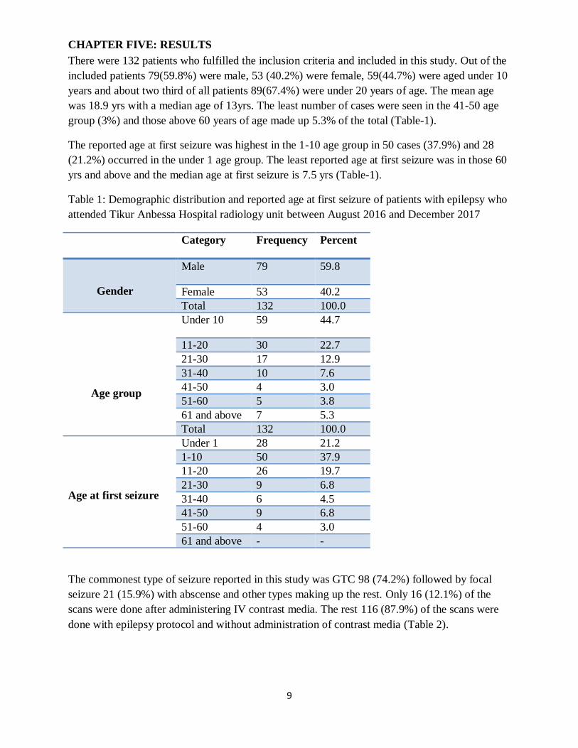
There were 132 patients who fulfilled the inclusion criteria and included in this study. Out of the
included patients 79(59.8%) were male, 53 (40.2%) were female, 59(44.7%) were aged under 10
years and about two third of all patients 89(67.4%) were under 20 years of age. The mean age
was 18.9 yrs with a median age of 13yrs. The least number of cases were seen in the 41-50 age
group (3%) and those above 60 years of age made up 5.3% of the total (Table-1).
The reported age at first seizure was highest in the 1-10 age group in 50 cases (37.9%) and 28
(21.2%) occurred in the under 1 age group. The least reported age at first seizure was in those 60
yrs and above and the median age at first seizure is 7.5 yrs (Table-1).
Table 1: Demographic distribution and reported age at first seizure of patients with epilepsy who
attended Tikur Anbessa Hospital radiology unit between August 2016 and December 2017
Category Frequency Percent
Gender
Male 79 59.8
Female 53 40.2
Total 132 100.0
Age group
Under 10 59 44.7
11-20 30 22.7
21-30 17 12.9
31-40 10 7.6
41-50 4 3.0
51-60 5 3.8
61 and above 7 5.3
Total 132 100.0
Age at first seizure
Under 1 28 21.2
1-10 50 37.9
11-20 26 19.7
21-30 9 6.8
31-40 6 4.5
41-50 9 6.8
51-60 4 3.0
61 and above - -
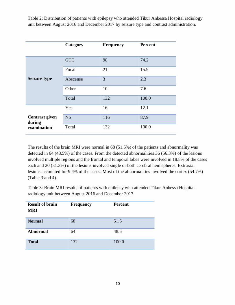
The commonest type of seizure reported in this study was GTC 98 (74.2%) followed by focal
seizure 21 (15.9%) with abscense and other types making up the rest. Only 16 (12.1%) of the
scans were done after administering IV contrast media. The rest 116 (87.9%) of the scans were
done with epilepsy protocol and without administration of contrast media (Table 2).
10
Table 2: Distribution of patients with epilepsy who attended Tikur Anbessa Hospital radiology
unit between August 2016 and December 2017 by seizure type and contrast administration.
Category Frequency Percent
Seizure type
GTC 98 74.2
Focal 21 15.9
Abscense 3 2.3
Other 10 7.6
Total 132 100.0
Contrast given
during
examination
Yes 16 12.1
No 116 87.9
Total 132 100.0
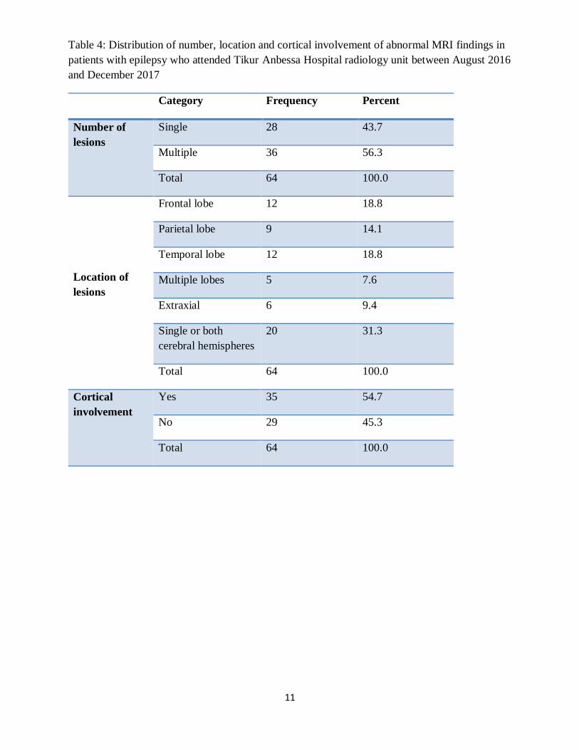
The results of the brain MRI were normal in 68 (51.5%) of the patients and abnormality was
detected in 64 (48.5%) of the cases. From the detected abnormalities 36 (56.3%) of the lesions
involved multiple regions and the frontal and temporal lobes were involved in 18.8% of the cases
each and 20 (31.3%) of the lesions involved single or both cerebral hemispheres. Extraxial
lesions accounted for 9.4% of the cases. Most of the abnormalities involved the cortex (54.7%)
(Table 3 and 4).
Table 3: Brain MRI results of patients with epilepsy who attended Tikur Anbessa Hospital
radiology unit between August 2016 and December 2017
Result of brain
MRI
Frequency Percent
Normal 68 51.5
Abnormal 64 48.5
Total 132 100.0
11
Table 4: Distribution of number, location and cortical involvement of abnormal MRI findings in
patients with epilepsy who attended Tikur Anbessa Hospital radiology unit between August 2016
and December 2017
Category Frequency Percent
Number of
lesions
Single 28 43.7
Multiple 36 56.3
Total 64 100.0
Location of
lesions
Frontal lobe 12 18.8
Parietal lobe 9 14.1
Temporal lobe 12 18.8
Multiple lobes 5 7.6
Extraxial 6 9.4
Single or both
cerebral hemispheres
20 31.3
Total 64 100.0
Cortical
involvement
Yes 35 54.7
No 29 45.3
Total 64 100.0
12
Table 5: Distribution of abnormal brain MRI findings in patients with epilepsy who attended
Tikur Anbessa Hospital radiology unit between August 2016 and December 2017
T
yp
e of
bra
in a
bn
orm
ali
ty
Age group
Under 10 11-20 21-60 61 and
above
Frequency
n(%)
Developmental cortical
malformation
2 1 1 0 4(6.3)
Gliosis/parenchymal
volume loss
18 5 3 3 29(45.3)
Mesial temporal
sclerosis
3 1 4 0 8(12.5)
Vascular malformation 0 0 3 0 3(4.7)
Brain tumor 1 0 5 2 8(12.5)
Infection 0 1 1 1 3(4.7)
Non-specific white
matter lesions
1 2 2 1 6(9.4)
Cerebellar atrophy 0 0 1 0 1(1.6)
others 2 0 0 0 2(3.1)
Total 27 10 20 7 64(100)
13
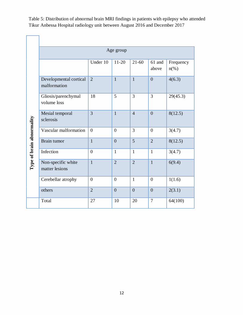
Among the brain abnormalities seen in this study 29 (45.3%) of the cases were
gliosis/brain parenchymal volume loss. The other abnormalities seen include
mesial temporal sclerosis 8(12.5%), brain tumor 8(12.5%), developmental
cortical malformations 4(6.3%) and vascular malformations and infection 3(4.7
%) each. There were also non-specific white matter lesions which were seen in
6(9.4%) of the cases (Table 5).
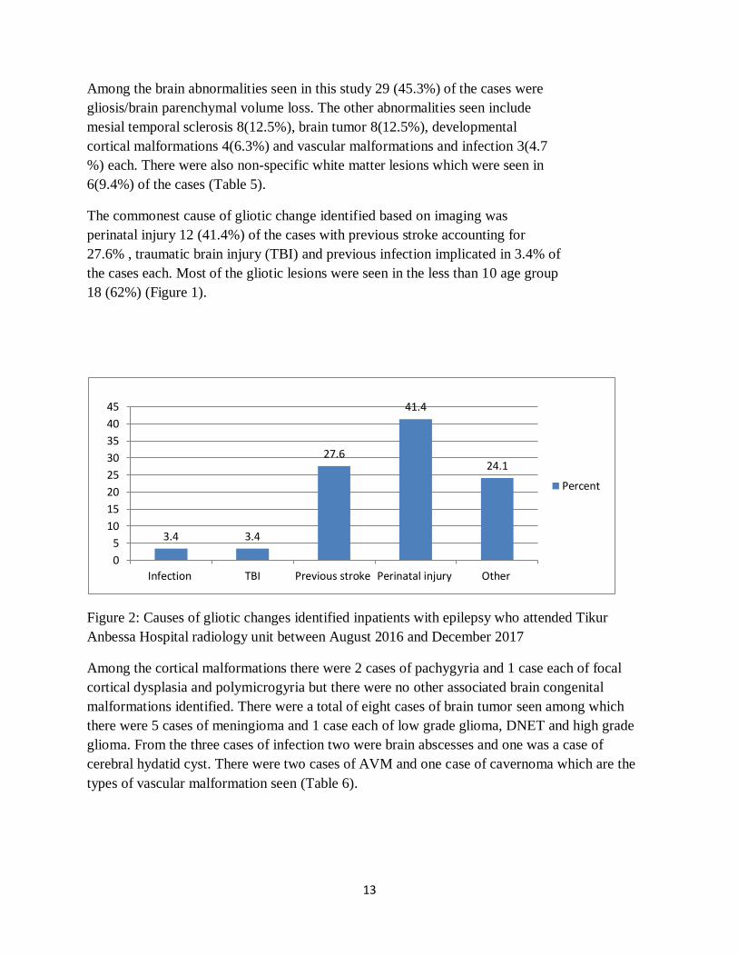
The commonest cause of gliotic change identified based on imaging was
perinatal injury 12 (41.4%) of the cases with previous stroke accounting for
27.6% , traumatic brain injury (TBI) and previous infection implicated in 3.4% of
the cases each. Most of the gliotic lesions were seen in the less than 10 age group
18 (62%) (Figure 1).
Figure 2: Causes of gliotic changes identified inpatients with epilepsy who attended Tikur
Anbessa Hospital radiology unit between August 2016 and December 2017
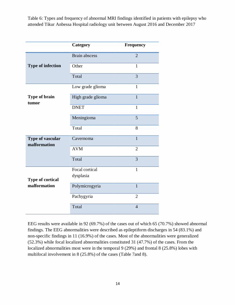
Among the cortical malformations there were 2 cases of pachygyria and 1 case each of focal
cortical dysplasia and polymicrogyria but there were no other associated brain congenital
malformations identified. There were a total of eight cases of brain tumor seen among which
there were 5 cases of meningioma and 1 case each of low grade glioma, DNET and high grade
glioma. From the three cases of infection two were brain abscesses and one was a case of
cerebral hydatid cyst. There were two cases of AVM and one case of cavernoma which are the
types of vascular malformation seen (Table 6).
3.4 3.4
27.6
41.4
24.1
0
5
10
15
20
25
30
35
40
45
Infection TBI Previous stroke Perinatal injury Other
Percent
14
Table 6: Types and frequency of abnormal MRI findings identified in patients with epilepsy who
attended Tikur Anbessa Hospital radiology unit between August 2016 and December 2017
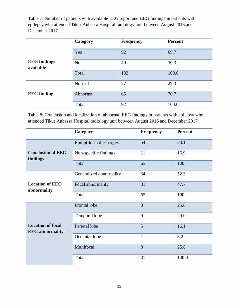
EEG results were available in 92 (69.7%) of the cases out of which 65 (70.7%) showed abnormal
findings. The EEG abnormalities were described as epileptiform discharges in 54 (83.1%) and
non-specific findings in 11 (16.9%) of the cases. Most of the abnormalities were generalized
(52.3%) while focal localized abnormalities constituted 31 (47.7%) of the cases. From the
localized abnormalities most were in the temporal 9 (29%) and frontal 8 (25.8%) lobes with
multifocal involvement in 8 (25.8%) of the cases (Table 7and 8).
Category Frequency
Type of infection
Brain abscess 2
Other 1
Total 3
Type of brain
tumor
Low grade glioma 1
High grade glioma 1
DNET 1
Meningioma 5
Total 8
Type of vascular
malformation
Cavernoma 1
AVM 2
Total 3
Type of cortical
malformation
Focal cortical
dysplasia
1
Polymicrogyria 1
Pachygyria 2
Total 4
15
Table 7: Number of patients with available EEG report and EEG findings in patients with
epilepsy who attended Tikur Anbessa Hospital radiology unit between August 2016 and
December 2017
EEG findings
available
Category Frequency Percent
Yes 92 69.7
No 40 30.3
Total 132 100.0
EEG finding
Normal 27 29.3
Abnormal 65 70.7
Total 92 100.0
Table 8: Conclusion and localization of abnormal EEG findings in patients with epilepsy who
attended Tikur Anbessa Hospital radiology unit between August 2016 and December 2017
Category Frequency Percent
Conclusion of EEG
findings
Epiliptiform discharges 54 83.1
Non-specific findings 11 16.9
Total 65 100
Location of EEG
abnormality
Generalized abnormality 34 52.3
Focal abnormality 31 47.7
Total 65 100
Location of focal
EEG abnormality
Frontal lobe 8 25.8
Temporal lobe 9 29.0
Parietal lobe 5 16.1
Occipital lobe 1 3.2
Multifocal 8 25.8
Total 31 100.0
16
CHAPTER SIX: DISCUSSION
Out of the included 132 patients, most (59.8%) were male and about two thirds of all patients
were aged less than 20 years. Most (37.9%) of the patients had their first seizure between the
ages of 1-10 where 21% had their first seizure while they were below one year of age. The
commonest type of seizure reported in this study was GTC and abnormalities were detected on
the brain MRI in about half of the cases. The commonest brain abnormality detected in this study
was gliosis/brain parenchymal volume loss (45.3%) which was predominantly caused by
perinatal insult (41.4%).
This study has shown that about two thirds of the patients with epilepsy were aged less than 20
years and very few patients were seen in the 40 years and above age group. This finding is
consistent with a Kenyan study which showed that people in the first two decades of life shared
the greatest number of epilepsy[9] The reported age at first seizure was in the under 20 yrs of
age group in 78.8% of the cases which is comparable to a review study done on epidemiology
and etiology of epilepsy in sub-Saharan Africa which found the reported age at first seizure onset
was less than 20 years in 60% of the cases. Similarly to this sub-Saharan study, our study also
did not show the bimodal distribution of age at first seizure onset seen in the developed
countries. The commonest seizure type seen in this study was GTC which is also similar to a
metanalysis done on epidemiology of epilepsy in sub-saharan Africa [10]. Most of the brain
scans done in this study were done without the administration of IV contrast media which is
consistent with the epilepsy protocol recommendations as most patients with chronic seizures or
patients who are not suspected of having brain tumor do not require contrast enhanced scans[15].
In our study abnormalities were detected in 48.5% of the scanned epileptic patients which is
higher than reported by a study done in Tikur Anbessa hospital and Yehuleshet higher clinic
(38%) which used 0.3T machine. The higher detection rate in the current study might be related
to the appropriate epilepsy protocol followed and the higher magnetic strength of the MRI
machine used in the present study (1.5T). From the detected abnormal lesions 56.3% involved
multiple regions and the frontal and temporal lobes were involved in 37.6% of the cases; these
findings were similar to the above mentioned study[17]. In addition significant number of lesions
involving single or bilateral cortical hemispheres and extraxial space were seen in our study.
The commonest brain abnormality seen in this study was gliosis/brain parenchymal volume loss
(45.3%) which was also the commonest lesion seen in the less than 10 yrs age group(66.6%)
with the predominant identified cause being perinatal injury(41.4%). These findings were higher
than other studies which show perinatal injury constitutes 1-36% of causes of epilepsy in
developing countries [28]. Another study from Tanzania also reported perinatal injury as a
common cause for epilepsy [29]. This high number is likely related to the low level of perinatal
care in developing countries including Ethiopia which will predispose the fetus/neonate to
perinatal hypoxia from conditions like prolonged labor and prematurity. Another common cause
of gliosis identified in the study was previous stroke, accounting for 27.6% of the cases where
most of the patients were in the older age groups (61 and above). This was also similar to other
studies which reported stroke as a common cause of epilepsy especially in the elderly
population[33, 34].
17
The other cause of epilepsy identified was mesial temporal sclerosis which accounted for 12.5%
of the cases and which was also more commonly seen in younger age groups (below 30 years).
These findings are supported by similar reports in different studies that show mesial temporal
sclerosis as one of the common causes of epilepsy [19-21]. The other identified cause in this
study was brain tumor, from which meningiomas were the commonest tumors. This finding is
supported by different studies which show significant number of meningioma cases present with
seizure as first symptom and also meningiomas are the commonest intracranial tumors. Different
studies reported low grade gliomas as the commonest type of brain tumor associated with
epilepsy which was not the case seen in our study and this might be due to the difference in the
epidemiology of the population studied and also the difference in the inclusion criteria for the
study as patients with other presenting symptoms like headache and weakness were not included
[19, 22, 26].
Vascular malformations constituted 4.7% of the abnormalities detected in this study and AVMs
are the commonest type which is similar to figures seen in the general epileptic population [13,
27]. The other causes identified in this study including cortical malformations and infections
constitute the least numbers which is lower than different studies including review studies from
sub-Saharan Africa. This difference might be due to the different epidemiology of the study
populations as well as that of some infectious processes like neurocysticercosis and the
preferential use of CT rather than MRI for assessment of acute infectious processes[19, 32].
EEG results were available in 69.7% of the cases with 70.7% abnormal findings which were
mostly generalized epileptiform discharges and the EEG findings showed poor correlation with
the MRI findings based on localization. This is similar to a study that correlated the clinical,
EEG and imaging features of patients with cerebral cortical dysgenesis but differs from another
study which studied the clinical correlation of MRI and EEG and found larger number of
concordance between the two in patients with hippocampal atrophy [18, 36].
18
LIMITATIONS
The major limitations faced in conducting this research were the loss of patient’s charts and lack
of proper recording of patient data in the MRI registry. The lack of image database at the
radiology unit was also an important limitation.
CONCLUSION
The commonest age group affected by epilepsy in this study was those aged below 20 years with
high number of children aged below one year. GTC was the commonest seizure type identified.
The most frequent type of brain abnormality seen was gliosis or brain parenchymal volume loss
predominantly caused by perinatal injury. The other lesions identified included mesial temporal
sclerosis, brain tumors, infections and cortical malformations.
RECOMMENDATION
For services and policy
More emphasis should be given for antenatal follow up and perinatal care to prevent
adverse pregnancy outcomes like hypoxic ischemic brain injuries and the results should
be communicated to the concerned service units such as pediatrics and Gynecology
departments and policy makers.
For research
Population based researches are recommended as it would give more reliable magnitude
of epilepsy in Ethiopia
Further prospective researches in collaboration with the neurology department should
also be undertaken especially on MRI and EEG correlation
The radiology department should start to develop its own imaging database for researches
that will be undertaken in the future
19
REFERENCES
1. Fisher, R.S., et al., Epileptic seizures and epilepsy: definitions proposed by the International
League Against Epilepsy (ILAE) and the International Bureau for Epilepsy (IBE). Epilepsia, 2005. 46(4): p. 470-472.
2. Daroff, R.B. and W.G. Bradley, in Bradley's neurology in clinical practice, B.W. Abou-khalil, m.J. Gallagher, and R.L. Macdonald, Editors. 2016, Elsevier/saunders: philadelphia, PA. p. 1563-1614.
3. Fiest, K.M., et al., Prevalence and incidence of epilepsy: a systematic review and meta-analysis of international studies. Neurology, 2017. 88(3): p. 296-303.
4. Banerjee, P.N., D. Filippi, and W.A. Hauser, The descriptive epidemiology of epilepsy—a review. Epilepsy research, 2009. 85(1): p. 31-45.
5. Lavados, J., et al., A descriptive study of epilepsy in the district of El Salvador, Chile, 1984–1988. Acta neurologica scandinavica, 1992. 85(4): p. 249-256.
6. Rwiza, H., et al., Prevalence and incidence of epilepsy in Ulanga, a rural Tanzanian district: a community‐based study. Epilepsia, 1992. 33(6): p. 1051-1056.
7. Sander, J. and S. Shorvon, Epidemiology of the epilepsies. Journal of neurology, neurosurgery, and psychiatry, 1996. 61(5): p. 433.
8. Paul, A., et al., An estimate of the prevalence of epilepsy in Sub–Saharan Africa: A systematic analysis. Journal of global health, 2012. 2(2).
9. Edwards, T., et al., Active convulsive epilepsy in a rural district of Kenya: a study of prevalence and possible risk factors. The Lancet Neurology, 2008. 7(1): p. 50-56.
10. Preux, P.-M. and M. Druet-Cabanac, Epidemiology and aetiology of epilepsy in sub-Saharan Africa. The Lancet Neurology, 2005. 4(1): p. 21-31.
11. Tekle‐Haimanot, R., L. Forsgren, and J. Ekstedt, Incidence of epilepsy in rural central Ethiopia. Epilepsia, 1997. 38(5): p. 541-546.
12. Krumholz, A., et al., Practice parameter: evaluating an apparent unprovoked first seizure in adults (an evidence-based review): report of the Quality Standards Subcommittee of the American Academy of Neurology and the American Epilepsy Society. Neurology, 2007. 69(21): p. 1996-2007.
13. Bronen, R.A., et al., Refractory epilepsy: comparison of MR imaging, CT, and histopathologic findings in 117 patients. Radiology, 1996. 201(1): p. 97-105.
14. Gaillard, W.D., et al., Guidelines for imaging infants and children with recent‐onset epilepsy. Epilepsia, 2009. 50(9): p. 2147-2153.
15. Commission, I.N., ILAE Neuroimaging Commission Recommendations for Neuroimaging of Patients with Epilepsy ILAE Neuroimaging Commission. Epilepsia, 1997. 38: p. 1-2.
16. Anson, B.G. and F. Barkhof, Neurodegenerative Diseases and Epilepsy. Grainer and Allison's Diagnostic Radiology: Neuroimaging, 2015: p. 162.
17. Mengistu, G., et al., Neuroimaging of Ethiopian patients with epilepsy: a retrospective review. Ethiopian medical journal, 2014. 52(2): p. 57-66.
18. Raymond, A., et al., Abnormalities of gyration, heterotopias, tuberous sclerosis, focal cortical dysplasia, microdysgenesis, dysembryoplastic neuroepithelial tumour and dysgenesis of the archicortex in epilepsy: clinical, EEG and neuroimaging features in 100 adult patients. Brain, 1995. 118(3): p. 629-660.
19. Vattipally, V.R. and R.A. Bronen, MR imaging of epilepsy: strategies for successful interpretation. Neuroimaging Clinics, 2004. 14(3): p. 349-372.
20. Berkovic, S.F., et al., Hippocampal sclerosis in temporal lobe epilepsy demonstrated by magnetic resonance imaging. Annals of Neurology: Official Journal of the American Neurological Association and the Child Neurology Society, 1991. 29(2): p. 175-182.
20
21. Kuzniecky, R., et al., Magnetic resonance imaging in temporal lobe epilepsy: pathological correlations. Annals of Neurology: Official Journal of the American Neurological Association and the Child Neurology Society, 1987. 22(3): p. 341-347.
22. Urbach, H., Epilepsy-Associated Brain Tumors: Diagnosis Using Magnetic Resonance Imaging, in Tumors of the Central Nervous System, Volume 4. 2012, Springer. p. 211-218.
23. Luyken, C., et al., The Spectrum of Long‐term Epilepsy–associated Tumors: Long‐term Seizure and Tumor Outcome and Neurosurgical Aspects. Epilepsia, 2003. 44(6): p. 822-830.
24. Rajneesh, K.F. and D.K. Binder, Tumor-associated epilepsy. Neurosurgical focus, 2009. 27(2): p. E4.
25. Lieu, A.-S. and S.-L. Howng, Intracranial meningiomas and epilepsy: incidence, prognosis and influencing factors. Epilepsy research, 1999. 38(1): p. 45-52.
26. Xue, H., et al., Intracranial meningiomas and seizures: a review of the literature. Acta neurochirurgica, 2015. 157(9): p. 1541-1548.
27. Bronen, R.A., et al., MR characteristics of neoplasms and vascular malformations associated with epilepsy. Magnetic resonance imaging, 1995. 13(8): p. 1153-1162.
28. Senanayake, N. and G.C. Román, Epidemiology of epilepsy in developing countries. Bulletin of the world health organization, 1993. 71(2): p. 247.
29. Burton, K.J., et al., Epilepsy in Tanzanian children: association with perinatal events and other risk factors. Epilepsia, 2012. 53(4): p. 752-760.
30. Annegers, J., et al., The risk of unprovoked seizures after encephalitis and meningitis. Neurology, 1988. 38(9): p. 1407-1407.
31. Salih, M.M.A., et al., Long term sequelae of childhood acute bacterial meningitis in a developing country: a study from the Sudan. Scandinavian journal of infectious diseases, 1991. 23(2): p. 175-182.
32. Assefa, G., et al., REVIEW OF THE CLINICAL, COMPUTERIZED TOMOGRAPHY SCAN AND/OR MAGNETIC RESONANCE IMAGING FINDINGS OF INTRACRANIAL TUBERCULOMA IN AN ETHIOPIAN TEACHING HOSPITAL. Ethiopian medical journal, 2016. 54(3).
33. Camilo, O. and L.B. Goldstein, Seizures and epilepsy after ischemic stroke. Stroke, 2004. 35(7): p. 1769-1775.
34. Bladin, C.F., et al., Seizures after stroke: a prospective multicenter study. Archives of neurology, 2000. 57(11): p. 1617-1622.
35. Mengesha, A.T., POST-STROKE SEIZURE IN PATIENTS ADMITTED TO TIKUR ANBASSA SPECIALIZED HOSPITAL. Ethiopian Medical Journal, 2017. 56(1).
36. Fish, D.R. and S.S. Spencer, Clinical correlations: MRI and EEG. Magnetic resonance imaging, 1995. 13(8): p. 1113-1117.
21
ANNEX -1 QUESTIONNAIRE
ADDIS ABEBA UNIVERSITY
SCHOOL OF GRADUATE STUDIES
DEPARTMENT OF RADIOLOGY
PATTERNS OF MRI FINDINGS IN EPILEPSY PATIENTS
1. Patient Card No. ___________________________
2. Age (In full years) _______
3. Sex 1. M ___ 2. F___
4. Age at first seizure ___________________
5. Seizure Type
1. Generalized tonic-clonic
2. Focal
3. Absence
4. Other
5. Year MRI was done (G.C.) 1. 2016 2. 2017
6. Contrast given: Yes_____ No_______
7. Result of the Brain MRI
1. Normal 2. Abnormal
8. If MRI result is abnormal,
1. Number of lesion/s
1. Single_____ 2. Multiple_____
2. Location of the lesion
1. Frontal lobe
2. Parietal lobe
3. Temporal lobe
4. Occipital lobe
5. Posterior fossa

22
6. Basal ganglia
7. Extaaxial
8. Frontal and parietal
9. Frontal and posterior fossa
10. Single or both cerebral hemispheres
11. Parietal and occipital
12. Parietal and temporal
3. Cortical involvement
1. Yes___ 2. No____
9. Type of brain abnormality described
1. Developmental cortical Malformations
2. Gliosis/brain parenchymal volume loss
3. Mesial temporal sclerosis
4. Vascular malformation
5. Brain tumor
6. Infection
7. Others
8. Non-specific white matter lesions
9. Cerebellar atrophy
10. If the abnormality is a Brain tumor
1. Type of tumor described ( First radiological differential diagnosis)
1. Low grade glioma
2. High grade glioma
3. Metastasis
4. DNET
5. Ganglioglioma
6. Oligodendroglioma
7. PXA

23
8. Meningioma
9. Other
11. If the abnormality is a cortical malformation,
1. Type of malformation
1. Focal cortical dysplasia
2. Polymicrogyria
3. schizencephaly
4. Grey matter hetrotopia
5. Cortical tubers (Tuberous sclerosis)
6. Pachygyria
7. Other
2. Are there other associated congenital anomalies
1. Yes-------- 2. No--------
3. If there are other associated congenital anomalies, type of anomaly
1. Chiari malformations
2. Dandy-walker malformation
3. Septo-optic dysplasia
4. Corpus callosum agenesis/dysgenesis
5. Holoprosencephaly
6. sturge-weber syndrome
7. Others
12. If the abnormality is a vascular malformation, type of malformation
1. Cavernoma
2. Arteriovenous malformation (AVM)
3. Developmental venous anomaly (DVA)
4. Mixed vascular malformation
13. If the abnormality is a gliotic change, cause of gliosis identified
1. Previous infection

24
2. Traumatic brain injury
3. Previous stroke (Ischemic or Hemorrhagic)
4. Perinatal injury
5. Post-surgery
6. Other
14. If the abnormality is Infection, type of infection identified
1. Tuberculoma
2. CNS Toxoplasmosis
3. Brain abscess
4. Other
15. EEG findings available
1. Yes ___ 2. No_____
16. If yes to Q 15,
1. The EEG finding is;
1. Normal ____ 2. Abnormal_________
2. If the EEG finding is abnormal, conclusion of the findings,
1. Epileptiform discharges
2. Non-specific findings
3. If the EEG finding is abnormal, localization of the abnormality
1. Generalized EEG abnormality
2. Focal EEG abnormality
4. If Focal EEG abnormality, location of the focal abnormality;
1. Frontal lobe
2. Temporal lobe
3. Parietal lobe
4. Occipital lobe
5. Does the EEG localization correlate with the MRI finding
1. Yes------- 2. No--------------
Recommended


