
Adenosine A2A Receptors Activation FacilitatesNeuromuscular Transmission in the Pre-SymptomaticPhase of the SOD1(G93A) ALS Mice, but Not in theSymptomatic PhaseFilipe Nascimento1,2, Paula A. Pousinha1,2¤a, AlexandraM. Correia2,3¤b, Rui Gomes2,4, AnaM. Sebastiao1,2,
Joaquim A. Ribeiro1,2*
1 Institute of Pharmacology and Neurosciences, Faculty of Medicine, University of Lisbon, Lisbon, Portugal, 2Unit of Neurosciences, Instituto de Medicina Molecular,
University of Lisbon, Lisbon, Portugal, 3National Museum of Natural History and Science, University of Lisbon, Lisbon, Portugal, 4 Faculty of Sciences, University of Lisbon,
Lisbon, Portugal
Abstract
Amyotrophic Lateral Sclerosis (ALS) is a neurodegenerative disease leading to motor neuron dysfunction resulting inimpairment of neuromuscular transmission. A2A adenosine receptors have already been considered as a potentialtherapeutical target for ALS but their neuromodulatory role at the neuromuscular junction in ALS remains to be clarified. Inthe present work, we evaluated the effects of A2A receptors on neuromuscular transmission of an animal model of ALS:SOD1(G93A) mice either in the pre-symptomatic (4–6 weeks old) or in the symptomatic (12–14 weeks old) stage.Electrophysiological experiments were performed obtaining intracellular recordings in Mg2+ paralyzed phrenic nerve-hemidiaphragm preparations. Endplate potentials (EPPs), quantal content (q. c.) of EPPs, miniature endplate potentials(MEPPs) and giant miniature endplate potential (GMEPPs) were recorded. In the pre-symptomatic phase of the disease (4–6weeks old mice), the selective A2A receptor agonist, CGS 21680, significantly enhanced (p,0.05 Unpaired t-test) the meanamplitude and q.c. of EPPs, and the frequency of MEPPs and GMEPPs at SOD1(G93A) neuromuscular junctions, the effectbeing of higher magnitude (p,0.05, Unpaired t-test) than age-matched control littermates. On the contrary, in symptomaticmice (12–14 weeks old), CGS 21680 was devoid of effect on both the amplitude and q.c. of EPPs and the frequency of MEPPsand GMEPPs (p,0.05 Paired t-test). The results herein reported clearly document that at the neuromuscular junction ofSOD1(G93A) mice there is an exacerbation of A2A receptor-mediated excitatory effects at the pre-symptomatic phase,whereas in the symptomatic phase A2A receptor activation is absent. The results thus suggest that A2A receptors functionchanges with ALS progression.
Citation: Nascimento F, Pousinha PA, Correia AM, Gomes R, Sebastiao AM, et al. (2014) Adenosine A2A Receptors Activation Facilitates NeuromuscularTransmission in the Pre-Symptomatic Phase of the SOD1(G93A) ALS Mice, but Not in the Symptomatic Phase. PLoS ONE 9(8): e104081. doi:10.1371/journal.pone.0104081
Editor: William Phillips, University of Sydney, Australia
Received April 3, 2014; Accepted July 5, 2014; Published August 5, 2014
Copyright: � 2014 Nascimento et al. This is an open-access article distributed under the terms of the Creative Commons Attribution License, which permitsunrestricted use, distribution, and reproduction in any medium, provided the original author and source are credited.
Data Availability: The authors confirm that all data underlying the findings are fully available without restriction. All relevant data are within the paper and itsSupporting Information files.
Funding: This work was supported by Fundacao do Ministerio da Ciencia e Tecnologia de Portugal [Grant PTDC/SAU-FAR/118787/2010] (http://www.fct.pt/index.phtml.pt). The funders had no role in study design, data collection and analysis, decision to publish, or preparation of the manuscript.
Competing Interests: The authors have declared that no competing interests exist.
* Email: [email protected]
¤a Current address: Institut de Pharmacologie Moleculaire et Cellulaire UMR7275 CNRS, Equipe Mecanismes moleculaires de la plasticite neuronale physiologiqueet pathologique, Sophia-Antipolis, Valbonne, France¤b Current address: Department of Ecology and Evolutionary Biology, Guyot Hall, Princeton University, Princeton, New Jersey, United States of America
Introduction
Amyotrophic Lateral Sclerosis (ALS) is an adult-onset progres-
sive neurodegenerative disease characterized by the selective loss
of motor neuron function leading to muscle atrophy and weakness.
After symptomatic onset disease progression lasts 4 to 5 years and
patients ultimately die due to bulbar failure. Most of the diagnosed
cases carry an unknown genetic link (sporadic ALS) and a few (5–
10%) are related to known mutations in specific proteins (familial
ALS). Both present similar pathological and clinical features [1,2].
The first gene associated with the inherited form of the disease was
the SOD1 gene encoding for the superoxide dismutase 1 enzyme
which accounts for 20% of the familial forms of ALS [3]. This led
to the design of the first animal model of ALS, the SOD1(G93A)
mouse, which currently is the most used and well characterized
rodent model for this disease [4]. Neuromuscular dysfunction at
symptomatic SOD1(G93A) mice has been reported [5,6]. We
recently showed that the SOD1(G93A) mice neuromuscular
transmission impairment starts long before symptomatic onset [7].
Adenosine is a key neuromodulator with implications in
pathological conditions [8]. At the neuromuscular junction it
can act on both A1 and A2A adenosine receptors, fine-tuning
acetylcholine (ACh) release [9]. A2A receptors are known to have a
neuroprotective role in some pathological conditions [8] and have
PLOS ONE | www.plosone.org 1 August 2014 | Volume 9 | Issue 8 | e104081

been considered as a potential therapeutical target for ALS [10–
12]. Some contradictory reports in the literature can however be
found [10,11] highlighting the need for an evaluation of the
influence of A2A receptors in ALS models where disease
progression and neuromuscular transmission impairment can be
taken into account.
Given the unexplored role of A2A receptors at the neuromus-
cular junction in ALS, and considering that the neuromuscular
transmission in the SOD1(G93A) mice starts to present alterations
long before symptoms onset [7], we considered of interest to
evaluate A2A receptor effects on neuromuscular transmission, in
both pre-symptomatic (4–6 weeks old) and symptomatic (12–14
weeks old) SOD1(G93A) ALS mice. The results now reported
show that the role of A2A receptors at the motor nerve terminals,
changes upon ALS progression. In the pre-symptomatic phase the
A2A receptor-mediated excitatory effects on neuromuscular
transmission are exacerbated, probably acting as a compensatory
mechanism towards delaying disease progression, whereas in the
symptomatic phase the A2A receptor excitatory action disappears.
Methods
Ethics statementThis study was performed in accordance with the European
Community guidelines (Directives 86/609/EU and 2010/63/EU,
Recommendation 2007/526/CE, European Convention for the
Protection of Vertebrate Animals used for Experimental or Other
Scientific Purposes ETS 123/Appendix A) and Portuguese Laws
on Animal Care (Decreto-Lei 129/92, Portaria 1005/92,
Portaria466/95, Decreto-Lei 197/96, Portaria 1131/97). All the
protocols carried in this study were under approval of the
Portuguese National Authority (General Direction of Veterinary)
and the Ethics Committee of the Instituto de Medicina Molecular
of the Faculty of Medicine, University of Lisbon, Lisbon, Portugal.
AnimalsTransgenic B6SJL-TgN (SOD1-G93A)1Gur/J males (Jackson
Laboratory, No. 002726) overexpressing the human SOD1 gene
carrying a glycine to alanine point mutation at residue 93 (G93A)
[4] and wild-type B6SJLF1/J females were purchased from The
Jackson Laboratory (Bar Harbor, ME, USA) and were breed at
IMM rodent facilities where a colony was established. Mice were
maintained on a background B6SJL by breeding SOD1(G93A)
transgenic males with non-transgenic females in a rotational
scheme. Males were crossed with non-transgenic females because
transgenic females are infertile. F1 offspring was used in all
experiments. Progeny was no longer used in breeding to avoid
mSOD1 gene copy number loss and therefore deviation from ALS
phenotype [4]. SOD1(G93A) mice were used to study pre-
symptomatic (4–6 weeks old) and symptomatic (12–14 weeks old)
phases of the disease. 4–6 and 12–14 weeks old wild type (WT)
animals served as controls. Both male and female mice were used.
The proportion of male and female mice was about the same in
the WT (18 males and 19 females in total) and SOD1(G93A) mice
(20 males and 17 females in total) groups. Furthermore, using the
same mice strain (B6SJL-Tg(SOD1-G93A)1Gur/J) no gender
influences over the intrinsic features of neuromuscular transmis-
sion have been detected [7], though gender differences could be
detected in other mice strains (B6.Cg-Tg-(SOD1-G93A)1Gur/J)
[6].
Littermates were identified by dermal ear punching and divided
into cages by gender. The ear tissue was used to genotype the
animals by polymerase chain reaction (PCR) [3]. Animals were
housed 4–5 mice/cage, under a 12 h light/12 h dark cycle, and
received food and water ad libitum.
Electrophysiological recordingsAnimals were anaesthetised using halothane and rapidly
decapitated. Both right and left phrenic-nerve attached to the
hemidiaphragm muscle were isolated. One preparation was placed
and stretched in a 3 mL Perspex chamber continuously perfused
via a roller pump (3 mL.min21) with a physiologic saline solution
(Krebs and Henseleit solution, see Drugs section) under contin-
uous oxygenation. The other phrenic-nerve hemidiaphragm
preparation was kept in a beaker with an oxygenated saline
solution before being set up the recording chamber. Since no
functional differences were found between right and left phrenic
nerve-hemidiaphragm muscles, different protocols were carried in
each preparation.
Intracellular recordings were performed in the conventional
way [13–15]. The phrenic-nerve was stimulated supramaximally
by a suction electrode (Cu/Cu2+) connected to a S48 square pulse
stimulator (Grass Tecnologies, West Warwick, RI, USA). Stimuli
were applied in a low frequency of 0.5 Hz with a current duration
of 20 ms. The reference electrode was an Ag-AgCl pellet placed in
the bath. The recording electrode was a glass microelectrode filled
with KCl (3 M) with resistance between 15–40 MV inserted into
the motor endplate. A Digidata 1440A digitizer (Molecular
Devices, Sunnyvale, CA, USA), designed to work with the
Axoclamp 2B amplifier (Molecular Devices, Sunnyvale, CA,
USA), performed data acquisition, allowing continuous monitor-
ing and digital storage of evaluated parameters with adequate
software (pCLAMP 10.3, Molecular Devices, Sunnyvale, CA,
USA).
Endplates with a resting potential between –65 to –85 mV were
chosen for experiment. Resting voltage was stable throughout all
experiments with less than 5% variation of its initial value.
Endplate Potentials (EPPs) amplitude was assessed as the average
amplitude of 60 consecutive EPPs (with amplitudes ranging
between 1 mV to 5 mV). To evaluate the percentage of the drug
effect, the mean averaged EPP amplitudes in the last 10 minutes
before adding any drug (control) was compared with the mean
averaged EPP amplitudes from the last 10 minutes of drug
perfusion (treatment). The quantal content (q. c.) of EPPs was
calculated as the ratio between the mean EPP amplitude and the
mean Miniature Endplate Potential (MEPP) amplitude acquired
during the same period with the same resting membrane potential.
MEPPs were recorded in gap-free intervals of 100 seconds before
adding the drug and at the end of drug perfusion. MEPP detection
threshold was set between 0.2 mV and 1 mV [7]. MEPP
amplitude was defined as the mean of all spontaneous events and
the frequency as the number of events registered during the
100 seconds. The minimum Giant Miniature Endplate Potential
(GMEPP) threshold amplitude was set in 1 mV [7]. This indirect
measure of spontaneous activity synchronism was analyzed as the
frequency of giant events in the 100 seconds gap-free acquisition
mode and the mean amplitude as the average of GMEPPs
magnitude in the same interval. Only if GMEPP frequency was
higher than 0.04 s21 before adding the drug, the percentage of
effect was considered for analysis. Evoked activity was analyzed
with Clampfit software (Molecular Devices, Sunnyvale, CA, USA)
and spontaneous events with Mini-Analysis software (Synaptosoft
Inc., Decatur, GA, USA). Whenever perfusing two drugs, the %
change was calculated by comparing acquired values with the ones
obtained from the first drug perfused (considered then as control).
Adenosine A2A Receptors and Neuromuscular Transmission in ALS
PLOS ONE | www.plosone.org 2 August 2014 | Volume 9 | Issue 8 | e104081

Figure 1. CGS 21680 facilitation of evoked activity is exacerbated in pre-symptomatic mice; (A) representative time-course changeof mean EPP amplitude throughout CGS 21680 (5 nM) perfusion and (B) representation of EPP amplitude increase in 4–6 weeks oldWT (n=5) and pre-symptomatic mice (n=10) upon A2A receptor activation (CGS 21680 at 5 nM); (C) concentration-response
Adenosine A2A Receptors and Neuromuscular Transmission in ALS
PLOS ONE | www.plosone.org 3 August 2014 | Volume 9 | Issue 8 | e104081

DrugsThe bathing solution was modified from Krebs and Henseleit
[16] (NaCl 117 mM; KCl 5 mM; NaHCO3 25 mM; NaH2PO4
1.2 mM; glucose 11 mM; CaCl2 2.5 mM; MgCl2 1.2 mM; pH
7.4) continuously gassed with 95% O2 and 5% CO2 kept at room
temperature (22–25uC). Muscle twitch was prevented by increas-
ing [Mg2+] to 18.5–19.5 mM in 4–6 weeks old animals and 20.0–
22.0 mM in 12–14 weeks old mice. This strategy reduces the q. c.
of EPPs but preserves the main features of neuromuscular
transmission [13].
Drugs used were: 2-p-(2-carboxyethyl) phenethylamino]-59-N-
ethylcarboxamido adenosinehydrochloride (CGS 21680) and 5-
Amino-7-(2-phenylethyl)-2-(2-furyl)-pyrazolo(4,3-e)-1,2,4-tria-zolo(1,5-c) pyrimidine (SCH 58261). Stock solutions (5 mM) were
made in dimethyl sulfoxide. To avoid compound precipitation
aliquots were kept frozen at –20uC until used. Dimethyl sulfoxide
was devoid of effect in the performed experiments like previously
reported [13].
Statistical analysisData are presented as mean 6 standard error of the mean in
each group, which n corresponds to the number of animals used (1
fiber per mouse).
Student’s t-test for independent samples (Unpaired t-test) wasused to compare drug effect between two groups. One way
analysis of variance (ANOVA) was applied whenever comparing
more than 2 means. If p,0.05, Tukey’s pos-test was applied to
compare drug-induced changes between different groups. Stu-
dent’s t-test for paired samples (Paired t-test) was used to compare
obtained measurement with the control parameter before adding
the drug (e.g. mean EPP amplitude before drug perfusion). Values
of p,0.05 were considered to represent statistically significant
differences.
Results
In pre-symptomatic SOD1(G93A) mice the excitatory A2A
receptor-mediated effects on neuromusculartransmission are exacerbatedThough the selective A2A receptor agonist CGS 21680 [17] has
been extensively used in research, namely at the neuromuscular
junction [9,13,18], there are only few studies reporting its effects at
the mouse neuromuscular junction, the existing being on K+-
evoked ACh-release (e.g. [19]). We performed a dose-response
study using 3, 5 to 10 nM of CGS 21680 in both pre-symptomatic
SOD1(G93A) mice and their age-matched healthy controls.
Figure 1A illustrates the time-course changes of mean EPP
amplitude in the presence of CGS 21680 (5 nM). It clearly shows
an exacerbation of the facilitatory effect of the A2A receptor
agonist on EPP amplitude, in the pre-symptomatic SOD1(G93A)
mice, a finding also illustrated in figure 1B. The difference
between the two groups started to be evident 20 min after drug
perfusion. As illustrated in Figure 1C, all the tested concentrations
enhanced the mean amplitude of EPPs, when compared to the
value measured before drug perfusion (p,0.05, Paired t-test). Toevaluate the role of A2A receptors on neuromuscular transmission,
while comparing the effect of CGS 21680 on the mean amplitude
of EPPs recorded in both groups of animals, it could be concluded
that at 3 nM there were no significant differences between groups
(p.0.05, Unpaired t-test) while at 5 nM and 10 nM the
facilitation caused by CGS 21680 on EPPs amplitude was
significantly higher in pre-symptomatic SOD1(G93A) mice
(5 nM: n= 13; 10 nM: n=5; p,0.05, Unpaired t-test), when
compared to the WT group (5 nM: n=14; 10 nM: n= 7). Since
the difference between groups was already pronounced at 5 nM,
we decided to use this concentration in the remaining experiments.
To exclude potential unspecific effects of CGS 21680, we
evaluated the effect of this drug in the presence of the selective
A2A receptor antagonist, SCH 58261 (50 nM) [20]. The blockade
of A2A receptors did not change the mean amplitude of EPP in
both groups of animals (data not shown, n= 5 for controls and
n=6 for SOD1G93A; p.0.05, Paired t-test) and effectively
prevented the facilitatory effects of CGS 21680 (5 nM) (Figure 1C,
n= 4 for controls and n= 6 for SOD1G93A; p,0.05, one-way
ANOVA followed by Tukey’s pos-hoc). These results suggest that
in the present experimental conditions A2A receptors are not
tonically activated by endogenous adenosine and, also, that the
effect of CGS 21680 (5 nM) results from specific A2A receptor
action upon neuromuscular transmission.
To evaluate changes in the q. c. of EPPs, MEPPs and EPPs were
recorded simultaneously. As illustrated in Figure 1D–E, when
tested in WT animals, CGS 21680 (5 nM) was devoid of effect on
both MEPPs amplitude and frequency (n= 10; p.0.05, Paired t-test). However, when applied to pre-symptomatic SOD1(G93A)
neuromuscular junctions it caused a significant increase in the
frequency of MEPPs, without changing its average amplitude
(n = 9; p,0.05, Unpaired t-test). As it occurred for evoked changes
in EPPs amplitude, the A2A receptor-mediated facilitatory effect
on the mean frequency of MEPPs was more pronounced in pre-
symptomatic SOD1(G93A) mice than in its age-matched healthy
controls (Figure 1E). Also, SCH 58261 (50 nM), per se, did not
significantly change MEPPs frequency in both studied animal
groups (data not shown, n = 8 for controls and n= 4 for
SOD1(G93A); p.0.05 Paired t-test), while preventing the
facilitatory action of the A2A receptor agonist upon MEPP
frequency in the pre-symptomatic SOD1(G93A) mice (n = 4, p,
0.05, one-way ANOVA followed by Tukey’s pos-hoc). Regarding
the q. c. of EPPs (Figure 1F) we observed that CGS 21680 (5 nM)
caused a significantly higher facilitation in pre-symptomatic
SOD1(G93A) mice (n = 12) than in its age-matched healthy mice
(n = 13; p,0.05, Unpaired t-test); this effect was prevented by
SCH 58261 (50 nM; n=4 for controls and n= 6 for
SOD1(G93A); p,0.05, one-way ANOVA followed by Tukey’s
pos-hoc).
GMEPPs arise from intracellular Ca2+ disturbances resulting in
a non-evoked ‘‘constitutive’’ secretion leading to abnormal
spontaneous events at mammalian neuromuscular junctions
[21,22] and pre-symptomatic SOD1(G93A) mice present higher
frequency of GMEPPs when compared to controls [7]. Consid-
changes in mean EPP amplitude in the presence of CGS 21680 (3 nM: n=7, WT, n=7, SOD1G93A; 5 nM: n=14, WT, n=13,SOD1G93A; 10 nM: n=7, WT, n=5, SOD1G93A) whose effect was blocked by SCH 58261 at 50 nM (n=5, WT, n=4, SOD1G93A); (D)raw recording of spontaneous release fluctuations from a 4–6 weeks old WT and pre-symptomatic SOD1G93A neuromuscularjunction promoted by CGS 21680 (5 nM); effect of CGS 21680 (5 nM) perfusion regarding (E) MEPP frequency (n=10, WT, n=9,SOD1(G93A), (F) quantal Content of EPPs (n=13, WT, n=12, SOD1(G93A)) and (G) GMEPP frequency (n=10, WT, n=11,SOD1(G93A)) in pre-symptomatic SOD1(G93A) mice and respective healthy controls; *p,0.05 Unpaired t-test; hp,0.05 one-wayANOVA with Tukey’s pos-hoc; #p,0.05 Paired t-test (as compared with control value before drug perfusion); control correspondsto 100% in all cases.doi:10.1371/journal.pone.0104081.g001
Adenosine A2A Receptors and Neuromuscular Transmission in ALS
PLOS ONE | www.plosone.org 4 August 2014 | Volume 9 | Issue 8 | e104081
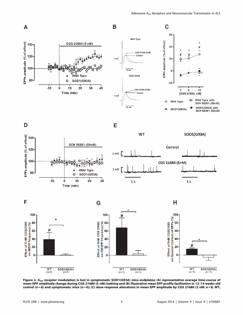
Figure 2. A2A receptor modulation is lost in symptomatic SOD1(G93A) mice endplates; (A) representative average time-course ofmean EPP amplitude change during CGS 21680 (5 nM) bathing and (B) illustrative mean EPP profile facilitation in 12–14 weeks oldcontrol (n=6) and symptomatic mice (n=6); (C) dose-response alterations in mean EPP amplitude by CGS 21680 (3 nM: n=8, WT,
Adenosine A2A Receptors and Neuromuscular Transmission in ALS
PLOS ONE | www.plosone.org 5 August 2014 | Volume 9 | Issue 8 | e104081

ering the role of adenosine receptors in Ca2+ modulation [23,24],
the effect of A2A receptor activation, with its selective agonist, on
the amplitude and frequency of giant spontaneous events was also
evaluated. As illustrated in Figure 1D and 1G, CGS 21680 (5 nM)
caused a significant increase in mean frequency of GMEPPs in
pre-symptomatic SOD1(G93A) mice (n = 11; p,0.05, Unpaired t-test), whereas in the WT group it was devoid of effect (n = 10, p.
0.05, Paired t-test). SCH 58261 (50 nM), per se, did not change the
mean frequency of GMEPPs (data not shown, n= 7 for controls
and n= 5 for SOD1(G93A)) and effectively prevented the
facilitatory effect caused by 5 nM CGS 21680 in pre-symptomatic
SOD1(G93A) mice (n = 5; p,0.05, one-way ANOVA). GMEPPs
amplitude remained unchanged upon CGS 21680 (5 nM)
perfusion and no statistical difference was found between the
studied groups of animals.
Together, the results suggest that the A2A receptor-mediated
facilitatory effects on neuromuscular transmission are exacerbated
in the pre-symptomatic phase of the disease.
In symptomatic SOD1(G93A) mice the excitatory A2A
receptor-mediated effects on neuromusculartransmission are absentAs in the pre-symptomatic phase of the disease, we performed a
concentration-response study using the same concentrations (3, 5
and 10 nM) of CGS 21680 in both symptomatic SOD1(G93A)
mice and their age-matched (12–14 weeks old) healthy controls.
Figure 2A shows the time course of mean EPP amplitude changes
in the symptomatic SOD1(G93A) mice throughout CGS 21680
(5 nM) perfusion. Figure 2B represents the profile of mean EPP
amplitude changes in WT and symptomatic mice by 5 nM of CGS
21680. As expected, all the tested concentrations enhanced the
mean averaged amplitude of EPPs in WT mice, when compared
to the measured value before drug perfusion (Figure 2C; 3 nM:
n=8; 5 nM: n= 10; 10 nM: n= 11; p,0.05, Paired t-test).Remarkably, when applied to the symptomatic SOD1(G93A)
neuromuscular junctions, none of the tested concentrations
modified the amplitude of EPPs (Figure 2C; 3 nM: n= 10;
5 nM: n= 7; 10 nM: n= 7; p.0.05, Paired t-test). As illustratedin Figure 2D, SCH 58261 (50 nM) was devoid of effect in the
mean EPP amplitude in both groups (WT: n= 4; SOD1(G93A):
n = 7; p.0.05 Paired t-test), suggesting the absence of tonic
activation of the A2A receptor agonist in symptomatic
SOD1(G93A) mice as well in WT mice.
As illustrated in figures 2E, 2F and 2G, CGS 21680 (5 nM)
caused an increase on MEPPs (n= 6) and GMEPPs frequency in
WT mice (n = 4; p,0.05, Paired t-test) but when applied to
symptomatic SOD1(G93A) neuromuscular junctions it was devoid
of effect on MEPPs (n= 5) and GMEPP frequency (n= 4; p,0.05,
Paired t-test). MEPPs amplitude remained unchanged in the
presence of the A2A receptor agonist in both groups (p.0.05,
Unpaired t-test). In relation to the q. c. of EPPs (Figure 2H), there
was a significant increase in the q. c. of EPPs in 12–14 weeks old
healthy mice, upon CGS 21680 (5 nM) perfusion (n= 9; p,0.05,
Unpaired t-test), which was prevented by SCH 58261 (50 nM;
n=4; p,0.05, one-way ANOVA followed by Tukey’s pos-hoc). In
contrast, the q. c. of EPPs was not modified by CGS 21680 (5 nM)
perfusion in symptomatic SOD1(G93A) mice (n = 7; p.0.05
Paired t-test).
Comparison between the effect of A2A receptorsactivation at SOD1(G93A) neuromuscular junctions upondisease progressionTo allow the assessment of the role of A2A receptors throughout
ALS progression, in Figure 3 are compared the effects of CGS
21680 (5 nM) in the pre-symptomatic and symptomatic
SOD1(G93A) mice. Age-matched healthy controls were also
subject of comparison to evaluate the maturation-associated
alterations of neuromuscular transmission in physiological condi-
tions. Figure 3A shows the superimposed time-course profiles of
mean EPP amplitude change throughout CGS 21680 (5 nM)
perfusion in pre-symptomatic and symptomatic SOD1(G93A)
neuromuscular junctions. By this figure one can find that the role
of A2A receptors dramatically changes with disease progression. It
is concluded that the A2A receptor selective agonist induced a
significantly higher enhancement of EPPs amplitude in 12–14
weeks old WT than in 4–6 weeks old control animals (p,0.05
Unpaired t-test) (Figure 3B), which was accompanied by a
significant increase in the q. c. of EPPs (p,0.05, Unpaired t-test)(Figure 3C). Interestingly, the effect of CGS 21680 (5 nM) in pre-
symptomatic SOD1(G93A) animals (4–6 weeks old) is similar to its
effect in the 12–14 weeks old WT controls. This might be related
to an ALS-associated early maturation process at the neuromus-
cular junction, as previously suggested [7].
In relation to the effect of A2A receptor activation on MEPPs
and GMEPPs frequency (figures 3D and 3E), there are some
similarities between the pre-symptomatic SOD1(G93A) mice and
the 12–14 weeks old wild-type controls. For example, the changes
on the frequency of MEPPs and GMEPPs caused by A2A receptor
activation, observed in pre-symptomatic SOD1(G93A) mice were
not statistically different from the values recorded in the 12–14
weeks old wild type controls (p,0.05 Unpaired t-test).
Discussion
The main finding of the present work was that the role of
adenosine A2A receptors at the neuromuscular junction of the ALS
SOD1(G93A) mouse model changes with disease progression. In
the pre-symptomatic phase, the magnitude of the excitatory effects
on neuromuscular transmission, caused by A2A receptor, is
enhanced compared to age-matched controls. In contrast, in the
symptomatic SOD1(G93A) mice, the A2A receptor-mediated
facilitation is absent.
The enhancement of neuromuscular transmission caused by the
selective A2A receptor agonist, CGS 21680, results from an
increase in the evoked release of ACh, since it increased the q. c. of
EPPs without affecting the average amplitude of MEPPs recorded
concomitantly. It is known that the activation of the adenosine A2A
receptors induces an enhancement of neuromuscular transmission,
which is hardly reversible [9,13] and apparently more robust in 3–
4 weeks old rats [13] than in 4–6 weeks old mice (present work).
n=10, SOD1G93A; 5 nM: n=10, WT, n=7, SOD1G93A; 10 nM: n=11, WT, n=7, SOD1G93A) were blocked by SCH 58261 at 50 nM inWT mice (n=4, WT, n=4, SOD1G93A); (D) SCH 58261 (50 nM) did not affect evoked activity throughout data acquisition (n=4, WT,n=7, SOD1G93A); (E) raw recording of spontaneous release variations from a 12–14 weeks old WT and symptomatic SOD1G93Aendplate upon CGS 21680 (5 nM) perfusion; effect of A2A receptor activation by CGS 21680 (5 nM) on (F) MEPP frequency (n=6,WT, n=5, SOD1(G93A)) (G) GMEPP frequency (n=4, WT, n=4, SOD1(G93A)) and (H) quantal content of EPPs (n=9, WT, n=7,SOD1(G93A)); *p,0.05 Unpaired t-test; hp,0.05 one-way ANOVA with Tukey’s pos-hoc; #p,0.05 Paired t-test (as compared withcontrol value before drug perfusion); control corresponds to 100% in all cases.doi:10.1371/journal.pone.0104081.g002
Adenosine A2A Receptors and Neuromuscular Transmission in ALS
PLOS ONE | www.plosone.org 6 August 2014 | Volume 9 | Issue 8 | e104081

Adenosine A2A Receptors and Neuromuscular Transmission in ALS
PLOS ONE | www.plosone.org 7 August 2014 | Volume 9 | Issue 8 | e104081

Interestingly, the A2A receptor signaling is apparently lost at the
neuromuscular junction of aged (70–80 weeks old) rats as it is in
the symptomatic SOD1(G93A) mice (present work), suggestive of
an a disease induced early-ageing of A2A receptor influence upon
neuromuscular transmission. The reason for the hardly reversible
adenosine A2A receptor-mediated action might be the transducing
system operated by the receptor, which binds to G-protein coupled
receptors [25] involving cyclic AMP formation and Protein kinase
A (PKA) activation [26] with subsequent protein phosphorylation,
causing a long-lasting increase in synaptic strength. In addition,
multiple interactions of A2A receptors with other proteins have
been described both in peripheral and central nervous system [8].
At the neuromuscular junction, A2A receptors are known to
interact with adenosine A1 receptors [13,18,27], presynaptic
nicotinic autofacilitatory receptors [28], tyrosin receptor kinase B
(TrkB) [29] or calcitonin gene-related peptide [30]. Furthermore,
SOD1(G93A) mice pathogenesis is characterized by increased
oxidative stress [2] and A2A receptors present redox-sensitive
synchronizing action at the neuromuscular synapse [31]. Interest-
ingly, in a recent work from our team where neuromuscular
transmission of the SOD1(G93A) mouse was studied [7], it was
show that ACh release at the neuromuscular junction is enhanced
in the pre-symptomatic phase of the disease, since the average
amplitude of EPPs recorded in the SOD(G93A) mice during the
pre-symptomatic phase (4–6 weeks old) was similar to the values
obtained in the healthy control group (12–14 weeks-old).
Interestingly, the levels of brain-derived neurotrophic factor
(BDNF) are strongly increased in post-mortem muscle samples of
early phase of ALS patients [32]. It is known that A2A receptors, at
motor nerve terminals, trigger the action of BDNF [29], which
enhances transmitter release at developing neuromuscular junc-
tions [33], improving neuromuscular transmission in the adult rat
diaphragm [34] and facilitating synaptic efficacy by increasing
presynaptic depolarization at the neuromuscular junction [35].
BDNF is also important for maintenance of ACh receptor
clustering in the endplate [36,37]. Whether the enhancement by
A2A receptor in the pre-symptomatic phase of the disease, could
account for the potentiation of endogenous BDNF actions that
might occur at the neuromuscular junction, therefore, enhancing
synaptic transmission and compensating an eventual early
denervation needs to be investigated. Nevertheless, data herein
reported suggests that activation of A2A receptors might be an
important mechanism involved in the scenarios of pathology that
leads to deficits in ACh release, like ALS.
Activation of A2A receptors with its selective agonist CGS 21680
markedly increased the frequency of spontaneous giant events in
SOD1(G93A) mice, when compared to age matched controls.
Interestingly, the magnitude of this effect in the pre-symptomatic
SOD1(G93A) mice (4–6 weeks old) was not different from the one
observed in the WT group with 12–14 weeks old, reinforcing the
‘‘early maturation’’ hypothesis [7]. Modulation of Ca2+ dynamics
by A2A receptors could also be considered as an adenosine-related
compensatory mechanism. In fact, it was shown that, at the mouse
[19] and rat [23] neuromuscular junction, activation of A2A
receptors can facilitate spontaneous and evoked ACh secretion by
independent mechanisms: as result of (1) an increase in cytosolic
nerve terminal Ca2+ concentration due to release of this ion from
intracellular Ca2+ stores or (2) by increase of extracellular Ca2+
entry into the terminals via L-type voltage gated Ca2+ channels
(VGCC). Both mechanisms lead to an intracellular Ca2+ rise that
in turn increases ACh release. Furthermore, muscle strength
depends on the firing frequency and motor unit recruitment [38]
and presynaptic changes in Ca2+ homeostasis may induce
adaptations to facilitate firing frequency, specifically during high-
frequency stimulation [39]. Fuchs and colleagues [40], using
visually guided patch-clamp recordings in combination with single
cell Ca2+ imaging of motor neurons throughout the complete
lifespan of the SOD1(G93A) ALS mouse, reported that the pre-
symptomatic motor terminals (70 days ,7 weeks) present
hyperexcitability in association with remodeling of Ca2+ handling.
In symptomatic mice, A2A receptors modulation of both evoked
and spontaneous activity was lost. Full occupancy of A2A receptors
by high levels of endogenous adenosine cannot account for this
lack of effect, because the selective antagonist was devoid of effect
on neuromuscular transmission suggesting that, A2A receptors
were not tonically activated by the endogenous ligand. Indeed, the
experimental conditions used to evaluate changes in the quantal
release of ACh (low frequency stimulation, low quantal content
and muscle twitching prevented) favor reduced levels of extracel-
lular adenosine at the endplate, since purines are released both
from the nerve endings, in part together with ACh, and from the
contracting muscle fibers [41,42]. In addition, the extracellular
levels of adenosine may be considerably decreased in ALS, as it
occurs in other disorders of the motor endplate [42].
The loss of excitatory effect while directly activating the A2A
receptors with the agonist in symptomatic SOD1(G93A) mice may
result from a decrease in the number and/or a decrease in the
affinity of the receptor to its ligand. A2A receptors expression was
shown to be decreased in the spinal cord of symptomatic
SOD1(G93A) animals [11]. Alterations in the transducing system
operated by A2A receptors may also be altered in ALS. Thus, ALS
patients have increased PKA expression (the intracellular target of
A2A receptor activation) in the spinal cord [43], which could
indicate a positive feedback response for a PKA saturation
mechanism, where different proteins trigger the cAMP – PKA
pathway, limiting A2A receptor effects. Also immunoglobulins
from ALS patients sera increased spontaneous release [44] by
rendering L-type VGCC sensitive to stimuli [45], the signaling
target of A2A receptor activation. This could lead to abnormal
interactions, resulting in impaired regulatory A2A receptor
recruitment of L-type VGCCs. For example, in Myasthenia
gravis, a deficient A2A modulation impairs recruitment of L-type
VGCC rendering animals susceptible to tetanic depression [42].
Interestingly, we could observe some similarities between the
symptomatic SOD1(G93A) mice (herein presented) and aged rats
(70–80 weeks old; [18]) in what respects to the effect of the A2A
receptor selective agonist, CGS21680. In both cases there is an
absence of effect of A2A receptors. It remains to be clarified what
are the consequences of the absence of A2A receptors actions for
fine-tuning of motor control and whether this relates to the age-
Figure 3. Comparison of A2A receptor function upon disease progression in SOD1(G93A) mice and healthy controls; average timecourse of mean EPP amplitude facilitation by CGS 21680 (5 nM) in (A) pre-(n=10) and symptomatic (n =6) SOD1(G93A) rodentsand (B) 4–6 weeks (n=5) and 12–14 weeks (n=6) old WT mice; effect of CGS 21680 perfusion at 5 nM on: (C) quantal content ofEPPs (4–6 weeks old: n=13, WT, n=12, SOD1(G93A); 12–14 weeks old: n=9, WT, n=7, SOD1(G93A)); (D) MEPP frequency (4–6weeks old: n=10, WT, n=9, SOD1(G93A); 12–14 weeks old: n=6, WT, n=5, SOD1(G93A)); and (E) GMEPP frequency (4–6 weeks old:n=10, WT, n=11, SOD1(G93A); 12–14 weeks old: n=4, WT, n=4, SOD1(G93A)) in both phases of the study from SOD1(G93A) miceand respective healthy controls; *p,0.05 Unpaired t-test; hp,0.05 one-way ANOVA with Tukey’s pos-hoc; #p,0.05 Paired t-test (ascompared with control value before drug perfusion); control corresponds to 100% in all cases.doi:10.1371/journal.pone.0104081.g003
Adenosine A2A Receptors and Neuromuscular Transmission in ALS
PLOS ONE | www.plosone.org 8 August 2014 | Volume 9 | Issue 8 | e104081

associated or ALS-related decline in neuromuscular control. So,
the reported loss of A2A receptor-mediated excitatory effects in
symptomatic SOD1(G93A) neuromuscular junctions could be an
adaptive shift to slow motor neuron degeneration. Further studies
designed to manipulate A2A receptors in vivo before or after
symptoms appearance may help to clarify whether A2A receptors
influence progression of the neuromuscular transmission deficits
observed in ALS patients or if these A2A receptor changes are a
consequence of the disease progression. The results herein
reported also pave the way for further studies designed to assess
whether A2A receptor changes occur in ALS patients and if so,
whether they are restricted to those with SOD1 gene mutations or
are present in all ALS forms.
Immuno-inflammatory processes are features present in ALS
patients and in the SOD1(G93A) mouse model [46]. A2A receptors
have a well described immunossupressive action on immune cells
[47] and their activation has proven beneficial in neuromuscular
inflammatory diseases such as experimental auto-immune myas-
thenia gravis [48]. Schwann cells participate in adenosinergic
modulation at the level of the neuromuscular junction [49] and
can also participate in the modulation of immune actions [50].
A2A receptors are also present on motor neurons, microglia and
astrocytes helping to fine-tune motor neuron responses and
participate in neuroinflammatory processes [8,47]. A2A receptors
are overexpressed in lymphocytes from ALS patients, resulting in
increased levels of intracellular cAMP [51], which highlights a
possible role for these receptors in immunosuppressive responses in
ALS. Whether the now documented A2A receptor functional
changes in the SOD1(G93A) also parallel with an immunological
based response and relate with the previously reported A2A
receptor-mediated delayed onset and reduced progression of
motor neuron dysfunction in this ALS model [11], awaits further
investigation.
In conclusion, the work herein reported clearly documents that
at the neuromuscular junction of SOD1(G93A) mice there is an
exacerbation of A2A receptor-mediated excitatory effects at the
pre-symptomatic phase, whereas in the symptomatic phase A2A
receptor activation is absent. The results thus suggest that A2A
receptors function changes with ALS progression.
Acknowledgments
We thank Mr. Joao Baiao for animals handling and the Rodent Facility
from the Instituto de Medicina Molecular, Faculty of Medicine University
of Lisbon.
Author Contributions
Conceived and designed the experiments: FN PAP AMC AMS JAR.
Performed the experiments: FN. Analyzed the data: FN. Contributed to
the writing of the manuscript: FN PAP AMC AMS JAR. Optimized and
performed the genotyping: RG.
References
1. Turner MR, Hardiman O, Benatar M, Brooks BR, Chio A, et al. (2013)
Controversies and priorities in amyotrophic lateral sclerosis. Lancet Neurol 12:
310–22.
2. Robberecht W, Philips T (2013) The changing scene of amyotrophic lateral
sclerosis. Nat Rev Neurosci 14: 248–64.
3. Rosen DR, Siddique T, Patterson D, Figlewicz DA, Sapp P, et al. (1993)
Mutations in Cu/Zn superoxide dismutase gene are associated with familial
amyotrophic lateral sclerosis. Nature 364: 362.
4. Gurney ME, Pu H, Chiu AY, Dal Canto MC, Polchow CY, et al. (1994) Motor
neuron degeneration in mice that express a human Cu, Zn superoxide dismutase
mutation. Science 264: 1772–5.
5. Kim YI, Joo C, Cheng CC, Davis CE, O’Shaughnessy TJ (1997) Neuromuscular
transmission in a transgenic animal model of motor neuron disease. Proceedings
of the 18th Annual International Conference of the Ieee Engineering in
Medicine and Biology Society, Vol 18, Pts 1–5 18: 1773–1774.
6. Naumenko N, Pollari E, Kurronen A, Giniatullina R, Shakirzyanova A, et al.
(2011) Gender-specific mechanism of synaptic impairment and its prevention by
GCSF in a mouse model of ALS. Frontiers in Cellular Neuroscience 5.
7. Rocha MC, Pousinha PA, Correia AM, Sebastiao AM, Ribeiro JA (2013) Early
Changes of Neuromuscular Transmission in the SOD1(G93A) Mice Model of
ALS Start Long before Motor Symptoms Onset. PLoS One 8: e73846.
8. Sebastiao AM, Ribeiro JA (2009) Adenosine receptors and the central nervous
system. Handb Exp Pharmacol 471–534.
9. Correia-de-Sa P, Sebastiao AM, Ribeiro JA (1991) Inhibitory and excitatory
effects of adenosine receptor agonists on evoked transmitter release from phrenic
nerve ending of the rat. Br J Pharmacol 103: 1614–20.
10. Beghi E, Pupillo E, Messina P, Giussani G, Chio A, et al. (2011) Coffee and
amyotrophic lateral sclerosis: a possible preventive role. Am J Epidemiol 174:
1002–8.
11. Potenza RL, Armida M, Ferrante A, Pezzola A, Matteucci A, et al. (2013) Effects
of chronic caffeine intake in a mouse model of amyotrophic lateral sclerosis.
J Neurosci Res 91: 585–92.
12. Yanpallewar SU, Barrick CA, Buckley H, Becker J, Tessarollo L (2012) Deletion
of the BDNF truncated receptor TrkB. T1 delays disease onset in a mouse model
of amyotrophic lateral sclerosis. PLoS One 7: e39946.
13. Pousinha PA, Correia AM, Sebastiao AM, Ribeiro JA (2010) Predominance of
adenosine excitatory over inhibitory effects on transmission at the neuromus-
cular junction of infant rats. J Pharmacol Exp Ther 332: 153–63.
14. Ribeiro JA, Sebastiao AM (1987) On the role, inactivation and origin of
endogenous adenosine at the frog neuromuscular junction. J Physiol 384: 571–
85.
15. Ribeiro JA, Walker J (1975) The effects of adenosine triphosphate and adenosine
diphosphate on transmission at the rat and frog neuromuscular junctions.
Br J Pharmacol 54: 213–8.
16. Krebs HA, Henseleit K (1932) Untersuchungen uber die Harnstoffbildung im
Tierkoper. Hoppe-Seyler’s Z Physiol Chem 210: 33–37.
17. Jarvis MF, Schulz R, Hutchison AJ, Do UH, Sills MA, et al. (1989) [H-3] Cgs-
21680, a Selective A2 Adenosine Receptor Agonist Directly Labels A2-
Receptors in Rat-Brain. Journal of Pharmacology and Experimental Therapeu-
tics 251: 888–893.
18. Pousinha PA, Correia AM, Sebastiao AM, Ribeiro JA (2012) Neuromuscular
transmission modulation by adenosine upon aging. Neurobiol Aging 33: 2869–
80.
19. Palma AG, Muchnik S, Losavio AS (2011) Excitatory effect of the A2A adenosine
receptor agonist CGS-21680 on spontaneous and K+-evoked acetylcholine
release at the mouse neuromuscular junction. Neuroscience 172: 164–76.
20. Fredholm BB, Lindstrom K, Dionisotti S, Ongini E (1998) [3H] SCH 58261, a
selective adenosine A2A receptor antagonist, is a useful ligand in autoradio-
graphic studies. J Neurochem 70: 1210–6.
21. Sellin L, Molgo J, Tornquist K, Hansson B, Thesleff S (1996) On the possible
origin of giant or slow-rising miniature end-plate potentials at the neuromuscular
junction. Pflugers Arch 431: 325–34.
22. Weinstein SP (1980) A comparative electrophysiological study of motor end-
plate diseased skeletal muscle in the mouse. J Physiol 307: 453–64.
23. Correia-de-Sa P, Timoteo MA, Ribeiro JA (2000) A(2A) adenosine receptor
facilitation of neuromuscular transmission: influence of stimulus paradigm on
calcium mobilization. J Neurochem 74: 2462–9.
24. Oliveira L, Timoteo MA, Correia-de-Sa P (2004) Tetanic depression is
overcome by tonic adenosine A(2A) receptor facilitation of L-type Ca(2+) influxinto rat motor nerve terminals. J Physiol 560: 157–68.
25. Lopes LV, Cunha RA, Ribeiro JA (1999) Increase in the number, G protein
coupling, and efficiency of facilitatory adenosine A2A receptors in the limbic
cortex, but not striatum, of aged rats. J Neurochem 73: 1733–8.
26. Correia-de-Sa P, Ribeiro JA (1994) Evidence that the presynaptic A2a-adenosine
receptor of the rat motor nerve endings is positively coupled to adenylate cyclase.
Naunyn Schmiedebergs Arch Pharmacol 350: 514–22.
27. Correia-de-Sa P, Timoteo MA, Ribeiro JA (1996) Presynaptic A1 inhibitory/
A2A facilitatory adenosine receptor activation balance depends on motor nerve
stimulation paradigm at the rat hemidiaphragm. J Neurophysiol 76: 3910–9.
28. Correia-de-Sa P, Ribeiro JA (1994) Tonic adenosine A2A receptor activation
modulates nicotinic autoreceptor function at the rat neuromuscular junction.
Eur J Pharmacol 271: 349–55.
29. Pousinha PA, Diogenes MJ, Ribeiro JA, Sebastiao AM (2006) Triggering of
BDNF facilitatory action on neuromuscular transmission by adenosine A2A
receptors. Neurosci Lett 404: 143–7.
30. Correia-de-Sa P, Ribeiro JA (1994) Potentiation by tonic A2a-adenosine receptor
activation of CGRP-facilitated [3H]-ACh release from rat motor nerve endings.
Br J Pharmacol 111: 582–8.
31. Tsentsevitsky A, Kovyazina I, Nikolsky E, Bukharaeva E, Giniatullin R (2013)
Redox-sensitive synchronizing action of adenosine on transmitter release at the
neuromuscular junction. Neuroscience 248: 699–707.
Adenosine A2A Receptors and Neuromuscular Transmission in ALS
PLOS ONE | www.plosone.org 9 August 2014 | Volume 9 | Issue 8 | e104081
32. Kust BM, Copray JC, Brouwer N, Troost D, Boddeke HW (2002) Elevated
levels of neurotrophins in human biceps brachii tissue of amyotrophic lateralsclerosis. Exp Neurol 177: 419–27.
33. Boulanger LM, Poo MM (1999) Presynaptic depolarization facilitates neurotro-
phin-induced synaptic potentiation. Nat Neurosci 2: 346–51.34. Mantilla CB, Zhan WZ, Sieck GC (2004) Neurotrophins improve neuromus-
cular transmission in the adult rat diaphragm. Muscle Nerve 29: 381–6.35. Huang EJ, Reichardt LF (2001) Neurotrophins: roles in neuronal development
and function. Annu Rev Neurosci 24: 677–736.
36. Belluardo N, Westerblad H, Mudo G, Casabona A, Bruton J, et al. (2001)Neuromuscular junction disassembly and muscle fatigue in mice lacking
neurotrophin-4. Mol Cell Neurosci 18: 56–67.37. Gonzalez M, Ruggiero FP, Chang Q, Shi YJ, Rich MM, et al. (1999) Disruption
of Trkb-mediated signaling induces disassembly of postsynaptic receptor clustersat neuromuscular junctions. Neuron 24: 567–83.
38. Norris FH Jr, Gasteiger EL (1955) Action potentials of single motor units in
normal muscle. Electroencephalogr Clin Neurophysiol 7: 115–25.39. Catterall WA, Few AP (2008) Calcium channel regulation and presynaptic
plasticity. Neuron 59: 882–901.40. Fuchs A, Kutterer S, Muhling T, Duda J, Schutz B, et al. (2013) Selective
mitochondrial Ca2+ uptake deficit in disease endstage vulnerable motoneurons of
the SOD1G93A mouse model of amyotrophic lateral sclerosis. J Physiol 591:2723–45.
41. Ribeiro JA, Cunha RA, Correia-de-Sa P, Sebastiao AM (1996) Purinergicregulation of acetylcholine release. Prog Brain Res 109: 231–41.
42. Noronha-Matos JB, Morais T, Trigo D, Timoteo MA, Magalhaes-Cardoso MT,et al. (2011) Tetanic failure due to decreased endogenous adenosine A(2A) tonus
operating neuronal Ca(v) 1 (L-type) influx in Myasthenia gravis. J Neurochem
117: 797–811.43. Hu JH, Zhang H, Wagey R, Krieger C, Pelech SL (2003) Protein kinase and
protein phosphatase expression in amyotrophic lateral sclerosis spinal cord.
J Neurochem 85: 432–42.44. Uchitel OD, Appel SH, Crawford F, Sczcupak L (1988) Immunoglobulins from
amyotrophic lateral sclerosis patients enhance spontaneous transmitter releasefrom motor-nerve terminals. Proc Natl Acad Sci U S A 85: 7371–4.
45. Fratantoni SA, Weisz G, Pardal AM, Reisin RC, Uchitel OD (2000)
Amyotrophic lateral sclerosis IgG-treated neuromuscular junctions developsensitivity to L-type calcium channel blocker. Muscle Nerve 23: 543–50.
46. Phani S, Re DB, Przedborski S (2012) The Role of the Innate Immune System inALS. Front Pharmacol 3: 150.
47. Hasko G, Linden J, Cronstein B, Pacher P (2008) Adenosine receptors:therapeutic aspects for inflammatory and immune diseases. Nat Rev Drug
Discov 7: 759–70.
48. Li N, Mu L, Wang J, Zhang J, Xie X, et al. (2012) Activation of the adenosineA2A receptor attenuates experimental autoimmune myasthenia gravis severity.
Eur J Immunol 42: 1140–51.49. Todd KJ, Robitaille R (2006) Purinergic modulation of synaptic signalling at the
neuromuscular junction. Pflugers Arch 452: 608–14.
50. Ydens E, Lornet G, Smits V, Goethals S, Timmerman V, et al. (2013) Theneuroinflammatory role of Schwann cells in disease. Neurobiol Dis 55: 95–103.
51. Vincenzi F, Corciulo C, Targa M, Casetta I, Gentile M, et al. (2013) A2A
adenosine receptors are up-regulated in lymphocytes from amyotrophic lateral
sclerosis patients. Amyotroph Lateral Scler Frontotemporal Degener 14: 406–13.
Adenosine A2A Receptors and Neuromuscular Transmission in ALS
PLOS ONE | www.plosone.org 10 August 2014 | Volume 9 | Issue 8 | e104081
Recommended


