
The Dissertation Committee for Barrett Rowland Harvey Certifies that this is the
approved version of the following dissertation:
Anchored Periplasmic Expression (APEx): A Versatile Technology for
the Flow Cytometric Selection of High Affinity Antibodies from
Escherichia coli Expressed Libraries
Committee:
George Georgiou, Supervisor
Brent L. Iverson
Charles F. Earhart
Andrew D. Ellington
David W. Hoffman
<Member's Name>

Anchored Periplasmic Expression (APEx): A Versatile Technology for
the Flow Cytometric Selection of High Affinity Antibodies from
Escherichia coli Expressed Libraries
by
Barrett Rowland Harvey, B.S.
Dissertation
Presented to the Faculty of the Graduate School of
The University of Texas at Austin
in Partial Fulfillment
of the Requirements
for the Degree of
Doctor of Philosophy
The University of Texas at Austin
December, 2003

iii
Anchored Periplasmic Expression (APEx): A Versatile Technology for
the Flow Cytometric Selection of High Affinity Antibodies from
Escherichia coli Expressed Libraries
Publication No._____________
Barrett Rowland Harvey, Ph.D.
The University of Texas at Austin, 2003
Supervisor: George Georgiou
Recombinant proteins are increasingly being used in health care over a broad
spectrum of applications, ranging from cancer treatment to microbial infections.
Antibodies in particular are showing promise as therapeutics and diagnostic tools. To
keep up with the demand for antibodies with tailored activity, evolutionary methods have
been utilized that allow for the discovery of protein function without the need for an
accurate understanding of structure to function relationship as required by rational design.
This dissertation describes the development and implementation of a new combinatorial
protein library screening strategy, referred to as APEx (for Anchored Periplasmic
Expression). APEx is based on anchoring proteins, such as single chain variable
fragment antibodies (scFv), to the periplasmic face of the inner membrane of Escherichia
coli using novel fusion strategies. For example, the first six amino acids of the mature
lipoprotein, new lipoprotein A (NlpA), which is fatty acylated and anchored to the

iv
outside of the inner membrane upon export, proved to be an ideal display partner. Upon
chemical permealization of the outer membrane, cells expressing NlpA(1-6)-scFv protein
fusions can be readily labeled with fluorescently conjugated antigens that include small
molecules, peptides and proteins that can be as large as 240 kDa in size. This allows for
scFv antibody libraries displayed by APEx to be screened by flow cytometry. Antibodies
to a bacterial toxin subunit, namely the Protective Antigen protein (PA) component of the
Bacillus anthracis toxin, and drugs of abuse such as methamphetamine derivatives, have
been engineered for affinity improvement. Notably, the affinity of an scFv antibody
specific for PA was enhanced 120 fold to 35pM, following one round of mutagenesis of
the parental antibody and screening of the resulting library by APEx. In addition to
affinity improvement, isolated antibodies exhibit excellent expression characteristics,
likely because bacterial expression is an implicit criterion in the selection process and the
short six amino acid residue extension used for APEx does not impact the expression
characteristics of the fused target protein.

v
Table of Contents
List of Figures ...................................................................................................... viii
Chapter 1: Antibody Engineering via Directed Evolution.......................................1 Introduction.....................................................................................................1
Hybridoma technology...........................................................................2 IgG structure ..........................................................................................3 Recombinant antibodies.........................................................................5 Rational Design......................................................................................5 Directed evolution..................................................................................6
Creating Genetic Diversity ...........................................................6 Selection methods .........................................................................7
Phage Display ........................................................................................8 Ribosome Display................................................................................11 Microbial Cell Display.........................................................................13
Go With the Flow........................................................................14 Phage Display and FC?...............................................................16 FC Limitations ............................................................................17
Microbial display methods...................................................................18 Yeast Surface Display.................................................................18 Gram Negative Bacterial Display ...............................................19
Ice Nucleation Protein Display ..............................................................................20
Autotransporter display..........................................................................................20
Peptidoglycan-associated-lipoprotein (PAL) display ............................................21
LppOmpA surface display technology ..................................................................23
Periplasmic Expression Cytometric Screening (PECS) technology......................24 Criteria for a display technology.................................................25
References.....................................................................................................27

vi
Chapter 2: Anchored Periplasmic Expression (APEx) .........................................33 Introduction...................................................................................................33 Results and Discussion .................................................................................35
APEx Design........................................................................................35 Display of scFv with APEx technology...............................................38 Signal-to-Noise with APEx..................................................................40 Outer Membrane Size Limitation of Antigen-Fluorophore Conjugates43 Labeling cells with larger antigens ......................................................46 ScFvs specifically target large antigens...............................................47 Recovering the dead.............................................................................48 Anchoring Larger Antibody Fragments...............................................49 Coupling APEx and Phage Display .....................................................51 Over expression of pIII fusions enhances FC signal ...........................53
Experimental Protocols.................................................................................55 Recombinant DNA techniques ............................................................55 Fluorescent Probes ...............................................................................56 Growth Conditions...............................................................................56 Flow Cytometry Conditions.................................................................57
References.....................................................................................................58
Chapter 3: APEx Affinity Maturation of Antibody Fragments to the Protective Antigen of Anthrax Toxin.............................................................................61 Introduction...................................................................................................61
E.coli expressing 14B7 scFv via APEx label specifically with PA-BODIPYTM..................................................................................62
Affinity Maturation of anti-PA scFv via APEx ...................................63 Surface Plasmon Resonance (SPR) Analysis ......................................66 Analysis of the affinity matured clones ...............................................69
Conclusions...................................................................................................73 Experimental Protocol ..................................................................................75
Synthesis of Fluorescent Probe............................................................75 Affinity Maturation of scFv library via APEx.....................................75

vii
Antibody fragment expression and purification ..................................77 Surface Plasmon Resonance Analysis .................................................78
References.....................................................................................................79
Chapter 4: Affinity Maturation and Cross-Reactive Analysis of Recombinant Antibody Fragments to Methamphetamine ..................................................81 Introduction...................................................................................................81 Results and discussion ..................................................................................83
From IgG to scFv.................................................................................83 Evaluation of Clones for ELISA Signal...............................................85 Expression of Meth scFv using APEx Technology .............................86 Affinity maturation of anti-methamphetamine scFv library via APEx87 APEx Analysis of Individual Meth Mutants........................................89 Expression and Analysis of Mutant Meth Antibody Fragments by SPR91 Cross-reactivity to OTC drugs .............................................................93 Cross Reactivity to Ethamphetamine...................................................95
Conclusions...................................................................................................98 Experimental Protocol ..................................................................................99
Expression of variable fragments and ELISA analysis........................99 Affinity maturation of Meth scFv using Anchored Periplasmic Expression
(APEx) flow cytometric (FC) selection. ...................................100 Protein Expression and Purification...................................................101 BIAcore for affinity determination and comparative cross reactivity
analysis of Meth clones to ephedrine and pseudoephedrine. ....102 References...................................................................................................103
Chapter 5: Conclusions and Future Directions ...................................................104 Conclusions.................................................................................................104 Future Directions ........................................................................................107 References...................................................................................................108
Bibliography ........................................................................................................109
Vita .....................................................................................................................118

viii
List of Figures
Figure 1-1. Schematic of an IgG antibody.............................................................4
Figure 1-2. Filamentous Phage Display.................................................................9
Figure 1-3. Schematic representation of filamentous phage display panning process.
...........................................................................................................10
Figure 1-4. Schematic of ribosome display. ........................................................13
Figure 1-5. Schematic diagram depicting steps in antibody selection using microbial
cell display and flow cytometry. ......................................................14
Figure 2-1. APEx1, the NlpA-fusion vector........................................................36
Figure 2-2. Schematic drawing of APEx display process. ..................................37
Figure 2-3. Comparing E.coli display platforms. ................................................39
Figure 2-4. Signal to noise resolution with APEx. ..............................................42
Figure 2-5. Limited permeability of E. coli labeled with dig-oligo-FL probes in
5xPBS. ..............................................................................................44
Figure 2-6. Limited permeability of E. coli labeled with dig-linker-FL probes in
5xPBS. ..............................................................................................45
Figure 2-7. IgG recognition of displayed scFvs. .................................................47
Figure 2-8. 240kDa probe specifically labels APEx displayed scFv...................48
Figure 2-9. Specific labeling of E. coli expressing NlpA-[scAb] fusions. ..........51
Figure 2-10. Comparison of NlpA and pIII anchoring strategies by FC. .............53
Figure 2-11. Single chain Fv pIII fusions over expressed with a T7 gene10 Shine
Delgarno sequence. ...........................................................................54
Figure 3-1. E. coli expressing NlpA-[14B7 scFv] label specifically with PA-
BODIPY™ probe..............................................................................63

ix
Figure 3-2. Analysis of mutant scFv of 14B7 compared to wildtype..................67
Figure 3-3. Mean fluorescent intensities of anti-PA scFvs displayed with APEx
technology.........................................................................................68
Figure 3-4. Comparison of the amino acid sequences of anti-PA scFvs. ............69
Figure 3-5. Model of anti-PA M18 scFv. ............................................................71
Figure 3-6. SPR data comparing anti-PA scAb mutants from APEx selection to
phage display scAb clone..................................................................72
Figure 4-1. Methamphetamine and its derivatives. .............................................82
Figure 4-2. From IgG to ScFv..............................................................................84
Figure 4-3. Scab ELISA signal on methamphetamine-BSA. ..............................86
Figure 4-4. Progress of sorting rounds. ...............................................................88
Figure 4-5. Allignment of Meth scFv to mutants selected from error prone library.
...........................................................................................................90
Figure 4-6. Molecular modeling of the Meth scFv..............................................91
Figure 4-7. SPR analysis of anti-methamphetamine scAb selected using APEx.93
Figure 4-8. Cross-reactivity analysis. ..................................................................95
Figure 4-9. Cross reactivity of anti-methamphetamine clones to ethamphetamine as
observed by APEx.............................................................................97
Figure 4-10. Cross reactivity of Meth scAb mutants to MDEA............................98

1
Chapter 1: Antibody Engineering via Directed Evolution
INTRODUCTION
The mammalian immune system has evolved the ability to generate antibody
molecules that recognize nearly any foreign antigen. Immunoglobulin G (IgG), the most
common type of antibody in serum, is generated by B-cells and can show specificity to a
number of different antigen types including proteins, peptides, carbohydrates and small
molecules.
In the laboratory setting, an immune response can be elicited in an animal model
by immunizing the animal with the antigen of interest. A heterogeneous antibody
population is produced within the serum of the animal as each B-cell provides IgG
antibody against different binding regions (epitopes) on the antigen target. Polyclonal
sera are useful in a number of applications where targeting multiple antigens or epitopes
is desired (Devi et al. 2002). In certain applications, however, it is advantageous to only
have antibodies specific to one target, and in these instances, polyclonal sera are not
adequate as they may also contain unwanted non-specific or cross-reacting antibodies. It
is difficult and time consuming to remove antibodies from a polyclonal population.
Absorption techniques utilized can result in the loss of both desired and undesired
antibodies, lessening the yield. Also, polyclonal sera are different from batch to batch, as
each animal will generate different antibodies during an immune response. An
alternative approach is to produce monospecific antibodies.

2
Hybridoma technology
Culturing individual B-cell clones in vitro for the production of monospecific IgG
antibodies is not feasible due to their short life span, making it difficult to maintain a cell
line. Hybridoma technology, developed in 1975 (Kohler and Milstein 1975), solved this
problem. It is based on the fusion of B-cells from immunized animals with a mylenoma
cell line, producing a chimera that expresses monoclonal antibodies in cell culture and
possesses the immortal growth properties of the mylenoma cell. The use of hybridoma
cells is still one of the core technologies for antibody production of diagnostic and
therapeutic IgG antibodies, but it has limitations.
One of the major obstacles in the clinical use of IgGs produced from hybridomas
is that they are generally rodent in origin and are therefore recognized as foreign by the
human immune system. This results in a human anti-mouse antibody (HAMA) response
which limits the half-life of the therapeutic in a human and also prevents multiple
applications. One of the most obvious answers to overcoming this dilemma is to generate
the hybridoma with antigen-primed B-cells from humans. This is difficult for a number
of reasons, the most obvious being that one cannot simply immunize a human with
antigens as done with rodents or other animals.
Rodent monoclonal antibodies can be “humanized” by removing those regions
that are rodent-specific and are not involved directly in the antibody’s binding ability and
replacing them with their human counterparts (Carter 2001; Winter and Harris 1993).
The complementary determining regions (CDRs), the region involved in the actual
binding to antigen, remain mouse in origin. A second alternative is to generate a fully
human antibody by immunization of a severe combined immunodeficiency mouse
(SCID-mouse) engineered to contain human B-cells. Upon immunization, B-cells can be

3
isolated that produce human IgG antibodies and can therefore be utilized to generate a
hybridoma that produces fully human IgG (He et al. 2002).
Regardless of the method to generate the hybridoma, they must ultimately be
screened for those that generate IgG with desired specificity, a challenge that can be
laborious. The most common technique for screening hybridomas is an enzyme linked
immunosorbant assay (ELISA) in which the antigen of interest is bound to a microtitre
plate, and supernatant from hybridoma culture is added. After incubation and washing,
bound IgG can be detected with an anti-isotype antibody linked to an enzyme, producing
a colorimetric reaction when a specific substrate is added. This technique is generally
run in a multi-microwell format in which sera from many hybridomas can be tested at
once. Finally, and most important, hybridoma technology does not provide a way to
manipulate the affinity or specificity of the antibodies produced. This requires an
understanding of the IgG structure and the use of recombinant DNA technology.
IgG structure
IgGs have a distinct ‘Y’ shape (Figure1-1) and are made up of four polypeptide
chains, 2 heavy and 2 light. Each half of the ‘Y’ consists of a heavy chain and light chain
associated by a disulfide bridge. The stem of the ‘Y’ is designated the Fc, or constant
fragment, and is known for its effector functions (such as complement activation and
macrophage receptor recognition) during an immune response. The Fab, or antigen
binding fragments, make up the arms of the ‘Y’ shape and can be separated from the Fc
region by proteolysis, producing monovalent antibody fragments. On the very end of the
Fab, responsible for antigen binding, are the variable fragment (Fv) regions consisting of
the variable heavy and light regions, VH and VL. The variable regions each have three

4
hypervariable loops, otherwise known as complementary determining regions (CDR),
which are responsible for the specific binding nature of the antibody and are flanked by
structural regions known as frameworks. Through the use of recombinant DNA
technologies, scientists are now able to manipulate the specificity and affinity parameters
of individual antibodies by manipulation of the variable regions at the DNA level.
Figure 1-1. Schematic of an IgG antibody. The constant domains (C) are highly conserved regions among IgG antibodies where the variable domains (V) differ between antibodies. The 2 heavy and 2 light chains of the IgG are designated H and L respectively. The constant fragment (Fc), antigen binding fragments (Fab) and variable fragments (Fv) are labeled accordingly.

5
Recombinant antibodies
The recombinant antibody revolution began with the development of techniques
in which antibody genes could be cloned and expressed in microbial cells. As it is the
binding domain of the antibody that we are interested in manipulating for affinity or
specificity maturation, it is common to only express this domain of the antibody in
microbial culture. Most often, the antibody fragment is produced by linkage of the
variable domains (VH and VL) by a flexible polypeptide linker to produce a single chain
variable fragment (scFv) (Bird et al. 1988; Raag and Whitlow 1995). The widespread use
of scFvs is due to : 1) antigen affinity of scFvs being typically the same as that of each of
the binding sites on an IgG (Glockshuber et al. 1990) and 2) their small size of less than
30kDa (compared to 150kDa IgG) allows for production and optimization in microbial
cells in batch at a fraction of the cost of hybridomas.
Rational Design
One method for the engineering of the antibody binding domain is through the use
of rational design (Webster et al. 1988). In rational design, changes are made in the
polypeptide sequence based on computer generated modeling predictions, point
mutagenesis or crystallography data analyzing the antibody / antigen interaction (Morea
et al. 2000). Rational design provides insight into the binding domain of an antibody,
functions to give predictions for antibody stability modifications (Nieba et al. 1997;
Steipe et al. 1994), is crucial in the humanization process of antibodies derived from
animals (Winter and Harris 1993), and plays an integral role in many antibody fusion
design strategies (Chowdhury et al. 1998). It may also be used in some cases for the
6
design of an antigen, to bind to a specific antibody (Johnson et al. 2003). Rational
design is currently limited by our finite understanding of structure-function relationships
involved in antibody/antigen interactions, although, with advances in technology, it will
undoubtedly be an integral part of antibody design in the future.
Recombinant antibodies are increasingly being used as therapeutic and diagnostic
tools over a broad spectrum of applications ranging from cancer treatment to microbial
infections. Thirteen antibodies are currently FDA approved, and 30 more are in late stage
clinical trials (Hudson and Souriau 2003). To keep up with this demand for antibodies
with tailored activity, the use of evolutionary methods has been utilized, allowing for the
discovery of protein function without the need for an accurate understanding of structure
to function relationship as required by rational design.
Directed evolution
Directed evolution of protein function requires: i) the introduction of sequence
diversity to generate a library and ii) the ability to link mutations made at the nucleotide
level (the genotype) to the protein activity (the phenotype) for which it encoded and
utilize a high throughput protein screening method to isolate variants with desired
function.
Creating Genetic Diversity
The first requirement of directed evolution is relatively straightforward, as
techniques utilized to introduce mutation or for generating an ensemble of mutagenized

7
genes are readily available. Specifically, sequence diversity can be created by: (i)
random mutagenesis, typically accomplished using error-prone PCR techniques
(Cadwell and Joyce 1994); (ii) homologous in vitro recombination (Crameri et al. 1998;
Stemmer 1994); (iii) non-homologous recombination. The last involves two families of
methods collectively known as Incremental Truncation for the Creation of Hybrid
enzYmes (ITCHY) (Lutz et al. 2001; Ostermeier et al. 1999) and Sequence-Homology
Independent Protein RECombination (SHIPREC) (Sieber et al. 2001).
Selection methods
Regardless of the means for generating sequence diversity, the next and more
technically challenging step in directed evolution is the screening of the resulting library
to isolate those members that are expressing a protein variant that exhibits the desired
function. Once this variant is found, it is useful to know the nucleotide sequence from
which it came. This involves maintaining a genotype to phenotype linkage while
performing the selection. In other words, the activity of the scFv must be linked with the
DNA from which it was encoded. Although there have been other methods for antibody
library selection (Huse et al. 1989; Watkins et al. 1998), when discussing techniques for
the screening of large library sizes greater than 105 for antibody engineering, there are
really only three major methods for linking genotype to phenotype which allow for high
throughput coverage.
8
Phage Display
The first and most commonly used display technology to date is phage display
utilizing filamentous bacteriophage. In filamentous bacteriophage display, the scFv is
tethered to a coat protein of the phage particle while the DNA sequence from which it
was made is packaged within the phage coat. Filamentous phage have 5 coat proteins, 4
which perform a capping function at the ends of the viral particle and are present at about
5 copies each (pIII, pVI, pVII, pIX), and one major coat protein (pVIII) which coats the
length of the phage and is present in about 3000 copies (Figure1-2).

9
Although pIII has been the most widely utilized coat protein for display (Hoess
2001; Hoogenboom 2002; Rodi and Makowski 1999), display on other capping coat
proteins has been demonstrated (Gao et al. 2002; Gao et al. 1999), as well as display on
the major coat protein pVIII. However, large proteins are not tolerated well on pVIII
without further manipulation (Sidhu et al. 2000). Selection generally occurs by
Figure 1-2. Filamentous Phage Display. A filamentous phage particle is a long rod-like structure about 65A by 9000A in size. It is made up of a single stranded circular genome encased in a major coat protein 8 (pVIII) and four minor coat proteins. Protein 7 (pVII) and protein 9 (pIX) are on the end that first protrudes from the bacterial cell upon export and protein 3 (pIII) and protein 6 (pVI) cap the other end. It is pIII that is most commonly fused to a carrier protein of interest to be displayed, in this case a scFv.

10
immobilizing the antigen of interest to a surface and allowing phage displayed libraries of
scFvs to bind. Through iterative rounds of adsorption and desorbsion, a scFv specific for
that ligand can be enriched in the library population, a technique known as “panning”
(Figure 1-3).
Figure 1-3. Schematic representation of filamentous phage display panning process.
Phage display that utilizes pIII for display can allow for 1-5 copies of the scFv to
be displayed per phage particle. Multivalency can complicate the panning process as it
introduces avidity effects that bias the selection, effectively selecting for clones with

11
more scFv displayed rather than affinity. To improve on these limitations, modifications
have been made, including: 1) generating phage that have one copy of an antibody (Ab)
fragment per particle to eliminate avidity effects (Hoogenboom 2002), and 2) selection of
phage on soluble antigen coupled to magnetic beads rather than immobilized on a surface
to potentially allow for better selection of high affinity clones due to less steric
interference (Hawkins et al. 1992). Nonetheless, even with these improvements the
generation of high affinity antibodies by phage remains a challenge. A second limitation
is that the library of phage display vectors must be transfected into Escherichia coli for
phage particle production, therefore the library can only be as large as the transformation
efficiency of the vector into E. coli. This limits library size to approximately 109 to 1010
clones. Phage display has been successful in the selection and affinity maturation of
many antibody fragments to date (Krebs et al. 2001; Nissim et al. 1994; Rubinstein et al.
2003). It has been demonstrated that sub-nanomolar antibody fragments against an array
of different targets can be isolated from human scFv libraries generated from the VH and
VL genes of non-immunized donors (Vaughan et al. 1996). This may someday eliminate
the need for the lengthy humanization processes necessary for antibodies derived from
rodent hybridomas.
Ribosome Display
Another method for linking genotype to phenotype is through the use of ribosome
display. Unlike phage display, ribosome display is an in vitro technique that does not use
whole E. coli, and therefore library size is independent of transformation efficiency. This
allows for libraries to be in excess of 1013 different members.

12
Briefly, in ribosome display, the scFv library is amplified by PCR and then
transcribed to make mRNA. The mRNA is translated in vitro in an E. coli S-30 system,
and translation is stopped by cooling the reaction while the ribosome complexes are
stabilized by increasing the magnesium concentration. This links the scFv to the mRNA
via the ribosome, and the library can be screened by panning in much the same fashion as
in phage display (Figure 1-4)(Hanes et al. 1998; Hanes and Pluckthun 1997). Ribosome
display, like phage display, also suffers from multivalency issues, as multiple functional
scFvs may be present near the end of the mRNA template (Mattheakis 1996). This will
allow for selection to be driven by avidity, due to the binding of multiple scFvs to antigen
targets rather than selection based on a monovalent interaction. An additional limitation
of ribosome display is that the selection must occur under conditions that do not interfere
with the stability of the ribosome complex.
Another method in which mRNA can be linked to the translational product is by
adding puromycin, a translation inhibitor, to the 3’ end of the mRNA template (Roberts
and Szostak 1997). Puromycin is an antibiotic that mimics the aminoacyl end of a tRNA.
When puromycin enters the ribosome A site and accepts the scFv, a stable amide linkage
is formed between the scFv and the mRNA. The complexes held together by this
covalent bond can then be panned against antigen. Although this technique seems to
overcome multivalency and complex stability issues, it requires the chemical synthesis of
puromycin-oligonucleotide linkers. To date, selections using the puromycin technology
have been limited to linear peptide libraries (Baggio et al. 2002) and to a 10kDa
fibronectin type III domain that was matured to bind TNF-alpha with high affinity (Xu et
al. 2002). No affinity improvement of antibody fragment / antigen interactions has yet
been reported.

13
Figure 1-4. Schematic of ribosome display.
Microbial Cell Display
The third method utilized to make the genotype to phenotype linkage is microbial
cell display. This approach utilizes the anchoring of protein libraries to bacteria or yeast
cells, most commonly Escherichia coli and Saccharomyces cerevisiae, respectively.
Unlike phage or ribosome display, the relatively large size of bacteria and yeast allows
screening by flow cytometry (FC) (Feldhaus et al. 2003; Georgiou 2000). For FC
screening of antibody libraries, microorganisms displaying the library are incubated with
a limiting amount of a fluorescently labeled antigen, and cells exhibiting a desired level
of fluorescence are isolated (Figure 1-5).

14
Figure 1-5. Schematic diagram depicting steps in antibody selection using microbial cell display and flow cytometry.
Go With the Flow
FC is a truly high throughput screening technique. As many as 1x10⁹ cells/hr can
be processed with state-of-the-art research instrumentation, and comparable rates are

15
attainable with top-of-the-line commercial instruments. The isolation of rare clones
represented within a heterogeneous population at frequencies as low as 1:106-1:107 has
been demonstrated (Daugherty et al. 1998; Leary 1994). FC is well suited for the
isolation of antibody fragments that bind to target antigens as binding equilibria, and
dissociation kinetics can be readily determined by FC of whole cells (Daugherty et al.
1998; Feldhaus et al. 2003). Multiple quantitative parameters for each cell can be
analyzed simultaneously, including fluorescence signals of various wavelengths as well
as forward and side light scattering. Recently, Perez and Nolan demonstrated the
simultaneous flow cytometric detection of 13 different cellular parameters (Perez and
Nolan 2002). Furthermore, multi-parameter FC can provide valuable information
regarding the function of each and every clone in the library in real time, thus helping to
guide the library construction process and optimize sorting conditions (Boder and
Wittrup 2000; Daugherty et al. 2000). In short, FC combines high throughput with real-
time, quantitative, multi-parameter analysis of each library member.
A second benefit of microbial display with FC selection is that since desired cells
are isolated on the basis of high fluorescence, the selection criteria can be satisfied either
by the affinity of the antibody fragment to antigen or by the number of soluble scFvs that
are displayed on each cell. This allows for expression to be used as a criteron for
selection. The selection of clones which can be highly expressed as soluble protein is
advantageous when scaled up production of the clone is desired.

16
Phage Display and FC?
Phage display could clearly benefit from the real-time, quantitative, and
multiparameter features of flow cytometric screening. However, at approximately 1000
by 10-15 nm, filamentous bacteriophage (the most common phage utilized for display)
are extremely small, making detection by scattered light difficult. It has been shown that
detection of individual virus particles, although none yet as small as filamentous phage,
can be identified by light scattering on specially designed flow cytometers (Hercher et
al., 1979). An alternative for detection may be to fluorescently label the phage, using
fluorescence as the event trigger. For example, the T4 head region can be labeled with
green fluorescent protein (GFP) (Mullaney and Black 1998), and viruses with genomes as
small as 7.4kb have been labeled with nucleic dyes for detection with flow cytometers
(Brussard et al. 2000). Aside from the size limitation, phage display is limited in that it
can only display a few copies of scFv per particle, and since FC cannot detect single
molecule fluorescence, there would have to be a way to amplify the antigen-fluorphore
signal. Advances in cytometer design could make the use of filamentous
phage/phagemid as FC library display platforms more common in the future. The power
of FC and the benefits of being able to select for expression and affinity concurrently
were the driving reasons efforts were focused on building a platform utilizing cellular
display.

17
FC Limitations
Sorting cells expressing ligand-binding proteins by FC is challenging for a
number of reasons. First, the hosts utilized for the vast majority of combinatorial library
screening experiments are bacteria or yeast, which are utilized because of their ease of
growth and manipulation, but due to their smaller size relative to mammalian cells, they
are more difficult to detect and sort. Second, the sorted populations are usually very
large, whereas the binding clones are rare (Daugherty et al. 1998). Third, since most
fluorescent ligands cannot permeate into the cytoplasm, library screening applications
require the use of an expression system that renders the protein of interest accessible to
the extracellular fluid or in some cases, the periplasmic space (Chen et al. 2001). If
protein expression sufficiently limits the number of scFvs displayed, the fluorescence
intensity of the labeled cells is often low, resulting in a low signal-to-noise ratio. For
example, it is not uncommon for the positive control, i.e. clones having high ligand
affinity, to exhibit only 5-fold higher fluorescence than the background of unlabeled
cells. The low signal-to-noise ratio complicates the isolation of rare clones. Finally, not
all cells in a population express a recombinant protein at the same level (Siegele and Hu
1997). As a result, clones expressing the same polypeptide exhibit a distribution of
fluorescent labeling intensities due to variations in protein levels, cell sizes, and effects
related to the mode of protein display such as the details of expression induction
(Daugherty et al. 1999).

18
Microbial display methods
Yeast Surface Display
S. cerevisiae protein display in combination with FC has been employed for the
engineering of high affinity antibodies to a variety of ligands (Feldhaus et al. 2003). In
the most widely used version of this display technology, proteins are displayed on the
surface of yeast as C-terminal fusions to the Aga2 mating adhesion receptor (Wittrup
2001). Yeast offers a number of advantages as a host for protein engineering purposes.
As a eukaryotic organism, it has a complex protein biosynthetic machinery that performs
certain post-translational modifications not carried out by bacteria. In yeast, most surface
displayed polypeptides are correctly folded, since unfolded molecules are largely
eliminated by the “quality control” mechanisms in the endoplasmic reticulum (Wittrup
2000). Preventing unfolded proteins from being displayed on the cell surface is desirable
in protein screening applications because exposed hydrophobic regions in unfolded
molecules can bind fluorescent probes non-specifically giving rise to false positive FC
signals. Finally, the larger size of yeast as compared to bacteria results in greater side
and forward scatter and makes it considerably easier to gate on the cells and distinguish
them from debris.
So far, yeast display has been used primarily for engineering immunological
proteins, including single chain antibodies and T cell receptors. In one study, three
rounds of mutagenesis and flow cytometric screening under increasingly stringent
conditions were used to improve the binding affinity of the anti-fluorescein 4-4-20
antibody by over four orders of magnitude (Boder et al. 2000). The main limitation of S.
cerevisiae is that their efficiency of DNA transformation is considerably lower than that
of E. coli. Therefore, it is much more difficult to construct very large libraries in S.

19
cerevisiae. Large libraries are desirable because they provide the means for exploring a
larger portion of protein sequence space, which in turn increases the probability of
isolating polypeptides with unique properties. Transformation efficiency, the
commonality of E. coli in most molecular biology labs, and the utility of bacteria in the
production of heterologous protein (Baneyx 1999), has motivated many laboratories to
research the development of a display technology platform utilizing E. coli as a host
platform.
Gram Negative Bacterial Display
Despite the large number of bacterial display platforms published, very few have
demonstrated the ability to successfully display antibody fragments that have the ability
to specifically bind antigen (Cornelis 2000; Lee et al. 2003). Reasons contributing to
these limitations are: 1) the size of an antibody fragment (approximately 27kD for a scFv)
complicates its location to the cell surface (Lee et al. 2003), 2) the technology used to
display the scFv interferes with its ability to fold (Veiga et al. 1999), and 3) protein
fusions may be located such that the antibody binding domain can be occluded from
interaction with the antigen (Chen and Georgiou 2002). To illustrate the need for
improvements in bacterial display technology, the details of those systems that have
successfully displayed scFv will be discussed.

20
Ice Nucleation Protein Display
The ice nucleation protein (INP), is an outer membrane protein found in
Pseudomonas syringae, a gram negative bacterium. The INP attaches to the cell surface
via a glucosylphosphatidylinositol (GPI) anchor, but unlike GPI anchors in eukaryotes,
its C-terminus is free which allows for carrier protein fusions. INP has been utilized in E.
coli for the display of a number of proteins (Jeong et al. 2001; Shimazu et al. 2001) and
has been shown to express proteins as large as 60kDa (Kwak et al. 1999). Bassi et al
reported the construction of INP N-terminally fused to a scFv that specifically recognizes
a peptide epitope of the c-myc protein, a protein whose expression is thought to be
correlated with certain carcinomas. Upon expression of the construct in E. coli, specific
labeling of cells expressing the anti-cmyc peptide scFv was demonstrated by incubation
with a fluorescent c-myc peptide conjugate and analysis of the cell population by FC
(Bassi et al. 2000). However, no specific labeling was recognized upon incubation of the
scFv displaying cells with anti-mouse IgG fluorescent antibody conjugates, suggesting
that the 150kDa IgG may not have access to the displayed scFv. Thus it appears that the
INP-scFv is expressed in a location that renders it accessible to peptides but not to full
length proteins. Importantly, no scFv enrichment studies were ever demonstrated with
INP fusions.
Autotransporter display
Another system that has demonstrated scFv display utilizes a unique type of
protein secretion known as autotransport. The IgA1 protease from Neisseria
gonorrhoeae is one of the most widely investigated member of this class of secreted
21
proteins (Henderson et al. 1998; Veiga et al. 2003). It carries within its single
polypeptide chain, the export signal, and machinery necessary for its passage out of the
cell. The signal sequence directs its export to the periplasm via the general secretory
pathway where it is then cleaved by a signal peptidase. The C-terminus contains a linker
domain and a translocation unit which forms an aqueous pore across the outer membrane.
The now N-terminal Iga1 protease and linker are then carried through the pore and
displayed on the surface where the protease autocleaves itself from its C-terminal anchor.
In the case of scFv display, Veiga et al. fused an anti-histidine tag scFv between an N-
terminal signal sequence and C-terminal translocation domain (Veiga et al. 1999). To
detect surface displayed scFv, an enzyme linked immunosorbant assay (ELISA) was
used, in which dihydrofolate reductase with a His tag (DHFR-6xHis) was coated on an
ELISA plate and specific binding of the E. coli expressing the scFv fusion to the tag was
detected. The main limitation of this system is that the presence of correctly folded scFv
was low due to the inability of the system to export correctly folded disulfide containing
scFv across the outer membrane. The low level that is correctly folded is speculated to
occur during transit. So far the utility of autodisplay for combinatorial protein library
screening applications has not been demonstrated.
Peptidoglycan-associated-lipoprotein (PAL) display
PAL display is a unique display format in that it allows anchoring of the scFv to
the peptidoglycan matrix of an E. coli cell. Secretion of PAL utilizes the general
secretory pathway in which its N-terminal leader directs export through the Sec pathway.
Cleavage of the leader exposes its N-terminal amino acid cysteine which is normally
modified by a lipid moiety for anchorage in the inside of the outer membrane. The C-

22
terminus of PAL associates with the peptidoglycan layer. It is this interaction that was
utilized for display of scFvs, and although only alluded to, it required EDTA treatment of
the cells to see passage of probe across the outer membrane (Fuchs et al. 1991). For the
display technology, the N-terminal leader and first mature amino acid cysteine were
replaced with the pectate lyase (pelB) leader sequence and a glycine residue as the first
amino acid in the mature polypeptide, thus removing the lipid moiety attachment site.
A scFv, against the c-myc peptide, was fused in frame behind the pelB sequence
and an N-terminal glycine, thus allowing pelB directed secretion across the inner
membrane via the Sec pathway and anchorage to the peptidoglycan through its C-
terminus. It was demonstrated that specific labeling of E. coli expressing the PAL
construct with an anti-phenoxyazalone (phOx) scFv could be visualized by incubation
with an ovalbumin-phOx fluorescent conjugate. Although the discrimination was only 2-
3 fold, this demonstration showed that there was potential for this system to bind large
antigen targets, as this conjugate is approximately 45kDa in size. In an additional
experiment, anti-c-myc scFv could be visualized by labeling of cells expressing the
construct with fluorescently conjugated c-myc peptide.
Enrichment was demonstrated using a 1:1 mixture of cells expressing the anti c-
myc peptide or an anti-phOx scFv and selectively sorting based on a fluorescently labeled
c-myc peptide signal on FC. An enrichment factor was not reported, as the enriched
population was simply subjected to PCR to amplify the scFv products and the DNA was
cleaved by restriction endonucleases to examine the cutting pattern on a gel. This
demonstrated that the majority of the cells sorted were anti-c-myc expressors (Fuchs et al.
1996). Despite this enrichment data, no affinity maturation was ever published with the
PAL technology.

23
To date, our lab has created the only gram negative bacterial display technologies
for antibody affinity selection (Chen et al. 2001; Daugherty et al. 1998).
LppOmpA surface display technology
The first example of a microbial protein display system suitable for library
screening applications was developed in the early 90s (Francisco et al. 1992). Proteins
were displayed on E. coli by utilizing the first nine amino acids of Braun’s lipoprotein
Lpp fused to a transmembrane domain of the outer membrane protein OmpA. (Lpp-
OmpA). The resulting Lpp-OmpA chimera contained a lipophilic anchor, derived from
Lpp, that attached to the interior of the E. coli outer membrane with the membrane
spanning regions of OmpA allowing for display of a C-terminal fusion to the outside of
the Escherichia coli cell (Earhart 2000).
To demonstrate affinity maturation, a high-affinity scFv specific to the cardiac
glycoside digoxin was successfully selected from an error prone library (using an anti-
digoxin scFv template) with a 3-fold improved dissociation constant from that of wild
type scFv template. This was accomplished by incubation of E. coli expressing the
LppOmpA-[scFv] library with fluorescently labeled digoxin and then selecting with one
round of sorting by FC (Daugherty et al. 1998). Although scFv selection to the small
molecule digoxin (approximately 800Da when conjugated with BODIPYTM fluorophore)
was demonstrated, display of protein binding scFvs has never been reported.
Microbial surfaces are chemically complex structures whose macromolecular
composition can interfere with protein:ligand recognition, limiting the size of the ligand
in which a displayed protein can interact (Chen and Georgiou 2002). This problem is

24
particularly manifest in Gram-negative bacteria because the presence of
lipopolysaccharides (LPS) on the outer membrane presents a steric barrier to
protein:ligand recognition, a fact that likely contributed to the evolution of specialized
appendages such as pili or fimbriae (Hultgren 1996). Despite the numerous display
strategies currently described in E. coli, none to date have demonstrated displayed
antibodies which can recognize protein antigens.
Periplasmic Expression Cytometric Screening (PECS) technology
Chemical treatments may be employed to render proteins localized in secretory
compartments accessible to fluorescent ligands without the protein actually being on the
cell “surface” in Gram negative bacteria. This is the basis for the second display
technology designed by the Georgiou/Iverson laboratories, a technique called PECS,
Periplasmic Expression with Cytometric Screening. PECS is a “display-less” method
whereby the protein of interest is secreted into the periplasm of the host E. coli cell while
the outer membrane is made more permeable to allow access of fluorescent ligands.
Chen et al. identified a combination of strain and incubation conditions that allowed
fluorescent molecules up to about 10 kDa to equilibrate into the periplasm without
significant loss of cell viability or leakage of proteins from the periplasmic space.
An important benefit of this system is that there is no fusion partner to complicate
the presentation of the protein. Additionally, selected proteins are expressed directly in
soluble form as needed for subsequent characterization and production purposes. Finally,
the periplasmic expression format is directly compatible with the use of many pre-
existing libraries constructed for phage display. This is because these libraries are

25
usually designed with an amber codon placed between the scFv and the gene III
sequence. In strains in which the amber codon is inefficiently suppressed, translation
stops at the end of the scFv, producing soluble scFv product without the pIII fusion
(Soderlind et al. 1992). Thus, scFv libraries that have been constructed for display in
filamentous bacteriophage can also be screened by PECS (Chen et al. 2001). This
approach, like LppOmpA surface display, is limited to small molecule and peptide
antigens and is not applicable to large antigens such as proteins. In PECS, because
soluble protein expressed is associated with the cell due to the confines of the periplasm,
conditions that allow the equilibration of high molecular weight species across the outer
membrane also result in the leakage of scFv from the periplasm. Therefore to target
scFvs that bind large antigens (such as protein targets) a new strategy will have to be
utilized.
Criteria for a display technology
In addition to designing a technology that will select for antibody fragment
binders against large protein antigens, other criteria to be considered included building a
selection process that will yield well-expressed clones. As discussed earlier, microbial
display coupled to FC does to some extent already accomplish this, as the selection of
antibodies based on fluorescence can be satisfied by either: 1) affinity to the fluorescent
probe or 2) expression level of antibody on the cell, each of which contribute to the
fluorescence of the event. Two additional criteria which were previously discussed for
selection of highly expressed clones (Chen et al. 2001) and which should be included in
a new platform were: 1) selection in the same cellular compartment in which over
expression of the soluble protein product will occur and 2) minimizing the size of the

26
anchoring domain on which the protein is tethered, as cleavage of large anchoring
domains could effect the solubility of a protein product and thus lessen protein yield.
Finally, it would be advantageous to demonstrate a method that could be utilized as a
“cross-over” technology to filamentous phage pIII display, due to its success and the
many libraries which exist for phage display technology.
In chaper 2 a new protein library screening technology is described, based on
Anchored Periplasmic Expression (APEx), that overcomes antigen access limitations of
previous E. coli display strategies, enabling the efficient isolation of antibodies to even
relatively large protein antigens. We have also demonstrated an alternative APEx
strategy that allows for flow cytometric analysis of the most common M13 bacteriophage
platform as well as offering an alternative C-terminal protein fusion anchor. By using
APEx, we have demonstrated the efficient isolation of well-expressed antibodies with
markedly improved ligand affinities.

27
REFERENCES Baggio R, Burgstaller P, Hale SP, Putney AR, Lane M, Lipovsek D, Wright MC, Roberts
RW, Liu R, Szostak JW, Wagner RW (2002) Identification of epitope-like consensus motifs using mRNA display. J Mol Recognit 15:126-34
Baneyx F (1999) Recombinant protein expression in Escherichia coli. Curr Opin Biotechnol 10:411-21
Bassi AS, Ding DN, Gloor GB, Margaritis A (2000) Expression of single chain antibodies (ScFvs) for c-myc oncoprotein in recombinant Escherichia coli membranes by using the ice-nucleation protein of Pseudomonas syringae. Biotechnol Prog 16:557-63
Bird RE, Hardman KD, Jacobson JW, Johnson S, Kaufman BM, Lee SM, Lee T, Pope SH, Riordan GS, Whitlow M (1988) Single-chain antigen-binding proteins. Science 242:423-6
Boder ET, Midelfort KS, Wittrup KD (2000) Directed evolution of antibody fragments with monovalent femtomolar antigen-binding affinity. Proc Natl Acad Sci U S A 97:10701-5
Boder ET, Wittrup KD (2000) Yeast surface display for directed evolution of protein expression, affinity, and stability. Methods Enzymol 328:430-44
Cadwell RC, Joyce GF (1994) Mutagenic PCR. PCR Methods Appl 3:S136-40
Carter P (2001) Improving the efficacy of antibody-based cancer therapies. Nat Rev Cancer 1:118-29
Chen G, Hayhurst A, Thomas JG, Harvey BR, Iverson BL, Georgiou G (2001) Isolation of high-affinity ligand-binding proteins by periplasmic expression with cytometric screening (PECS). Nat Biotechnol 19:537-42
Chen W, Georgiou G (2002) Cell-Surface display of heterologous proteins: From high-throughput screening to environmental applications. Biotechnol Bioeng 79:496-503
Chowdhury PS, Vasmatzis G, Beers R, Lee B, Pastan I (1998) Improved stability and yield of a Fv-toxin fusion protein by computer design and protein engineering of the Fv. J Mol Biol 281:917-28

28
Cornelis P (2000) Expressing genes in different Escherichia coli compartments. Curr Opin Biotechnol 11:450-4
Crameri A, Raillard SA, Bermudez E, Stemmer WP (1998) DNA shuffling of a family of genes from diverse species accelerates directed evolution. Nature 391:288-91
Daugherty PS, Chen G, Iverson BL, Georgiou G (2000) Quantitative analysis of the effect of the mutation frequency on the affinity maturation of single chain Fv antibodies. Proc Natl Acad Sci U S A 97:2029-34
Daugherty PS, Chen G, Olsen MJ, Iverson BL, Georgiou G (1998) Antibody affinity maturation using bacterial surface display. Protein Eng 11:825-32
Daugherty PS, Olsen MJ, Iverson BL, Georgiou G (1999) Development of an optimized expression system for the screening of antibody libraries displayed on the Escherichia coli surface. Protein Eng 12:613-21
Devi CM, Bai MV, Lal AV, Umashankar PR, Krishnan LK (2002) An improved method for isolation of anti-viper venom antibodies from chicken egg yolk. J Biochem Biophys Methods 51:129-38
Earhart CF (2000) Use of an Lpp-OmpA fusion vehicle for bacterial surface display. Methods Enzymol 326:506-16
Feldhaus MJ, Siegel RW, Opresko LK, Coleman JR, Feldhaus JM, Yeung YA, Cochran JR, Heinzelman P, Colby D, Swers J, Graff C, Wiley HS, Wittrup KD (2003) Flow-cytometric isolation of human antibodies from a nonimmune Saccharomyces cerevisiae surface display library. Nat Biotechnol 21:163-70
Francisco JA, Earhart CF, Georgiou G (1992) Transport and anchoring of beta-lactamase to the external surface of Escherichia coli. Proc Natl Acad Sci U S A 89:2713-7
Fuchs P, Breitling F, Dubel S, Seehaus T, Little M (1991) Targeting recombinant antibodies to the surface of Escherichia coli: fusion to a peptidoglycan associated lipoprotein. Biotechnology (N Y) 9:1369-72
Fuchs P, Weichel W, Dubel S, Breitling F, Little M (1996) Separation of E. coli expressing functional cell-wall bound antibody fragments by FACS. Immunotechnology 2:97-102
Gao C, Mao S, Kaufmann G, Wirsching P, Lerner RA, Janda KD (2002) A method for the generation of combinatorial antibody libraries using pIX phage display. Proc Natl Acad Sci U S A 99:12612-6

29
Gao C, Mao S, Lo CH, Wirsching P, Lerner RA, Janda KD (1999) Making artificial antibodies: a format for phage display of combinatorial heterodimeric arrays. Proc Natl Acad Sci U S A 96:6025-30
Georgiou G (2000) Analysis of large libraries of protein mutants using flow cytometry. Adv Protein Chem 55:293-315
Glockshuber R, Malia M, Pfitzinger I, Pluckthun A (1990) A comparison of strategies to stabilize immunoglobulin Fv-fragments. Biochemistry 29:1362-7
Hanes J, Jermutus L, Weber-Bornhauser S, Bosshard HR, Pluckthun A (1998) Ribosome display efficiently selects and evolves high-affinity antibodies in vitro from immune libraries. Proc Natl Acad Sci U S A 95:14130-5
Hanes J, Pluckthun A (1997) In vitro selection and evolution of functional proteins by using ribosome display. Proc Natl Acad Sci U S A 94:4937-42
Hawkins RE, Russell SJ, Winter G (1992) Selection of phage antibodies by binding affinity. Mimicking affinity maturation. J Mol Biol 226:889-96
He Y, Honnen WJ, Krachmarov CP, Burkhart M, Kayman SC, Corvalan J, Pinter A (2002) Efficient isolation of novel human monoclonal antibodies with neutralizing activity against HIV-1 from transgenic mice expressing human Ig loci. J Immunol 169:595-605
Henderson IR, Navarro-Garcia F, Nataro JP (1998) The great escape: structure and function of the autotransporter proteins. Trends Microbiol 6:370-8
Hoess RH (2001) Protein design and phage display. Chem Rev 101:3205-18
Hoogenboom HR (2002) Overview of antibody phage-display technology and its applications. Methods Mol Biol 178:1-37
Hudson PJ, Souriau C (2003) Engineered antibodies. Nat Med 9:129-34
Hultgren SJ, Jones, C.H., Normark, S. (1996) Bacterial Adhesins and their Assembly
Huse WD, Sastry L, Iverson SA, Kang AS, Alting-Mees M, Burton DR, Benkovic SJ, Lerner RA (1989) Generation of a large combinatorial library of the immunoglobulin repertoire in phage lambda. Science 246:1275-81
Jeong H, Yoo S, Kim E (2001) Cell surface display of salmobin, a thrombin-like enzyme from Agkistrodon halys venom on Escherichia coli using ice nucleation protein. Enzyme Microb Technol 28:155-160

30
Johnson MA, Eniade AA, Pinto BM (2003) Rational design and synthesis of peptide ligands for an anti-carbohydrate antibody and their immunochemical characterization. Bioorg Med Chem 11:781-8
Kohler G, Milstein C (1975) Continuous cultures of fused cells secreting antibody of predefined specificity. Nature 256:495-7
Krebs B, Rauchenberger R, Reiffert S, Rothe C, Tesar M, Thomassen E, Cao M, Dreier T, Fischer D, Hoss A, Inge L, Knappik A, Marget M, Pack P, Meng XQ, Schier R, Sohlemann P, Winter J, Wolle J, Kretzschmar T (2001) High-throughput generation and engineering of recombinant human antibodies. J Immunol Methods 254:67-84
Kwak YD, Yoo SK, Kim EJ (1999) Cell surface display of human immunodeficiency virus type 1 gp120 on Escherichia coli by using ice nucleation protein. Clin Diagn Lab Immunol 6:499-503
Leary JF (1994) Strategies for rare cell detection and isolation. Methods Cell Biol 42 Pt B:331-58
Lee SY, Choi JH, Xu Z (2003) Microbial cell-surface display. Trends Biotechnol 21:45-52
Lutz S, Ostermeier M, Moore GL, Maranas CD, Benkovic SJ (2001) Creating multiple-crossover DNA libraries independent of sequence identity. Proc Natl Acad Sci U S A 98:11248-53
Morea V, Lesk AM, Tramontano A (2000) Antibody modeling: implications for engineering and design. Methods 20:267-79
Nieba L, Honegger A, Krebber C, Pluckthun A (1997) Disrupting the hydrophobic patches at the antibody variable/constant domain interface: improved in vivo folding and physical characterization of an engineered scFv fragment. Protein Eng 10:435-44
Nissim A, Hoogenboom HR, Tomlinson IM, Flynn G, Midgley C, Lane D, Winter G (1994) Antibody fragments from a 'single pot' phage display library as immunochemical reagents. Embo J 13:692-8
Ostermeier M, Shim JH, Benkovic SJ (1999) A combinatorial approach to hybrid enzymes independent of DNA homology. Nat Biotechnol 17:1205-9
Perez OD, Nolan GP (2002) Simultaneous measurement of multiple active kinase states using polychromatic flow cytometry. Nat Biotechnol 20:155-62
Raag R, Whitlow M (1995) Single-chain Fvs. Faseb J 9:73-80

31
Rodi DJ, Makowski L (1999) Phage-display technology--finding a needle in a vast molecular haystack. Curr Opin Biotechnol 10:87-93
Rubinstein JL, Holt LJ, Walker JE, Tomlinson IM (2003) Use of phage display and high-density screening for the isolation of an antibody against the 51-kDa subunit of complex I. Anal Biochem 314:294-300
Shimazu M, Mulchandani A, Chen W (2001) Cell surface display of organophosphorus hydrolase using ice nucleation protein. Biotechnol Prog 17:76-80
Sidhu SS, Weiss GA, Wells JA (2000) High copy display of large proteins on phage for functional selections. J Mol Biol 296:487-95
Sieber V, Martinez CA, Arnold FH (2001) Libraries of hybrid proteins from distantly related sequences. Nat Biotechnol 19:456-60
Soderlind E, Simonsson AC, Borrebaeck CA (1992) Phage display technology in antibody engineering: design of phagemid vectors and in vitro maturation systems. Immunol Rev 130:109-24
Steipe B, Schiller B, Pluckthun A, Steinbacher S (1994) Sequence statistics reliably predict stabilizing mutations in a protein domain. J Mol Biol 240:188-92
Stemmer WP (1994) DNA shuffling by random fragmentation and reassembly: in vitro recombination for molecular evolution. Proc Natl Acad Sci U S A 91:10747-51
Vaughan TJ, Williams AJ, Pritchard K, Osbourn JK, Pope AR, Earnshaw JC, McCafferty J, Hodits RA, Wilton J, Johnson KS (1996) Human antibodies with sub-nanomolar affinities isolated from a large non-immunized phage display library. Nat Biotechnol 14:309-14
Veiga E, de Lorenzo V, Fernandez LA (1999) Probing secretion and translocation of a beta-autotransporter using a reporter single-chain Fv as a cognate passenger domain. Mol Microbiol 33:1232-43
Veiga E, de Lorenzo V, Fernandez LA (2003) Autotransporters as scaffolds for novel bacterial adhesins: surface properties of Escherichia coli cells displaying Jun/Fos dimerization domains. J Bacteriol 185:5585-90
Watkins JD, Beuerlein G, Wu H, McFadden PR, Pancook JD, Huse WD (1998) Discovery of human antibodies to cell surface antigens by capture lift screening of phage-expressed antibody libraries. Anal Biochem 256:169-77
Webster DM, Roberts S, Cheetham JC, Griest R, Rees AR (1988) Engineering antibody affinity and specificity. Int J Cancer Suppl 3:13-6

32
Winter G, Harris WJ (1993) Humanized antibodies. Immunol Today 14:243-6
Wittrup KD (2000) The single cell as a microplate well. Nat Biotechnol 18:1039-40
Wittrup KD (2001) Protein engineering by cell-surface display. Curr Opin Biotechnol 12:395-9
Xu L, Aha P, Gu K, Kuimelis RG, Kurz M, Lam T, Lim AC, Liu H, Lohse PA, Sun L, Weng S, Wagner RW, Lipovsek D (2002) Directed evolution of high-affinity antibody mimics using mRNA display. Chem Biol 9:933-42

33
Chapter 2: Anchored Periplasmic Expression (APEx)
INTRODUCTION
Escherichia coli and other Gram negative bacteria are enclosed by two discrete
membranes, the outer membrane and inner or cytoplasmic membrane. The peptidoglycan
layer, also know as the cell wall, is located between the membranes. The membranes
contain a group of proteins designated lipoproteins, identified by an amino-terminal lipid
modification that anchors them to either the inner or outer membrane. Produced as
secretory precursors (prolipoproteins) in the cytoplasm with a leader sequence on their N-
terminus, lipoproteins are exported across the membrane via the Sec pathway, the main
secretion pathway in Gram negative bacteria. Once in the periplasm, a diacylglyceride
group is attached through a thioether bond to a cysteine residue on the C-terminal side of
the signal sequence. The signal peptide is then cleaved by signal peptidase II, the protein
is fatty acylated at the modified cysteine residue, and finally, the lipophilic fatty acid
inserts into the membrane, thereby anchoring the protein (Inouye 1979; Pugsley 1993;
Seydel et al. 1999; Yakushi et al. 2000).
The strategy pursued in the design of a display technology for protein evolution
was to utilize lipoproteins for targeting and anchoring fusions to the outside of the inner
membrane of Escherichia coli. A lipoprotein targeted to the inner membrane of E. coli
represents an excellent candidate for anchoring antibody fragments (or other protein
fusions) to the cell membrane for a number of reasons: 1) Minimal N-terminal fusion is

34
all that is required for anchoring, consisting of a leader sequence (for inner membrane
secretion via the Sec pathway) and the first few amino acids of the mature lipoprotein for
the N-terminal lipid modification and membrane targeting. 2) The antibody fragment
displayed only has to cross one membrane (the inner membrane) for display. 3) There is
no LPS on the inner membrane to interfere with large antigen binding. 4) The
lipoprotein anchor may provide a more uniform display of antibody on the surface in
comparison to soluble expression (PECS), thus potentially improving signal in FC
analysis. 5) The outer membrane can be permeabilized without loss of displayed
antibody from the cell because the antibody is now anchored to the inner membrane, thus
maintaining the link between the DNA in the cytoplasm and the displayed protein.
The only other known strategy utilized for inner membrane anchoring was
recently reported by Hoischen et al. The study utilized an L-form of E. coli, a strain
incapable of cell wall synthesis and therefore only surrounded by a cytoplasmic
membrane. The integral inner membrane proteins, lactose permease LacY and preprotein
translocase SecY, were used as anchor domains (Hoischen et al. 2002). Each could
anchor the 135 amino acid reporter protein staphylokinase (Sak) to the cytoplasmic
membrane, a protein previously shown to be easily overexpressed in L-forms of E. coli
(Gumpert and Hoischen 1998). There are several possible reasons why no selection
strategies have been performed with this system to date. These include toxicity issues, as
over expression of transmembrane domains has been shown to have toxic effects in
bacteria (Mingarro et al. 1997; Miroux and Walker 1996), structural integrity of the L-
forms may be compromised upon overexpression of other proteins, and the lack of a
periplasmic compartment prevents these strains from retaining the protein folding
machinery that aids in the folding of the displayed protein.

35
RESULTS AND DISCUSSION
APEx Design
NlpA also known as lipoprotein 28, is a non-essential E. coli lipoprotein that
exclusively localizes to the inner membrane. An NlpA deletion strain shows no
detectable phenotype, and therefore NlpA has been deemed non-essential for E. coli
growth (Yamaguchi et al. 1988; Yu et al. 1986). Notably, the aspartate residue adjacent
to the fatty acylated cysteine residue is thought to be required for inner membrane
targeting (Yamaguchi et al. 1988).
A sequence encoding the leader peptide and first six amino acids of the mature
NlpA (containing the putative fatty acylation and inner membrane targeting sites) was
employed for anchoring scFv antibodies to the periplasmic face of the inner membrane in
a new display technology called APEx for Anchored Periplasmic Expression. Briefly,
the leader sequence and first six amino acids of NlpA gene in E. coli were ligated into
vector MoPac1 (Hayhurst et al. 2003), replacing the pectate lyase (pelB) leader sequence.
This produced the pAPEx1 plasmid in which a heterologous protein such as a scFv can
be easily cloned downstream of the NlpA sequence in frame utilizing SfiI sites, thus
replacing tetracycline resistant stuffer cassettes (Figure 2-1).

36
Upon transformation of E. coli, proteins expressed as NlpA fusions from the
pAPEx1 plasmid are targeted to the periplasm where the leader sequence is removed, and
they become tethered to the inner membrane via lipidation of the small N-terminal six
amino acid fusion of mature NlpA, allowing for anchoring. Following
chemical/enzymatic permeabilization of the bacterial outer membrane, E. coli cells
expressing anchored scFv antibodies can be specifically labeled with fluorescent antigens
and analyzed by FC (Figure 2-2).
Figure 2-1. APEx1, the NlpA-fusion vector. Key elements of this vector include the lactose promoter Plac, to drive expression, the NlpA leader and 6 amino acids of the mature protein which can be fused to proteins ligated in frame in place of the tetracycline cassettes, tetR and tet A. Downstream fusion tags include the c-myc and his 6. The vector provides chloramphinocol resistance due to a copy of the chloramphenicol acetyl transferase gene, cat.

37
Figure 2-2. Schematic drawing of APEx display process. 1) pAPEx1 plasmid for NlpA-[scFv] fusions is over expressed via a lac promoter. 2) NlpA leader allows for export of fusion via the Sec pathway. 3) Leader is cleaved by signal peptidase II and N’ terminal cysteine is modified with a lipophilic anchor to attach the fusion to the periplasmic face of the inner membrane.

38
Display of scFv with APEx technology
In an initial experiment, we wanted to compare the APEx technology for display
of scFvs to previous display formats designed in our lab (as discussed in Chapter 1). To
do this, we constructed vectors that would express our model 26-10 scFv (Dig scFv) in E.
coli, using the APEx, PECS (soluble periplasmic display)(Chen et al. 2001) or LppOmpA
(surface display) (Daugherty et al. 1998) technology under lac promoters. The Dig scFv,
originally isolated from a mouse monoclonal IgG antibody (Huston et al. 1988),
specifically binds to cardiac glycosides such as digoxin and digoxigenin with affinities of
0.9nM and 2.4nM respectively (Chen et al. 1999).
Following assembly of these constructs and transformation into ABLEC™
(Stratagene) E. coli, cells were grown at 25oC and induced with isopropyl-beta-D-
thiogalactopyranoside (IPTG). Prior to labeling, the outer membrane of the cells
expressing the APEx and PECS constructs were disrupted with 5x phosphate buffered
saline (5xPBS) which has previously been shown to allow small molecule probes access
to the periplasm of E. coli without significant effect on the cells viability (Chen et al.
2001). Cells expressing the LppOmpA construct were incubated in 1xPBS during
labeling (5xPBS actually decreases signal when used during labeling for surface display,
data not shown). Cells were labeled with a 200nM concentration of the fluorescent
antigen conjugate, BODIPYTM-digoxigenin, and incubated at room temperature for 45
minutes. Prior to FC analysis, each culture was also labeled with propidium iodide (PI)
at a concentration of 5ug/ml. PI is a cytoplasmic membrane impermeable nucleic acid
stain that can be used to monitor loss of inner membrane integrity of E. coli as it
fluoresces only if intercalated in nucleic acids (Daugherty et al. 2000; Wickens et al.
2000).

39
The data from these initial results is depicted in the form of histograms showing
the BODIPY™ fluorescence emission of 10,000 cellular events from each population
(Figure 2-3). In comparing the fluorescent labeling intensities of the three technologies,
APEx regularly provided a slightly higher fluorescent signal than the other two and also
had the smallest the coefficient of variation (CV), a measurement of the uniformity of the
population. In addition, we compared each cellular population in two different
fluorescent emission spectra, one monitoring the digoxigenin-BODIPYTM fluorescence
(FL-1) and the other PI emission (FL-2) in the form of scatter plots (Figure 2-3). We see
that PECS and APEx show less staining in FL-2 than that of surface display, indicating
less PI crossing the inner membrane and intercalating into the DNA of the cell, thus
suggesting better inner membrane integrity.
Figure 2-3. Comparing E.coli display platforms. A) Mean fluorescent intensity comparison of 10,000 cellular events expressing the Dig scFv with PECS, LppOmpA, or APEx technology labeled with 200nM digoxigenin-BODIPYTM and emission detected as FL1-Height. B) Dot plot representation of same 10,000 events monitoring digoxigenin-BODIPYTM fluorescence on the X axis and propidium iodide staining, indicating inner membrane integrity on the Y axis.

40
Signal-to-Noise with APEx
In evaluating the fluorescent signal from a labeled population of cells expressing a
specific scFv to the label, it is important to compare the mean fluorescence generated to
that of a population of cells expressing a non-specific scFv target. This allows for an
evaluation of the signal-to-noise ratio of the technology, a high signal-to-noise ratio
improving the likelihood of success of a selection (Daugherty et al. 2000). Factors that
affect this parameter are those that contribute to non-specific binding of the probe to the
cell. These include: the hydrophobicity of the probe, composition of the microbial
surface, and the affinity of the antibody / antigen interaction.
To demonstrate signal-to-noise resolution of the APEx technology against small
hapten and peptide targets, NlpA fusions (leader + first 6aa) utilizing the pAPEx1 vector
were made to Dig scFv, an anti-peptide scFv (7C2 scFv), and an anti-methamphetamine
scFv (Meth scFv) and transformed into E. coli. The anti-peptide scFv specifically binds
an 18 amino acid peptide, a domain from the integrin protein Mac-1 with an affinity of
127nM. The Meth scFv was constructed as described in Chapter 4 and binds to
methamphetamine, a small molecule drug with an affinity of 11.5nM. Following
induction of these NlpA-[scFv] fusion vectors using IPTG, the cells were incubated with
5xPBS to disrupt the outer membrane and mixed with a 200nM of their respective
antigens conjugated to fluorescent molecules (BODIPYTM for the digoxigenin probe and
fluorescein (FL) for the peptide and methamphetamine probes). Both fluorescent probes
used emit strongly in the FL-1 range (~530nM). The mean fluorescence intensity at
530nM of each population was then determined by FC. Treated cells expressing the

41
NlpA-[Meth scFv], NlpA-[7C2 scFv], and the NlpA-[Dig scFv] exhibited an approximate
12 fold, 18 fold, and 11 fold higher mean fluorescence intensity respectively, compared
to background fluorescence when the cells were mixed with unrelated fluorescent antigen
(Figure 2-4). The mean fluorescent intensities of these populations are presented as
representative scans demonstrating the ability of scFv anchored via APEx to be resolved
from non-specific scFv. Four scans of the NlpA-[Meth scFv] performed on separate days
as described in the previous experiment gave an average difference in signal-to-noise of
15 fold ± 3.2 which demonstrates the reproducibility of the resolution of positive signal
over background. This shows that resolution of cellular populations expressing these
different NlpA anchored scFvs that bind either small molecules or peptides can clearly be
distinguished from non-specific scFv binders.

42
Figure 2-4. Signal to noise resolution with APEx. A) NlpA-[scFv] fusions labeled with 200nM Methamphetamine-fluorescein, demonstrate that the mean fluorescent intensity of 10,000 labeled E.coli expressing the NlpA-[Meth scFv] fusion is over 12 fold higher (mean of 63) compared to an average mean of 5 for cell populations expressing non-methamphetamine specific scFv fusions. B) NlpA-[scFv] fusions labeled with 200nM 7C2 peptide-fluorescein, demonstrate that the mean fluorescent intensity of 10,000 labeled E.coli expressing the NlpA-[7C2 scFv] fusion is over 18 fold higher (mean of 144) compared to an average mean of 8 for cell populations expressing non-7C2 peptide specific scFv fusions. C) NlpA-[scFv] fusions labeled with 200nM digoxigenin-BODIPYTM, demonstrate that the mean fluorescent intensity of 10,000 labeled E.coli expressing the NlpA-[Dig scFv] fusion is over 10 fold higher (mean of 150) compared to an average mean of 14 for cell populations expressing non-digoxigenin specific scFv fusions.

43
Outer Membrane Size Limitation of Antigen-Fluorophore Conjugates
In the experiments described in the last section, a 5xPBS incubation step was
employed to increase the permeability of the outer membrane to fluorescent ligants.
Utilization of 5xPBS for labeling conditions was based on previous work that identified
these conditions as useful for sufficient labeling in the PECS platform, for labeling
soluble scFv antibodies, but not so harsh as to significantly effect viability of the E. coli
(Chen et al. 2001). We previously demonstrated using the PECS technology that under
these conditions, cell populations expressing soluble Dig scFv, when incubated with
increasing size oligonucleotide probes flanked with digoxigenin and fluorescein, were
less fluorescent as the size of the probe increased. This demonstrated a limitation in
accessing the periplasmically displayed scFv due to outer membrane integrity. As
expected, when cells expressing the NlpA-[Dig scFv] pAPEx construct were subjected to
the same labeling regiment, they also showed a decrease in labeling as the probe size
increased. For the dig-20mer-FL probe (mw. 8,727Da) the mean fluorescence intensity
of the cells was only 20% of the value obtained when labeling was performed with the
dig-5mer-FL probe (mw. 2,384Da) This result indicates that the hyperosmotic treatment
(i.e. with 5xPBS) results in only a limited permeabilization of the outer membrane
(Figure 2-5).

44
An additional experiment utilized to demonstrate permeability limitations of the
outer membrane of E. coli under 5xPBS labeling conditions, used a set of digoxigenin-
BODIPYTM conjugates, each different in size due to the linker region between
digoxigenin and BODIPYTM (Designed and synthesized by Jonsik Gam). By incubating
each of these probes with cells expressing the NlpA-[Dig scFv] construct, the same
labeling pattern was evident. As the size increased of the fluorescent antigen conjugate
increased, the signal decreased (Figure 2-6). Although there are other factors involved
Figure 2-5. Limited permeability of E. coli labeled with dig-oligo-FL probes in 5xPBS. E. coli populations of 10,000 events expressing the NlpA-[Dig scFv] APEx construct are labeled with fluorescein-digoxigenin labeled oligonucleotides that are 5 (5mer), 10 (10mer), and 20 (20mer) bases long. Also provided is NlpA-[Meth scFv] labeled with the 10mer as a negative control.

45
in how well these sets of probes labeled cells expressing scFv via APEx (charge, 3D
shape etc.), the overwhelming pattern of these results suggested that an alternative
labeling strategy was necessary if we planned to display antibody fragments that target
antigens larger than 10kDa.
Figure 2-6. Limited permeability of E. coli labeled with dig-linker-FL probes in 5xPBS.

46
Labeling cells with larger antigens
Treatment of E. coli with Tris buffer, EDTA and lysozyme (Tris-EDTA-
lysozyme) is a common method for the disruption of the outer membrane of E. coli (Neu
and Heppel 1965). To evaluate this method of membrane disruption for the APEx
technology in order to target larger antigens, we designed an experiment utilizing the
NlpA-[Dig scFv] and NlpA-[7C2 scFv] constructs. The Dig-scFv is a recombinant
antibody fragment with mouse framework regions, whereas the 7C2 scFv has human
frameworks. We postulated that if the Tris-EDTA-lysozyme treatment disrupted the outer
membrane/peptidoglycan matrix of the cell adequately, we should be able to label the
cells with IgG antibodies that specifically bind to mouse or human antibody framework
regions. To test this hypothesis, E. coli populations expressing the NlpA-[Dig scFv] or
NlpA-[7C2 scFv] constructs were labeled with either anti-mouse IgG (H+L)-Alexa-FL, a
fluorescently labeled polyclonal IgG serum specific for mouse antibody framework
regions, or anti-human IgG (Fab)-FITC, a fluorescently labeled polyclonal IgG serum
specific for human antibody framework regions. When analyzed by FC, cells expressing
the NlpA-[Dig scFv] were specifically labeled with the anti-mouse IgG polyclonal serum,
whereas cells expressing NlpA-[7C2 scFv] were specifically labeled with the anti-human
IgG polyclonal serum. This result demonstrates that IgG molecules, with a molecular
weight of 150kDa are able to gain access to scFv antibodies anchored on the cytoplasmic
membrane, following treatment with Tris-EDTA-lysozyme (Figure 2-7).

47
ScFvs specifically target large antigens
To evaluate the ability of antibody fragments anchored on the cytoplasmic
membrane to bind large antigens, we examined the ability of the NlpA-[Dig scFv] to
recognize digoxigenin conjugated to the 240kDa fluorescent protein phycoerythrin (PE).
The conjugate was mixed with cells at a 200nM concentration, expressing NlpA-[Dig
scFv] or NlpA-[7C2 scFv] (as a negative control) and treated with Tris-EDTA-lysozyme.
As an additional control, additional aliquots of both cell populations were treated with the
805Da digoxigenin-BODIPYTM probe to demonstrate that, upon Tris-EDTA-lysozyme
treatment, small probes are able to specifically bind scFv targets. An increase in cellular
fluorescence of over 35 fold was observed in populations of cells expressing the NlpA-
[Dig scFv] construct compared to those expressing the NlpA-[7C2 scFv] when labeled
with the digoxigenin-PE conjugate, and a fluorescence of over 50 fold was observered
when labeled with the small digoxigenin-BODIPYTM. These results indicate that ligands
Figure 2-7. IgG recognition of displayed scFvs. Specific labeling of Dig scFv and 7C2 scFv anchored on the outside of the inner membrane of E. coli with anti-mouse IgG (H+L)-Alexa FL and anti-human IgG (Fab)-FITC.

48
up to at least 240kDa can readily bind to scFvs anchored on the periplasmic membrane
following treatment with Tris-EDTA-lysozyme (Figure 2-8).
Recovering the dead
Typically, following sorting of a library population, the selected cells that are
enriched for are amplified by growth in the appropriate media. High level expression of
heterologous proteins is taxing for cells and generally results in slower growth rate or
even cell death. If the synthesis of the expressed protein is not repressed during the
amplification step, the desired recombinant clones can be at a substantial growth
disadvantage compared to cells that have lost the ability to produce the protein. This can
result in de-enrichment of the positive clones during regrowth following a successful sort.
Fine-tuning the expression system and the parameters related to induction and gene
expression, namely the concentration of inducer and time before the cells are harvested,
Figure 2-8. 240kDa probe specifically labels APEx displayed scFv. E. coli populations expressing the NlpA-[Dig scFv] construct specifically label with digoxigenin-BODIPYTM (left) or digoxigenin-phycoerythrin (right) when treated with Tris-EDTA-lysozyme.

49
can sometimes circumvent these problems (Daugherty et al. 1999), and we have shown
that the APEx platform does not appear as taxing on the cell (based on PI staining, Figure
1-3) as the LppOmpA surface display technology. Inevitably though, well expressed,
high affinity clones can be lost from the library simply because they cannot be recovered
following the induction/sorting process.
To circumvent this problem, the DNA from the sorted sample was recovered and
amplified by PCR. This amplified DNA could then be ligated into the pAPEx vector and
re-transformed into E. coli for the next round of sorting. To aid further in the recovery of
clones, these freshly transformed E. coli were plated rather than recovered in liquid
media to give spatial separation for growth, thus reducing competition between cell
growth rates. In addition, glucose was added to inhibit scFv induction through the lac
promoter.
To demonstrate this recovery strategy, we established that in a mixed population
of cells expressing either NlpA-[Dig scFv] or NlpA-[14B7scFv] (a protein binding scFv
to be discussed in Chapter 3), a scFv with unrelated antigen specificity, treated with Tris-
EDTA-lysozyme and labeled with digoxigenin-PE, the population of digoxigenin
“binders” could be enriched over 500-fold with a single round of sorting (data not
shown). This inspired enough confidence in this labeling and recovery strategy to
proceed with an antibody selection for a protein antigen (Chapter 3).
Anchoring Larger Antibody Fragments
It has previously been demonstrated that the fusion of a human kappa constant
domain (Ck) in-frame downstream of scFvs, giving rise to an antibody fragment known
as a single chain antibody fragment (scAb) greatly improves its expression in bacteria

50
(Hayhurst 2000; McGregor et al. 1994). The addition of the Ck region to the C-terminal
end of a scFv increases the overall size of the fusion to be around 45kDa, depending on
the scFv. To determine whether these larger antibody fragments could be anchored to the
inner membrane with APEx technology, NlpA-[scAb] fusions were made by modifying
the pAPEx1 plasmid to include a Ck region downstream of the tetracycline stuffer
cassettes. This allowed for the creation of NlpA-[Dig scAb] and NlpA-[Meth scAb]
constructs.
Each of these constructs was transformed into ABLEC™ E. coli and induced as
previously described. Labeling was carried out in 5xPBS with 200nM concentrations of
the methamphetamine-flourescein or digoxigenin-BODIPYTM conjugates described
earlier. As before, each served as the other’s negative control by labeling a NlpA-[Dig
scAb] expressing population with methamphetamine-fluorescein and labeling a NlpA-
[Meth scAb] expressing population with digoxigenin-BODIPYTM. Analysis of 10,000
events by FC demonstrated that each NlpA-[scAb] construct specifically labeled with its
respective fluorescent antigen with very little background from non-specific labeling
(Figure 2-9). This shows that the 45kDa scAb antibody format is compatible with APEx.

51
Coupling APEx and Phage Display
Numerous antibody fragments to important therapeutic and diagnostic targets
have been isolated from repertoire libraries screened by phage display. It is desirable to
develop a means for rapid antigen binding analysis and affinity maturation of such
antibodies without the need for time consuming subcloning steps. Antibodies are most
commonly displayed on filamentous phage via C-terminal fusion to the N-terminus of the
phage gene III minor coat protein (pIII) (Barbas et al. 1991; McCafferty et al. 1990;
Rakonjac et al. 1999; Rakonjac and Model 1998). During phage morphogenesis, pIII
becomes transiently attached to the inner membrane via its extreme C-terminus before it
Figure 2-9. Specific labeling of E. coli expressing NlpA-[scAb] fusions. A 200nM concentration of digoxigenin-BODIPY™ specifically labeled a population of cells expressing the NlpA-[Dig scAb] construct, compared to background labeling of cells expressing the NlpA-[Meth scAb] construct (left). A 200nM concentration of methamphetamine-fluorescein specifically labeled a population of cells expressing the NlpA-[Meth scAb] construct, compared to background labeling of cells expressing the NlpA-[Dig scAb] construct (right).

52
can be incorporated onto the growing virion (Boeke and Model 1982). The antibody
fragments are thus both anchored and displayed in the periplasmic compartment.
Therefore, we evaluated whether pIII fusion proteins can be exploited for antibody
library screening purposes using the APEx format.
The high affinity anti-PA M18 scFv, which binds the protective antigen
component of anthrax toxin (discussed in detail in Chapter 3), the anti-
digoxin/digoxigenin Dig scFv, and an anti-methamphetamine, Meth scFv were cloned in
frame to the N-terminus of pIII downstream from a lac promoter in phagemid pAK200.
This vector is widely used for phage display purposes and utilizes a short variant of gene
III for pIII display (Krebber et al. 1997). Following induction with IPTG, cells
expressing scFv-pIII fusions were permeabilized by Tris-EDTA-lysozyme and labeled
with the respective fluorescent antigens. High fluorescence was obtained for all three
scFvs only when incubated with their respective antigens. Significantly, the mean
fluorescence intensity of the scFvs fused to the N-terminus of pIII was comparable to that
obtained by fusion to the C-terminus of the NlpA anchor (Figure 2-10).

53
Figure 2-10. Comparison of NlpA and pIII anchoring strategies by FC.
Over expression of pIII fusions enhances FC signal
The pAK200 vector was designed for moderate expression of pIII fusions to avoid
toxicity associated with their over expression (Krebber et al. 1997). To increase
expression levels and analyze the effect on labeling, we replaced the Shine Delgarno
(SD) sequence, the translation initiation region, in pAK200 with a much stronger SD
sequence from phage T7 gene10. Dig scFv and Meth scFv were then inserted into the

54
modified construct and expressed in E. coli as described. In comparing these constructs
to the original pAK200 constructs, we demonstrated that anchored protein expression was
significantly increased by the addition of the T7 gene10 SD, monitored by a fluorescence
increase of these populations when incubated with their respective flourophore and
analyzed by FC. We did, however, see nearly a 2-fold increase in the mean fluorescent
intensity of propidium iodide (PI) emission when these populations were stained for
viability (data not shown). PI only emits if it gains passage through the normally
impermeable inner membrane. These results are not surprising because increasing
protein expression of pIII fusions has previously been shown to decrease cell viability
(Krebber et al. 1997).
Figure 2-11. Single chain Fv pIII fusions over expressed with a T7 gene10 Shine Delgarno sequence. By exchanging the weak SD sequence in the pAK200 vectors with a stronger one we have demonstrated overexpression of the scFv pIII fusions, yielding a higher FL signal as shown on the X axis.

55
The results in Figure 2-10 and Figure 2-11 demonstrate that: (i) large soluble
domains can be tethered N-terminal of a membrane anchor; (ii) antibody fragments
cloned into phagemids for display on filamentous phage can be readily analyzed by flow
cytometry using the APEx format, and (iii) scFv antibodies can be anchored on the
cytoplasmic membrane either as N- or C-terminal fusions without loss of antigen binding.
EXPERIMENTAL PROTOCOLS
Recombinant DNA techniques
The leader peptide and first six amino acids of the mature NlpA protein flanked
by NdeI and SfiI sites was amplified by whole cell PCR of XL1-Blue (Stratagene, CA)
using primers BRH#08 5’-
GAAGGAGATATACATATGAAACTGACAACACATCATCTA-3’ and BRH#09 5’-
CTGGGCCATGGCCGGCTGGGCCTCGCTGCTACTCTGGTCGCAACC-3’.
The resulting NlpA fragment was used to replace the pectate lyase pelB leader sequence
of pMoPac1(Hayhurst et al. 2003) via NdeI and SfiI to generate pAPEx1. scFv specific
for digoxigenin (Chen et al. 1999), an anti-peptide scFv against a peptide sequence of the
Mac-1 I domain integrin protein (CFTFKEFQNNPNPRSLVK) (provided by Morphosis),
and an anti-methamphetamine scFv (engineered from a hybridoma provided by Roche)
were inserted downstream of the NlpA fragment in pAPEx1 via the non-compatible Sfi1
sites.
56
Fluorescent Probes
The synthesis of digoxigenin-BODIPY™ has been described previously
(Daugherty et al. 1999). Phosphorothioate linkage oligonucleotides with a 5’ digoxigenin
ester and a 3’ FITC were purchased from Integrated DNA Technologies (Coralville, IA).
To synthesize digoxigenin-phycoerythrin, R-phycoerythrin and 3-amino-3-
dioxydigxigenin hemisuccinamide, succinimidyl ester (Molecular Probes) were
conjugated at a 1 to 5 molar ratio according to the manufacturers instructions. Free
digoxigenin was removed by dialysis in excess PBS. Methamphetamine-fluorescein
conjugate was a gift from Roche Diagnostics. 7C2 peptide-fluorescein was a gift from
Morphosis.
Growth Conditions
E. coli ABLE CTM (Stratagene) was the host strain used throughout. E. coli
transformed with the pAPEx1 vector were inoculated in SOC media supplemented with
2% glucose and chloramphenicol (Cm) at 30ug/ml to an OD600 of 0.1. Cells were
grown for 2 hours at 37°C to an OD600 ~ 0.6 before being cooled for 30 minutes at 25°C.
Cultures were then induced using 1mM IPTG and incubated for an additional 4 hours at
25°C with agitation. Following induction, the cellular outer membrane was
permeabilized with 5xPBS as previously described (Chen et al. 2001) or via Tris-EDTA-
lysozyme treatment. Briefly, cells (equivalent to approx 1ml of 20 OD600) were pelleted
and resuspended in 350ul of ice-cold solution of 0.75M sucrose, 0.1M Tris-HCl pH 8.0,

57
100ug/ml hen egg lysozyme. 700ul of ice-cold 1mM EDTA was gently added and the
suspension left on ice for 10 min. 50ul of 0.5M MgCl2 was added and the mix left on ice
for a further 10min. The resulting cells were gently pelleted and resuspended in
phosphate buffered saline (1xPBS) with 200nM antigen labeled fluorophore at room
temperature for 45 minutes, before evaluation by FC.
Flow Cytometry Conditions
Scans were performed on a Becton Dickenson FacsSortTM with the following
settings in log format: Forward scatter E01, Side scatter trigger 400volts, FL-1 700volts,
FL-2 700volts. Distilled water was run as sheath for all scans. The enrichment
experiment involving the digoxigenin-phycoerythrin probe was run on a MoFlo
(Cytomation, Fort Collins, CO) droplet deflection flow cytometer using 488nm Argon
laser for excitation. Cells were selected based on improved fluorescence in the
phycoerythrin emission spectrum detecting through a 570/40 band pass filter.

58
REFERENCES
Barbas CF, 3rd, Kang AS, Lerner RA, Benkovic SJ (1991) Assembly of combinatorial antibody libraries on phage surfaces: the gene III site. Proc Natl Acad Sci U S A 88:7978-82
Boeke JD, Model P (1982) A prokaryotic membrane anchor sequence: carboxyl terminus of bacteriophage f1 gene III protein retains it in the membrane. Proc Natl Acad Sci U S A 79:5200-4
Chen G, Dubrawsky I, Mendez P, Georgiou G, Iverson BL (1999) In vitro scanning saturation mutagenesis of all the specificity determining residues in an antibody binding site. Protein Eng 12:349-56
Chen G, Hayhurst A, Thomas JG, Harvey BR, Iverson BL, Georgiou G (2001) Isolation of high-affinity ligand-binding proteins by periplasmic expression with cytometric screening (PECS). Nat Biotechnol 19:537-42
Daugherty PS, Chen G, Olsen MJ, Iverson BL, Georgiou G (1998) Antibody affinity maturation using bacterial surface display. Protein Eng 11:825-32
Daugherty PS, Iverson BL, Georgiou G (2000) Flow cytometric screening of cell-based libraries. J Immunol Methods 243:211-27
Daugherty PS, Olsen MJ, Iverson BL, Georgiou G (1999) Development of an optimized expression system for the screening of antibody libraries displayed on the Escherichia coli surface. Protein Eng 12:613-21
Gumpert J, Hoischen C (1998) Use of cell wall-less bacteria (L-forms) for efficient expression and secretion of heterologous gene products. Curr Opin Biotechnol 9:506-9
Hayhurst A (2000) Improved expression characteristics of single-chain Fv fragments when fused downstream of the Escherichia coli maltose-binding protein or upstream of a single immunoglobulin-constant domain. Protein Expr Purif 18:1-10
Hayhurst A, Happe S, Mabry R, Koch Z, Iverson BL, Georgiou G (2003) Isolation and expression of recombinant antibody fragments to the biological warfare pathogen Brucella melitensis. J Immunol Methods 276:185-96

59
Hoischen C, Fritsche C, Gumpert J, Westermann M, Gura K, Fahnert B (2002) Novel bacterial membrane surface display system using cell wall-less L-forms of Proteus mirabilis and Escherichia coli. Appl Environ Microbiol 68:525-31
Huston JS, Levinson D, Mudgett-Hunter M, Tai MS, Novotny J, Margolies MN, Ridge RJ, Bruccoleri RE, Haber E, Crea R, et al. (1988) Protein engineering of antibody binding sites: recovery of specific activity in an anti-digoxin single-chain Fv analogue produced in Escherichia coli. Proc Natl Acad Sci U S A 85:5879-83
Inouye M (1979) Lipoprotein of the outer membrane of Escherichia coli. Biomembranes 10:141-208
Krebber A, Bornhauser S, Burmester J, Honegger A, Willuda J, Bosshard HR, Pluckthun A (1997) Reliable cloning of functional antibody variable domains from hybridomas and spleen cell repertoires employing a reengineered phage display system. J Immunol Methods 201:35-55
McCafferty J, Griffiths AD, Winter G, Chiswell DJ (1990) Phage antibodies: filamentous phage displaying antibody variable domains. Nature 348:552-4
McGregor DP, Molloy PE, Cunningham C, Harris WJ (1994) Spontaneous assembly of bivalent single chain antibody fragments in Escherichia coli. Mol Immunol 31:219-26
Mingarro I, von Heijne G, Whitley P (1997) Membrane-protein engineering. Trends Biotechnol 15:432-7
Miroux B, Walker JE (1996) Over-production of proteins in Escherichia coli: mutant hosts that allow synthesis of some membrane proteins and globular proteins at high levels. J Mol Biol 260:289-98
Neu HC, Heppel LA (1965) The release of enzymes from Escherichia coli by osmotic shock and during the formation of spheroplasts. J Biol Chem 240:3685-92
Pugsley AP (1993) The complete general secretory pathway in gram-negative bacteria. Microbiol Rev 57:50-108
Rakonjac J, Feng J, Model P (1999) Filamentous phage are released from the bacterial membrane by a two-step mechanism involving a short C-terminal fragment of pIII. J Mol Biol 289:1253-65
Rakonjac J, Model P (1998) Roles of pIII in filamentous phage assembly. J Mol Biol 282:25-41

60
Seydel A, Gounon P, Pugsley AP (1999) Testing the '+2 rule' for lipoprotein sorting in the Escherichia coli cell envelope with a new genetic selection. Mol Microbiol 34:810-21
Wickens HJ, Pinney RJ, Mason DJ, Gant VA (2000) Flow cytometric investigation of filamentation, membrane patency, and membrane potential in Escherichia coli following ciprofloxacin exposure. Antimicrob Agents Chemother 44:682-7
Yakushi T, Masuda K, Narita S, Matsuyama S, Tokuda H (2000) A new ABC transporter mediating the detachment of lipid-modified proteins from membranes. Nat Cell Biol 2:212-8
Yamaguchi K, Yu F, Inouye M (1988) A single amino acid determinant of the membrane localization of lipoproteins in E. coli. Cell 53:423-32
Yu F, Inouye S, Inouye M (1986) Lipoprotein-28, a cytoplasmic membrane lipoprotein from Escherichia coli. Cloning, DNA sequence, and expression of its gene. J Biol Chem 261:2284-8

61
Chapter 3: APEx Affinity Maturation of Antibody Fragments to the Protective Antigen of Anthrax Toxin
INTRODUCTION
The use of anthrax as an agent of biowarfare is a reality in our modern world.
Anthrax is caused by the Gram positive bacterium, Bacillius anthracis. It is rarely
naturally contracted by humans, and such cases are usually limited to individuals in
contact with domestic livestock (Laforce 1978). Epidemic outbreaks of pulmonary
(inhalation) anthrax, however, can be caused by massive amounts of spores released into
the atmosphere, as evident with the Sverdlovsk outbreak in 1979 (Brookmeyer et al.
2001; Meselson et al. 1994) or the recent anthrax mail ordeal. Once inhaled, the spores
germinate within the alveolar macrophages and then reside in the lymph nodes, where
they multiply and enter the bloodstream. The rapidly growing B. anthracis cells secrete
three toxic components. The protective antigen (PA), originally named for its ability to
bestow protection through immunization during experimental anthrax challenges,
functions to deliver the enzymatic components of the toxin, lethal factor (LF) and edema
factor (EF), to the cytoplasm of the targeted host cell via a recently identified receptor
(Bradley et al. 2001).
Our lab has previously demonstrated that an affinity matured antibody fragment
that specifically targets PA and blocks its ability to interact with the host cell receptor can
confer better protection against toxin challenge in rodents than the parental antibody
fragment of lower affinity. The parental scFv affinity matured in this study, 14B7 (Little

62
et al. 1988), demonstrated nearly a 30-fold improvement in affinity to PA and was
designated 1H (Maynard et al. 2002). The affinity maturation of the antibody was
performed using filamentous pIII phage display. Briefly, a mutagenic PCR library of
nearly 106 variants was screened through five iterative rounds of panning under
conditions that resulted in the enrichment of clones with improved affinity. These clones
were then used to create a second library which again was screened by five additional
rounds of the panning process (see Chapter 1 for phage display details). In this chapter it
is shown that after one round of mutagenesis, screening of an error prone PCR library of
the parental 14B7scFv by APEx resulted in the isolation of high affinity antibodies
superior to those obtained earlier through phage display.
E.coli expressing 14B7 scFv via APEx label specifically with PA-BODIPYTM
An NlpA fusion to the anti-B. anthracis protective antigen (PA) 14B7 scFv was
constructed and expressed from the lac promoter of the pAPEx1 vector (see Chapter 2).
This construct was then transformed into ABLEC™ E. coli. The pAPEx1 vector
encoding the NlpA-[Dig scFv] in ABLEC™ cells was used as a negative control.
Following induction of NlpA-[scFv] using IPTG, the cells were grown for 4 hours at
25°C in teriffic broth (TB) liquid media before they were decanted and incubated with
Tris-EDTA-lysozyme to disrupt the outer membrane and peptidoglycan of the E. coli
population (described in detail in chapter 2). The permeabilized cells were mixed with
PA conjugated to the fluorescent dye BODIPYTM (200 nM) and cellular fluorescence was
determined by flow cytometry (FC). PA-BODIPY™ probe was generated by reacting a
succinimidyl ester modified BODIPY™-FL fluorphore with amine residues on surface-
exposed lysines of the PA toxin at a 7:1 molar ratio of fluorophore to toxin. Excess

63
fluorophore was removed by dialysis. Treated cells expressing the NlpA-[14B7 scFv]
exhibited approximately 9-fold higher mean fluorescence intensity, compared to the
NlpA-[Dig scFv] control cells (Figure 3-1). Only background fluorescence was detected
when the NlpA-[14B7] expressing cells were mixed with the unrelated fluorescent
antigen, digoxigenin-BODIPY™, indicating negligible background binding under the
conditions of the experiment.
Affinity Maturation of anti-PA scFv via APEx
The anti-PA 14B7 scFv was used as template in an error-prone PCR reaction to
create diversity in the sequence for construction of a mutagenic library (Fromant et al.
1995). The PCR product was ligated into the pAPEx1 vector, in frame and downstream
Figure 3-1. E. coli expressing NlpA-[14B7 scFv] label specifically with PA-BODIPY™ probe. A) E. coli expressing NlpA-[14B7 scFv] and treated with tris-EDTA-lysozyme are specifically labeled (FL-1 axis) with a PA-BODIPYTM conjugated fluorescent probe compared to cells expressing non-specific scFv. B) Negative control demonstrating that cells expressing NlpA-[14B7 scFv] does not specifically label with a digoxigenin-BODIPYTM conjugate.

64
of the NlpA membrane anchoring sequence. The ligation product was transformed into
E. coli and grown on large SOC medium plates overnight. The transformation yielded a
library of approximately 2x10⁷ clones, and DNA sequencing of 12 clones selected at
random revealed an average of 2% nucleotide substitutions per gene. Following growth
in liquid culture to log phase, 1mM IPTG was used to induction the NlpA-[14B7 mutant
scFv] library. The cells were grown for four hours at 25°C before being decanted,
treated with Tris-EDTA-lysozyme, washed, and labeled with 200 nM PA-BODIPYTM. In
parallel, inner membrane integrity was monitored by staining with propidium iodide (PI).
A total of 2 x 10⁸ bacteria were sorted using an ultra-high throughput Cytomation
Inc. MoFlo droplet deflection flow cytometer selectively gating for low PI fluorescence
(630nm emission) and high BODIPYTM fluorescence (530nm emission). Approximately
5% of the cells sorted with the highest 530nm fluorescence (FL-1) were collected and
resorted as above with increased stringency so as to only select the most fluorescent
events. The time required for the sort /resort was approximately three hours. Since no
antigen was added during this second sorting cycle, only cells expressing antibodies with
slow dissociation kinetics remain fluorescent during the course of the experiment. The
plating efficiency of this population was low, presumably due to a combination of
potential scFv toxicity (Hayhurst and Harris 1999; Somerville et al. 1994) and Tris-
EDTA-lysozyme treatment.
Therefore, to avoid loss of potentially high affinity clones, DNA encoding scFvs
from the sorted population was rescued by PCR amplification of the approximately 1 x
104 fluorescent events recovered by sorting. This was performed by washing the
recovery tube with 400ul of a standard PCR mix consisting of 2.5mM of each nucleotide
triphosphate, 1x PCR reaction buffer, water, and primers for the scFv amplification. The
solution was then distributed equally to four PCR tubes, and Taq polymerase was added

65
immediately before the PCR reaction. This procedure minimized loss of clones due to
tube transfer. Alternatively, one may choose to sort directly into PCR tubes, although
there is some risk of losing events by having them miss or bounce out of these shallow
receptacles.
It should be noted that the conditions used for PCR amplification result in the
quantitative release of cellular DNA from the cells that have partially hydrolyzed cell
walls due to the Tris-EDTA-lysozyme treatment during labeling. Following 30 rounds of
PCR amplification, the DNA was ligated into pAPEx1 and transformed into fresh
ABLEC™ E. coli. A second round of sorting was performed exactly as above, except
that in this case, only the most fluorescent 2% of the population was collected and then
immediately resorted to yield approximately 5,000 fluorescent events.
The scFv DNA from the second round was amplified by PCR and ligated into
pMoPac16 (Hayhurst et al. 2003) for expression of the soluble antibody fragments in the
scAb format. A scAb antibody fragment is comprised of an scFv which is fused to a
human kappa constant region (Ck). This antibody fragment format has been found to
exhibit better periplasmic solubility compared to scFvs (Chapter 2)(Hayhurst 2000;
Maynard et al. 2002). In addition, the pMoPac16 vector expresses the periplasmic
chaperonin Skp, which has also been shown to increase the expression of soluble
antibody fragments in the periplasm (Hayhurst and Harris 1999).
Twenty clones containing plasmid that express scFvs in the scAb format were
picked at random and grown in liquid cultures. Following induction with IPTG, the
cultures were grown for four hours, decanted, and osmotically shocked for the release of
periplasmic proteins. 14B7scAb was also grown up in parallel as a positive control. This
crude supernatant was then diluted into HBS-EP (BIACore) running buffer. The scAb
protein in this solution was analyzed over a PA coated chip and rank-ordered with respect
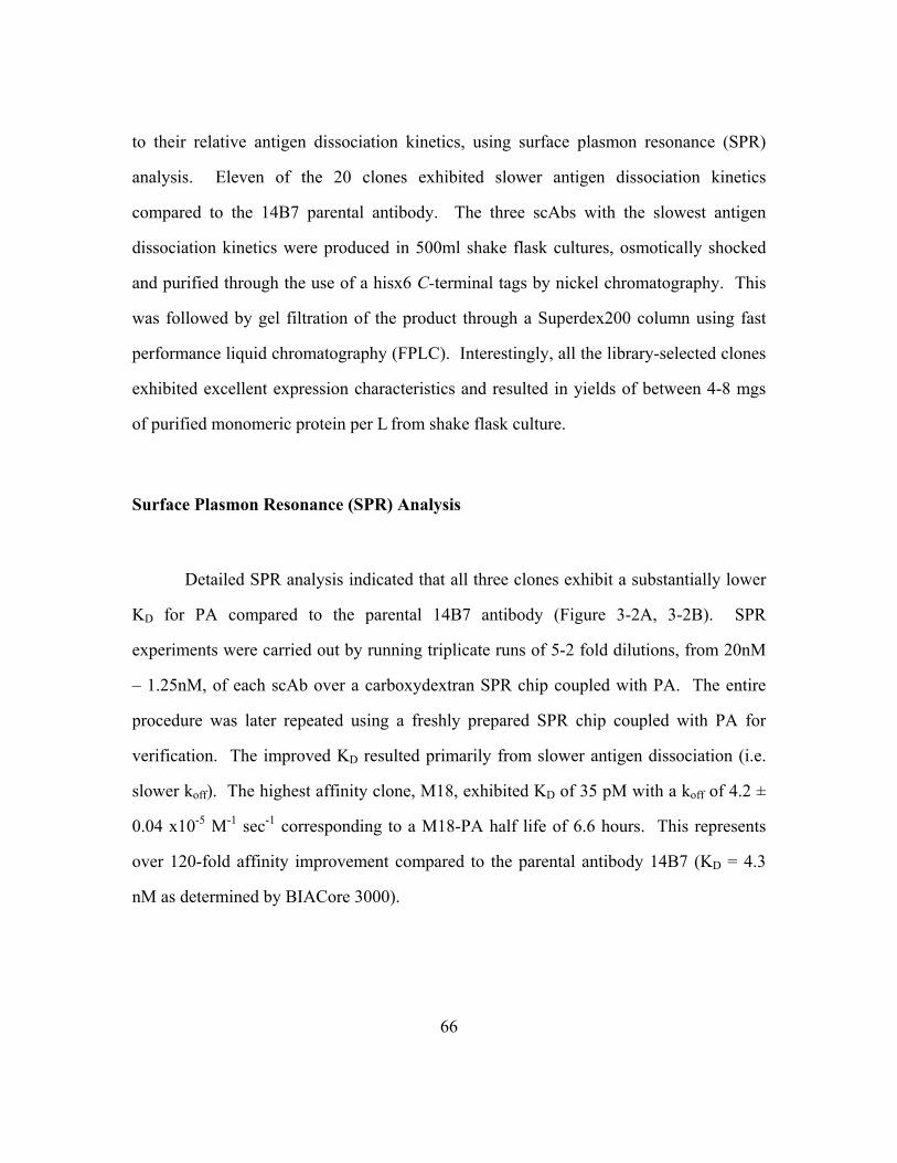
66
to their relative antigen dissociation kinetics, using surface plasmon resonance (SPR)
analysis. Eleven of the 20 clones exhibited slower antigen dissociation kinetics
compared to the 14B7 parental antibody. The three scAbs with the slowest antigen
dissociation kinetics were produced in 500ml shake flask cultures, osmotically shocked
and purified through the use of a hisx6 C-terminal tags by nickel chromatography. This
was followed by gel filtration of the product through a Superdex200 column using fast
performance liquid chromatography (FPLC). Interestingly, all the library-selected clones
exhibited excellent expression characteristics and resulted in yields of between 4-8 mgs
of purified monomeric protein per L from shake flask culture.
Surface Plasmon Resonance (SPR) Analysis
Detailed SPR analysis indicated that all three clones exhibit a substantially lower
KD for PA compared to the parental 14B7 antibody (Figure 3-2A, 3-2B). SPR
experiments were carried out by running triplicate runs of 5-2 fold dilutions, from 20nM
– 1.25nM, of each scAb over a carboxydextran SPR chip coupled with PA. The entire
procedure was later repeated using a freshly prepared SPR chip coupled with PA for
verification. The improved KD resulted primarily from slower antigen dissociation (i.e.
slower koff). The highest affinity clone, M18, exhibited KD of 35 pM with a koff of 4.2 ±
0.04 x10⁻⁵ M-1 sec-1 corresponding to a M18-PA half life of 6.6 hours. This represents
over 120-fold affinity improvement compared to the parental antibody 14B7 (KD = 4.3
nM as determined by BIACore 3000).

67
The three high affinity antibody fragments, M5, M6, and M18 were expressed as
scFv fusions to NlpA. The fluorescence of cells expressing these NlpA-[high affinity
scFvs] was compared to the 14B7 parental as previously described. The fluorescence
intensity of Tris-EDTA-lysozyme permeabilized cells expressing NlpA-[scFv] fusions
and labeled with 200nM PA-BODIPY™ was higher for all mutants compared to the
parental 14B7 scFv (Figure 3-4). This trend persisted even after incubation for 1.5 hours
in PBS in the absence of probe. Thus as expected from the in vitro antigen binding
Figure 3-2. Analysis of mutant scFv of 14B7 compared to wildtype. A) SPR Plot of Anti-PA scAbs interaction with immobilized PA on SPR chip. B) Table of affinity data from SPR.

68
kinetics, the three high affinity anti-PA mutants remain bound to the antigen for over 1.5
hours. In contrast, 14B7 becomes dissociated from the fluorescent PA. It is interesting to
note that the pattern of fluorescence ranked the clones in the order of their affinity as
determined with soluble scAb (Figure 3-2). This of course may not always be the case,
as expression levels of scFv on the cell also effect the cell fluorescence. It is speculated
that with extremely well expressed scFvs, as the case here, the level of protein synthesis
for all selected clones can be comparable, and therefore whole cell fluorescence can be
employed to rank the antibodies on the basis of their respective affinities.
Figure 3-3. Mean fluorescent intensities of anti-PA scFvs displayed with APEx technology. NlpA-[Meth scFv] was also subjected to PA-BODIPY™ labeling as a negative control.

69
Analysis of the affinity matured clones
The 3 clones analyzed in detail, M5, M6 and M18, contained 7, 12, and 11 amino
acid substitutions, respectively (Figure 3-4).
In earlier studies using phage display (Maynard et al. 2002), a variant of the 14B7 scFv
was isolated through three cycles, each consisting of: 1) mutagenic error prone PCR, 2)
five rounds of phage panning, and 3) DNA shuffling of the post-panning clones. The
best clone isolated in that study, 1H, contained two substitutions, Q55L and S56P, and
exhibited a KD of 150 pM (as determined by a BIACore3000). These two mutations
likely increase the hydrophobicity of the binding pocket, adding to the mounting
evidence that an increase in hydrophobic interactions is a dominant effect in antibody
affinity maturation (Li et al. 2003). Although the Q55L mutation was not found in the
Figure 3-4. Comparison of the amino acid sequences of anti-PA scFvs. The amino acid sequence of 14B7 parental scFv is compared to the 3 mutant sequences obtained from the APEx selection. Framework regions are designated in black, CDR regions in white and the glycine-serine linker sequence in gray.

70
highest affinity clone selected from the APEx library, M18, it did however posses the
S56P mutation in common with 1H. Figure 3-5 shows a model of M18, obtained through
The Universitity of Bath’s Center for Protein Analysis and Design’s web antibody
modeling (WAM) (Whitelegg and Rees 2000) software to create a structure model of the
scFv. The location of the S56P mutation is labeled in red and all other amino acid
changes from 14B7 parental labeled in blue. Importantly, most of the mutations appeared
to be surface exposed.

71
The Q55L and S56P amino acid substitutions of the phage-derived antibody were also
found in the M5 and M6 clones isolated by APEx. However, the presence of the
additional mutations in these two clones must have conferred an even higher affinity
(Figure 3-6) as well as improved expression levels in our scAb format, relative to the
phage derived 1H clone (4-8mg/L compared to < 1mg/L based on production in 500ml
liquid culture).
Figure 3-5. Model of anti-PA M18 scFv. The S56P mutation is designated in red, while all other mutations are in blue. Linker region is not included in model.

72
It is noteworthy that the M5, M6, and M18 were isolated after only one round of error
prone PCR, followed by two sorting rounds, yet they all had higher affinity relative to the
best antibody that could be isolated by phage display, 1H, even after multiple rounds of
mutagenesis and selection (Maynard et al. 2002).
M18, the highest affinity clone isolated by APEx, contained the S56P mutation
but lacked the Q55L substitution found in 1H, M5, and M6. When the Q55L substitution
Figure 3-6. SPR data comparing anti-PA scAb mutants from APEx selection to phage display scAb clone. At 400s the off rate from PA of each scAb is monitored as the relative decrease in response over time, correlating to the loss of scAb from the PA coated chip during that time interval.

73
was introduced into M18 by site specific mutagenesis, the resulting scAb exhibited a
further improvement in antigen binding (KD=21 pM) with a kon of 1.1 ± 0.03 x 10⁶ M-1
sec-1 and a koff of 2.5 ± 0.07 x 10⁻⁵ sec-1, corresponding to a antibody:antigen complex
half life of 11.6 hours. However, the introduction of this mutation reduced the yield of
purified protein more than 5-fold to 1.2 mg/L in shake flask cultures, suggesting a
possible reason that this set of mutations was not selected during the sorting process.
CONCLUSIONS
After two rounds of selection via APEx, the affinity of a neutralizing antibody to
the Bacillus anthracis protective antigen (PA) was improved over 120-fold, exhibiting a
final KD=35 pM, with additional engineering producing scFv exhibiting a KD of 21 pM.
This makes them excellent candidates for humanization, conversion to full length IgG,
and evaluation of its neutralizing potential in preclinical trials. Discovery of a 35pM
antibody utilizing the APEx technology is in line with some of the best antibodies
discovered using preexisting technologies, including binders from phage display (Schier
et al. 1996; Thompson et al. 1996), ribosome display (Hanes et al. 1998) and yeast
display (Boder et al. 2000; Feldhaus et al. 2003). Although all APEx clones discovered
to date exhibit expression characteristics as good or better than the parental clone from
which they arose, it is difficult to compare them to the expression characteristics of
clones from other technologies as direct comparative studies have never been done.
The results presented in this chapter demonstrated the effectiveness of PCR
recovery as a means to rescue sorted cells. An advantage of this approach is that it
enables the isolation of clones that are no longer viable due to the combination of

74
potential scFv toxicity and Tris-EDTA-lysozyme disruption. Yet another advantage of
PCR rescue is that the amplification of DNA from pooled cells can be carried out under
mutagenic conditions prior to subcloning. Thus, following each round of selection,
random mutations can be introduced into the isolated genes, simplifying further rounds of
directed evolution. Furthermore, PCR conditions that favor template switching among
the protein encoding genes in the pool may be employed during the amplification step to
allow recombination among the selected clones. PCR rescue has been shown to be
advantageous in other library screening formats as well (Hanes and Pluckthun 1997).
The ability to resort the population without amplification is an advantage of using
droplet deflection sorters compared to mechanical sorters. With mechanical sorters, such
as the Becton-Dickinson FACSCalibur, the sort solution is very dilute and cannot be
directly applied back onto the instrument for resorting. In droplet deflection sorters, such
as the MoFlo™ by Cytomation, the cells are collected in a small volume of fluid, and
therefore, the sort solution can be resorted immediately without the need for regrowth.
This feature expedites the library screening process, as was demonstrated here, and can
also favor the selection of clones with slower ligand dissociation rates due to the time
between resorts. However, the downside is that without amplification, rare clones
represented only by a single cell can be more easily lost, a problem resolved by PCR
recovery.

75
EXPERIMENTAL PROTOCOL
The anti-Bacillus anthracis protective antigen PA scFV 14B7 (Maynard et al.
2002), as well as the error prone PCR library generated from 14B7 scFv, were cloned
into pAPEx1 vector via its non compatible Sfi1 sites.
Synthesis of Fluorescent Probe
Purified PA protein, kindly provided by S. Leppla NIH, was conjugated to
BODIPYTM (Molecular Probes, Eugene, OR) at a 1:7 molar ratio with BODIPY™ FL SE
D-2184, according to the manufacturers instructions. Unconjugated BODIPYTM was
removed by dialysis.
Affinity Maturation of scFv library via APEx
Libraries were made from the 14B7 parental scFv using error prone PCR standard
techniques (Fromant et al. 1995) and cloned into the pAPEx1 expression vector. After
transformation into ABLEC™ E. coli, the cells were grown on SOC 2% glucose (2%glc)
+ chloramphenicol at 30ug/ml (Cm30) on large agar plates overnight at 30°C. The plates
were then scraped and the library frozen in SOC + 15% glycerol aliquots. The next
morning, the library was inoculated into teriffic broth (TB) media supplemented with 2%

76
glc and Cm30 to an OD600 of 0.1. Cells were grown for 2 hours at 37°C to an OD600 ~
0.6 before being cooled for 30 minutes at 25°C. Cultures were then induced using 1mM
IPTG and incubated for an additional four hours at 25°C with agitation. Following
induction, the cellular outer membrane was permeabilized with Tris-EDTA-lysozyme
treatment.
Briefly, cells (equivalent to approx 1ml of 20 OD600) were pelleted and
resuspended in 350ul of ice-cold solution of 0.75M sucrose, 0.1M Tris-HCl pH 8.0,
100ug/ml hen egg lysozyme. 700ul of ice-cold 1mM EDTA (ethylenediaminetetraacetic
acid) was gently added and the suspension left on ice for 10 min. 50ul of 0.5M MgCl2
was added and the mix left on ice for an additional 10 min. The resulting cells were
gently pelleted and resuspended in phosphate buffered saline (1xPBS) with 200nM
antigen labeled fluorophore at room temperature for 45 minutes, before evaluation by FC.
induction, and labeling, the cells were then stained with propidium iodide (PI emission
617nm) to monitor inner membrane integrity. Cells were sorted on a MoFlo
(Cytomation, Fort Collins, CO) droplet deflection flow cytometer using 488nm Argon
laser for excitation. Cells were selected based on improved fluorescence in the
Fluorescein/BODIPY™ FL emission spectrum detecting through a 530/40 band pass
filter and for the absence of labeling in PI emission detecting through a 630/40 band pass
filter.
E. coli captured after the first sort were immediately resorted through the flow
cytometer. Subsequently, the scFv genes in the sorted cell suspension were amplified by
PCR. Once amplified, the mutant scFv genes were then recloned into pAPEx1 vector,
retransformed into ABLEC™ E. coli cells and then grown overnight on SOC 2%glc +
Cm30 agar plates at 30oC. The resulting clones were subjected to a second round of

77
sorting plus resorting as above, before scFv genes were again amplified by PCR and
subcloned into pMoPac16 (Hayhurst et al. 2003) for expression of scAb protein.
Antibody fragment expression and purification
The mutant scFvs were cloned into the pMoPac16 vector via its non compatible
Sfi1 sites and the ligation was transformed into ABLEC™ E. coli for over-expression of
scAb fragments (Hayhurst et al. 2003). 500ml overnight cultures of TB media + 2%glc +
ampicillin at 200ug/ml (Amp200) in a 2L flask was inoculated and grown overnight at
30°C. Cultures were centrifuged for 10 minutes at 3200rpms in sterile 500ml centrifuge
bottles. They were then resuspended in fresh TB + Amp200 media (no glucose) and
incubated at 25°C, 240rpm for two hours. Cells were then induced with 1mM IPTG and
continued to incubate for an additional four hours.
The cultures were then centrifuged again at 3200rpms for 10 minutes and
resuspended in 16mls of ice cold 0.75M sucrose 0.1M Tris, pH 8. 1.6mls of 10mg/ml
lysozyme in sucrose-Tris buffer was added. The bottles were then gently shaken at
100rpms while 32mls of 1mM EDTA was added dropwise and the solution was stirred
gently for 10 minutes. 1.5mls of 0.5M MgCl2 was added and the incubation continued
for an additional 10 minutes. The solution was transferred to 50ml Falcon tubes and spun
at 8000 rpms for 20 minutes. The supernatant was dialyzed overnight against 10mM Tris
pH 8, 0.5M NaCl. The scAb was purified from the supernatant using an immobilized
metal affinity chromatography (IMAC) column followed by size-exclusion FPLC on a
Superdex 200, (Amersham Pharmacia, Piscataway, NJ) for monomeric scAb purification.

78
Surface Plasmon Resonance Analysis
Affinity measurements were obtained via SPR using a BIACore3000 instrument.
A CM-5 Carboxymethylated dextran matrix chip (BIAcore Inc, Uppsala, Sweden) was
activated by mixing equal volumes of 100mM NHS (N-hydroxysuccinimide) and 400mM
EDC (N-ethyl-N-(dimethyl-aminopropyl) carbodiimide hydrochloride) as described by
the BIAcore Application Wizard software. 5ug/ml of the PA diluted into sodium acetate
buffer pH5 was used to couple the chip. BSA was conjugated in parallel as an in line
control. Approximately 500RUs of PA was coupled to the CM5 chip using the
EDC/NHS chemistry. BSA was similarly coupled and used for in line subtraction.
Kinetic analysis was performed at a flow rate of 100ul/min, to reduce mass transport
limitations. Injections were performed as 5 two-fold dilutions in duplicate with scAb
concentrations ranging from 20nM-1.25nM in HEPES-buffered saline with 3mM EDTA
and 0.005% tween (HBS-EP). Surface was regenerated with 4M MgCl2 to disrupt the
scAb / PA interaction, before the next run.
79
REFERENCES
Boder ET, Midelfort KS, Wittrup KD (2000) Directed evolution of antibody fragments with monovalent femtomolar antigen-binding affinity. Proc Natl Acad Sci U S A 97:10701-5
Bradley KA, Mogridge J, Mourez M, Collier RJ, Young JA (2001) Identification of the cellular receptor for anthrax toxin. Nature 414:225-9
Brookmeyer R, Blades N, Hugh-Jones M, Henderson DA (2001) The statistical analysis of truncated data: application to the Sverdlovsk anthrax outbreak. Biostatistics 2:233-47
Feldhaus MJ, Siegel RW, Opresko LK, Coleman JR, Feldhaus JM, Yeung YA, Cochran JR, Heinzelman P, Colby D, Swers J, Graff C, Wiley HS, Wittrup KD (2003) Flow-cytometric isolation of human antibodies from a nonimmune Saccharomyces cerevisiae surface display library. Nat Biotechnol 21:163-70
Fromant M, Blanquet S, Plateau P (1995) Direct random mutagenesis of gene-sized DNA fragments using polymerase chain reaction. Anal Biochem 224:347-53
Hanes J, Jermutus L, Weber-Bornhauser S, Bosshard HR, Pluckthun A (1998) Ribosome display efficiently selects and evolves high-affinity antibodies in vitro from immune libraries. Proc Natl Acad Sci U S A 95:14130-5
Hanes J, Pluckthun A (1997) In vitro selection and evolution of functional proteins by using ribosome display. Proc Natl Acad Sci U S A 94:4937-42
Hayhurst A (2000) Improved expression characteristics of single-chain Fv fragments when fused downstream of the Escherichia coli maltose-binding protein or upstream of a single immunoglobulin-constant domain. Protein Expr Purif 18:1-10
Hayhurst A, Happe S, Mabry R, Koch Z, Iverson BL, Georgiou G (2003) Isolation and expression of recombinant antibody fragments to the biological warfare pathogen Brucella melitensis. J Immunol Methods 276:185-96
Hayhurst A, Harris WJ (1999) Escherichia coli skp chaperone coexpression improves solubility and phage display of single-chain antibody fragments. Protein Expr Purif 15:336-43
Laforce FM (1978) Woolsorters' disease in England. Bull N Y Acad Med 54:956-63

80
Li Y, Li H, Yang F, Smith-Gill SJ, Mariuzza RA (2003) X-ray snapshots of the maturation of an antibody response to a protein antigen. Nat Struct Biol 10:482-8
Little SF, Leppla SH, Cora E (1988) Production and characterization of monoclonal antibodies to the protective antigen component of Bacillus anthracis toxin. Infect Immun 56:1807-13
Maynard JA, Maassen CB, Leppla SH, Brasky K, Patterson JL, Iverson BL, Georgiou G (2002) Protection against anthrax toxin by recombinant antibody fragments correlates with antigen affinity. Nat Biotechnol 20:597-601
Meselson M, Guillemin J, Hugh-Jones M, Langmuir A, Popova I, Shelokov A, Yampolskaya O (1994) The Sverdlovsk anthrax outbreak of 1979. Science 266:1202-8
Schier R, McCall A, Adams GP, Marshall KW, Merritt H, Yim M, Crawford RS, Weiner LM, Marks C, Marks JD (1996) Isolation of picomolar affinity anti-c-erbB-2 single-chain Fv by molecular evolution of the complementarity determining regions in the center of the antibody binding site. J Mol Biol 263:551-67
Somerville JE, Jr., Goshorn SC, Fell HP, Darveau RP (1994) Bacterial aspects associated with the expression of a single-chain antibody fragment in Escherichia coli. Appl Microbiol Biotechnol 42:595-603
Thompson J, Pope T, Tung JS, Chan C, Hollis G, Mark G, Johnson KS (1996) Affinity maturation of a high-affinity human monoclonal antibody against the third hypervariable loop of human immunodeficiency virus: use of phage display to improve affinity and broaden strain reactivity. J Mol Biol 256:77-88
Whitelegg NR, Rees AR (2000) WAM: an improved algorithm for modelling antibodies on the WEB. Protein Eng 13:819-24

81
Chapter 4: Affinity Maturation and Cross-Reactive Analysis of Recombinant Antibody Fragments to Methamphetamine
INTRODUCTION
“Amphetamines” are a group of compounds that are powerful stimulants of the
central nervous system. The two key compounds, amphetamine and methamphetamine,
can be derivatized to create a number of illegal drugs of abuse while other derivatives of
these compounds can be found in over-the-counter (OTC) and prescription medication
(Buchanan and Brown 1988). The common derivatives of methamphetamine which have
been used as drugs of abuse include 3,4-methylenedioxymethamphetamine (MDMA) and
3,4-methylenedioxy-N-ethylamphetamine (MDEA). Common OTC and prescription
derivatives of methamphetamine include ephedrine and pseudoephedrine (Figure 4-1).

82
The use of immunoassays is a common method for urine screenings for
methamphetamines in order to differentiate between negative and potentially positive
samples. Samples that test positive by immunoassay are most commonly analyzed
further by gas chromatography-mass spectrometry (GC-MS) (Goldberger and Cone
1994), due to the highly specific and sensitive nature of these techniques. However, GC-
MS steps are time-consuming and expensive. Improving the sensitivity of immunoassays
by modifying affinity or specificity of the antibodies used for detection could drastically
reduce the number of false positives and thus save time and money required for further
sample evaluation. Specifically, diagnostic antibodies for amphetamines can be
Figure 4-1. Methamphetamine and its derivatives. Drugs of abuse MDMA and MDEA are on the left, and OTC drugs (-)-ephedrine and (+)-pseudoephedrine are pictured on the right.

83
improved for: 1) higher affinity to target drugs, 2) high cross reactivity to derivitized
variants of the target drug, and 3) low cross reactivity to over-the-counter (OTC) drugs.
In this study, a recombinant single-chain variable fragment (scFv) antibody to
methamphetamine (Meth scFv) was cloned using the genetic material from hybridoma
cells, which produce an IgG against methamphetamine. This hybridoma cell line is used
to produce monoclonal IgG that is used in commercial diagnostic arrays. This Meth scFv
was affinity matured using the APEx technology. Cross reactivity of the matured scFv
variants to over-the-counter (OTC) drugs, ephedrine and pseudoephedrine, was
determined by a comparative on-rate study. The mutant antibodies were incubated with
increasing levels of these OTC drugs and analyzed for their ability to bind
methamphetamine using surface plasmon resonance (SPR) technology. Finally, it was
demonstrated that some of the matured scFv variants had higher affinity to not only
methamphetamine but also to an ethamphetamine variant, 3,4-methylenedioxy-N-
ethylamphetamine (MDEA).
RESULTS AND DISCUSSION
From IgG to scFv
Roche diagnostics provided our lab with a hybridoma which produced their best
IgG antibody to methamphetamine. The hybridoma was originally produced by
immunizing a mouse with the keyhole limpet hemocyanin -methamphetamine conjugate
depicted below (KLH-Meth) (Figure 4-1). Using standard protocol from a Totally
RNA™ kit from Ambion, Inc. (Austin, TX), RNA was purified from the anti-

84
methamphetamine IgG expressing hybridoma and reverse transcribed to cDNA. The VH
and VL chains of the IgG were PCR amplified from the cDNA using the Krebber murine
primer set (Krebber et al. 1997). This set of primers has been optimized for selection of
mouse variable region genes and have flanking restriction endonuclease sites for facile
cloning as well as a region for variable domain overlap to be utilized in the next step.
Once amplified, the variable regions were then overlapped by PCR, which by design adds
a flexible glycine-serine linker was introduced between the VH and VL, producing a scFv
gene. The overlap PCR reaction of the antibody variable regions was cloned into
pMoPac1 (Hayhurst et al. 2003), a vector for soluble periplamic expression (Figure 4-2).
Figure 4-2. From IgG to ScFv. The hybridoma used in this study, provided by Roche Diagnostics was derived from a mouse immunized with a KLH-methamphetamine conjugate. mRNA was isolated from the hybridoma cells, reverse transcribed to cDNA and the cDNA was used as template for PCR amplification of the VH and VL regions. The variable regions were then overlapped by PCR and cloned into the MoPac1 soluble expression vector for evaluation.

85
Evaluation of Clones for ELISA Signal
The MoPac1 expression vectors containing the scFv genes were transformed into
E. coli and plated on SOC solid media plates containing 30ug/ml chloramphenicol to
select for cells that retained the MoPac1 vector. To find clones that contained the correct
scFv in MoPac1, 10 clones were randomly selected, grown in liquid culture, and induced
for expression of the scFv protein. Cultures were then normalized based on OD600,
osmotically shocked (as described in experimental protocol at the end of this chapter),
and the supernatant was collected. An ELISA assay was run which identified two clones
that gave positive ELISA signal against a methamphetamine-BSA conjugate (Meth-
BSA). Purification and sequencing of the plasmid DNA of these two clones showed that
they had identical sequence and were designated Meth scFv. Meth scFv was subcloned
into MoPac16 (Hayhurst et al. 2003) for expression in the scAb format (as discussed in
Chapter 3). After transformation of MoPac16: Meth scAb into ABLEC™ E. coli, the
culture was grown, induced for expression, and osmotically shocked as previously
discussed. ELISA results demonstrate that supernatant containing the Meth scAb showed
specific binding to a 96 well plate coated with Meth-BSA, with no cross-reactivity to
BSA alone (Figure 4-3).

86
Expression of Meth scFv using APEx Technology
As described in Chapter 2, the Meth scFv was cloned into the pAPEx1 expression
vector to generate a NlpA-[Meth scFv] fusion. Expression of the fusion protein in E.
coli, as an anchored chimera on the outside of the inner membrane, allowed for specific
labeling of these cells when incubated in 5xPBS and labeled with 200nM of a
methamphetamine-fluorescein (Meth-FL) conjugate provided by Roche Diagnostics
(Figure 2-4, Chapter 2). Due to the small size of the Meth-FL probe, 5xPBS for
membrane disruption during labeling was adequate for outer membrane penetration.
Cells expressing unrelated scFv fusions, namely the NlpA-[Dig scFv] or the NlpA-[7C2
scFv] in E. coli, and labeled with the Meth-FL probe did not give a fluorescent signal
Meth scAb ELISA
00.20.40.60.8
11.21.41.6
2 4 8 16 32 64
Dilutions
Abs
at 4
90nm Meth scAb on Meth-
BSAMeth scAb on BSA
Figure 4-3. Scab ELISA signal on methamphetamine-BSA. ELISA depicting specific binding of Meth scAb to Meth-BSA conjugate with no cross-reactivity to wells coated with BSA alone.

87
above background. This demonstrated the ability of the APEx technology to resolve
populations of cells expressing anchored Meth scFvs with a Meth-FL probe from those
expressing anchored scFvs non-specific to methamphetamine. The next step was to
demonstrate that APEx could select for affinity improved variants of scFvs to Meth-FL
from a mutagenic library.
Affinity maturation of anti-methamphetamine scFv library via APEx
Using the Meth scFv as template, an error prone PCR reaction was performed
(Fromant et al. 1995). The PCR products were then ligated into the pAPEx1 vector and
transformed into ABLEC™ E. coli. The transformation mixture was plated on SOC agar
media with 2% glucose (2%glc) to inhibit the lac promoter and 30ug/ml chloramphenicol
(Cm30) to select for vector transformants. The library size was estimated to be
approximately 4x10⁵ clones. The library was grown to log phase (OD600~0.6), induced
for 4hrs at 25°C, and labeled in 5xPBS with 200nM of the Meth-FL probe. After 45
minutes of shaking at room temperature, the cells were decanted and resuspended in
5xPBS. This was then stained with 5ug/ml propidium iodide (PI) so that inner membrane
integrity could be monitored. PI is a cytoplasmic membrane impermeable nucleic acid
stain which can be used to monitor loss of inner membrane integrity of E. coli, as it
fluoresces only if intercalated in nucleic acids (Daugherty et al. 2000; Wickens et al.
2000). The library of cells was then sorted on a droplet deflection MoFlo (Cytomation,
Fort Collins, CO) flow cytometer. Using an argon laser with 488nm emission,
approximately 1% of the population was sorted based on a gate setup to include the most
fluorescent events which emitted in the FL-1, monitoring fluorescein emission (530/40
band pass filter), but had little to no fluorescence in the FL-2 monitoring PI emission
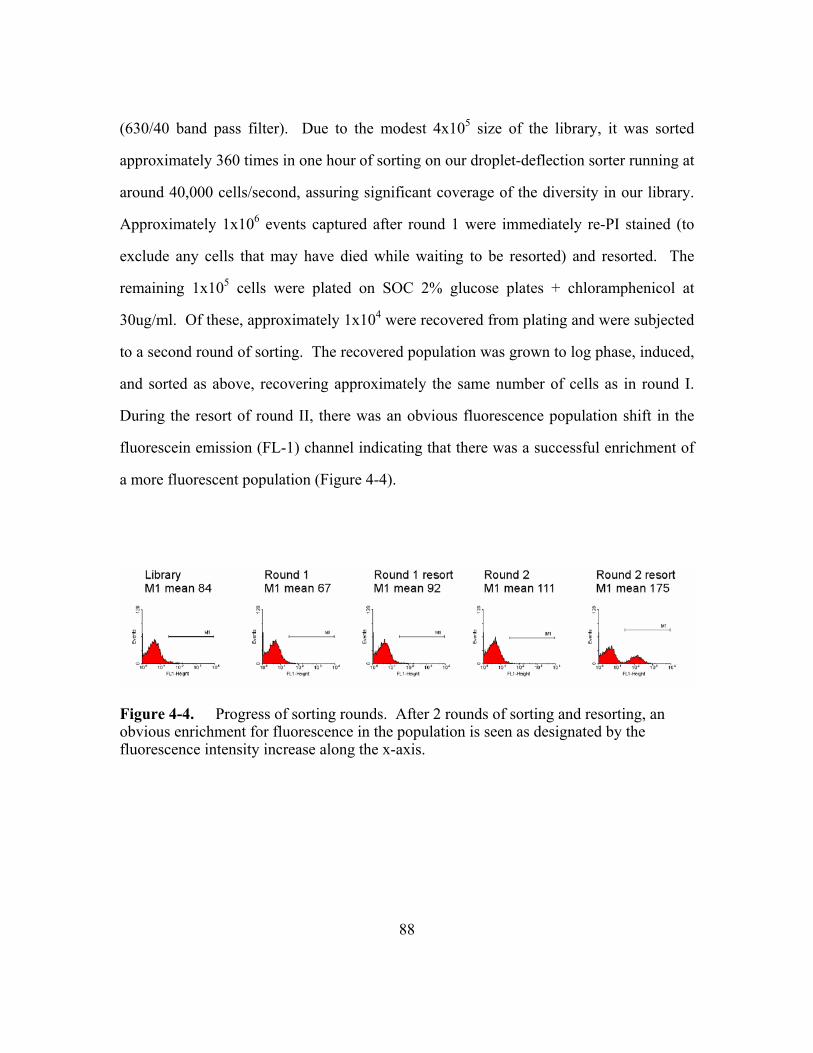
88
(630/40 band pass filter). Due to the modest 4x10⁵ size of the library, it was sorted
approximately 360 times in one hour of sorting on our droplet-deflection sorter running at
around 40,000 cells/second, assuring significant coverage of the diversity in our library.
Approximately 1x10⁶ events captured after round 1 were immediately re-PI stained (to
exclude any cells that may have died while waiting to be resorted) and resorted. The
remaining 1x10⁵ cells were plated on SOC 2% glucose plates + chloramphenicol at
30ug/ml. Of these, approximately 1x10⁴ were recovered from plating and were subjected
to a second round of sorting. The recovered population was grown to log phase, induced,
and sorted as above, recovering approximately the same number of cells as in round I.
During the resort of round II, there was an obvious fluorescence population shift in the
fluorescein emission (FL-1) channel indicating that there was a successful enrichment of
a more fluorescent population (Figure 4-4).
Figure 4-4. Progress of sorting rounds. After 2 rounds of sorting and resorting, an obvious enrichment for fluorescence in the population is seen as designated by the fluorescence intensity increase along the x-axis.

89
APEx Analysis of Individual Meth Mutants
Fifteen individual clones, randomly picked from the approximately 1x10⁴
colonies grown on solid SOC media plates (Cm30 + 2% glc) after the round II resort,
were individually grown in SOC liquid media (Cm30 + 2% glc), induced with 1mM
IPTG, and labeled with 200nM Meth-FL in 5xPBS as previously described. Of the 15
clones, 9 had a higher FL intensity than that of parental Meth scFv. The pAPEx1 plasmid
DNA from these 9 clones was prepped and sequenced. From the sequencing reactions,
three unique sequences were discovered. The clones were designated Meth M9 scFv,
Meth M12 scFv, and Meth M15 scFv which were present in this population 3, 1 and 5
times respectively (Figure 4-5).

90
Sequence comparison of the three clones yielded some interesting information. In
the CDR H1 domain, phenylalanine is changed to a serine in both Meth M9 scFv and
Meth M12 scFv. Even more interesting is that, an isoleucine in CDR3 of 7.3 Meth scFv,
is changed to a threonine in Meth M9 scFv and a valine in Meth M15 scFv. The
sequence of the Meth scFv was sent to the Universitity of Bath’s Center for Protein
Analysis and Design where a group run by A.R. Rees utilized web antibody modeling
(WAM) (Whitelegg and Rees 2000) software to create a structure model of the scFv.
Modeling predictions from the WAM suggests that the isoleucine amino acid is within
the pocket binding site of the scFv (Figure 4-6). It is interesting that, in such a small
library with such an enormous sequence space to cover, the selection found two
mutations at this site for potential improvement in scFv activity.
Figure 4-5. Allignment of Meth scFv to mutants selected from error prone library. Of particular interest is the phenylalanine to serine change in CDR H1 of both Meth M9 scFv and Meth M12 scFv, as well as the isoleucine to threonine or valine changes in CDR H3 to Meth M9 scFv and Meth M15 scFv respectively.

91
Expression and Analysis of Mutant Meth Antibody Fragments by SPR
For soluble expression, the Meth scFv and the three mutants were each fused
upstream of a human kappa constant region (Hayhurst et al. 2003) to produce scAb
antibody fragments. This was done for ease of purification as it has been previously
shown to improve soluble expression yields (Hayhurst 2000). These vectors were
transformed into ABLEC™ E. coli and grown in 500ml shake flask cultures. After
induction with 1mM IPTG and growth for four hours at 25°C, the cultures were
osmotically shocked, and the supernatant was run over an immobilized metal affinity
chromatography (IMAC) column for purification of the scAb via nickel affinity to the
Figure 4-6. Molecular modeling of the Meth scFv. Isoleucine in CDR H3 designated in red. Phenylalanine in CDR H1 designated in blue.

92
hisx6 tag. Eluate from IMAC was then run on fast performance liquid chromatography
(FPLC) in 1xPBS buffer for pure monomeric scAb purification. All mutants could be
expressed as soluble protein at levels equivalent or better than that of the parental Meth
scFv in the scAb format. The yield of the purified scAb mutants ranged from 1.56mg-
2.90mg/L in comparison to the 1.30mg/L obtained from the Meth scAb. The fact that all
the mutants expressed soluble proteins at yields equal to or better than the original Meth
scFv is probably because the fluorescence criteria imposed by the sort can either be met
by improved affinity, improved expression levels in the E. Coli host, or a combination of
both.
All three mutants showed higher affinity to methamphetamine as analyzed by
kinetic analysis of purified scAb over the methamphetamine-BSA conjugate by surface
plasmon resonance (SPR). Each scAb mutant was diluted into BIACore HBS-EP
running buffer to concentrations of 20, 10, 5, 2.5 and 1.25nM. Briefly, each dilution was
run in duplicate over a standard Cm-5 BIACore chip with Meth-BSA coupled to its
surface. Compared to Meth scFv affinity of 11.5nM (kon 2.6 ± 0.13 x 10⁴, koff 3.0 ± 0.21
x 10-4) the mutants from the post round II resort showed affinities of 3.0nM (kon 2.2 ±
0.14 x 10⁴, koff 6.8 ± 0.02 x 10⁻⁵), 3.6nM (kon 2.2 ± 0.02 x 10⁴, koff 7.9 ± 0.04 x 10⁻⁵), and
4.7nM (kon 3.6 ± 0.03 x10⁴, koff 1.7 ± 0.03 x 10⁻⁴) for Meth M9 scAb, Meth M12 scAb
and Meth M15 scAb respectively (Figure 4-7). Interestingly, Meth M15 scAb showed
only a slightly better off-rate to that of Meth scAb, and most of its improvement was in
the on-rate.

93
Cross-reactivity to OTC drugs
To compare relative cross reactivity of these clones to the common derivatives of
methamphetamine commonly found in OTC medications, a 300nM concentration of each
scAb was incubated with different concentrations of free (-)-ephedrine or (+)-
pseudoephedrine. Each scAb mixed with 15000nM, 7500nM, 3750nM, 1875nM and
0nM concentrations of free (-)-ephedrine or (+)-pseudoephedrine were left at room
temperature for 30 minutes. Each was then run over a Meth-BSA coated CM-5 chip on
SPR, and their on-rates recorded. Figure 4-8 is a summary of the results of this
Figure 4-7. SPR analysis of anti-methamphetamine scAb selected using APEx. Table of affinity data aquired by SPR.

94
experiment. It provided information on the relative ranking of each of these mutants for
their cross reactivity to (-)-ephedrine or (+)-pseudoephedrine. On-rate is a good measure
of cross reactivity because it is concentration dependant. If there is high cross reactivity
between the scAb and the OTC drug, the concentration of free scAb will be less in the
solution, correlating to a slower observed on-rate with SPR. It appears that the highest
affinity clone to methamphetamine, Meth M9 scAb also has the highest cross reactivity to
ephedrine and pseudoephedrine, suggesting that the affinity improvement mutations
decreased the specificity of the antibody fragment. Also, it appears that the the mutations
of Meth M15 scAb, did not significantly increase cross reactivity to the OTC drugs as
was seen in Meth M9 and Meth M12.

95
Cross Reactivity to Ethamphetamine
The cross reactivity of the antibody mutants to ethamphetamine was of interest
because ethamphetamine can be derivatized into 3,4-methylenedioxy-N-
ka of scAb 300nM + ephedrine competition
0.00E+00
5.00E+03
1.00E+04
1.50E+04
2.00E+04
2.50E+04
3.00E+04
3.50E+04
WT M9 M12 M15
Meth scAbs
ka (1
/Ms)
0nM Ephedrine
1875nM Ephedrine
3750nM Ephedrine
7500nM Ephedrine
15000nM Ephedrine
ka of scAb300nM + psuedoephedrine competition
0.00E+00
5.00E+03
1.00E+04
1.50E+04
2.00E+04
2.50E+04
3.00E+04
3.50E+04
WT M9 M12 M15
Meth scAbs
ka (1
/Ms)
0nM PEphedrine
1875nM PEphedrine
3750nM PEphedrine
7500nM PEphedrine
15000nM PEphedrine
Figure 4-8. Cross-reactivity analysis. Effect of on-rate of scAb due to increasing levels of competition by free ephedrine or pseudoephedrine as measured by BIACore.

96
ethylamphetamine (MDEA), a designer drug of abuse. Cells expressing each of the
mutant scFvs as fusions to NlpA were grown to log phase, induced with 1mM IPTG, and
grown for an additional four hours at 25°C. Following induction, the cells were pelleted
and resuspended in 500ul of 5xPBS with a 200nM concentration of an ethamphetamine-
flourescein probe (Eth-FL). After incubation at room temperature with gentle shaking for
45 minutes, the cells were analyzed by FC.
Analysis of each population demonstrated that each of the mutant antibodies
conferred a higher mean fluorescence with Eth-FL than that of the parental Meth scFv.
Meth M9 scFv and Meth M12 scFv exhibited the highest mean fluorescence at about
three times that of the cells expressing parental Meth scFv via APEx. This finding
suggests that the clones not only bind methamphetamine but also have improved affinity
to ethamphetamine (Figure 4-9). This was confirmed by SPR analysis of each Meth
scAb in duplicate on BIACore using a CM-5 carboxydextran chip coupled with
ethamphetamine-BSA. The following values were obtained for KD of each Meth scAb to
ethamphetamine: Meth scAb: 80nM, Meth M9 scAb: 35nM, Meth M12 scAb: 41nM,
Meth M15 scAb: 45nM.

97
The cross reactivity characteristics of the Meth scAb mutants to ethamphetamine
in the presence of increasing levels of MDEA was evaluated. Briefly, 300nM
concentrations of each Meth scAb were incubated with 450 or 225nM concentrations of
MDEA. After 30 minutes at room temperature, the mixture was run on BIACore for on-
rate analysis to a Meth-BSA conjugate coupled to the chip. Figure 4-10 demonstrates
that on-rates were significantly decreased for the Meth M9 scAb as well as the Meth M12
scAb upon increasing the amount of MDEA competitor (Figure 4-10). This is in
agreement with both the FC data and the affinity measurements.
Figure 4-9. Cross reactivity of anti-methamphetamine clones to ethamphetamine as observed by APEx. The histograms depict the mean fluorescent intensity of 10,000 events from NlpA-[Meth scFv] expressing cells upon labeling with 200nM ethamphetamine-fluorescein.
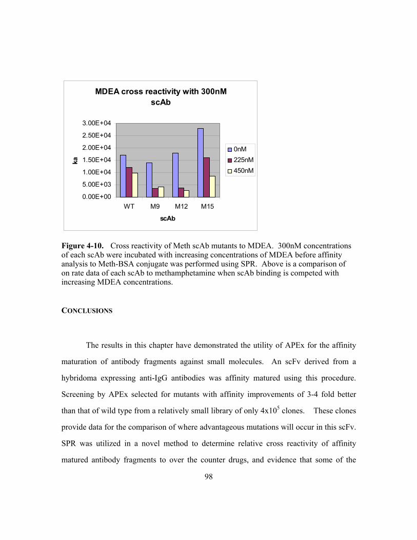
98
CONCLUSIONS
The results in this chapter have demonstrated the utility of APEx for the affinity
maturation of antibody fragments against small molecules. An scFv derived from a
hybridoma expressing anti-IgG antibodies was affinity matured using this procedure.
Screening by APEx selected for mutants with affinity improvements of 3-4 fold better
than that of wild type from a relatively small library of only 4x10⁵ clones. These clones
provide data for the comparison of where advantageous mutations will occur in this scFv.
SPR was utilized in a novel method to determine relative cross reactivity of affinity
matured antibody fragments to over the counter drugs, and evidence that some of the
MDEA cross reactivity with 300nM scAb
0.00E+00
5.00E+03
1.00E+04
1.50E+04
2.00E+04
2.50E+04
3.00E+04
WT M9 M12 M15
scAb
ka
0nM225nM450nM
Figure 4-10. Cross reactivity of Meth scAb mutants to MDEA. 300nM concentrations of each scAb were incubated with increasing concentrations of MDEA before affinity analysis to Meth-BSA conjugate was performed using SPR. Above is a comparison of on rate data of each scAb to methamphetamine when scAb binding is competed with increasing MDEA concentrations.
99
clones have a high cross reactivity for ethamphetamine and ethamphetamine-like drugs
was demonstrated, based on FC and BIACore data.
EXPERIMENTAL PROTOCOL
The hybridoma cells and all methamphetamine and ethamphetamine conjugates
were provided by Roche Diagnostics. (-)-Ephedrine and (+)-Pseudoephedrine were
purchased from Alltech-Applied Science (State College, PA). BIAcore 3000TM and CM5
sensor chips were obtained from BIAcore AB (Uppsala, Sweden).
Expression of variable fragments and ELISA analysis
A recombinant single-chain variable fragment (scFv) antibody to
methamphetamine was cloned from a hybrodoma cell line that had been created from the
spleen cells of mice immunized with a methamphetamine-keyhole limpet hemocyanin
(Meth-KLH) conjugate. RNA purified using Totally™ RNA (Ambion, Austin, TX) was
used to first produce cDNA which was used for the amplification of the immunoglobulin
light (VL) and heavy (VH) chain genes using the Krebber murine primer set (Krebber et
al.). Following gel purification of the bands, they were used as template in an overlap
extension PCR reaction and cloned into MoPac1 (Hayhurst et al. 2003) for soluble
expression. Individual colonies were grown overnight in 2ml volumes of terrific broth
2% glucose (TB 2%glc) media with 30ug/ml chloramphenicol (Cm30) at 30°C. Cultures
were diluted in fresh TB 2%glc + Cm30 media at 1:100 for 2 hrs at 37°C, cooled to 25°C
and induced with 0.1mM IPTG for 4 hrs. They were then osmotically shocked as

100
previously described(Chen et al.) and the osmotic shockate from each clone was screened
by ELISA. 100ul of 0.1ug/ml methamphetamine-bovine serum albumin (Meth-BSA)
was used to coat a Costar High binding 96 well plate (Corning Incorporated, Corning,
NY) overnight at 4C. The next morning the plate was blocked with 2% milk (Carnation,
Nestle Inc., Solon, OH) phosphate buffered saline (2%MPBS) for 1 hour at room
temperature (RT). Osmotic shockate was then diluted 2-fold down the plate in 2%MPBS
and incubated at RT for 1 hour. Following through washing with 1xPBS and 1xPBS
0.1%tween20, the primary antibody anti-Histadine (Sigma) was applied at a 1:5000
dilution to detect the his tag from the pMoPac1 vector. After RT incubation for 1 hour
and subsequent washing the secondary antibody was applied at a 1:2000 dilution of goat
anti-mouse horse radish peroxidase (HRP) (Bio-rad). Signal of HRP was detected using
OPD substrate (Sigma) as described in product literature to determine positive clones.
Sequence confirmed by sequencing from pMoPac1 plasmid and designated Meth
7.3scFv.
Affinity maturation of Meth scFv using Anchored Periplasmic Expression (APEx) flow cytometric (FC) selection.
The Meth scFv sequence was cloned into the pAPEx1 (Chapter 2) expression
vector, induced and labeled with 200nM concentration of Methamphetamine-fluorescein
(Meth-FL) in 5xPBS. Dig scFv, an anti-digoxin scFv (Chen et al. 1999) was run and
labeled with Meth-FL in parallel as a negative control. Cells were analyzed for mean
fluorescence intensity of 10,000 events using a BD FacSort (BD Biosciences, San Jose,
CA).

101
A small 1% error rate mutagenic library of 4x10⁵ scFv members was made using
Meth scFv as the parental template using standard error prone techniques (Fromant et al.
1995) and was cloned into pAPEx1 for transformation into E.coli. After transformation
the cells were plated on 150 x 15mm Petri dishes and left to grow overnight at 30°C.
Plates were scraped in TB media and frozen in 15% glycerol aliquots, stored at -80°C.
The library was inoculated from an aliquot of frozen stock to an OD600 of 0.1 in 30mls of
TB 2% glc Cm30. After growth for 2 hours at 37°C, cells were cooled to 25°C for 1hr
with shaking and then induced with 1mM IPTG for 4 hours. Cells were then resuspended
in 5xPBS and labeled with 200nM Methamphetamine-Fluorescein (Meth-FL) for 45
minutes at room temperature with shaking in 5xPBS. Cells were then PI stained to
monitor inner membrane integrity and sorted on a MoFlo flow cytometer(Cytomation,
Fort Collins, CO). Cells were detected by triggering on side scatter using 100mW of
488nm light from an argon laser. Detection of Meth-FL binding was monitored by
518nm emission via a 530/40 band pass filter (FL-1). PI emission was monitored with a
630/40 band pass filter (FL-2). By gating for cells showing high FL-1 signal and low FL-
2 signal, the library of scFvs were subjected to two rounds of sorting and re-sorting
followed by overnight growth on SOC 2%glc + Cm30 plates.
Clones from post-round 2 selection were monoclonally grown, induced, labeled
with 200nM Meth-FL and analyzed by FC.
Protein Expression and Purification
For soluble protein expression all scFvs were cloned upstream of a human kappa
constant domain (HuCk) in the vector pMoPac16 (Hayhurst et al. 2003) to produce scAb
fragments. It was previously shown that fusion of the HuCk domain downstream of

102
scFvs improved expression levels of antibody fragments (Hayhurst 2000). Following
cell fractionation, IMAC and FPLC purification as described in chapter 3, protein was
recovered in 1xPBS buffer for dilution into BIAcore buffer HBS-EP for BIAcore
analysis.
BIAcore for affinity determination and comparative cross reactivity analysis of Meth clones to ephedrine and pseudoephedrine.
A CM-5 Carboxymethylated dextran matrix chip (BIAcore) was activated by
mixing equal volumes of 100mM NHS (N-hydroxysuccinimide) and 400mM EDC (N-
ethyl-N-(dimethyl-aminopropyl) carbodiimide hydrochloride) as described by the
BIAcore Application Wizard software. 5ug/ml of the Meth-BSA conjugate diluted into
sodium acetate buffer pH5 was used to couple the chip. BSA was conjugated in parallel
as an in line control. Approximately 500 RUs of each conjugate were bound by the
activated surface. Concentrated scAb purified in 1xPBS were each diluted into HBS-EP
buffer for BIAcore analysis. 2 fold dilutions of scAB in HBS-EP buffer from 600nM to
75nM were run in duplicate at a flow rate of 60ul/min over the chip for affinity analysis
of each clone. For cross reactivity comparisons, each clone at 300nM concentration was
incubated with 50,25,12.5 and 6.25 fold excess concentrations (15000nM to 1875nM) of
free (-)-ephedrine or (+)-pseudoephedrine and then run over the Meth-BSA surface at a
flow rate of 60ul/min to determine on rate changes.

103
REFERENCES
Buchanan JF, Brown CR (1988) 'Designer drugs'. A problem in clinical toxicology. Med
Toxicol Adverse Drug Exp 3:1-17
Chen G, Dubrawsky I, Mendez P, Georgiou G, Iverson BL (1999) In vitro scanning saturation mutagenesis of all the specificity determining residues in an antibody binding site. Protein Eng 12:349-56
Daugherty PS, Iverson BL, Georgiou G (2000) Flow cytometric screening of cell-based libraries. J Immunol Methods 243:211-27
Fromant M, Blanquet S, Plateau P (1995) Direct random mutagenesis of gene-sized DNA fragments using polymerase chain reaction. Anal Biochem 224:347-53
Goldberger BA, Cone EJ (1994) Confirmatory tests for drugs in the workplace by gas chromatography-mass spectrometry. J Chromatogr A 674:73-86
Hayhurst A (2000) Improved expression characteristics of single-chain Fv fragments when fused downstream of the Escherichia coli maltose-binding protein or upstream of a single immunoglobulin-constant domain. Protein Expr Purif 18:1-10
Hayhurst A, Happe S, Mabry R, Koch Z, Iverson BL, Georgiou G (2003) Isolation and expression of recombinant antibody fragments to the biological warfare pathogen Brucella melitensis. J Immunol Methods 276:185-96
Krebber A, Bornhauser S, Burmester J, Honegger A, Willuda J, Bosshard HR, Pluckthun A (1997) Reliable cloning of functional antibody variable domains from hybridomas and spleen cell repertoires employing a reengineered phage display system. J Immunol Methods 201:35-55
Whitelegg NR, Rees AR (2000) WAM: an improved algorithm for modelling antibodies on the WEB. Protein Eng 13:819-24
Wickens HJ, Pinney RJ, Mason DJ, Gant VA (2000) Flow cytometric investigation of filamentation, membrane patency, and membrane potential in Escherichia coli following ciprofloxacin exposure. Antimicrob Agents Chemother 44:682-7

104
Chapter 5: Conclusions and Future Directions
CONCLUSIONS
The work presented describes the development of a protein library screening
technology in Escherichia coli for high throughput screening of proteins libraries and the
isolation of polypeptides exhibiting ligand binding. The new strategy, referred to as
APEx (for Anchored Periplasmic Expression) is based on anchoring proteins such as
single chain variable fragments (scFv) to the periplasmic face of the inner membrane of
E. coli using protein fusion strategies. The leader sequence and first six amino acids of
the mature lipoprotein NlpA, which becomes fatty acylated upon export and anchored the
outer face of the inner membrane, proved to be ideal for this purpose. Upon chemical
permealization of the outer membrane, cells expressing NlpA(1-6)-[scFv] protein fusions
could be readily labeled with fluorescently conjugated antigens and screened by flow
cytometry.
The utilization of a lipoprotein fusion for targeting and anchoring of protein
fusions to the outside of the inner membrane of Escherichia coli proved to be an
excellent choice for a number of reasons: 1) The minimal N-terminal fusion which
consisted of a leader sequence for secretion to the periplasm via the general secretory
pathway, and the first few amino acids of the mature lipoprotein for the N-terminal lipid
modification and membrane targeting, allowed the over expression of anchored scFvs to
the inner membrane without any adverse effects on cell growth. 2) The antibody
fragment displayed only has to cross one membrane (the inner membrane) for display
without the extra burden of secretion across the outer membrane, common to Gram

105
negative bacteria surface display technologies. 3) There is no LPS on the inner
membrane to interfere with large antigen binding, another problem faced with surface
display technologies. 4) The outer membrane can be permeabilized without loss of
displayed antibody from the cell, with the inner membrane fusion maintaining the link
between the DNA in the cytoplasm and the displayed protein. This feature overcame the
antigen size limitation seen in soluble periplasmic display (Chen et al. 2001), as the scFv
could not dissociate from the cell upon outer membrane disruption. In summary, the
technology utilized a minimal carrier domain which allowed antibody fragments, 30-
45kDa (scFvs and scabs) in size, to be displayed freely where they could interact with
small molecule, peptide, and even large protein antigens up to 240kDa in size (Chapter
2). It has been demonstrated that libraries of scFv antibodies displayed by APEx can be
readily screened by flow cytometry and isolation of affinity improved variants of scFv to
bacterial toxins (Chapter 3) and drugs of abuse have been discussed (Chapter 4).
In addition, the power of droplet deflection flow cytometry was exploited, which
not only allows for high throughput of library members at rates over 50,000/s but also
allows the events to be collected in a very manageable volume. This can be conveniently
transferred to PCR tubes for whole cell PCR recovery of events. PCR has previously
been utilized for recovery in a ribosome display in vitro system and has numerous
advantages (Hanes and Pluckthun 1997). One advantage of this approach is that it
enables the isolation of clones that are no longer viable due to the combination of
potential scFv toxicity or the Tris-EDTA-lysozyme disruption conditions used to
permeabilize the outer membrane. In addition, the PCR recovery step can be
incorporated with DNA shuffling (Stemmer 1994) or error prone PCR techniques
(Cadwell and Joyce 1994; Fromant et al. 1995). Thus, following each round of selection,

106
random mutations can be introduced into the isolated genes, simplifying further rounds of
directed evolution.
Finally, there have been numerous antibody fragments to important therapeutic
and diagnostic targets isolated from antibody libraries screened by phage display. Due to
the commonality of phage display, it was desirable to develop a means for rapid antigen
binding analysis and affinity or expression maturation of such antibodies without the
need for time consuming subcloning steps.
It was demonstrated that the mean fluorescence intensity of cells displaying scFvs
that were fused to the N-terminus of the pIII phage coat protein and anchored on the
outside of the inner membrane, was comparable to that obtained by fusion to the C-
terminus of the NlpA anchor. These results showed that: (i) large soluble domains can be
tethered N-terminally to a membrane anchor; (ii) antibody fragments cloned into
phagemids for display on filamentous phage can be readily analyzed by flow cytometry
using the APEx format, and (iii) scFv antibodies can be anchored on the cytoplasmic
membrane either as N- or C-terminal fusions without loss of antigen binding (Chapter 2).

107
FUTURE DIRECTIONS
There is almost a limitless number of future projects to which the APEx
technology can be applied. APEx is currently being utilized for the affinity maturation of
scFv to a number of antigen targets, including 1) the Stx2 shiga toxin, secreted from
0157:H7 E. coli and responsible for hemolytic-uremic syndrome (HUS), the leading
cause of renal failure in children (Griffin and Tauxe 1991) and 2) the gp120 coat protein
of the human immunodeficiency virus (HIV). It is also being used for the selection of
scFv from immunized libraries which target 1) tri-nitro-benzoate (TNB), an analog of the
explosive, tri-nitro-toluene (TNT), and 2) a peptide epitope sequence of human gastrin I.
Future experiments will undoubtedly also include selection of antibodies from large naïve
libraries. Also, with the promising results of the pIII anchoring scheme, selection of
antibodies from pre-existing phage display libraries is also expected.
In addition to antibody selection and maturation experiments, current
investigations with alternative uses for the APEx technology include: 1) using APEx for
the selection of enzyme variants, in which the periplasmic space can be utilized for the
capture of enzymatic product, 2) selection based on improved folding characteristics of a
displayed moiety, for selection of improved variants of the E. coli periplasmic chaperone
disulfide bond isomerase, DsbC, and 3) determining transmembrane domains of integral
proteins by monitoring display of fusions of scFv to different size truncated domains of
proteins in the membrane.
108
REFERENCES
Cadwell RC, Joyce GF (1994) Mutagenic PCR. PCR Methods Appl 3:S136-40
Chen G, Hayhurst A, Thomas JG, Harvey BR, Iverson BL, Georgiou G (2001) Isolation of high-affinity ligand-binding proteins by periplasmic expression with cytometric screening (PECS). Nat Biotechnol 19:537-42
Fromant M, Blanquet S, Plateau P (1995) Direct random mutagenesis of gene-sized DNA fragments using polymerase chain reaction. Anal Biochem 224:347-53
Griffin PM, Tauxe RV (1991) The epidemiology of infections caused by Escherichia coli O157:H7, other enterohemorrhagic E. coli, and the associated hemolytic uremic syndrome. Epidemiol Rev 13:60-98
Hanes J, Pluckthun A (1997) In vitro selection and evolution of functional proteins by using ribosome display. Proc Natl Acad Sci U S A 94:4937-42
Stemmer WP (1994) DNA shuffling by random fragmentation and reassembly: in vitro recombination for molecular evolution. Proc Natl Acad Sci U S A 91:10747-51

109
BIBLIOGRAPHY
Baggio R, Burgstaller P, Hale SP, Putney AR, Lane M, Lipovsek D, Wright MC, Roberts
RW, Liu R, Szostak JW, Wagner RW (2002) Identification of epitope-like consensus motifs using mRNA display. J Mol Recognit 15:126-34
Baneyx F (1999) Recombinant protein expression in Escherichia coli. Curr Opin Biotechnol 10:411-21
Barbas CF, 3rd, Kang AS, Lerner RA, Benkovic SJ (1991) Assembly of combinatorial antibody libraries on phage surfaces: the gene III site. Proc Natl Acad Sci U S A 88:7978-82
Bassi AS, Ding DN, Gloor GB, Margaritis A (2000) Expression of single chain antibodies (ScFvs) for c-myc oncoprotein in recombinant Escherichia coli membranes by using the ice-nucleation protein of Pseudomonas syringae. Biotechnol Prog 16:557-63
Bird RE, Hardman KD, Jacobson JW, Johnson S, Kaufman BM, Lee SM, Lee T, Pope SH, Riordan GS, Whitlow M (1988) Single-chain antigen-binding proteins. Science 242:423-6
Boder ET, Midelfort KS, Wittrup KD (2000) Directed evolution of antibody fragments with monovalent femtomolar antigen-binding affinity. Proc Natl Acad Sci U S A 97:10701-5
Boder ET, Wittrup KD (2000) Yeast surface display for directed evolution of protein expression, affinity, and stability. Methods Enzymol 328:430-44
Boeke JD, Model P (1982) A prokaryotic membrane anchor sequence: carboxyl terminus of bacteriophage f1 gene III protein retains it in the membrane. Proc Natl Acad Sci U S A 79:5200-4
Bradley KA, Mogridge J, Mourez M, Collier RJ, Young JA (2001) Identification of the cellular receptor for anthrax toxin. Nature 414:225-9
Brookmeyer R, Blades N, Hugh-Jones M, Henderson DA (2001) The statistical analysis of truncated data: application to the Sverdlovsk anthrax outbreak. Biostatistics 2:233-47
Buchanan JF, Brown CR (1988) 'Designer drugs'. A problem in clinical toxicology. Med Toxicol Adverse Drug Exp 3:1-17

110
Cadwell RC, Joyce GF (1994) Mutagenic PCR. PCR Methods Appl 3:S136-40
Carter P (2001) Improving the efficacy of antibody-based cancer therapies. Nat Rev Cancer 1:118-29
Chen G, Dubrawsky I, Mendez P, Georgiou G, Iverson BL (1999) In vitro scanning saturation mutagenesis of all the specificity determining residues in an antibody binding site. Protein Eng 12:349-56
Chen G, Hayhurst A, Thomas JG, Harvey BR, Iverson BL, Georgiou G (2001) Isolation of high-affinity ligand-binding proteins by periplasmic expression with cytometric screening (PECS). Nat Biotechnol 19:537-42
Chen W, Georgiou G (2002) Cell-Surface display of heterologous proteins: From high-throughput screening to environmental applications. Biotechnol Bioeng 79:496-503
Chowdhury PS, Vasmatzis G, Beers R, Lee B, Pastan I (1998) Improved stability and yield of a Fv-toxin fusion protein by computer design and protein engineering of the Fv. J Mol Biol 281:917-28
Cornelis P (2000) Expressing genes in different Escherichia coli compartments. Curr Opin Biotechnol 11:450-4
Crameri A, Raillard SA, Bermudez E, Stemmer WP (1998) DNA shuffling of a family of genes from diverse species accelerates directed evolution. Nature 391:288-91
Daugherty PS, Chen G, Iverson BL, Georgiou G (2000a) Quantitative analysis of the effect of the mutation frequency on the affinity maturation of single chain Fv antibodies. Proc Natl Acad Sci U S A 97:2029-34
Daugherty PS, Chen G, Olsen MJ, Iverson BL, Georgiou G (1998) Antibody affinity maturation using bacterial surface display. Protein Eng 11:825-32
Daugherty PS, Iverson BL, Georgiou G (2000b) Flow cytometric screening of cell-based libraries. J Immunol Methods 243:211-27
Daugherty PS, Olsen MJ, Iverson BL, Georgiou G (1999) Development of an optimized expression system for the screening of antibody libraries displayed on the Escherichia coli surface. Protein Eng 12:613-21
Devi CM, Bai MV, Lal AV, Umashankar PR, Krishnan LK (2002) An improved method for isolation of anti-viper venom antibodies from chicken egg yolk. J Biochem Biophys Methods 51:129-38

111
Earhart CF (2000) Use of an Lpp-OmpA fusion vehicle for bacterial surface display. Methods Enzymol 326:506-16
Feldhaus MJ, Siegel RW, Opresko LK, Coleman JR, Feldhaus JM, Yeung YA, Cochran JR, Heinzelman P, Colby D, Swers J, Graff C, Wiley HS, Wittrup KD (2003) Flow-cytometric isolation of human antibodies from a nonimmune Saccharomyces cerevisiae surface display library. Nat Biotechnol 21:163-70
Francisco JA, Earhart CF, Georgiou G (1992) Transport and anchoring of beta-lactamase to the external surface of Escherichia coli. Proc Natl Acad Sci U S A 89:2713-7
Fromant M, Blanquet S, Plateau P (1995) Direct random mutagenesis of gene-sized DNA fragments using polymerase chain reaction. Anal Biochem 224:347-53
Fuchs P, Breitling F, Dubel S, Seehaus T, Little M (1991) Targeting recombinant antibodies to the surface of Escherichia coli: fusion to a peptidoglycan associated lipoprotein. Biotechnology (N Y) 9:1369-72
Fuchs P, Weichel W, Dubel S, Breitling F, Little M (1996) Separation of E. coli expressing functional cell-wall bound antibody fragments by FACS. Immunotechnology 2:97-102
Gao C, Mao S, Kaufmann G, Wirsching P, Lerner RA, Janda KD (2002) A method for the generation of combinatorial antibody libraries using pIX phage display. Proc Natl Acad Sci U S A 99:12612-6
Gao C, Mao S, Lo CH, Wirsching P, Lerner RA, Janda KD (1999) Making artificial antibodies: a format for phage display of combinatorial heterodimeric arrays. Proc Natl Acad Sci U S A 96:6025-30
Georgiou G (2000) Analysis of large libraries of protein mutants using flow cytometry. Adv Protein Chem 55:293-315
Glockshuber R, Malia M, Pfitzinger I, Pluckthun A (1990) A comparison of strategies to stabilize immunoglobulin Fv-fragments. Biochemistry 29:1362-7
Goldberger BA, Cone EJ (1994) Confirmatory tests for drugs in the workplace by gas chromatography-mass spectrometry. J Chromatogr A 674:73-86
Griffin PM, Tauxe RV (1991) The epidemiology of infections caused by Escherichia coli O157:H7, other enterohemorrhagic E. coli, and the associated hemolytic uremic syndrome. Epidemiol Rev 13:60-98
Gumpert J, Hoischen C (1998) Use of cell wall-less bacteria (L-forms) for efficient expression and secretion of heterologous gene products. Curr Opin Biotechnol 9:506-9

112
Hanes J, Jermutus L, Weber-Bornhauser S, Bosshard HR, Pluckthun A (1998) Ribosome display efficiently selects and evolves high-affinity antibodies in vitro from immune libraries. Proc Natl Acad Sci U S A 95:14130-5
Hanes J, Pluckthun A (1997) In vitro selection and evolution of functional proteins by using ribosome display. Proc Natl Acad Sci U S A 94:4937-42
Hawkins RE, Russell SJ, Winter G (1992) Selection of phage antibodies by binding affinity. Mimicking affinity maturation. J Mol Biol 226:889-96
Hayhurst A (2000) Improved expression characteristics of single-chain Fv fragments when fused downstream of the Escherichia coli maltose-binding protein or upstream of a single immunoglobulin-constant domain. Protein Expr Purif 18:1-10
Hayhurst A, Happe S, Mabry R, Koch Z, Iverson BL, Georgiou G (2003) Isolation and expression of recombinant antibody fragments to the biological warfare pathogen Brucella melitensis. J Immunol Methods 276:185-96
Hayhurst A, Harris WJ (1999) Escherichia coli skp chaperone coexpression improves solubility and phage display of single-chain antibody fragments. Protein Expr Purif 15:336-43
He Y, Honnen WJ, Krachmarov CP, Burkhart M, Kayman SC, Corvalan J, Pinter A (2002) Efficient isolation of novel human monoclonal antibodies with neutralizing activity against HIV-1 from transgenic mice expressing human Ig loci. J Immunol 169:595-605
Henderson IR, Navarro-Garcia F, Nataro JP (1998) The great escape: structure and function of the autotransporter proteins. Trends Microbiol 6:370-8
Hoess RH (2001) Protein design and phage display. Chem Rev 101:3205-18
Hoischen C, Fritsche C, Gumpert J, Westermann M, Gura K, Fahnert B (2002) Novel bacterial membrane surface display system using cell wall-less L-forms of Proteus mirabilis and Escherichia coli. Appl Environ Microbiol 68:525-31
Hoogenboom HR (2002) Overview of antibody phage-display technology and its applications. Methods Mol Biol 178:1-37
Hudson PJ, Souriau C (2003) Engineered antibodies. Nat Med 9:129-34
Hultgren SJ, Jones, C.H., Normark, S. (1996) Bacterial Adhesins and their Assembly

113
Huse WD, Sastry L, Iverson SA, Kang AS, Alting-Mees M, Burton DR, Benkovic SJ, Lerner RA (1989) Generation of a large combinatorial library of the immunoglobulin repertoire in phage lambda. Science 246:1275-81
Huston JS, Levinson D, Mudgett-Hunter M, Tai MS, Novotny J, Margolies MN, Ridge RJ, Bruccoleri RE, Haber E, Crea R, et al. (1988) Protein engineering of antibody binding sites: recovery of specific activity in an anti-digoxin single-chain Fv analogue produced in Escherichia coli. Proc Natl Acad Sci U S A 85:5879-83
Inouye M (1979) Lipoprotein of the outer membrane of Escherichia coli. Biomembranes 10:141-208
Jeong H, Yoo S, Kim E (2001) Cell surface display of salmobin, a thrombin-like enzyme from Agkistrodon halys venom on Escherichia coli using ice nucleation protein. Enzyme Microb Technol 28:155-160
Johnson MA, Eniade AA, Pinto BM (2003) Rational design and synthesis of peptide ligands for an anti-carbohydrate antibody and their immunochemical characterization. Bioorg Med Chem 11:781-8
Kohler G, Milstein C (1975) Continuous cultures of fused cells secreting antibody of predefined specificity. Nature 256:495-7
Krebber A, Bornhauser S, Burmester J, Honegger A, Willuda J, Bosshard HR, Pluckthun A (1997) Reliable cloning of functional antibody variable domains from hybridomas and spleen cell repertoires employing a reengineered phage display system. J Immunol Methods 201:35-55
Krebs B, Rauchenberger R, Reiffert S, Rothe C, Tesar M, Thomassen E, Cao M, Dreier T, Fischer D, Hoss A, Inge L, Knappik A, Marget M, Pack P, Meng XQ, Schier R, Sohlemann P, Winter J, Wolle J, Kretzschmar T (2001) High-throughput generation and engineering of recombinant human antibodies. J Immunol Methods 254:67-84
Kwak YD, Yoo SK, Kim EJ (1999) Cell surface display of human immunodeficiency virus type 1 gp120 on Escherichia coli by using ice nucleation protein. Clin Diagn Lab Immunol 6:499-503
Laforce FM (1978) Woolsorters' disease in England. Bull N Y Acad Med 54:956-63
Leary JF (1994) Strategies for rare cell detection and isolation. Methods Cell Biol 42 Pt B:331-58
Lee SY, Choi JH, Xu Z (2003) Microbial cell-surface display. Trends Biotechnol 21:45-52

114
Li Y, Li H, Yang F, Smith-Gill SJ, Mariuzza RA (2003) X-ray snapshots of the maturation of an antibody response to a protein antigen. Nat Struct Biol 10:482-8
Little SF, Leppla SH, Cora E (1988) Production and characterization of monoclonal antibodies to the protective antigen component of Bacillus anthracis toxin. Infect Immun 56:1807-13
Lutz S, Ostermeier M, Moore GL, Maranas CD, Benkovic SJ (2001) Creating multiple-crossover DNA libraries independent of sequence identity. Proc Natl Acad Sci U S A 98:11248-53
Maynard JA, Maassen CB, Leppla SH, Brasky K, Patterson JL, Iverson BL, Georgiou G (2002) Protection against anthrax toxin by recombinant antibody fragments correlates with antigen affinity. Nat Biotechnol 20:597-601
McCafferty J, Griffiths AD, Winter G, Chiswell DJ (1990) Phage antibodies: filamentous phage displaying antibody variable domains. Nature 348:552-4
McGregor DP, Molloy PE, Cunningham C, Harris WJ (1994) Spontaneous assembly of bivalent single chain antibody fragments in Escherichia coli. Mol Immunol 31:219-26
Meselson M, Guillemin J, Hugh-Jones M, Langmuir A, Popova I, Shelokov A, Yampolskaya O (1994) The Sverdlovsk anthrax outbreak of 1979. Science 266:1202-8
Mingarro I, von Heijne G, Whitley P (1997) Membrane-protein engineering. Trends Biotechnol 15:432-7
Miroux B, Walker JE (1996) Over-production of proteins in Escherichia coli: mutant hosts that allow synthesis of some membrane proteins and globular proteins at high levels. J Mol Biol 260:289-98
Morea V, Lesk AM, Tramontano A (2000) Antibody modeling: implications for engineering and design. Methods 20:267-79
Neu HC, Heppel LA (1965) The release of enzymes from Escherichia coli by osmotic shock and during the formation of spheroplasts. J Biol Chem 240:3685-92
Nieba L, Honegger A, Krebber C, Pluckthun A (1997) Disrupting the hydrophobic patches at the antibody variable/constant domain interface: improved in vivo folding and physical characterization of an engineered scFv fragment. Protein Eng 10:435-44
115
Nissim A, Hoogenboom HR, Tomlinson IM, Flynn G, Midgley C, Lane D, Winter G (1994) Antibody fragments from a 'single pot' phage display library as immunochemical reagents. Embo J 13:692-8
Ostermeier M, Shim JH, Benkovic SJ (1999) A combinatorial approach to hybrid enzymes independent of DNA homology. Nat Biotechnol 17:1205-9
Perez OD, Nolan GP (2002) Simultaneous measurement of multiple active kinase states using polychromatic flow cytometry. Nat Biotechnol 20:155-62
Pugsley AP (1993) The complete general secretory pathway in gram-negative bacteria. Microbiol Rev 57:50-108
Raag R, Whitlow M (1995) Single-chain Fvs. Faseb J 9:73-80
Rakonjac J, Feng J, Model P (1999) Filamentous phage are released from the bacterial membrane by a two-step mechanism involving a short C-terminal fragment of pIII. J Mol Biol 289:1253-65
Rakonjac J, Model P (1998) Roles of pIII in filamentous phage assembly. J Mol Biol 282:25-41
Rodi DJ, Makowski L (1999) Phage-display technology--finding a needle in a vast molecular haystack. Curr Opin Biotechnol 10:87-93
Rubinstein JL, Holt LJ, Walker JE, Tomlinson IM (2003) Use of phage display and high-density screening for the isolation of an antibody against the 51-kDa subunit of complex I. Anal Biochem 314:294-300
Schier R, McCall A, Adams GP, Marshall KW, Merritt H, Yim M, Crawford RS, Weiner LM, Marks C, Marks JD (1996) Isolation of picomolar affinity anti-c-erbB-2 single-chain Fv by molecular evolution of the complementarity determining regions in the center of the antibody binding site. J Mol Biol 263:551-67
Seydel A, Gounon P, Pugsley AP (1999) Testing the '+2 rule' for lipoprotein sorting in the Escherichia coli cell envelope with a new genetic selection. Mol Microbiol 34:810-21
Shimazu M, Mulchandani A, Chen W (2001) Cell surface display of organophosphorus hydrolase using ice nucleation protein. Biotechnol Prog 17:76-80
Sidhu SS, Weiss GA, Wells JA (2000) High copy display of large proteins on phage for functional selections. J Mol Biol 296:487-95
Sieber V, Martinez CA, Arnold FH (2001) Libraries of hybrid proteins from distantly related sequences. Nat Biotechnol 19:456-60

116
Soderlind E, Simonsson AC, Borrebaeck CA (1992) Phage display technology in antibody engineering: design of phagemid vectors and in vitro maturation systems. Immunol Rev 130:109-24
Somerville JE, Jr., Goshorn SC, Fell HP, Darveau RP (1994) Bacterial aspects associated with the expression of a single-chain antibody fragment in Escherichia coli. Appl Microbiol Biotechnol 42:595-603
Steipe B, Schiller B, Pluckthun A, Steinbacher S (1994) Sequence statistics reliably predict stabilizing mutations in a protein domain. J Mol Biol 240:188-92
Stemmer WP (1994) DNA shuffling by random fragmentation and reassembly: in vitro recombination for molecular evolution. Proc Natl Acad Sci U S A 91:10747-51
Thompson J, Pope T, Tung JS, Chan C, Hollis G, Mark G, Johnson KS (1996) Affinity maturation of a high-affinity human monoclonal antibody against the third hypervariable loop of human immunodeficiency virus: use of phage display to improve affinity and broaden strain reactivity. J Mol Biol 256:77-88
Vaughan TJ, Williams AJ, Pritchard K, Osbourn JK, Pope AR, Earnshaw JC, McCafferty J, Hodits RA, Wilton J, Johnson KS (1996) Human antibodies with sub-nanomolar affinities isolated from a large non-immunized phage display library. Nat Biotechnol 14:309-14
Veiga E, de Lorenzo V, Fernandez LA (1999) Probing secretion and translocation of a beta-autotransporter using a reporter single-chain Fv as a cognate passenger domain. Mol Microbiol 33:1232-43
Veiga E, de Lorenzo V, Fernandez LA (2003) Autotransporters as scaffolds for novel bacterial adhesins: surface properties of Escherichia coli cells displaying Jun/Fos dimerization domains. J Bacteriol 185:5585-90
Watkins JD, Beuerlein G, Wu H, McFadden PR, Pancook JD, Huse WD (1998) Discovery of human antibodies to cell surface antigens by capture lift screening of phage-expressed antibody libraries. Anal Biochem 256:169-77
Webster DM, Roberts S, Cheetham JC, Griest R, Rees AR (1988) Engineering antibody affinity and specificity. Int J Cancer Suppl 3:13-6
Whitelegg NR, Rees AR (2000) WAM: an improved algorithm for modelling antibodies on the WEB. Protein Eng 13:819-24
Wickens HJ, Pinney RJ, Mason DJ, Gant VA (2000) Flow cytometric investigation of filamentation, membrane patency, and membrane potential in Escherichia coli following ciprofloxacin exposure. Antimicrob Agents Chemother 44:682-7

117
Winter G, Harris WJ (1993) Humanized antibodies. Immunol Today 14:243-6
Wittrup KD (2000) The single cell as a microplate well. Nat Biotechnol 18:1039-40
Wittrup KD (2001) Protein engineering by cell-surface display. Curr Opin Biotechnol 12:395-9
Xu L, Aha P, Gu K, Kuimelis RG, Kurz M, Lam T, Lim AC, Liu H, Lohse PA, Sun L, Weng S, Wagner RW, Lipovsek D (2002) Directed evolution of high-affinity antibody mimics using mRNA display. Chem Biol 9:933-42
Yakushi T, Masuda K, Narita S, Matsuyama S, Tokuda H (2000) A new ABC transporter mediating the detachment of lipid-modified proteins from membranes. Nat Cell Biol 2:212-8
Yamaguchi K, Yu F, Inouye M (1988) A single amino acid determinant of the membrane localization of lipoproteins in E. coli. Cell 53:423-32
Yu F, Inouye S, Inouye M (1986) Lipoprotein-28, a cytoplasmic membrane lipoprotein from Escherichia coli. Cloning, DNA sequence, and expression of its gene. J Biol Chem 261:2284-8

118
Vita
Barrett Rowland Harvey was born in Houston, Texas on December 5th, 1974, the
son of Rowland and Doris Harvey. After attending Elsik High School in Houston, he
enrolled in The University of Texas at Austin in 1993. Upon receiving his Bachelor of
Science in Microbiology in 1998, he immediately was accepted into the Institute of
Cellular and Molecular Biology at U.T. Austin to pursue a doctoral degree in Molecular
Biology.
Permanent address: 50 East Meadowlark, Port Lavaca, Texas 77979
This dissertation was typed by Barrett R. Harvey.
Recommended


