BIOCONTROL POTENTIALS OF… Sulaiman., Haya. and Terna FJS
FUDMA Journal of Sciences (FJS) Vol. 3 No. 2, June, 2019, pp 354 - 363 354
BIOCONTROL POTENTIALS OF SELECTED PLANTS AGAINST SOME POST-HARVEST FUNGAL
PATHOGENS OF YAM (DIOSCOREA ROTUNDATA P.) IN LAFIA, NASARAWA STATE, NIGERIA
*1Sulaiman, B., 2Hayatu, M. and 1Terna, T. P.
1Department of Botany, Federal University Lafia, Nasarawa State, Nigeria. 2Department of Plant Biology, Bayero University, Kano.
*Corresponding Author’s Email: [email protected]
ABSTRACT
Ethanolic leaf extracts of Ficus sycomorus, Guiera senegalensis, Khaya senegalensis, Sclerocarya birrea,
Azadirachta indica, Jatropha curcas and Tamarindus indica were evaluated for biocontrol potentials against
selected post-harvest rot fungi of yam (Dioscorea rotundata) in Lafia, Nasarawa State, Nigeria. Rot fungi were
isolated from decayed yam tissues by direct plating method. Pathogenicity test revealed that Aspergillus niger,
Aspergillus flavus, Aspergillus fumigatus, Fusarium sp., Rhizoctonia sp., and Mucor sp, induced rots in healthy
yam tubers after 10 days of inoculation. Aspergillus niger was the most virulent, causing 18.7% tissue damage
in infected yam tissues. All plant extracts showed effective inhibition of radial growths ranging from 89.51%
to 93.77% on mycelia of Rhizoctonia sp. and Mucor sp., but all plant extracts showed moderate to effective
inhibition ranging from 46.28% to 89.33% on mycelial growth of A. niger, A. flavus, A. fumigatus and Fusarium
sp. The most significant fungitoxic effects of the extracts (P< 0.05) were observed with leaf extracts of F.
sycomorus, G. senegalensis and A. indica at 10g/ml on all tested fungi. Phytochemical screening of ethanolic
leaf extracts revealed the presence of saponins, tannins, flavaniods, alkaloids, and phenols in all evaluated
plants except T. indica which showed the presence only of saponins and tannins. The biocontrol potentials of
ethanolic leaf extracts of A. indica, F. sycomorus, G. senegalensis, J. curcus, K. senegalensis, S. birrea and T.
indica, in the effective growth inhibition of the studied rot fungi is an affirmation of the possibility of
incorporating these plant materials in the protection of mechanically injured yam tubers against rot fungi during
storage.
Keywords: Biocontrol, Botanicals, Fungi, Post-harvest Rot, Yam
INTRODUCTION
Yam (Dioscorea spp.) belongs to the family Dioscoreaceae
(IITA, 1993). These are perennial herbaceous vines cultivated
for the consumption of their starchy tubers in West Africa, Asia,
Latin-America, South East Asia, India, the Caribean and part of
the Brazil. The most cultivated species in Nigeria are the
Dioscorea rotundata (white yam), Dioscorea cayanesis (yellow
yam) and Dioscorea alata (water yam) (Amusa, 1999). They are
large plants; the vines can be as long 10-12m. The tubers often
weigh about 2.5-5kg (6-12 pounds) (IITA, 1993).
Nutritionally, yams are mainly carbohydrate foods but contain
about 1-2% dietary protein, which is high compared with other
tropical root crops (Coursey, 1967). Yams are therefore able to
provide a good proportion of protein requirement for man when
consumed in large quantities (Coursey, 1967).
Out of the world production of over 30milloin tones per annum,
Nigeria alone produces 22million tones (FAO, 1998). Despite
this, the demand for yam tubers in Nigeria has always exceeded
the supply. In Nigeria, it is eaten as boiled yam, yam pottage,
fried yam, roasted yam, pounded yam and as Amala (Yoruba).
It is also used as basic ingredient for snacks or made into floor
for making into puree (Coursey, 1983; Okaka and Okechukwu,
1987).
Yam is prone to infections right from the seedling stage through
harvesting and even after harvesting in storage. However, it has
been estimated that an average of over 25% of the yield is lost
annually to diseases and pests (Arene, 1987; Ezeh, 1998, FAO,
1998). Onayemi (1983) also reported that over 50% of the yam
tubers produced and harvested in Nigeria are lost in storage. The
disease causing agents reduce the quantity of yam produced and
also reduce the quality by making them unappealing to the
consumers. Different genera of fungi have been reported in
association with storage deterioration of yam tubers. Post-
harvest loss of root and tuber crops has been a very serious
problem to farmers as more than 40% of their harvest may be
lost due to decay (Olurinola et al., 1992), and studies have
shown that fungal rot is the greatest cause of roots and tubers
loss in storage (IITA, 1993).
The principal fungal species associated with yam rot in Nigeria
include Botryodiplodia theobromate, Aspergillus tamari,
Penicillim oxalicum, P. cyclopium, P. italicum, Fusarium
oxysporium, F. solani, Rhizopus nigricans, Sclerotium rolfsii,
FUDMA Journal of Sciences (FJS)
ISSN online: 2616-1370
ISSN print: 2645 - 2944
Vol. 3 No. 2, June, 2019, pp 354 - 363
BIOCONTROL POTENTIALS OF… Sulaiman., Haya. and Terna FJS
FUDMA Journal of Sciences (FJS) Vol. 3 No. 2, June, 2019, pp 354 - 363 355
Muccor circinelloides, and Trichoderma viridae (Amusa and
Baiyewu, 1999). In Nasarawa State, dry rot of yam caused by
fungi is considered the most devastating of the entire storage
diseases of yam (Ogaraku and Usman, 2008).
The use of chemical fungicides has helped in the control of post-
harvest rots, but the attendant problems of their non-
biodegradability, phytoxicity, environmental pollution,
development of resistance in target organisms and high cost
(Okigbo and Odunikwe, 2009), have called for cheaper,
sustainable and more environmentally friendly alternatives.
Thus, this study explores the use of botanicals (plant extracts) in
the control of fungal rot of yam tubers in Nasarawa State,
Nigeria.
MATERIALS AND METHODS
Sources of Plant Material
Rotted Yam Tubers
Yam (Dioscorea rotundata) tubers showing symptoms of soft
and dry rots were obtained from four different yam storage
facilities in Lafia, Nassarawa State. The diseased yam tubers
were packaged in polyethylene bags and taken to the
Postgraduate Laboratory of the Department of Plant Biology,
Bayero University, Kano.
Botanicals
Disease-free leaves of 7 plants, namely; Azadirachta indica L.,
Sclerocarya birrea, Ficus sycomorus L., Guiera senesgalensis
L., Jaftropha curcas L., Khaya senegalensis L., and Tamarindus
indica used in this study were collected from wild and cultivated
plants within Kano Metropolis, Kano State. Sampled plants
were identified in the Herbarium Unit of Department of Plant
Biology, Bayero University Kano, using taxonomic keys
(Harrington, 1957).
Isolation and Identification of Fungal Pathogens
The isolation techniques were similar to those reported by
Ritchie (1991) and Mahmoud and Al-Ani (2016). Small sections
(2mm) of yam tissues containing the advancing margin of rot
and adjoining healthy tissues were surface sterilized by
immersion in 0.1% mercuric chloride solution for 1-2 minutes,
and rinsed three times in sterile distilled water. The peeled and
sliced periderm of the rotted yams were plated on Potato
Dextrose Agar (PDA) and incubated at 270C for 7 days.
Fungal growths were sub-cultured on Potato Dextrose Agar
(PDA) to obtain pure cultures. The resulting pure cultures were
characterized and identified using cultural and microscopic
examination of their growth morphology (Terna et al., 2019).
Pathogenicity Tests
Inoculation of Fungal Pathogens
Healthy yam tubes were washed with running tap water to
remove soil and other debris. The tubers were surface sterilized
with 1% sodium hypochlorite for 2 minutes, rinsed with sterile
distilled water, and allowed to drain for 30 minutes. Both ends
of surface sterilized yam tubers were each wounded once to a
depth of 1cm using a sterilized 5mm diameter cork borer. Four
millimeter discs of 7 days old pure cultures of fungal isolates
were used to plug the holes created in the tubers. Inoculated
points were plugged with 7mm diameter yam plugs previously
removed during wounding of yam tissues, and sealed air-tight
with petroleum jelly. Surface sterilized yam tubers inoculated
with 1ml of sterile distilled water served as control. Inoculated
tubers were each enclosed in sterile polyethylene bags and
incubated for 10 days at room temperature (25-37°C) in a micro-
humid environment provided by enclosing a sterile water soaked
adsorbent cotton wool in each setup. The experiment was
conducted in 3 replicates.
Assessment of Yam Tissue Rot
At the end of the 10 days incubation period, inoculated tubers
were cut open along the points of inoculation using a sterile
knife to obtain identical halves. Where positive, the length and
girth of the rot area were measured in millimeters (mm) using a
transparent meter rule (Okigbo and Ogbonna, 2006).
Preparation of Plant Materials for Extraction
Leaf samples were properly washed with tap water, rinsed with
sterile distilled water and shade-dried. Dried leaf samples were
separately pulverize in a laboratory mill, and sieved to obtain
finer particles with higher surface area for extraction.
Extraction of Active Ingredients from Leaf Samples
The cold solvent extraction method reported by Harbone (1984)
was used as follows; 25g, 50g, 75g and 100g portions of each
powdered processed leaf samples were separately soaked in
100ml of absolute ethanol for 48 hours to yield 2.5g/ml, 5.0g/ml,
7.5g/ml and 10g/ml extract concentrations respectively. The
extracts were filtered using Whatman No. 1 filter paper, and the
filtrates recovered and stored in sterile conical flasks for biassay.
In vitro Antifungal Assay of Plant Extracts
Effect of plant extracts on mycelia growth of test fungi was
studied using the food poisoning technique reported by Salhi et
al. (2017) with slight modifications. One milliliter of each plant
extract concentration (2.5g/ml, 5.0g/ml, 7.5g/ml and 10g/ml)
was dispensed per petri dish, and 9ml of sterile molten PDA
added and gently mixed with plant extracts in each of the petri
dishes, to give rise to PDA-extract mixture with corresponding
2.5g/ml 5.0g/ml 7.5g/ml and 10g/ml extract concentrations
respectively. The PDA-extract mixtures were allowed to
solidify and then separately inoculated at the center with a 4mm
diameter mycelia disc obtained from the colony edge of 7 day
old pure cultures of each of the tests fungi. The control
experiment consisted of test fungi inoculated on sterile molten
PDA mixed with sterile distilled water. All inoculated plates
were incubated at 270C, and the radial growth diameters of test
fungi measured daily for 5 days, with a transparent meter rule.
Colony diameter was taken as the mean growth along two
directions on two perpendicular lines drawn on the reverse side
of the plates. Inhibition of fungal growth by plant extracts was
determined using a modification of the formula described by
Himratul-Aznita et al. (2011) as follows:
Percentage Inhibition = R1−R2
R1× 100
Where R1 = Radial growth diameter of pathogen in control
BIOCONTROL POTENTIALS OF… Sulaiman., Haya. and Terna FJS
FUDMA Journal of Sciences (FJS) Vol. 3 No. 2, June, 2019, pp 354 - 363 356
R2 = Radial distance of pathogen in extract-incorporated agar
plates.
Extracts were rated for their inhibitory effects using the scale
described by Sangayomi (2004) as follows:
0% = Not effective
> 0 – 20% = Slightly effective
>20 – 50% = Moderately effective
>50 – <100% = Effective
100% inhibition = Highly effective
Experimental Design
The experimental units were laid out in Randomized Complete
Block Design (RCBD) comprising 6 fungal isolates, 7 plant
extracts and four extract concentrations (6×7×4), administered
in 3 replicates.
Data Analysis
The data collected were subjected to Analysis of Variance
(ANOVA) and significantly different means were separated
using Least Significant Difference (LSD) at 5% level of
probability.
Phytochemical Screening of Leaf Extracts
The phytochemical components of leaf extracts were screened
using the methods reported by Harbone (1984) and Trease and
Evans (1989), as follows:
Test for Flavonoids:
Five milliliters of 10% dilute ammonia, followed by 1ml
concentrated sulphuric acid were added to a portion of extract.
The appearance of a yellow colouration that disappeared on
standing indicated the presence of flavonoids.
Test for Saponins:
To 0.5g of extract was added 5ml of distilled water in a test
tube. The solution was shaken vigorously and observed for a
stable persistent froth. The frothing was mixed with 3 drops of
olive oil, shaken vigorously and observed for the formation of
an emulsion.
Test for Tannins
About 0.5g of the extract was boiled in 10ml of distilled water
in a test tube and filtered. A few drops of 0.1% ferric chloride
were added and observed for brownish-green or a blue-black
colouration.
Test for Alkaloids:
About 0.5g of extract was diluted in 10ml of 1% aqueous
hydrochloric acid, boiled and filtered through a Whatman No. 1
filter paper. Two milliliters of dilute ammonia, followed by 5ml
of chloroform were added to 5ml of the filtrate, and shaken
gently to extract the alkaloidal base. The chloroform layer was
extracted with 10ml of acetic acid, followed by addition of 1ml
of Draggendroff’s reagent. The formation of a reddish brown
precipitate was indicative of the presence of alkaloids.
Test for Phenolic Compounds
Small amounts of various extracts were taken separately in
water and tested for the presence of phenolic compounds in 2ml
dilute ferric chloride solution. The appearance of violet color
indicated the presence of phenolic compounds.
Test for Phytosterols
One gram of each of the extracts were dissolved in 10 ml of
chloroform and filtered. The filtrates were treated with few
drops (3-4) of concentrated sulphuric acid, shaken and allowed
to stand. Appearance of golden yellow colour indicated the
presence of phytosterols.
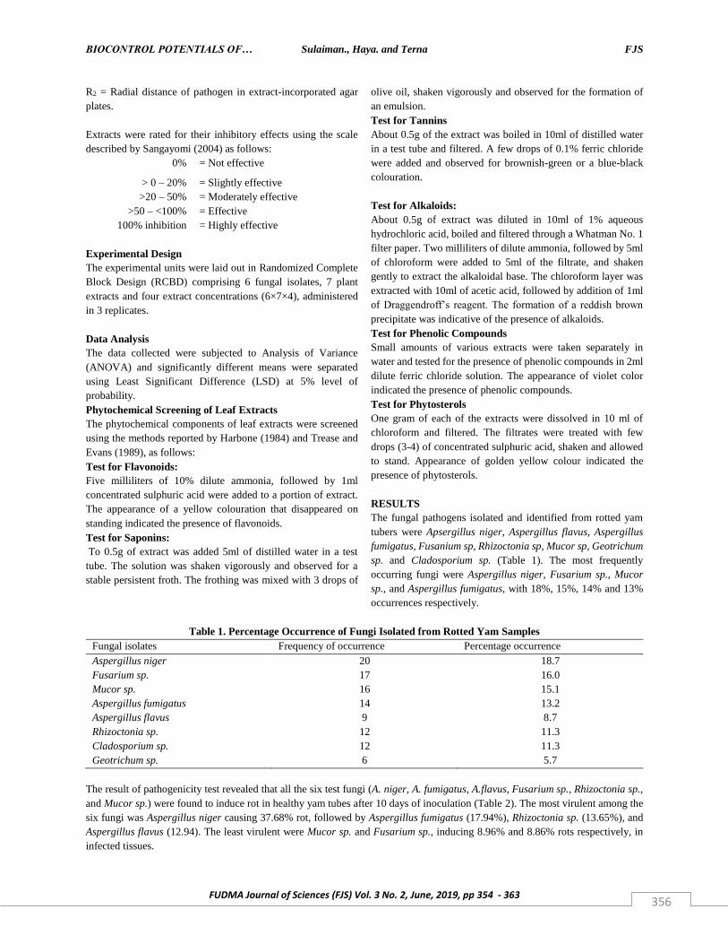
RESULTS
The fungal pathogens isolated and identified from rotted yam
tubers were Apsergillus niger, Aspergillus flavus, Aspergillus
fumigatus, Fusanium sp, Rhizoctonia sp, Mucor sp, Geotrichum
sp. and Cladosporium sp. (Table 1). The most frequently
occurring fungi were Aspergillus niger, Fusarium sp., Mucor
sp., and Aspergillus fumigatus, with 18%, 15%, 14% and 13%
occurrences respectively.
Table 1. Percentage Occurrence of Fungi Isolated from Rotted Yam Samples
Fungal isolates Frequency of occurrence Percentage occurrence
Aspergillus niger 20 18.7
Fusarium sp. 17 16.0
Mucor sp. 16 15.1
Aspergillus fumigatus 14 13.2
Aspergillus flavus 9 8.7
Rhizoctonia sp. 12 11.3
Cladosporium sp. 12 11.3
Geotrichum sp. 6 5.7
The result of pathogenicity test revealed that all the six test fungi (A. niger, A. fumigatus, A.flavus, Fusarium sp., Rhizoctonia sp.,
and Mucor sp.) were found to induce rot in healthy yam tubes after 10 days of inoculation (Table 2). The most virulent among the
six fungi was Aspergillus niger causing 37.68% rot, followed by Aspergillus fumigatus (17.94%), Rhizoctonia sp. (13.65%), and
Aspergillus flavus (12.94). The least virulent were Mucor sp. and Fusarium sp., inducing 8.96% and 8.86% rots respectively, in
infected tissues.
BIOCONTROL POTENTIALS OF… Sulaiman., Haya. and Terna FJS
FUDMA Journal of Sciences (FJS) Vol. 3 No. 2, June, 2019, pp 354 - 363 357
Table 2: Percentage Rot Caused by Test Fungi on
Healthy Yam Tubers
Fungi Percentage rot
Rhizoctonia sp. 13.65
Aspergillus fumigatus 17.91
Aspergillus niger 37.68
Fusarium sp. 8.86
Mucor sp. 8.96
Aspergillus flavus 12.94
Radial growth of A. flavus was inhibited by all extracts of the evaluated plants (Table 3). Inhibition of fungal growth was
concentration dependent, yielding the highest inhibitory activities at 10.0g/ml and the least at 2.50g/ml in all evaluated leaf extracts.
Ethanolic leaf extracts of Guiera senegalensis gave the highest inhibition of the radial growth of A. flavus (62.72%). Differences
in inhibitory activity of the evaluated plant extracts were significant (P≤0.05).
Table 3: Percentage Mean Radial Growth Inhibition of
Aspergillus flavus by Different Plant Extracts 7 Days after Inoculation
Concentration (g/ml)
Plant Extract 2.50 5.00 7.50 10.00
Ficus sycomorus
51.58 55.06 57.14 61.13
Guiera senegalensis
47.88 55.54 59.53 62.72
Jatrapha curcas
48.36 52.63 55.30 57.94
Khaya senegalensis
51.07 54.26 55.86 56.34
Azadirachta indica
47.32 52.11 55.30 59.53
Sclerocarya birrea
46.28 53.71 56.10 60.09
Tamarindus indica
46.76 52.11 55.86 58.49
LSD 12.09 1.59 6.68 2.37
The inhibitory effect of ethanolic leaf extracts of the evaluated plants differed against A. niger (Table 4), and increased at higher
extract concentrations. Ethanolic leaf extracts of Ficus sycomorus, Guiera senegalensis and Sclerocarya birrea at 10.0g/ml yielded
the highest radial growth inhibition of A. niger. Differences in radial growth inhibition of A. niger by ethanolic leaf extracts of the
evaluated plants were significant (P≤0.05).
Table 4: Percentage Mean Radial Growth Inhibition of
Aspergillus niger by Different Plant Extracts 7 Days after Inoculation
Concentration (g/ml)
Plant Extract 2.50 5.00 7.50 10.00
Ficus sycomorus
83.90 87.73 88.23 89.33
Guiera senegalensis
85.89 87.31 87.98 89.33
Jatrapha curcas
82.91 85.39 86.49 88.85
Khaya senegalensis 80.59 86.14 87.56 89.02
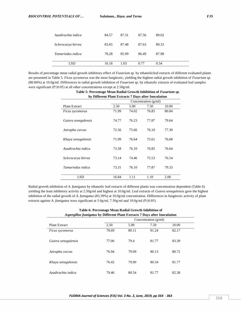
BIOCONTROL POTENTIALS OF… Sulaiman., Haya. and Terna FJS
FUDMA Journal of Sciences (FJS) Vol. 3 No. 2, June, 2019, pp 354 - 363 358
Azadirachta indica
84.57 87.31 87.56 89.02
Sclerocarya birrea
83.65 87.48 87.63 89.33
Tamarindus indica
78.28 85.99 86.49 87.98
LSD 16.18 1.03 0.77 0.54
Results of percentage mean radial growth inhibitory effect of Fusarium sp. by ethanolicleaf extracts of different evaluated plants
are presented in Table 5. Ficus sycomorus was the most fungitoxic, yielding the highest radial growth inhibition of Fusarium sp.
(80.84%) at 10.0g/ml. Differences in radial growth inhibition of Fusarium sp. by ethanolic extracts of evaluated leaf samples
were significant (P≤0.05) at all other concentrations except at 2.50g/ml.
Table 5: Percentage Mean Radial Growth Inhibition of Fusarium sp.
by Different Plant Extracts 7 Days after Inoculation
Concentration (g/ml)
Plant Extract 2.50 5.00 7.50 10.00
Ficus sycomorus
71.99 74.02 76.85 80.84
Guiera senegalensis
74.77 76.23 77.87 79.64
Jatrapha curcas
72.56 75.66 76.10 77.30
Khaya senegalensis
71.99 76.64 75.61 76.68
Azadirachta indica
73.58 76.10 76.85 76.64
Sclerocarya birrea
73.14 74.46 75.53 76.54
Tamarindus indica
73.31 76.10 77.87 79.33
LSD 16.84 1.11 1.10 2.00
Radial growth inhibition of A. fumigatus by ethanolic leaf extracts of different plants was concentration dependent (Table 6)
yielding the least inhibitory activity at 2.50g/ml and highest at 10.0g/ml. Leaf extracts of Guiera senegalensis gave the highest
inhibition of the radial growth of A. fumigatus (83.39%) at 10.0g/ml concentration. Differences in fungitoxic activity of plant
extracts against A. fumigatus were significant at 5.0g/ml, 7.50g/ml and 10.0g/ml (P≤0.05).
Table 6: Percentage Mean Radial Growth Inhibition of
Aspergillus fumigatus by Different Plant Extracts 7 Days after Inoculation
Concentration (g/ml)
Plant Extract 2.50 5.00 7.50 10.00
Ficus sycomorus
78.69 80.11 81.24 82.17
Guiera senegalensis
77.06 79.4 81.77 83.39
Jatrapha curcas
76.94 79.09 80.13 80.72
Khaya senegalensis
76.42 79.00 80.54 81.77
Azadirachta indica
79.46 80.54 81.77 82.38
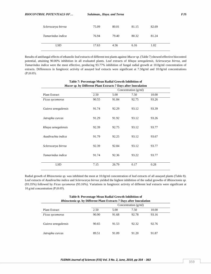
BIOCONTROL POTENTIALS OF… Sulaiman., Haya. and Terna FJS
FUDMA Journal of Sciences (FJS) Vol. 3 No. 2, June, 2019, pp 354 - 363 359
Sclerocarya birrea
75.09 80.01 81.15 82.69
Tamarindus indica
76.94 79.40 80.32 81.24
LSD 17.63 4.56 6.16 1.02
Results of antifungal effects of ethanolic leaf extracts of different test plants against Mucor sp. (Table 7) showed effective biocontrol
potential, attaining 90.00% inhibition in all evaluated plants. Leaf extracts of Khaya senegalensis, Sclerocarya birrea, and
Tamarindus indica were the most effective, producing 93.77% inhibition of fungal radial growth at 10.0g/ml concentration of
extracts. Differences in fungitoxic activity of assayed leaf extracts were significant at 7.50g/ml and 10.0g/ml concentrations
(P≤0.05).
Table 7: Percentage Mean Radial Growth Inhibition of
Mucor sp. by Different Plant Extracts 7 Days after Inoculation
Concentration (g/ml)
Plant Extract 2.50 5.00 7.50 10.00
Ficus sycomorus
90.55 91.84 92.75 93.26
Guiera senegalensis
91.74 92.29 93.12 93.39
Jatrapha curcas
91.29 91.92 93.12 93.26
Khaya senegalensis
92.39 92.75 93.12 93.77
Azadirachta indica
91.79 92.25 93.12 93.67
Sclerocarya birrea
92.39 92.84 93.12 93.77
Tamarindus indica
91.74 92.36 93.22 93.77
LSD 7.15 26.79 0.17 0.28
Radial growth of Rhizoctonia sp. was inhibited the most at 10.0g/ml concentration of leaf extracts of all assayed plants (Table 8).
Leaf extracts of Azadirachta indica and Sclerocarya birrea yielded the highest inhibition of the radial growths of Rhizoctonia sp.
(93.35%) followed by Ficus sycomorus (93.16%). Variations in fungitoxic activity of different leaf extracts were significant at
10.g/ml concentration (P≤0.05).
Table 8: Percentage Mean Radial Growth Inhibition of
Rhizoctonia sp. by Different Plant Extracts 7 Days after Inoculation
Concentration (g/ml)
Plant Extract 2.50 5.00 7.50 10.00
Ficus sycomorus
90.90 91.68 92.78 93.16
Guiera senegalensis
90.65 91.53 92.32 92.76
Jatrapha curcas
89.51 91.09 91.20 91.87
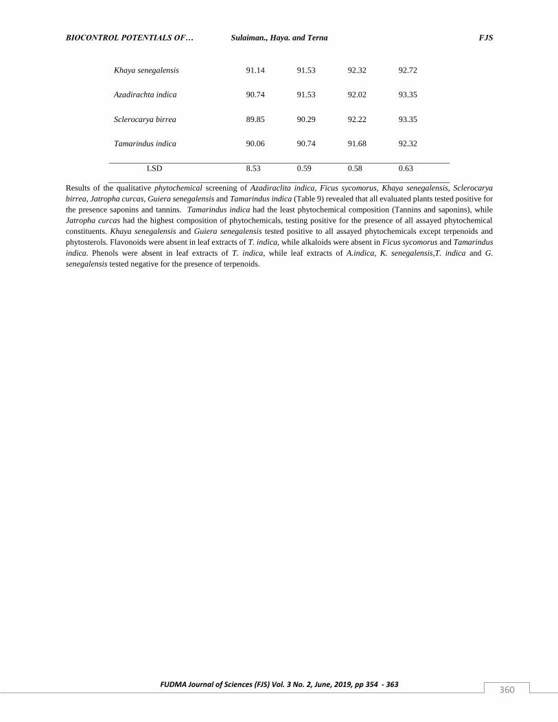
BIOCONTROL POTENTIALS OF… Sulaiman., Haya. and Terna FJS
FUDMA Journal of Sciences (FJS) Vol. 3 No. 2, June, 2019, pp 354 - 363 360
Khaya senegalensis
91.14 91.53 92.32 92.72
Azadirachta indica
90.74 91.53 92.02 93.35
Sclerocarya birrea
89.85 90.29 92.22 93.35
Tamarindus indica
90.06 90.74 91.68 92.32
LSD 8.53 0.59 0.58 0.63
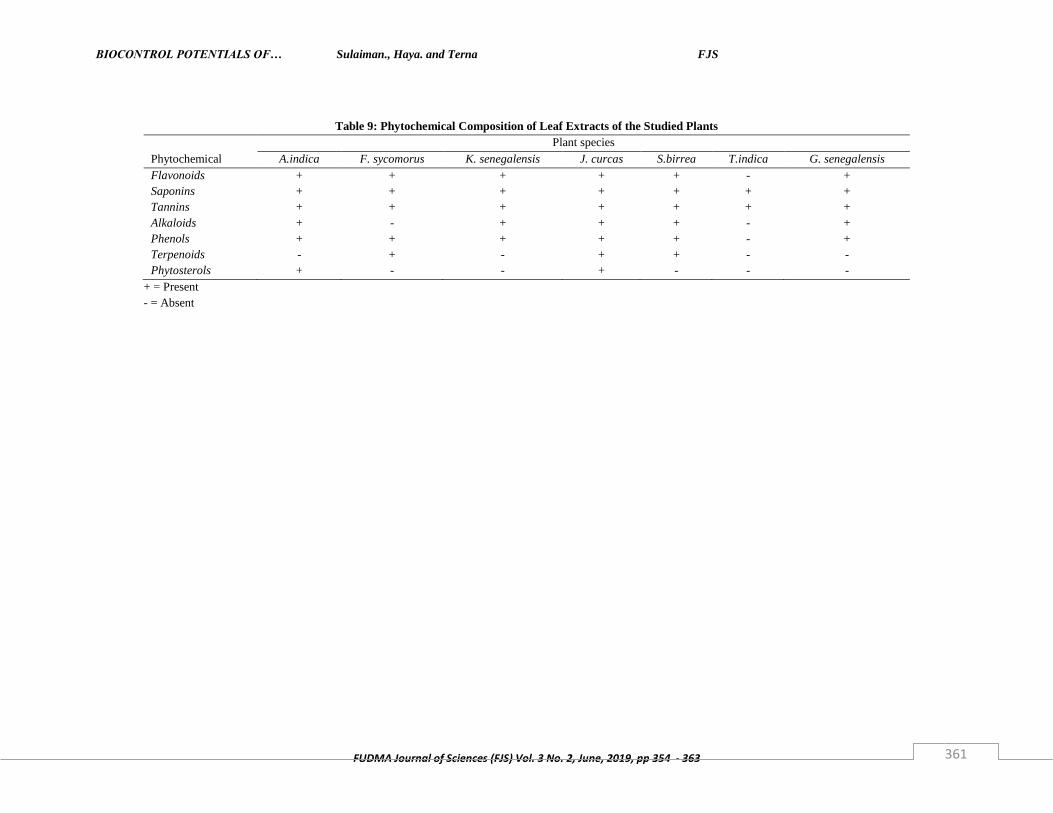
Results of the qualitative phytochemical screening of Azadiraclita indica, Ficus sycomorus, Khaya senegalensis, Sclerocarya
birrea, Jatropha curcas, Guiera senegalensis and Tamarindus indica (Table 9) revealed that all evaluated plants tested positive for
the presence saponins and tannins. Tamarindus indica had the least phytochemical composition (Tannins and saponins), while
Jatropha curcas had the highest composition of phytochemicals, testing positive for the presence of all assayed phytochemical
constituents. Khaya senegalensis and Guiera senegalensis tested positive to all assayed phytochemicals except terpenoids and
phytosterols. Flavonoids were absent in leaf extracts of T. indica, while alkaloids were absent in Ficus sycomorus and Tamarindus
indica. Phenols were absent in leaf extracts of T. indica, while leaf extracts of A.indica, K. senegalensis,T. indica and G.
senegalensis tested negative for the presence of terpenoids.
BIOCONTROL POTENTIALS OF… Sulaiman., Haya. and Terna FJS
FUDMA Journal of Sciences (FJS) Vol. 3 No. 2, June, 2019, pp 354 - 363 361
Table 9: Phytochemical Composition of Leaf Extracts of the Studied Plants
Plant species
Phytochemical A.indica F. sycomorus K. senegalensis J. curcas S.birrea T.indica G. senegalensis
Flavonoids + + + + + - +
Saponins + + + + + + +
Tannins + + + + + + +
Alkaloids + - + + + - +
Phenols + + + + + - +
Terpenoids - + - + + - -
Phytosterols + - - + - - -
+ = Present
- = Absent
BIOCONTROL POTENTIALS OF… Sulaiman., Haya. and Terna FJS
FUDMA Journal of Sciences (FJS) Vol. 3 No. 2, June, 2019, pp 354 - 363 362
DISCUSSION
The fungal pathogens isolated from the rotted yam tubers in this
study were similar to those reported by Ogaraku and Usman
(2008) from rotted yam tubers in Keffi, Nasarrawa State.
Several workers have also reported the isolation of these fungi
from post-harvest rotted yam tubers (Amusa and Baiyewu,
1999; Eze and Madu, 1990). The isolation of more than one
pathogenic organism from a particular yam tuber confirms the
possibility of multiple infections whose effect may cause rapid
rottening of root and tuber crops, as reported by Sangayomi
(2004). In several instances, fungi gain entrance into yam tubers
through natural openings and wounds created during harvesting,
transportation, handling and marketing. However, Okigbo and
Odunikwe (2009) noted that root and tuber crops at time of
harvest may already be infested by pathogens derived from
disease foliage, roots or mother tubers.
The present study showed the presence of fungitoxic
compounds in A.indica, F. sycomonis, G. senegalensis, J.
curcus, K. senegalensis, S. birrea and T. indica, since leaf
extracts of these plants were able to inhibit the growth of the test
fungi. Phytochemical screening of the plant extracts revealed
the presence of alkaloids, flavonoids, saponins, tannins,
phenols, terpenoids, and phytosterols. The medicinal and
pharmacological potentials of these phytochemicals have been
affirmed by the reports of several workers (Okigbo et al., 2009;
Okwu, 2004). The presence of bioactive substances also confer
resistance to plants against bacteria, fungi and pests (Srinivasan
et al., 2001; Okigbo and Ajalie, 2005; Okwu and Joshia, 2006).
CONCLUSION
Aspergillus niger, Aspergillus flavus, Aspergillus fumigatus,
Fusarium sp., Rhizoctonia sp., and Mucor sp. accounted for
significant post-harvest spoilage of yam tubers in the study area.
The biocontrol potentials of ethanolic leaf extracts of A.indica,
F. sycomonis, G. senegalensis, J. curcus, K. senegalensis, S.
birrea and T. indica, in the effective growth inhibition of the
studied rot fungi is an affirmation of the possibility of
incorporating these plant materials in the protection of
mechanically injured yam tubers against rot fungi during
storage.
REFERENCES
Amusa, N.A. (1999). Concentric lead spot of yam (dioscorea
spp) in South Western Nigeria. Mycopathologia., 148:113-120.
Amusa, N.A. and Baiyewu, R.A. (1999). Storage and Market
Disease of Yam Tubers in South Western Nigeria. Ogun Journal
of Agriculture Research, 11:211-225.
Arene, O.B. (1987). Advanced Integrated Control of Economic
Diseases of Cassava in Nigeria in: Hahn, S.K., Cavenes, F.E.
(Eds). Integrated Pest Management for Tropical Root and Tuber
Crops. Pp. 167-175.
Coursey, D.G. (1967). Yam Storage. A Review of Storage
Practices and Information on Storage Losses. Journal of Stored
Product Research, 2:227-244.
Coursey, D.G. (1983). Potential Utilization of Major Root Crops
with special emphasis on Human, Animal and Industrial uses.
Proceedings of the 2nd Triennial Symposium of the ISTRC
Cameroun. Pp. 25-35.
Eze, C.C., and Madu, J.N.C. (1990). Relation of Traditional
Methods to The Magnitude of Storage Losses of Cocoyam
(Colocasia esculental schott). Nigeria Journal of Plant
protection, 13: 2684.
Ezeh, N.O. (1998). Economics of Production and Post-harvest
Technology in: Orkwor, G.C., Asiedu, R., and Ekanayenke, I.J.
(Eds) Food Yam; Advances in Research IITA and NRCRI,
Nigeria. Pp. 187-214.
FAO. (1998). Food and Agriculture Organization Production
year Book FAO Rome.
Harbone, J.B. (1984). Phytochemical Methods: A Guide to
Modern Technique of Plants Analysis. Pp. 126-129.
Harrington, H.D. (1957). How to Identify Plants. USA: Swallow
Press. 203Pp.
Himratul-Aznita, W.H., Mohd-Al-Faisal, N., and Fathilah, A.R.
(2011). Determination of the percentage inhibition of diameter
growth (PIDG) of Piper betle crude aqueious extract against
oral Candida species.
BIOCONTROL POTENTIALS OF… Sulaiman., Haya. and Terna FJS
FUDMA Journal of Sciences (FJS) Vol. 3 No. 2, June, 2019, pp 354 - 363 363
IITA. (1993). Yam, Dioscorea Spp. Archival Reports (1989-93)
part III, Root and Tuber Improvement Programme, Crop
Improvement Division. IITA Ibadan. Pp. 20-85.
Ogaraku, A.O., and Usman, H.O. (2008). Storage Root of some
Yams (Dioscorea spp) in Keffi and Environs, Nasarawa State.
PAT., 4 (2): 22-27.
Okaka, J.C., and Okechukwu, P.E. (1987). Yam Processing
Problems and Prospects in: Quality Evaluation of Sundried Yam
chips. Tropical Science, 30:265-275.
Okigbo, R.N., and Ajalie, A.N. (2005). Inhibition of some
Human Pathogens with Tropical Plant Extracts: Chromolena
odorata and Citrus aurantifolia and some Antibiotics. Journal
of Molecular Medicine and Advances in Science, 1(1): 34-40.
Okigbo, R.N. and Ogbonna, U.O. (2006). Antifungal Effects of
two Tropical Plant Leaf Extracts (Occimum gratissimum and
Afromonium meleguata on post-harvest yam (Discorea sp).
African Journal of Biotechnology, 5: 727 – 731.
Okigbo, R., and Igwe, D. (2007). The Antimicrobial Effect of
Piper Guineese “Uziza” And Phyllantus Amanus” Ebe Benizo
on Candida Albican and Streptococcus Faecalis. Acta
Microbiological Immunologica Hungarica, 54(4): 353-366.
Okigbo, R.N., Eme, U.E., Aseidu, R., and Ramesh, P. (2009).
Effect of Crude Extracts of Allium sativum linn, Cymbopogon
citrates C.D Stapf and Terminalia catappa in Rot Causing Fungi
of Dioscorea species.
Okigbo, R.N., and Odunikwe, C.N. (2009). Occurrence and
Control Of Fungal Rot Pathogens of Yam (Dioscorea Rutundata
Pair) With Leaf Extracts of Chormolena Odorata, Carica Papaya
and Aspillia Africana. Nigerian Journal of Mycology, 2(1): 154-
165.
Okwu, D.E. (2004). Phytochemical and Vitamin Content of
Indigenous Species of South Eastern Nigeria. Nigerian Journal
of sustainable Agricculture and Environment, 6(1): 30-37.
Okwu, D.E. and Joshia, C. (2006). Evaluation of the Chemical
Composition of Two Nigeria Medicinal Plants. African Journal
of Biotechnology, 5(4): 361.
Olurinola, P.E., Ehimmadu, J.O., and Bonire, S.S. (1992).
Antifungal Activity of N-Ributylin Acetate Aagians Some
Common Yam Rot Fungi. Applied and Environmental
Microbiology, 58(2): 758-760.
Onayemi, O. (1983). Observation on the development
characteristics of different varieties of yam and cocoyam.
Abstract of 6th symposium of the International Society For
Tropical Crops. Peru.
Ritchie, B. (1991). Practical Techniques in Plant Pathology.
Wallingford U.K.: CABI.
Mahmoud, S.N., and Al-Ani, N.K. (2016). Effect of Different
Sterilization Methods on Contamination and Viability of Nodal
Segments of Cestrum nocturnum L. International Journal of
Research Studies in Biosciences, 4(1): 4-9.
Sangayomi, T.E. (2004). Post-Harvest Fungal Deterioration of
Yam (Dioscorea rotundata) and its Control. PHD thesis IITA,
Ibadan Nigeria. 179pp.
Salhi, N., Saghir, S.A.M., Terzi, V., Brahmi, I., Ghedairi, N.,
and Bissati, S. (2017). Antifungal Activity of Aqueous Extracts
of Some Dominant Algerian Medicinal Plants. Biomed
Research International, 2017: 1-6.
Srinivasan, D., Perumalsamy, L.P., and Nathan, S.T. (2001).
Antimicrobial Activity of Certain Indian Medicinal Plants used
in Folkloric Medicine. Journal of ethnopharmacy, 94: 217-222.
Terna, T.P., Audi, Y.A. and Terna, F.C. (2019). Isolation and
Identification of Fungi Associated with Stored Sorghum
(Sorghum bicolor L. Moench) Seeds in Lafia, Nasarawa State,
Nigeria. FUDMA Journal of Sciences, 3(1): 33-38.
Trease, G.E., and Evans, W.C. (1989). Pharmacognosy (13th
Ed). Bailliere Tindall, Britain: English Language Book Society.
Pp. 378, 386-480.
Recommended


