Jour
nal o
f Cel
l Sci
ence
RESEARCH ARTICLE
Btn3 regulates the endosomal sorting function of the yeast Ent3epsin, an adaptor for SNARE proteins
Joelle Morvan, Johan-Owen de Craene, Bruno Rinaldi, Vanessa Addis, Cedric Misslin and Sylvie Friant*
ABSTRACT
Ent3 and Ent5 are yeast epsin N-terminal homology (ENTH)
domain-containing proteins involved in protein trafficking between
the Golgi and late endosomes. They interact with clathrin, clathrin
adaptors at the Golgi (AP-1 and GGA) and different SNAREs (Vti1,
Snc1, Pep12 and Syn8) required for vesicular transport at the Golgi
and endosomes. To better understand the role of these epsins in
membrane trafficking, we performed a protein–protein interaction
screen. We identified Btn3 (also known as Tda3), a putative
oxidoreductase, as a new partner of both Ent3 and Ent5. Btn3 is
a negative regulator of the Batten-disease-linked protein Btn2
involved in the retrieval of specific SNAREs (Vti1, Snc1, Tlg1 and
Tlg2) from the late endosome to the Golgi. We show that Btn3
endosomal localization depends on the epsins Ent3 and Ent5. We
demonstrated that in btn3D mutant cells, endosomal sorting of
ubiquitylated cargos and endosomal recycling of the Snc1 SNARE
are delayed. We thus propose that Btn3 regulates the sorting
function of two adaptors for SNARE proteins, the epsin Ent3 and the
Batten-disease-linked protein Btn2.
KEY WORDS: Epsin, Multivesicular body, Ubiquitin, ESCRT, Yeast
INTRODUCTIONEndosomes are key compartments where endocytic, biosynthetic
and exocytic pathways intersect. At the late endosome or
multivesicular body (MVB), proteins undergo crucial sorting
(Hurley, 2008). Membrane proteins destined to the vacuolar or
lysosomal lumen are sorted in MVB intraluminal vesicles and are
subsequently delivered to the vacuolar lumen upon fusion of the
MVB with the vacuole. Proteins remaining at the limiting
membrane of the MVB are either delivered to the vacuolar
membrane or recycled to the Golgi or the plasma membrane.
Several studies have shown that cargo modification by ubiquitin
serves as a signal that causes cargo to enter the vesicles of the
MVB (Hicke and Dunn, 2003; Lauwers et al., 2010).
In the yeast Saccharomyces cerevisiae, several screens have
led to the identification of vps (vacuolar protein sorting) mutants
grouped in six classes, A to F, based on their phenotypes
(Raymond et al., 1992). The class E vps mutants are defective in
MVB sorting and accumulate an aberrant endosomal structure
(the class E compartment) in which cargos and endosomal
proteins are trapped (Odorizzi et al., 1998). Some class E Vps
proteins are associated in complexes termed ESCRT (endosomal
sorting complex required for transport) (Henne et al., 2011).
In yeast, MVB sorting is regulated by two types of endosomal
lipids, phosphatidylinositol 3-phosphate (PtdIns3P) and
phosphatidylinositol 3,5-bisphosphate [PtdIns(3,5)P2]. PtdIns3P,
synthesized by the Vps34 lipid kinase, mediates the specific
recruitment of Vps27 through its FYVE domain (Katzmann et al.,
2003). PtdIns(3,5)P2, synthesized by the Fab1 lipid kinase from
PtdIns3P, controls the MVB sorting of ubiquitylated cargos like
the carboxypeptidase S (Cps1) or the endopolyphosphatase
(Phm5) (Odorizzi et al., 1998; Reggiori and Pelham, 2002).
Yeast epsins Ent3 and Ent5 are PtdIns(3,5)P2 effectors
participating in MVB sorting (Eugster et al., 2004; Friant et al.,
2003).
In yeast there are five epsins named Ent1 to Ent5 (Duncan
et al., 2003; Wendland et al., 1999). Epsins are cytoplasmic
proteins, recruited to membranes through their ENTH (epsin N-
terminal homology) domain (De Camilli et al., 2002; Kay et al.,
1999). Ent3 and Ent5 were first identified as interactors of the
Golgi-localized clathrin adaptors the AP-1 complex and GGA
(Golgi-localizing, gamma-adaptin ear homology domain, ARF-
binding) proteins (Duncan et al., 2003). However, they have
broad functions at the trans Golgi network (TGN) and endosome
level. Indeed, ent3D ent5D double-mutant cells display several
trafficking defects: (1) in Golgi-to-endosome trafficking of some
endosomal SNAREs [SNAP (soluble NSF attachment protein)
receptors] and vacuolar carboxypeptidases, (2) in MVB sorting of
ubiquitylated cargos and (3) in endosome-to-Golgi retrograde
transport of specific cargos like the SNARE Snc1 or the casein
kinase Yck2 (Chidambaram et al., 2004; Chidambaram et al.,
2008; Copic et al., 2007; Duncan et al., 2003; Eugster et al., 2004;
Zimmermann et al., 2010). However, Ent3 and Ent5 proteins also
display some specific functions. Ent3 acts principally in the
GGA-mediated TGN sorting (Copic et al., 2007; Costaguta et al.,
2006) and only the ENTH domain of Ent3 binds to the endosomal
SNAREs Vti1, Pep12 and Syn8 (Chidambaram et al., 2004;
Chidambaram et al., 2008; Copic et al., 2007). Ent5 (but not
Ent3) binds to the chitin synthase Chs3, but Ent3 and Ent5 are
both required for its trafficking from the Golgi (Copic et al.,
2007). All these results show that Ent3 and Ent5 are key players
in membrane trafficking at the TGN and endosomal levels; it is
therefore important to better understand their regulation. Indeed,
in vitro quantitative studies have shown using surface plasmon
resonance (Biacore) that the ENTH domain of both Ent3 and Ent5
binds to PtdIns(3,5)P2 as efficiently as to PtdIns(4,5)P2 (Narayan
and Lemmon, 2006), suggesting that some additional protein
interactions are required to mediate their PtdIns(3,5)P2-dependent
specific functions in MVB sorting (Eugster et al., 2004; Friant
et al., 2003). Here, we performed a yeast proteome microarray
screen to identify new Ent3 and Ent5 binding partners. Among
Department of Molecular and Cellular Genetics, UMR7156, Centre National deRecherche Scientifique (CNRS), Universite de Strasbourg, Strasbourg 67084,France.
*Author for correspondence ([email protected])
Received 17 July 2014; Accepted 5 December 2014
� 2015. Published by The Company of Biologists Ltd | Journal of Cell Science (2015) 128, 706–716 doi:10.1242/jcs.159699
706
Jour
nal o
f Cel
l Sci
ence
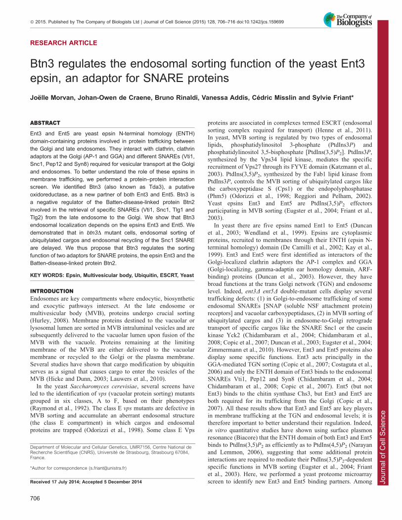
the different proteins identified, we chose to further characterizeBtn3. Btn3 was previously linked to endosomal trafficking and
identified as a regulator of the Batten disease protein Btn2, whichinteracts with different SNAREs (Kama et al., 2007; Kannegantiet al., 2011). We show that the epsins Ent3 and Ent5 are requiredfor the endosomal localization of Btn3. Moreover, in btn3Dmutant cells, endosomal sorting of ubiquitylated cargos andrecycling of the Snc1 SNARE are delayed. Our results suggestthat Btn3 is a cytoplasmic protein regulating two adaptors for
SNARE proteins, the epsin Ent3 and the Batten-disease-linkedprotein Btn2.
RESULTSIdentification of Btn3 as a new direct Ent3 and Ent5interactorTo identify new direct interactors of Ent3 and/or Ent5, we carriedout a protein–protein interaction screen using yeast proteinmicroarrays (Yeast ProtoArrayH Invitrogen) (Zhu et al., 2001).Ent3 and Ent5 were N- or C-terminally tagged with V5-6xHis to
avoid missing interactions owing to the tag position and reducethe number of false-positive hits. The fusion proteins wereproduced in Escherichia coli and purified using Ni-NTA–agarose
beads. Yeast ProtoArrayH chips were incubated in the presence ofpurified recombinant V5-6xHis-tagged Ent3 and Ent5 proteinsand the interactions were revealed using fluorescently tagged
anti-V5 antibody. A Z-score, representing the number of standarddeviations away from the mean fluorescence of the entire array,was calculated for each signal (ProtoArrayH Prospector;
Invitrogen). A significant hit was characterized by a Z-score of§3 and a coefficient of variance for signals from the tworeplicates (each protein is spotted in duplicate on the chip) of,0.5. This resulted in the identification of 53 direct hits for N-
and C-terminally tagged Ent3, and 31 for N- and C-terminallytagged Ent5 (supplementary material Table S1). Among thesignificant hits, 17 were common to Ent3 and Ent5
(supplementary material Table S1), including the gammasubunit of the AP-1 adaptor complex Apl4 (also known asYpr029c), previously identified by yeast two-hybrid (Duncan
et al., 2003), thus validating our approach (Fig. 1A). Btn3 wasalso found as a common hit and was of particular interest. Indeed,Btn3 (also known as Tda3 or Yhr009c) was shown to interact in
vitro with the ubiquitin ligase Rsp5 (Hesselberth et al., 2006)
required for MVB sorting (Katzmann et al., 2004; Morvan et al.,2004). Btn3 was recently identified as a binding partner of theBtn2 protein (Kanneganti et al., 2011). Btn2, a Batten-disease-
related protein, binds to endocytic SNARE complexes (Snc1,Snc2, Tlg1, Tlg2 and Vti1) and regulates late endosomal sortingof some specific cargos (Kama et al., 2007). Btn3 negatively
regulates Btn2 sorting function by sequestering Btn2 away fromits cargos (Kanneganti et al., 2011). This was of particular interestfor our study, as Ent3 is also involved in SNARE (Vti1, Pep12 or
Snc1) binding and trafficking. Surprisingly, the BTN3 gene wasidentified in a screen for deletion mutants sensitive to theoverexpression of the DNA topoisomerase I top1-T722A mutant,and was thus named TDA3 (topoisomerase I damage affected)
(Reid et al., 2011). Interestingly, the largest class of mutantsidentified in this screen affects the endosomal trafficking,including ESCRT subunits or the Fab1 lipid kinase. In
conclusion, all these results point towards a functional linkbetween Btn3 and Ent3.
To confirm the interactions between Btn3 and Ent3 and
Ent5, we performed GST (gluthatione-S-transferase) pull-down
experiments using GST, GST–Ent3 or GST–Ent5 recombinantproteins and a protein extract from cells expressing
chromosomally GFP-tagged Btn3 (Fig. 1B). Btn3–GFP was notretained on GST beads, but specifically interacted with GST–Ent3 and GST–Ent5, with a higher affinity for Ent5. Similarresults were obtained for the protein microarray interactions
showing a better Z-score for the Btn3–Ent5 interaction (Fig. 1A).To determine whether Ent3 could be associated with Btn3 in
yeast protein extracts, we carried out a co-immunoprecipitation
(co-IP) assay. Ent3–HA was expressed in a wild-type yeast strainexpressing or not expressing Btn3–GFP at the locus. Ent3–HAproteins were immunoprecipitated with anti-HA antibodies on c-
bind Sepharose beads and pull down of Btn3–GFP was assessedby immunoblotting (Fig. 1C). Under these conditions, Btn3–GFPwas co-immunoprecipitated by Ent3–HA (Fig. 1C), showing that
Btn3 interacts with Ent3 in wild-type cells. As a control, we alsotested the non-specific binding of Btn3–GFP to the beads in theabsence of Ent3–HA [Fig. 1C, lane ‘WT Btn3-GFP+pHAC’(pHAC, empty vector)], the results show that Btn3–GFP was not
retained on the beads. The microarray interaction screen suggeststhat Btn3 binds to Ent5 with a higher affinity than that its affinityfor Ent3 (Fig. 1A). To determine whether Ent5 competes with
Ent3 for Btn3 binding, we performed co-IP from an ent5D cellextract. We observed similar binding efficiency of Btn3–GFP toEnt3–HA, showing that Ent5 is neither required nor competing
with Ent3 for Btn3 binding (Fig. 1C). We next determinedwhether Btn2 competes with Ent3 for Btn3 binding by analyzingtheir interaction in btn2D cells, because Btn3 has been shown to
interact with Btn2 (Kanneganti et al., 2011). Interestingly, in abtn2D cell extract, about two times more Btn3–GFP co-immunoprecipitated with Ent3–HA (Fig. 1C); indeed, the meanpercentage of Btn3–GFP input protein co-immunoprecipitated by
Ent3–HA (two or three different experiments) was 1.18% for thewild-type strain, 2.12% for btn2D strain and 1.06% for thevps27D strain. This suggests that Btn2 competes with Ent3 for its
interaction with Btn3. We also tested whether the absence ofVps27 interacting with Ent3 and Ent5 (Eugster et al., 2004)affected the interaction between Btn3–GFP and Ent3–HA. In
vps27D mutant cells, Btn3–GFP also co-immunoprecipitated withEnt3–HA (Fig. 1C; 1.06% of coIP), showing that Vps27 is notrequired for this interaction. Taken together, these results showthat Btn3 is a direct interactor of the Ent3 epsin and that this
interaction does not require Vps27 and competes with the Btn2interaction.
Although Btn2 was not found as a hit in our microarray screen
for Ent3 interactors, it could be associated with the Btn3–Ent3protein complex. We thus analyzed the co-IP between Ent3–HAand Btn2–GFP using Pgk1 as a negative control (Fig. 1D).
Compared to the co-IP performed on protein extracts from cellslacking Ent3–HA (Fig. 1D, lane ‘WT+pHAC’) that was used as anegative control, Btn2–GFP was not retained on the Ent3–HA
beads, regardless of the presence of Btn3 in the extract (fromwild-type or btn3D cells), showing that Btn2 was not associatedwith the Ent3 protein.
We then asked whether the Ent3 and Ent5 epsins are required
for the Btn2–Btn3 interaction described previously (Kannegantiet al., 2011). We performed a co-IP between Btn3–HA and Btn2–GFP in extracts from wild-type or ent3D ent5D mutant cells
(Fig. 1E). In both cell lysates, Btn2–GFP similarly interacts withBtn3–HA. Given that Btn2 facilitates the recruitment of Btn3 toendosomal structures (Kanneganti et al., 2011), we also analyzed
the co-IP between Btn3–HA and Btn2–GFP in the vps27D
RESEARCH ARTICLE Journal of Cell Science (2015) 128, 706–716 doi:10.1242/jcs.159699
707
Jour
nal o
f Cel
l Sci
ence
mutant. We observed that more Btn2–GFP was retained on theBtn3–HA beads, as expected if the interaction occurred at theendosomal membrane (Fig. 1E). This shows that the Btn3
interaction with Btn2 is not mediated by the Ent3 and Ent5epsins.
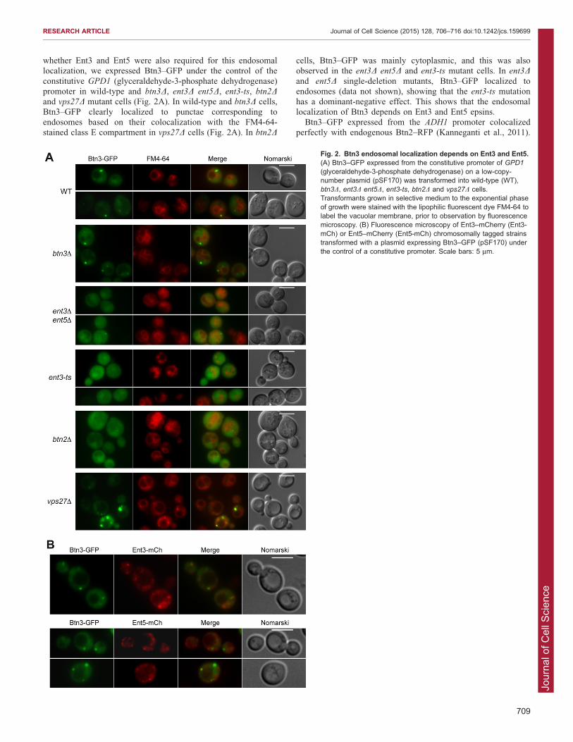
Btn3 endosomal localization depends on the Ent3 andEnt5 epsinsIt was shown that Btn3 expressed from the ADH1 (alcohol
dehydrogenase) promoter localizes to endosomes in a Btn2-dependent manner (Kanneganti et al., 2011). To determine
Fig. 1. Btn3 interacts with Ent5 and Ent3 in vitro and in vivo. (A) Interactions between V5-6xHis-tagged Ent3 or Ent5 and the proteins spotted on the arrayswere determined by a Z-score calculated by using the ProtoArrayH Prospector (Invitrogen) software. A Z-Score .3 identifies the statistically significant interactors.(B) Yeast lysate from BY4741 expressing Btn3–GFP was incubated with GST alone, GST–Ent3 or GST–Ent5 on gluthatione-Sepharose beads. Beads werepelleted and the supernatant removed. After washing, proteins attached to beads were eluted with SDS buffer and analyzed by SDS-PAGE. Total yeast extractrepresents 5% of the protein amount used for the GST-pull down. Btn3–GFP was revealed with anti-GFP antibodies. Ponceau staining of the nitrocellulosemembrane shows the amount of GST fusion protein present on the beads. MW indicates the molecular mass marker lane. (C) Yeast cytosol from wild-type (WT)Btn3–GFP (BY4741 Btn3–GFP) cells transformed with either the Ent3–HA expression vector (pFL575) or the empty vector (pHAC, negative control) wasimmunoprecipitated with anti-HA antibodies and subjected to western blotting with anti-GFP (to detect Btn3–GFP) and anti-Pgk1 phosphoglycerate kinase (loadingcontrol). The same co-IP experiment was performed on wild-type (BY4741), wild-type Btn3-GFP (BY4741 Btn3-GFP), btn2D Btn3-GFP (SFY127), ent3D ent5D
Btn3-GFP (SFY23) or vps27D Btn3-GFP (SFY25) strains expressing HA-tagged Ent3 (pFL575). Input represents 2.5% of the cytosol used for immunoprecipitation.The asterisk shows a non-specific band detected with the anti-GFP antibodies. (D) Coimmunoprecipitation experiments between Btn2–GFP (pAD6-Btn2-GFP)and Ent3–HA (pFL575) expressed from a wild-type or btn3D strain. The wild-type strain bearing Btn2–GFP (pAD6-Btn2-GFP) and the empty vector (pHAC) wasused to control for the non-specific binding of Btn2–GFP to the beads. Pgk1 was used as a negative control. (E) The interaction between Btn2–GFP (pAD6-Btn2-GFP) and Btn3–HA (pSF191) was monitored by co-IP with anti-HA antibodies in different yeast strains, wild type, ent3D ent5D (SFY2) and vps27D.
RESEARCH ARTICLE Journal of Cell Science (2015) 128, 706–716 doi:10.1242/jcs.159699
708
Jour
nal o
f Cel
l Sci
ence
whether Ent3 and Ent5 were also required for this endosomallocalization, we expressed Btn3–GFP under the control of the
constitutive GPD1 (glyceraldehyde-3-phosphate dehydrogenase)promoter in wild-type and btn3D, ent3D ent5D, ent3-ts, btn2Dand vps27D mutant cells (Fig. 2A). In wild-type and btn3D cells,Btn3–GFP clearly localized to punctae corresponding to
endosomes based on their colocalization with the FM4-64-stained class E compartment in vps27D cells (Fig. 2A). In btn2D
cells, Btn3–GFP was mainly cytoplasmic, and this was alsoobserved in the ent3D ent5D and ent3-ts mutant cells. In ent3Dand ent5D single-deletion mutants, Btn3–GFP localized toendosomes (data not shown), showing that the ent3-ts mutationhas a dominant-negative effect. This shows that the endosomallocalization of Btn3 depends on Ent3 and Ent5 epsins.
Btn3–GFP expressed from the ADH1 promoter colocalizedperfectly with endogenous Btn2–RFP (Kanneganti et al., 2011).
Fig. 2. Btn3 endosomal localization depends on Ent3 and Ent5.(A) Btn3–GFP expressed from the constitutive promoter of GPD1
(glyceraldehyde-3-phosphate dehydrogenase) on a low-copy-number plasmid (pSF170) was transformed into wild-type (WT),btn3D, ent3D ent5D, ent3-ts, btn2D and vps27D cells.Transformants grown in selective medium to the exponential phaseof growth were stained with the lipophilic fluorescent dye FM4-64 tolabel the vacuolar membrane, prior to observation by fluorescencemicroscopy. (B) Fluorescence microscopy of Ent3–mCherry (Ent3-mCh) or Ent5–mCherry (Ent5-mCh) chromosomally tagged strainstransformed with a plasmid expressing Btn3–GFP (pSF170) underthe control of a constitutive promoter. Scale bars: 5 mm.
RESEARCH ARTICLE Journal of Cell Science (2015) 128, 706–716 doi:10.1242/jcs.159699
709
Jour
nal o
f Cel
l Sci
ence
To determine whether the epsins Ent3 and Ent5 behave similarlyto Btn2, we analyzed the localization of Btn3–GFP expressed
from the constitutive GPD1 promoter in a yeast strain bearingendogenous mCherry (mCh)-tagged Ent3 or Ent5 proteins(Fig. 2B). Ent3–mCh and Ent5–mCh did not colocalize withBtn3–GFP, even though some Ent3–mCh and Ent5–mCh are
localized next to Btn3–GFP positive structures (Fig. 2B). Thisshows that Ent3 does not behave like Btn2, despite the twoproteins being able to bind to Btn3 and SNAREs (Vti1 and Snc1).
The endosomal localization of Btn2 does not require Btn3(Kanneganti et al., 2011), but could be dependent on the Ent3 andEnt5 epsins. We analyzed the intracellular localization of Btn2–
GFP expressed under the control of the constitutive ADH1
promoter in wild-type and btn2D, btn3D, ent3-ts, ent3D ent5Dand vps27D mutant cells (supplementary material Fig. S1A). In
all these cells, Btn2–GFP was localized to the endosomes.However, in ent3-ts and ent3D ent5D mutants, the Btn2–GFP-labeled punctate structures were smaller (supplementary materialFig. S1A). This was previously observed for other mutants, such
as the snc1D snc2D cells that are defective in endocytosis andsecretion and also have fragmented vacuoles (Kama et al., 2007).To determine whether Btn2–GFP colocalizes with Ent3 or Ent5,
we expressed Btn2–GFP in yeast strains bearing locus mCh-tagged Ent3 and Ent5 (supplementary material Fig. S1B). Btn2–GFP did not colocalize with Ent3–mCh nor with Ent5–mCh,
showing that constitutively expressed Btn2 does not recruit Ent3or Ent5 to endosomes and supporting our findings that theseproteins belong to different complexes (Fig. 1). Moreover, in
btn3D cells, the Ent3–mCh and Ent5–mCh localization tointracellular punctae (Golgi and endosomes) was similar to thatobserved in wild-type cells (supplementary material Fig. S1B).Thus neither Btn2, nor Ent3 and Ent5 intracellular localizations
depend on their interaction with Btn3 (supplementary materialFig. S1B; Kanneganti et al., 2011).
The endosomal sorting of Ent3-dependent cargos requiresBtn3Ent3 and Ent5 play multiple roles in membrane trafficking, one at
the Golgi level in coordination with AP-1 and GGA adaptors andone at the endosomes for either retrograde Snc1 trafficking,SNARE (Vti1, Pep12 and Syn8) sorting or MVB sorting of Cps1(Chidambaram et al., 2008; Copic et al., 2007; Duncan et al., 2003;
Eugster et al., 2004; Zimmermann et al., 2010). We tested whetherBtn3 was involved in the same trafficking steps as Ent3 and Ent5.We analyzed the vacuolar trafficking of the dipeptidyl
aminopeptidase B (DPAP-B or Dap2) and, as a control, thevacuolar SNARE Vam3, which is transported from the Golgi to thevacuolar membrane through the AP-3 adaptor complex (along the
endosome-independent alkaline phosphatase pathway) (Darsowet al., 1998). In btn3D mutant cells, GFP–DPAP-B and GFP–Vam3are localized at the vacuolar membrane as in wild-type cells, showing
that there is no defect in their trafficking (supplementary materialFig. S2A). We tested the VPS pathway by analyzing the secretion ofcarboxypeptidase Y (CPY) into the extracellular medium, and couldnot observe any significant CPY accumulation in the btn3D mutant
cells, contrary to what was observed for ent3Dent5D, fab1D orvps27D mutants (supplementary material Fig. S2B). Thus, Btn3 isnot required for GGA-dependent CPY trafficking.
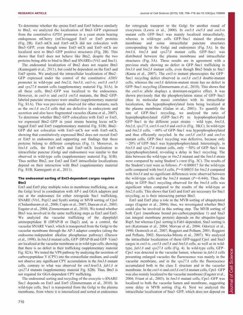
The endosomal sorting and recycling of the exocytic v-SNARESnc1 depends on Ent3 and Ent5 (Zimmermann et al., 2010). Inwild-type cells, Snc1 is transported from the Golgi to the plasma
membrane, from where it is internalized and sorted at endosomes
for retrograde transport to the Golgi for another round ofexocytosis (Lewis et al., 2000). In ent3D ent5D and ent3-ts
mutant cells GFP–Snc1 was mainly localized intracellularly,whereas in wild-type cells GFP–Snc1 stained the plasmamembrane and some intracellular punctate structurescorresponding to the Golgi and endosomes (Fig. 3A). In the
btn3D, btn2D and vps27D mutant cells, GFP–Snc1 wasdistributed between the plasma membrane and intracellularstructures (Fig. 3A). These results are in agreement with a
previous study showing no defect in GFP–Snc1 trafficking inbtn3D and btn2D mutant cells, even though Btn2 binds to Snc1(Kama et al., 2007). The ent3-ts mutant phenocopies the GFP–
Snc1 recycling defect observed in ent3D ent5D double-mutantcells, whereas the ent3D deletion-mutant strain has no defect inGFP–Snc1 recycling (Zimmermann et al., 2010). This shows that
the ent3-ts allele displays a dominant-negative effect. It wasshown previously that the phosphorylation status of GFP–Snc1(thus its molecular mass) correlates with its intracellularlocalization, the hyperphosphorylated form being localized at
the plasma membrane (Galan et al., 2001). To quantify therate of GFP–Snc1 recycling, we analyzed the ratio ofhyperphosphorylated (GFP–Snc1-P) to hypophosphorylated
GFP–Snc1 in the different yeast strains – wild type, btn3D,btn2D, vps27D, ent3D ent5D and ent3-ts (Fig. 3B,C). In wild-typeand btn2D cells, ,60% of GFP–Snc1 was hyperphosphorylated
and thus efficiently recycled. In the ent3D ent5D and ent3-ts
mutant cells, GFP–Snc1 recycling was defective, because only,20% of GFP–Snc1 was hyperphosphorylated. Interestingly, in
btn3D and vps27D mutant cells, only ,50% of GFP–Snc1 washyperphosphorylated, revealing a delay in Snc1 recycling. Thedata between the wild-type or btn2D mutant and the btn3D strainwere compared by using Student’s t-test (Fig. 3C). The results of
the Student’s test were as follows: P50.000017 for the wild-typecells compared with btn3D cells, P50.00033 for btn2D comparedwith btn3D and no significant differences were observed between
the wild-type cells and the btn2D mutant (P50.444). Thus, thedelay in GFP–Snc1 recycling observed for the btn3D cells wassignificant when compared to the results of the wild-type or
btn2D cells. This shows that Ent3 and Ent5 are necessary for Snc1recycling, as is their interacting partner Btn3.
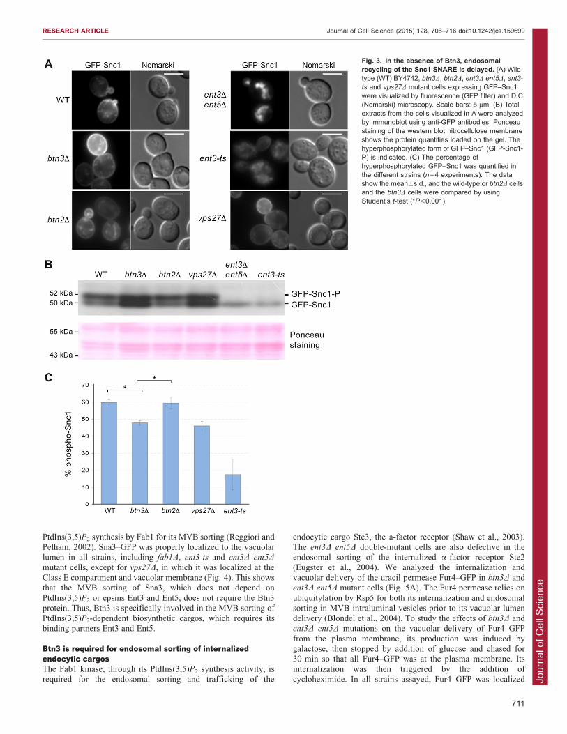
Ent3 and Ent5 play a role in the MVB sorting of ubiquitylatedcargo (Eugster et al., 2004); thus, we investigated whether Btn3
could also be involved in this sorting step. The MVB sorting ofboth Cps1 (membrane bound pre-carboxypeptidase 1) and Sna3(an integral membrane protein) depends on the ubiquitin-ligase
Rsp5, but whereas Cps1 sorting is Fab1-dependent, Sna3 sorting isnot (Katzmann et al., 2004; Morvan et al., 2004; Odorizzi et al.,1998; Oestreich et al., 2007; Reggiori and Pelham, 2001; Reggiori
and Pelham, 2002; Stawiecka-Mirota et al., 2007). We analyzedthe intracellular localization of these GFP-tagged Cps1 and Sna3cargos in ent3-ts, ent3D ent5D and btn3D cells, as well as in wild-
type, fab1D and vps27D cells (Fig. 4). In wild-type cells, GFP–Cps1 was detected in the vacuolar lumen, whereas in fab1D cellspresenting enlarged vacuoles the fluorescence was mainly in thevacuolar membrane, and in the vps27D cells the fluorescence
accumulated both in the class E structure and in the vacuolarmembrane. In the ent3-ts and ent3D ent5D mutant cells, Cps1–GFPwas also mainly localized to the vacuolar membrane (Eugster et al.,
2004; Friant et al., 2003). In btn3D mutant cells, Cps1–GFP waslocalized to both the vacuolar lumen and membrane, suggestingsome delay in MVB sorting (Fig. 4). Next we analyzed the
intracellular localization of Sna3–GFP, which does not depend on
RESEARCH ARTICLE Journal of Cell Science (2015) 128, 706–716 doi:10.1242/jcs.159699
710
Jour
nal o
f Cel
l Sci
ence
PtdIns(3,5)P2 synthesis by Fab1 for its MVB sorting (Reggiori andPelham, 2002). Sna3–GFP was properly localized to the vacuolarlumen in all strains, including fab1D, ent3-ts and ent3D ent5Dmutant cells, except for vps27D, in which it was localized at theClass E compartment and vacuolar membrane (Fig. 4). This showsthat the MVB sorting of Sna3, which does not depend on
PtdIns(3,5)P2 or epsins Ent3 and Ent5, does not require the Btn3protein. Thus, Btn3 is specifically involved in the MVB sorting ofPtdIns(3,5)P2-dependent biosynthetic cargos, which requires its
binding partners Ent3 and Ent5.
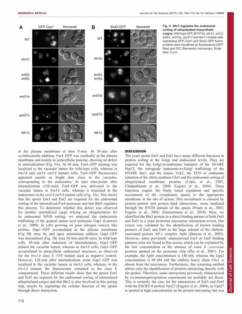
Btn3 is required for endosomal sorting of internalizedendocytic cargosThe Fab1 kinase, through its PtdIns(3,5)P2 synthesis activity, isrequired for the endosomal sorting and trafficking of the
endocytic cargo Ste3, the a-factor receptor (Shaw et al., 2003).The ent3D ent5D double-mutant cells are also defective in theendosomal sorting of the internalized a-factor receptor Ste2
(Eugster et al., 2004). We analyzed the internalization andvacuolar delivery of the uracil permease Fur4–GFP in btn3D andent3D ent5D mutant cells (Fig. 5A). The Fur4 permease relies on
ubiquitylation by Rsp5 for both its internalization and endosomalsorting in MVB intraluminal vesicles prior to its vacuolar lumendelivery (Blondel et al., 2004). To study the effects of btn3D and
ent3D ent5D mutations on the vacuolar delivery of Fur4–GFPfrom the plasma membrane, its production was induced bygalactose, then stopped by addition of glucose and chased for30 min so that all Fur4–GFP was at the plasma membrane. Its
internalization was then triggered by the addition ofcycloheximide. In all strains assayed, Fur4–GFP was localized
Fig. 3. In the absence of Btn3, endosomalrecycling of the Snc1 SNARE is delayed. (A) Wild-type (WT) BY4742, btn3D, btn2D, ent3D ent5D, ent3-ts and vps27D mutant cells expressing GFP–Snc1were visualized by fluorescence (GFP filter) and DIC(Nomarski) microscopy. Scale bars: 5 mm. (B) Totalextracts from the cells visualized in A were analyzedby immunoblot using anti-GFP antibodies. Ponceaustaining of the western blot nitrocellulose membraneshows the protein quantities loaded on the gel. Thehyperphosphorylated form of GFP–Snc1 (GFP-Snc1-P) is indicated. (C) The percentage ofhyperphosphorylated GFP–Snc1 was quantified inthe different strains (n54 experiments). The datashow the mean6s.d., and the wild-type or btn2D cellsand the btn3D cells were compared by usingStudent’s t-test (*P,0.001).
RESEARCH ARTICLE Journal of Cell Science (2015) 128, 706–716 doi:10.1242/jcs.159699
711
Jour
nal o
f Cel
l Sci
ence
at the plasma membrane at time 0 min. At 30 min aftercycloheximide addition, Fur4–GFP was residually at the plasmamembrane and mainly in intracellular punctae, showing no defect
in internalization (Fig. 5A). At 60 min, Fur4–GFP staining waslocalized to the vacuolar lumen for wild-type cells, whereas inbtn3D and ent3D ent5D mutant cells, Fur4–GFP fluorescenceappeared mainly as bright dots close to the vacuoles,
corresponding to the endosomes. At later time-points afterinternalization (120 min), Fur4–GFP was delivered to thevacuolar lumen in btn3D cells, whereas it remained at the
endosomes in the ent3D ent5D mutant cells (Fig. 5A). This showsthat the epsins Ent3 and Ent5 are required for the endosomalsorting of the internalized Fur4 permease and that Btn3 regulates
this process. To determine whether this defect was observedfor another internalized cargo relying on ubiquitylation forits endosomal MVB sorting, we analyzed the endocytosis
trafficking of the general amino acid permease Gap1 (Lauwerset al., 2009). In cells growing on poor nitrogen sources likeproline, Gap1–GFP accumulated at the plasma membrane(Fig. 5B, time 0), and upon ammonium addition Gap1–GFP
was internalized (Fig. 5B, time 30 min and 60 min). In wild-typecells, 60 min after induction of internalization, Gap1–GFPstained the vacuolar lumen, whereas in btn3D cells, Gap1–GFP
accumulated in intracellular endosomal structures, as observedfor the bro1D class E VPS mutant used as negative control.However, 120 min after internalization, some Gap1–GFP was
localized to the vacuolar lumen in btn3D cells, whereas, in thebro1D mutant, the fluorescence remained in the class Ecompartment. These different results show that the epsins Ent3
and Ent5 are required for the endosomal sorting of internalizedubiquitylated cargos and that Btn3 is also involved in this sortingstep, maybe by regulating the cellular function of the epsinsthrough direct interaction.
DISCUSSIONThe yeast epsins Ent3 and Ent5 have many different functions inprotein sorting at the Golgi and endosomal levels. They are
required for the Golgi-to-endosome transport of the SNAREPep12, the retrograde endosome-to-Golgi trafficking of theSNARE Snc1 and the kinase Yck2, the TGN or endosomeretention of the chitin synthase Chs3 and the endosomal sorting of
ubiquitylated membrane proteins (Copic et al., 2007;Chidambaram et al., 2008; Eugster et al., 2004). Thesefunctions require the finely tuned regulation and specific
recruitment of the cytoplasmic epsins to the appropriatemembrane at the site of action. This recruitment is ensured byprotein–protein and protein–lipid interactions, some mediated
through the ENTH domain of the epsins (Friant et al., 2003;Eugster et al., 2004; Zimmermann et al., 2010). Here, weidentified the Btn3 protein as a direct binding partner of both Ent5
and Ent3 in a yeast proteome microarray interaction screen. Ourscreen was validated by the identification of known bindingpartners of Ent3 and Ent5 as the large subunit of the clathrin-associated protein AP-1 complex Apl4 (Duncan et al., 2003).
However, some previously characterized Ent3 or Ent5 bindingpartners were not found in this screen, which can be explained bythe low concentration or the absence of some S. cerevisiae
proteins spotted on the proteome chip (Zhu et al., 2001). Forexample, the Apl4 concentration is 140 nM, whereas the Gga2concentration is 60 nM and the clathrin heavy chain Chc1 is
absent from the microarray. Furthermore, this screening methodallows only the identification of proteins interacting directly withthe probes. Therefore, some interactions previously characterized
by co-immunoprecipitation experiments are probably not direct.This is certainly the case for the interactions of Ent3 and Ent5with the ESCRT-0 protein Vps27 (Eugster et al., 2004), as Vps27is spotted at high concentration on the protein microarray but was
Fig. 4. Btn3 regulates the endosomalsorting of ubiquitylated biosyntheticcargos. Wild-type (WT) BY4742, btn3D, ent3D
ent5D, ent3-ts, vps27D and fab1D mutant cellsexpressing GFP–Cps1 and Sna3–GFP fusionproteins were visualized by fluorescence (GFPfilter) and DIC (Nomarski) microscopy. Scalebars: 5 mm.
RESEARCH ARTICLE Journal of Cell Science (2015) 128, 706–716 doi:10.1242/jcs.159699
712
Jour
nal o
f Cel
l Sci
ence
not revealed by Ent3 or Ent5. This was already suggested based
on tandem affinity purification (TAP) assays of Ent3 and Ent5(Copic et al., 2007).
Two recent studies pointed to a role of Btn3 in membrane
trafficking. First, Btn3 was identified as interacting with Btn2(Kanneganti et al., 2011), a protein linked to Batten disease andrequired for the endosome-to-Golgi recycling of some specificcargos like Snc1 (Kama et al., 2007). Second, Btn3 was also
identified in a screen for deletion mutants sensitive to DNAtopoisomerase I top1-T722A mutant overexpression and was thusnamed Tda3 (Reid et al., 2011). In this study, the authors could not
assign a function to Tda3/Btn3 but, interestingly, the largest classof mutants identified in this screen affects MVB sorting, with genesencoding ESCRT subunits or the Fab1 lipid kinase. Their isolation
in this screen was linked to their defect in the processing of the
small ubiquitin-like modifier (SUMO)-modified form of Top1
topoisomerase, because mutation of the three lysine residues intop1-T722A mutant suppressed the ESCRT mutant sensitivityphenotype. Interestingly, SUMOylation and ubiquitylation both
occur on lysine residues and recent studies have shown that thesetwo modifications intersect and can even be found on the sameprotein (Denuc and Marfany, 2010). Here, we show that in theabsence of Btn3/Tda3, the endosomal sorting of Fab1-, ubiquitin-
and ESCRT-dependent cargos like Cps1, Fur4 or Gap1 is delayed.Moreover, Btn3 is localized to the Class E compartment in vps27Dcells and its endosomal localization requires the Ent3 and Ent5
epsins (Fig. 2A). However, the Btn3 protein is not required for theEnt3 or Ent5 endosomal and Golgi localization (supplementarymaterial Fig. S1B). Taken together, these results point towards a
regulatory role for Btn3 in ubiquitin-dependent endosomal sorting.
Fig. 5. Btn3 is required forthe endosomal sorting ofendocytic cargos. (A) Wild-type (WT), btn3D and ent3D
ent5D cells transformed withpFL38-GAL-FUR4-GFP weregrown exponentially insucrose medium, then Fur4–GFP expression was inducedby addition of galactose for2 h before being chased for30 min with glucose.Internalization was inducedby the addition ofcycloheximide (T0) and thetimecourse (30, 60, 90 and120 min) of Fur4–GFPendocytosis was followed byfluorescence microscopy.(B) Wild-type, btn3D andbro1D cells transformed withGAP1-GFP plasmid weregrown exponentially onProline medium toaccumulate Gap1–GFP at theplasma membrane (T0), andammonium was added to theculture to induce Gap1–GFPendocytosis that wasexamined at different time-points (30, 60 and 120 min)by fluorescence microscopy.Merge indicates an imageresulting from superimpositionof the GFP and DIC images.Scale bars: 5 mm.
RESEARCH ARTICLE Journal of Cell Science (2015) 128, 706–716 doi:10.1242/jcs.159699
713
Jour
nal o
f Cel
l Sci
ence
Btn3 might regulate the endosomal sorting function of Ent3 andEnt5 epsins through direct protein–protein interactions.
Interestingly, another direct interactor of Btn3, the Btn2 protein,shares cellular functions with Ent3 (Kanneganti et al., 2011).Indeed, Ent3 and Btn2 bind to the Vti1 and Snc1 SNAREs. Here,we confirm that Ent3 and Ent5 epsins are required for the
endosome-to-Golgi recycling of the SNARE Snc1 (Zimmermannet al., 2010). Btn2 is required for the endosome-to-Golgi recyclingof the Yif1 protein (Kama et al., 2007). Despite sharing similar
cellular functions and interacting directly with Btn3, Btn2 and theepsins Ent3 and Ent5 are certainly not in the same protein complexbecause they do not interact by co-immunoprecipitation (Fig. 1D)
and they do not colocalize (supplementary material Fig. S2B).Btn3 interacts with Btn2 and colocalizes with Btn2 at theendosomes; however, this endosomal localization of Btn3 does
not depend on its direct interaction with Btn2, as Btn3 mutantslacking the Btn2-interaction site colocalize with Btn2 (Kannegantiet al., 2011). We show that Btn3 interacts with the Golgi-endosomal epsins Ent3 and Ent5 (Fig. 1). Btn3 is also interacting
genetically with the SNARE Vti1 that requires the epsin Ent3 forits trafficking (Kanneganti et al., 2011; Chidambaram et al., 2004).Indeed, BTN3 overexpression inhibits the growth of the
temperature-sensitive vti1-11 mutant cells, and this suggests thatBtn3 is acting as an inhibitor in endosome-to-Golgi transport(Kanneganti et al., 2011). We show that the endosome-to-Golgi
recycling of the Snc1 SNARE is delayed in btn3D mutantcells (Fig. 3). Moreover, overexpression of Btn3 results inmislocalization into the vacuole and the late endosomes of two
different Golgi cargos (Yif1 and Kex2), indicating that Btn3 isinvolved in protein retrieval from the late endosomes to the Golgi(Kanneganti et al., 2011). Thus, Btn3 overexpression mimics thetrafficking phenotypes observed for the btn2D deletion strain,
indicating that Btn3 is a negative regulator of the Btn2 protein(Kama et al., 2007; Kanneganti et al., 2011). Moreover, Btn3clusters Btn2 away from its cargos (Kanneganti et al., 2011). These
results suggest that Btn3 might regulate the endosomal sortingfunction of two SNARE-interacting proteins, the Batten-disease-related protein Btn2 and the epsins Ent3 and Ent5. This also
suggests that Batten disease might be linked to defects in theendosomal recycling of proteins involved in the vesicular fusion.
MATERIALS AND METHODSPlasmids, yeast Strains, media and genetic manipulationspGO45 (pRS426-GFP-CPS1), pGO89 (pRS426-GFP-DPAP-B) and
pRS426-GFP-VAM3 were a gift from Scott D. Emr (Darsow et al.,
1998; Odorizzi et al., 1998), pRS416-TPI1-GFP-PHM5 pRS416-TPI1-
SNA3-GFP and pRS416-TPI1-GFP-SNC1 were a gift from Hugh R.
Pelham (Lewis et al., 2000; Reggiori and Pelham, 2001), pFL38-Gal-Fur4-
GFP was a gift from Rosine Haguenauer-Tsapis (Blondel et al., 2004),
pFL38-GAP1-GFP was a gift from Bruno Andre (Lauwers et al., 2009),
pAD6-Btn2-GFP was a gift from Jeffrey E. Gerst (Kama et al., 2007) and
pHAC (YCplac33-3XHA) and pFL575 (YCplac33-promENT3-ENT3-
3XHA) were as described previously (Friant et al., 2003).
BTN3, ENT3 and ENT5 were amplified from wild-type BY4742 S.
cerevisiae genomic DNA using the polymerase chain reaction (PCR)
with Phusion High-Fidelity DNA polymerase (Thermo Scientific). ENT3
and ENT5 were cloned into pET101/D-TOPO and pET151/D-TOPO
vector (Invitrogen) for V5-6xHis tagging to obtain the pSF37 (pET101-
ENT3-6xHIS-V5), pSF38 (pET151-6xHIS-V5-ENT3), pSF39 (pET101-
ENT5-6xHIS-V5) and pSF40 (pET151-6xHIS-V5-ENT5) plasmids.
BTN3 was cloned into the pDONR207 entry vector by GatewayH BP
reaction (Invitrogen), followed by recombination using the GatewayH LR
reaction (Invitrogen) into yeast destination vectors (Addgene plasmid
numbers 14196 and 14252; Alberti et al., 2007) to obtain the pSF170
(pAG416-promGPD-BTN3-eGFP) and pSF191 (pAG426-promGPD-
BTN3-3xHA) plasmids.
S. cerevisiae strains used in this study are BY4742 WT (MATa leu2D0
ura3D0 his3D0 lys2D0), btn3D (BY4742 btn3::kanMX), btn2D (BY4742
btn2::kanMX), vps4D (BY4742 vps4::kanMX), vps27D (BY4742
vps27::kanMX), fab1D (BY4742 fab1::kanMX), SFY2 ent3D ent5D(MATa his3 leu2 ura3 ent3::HIS3MX ent5::KanMX), ent5D (BY4742
ent5::kanMX) and FLY639 ent3-ts (MATa bar1 ura3 leu2 lys2 ent3-1); the
wild-type 27061 (MATa ura3 trp1) and its isogenic npi1/rsp5 mutant strains
were a gift from Bruno Andre (Universite Libre de Bruxelles, Belgium). The
Ent3-mCherry (LRY4-YVD236, MATa his3D0 leu2D0 met15D0 ura3D0
ENT3-mCherry:HIS3MX) and Ent5-mCherry (LRY6-YVD247, MATa
his3D0 leu2D0 met15D0 ura3D0 ENT5-mCherry:HIS3MX) strains were
constructed in the BY4741 background using standard techniques (Sherman,
1991) and were a kind gift from Vincent Dalibard, Gladys Mirey and
Barbara Winsor (University of Strasbourg, France). The strains deleted for
BTN3 and expressing Ent3–mCherry or Ent5–mCherry were constructed by
crossing the btn3D deletion strain in the BY4742 background with BY4741
Ent3-mCh (LRY4-YVD236) or Ent5-mCh (LRY6-YVD247), followed by
sporulation of the resulting diploid and tetrad dissection to obtain haploid
strains (SFY153 btn3D Ent3-mCh and SFY154 btn3D Ent5-mCh) using
standard techniques (Sherman, 1991). The chromosomally GFP-tagged
BTN3 BY4741 yeast strain used in this study (MATa his3D0 leu2D0
met15D0 ura3D0 BTN3-GFP:HIS3MX) (Huh et al., 2003) was termed Btn3-
GFP and was a gift from Sebastien Leon (Institut Jacques Monod, France).
Strains deleted for BTN2, ENT5, VPS27 or VPS4 and expressing Btn3–GFP
were constructed by using standard techniques (Sherman, 1991).
The indicated yeast strains were grown at 30 C to mid-exponential
growth phase in rich medium (YPD: 1% yeast extract, 2% peptone, 2%
glucose) or in synthetic medium (SD: 0.67% yeast nitrogen base without
amino acids, 2% glucose and the appropriate dropout mix). Yeast cells
were transformed using the modified lithium acetate method (Gietz et al.,
1992).
Expression and purification of V5-6xHis-tagged proteinsThe production of V5-6xHis-tagged Ent3 and Ent5 proteins was induced
in E. coli BL21 (DE3) (Invitrogen) at 30 C for 4 h with 0.5 mM
isopropyl-b-D-thiogalactopyranoside (IPTG). Cells were harvested by
centrifugation and lysed by sonication in PBS buffer (three times for 30 s
each). Recombinant Ent3 and Ent5 were purified using Ni-NTA–agarose
beads (Qiagen, Hilden, Germany). Purification was assessed by
Coomassie staining after PAGE. Protein concentration was determined
by the Bradford method using Bio-Rad Protein Assay (Bio-Rad).
Binding assay using protein microarraysThe ProtoArrayH Yeast Proteome Microarray was manufactured by
Invitrogen. It contains 4088 S. cerevisiae open reading frames expressed
as 59-GST fusions, purified and spotted in duplicate on a 163 inch
nitrocellulose-coated slide.
Microarray experiments were all carried out in a cold room (4 C) as
described by the manufacturer (Invitrogen). The probing procedures and
the binding specificity of the N- or C-terminally tagged Ent3 or Ent5
probes were validated by the ProtoArrayH Control Protein microarrays,
before assaying the Yeast Proteome microarrays. The arrays were
blocked for 1 h in PBS-Tween-BSA 1%. Each array was incubated for
2 h with 6 mg of purified N- or C-terminally V5-6xHis-tagged Ent3 or
Ent5. After several washes, the microarrays were incubated with anti-
V5–Alexa-Fluor-647H antibodies for 30 min, and after several washes
the arrays were dried before being scanned. Fluorescent scans of each
protein microarray were obtained using an Axon GenePixH scanner, data
were acquired using GenePixH Pro (Molecular Devices, Sunnyvale, CA)
and analyzed with ProtoArrayH Prospector (Invitrogen).
GST pull-down experimentsYeast cells (Btn3-GFP BY4741) were grown in YPD medium to an
OD600 of 0.6, harvested and resuspended in cold PBS, 10 mM EDTA and
protease inhibitor cocktail (Roche Diagnostics) prior to lysis with glass
beads (Sigma). Production of glutathione-S-transferase (GST)-fused Ent3
RESEARCH ARTICLE Journal of Cell Science (2015) 128, 706–716 doi:10.1242/jcs.159699
714
Jour
nal o
f Cel
l Sci
ence
and Ent5 protein was induced at 37 C in E. Coli Rosetta II BL21
(Novagen, Merck Chemicals Ltd, Nottingham, UK) for 2 h with 0.1 mM
IPTG. Cells were harvested, resuspended in PBS with protease inhibitor
cocktail (Roche Diagnostics) and lysed on ice by sonication. A total of
250 mg of GST fusion proteins pre-bound to 50 ml of glutathione–
Sepharose beads (GE Healthcare) were incubated with 300 mg of total
yeast extract in PBS buffer containing protease inhibitors. Reactions were
incubated at 4 C overnight under gentle agitation and then washed four
times with PBS-protease inhibitors. Proteins bound to the Sepharose
beads were eluted in 100 ml of Laemmli sample buffer and boiled
for 5 min. Proteins were separated on 10% SDS-PAGE prior to
immunoblotting with mouse monoclonal anti-GFP (Roche Diagnostic).
CoimmunoprecipitationWhole-cell lysates were prepared by glass bead lysis with FastPrep (MP
Biomedicals, Illkirch, France) in cold PBS, 0.25 M sorbitol, 1 mM PMSF
and protease inhibitor cocktail (Roche Diagnostics) and subjected to
clarification by a 10 min spin at 13,000 g. The resulting yeast cytosols
(1 mg) were immunoprecipitated overnight at 4 C with rat monoclonal anti-
HA antibodies (Roche Diagnostics) bound to Gamma-bind Sepharose beads
(GE Healthcare) and then subjected to immunoblotting with mouse
monoclonal anti-GFP (Roche Diagnostics), anti-HA (Roche Diagnostics)
and anti-Pgk1 (Invitrogen) as indicated in the figure legends. The
percentage of co-IP was determined by quantification of the resulting
western blot using the ImageJ software (Rasband W.S., ImageJ, U.S.
National Institutes of Health, Bethesda, Maryland, http://imagej.nih.gov/ij/).
Membrane trafficking assays and fluorescence microscopyLiving cells expressing Btn3–GFP, GFP–Cps1, Sna3–GFP, GFP–Phm5,
GFP–DPAP-B, GFP–Snc1 or GFP–Vam3 were harvested at an OD600 of
0.5–1 and resuspended in synthetic complete yeast medium before
visualization. For Fur4–GFP and Gap1–GFP internalization assays, the
experiments were performed as described previously (Blondel et al., 2004;
Lauwers et al., 2009). For FM4-64 (Invitrogen) staining, the indicated yeast
strain was harvested by a 500 g centrifugation for 1 min, resuspended in
50 ml of YPD medium and stained with 2 ml of FM4-64 (200 mM) for
15 min at 30 C, prior to washing with 900 ml of YPD and chasing by
incubation at 30 C for 10 min followed by a second washing step in PBS.
The stained cells were then observed by fluorescence microscopy.
Observation was performed with 1006 objective (Zeiss, 1.45 oil, ‘/0.17)
on a fluorescence Axiovert200 microscope (Zeiss) equipped with GFP,
DAPI and Rhodamine filters, and DIC optics. Images were captured with a
CoolSnap HQ2 photometrix camera (Roper Scientific, Evry, France) and
analyzed by using ImageJ. The CPY secretion assay was performed as
described previously using anti-CPY antibodies (gift of Howard Riezman,
University of Geneva, Switzerland) (Morvan et al., 2012). For analysis of the
phosphorylation state of GFP–Snc1, total yeast extracts were obtained by
NaOH lysis followed by TCA precipitation as described previously (Volland
et al., 1994). The equivalent of 1.5 OD600 units of yeast cells was
resuspended in 50 ml of 26Laemmli buffer plus Tris Base. Samples were
incubated for 5 min at 37 C and analyzed by 10% SDS-PAGE followed by
immunoblotting with anti-GFP (Roche Diagnostics) using standard
procedures. The percentage of hyperphosphorylated GFP–Snc1 was
determined by quantification of the resulting western blot using the
ImageJ software; the statistical analysis of the data (obtained for four
independent experiments) was performed with the Microsoft Excel software.
AcknowledgementsWe thank B. Andre, V. Dalibard, S.D. Emr, J.E. Gerst, R. Haguenauer-Tsapis, S.Leon, G. Mirey, H.R. Pelham, H. Riezman and B. Winsor for sharing strains,plasmids and reagents.
Competing interestsThe authors declare no competing or financial interests.
Author contributionsExperiments were carried out by V.A., J.M., B.R., C.M. and J.-O.D.C. Resultswere analyzed by J.M., J.-O.D.C. and S.F. The article was written by J.M.,J.-O.D.C. and S.F.
FundingThis work was supported by an Association Nationale pour la Recherche contre leSIDA fellowship to V.A.; and by grants from the Association pour la Recherche surle Cancer [grant number JR/MLD/MDV-CR306/7901]; Fondation pour laRecherche Medicale [grant numbers FRM INE20051105238 and FRM-ComiteAlsace 2006CX67-1 to S.F.] and FRM Postdoctoral fellowship to J.-O.D.C.;Sidaction [grant number 13065-01-00/AO016-1]; Centre National de la RechercheScientifique [grant numbers ATIP-CNRS 05-00932 and ATIP-Plus 2008-3098];and Agence Nationale de la Recherche [grant numbers ANR-07-BLAN-0065 andANR-13-BSV2-0004].
Supplementary materialSupplementary material available online athttp://jcs.biologists.org/lookup/suppl/doi:10.1242/jcs.159699/-/DC1
ReferencesAlberti, S., Gitler, A. D. and Lindquist, S. (2007). A suite of Gateway cloningvectors for high-throughput genetic analysis in Saccharomyces cerevisiae.Yeast 24, 913-919.
Blondel, M. O., Morvan, J., Dupre, S., Urban-Grimal, D., Haguenauer-Tsapis,R. and Volland, C. (2004). Direct sorting of the yeast uracil permease to theendosomal system is controlled by uracil binding and Rsp5p-dependentubiquitylation. Mol. Biol. Cell 15, 883-895.
Chidambaram, S., Mullers, N., Wiederhold, K., Haucke, V. and von Mollard,G. F. (2004). Specific interaction between SNAREs and epsin N-terminalhomology (ENTH) domains of epsin-related proteins in trans-Golgi network toendosome transport. J. Biol. Chem. 279, 4175-4179.
Chidambaram, S., Zimmermann, J. and von Mollard, G. F. (2008). ENTHdomain proteins are cargo adaptors for multiple SNARE proteins at the TGNendosome. J. Cell Sci. 121, 329-338.
Copic, A., Starr, T. L. and Schekman, R. (2007). Ent3p and Ent5p exhibit cargo-specific functions in trafficking proteins between the trans-Golgi network and theendosomes in yeast. Mol. Biol. Cell 18, 1803-1815.
Costaguta, G., Duncan, M. C., Fernandez, G. E., Huang, G. H. and Payne, G. S.(2006). Distinct roles for TGN/endosome epsin-like adaptors Ent3p and Ent5p.Mol. Biol. Cell 17, 3907-3920.
Darsow, T., Burd, C. G. and Emr, S. D. (1998). Acidic di-leucine motif essentialfor AP-3-dependent sorting and restriction of the functional specificity of theVam3p vacuolar t-SNARE. J. Cell Biol. 142, 913-922.
De Camilli, P., Chen, H., Hyman, J., Panepucci, E., Bateman, A. and Brunger,A. T. (2002). The ENTH domain. FEBS Lett. 513, 11-18.
Denuc, A. and Marfany, G. (2010). SUMO and ubiquitin paths converge.Biochem. Soc. Trans. 38, 34-39.
Duncan, M. C., Costaguta, G. and Payne, G. S. (2003). Yeast epsin-relatedproteins required for Golgi-endosome traffic define a gamma-adaptin ear-binding motif. Nat. Cell Biol. 5, 77-81.
Eugster, A., Pecheur, E. I., Michel, F., Winsor, B., Letourneur, F. and Friant, S.(2004). Ent5p is required with Ent3p and Vps27p for ubiquitin-dependent proteinsorting into the multivesicular body. Mol. Biol. Cell 15, 3031-3041.
Friant, S., Pecheur, E. I., Eugster, A., Michel, F., Lefkir, Y., Nourrisson, D. andLetourneur, F. (2003). Ent3p Is a PtdIns(3,5)P2 effector required for proteinsorting to the multivesicular body. Dev. Cell 5, 499-511.
Galan, J. M., Wiederkehr, A., Seol, J. H., Haguenauer-Tsapis, R., Deshaies,R. J., Riezman, H. and Peter, M. (2001). Skp1p and the F-box protein Rcy1pform a non-SCF complex involved in recycling of the SNARE Snc1p in yeast.Mol. Cell. Biol. 21, 3105-3117.
Gietz, D., St Jean, A., Woods, R. A. and Schiestl, R. H. (1992). Improvedmethod for high efficiency transformation of intact yeast cells. Nucleic AcidsRes. 20, 1425.
Henne, W. M., Buchkovich, N. J. and Emr, S. D. (2011). The ESCRT pathway.Dev. Cell 21, 77-91.
Hesselberth, J. R., Miller, J. P., Golob, A., Stajich, J. E., Michaud, G. A. andFields, S. (2006). Comparative analysis of Saccharomyces cerevisiae WWdomains and their interacting proteins. Genome Biol. 7, R30.
Hicke, L. and Dunn, R. (2003). Regulation of membrane protein transport byubiquitin and ubiquitin-binding proteins. Annu. Rev. Cell Dev. Biol. 19, 141-172.
Huh, W. K., Falvo, J. V., Gerke, L. C., Carroll, A. S., Howson, R. W., Weissman,J. S. and O’Shea, E. K. (2003). Global analysis of protein localization in buddingyeast. Nature 425, 686-691.
Hurley, J. H. (2008). ESCRT complexes and the biogenesis of multivesicularbodies. Curr. Opin. Cell Biol. 20, 4-11.
Kama, R., Robinson, M. and Gerst, J. E. (2007). Btn2, a Hook1 ortholog andpotential Batten disease-related protein, mediates late endosome-Golgi proteinsorting in yeast. Mol. Cell. Biol. 27, 605-621.
Kanneganti, V., Kama, R. and Gerst, J. E. (2011). Btn3 is a negative regulator ofBtn2-mediated endosomal protein trafficking and prion curing in yeast.Mol. Biol.Cell 22, 1648-1663.
Katzmann, D. J., Stefan, C. J., Babst, M. and Emr, S. D. (2003). Vps27 recruitsESCRT machinery to endosomes during MVB sorting. J. Cell Biol. 162, 413-423.
Katzmann, D. J., Sarkar, S., Chu, T., Audhya, A. and Emr, S. D. (2004).Multivesicular body sorting: ubiquitin ligase Rsp5 is required for the modificationand sorting of carboxypeptidase S. Mol. Biol. Cell 15, 468-480.
RESEARCH ARTICLE Journal of Cell Science (2015) 128, 706–716 doi:10.1242/jcs.159699
715
Jour
nal o
f Cel
l Sci
ence
Kay, B. K., Yamabhai, M., Wendland, B. and Emr, S. D. (1999). Identification of anovel domain shared by putative components of the endocytic and cytoskeletalmachinery. Protein Sci. 8, 435-438.
Lauwers, E., Jacob, C. and Andre, B. (2009). K63-linked ubiquitin chains as aspecific signal for protein sorting into the multivesicular body pathway. J. CellBiol. 185, 493-502.
Lauwers, E., Erpapazoglou, Z., Haguenauer-Tsapis, R. and Andre, B. (2010).The ubiquitin code of yeast permease trafficking. Trends Cell Biol. 20, 196-204.
Lewis, M. J., Nichols, B. J., Prescianotto-Baschong, C., Riezman, H. andPelham, H. R. (2000). Specific retrieval of the exocytic SNARE Snc1p fromearly yeast endosomes. Mol. Biol. Cell 11, 23-38.
Morvan, J., Froissard, M., Haguenauer-Tsapis, R. and Urban-Grimal, D.(2004). The ubiquitin ligase Rsp5p is required for modification and sorting ofmembrane proteins into multivesicular bodies. Traffic 5, 383-392.
Morvan, J., Rinaldi, B. and Friant, S. (2012). Pkh1/2-dependent phosphorylationof Vps27 regulates ESCRT-I recruitment to endosomes.Mol. Biol. Cell 23, 4054-4064.
Narayan, K. and Lemmon, M. A. (2006). Determining selectivity ofphosphoinositide-binding domains. Methods 39, 122-133.
Odorizzi, G., Babst, M. and Emr, S. D. (1998). Fab1p PtdIns(3)P 5-kinasefunction essential for protein sorting in the multivesicular body. Cell 95, 847-858.
Oestreich, A. J., Aboian, M., Lee, J., Azmi, I., Payne, J., Issaka, R., Davies,B. A. and Katzmann, D. J. (2007). Characterization of multiple multivesicularbody sorting determinants within Sna3: a role for the ubiquitin ligase Rsp5. Mol.Biol. Cell 18, 707-720.
Raymond, C. K., Howald-Stevenson, I., Vater, C. A. and Stevens, T. H. (1992).Morphological classification of the yeast vacuolar protein sorting mutants:evidence for a prevacuolar compartment in class E vps mutants. Mol. Biol. Cell3, 1389-1402.
Reggiori, F. and Pelham, H. R. (2001). Sorting of proteins into multivesicular bodies:ubiquitin-dependent and -independent targeting. EMBO J. 20, 5176-5186.
Reggiori, F. and Pelham, H. R. (2002). A transmembrane ubiquitin ligase requiredto sort membrane proteins into multivesicular bodies. Nat. Cell Biol. 4, 117-123.
Reid, R. J., Gonzalez-Barrera, S., Sunjevaric, I., Alvaro, D., Ciccone, S.,Wagner, M. and Rothstein, R. (2011). Selective ploidy ablation, a high-throughput plasmid transfer protocol, identifies new genes affectingtopoisomerase I-induced DNA damage. Genome Res. 21, 477-486.
Shaw, J. D., Hama, H., Sohrabi, F., DeWald, D. B. and Wendland, B. (2003).PtdIns(3,5)P2 is required for delivery of endocytic cargo into the multivesicularbody. Traffic 4, 479-490.
Sherman, F. (1991). Getting started with yeast. Methods Enzymol. 194, 3-21.Stawiecka-Mirota, M., Pokrzywa, W., Morvan, J., Zoladek, T., Haguenauer-Tsapis, R., Urban-Grimal, D. and Morsomme, P. (2007). Targeting of Sna3p tothe endosomal pathway depends on its interaction with Rsp5p andmultivesicular body sorting on its ubiquitylation. Traffic 8, 1280-1296.
Volland, C., Urban-Grimal, D., Geraud, G. and Haguenauer-Tsapis, R. (1994).Endocytosis and degradation of the yeast uracil permease under adverseconditions. J. Biol. Chem. 269, 9833-9841.
Wendland, B., Steece, K. E. and Emr, S. D. (1999). Yeast epsins contain anessential N-terminal ENTH domain, bind clathrin and are required forendocytosis. EMBO J. 18, 4383-4393.
Zhu, H., Bilgin, M., Bangham, R., Hall, D., Casamayor, A., Bertone, P., Lan, N.,Jansen, R., Bidlingmaier, S., Houfek, T. et al. (2001). Global analysis ofprotein activities using proteome chips. Science 293, 2101-2105.
Zimmermann, J., Chidambaram, S. and Fischer von Mollard, G. (2010).Dissecting Ent3p: the ENTH domain binds different SNAREs via distinct aminoacid residues while the C-terminus is sufficient for retrograde transport fromendosomes. Biochem. J. 431, 123-134.
RESEARCH ARTICLE Journal of Cell Science (2015) 128, 706–716 doi:10.1242/jcs.159699
716
Supplementary Table S1: ProtoArray chips interaction results
Yeast ProtoArray® chips were incubated with purified recombinant Ent3 and Ent5 proteins
fused at their N- or C-terminus with a V5-6xHis tag and the interactions were revealed using
anti-V5-Alexa Fluor 647® antibodies. Each protein microarray was scanned and the data
were analyzed with ProtoArray® Prospector which calculated a Z-score for each signal. The
Z score represents the number of standard deviations away from the mean value of the entire
array. A significant hit was characterized by a Z-score of at least 3 and a coefficient of
variation (CV) between the two replicates (since each protein is spotted in duplicate on the
ProtoArray®) of less than 0.5. The significant hits were identified and classified between
those interacting with the four baits tested (Ent3 and Ent5 tagged at their N- and C-terminus)
and those interacting only with one of the bait.
Download Table S1
Journal of Cell Science | Supplementary Material
Journal of Cell Science | Supplementary Material
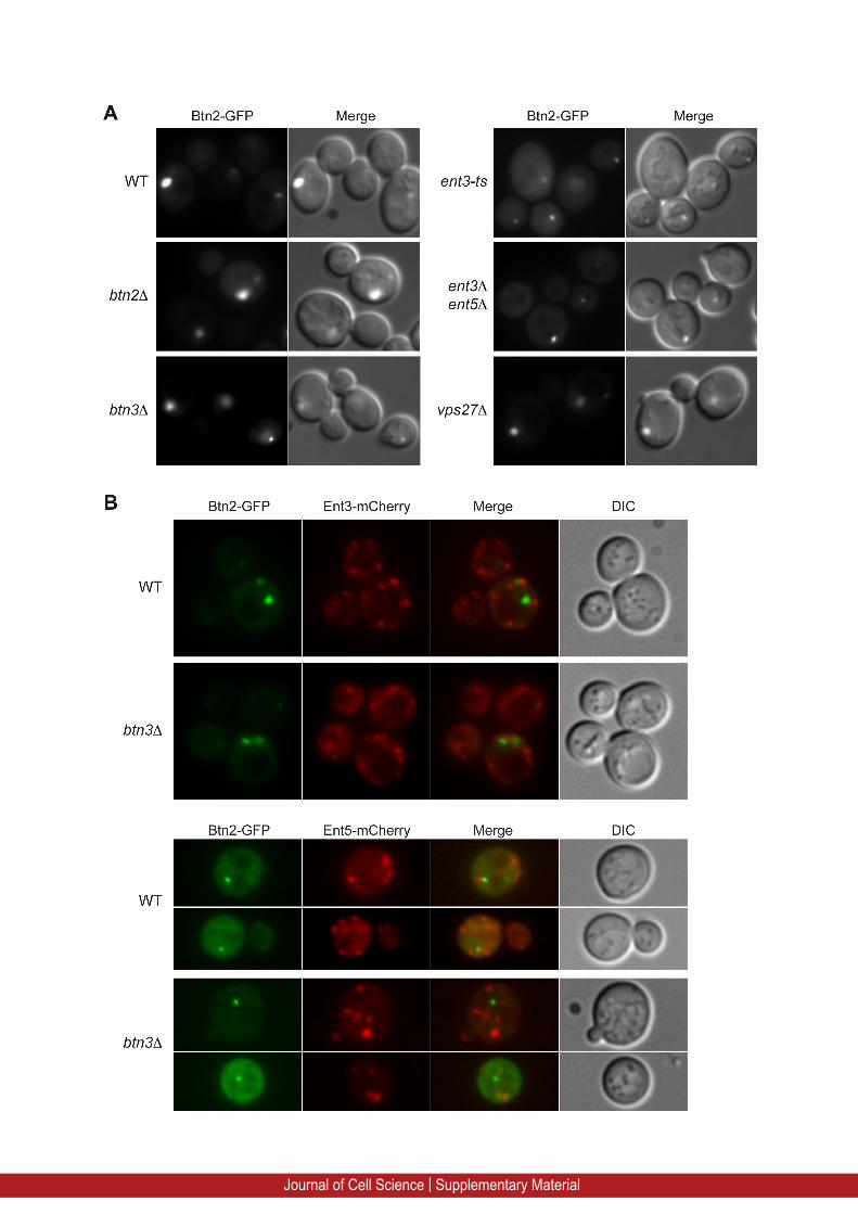
Supplementary Figure 1: Btn2 endomal localization does not require the presence of the epsins Ent3 and Ent5.
(A) Fluorescence microscopy of Btn2-GFP expressed under the control of the constitutive
ADH1 (Alcohol dehydrogenase) promoter in wild type (WT, BY4742), btn2Δ, btn3Δ, ent3-ts,
ent3Δ ent5Δ and vps27Δ cells. (B) The intracellular localization of Btn2-GFP expressed from
ADH1 promoter was observed by fluorescence microscopy in yeast strains bearing
chromosomally mCherry-tagged versions of Ent3 (Ent3-mCherry) or Ent5 (Ent5-mCherry) in
wild type (WT) or btn3Δ cells.
Journal of Cell Science | Supplementary Material
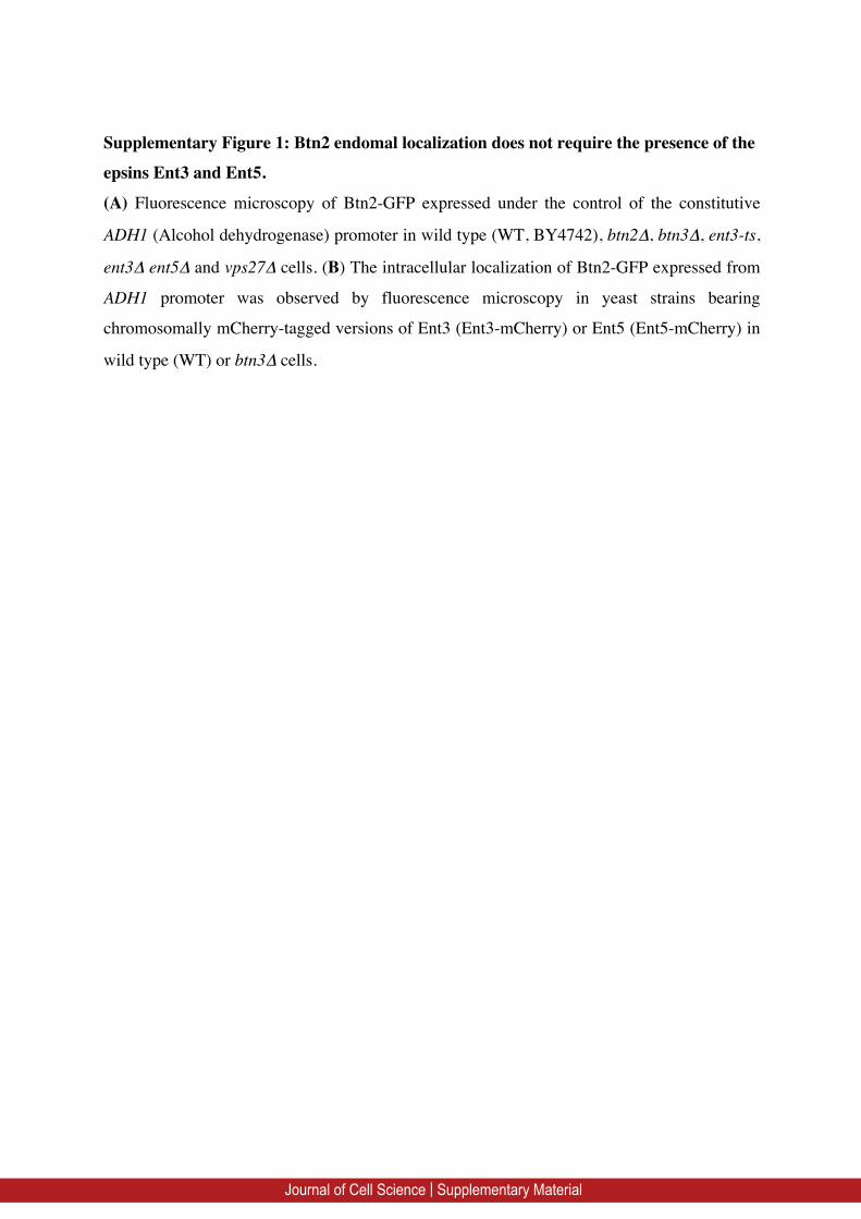
Supplementary Figure 2: Btn3 is not involved in the vacuolar protein sorting VPS
pathway
(A) The GFP-Vam3 and GFP-DPAP-B fusion proteins were expressed in wild type cells
(WT, BY4742) and in btn3Δ mutant cells and were visualized by fluorescence (GFP filter)
and DIC (Nomarski) microscopy. (B) Wild type (BY4742), vps27∆, fab1∆, ent3∆ ent5∆ and
btn3∆ cells were grown until exponential phase of growth. 5µl drop of culture at OD600nm=0.4
were spotted on YPD plate and then covered with a water hydrated nitrocellulose membrane.
After 48 hr of growth at 30°C the membrane was removed, rinsed with water and
Journal of Cell Science | Supplementary Material
carboxypeptidase Y (CPY) secretion was revealed by immunoblotting with rabbit polyclonal
anti-CPY antibodies (a gift from H. Riezman).
Journal of Cell Science | Supplementary Material
Recommended


