ORIGINAL ARTICLE
Pedro P. Pereira-Junior Æ Elen A. Chaves
Ricardo H. Costa-e-Sousa Æ Masako O. Masuda
Antonio C. Campos de Carvalho
Jose H. M. Nascimento
Cardiac autonomic dysfunction in rats chronically treatedwith anabolic steroid
Accepted: 17 November 2005 / Published online: 13 December 2005� Springer-Verlag 2005
Abstract To date no published data exist regarding theeffects of chronic high-dose anabolic-androgenic steroidadministration on tonic cardiac autonomic control. Theaim of this study was to evaluate, by power spectralanalysis of heart rate variability (HRV), the effects ofchronic treatment with supraphysiological doses ofnandrolone decanoate (DECA) on tonic cardiacautonomic regulation in sedentary rats. Male Wistar ratswere treated weekly with 10 mg kg�1 of DECA (n=7)or vehicle (CONTROL, n=7) for 10 weeks. At the 8thweek of treatment, electrocardiogram was recorded inthe conscious state, for time- and frequency-domainHRV analysis. Parasympathetic indexes were reduced inDECA group: high-frequency power (CONTROL=11.1±3.0 ms2 vs. DECA=3.8±0.6 ms2, P<0.05),RMSSD (CONTROL=5.9±0.9 ms vs. DECA 3.5±0.3ms; P<0.05) and pNN5 (CONTROL=31.5±7.5 msvs. DECA=13.2±2.6 ms; P<0.05). The sympatheticindex LF/HF tended to be higher in DECA group(CONTROL=0.65±0.15 vs. DECA=1.17±0.26, P=0.0546). In conclusion, chronic treatment with DECA,in rats, impairs tonic cardiac autonomic regulation,which may provide a key mechanism for anabolic ste-roid-induced arrhythmia and sudden cardiac death.
Keywords Heart rate variability Æ Spectral analysis ÆAutonomic nervous system Æ Anabolic-androgenicsteroid Æ Nandrolone decanoate
Introduction
Anabolic-androgenic steroids (AAS) are synthetic tes-tosterone derivatives developed to maximize anabolicand minimize androgenic action (Strauss and Yesalis1991; Schanzer 1996), being used in the clinical setting totreat catabolic conditions such as osteoporosis and renalfailure (Strauss and Yesalis 1991; Pavlatos et al. 2001).
Although various side effects secondary to high-doseAAS consumption are reported, illicit AAS abuse iswidespread among young athletes and non-athletesaiming to optimize strength and muscle mass gain(Kennedy and Lawrence 1993; Dickerman et al. 1995;NIDA 2000), being nandrolone decanoate (DECA) oneof the most frequently consumed AAS among thispopulation (NIDA 2000). AAS abuse has become anissue of public health (Payne et al. 2004) and, in the lastyears, special attention has been given to its role in thedevelopment of cardiovascular abnormalities, withpublished data pointing to the occurrence of hyperten-sion, heart failure, cardiomyopathy, arrhythmia andsudden cardiac death (Kennedy and Lawrence 1993;Sullivan et al. 1998; Fineschi et al. 2001).
Heart rate variability (HRV) analysis has been usedas a powerful tool for assessment of cardiac autonomiccontrol (Akselrod et al. 1981; Task Force 1996; Cohenand Taylor 2002). Cardiac vagal impairment, detectedby reduced high-frequency (HF) power in spectralanalysis of HRV, is a marker of cardiac electricalinstability, and has been shown to constitute an inde-pendent prognostic factor for ventricular arrhythmiaand sudden cardiac death in cardiac patients (Tsuji et al.1996; Bigger et al. 1992; Kleiger et al. 1987; Routledgeet al. 2002). In rats, power spectral analysis of HRV hasbeen shown to be an effective method of detecting dis-turbances in cardiac autonomic control in some experi-
P. P. Pereira-Junior Æ E. A. Chaves Æ M. O. MasudaJ. H. M. Nascimento (&)Laboratorio de Eletrofisiologia Cardıaca Antonio Paes deCarvalho, Instituto de Biofisica Carlos Chagas Filho,UFRJ, CCS, Bloco G, Ilha do Fundao,21949-900 Rio de Janeiro RJ, BrazilE-mail: [email protected].: +55-21-25626555Fax: +55-21-22808193
R. H. Costa-e-Sousa Æ A. C. C. de CarvalhoLaboratorio de Cardiologia Celular e Molecular,Instituto de Biofisica Carlos Chagas Filho,UFRJ, CCS, Bloco G, Ilha do Fundao,21949-900 Rio de Janeiro RJ, Brazil
Eur J Appl Physiol (2006) 96: 487–494DOI 10.1007/s00421-005-0111-7
mental models of pathologic conditions, such as myo-cardial infarction and diabetic neuropathy (Kruger et al.2000; Sanyal et al. 2002).
Despite the prevailing association between AASabuse, ventricular arrhythmia and sudden cardiac death,to date no published data exist regarding the effects ofchronic administration of supraphysiological doses ofAAS on tonic cardiac autonomic control.
Vascular and structural changes within ventricularmyocardium are postulated as the most plausiblemechanism that could account for arrhythmogenesisand sudden cardiac death in this population (McNuttet al. 1988; Kennedy and Lawrence 1993; Dickermanet al. 1995). Nevertheless, a more clear understanding ofthe mechanisms of heart diseases associated to AASabuse is needed (Payne et al. 2004). Few animal modelstudies have been conducted in order to evaluate theimpact of AAS supraphysiological doses on the cardio-vascular system, and human studies still fail to clearlyestablish a direct cause–effect relationship between AASabuse and sudden cardiac death (Kennedy andLawrence 1993; Fineschi et al. 2001).
The aim of this study was to evaluate, by powerspectral analysis of HRV, the effects of chronic treat-ment with supraphysiological doses of DECA on toniccardiac autonomic regulation in sedentary rats.
Methods
Experimental animals
The study was in accordance with the ‘‘Principles oflaboratory animal care’’ (NIH publication No. 85-23,revised 1985), and was approved by the Institution’sAnimal Care and Use Committee. Experiments wereconducted on 14 male Wistar rats, housed in individualcages floored with wood shavings, in a room with con-stant temperature (23�C) and 12-h light–dark cycle(lights on at 07:00 h). All animals had access to food andwater ad libitum. The animals were divided into twogroups: DECA (n=7), which were treated weekly with10 mg kg–1 body weight of DECA (Deca Durabolin�,Organon Inc.) and CONTROL (n=7) which weretreated weekly with the vehicle for the drug (peanut oilwith benzyl alcohol, 90:10, v/v). The dose of DECA usedin this study was based on the studies of Trifunovic et al.(1995) and Woodiwiss et al. (2000). DECA and VEHI-CLE were administered by a single injection in the glu-teus medium muscle once a week for 10 weeks.
Biometrics measures
Body weights of animals were determined weekly. At theend of AAS treatment period, the animals were etherizedand sacrificed by cervical dislocation. The hearts wereremoved to determine heart weight. The heart weight/
body weight ratio was used as an index of cardiachypertrophy.
Electrocardiogram recording
By the start of the 8th week, rats were anesthetized(ketamine/xylazine: 80/20 mg kg–1, i.p.), and by a sur-gical procedure two steel electrodes were implantedsubcutaneously near to the apex and base of the heart.Electrodes were exteriorized through the back of theneck and terminated in a custom-made connector.Electrocardiogram (ECG) recordings in the consciousstate were started 72 h after surgical procedure, beingconducted while unrestricted animals rested quietly inindividual cages, without walking inside it. Records werealways started 15 min after linking the connector,through flexible cables, to the data acquisition system(3A9 amplifier, Tektronix/TL-2 A/D Interface, AxonInstruments), and were conducted, in all animals, for3 min and 10 s (these additional 10 s were recorded toensure 180 s of artifact-free tachograms, nevertheless,when RR edition was conducted, it never reached over1% of detected intervals). ECG signals were acquiredwith the sampling rate of 10-kHz and amplitude reso-lution of 12 bits. In order to reduce the effect of heartrate circadian fluctuations, all records were conducted ina constant environment, at the same period of the day(13:00–15:00 h). Forty-eight hours after basal ECGrecording, 10 of the 14 animals (CONTROL n=5;DECA n=5) were treated with the muscarinic antago-nist atropine sulfate (2 mg kg–1 i.p.) and new ECGrecords were carried out.
Heart rate variability
All HRV signals processing in the present study wasdone using Matlab-based algorithms. After R wave peakdetection, 180-s tachograms were generated, containingall heart period fluctuations within this time segment. Inthe time-domain, the following indexes were obtained:RR (mean RR interval), SDNN (standard deviation ofRR intervals), RMSSD (square root of the meansquared differences of successive RR intervals) andpNN5 [percentage of successive RR interval differencesgreater than 5 ms (Aubert et al. 1999)]. For spectral(frequency-domain) analysis of HRV, tachograms wereresampled to equal intervals by spline cubic interpola-tion method at 10 Hz, and the linear trend was removed.Power spectrum was obtained with a fast Fouriertransform based method (Welch’s periodogram: 256points, 50% overlap, and Hamming window). Two fre-quency bands were determined: low frequency (LF: 0.2–0.8 Hz), and high frequency (HF: 0.8–2.5 Hz). Power (inms2) was estimated as the area under the spectrumwithin these frequency ranges. Muscarinic blockade datawere expressed as percent reductions in HF power afteratropine sulfate administration.
488
Statistical analysis
All data are expressed as mean±standard error of themean (SEM). Statistical analysis was performed usingGraphPad Prism 4 (GraphPad Software, Inc.). Com-parisons between groups were carried out by Student’sunpaired t test. A paired t test was conducted to com-pare HF power values prior to and post-atropineadministration. Welch’s correction was applied whenvariances were significantly different between groups.For body weight comparisons, two-way analysis ofvariance (ANOVA) was applied, followed byBonferroni’s multiple comparison post-test. Statisticalsignificance was established at the P<0.05 level.
Results
Effects of AAS treatment on body and heart weight
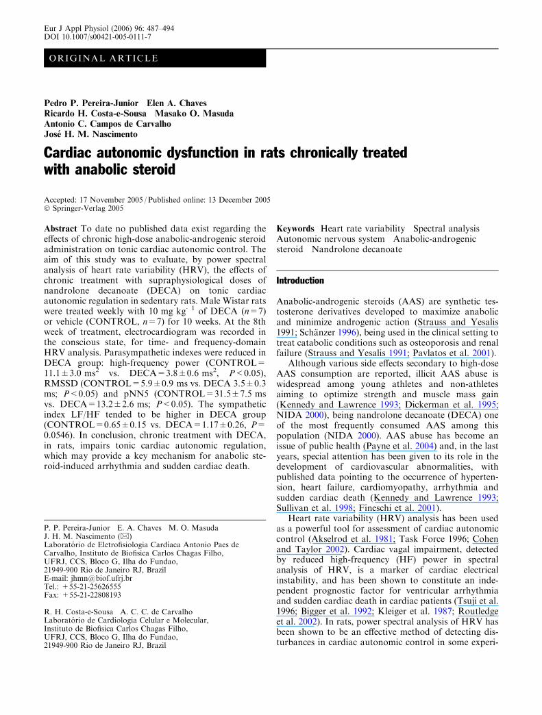
The effect of AAS treatment on body weight of rats isillustrated in Fig. 1. There was no significant differencebetween the initial body weights of both groups. DECAimpaired the growth of rats, so statistical difference inmean body weight between the two groups occurredfrom the 4th week to the end of treatment.
The final heart weight was not different betweengroups when compared in absolute values, but wassignificantly increased in the animals treated with su-praphysiological doses of AAS when expressed in rela-tion to final body weight (Table 1).
Effects of AAS treatment on time-domain measuresof HRV in conscious rats
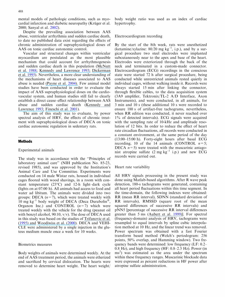
Figure 2 shows time-domain HRV indexes for CON-TROL and DECA groups. No significant differencesbetween groups were observed for RR and for SDNN.
The time-domain indexes of parasympathetic modula-tion RMSSD and pNN5 were significantly lower inDECA group (P<0.05).
Effects of AAS on frequency-domain measuresof HRV in conscious rats
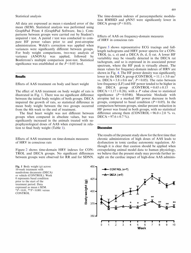
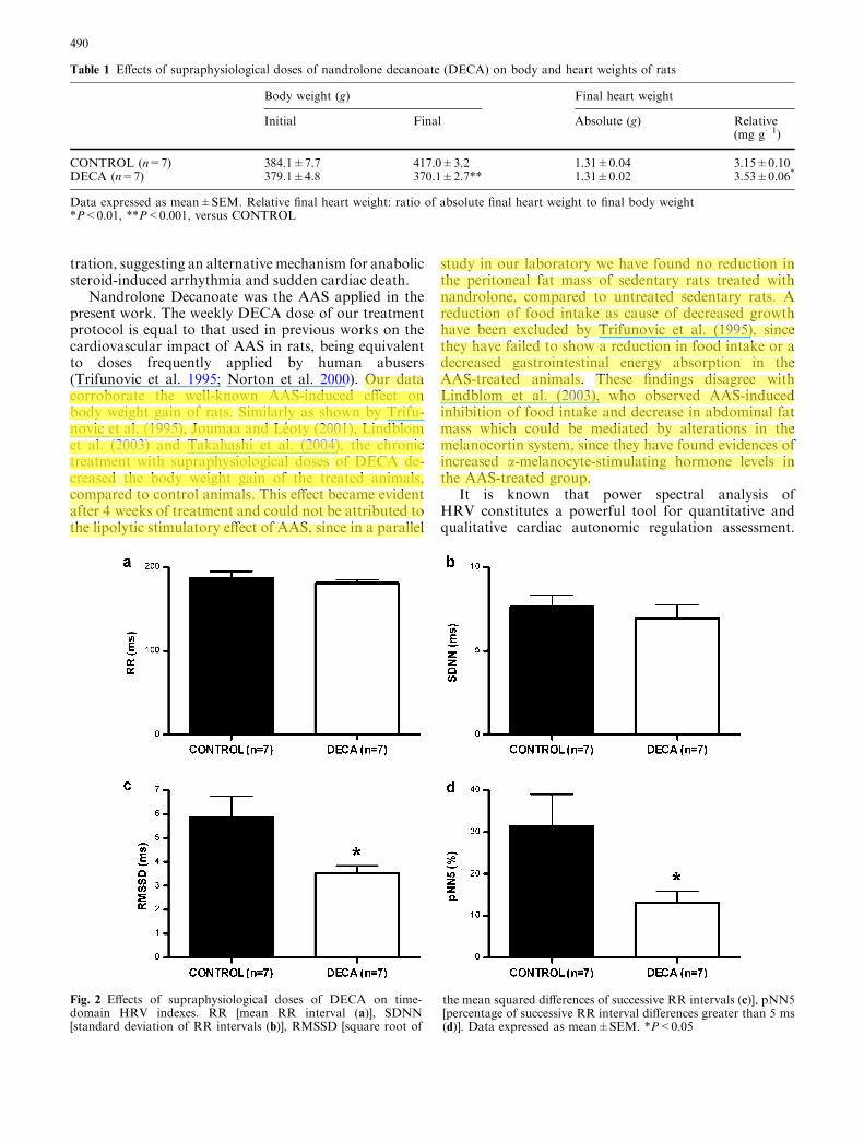
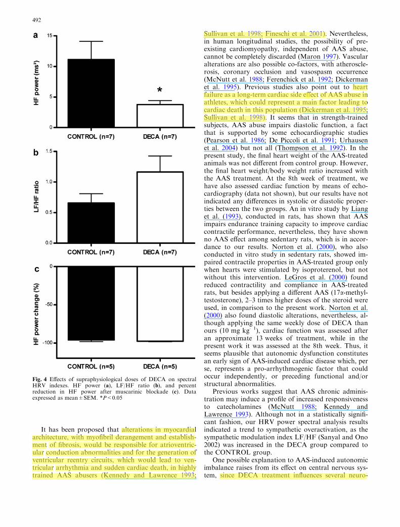
Figure 3 shows representative ECG tracings and full-length tachograms and HRV power spectra for a CON-TROL (a, c, e) and a DECA (b, d, f) rat. A lower HFvariability may be visually detected in the DECA rattachogram, and so is expressed in its associated powerspectrum, where the HF peak is virtually absent. Themean values for frequency-domain indexes of HRV areshown in Fig. 4. The HF power density was significantlylower in the DECA group (CONTROL=11.1±3.0 ms2
vs. DECA=3.8±0.6 ms2, P<0.05). The ratio betweenlow-frequency (LF) and HF power tended to be higher inthe DECA group (CONTROL=0.65±0.15 vs.DECA=1.17±0.26), with a P value close to statisticalsignificance (P=0.0546). Muscarinic blockade withatropine led to a marked HF power decrease in bothgroups, compared to basal condition (P<0.05). In thecomparison between groups, similar percent reduction inHF power was found in both groups, with no statisticaldifference among them (CONTROL=96.0±2.0 % vs.DECA=97.6±0.7 %).
Discussion
The results of the present study show for the first time thatchronic administration of high doses of AAS leads todysfunction in tonic cardiac autonomic regulation. Al-though it is clear that caution should be applied whenextrapolating animal model data to human physiology,we believe that the present study may provide further in-sight on the cardiac impact of high-dose AAS adminis-
Fig. 1 Body weight (g) across10-week treatment withnandrolone decanoate (DECA)or vehicle (CONTROL). Week0 represents basal conditionprior to the start of thetreatment period. Dataexpressed as mean±SEM.*P<0.01, **P<0.001 versusCONTROL
489
tration, suggesting an alternative mechanism for anabolicsteroid-induced arrhythmia and sudden cardiac death.
Nandrolone Decanoate was the AAS applied in thepresent work. The weekly DECA dose of our treatmentprotocol is equal to that used in previous works on thecardiovascular impact of AAS in rats, being equivalentto doses frequently applied by human abusers(Trifunovic et al. 1995; Norton et al. 2000). Our datacorroborate the well-known AAS-induced effect onbody weight gain of rats. Similarly as shown by Trifu-novic et al. (1995), Joumaa and Leoty (2001), Lindblomet al. (2003) and Takahashi et al. (2004), the chronictreatment with supraphysiological doses of DECA de-creased the body weight gain of the treated animals,compared to control animals. This effect became evidentafter 4 weeks of treatment and could not be attributed tothe lipolytic stimulatory effect of AAS, since in a parallel
study in our laboratory we have found no reduction inthe peritoneal fat mass of sedentary rats treated withnandrolone, compared to untreated sedentary rats. Areduction of food intake as cause of decreased growthhave been excluded by Trifunovic et al. (1995), sincethey have failed to show a reduction in food intake or adecreased gastrointestinal energy absorption in theAAS-treated animals. These findings disagree withLindblom et al. (2003), who observed AAS-inducedinhibition of food intake and decrease in abdominal fatmass which could be mediated by alterations in themelanocortin system, since they have found evidences ofincreased a-melanocyte-stimulating hormone levels inthe AAS-treated group.
It is known that power spectral analysis ofHRV constitutes a powerful tool for quantitative andqualitative cardiac autonomic regulation assessment.
Table 1 Effects of supraphysiological doses of nandrolone decanoate (DECA) on body and heart weights of rats
Body weight (g) Final heart weight
Initial Final Absolute (g) Relative(mg g�1)
CONTROL (n=7) 384.1±7.7 417.0±3.2 1.31±0.04 3.15±0.10DECA (n=7) 379.1±4.8 370.1±2.7** 1.31±0.02 3.53±0.06*
Data expressed as mean±SEM. Relative final heart weight: ratio of absolute final heart weight to final body weight*P<0.01, **P<0.001, versus CONTROL
Fig. 2 Effects of supraphysiological doses of DECA on time-domain HRV indexes. RR [mean RR interval (a)], SDNN[standard deviation of RR intervals (b)], RMSSD [square root of
the mean squared differences of successive RR intervals (c)], pNN5[percentage of successive RR interval differences greater than 5 ms(d)]. Data expressed as mean±SEM. *P<0.05
490

Recently, a validation study of HRV analysis in rats hasbeen published (Aubert et al. 1999). Other reports haveshown that altered HRV patterns in rats are consistentwith those presented in human studies for well-estab-lished cardiac autonomic dysfunction conditions, suchas diabetes mellitus and myocardial infarction (Krugeret al. 2000; Sanyal et al. 2002), which are associated withventricular arrhythmia and sudden cardiac death. As inthese pathological conditions, our group of rats treatedwith AAS presented a marked impairment of parasym-pathetic cardiac modulation with decreased HF powerof HRV compared to the control group. In the AASgroup, time-domain parasympathetic indexes RMSSDand pNN5 (Aubert et al. 1999; El-Mas MM and Abdel-Rahman 2004) were also reduced, which corroboratesthe parasympathetic cardiac dysfunction shown byspectral analysis. Muscarinic blockade data reinforce therole of parasympathetic modulation over HF power,confirming the reliability of our HRV analysis method-ology (Ramaekers et al. 2002a).
The body of evidence supporting a direct cause–effectrelationship between AAS abuse, ventricular arrhythmiaand sudden cardiac death has not yet been clearlyestablished, and not much is known about the possiblemechanisms responsible for arrhythmogenesis in AASabusers. Studies on the cardiovascular impact of chronicAAS administration consist mainly of case reports withfew animal models available. In one of these animal-model-based studies, it has been shown that chronictreatment with AAS reduces markedly the life span ofmale mice (Bronson and Matherne 1997), mortalitybeing assumed to occur mainly by liver and kidneydamage, with cardiac involvement representing a sec-ondary cause. In humans a 12-year longitudinal studycomparing a group of body builders who abused of AASto an age matched control group, constituted by generalpopulation individuals, indicated a 4.6 higher mortalityin the former group, with cardiovascular damage poin-ted as the cause of death in 37.5 % of these individuals(Parssinen et al. 2000).
Fig. 3 Representative ECGtracing, tachogram and powerspectrum from a CONTROL(a, c, e) and a DECA animal(b, d, f)
491
It has been proposed that alterations in myocardialarchitecture, with myofibril derangement and establish-ment of fibrosis, would be responsible for atrioventric-ular conduction abnormalities and for the generation ofventricular reentry circuits, which would lead to ven-tricular arrhythmia and sudden cardiac death, in highlytrained AAS abusers (Kennedy and Lawrence 1993;
Sullivan et al. 1998; Fineschi et al. 2001). Nevertheless,in human longitudinal studies, the possibility of pre-existing cardiomyopathy, independent of AAS abuse,cannot be completely discarded (Maron 1997). Vascularalterations are also possible co-factors, with atheroscle-rosis, coronary occlusion and vasospasm occurrence(McNutt et al. 1988; Ferenchick et al. 1992; Dickermanet al. 1995). Previous studies also point out to heartfailure as a long-term cardiac side effect of AAS abuse inathletes, which could represent a main factor leading tocardiac death in this population (Dickerman et al. 1995;Sullivan et al. 1998). It seems that in strength-trainedsubjects, AAS abuse impairs diastolic function, a factthat is supported by some echocardiographic studies(Pearson et al. 1986; De Piccoli et al. 1991; Urhausenet al. 2004) but not all (Thompson et al. 1992). In thepresent study, the final heart weight of the AAS-treatedanimals was not different from control group. However,the final heart weight/body weight ratio increased withthe AAS treatment. At the 8th week of treatment, wehave also assessed cardiac function by means of echo-cardiography (data not shown), but our results have notindicated any differences in systolic or diastolic proper-ties between the two groups. An in vitro study by Lianget al. (1993), conducted in rats, has shown that AASimpairs endurance training capacity to improve cardiaccontractile performance, nevertheless, they have shownno AAS effect among sedentary rats, which is in accor-dance to our results. Norton et al. (2000), who alsoconducted in vitro study in sedentary rats, showed im-paired contractile properties in AAS-treated group onlywhen hearts were stimulated by isoproterenol, but notwithout this intervention. LeGros et al. (2000) foundreduced contractility and compliance in AAS-treatedrats, but besides applying a different AAS (17a-methyl-testosterone), 2–3 times higher doses of the steroid wereused, in comparison to the present work. Norton et al.(2000) also found diastolic alterations, nevertheless, al-though applying the same weekly dose of DECA thanours (10 mg kg�1), cardiac function was assessed afteran approximate 13 weeks of treatment, while in thepresent work it was assessed at the 8th week. Thus, itseems plausible that autonomic dysfunction constitutesan early sign of AAS-induced cardiac disease which, perse, represents a pro-arrhythmogenic factor that couldoccur independently, or preceding functional and/orstructural abnormalities.
Previous works suggest that AAS chronic adminis-tration may induce a profile of increased responsivenessto catecholamines (McNutt 1988; Kennedy andLawrence 1993). Although not in a statistically signifi-cant fashion, our HRV power spectral analysis resultsindicated a trend to sympathetic overactivation, as thesympathetic modulation index LF/HF (Sanyal and Ono2002) was increased in the DECA group compared tothe CONTROL group.
One possible explanation to AAS-induced autonomicimbalance raises from its effect on central nervous sys-tem, since DECA treatment influences several neuro-
Fig. 4 Effects of supraphysiological doses of DECA on spectralHRV indexes. HF power (a), LF/HF ratio (b), and percentreduction in HF power after muscarinic blockade (c). Dataexpressed as mean±SEM. *P<0.05
492
transmitter systems, including dopaminergic, serotoner-gic and adrenergic (Thiblin et al. 1999; Schlussman et al.2000; Kindlundh et al. 2003; Lindblom et al. 2003; Ta-maki et al. 2003; Kindlundh et al. 2004). It is of specialinterest to detach the work of Tamaki et al. (2003), whodemonstrated that nandrolone enhances both norepi-nephrine and its metabolite 4-hydroxy-3-methoxyphe-nylglycol levels in hypothalamus. Moreover, increasedhypothalamic norepinephrine turnover is associated toan increase in systemic arterial pressure (reviewed in DeWardener 2001) and to a systemic hyperadrenergic state(Ishac et al. 1987). As previously cited, it has beenproposed that high-dose AAS administration could in-crease a-melanocyte-stimulating hormone levels(Lindblon et al. 2003). Thus, an involvement of mela-nocortin system in AAS-induced autonomic changesmay also be suggested, since it has been shown thatcentral administration of melanocotins, in rats, can elicitincreases in blood pressure and LF power of bloodpressure variability, indicating increased sympatheticactivity (Ramaekers et al. 2002b). It is known thatsympathetic and vagal influences counteract in a mannerthat a sympathetic hyperactivation state could lead to adecrease in vagal cardiac control or vice versa (Levy1971). Moreover, in a recent work Hamson et al. (2004)have shown the presence of androgen receptors in nu-cleus ambiguus, a brain center related to cardiac vagalcontrol. This way, the existence of some kind of directandrogen-mediated vagal effect on cardiac autonomicregulation can also be hypothesized. Nevertheless, it isclear that further research needs to be conducted inorder to evaluate the possible mechanisms involved inandrogen central effects on cardiac autonomic regula-tion.
Several potential limitations of the present studyshould be addressed. Concomitantly to HRV assess-ment of autonomic function, it would have been ofinterest to assess the impact of the AAS treatmentprotocol that we applied, on hemodynamic variables,such as blood pressure and total peripheral resistance.It is also not clear whether the AAS treatment appliedhad direct effects on respiratory rate or tidal volume,which could possibly affect HRV parasympathetic in-dexes. Possible changes in free-running rhythmicity(e.g., in baroreflex), could also account for alterationsin measured indexes of HRV. It is important to re-mark that the present study was conducted in ananimal model of sedentary rats, and that, to ourknowledge, no human study has assessed this issue onsedentary individuals but, in contrast, generally onhighly strength trained subjects. Thus, some of theresults found by us may consist of model-specific re-sponses that could possibly not be reproducible inhumans. Besides assessing how exercise training wouldmodulate autonomic effect of AAS, in the presentmodel, it is clear that future research is also needed inorder to assess cardiac autonomic regulation in humanAAS abusers.
In summary, the results of the present study show, forthe first time, that chronic high-dose AAS administra-tion leads to dysfunction in tonic cardiac autonomicregulation, with marked impairment of parasympatheticcontrol, which may provide a key mechanism for AAS-induced arrhythmia and sudden cardiac death. Inaddiction, autonomic dysfunction seems to occur inde-pendently, or to precede any changes in cardiac systolicor diastolic function.
Acknowledgements PPPJ was recipient of a CNPq scholarship, andthis work is part of his MSc manuscript.
References
Akselrod S, Gordon D, Ubel FA, Shannon DC, Berger AC, CohenRJ (1981). Power spectrum analysis of heart rate fluctuation: aquantitative probe of beat-to-beat cardiovascular control.Science 213:220–222
Aubert AE, Ramaekers D, Beckers F, Breem R, Denef C, Van DeWerf F, Ector H (1999) The analysis of heart rate variability inunrestrained rats. Validation of method and results. ComputMethods Programs Biomed 60:197–213
Bigger JT Jr, Fleiss JL, Steinman RC, Rolnitzky LM, Kleiger RE,Rottman JN (1992) Frequency-domain measures of heart per-iod variability and mortality after myocardial infarction.Circulation 85:164–171
Bronson FH, Matherne CM (1997) Exposure to anabolic-andro-genic steroids shortens life span of male mice. Med Sci SportsExerc 29:615–619
CohenMA, Taylor JA (2002) Short-term cardiovascular oscillationsin man: measuring and modelling the physiologies. J Physiol542:669–683
De Piccoli B, Giada F, Benettin A, Sartori F, Piccolo E (1991)Anabolic steroid use in body builders: an echocardiographicstudy of left ventricle morphology and function. Int J SportsMed 12:408–412
De Wardener HE (2001) The hypothalamus and hypertension.Physiol Rev 81:1599–1658
Dickerman RD, Schaller F, Prather I, McConathy WJ (1995)Sudden cardiac death in a 20-year-old bodybuilder usinganabolic steroids. Cardiology 86:172–173
El-Mas MM, Abdel-Rahman AA (2004) Differential modulationby estrogen of alpha2-adrenergic and I1-imidazoline receptor-mediated hypotension in female rats. J Appl Physiol97:1237–1244
Ferenchick GS, Adelman S (1992) Myocardial infarction associ-ated with anabolic steroid use in a previously healthy 37-year-old weight lifter. Am Heart J 124:507–508
Fineschi V, Baroldi G, Monciotti F, Paglicci Reattelli L, TurillazziE (2001) Anabolic steroid abuse and cardiac sudden death: apathologic study. Arch Pathol Lab Med 125:253–255
Hamson DK, Jones BA, Watson NV (2004) Distribution ofandrogen receptor immunoreactivity in the brainstem of malerats. Neuroscience 127:797–803
Ishac EJ, Eskay R, Hirata F, Axelrod J, Kunos G (1987) Adren-ergic regulation of beta-endorphin secretion from anteriorpituitary in conscious rats: effects of thyroid state. Endocri-nology 120:1073–1078
Joumaa WH, Leoty C (2001) Differential effects of nandrolonedecanoate in fast and slow rat skeletal muscles. Med Sci SportsExerc 33:397–403
Kennedy MC, Lawrence C (1993) Anabolic steroid abuse andcardiac death. Med J Aust 158:346–348
Kindlundh AMS, Lindblom J, Bergstrom L, Nyberg F (2003) Theanabolic-andogenic steroid nandrolone induces alteration in thedensity of serotonergic 5HT1B and 5HT2 receptors in the malerat brain. Neuroscience 119:113–120
493

Kindlundh AMS, Rahman S, Lindblom J, Nyberg F (2004) In-creased dopamine transporter density in the male rat brainfollowing chronic nandrolone decanoate administration. Neu-rosci Lett 356:131–134
Kleiger RE, Miller JP, Bigger JT, Moss AJ, The Multicenter Post-Infarction Research Group (1987) Decreased heart rate vari-ability and its association with increased mortality after acutemyocardial infarction. Am J Cardiol 59:256–262
Kruger C, Landerer V, Zugck C, Ehmke H, Kubler W, Haass M(2000) The bradycardic agent zatebradine enhances baroreflexsensitivity and heart rate variability in rats early after myo-cardial infarction. Cardiovasc Res 45:900–912
LeGros T, McConnell D, Murry T, Edavettal M, Racey-Burns LA,Shepherd RE, Burns AH (2000) The effects of 17 alpha-meth-yltestosterone on myocardial function in vitro. Med Sci SportsExerc 32:897–903
Levy MN (1971) Sympathetic-parasympathetic interactions in theheart. Circ Res 29:437–445
Liang MTC, Paulson DJ, Kopp SJ, Glonek T, Meneses P,Gierke LW, Schwartz FN (1993) Effects of anabolic steroidsand endurance exercise performance. Int J Sports Med14:324–329
Lindblom J, Kindlundh AMS, Nyberg F, Bergstrom L, WikbergJES (2003) Anabolic androgenic steroid nandrolone decanoatereduces hypothalamic proopiomelanocortin mRNA levels.Brain Res 986:139–147
Maron BJ (1997) Hypertrophic cardiomyopathy. Lancet350:127–133
McNutt RA, Ferenchick GS, Kirlin PC, Hamlin NJ (1988) Acutemyocardial infarction in a 22-year-old world class weight lifterusing anabolic steroids. Am J Cardiol 62:164
NIDA - National Institute on Drug Abuse. Research report series:Anabolic steroid abuse (2000) [Online]. U.S. Department ofHealth and Human Services, National Institutes of Health.http://www.nida.nih.gov/PDF/RRSteroi.pdf [14 Jul. 2004]
Norton GR, Trifunovic B, Woodiwiss AJ (2000) Attenuated beta-adrenoceptor-mediated cardiac contractile responses followingandrogenic steroid administration to sedentary rats. Eur J ApplPhysiol 81:310–316
Parssinen M, Kujala U, Vartiainen E, Sarna S, Seppala T (2000)Increased premature mortality of competitive powerlifters sus-pected to have used anabolic agents. Int J Sports Med 21:225–227
Pavlatos AM, Fultz O, Monberg MJ, Vootkur A (2001) Review ofoxymetholone: a 17a-alkylated anabolic-androgenic steroid.Clin Ther 23:789–801
Payne JR, Kotwinski PJ, Montgomery HE (2004) Cardiac effectsof anabolic steroids. Heart 90:473–475
Pearson AC, Schiff M, Mrosek D, Labovitz AJ, Williams GA(1986) Left ventricular diastolic function in weight lifters. Am JCardiol 58:1254–1259
Ramaekers D, Beckers F, Demeulemeester H, Aubert AE (2002a)Cardiovascular autonomic function in conscious rats: a novelapproach to facilitate stationary conditions. Ann NoninvasiveElectrocardiol 7:307–318
Ramaekers D, Beckers F, Demeulemeester H, Bert C, Denef C,Aubert AE (2002b) Effects of melanocortins on cardiovascularregulation in rats. Clin Exp Pharmacol Physiol 29:549–558
Routledge HC, Chowdhary S, Townend JN (2002) Heart ratevariability–a therapeutic target? J Clin Pharm Ther 27:85–92
Sanyal SN, Ono K (2002) Derangement of autonomic nerve con-trol in rat with right ventricular failure. Pathophysiology 8:197–203
Sanyal SN, Arita M, Ono K (2002) Inhomogeneous derangementof cardiac autonomic nerve control in diabetic rats. Circ J66:283–288
Schanzer W (1996) Metabolism of anabolic androgenic steroids.Clin Chem 42:1001–1020
Schlussman SD, Zhou Y, Johansson P, Kiuru A, Ho A, Nyberg F,Kreek MJ (2000) Effects of the androgenic anabolic steroid, nan-drolone decanoate, on adrenocorticotropin hormone, corticoste-rone and proopiomelanocortin, corticotrophin releasing factos(CRF) and CRF receptor1 mRNA levels in the hypothalamus,pituitary and amygdala of the rat. Neurosci Lett 284:190–194
Strauss RH, Yesalis CE (1991) Anabolic steroids in the athlete.Annu Rev Med 42:449–457
Sullivan ML, Martinez CM, Gennis P, Gallagher EJ (1998) Thecardiac toxicity of anabolic steroids. Prog Cardiovasc Dis 41:1–15
Takahashi M, Tatsugi Y, Kohno T (2004) Endocrinological andpathological effects of anabolic-androgenic steroid in male rats.Endocr J 51:425–434
Tamaki T, Shiraishi T, Takeda H, Matsumiya T, Roy RR, Edg-erton VR (2003) Nandrolone decanoate enhances hypothalamicbiogenic amines in rats. Med Sci Sports Exerc 35:32–38
Task Force of the European Society of Cardiology, the NorthAmerican Society of Pacing and Electrophysiology (1996)Heart rate variability: standards of measurement, physiologicalinterpretantion and clinical use. Circulation 93:1043–1065
Thiblin I, Finn A, Ross SB, Stenfors C (1999) Increased dopami-nergic and 5-hydroxytryptaminergic activities in male rat brainfollowing long-term treatment with anabolic androgenic ste-roids. Br J Pharmacol 126:1301–1306
Thompson PD, Sadaniantz A, Cullinane EM, Bodziony KS, CatlinDH, Torek-Both G, Douglas PS (1992) Left ventricular func-tion is not impaired in weight-lifters who use anabolic steroids.J Am Coll Cardiol 19:278–282
Trifunovic B, Norton GR, Duffield MJ, Avraam P, Woodiwiss AJ(1995) An androgenic steroid decreases left ventricular com-pliance in rats. Am J Physiol 268:H1096–1105
Tsuji H, Larson MG, Venditti FJ Jr, Manders ES, Evans JC,Feldman CL, Levy D (1996) Impact of reduced heart ratevariability on risk for cardiac events—the Framingham heartstudy. Circulation 94:2850–2855
Urhausen A, Albers T, Kinderman W (2004) Are the cardiac effectsof anabolic steroid abuse in strength athletes reversible? Heart90:496–501
Woodiwiss AJ, Trifunovic B, Philippides M, Norton GR (2000)Effects of an androgenic steroid on exercise-induced cardiacremodeling in rats. J Appl Physiol 88:409–415
494
Recommended


