
HAL Id: tel-01296967https://tel.archives-ouvertes.fr/tel-01296967
Submitted on 1 Apr 2016
HAL is a multi-disciplinary open accessarchive for the deposit and dissemination of sci-entific research documents, whether they are pub-lished or not. The documents may come fromteaching and research institutions in France orabroad, or from public or private research centers.
L’archive ouverte pluridisciplinaire HAL, estdestinée au dépôt et à la diffusion de documentsscientifiques de niveau recherche, publiés ou non,émanant des établissements d’enseignement et derecherche français ou étrangers, des laboratoirespublics ou privés.
Cellulose nanocrystals : surface modification andadvanced materials
Ning Lin
To cite this version:Ning Lin. Cellulose nanocrystals : surface modification and advanced materials. Chemical and ProcessEngineering. Université de Grenoble, 2014. English. �NNT : 2014GRENI034�. �tel-01296967�

THÈSE Pour obtenir le grade de
DOCTEUR DE L’UNIVERSITÉ DE GRENOBLE Spécialité : Matériaux, Mécanique, Génie civil, Electrochimie
Arrêté ministériel : 7 août 2006
Présentée par
Ning LIN Thèse dirigée par Professeur Alain DUFRESNE préparée au sein du Laboratoire du Génie des Procédés Papetiers de l’Ecole Internationale du Papier, de la Communication Imprimée est des Biomatériaux (LGP2-UMR 5518) dans l'École Doctorale Ingénierie – Matériaux, Mécanique, Energétique, Environnement, Procédés de Production
Cellulose Nanocrystals: Surface Modification and Advanced Materials
Thèse soutenue publiquement le 24th June 2014, devant le jury composé de :
Monsieur Etienne FLEURY Professeur, INSA de Lyon, France (Président)
Monsieur Christoph WEDER Professeur, l’Université de Fribourg, Switzerland (Rapporteur)
Monsieur Jean-Marie RAQUEZ Dr., l’Université de Mons, Belgium (Rapporteur)
Monsieur Jin HUANG Professeur, Wuhan University of Technology, China (Membre)
Madame Alessandra de ALMEIDA LUCAS Dr., Federal University of SaoCarlos, Brazil (Membre)
Monsieur Alain DUFRESNE Professeur, Grenoble INP, France (Membre)


“天行健,君子以自强不息;地势坤,君子以厚德载物。”
——《周易·乾》
To my parents who gave and supported my life,
and Yuan for her love and companion


Acknowledgements
Ning LIN −−−− 2014 I
Acknowledgements
The research presented in this thesis was carried out in the Multiscaled Biobase
Materials group at the Department of Grenoble Institute of Technology-Pagora
(Institut Polytechnique de Grenoble) in France. Three-year study in Pagora is a long
but pleasant journey to explore the research on cellulose nanocrystals.
First and foremost, I am grateful to my PhD supervisor, Professor Alain Dufresne, for
his help, support, and patience throughout last three years. He encouraged me to
propose any idea during the study, and was always available when I needed his
advice. Without his instruction and contribution, this thesis would not have turned
out as it is. Alain’s never-ending thoughts and enthusiasm for science give me an
impression and good example. He has my utmost respect as an excellent scientist,
and my deepest gratitude as a good friend.
I am grateful to the dissertation committee, Professor Etienne Fleury, Professor
Christoph Weder, Dr. Jean-Marie Raquez, Professor Jin Huang, and Dr. Alessandra de
Almeida Lucas, for their interest in my research, taking time to check my thesis, and
providing valuable suggestions and insights.
I much appreciate my Master supervisor, Professor Jin Huang from Wuhan University
of Technology, China. He led me into the science world, and showed me what
cellulose nanocrystal is. Even after my graduation from Master study, we frequently
discussed hot research topics on diverse fields of science. His remarks or comments
always provided me the promising inspiration on my study.
I would like to thank Professor Jean-Luc Putaux (CERMAV-CNRS), Dr. Gregory
Berthome (INPG-SIMaP), and Prof. Yun Chen (School of Basic Medical
Sciences-Wuhan University) for their help and discussion on instrument
characterizations. Special thanks go to Cécile Sillard and David Dallerac, who
provided access and instructions on the laboratory equipment and chemicals
reservation.

Acknowledgements
Ning LIN −−−− 2014 II
I am grateful to all the professors and students (and post-doctors) in the team of
LGP2 for their kind help and suggestions. Thanks a lot my colleagues and friends,
Oussama El Baradai, Ahlem Romdhane, Karima Benhamou, who always cheered me
up and encouraged me in difficult periods. Special thanks to Imtiaz Ali, you are my
first friend who lent me a helping hand when I arrived at Grenoble. I really feel lucky
that during my study in France, all of us can become the good friends.
I dedicate my dissertation to my parent in China, for their unlimited love, support
and trust for all my life.
My utmost thanks go to my true love, Yuan Li. She is the kindest and most patient girl
I have ever met. Her constant support and encouragement, and her loving care gave
me the energy to carry out the experiment and study. Thank God for giving me the
best wife of the world and a very happy life.
One day in the September of three years ago, when I walked on the “Rue de la
Chimie” of Grenoble, I suddenly realized it was the first time I lived in abroad; an
unknown experience for the new life was waiting for me. At that time, I really didn’t
know this experience would be interesting, exciting or hard or lonely. Now I am
about to finish the PhD study. I know there is a long list for the people deserving my
gratitude, and I can’t write all their names in here. But I will definitely write your
names in my heart and remember all of your kind helps!
08th, April 2014 at Grenoble

Table of Contents
Ning LIN −−−− 2014 III
Table of Contents
Acknowledgements ............................................................................................................ I
Table of Contents ............................................................................................................. III
Scientific Publications ...................................................................................................... V
General Introduction ......................................................................................................... 1
Chapter 1. Literature Review ........................................................................................... 9
1.1 Extraction and Production of Cellulose Nanocrystals .................................................... 15
1.2 Structure and Properties of Cellulose Nanocrystals ....................................................... 18
1.3 Surface Modification of Cellulose Nanocrystals ............................................................. 34
1.4 Nano-Reinforcement of Cellulose Nanocrystals in Plastic Composites .......................... 57
1.5 Advanced Functional Materials based on Cellulose Nanocrystals ................................. 69
1.6 Conclusions ..................................................................................................................... 90
1.7 References ...................................................................................................................... 91
Chapter 2. Surface Chemistry, Morphological Analysis and Properties of
Cellulose Nanocrystals with Gradiented Sulfation Degrees ........................................ 111
2.1 Introduction .................................................................................................................. 123
2.2 Experimental Methodology .......................................................................................... 126
2.3 Spectroscopic Proofs for Gradient Sulfation of Cellulose Nanocrystals ....................... 130
2.4 Morphological Observation and Dimensions ............................................................... 134
2.5 Morphological Models and Surface Chemistry of Cellulose Nanocrystals ................... 137
2.6 Effect of Sulfate Groups on the Properties of Cellulose Nanocrystals ......................... 142
2.7 Conclusions ................................................................................................................... 148
2.8 References .................................................................................................................... 148
Chapter 3. Physical and/or Chemical Compatibilization of Extruded Cellulose
Nanocrystal Reinforced Polystyrene Nanocomposites ................................................ 153
3.1 Introduction .................................................................................................................. 165
3.2 Experimental Methodology .......................................................................................... 167

Table of Contents
Ning LIN −−−− 2014 IV
3.3 Surface Modification and Properties of Cellulose Nanocrystals .................................. 175
3.4 Structure and Properties of Extruded Nanocomposites .............................................. 184
3.5 Conclusions ................................................................................................................... 196
3.6 References .................................................................................................................... 197
Chapter 4. TEMPO-Oxidized Nanocellulose Participating as Crosslinking Aid for
Alginate-Based Sponges ..................................................................................................... 201
4.1 Introduction .................................................................................................................. 213
4.2 Experimental Methodology .......................................................................................... 215
4.3 TEMPO-Mediated Oxidation of Nanocellulose ............................................................. 222
4.4 Structure and Properties of Crosslinked Sponges ........................................................ 227
4.5 Roles and Mechanisms of Nanocelluloses in Alginate-Based Sponges ........................ 236
4.6 Conclusions ................................................................................................................... 238
4.7 References .................................................................................................................... 239
Chapter 5. Supramolecular Hydrogels from In Situ Host−Guest Inclusion
between Chemically Modified Cellulose Nanocrystals and Cyclodextrin ................. 245
5.1 Introduction .................................................................................................................. 257
5.2 Experimental Methodology .......................................................................................... 259
5.3 Chemical Modification of Cellulose Nanocrystals ........................................................ 267
5.4 Structure and Properties of Supramolecular Hydrogels............................................... 271
5.5 Drug Release and Mechanism Study ............................................................................ 279
5.6 Conclusions ................................................................................................................... 281
5.7 References .................................................................................................................... 282
Chapter 6. General Conclusions and Perspectives for Future Work ..................... 287
6.1 General Conclusions ..................................................................................................... 289
6.2 Possible Research Points for Further Study ................................................................ 294
6.3 Perspectives and Challenges in Future ....................................................................... 296
Résumé en Français ....................................................................................................... 299
Appendix and Abbreviations ....................................................................................... 317

Scientific Publications
Ning LIN −−−− 2014 V
Scientific Publications (2012−−−−2014)
Articles in Scientific Journals
1. Ning Lin, Alain Dufresne, “Surface Chemistry, Morphological Analysis and
Properties of Cellulose Nanocrystals with Gradiented Sulfation Degrees”,
Nanoscale, 2014, 6(10), 5384–5393.
2. Ning Lin, Alain Dufresne, “Physical and/or Chemical Compatibilization of
Extruded Cellulose Nanocrystal Reinforced Polystyrene Nanocomposites”,
Macromolecules, 2013, 46(14), 5570–5583.
3. Ning Lin, Alain Dufresne, “Supramolecular Hydrogels from in situ Host-Guest
Inclusion between Chemically Modified Cellulose Nanocrystals and Cyclodextrin”,
Biomacromolecules, 2013, 14(3), 871–880.
4. Ning Lin, Cécile Bruzzese, Alain Dufresne, “TEMPO-Oxidized Nanocellulose
Participating as Crosslinking Aid for Alginate-Based Sponges”, ACS Applied
Materials & Interfaces, 2012, 4(9), 4948–4959.
5. Ning Lin, Jin Huang, Alain Dufresne, “Preparations, Properties and Applications
of Polysaccharide Nanocrystals in Advanced Functional nanomaterials: A Review”,
Nanoscale, 2012, 4(11), 3274–3294.
6. Ning Lin, Alain Dufresne, “Nanocellulose in Biomedicine: Current Status and
Future Prospect”, European Polymer Journal, 2014, in press.
Invited Book Chapters
1. Ning Lin, Alain Dufresne: “Chapter 3. Surface Modification of Polysaccharide
Nanocrystals”;
Ning Lin, Jin Huang, Alain Dufresne: “Chapter 6. Polysaccharide
Nanocrystals-Based Materials for Advanced Applications”;

Scientific Publications
Ning LIN −−−− 2014 VI
Alain Dufresne, Ning Lin: “Chapter 7. Characterization of Polysaccharide
Nanocrystal-Based Materials”;
Ed. Jin Huang, Peter R. Chang, Ning Lin, Alain Dufresne, “Biobased
Polysaccharide Nanocrystals: Chemistry and Applications”, John Wiley & Sons,
USA, 2014/12, in press. (Both in English and Chinese)
2. Ning Lin, Alain Dufresne: “Nanocellulose: Biomedical Nanomaterial
Applications”, Entry No. 120050596; Ed. Munmaya Mishra, “Encyclopedia of
Biomedical Polymers and Polymeric Biomaterials”, CRC Press, USA, 2015/01.
3. Ning Lin, Alain Dufresne, “In situ Conjunction of Cellulose Nanocrystals in
Supramolecular Hydrogels by the Aid of Host-Guest Inclusion Complexation”,
pp. 123–126; Ed. Michael T. Postek, Robert J. Moon, Alan W. Rudie, Michael A.
Bilodeau, “Production and Application of Cellulose Nanomaterials”, TAPPI Press,
2013/06.
Refereed Conference Proceedings
1. Ning Lin (2013/12). Oral Presentation. “Properties and Electrostatic Interactions
from Cellulose Nanocrystals with Gradient Surface Sulfate Groups”. Winter
Training School Conference for “Use of Nanopolysaccharides in Packaging”,
Grenoble, France.
2. Ning Lin (2013/06). Oral Presentation. “Structure Properties and Interface in
Polystyrene Nanocomposites Based on Cellulose Nanocrystals with Physical and
Chemical Modifications from Non-Covalent and Covalent PEG Compatibilization”.
2013 TAPPI International Conference on Nanotechnology for Renewable
Materials, Stockholm, Sweden.
3. Ning Lin, Alain Dufresne (2013/06). Poster. “Supramolecular hydrogels from in
situ host−guest inclusion between chemically modified cellulose nanocrystals

Scientific Publications
Ning LIN −−−− 2014 VII
and cyclodextrin”. 2013 TAPPI International Conference on Nanotechnology for
Renewable Materials, Stockholm, Sweden.
4. Ning Lin, Cécile Bruzzese, Alain Dufresne (2013/06). Poster. “TEMPO-oxidized
nanocellulose participating as crosslinking aid for alginate-based sponges”. 2013
TAPPI International Conference on Nanotechnology for Renewable Materials,
Stockholm, Sweden.

Scientific Publications
Ning LIN −−−− 2014 VIII
Scientific Publications not Included in This Thesis (2009−−−−2011)
1. Ning Lin, Jin Huang, Peter R. Chang, Jiwen Feng, Jiahui Yu, “Surface Acetylation
of Cellulose Nanocrystal and Its Reinforcing Function for Poly(lactic acid)”,
Carbohydrate Polymers, 2011, 83(4), 1834–1842.
2. Ning Lin, Jin Huang, Peter R. Chang, Liangdong Feng, Jiahui Yu, “Effect of
Polysaccharide Nanocrystals on Structure, Properties and Drug Release Kinetics
of Alginate-based Microsphere”, Colloids and Surfaces B: Biointerfaces, 2011,
85(2), 270–279.
3. Ning Lin, Jin Huang, Peter R. Chang, Debbie P. Anderson, Jiahui Yu, “Preparation,
Modification, and Application of Starch Nanocrystals in Nanomaterials: A
Review”, Journal of Nanomaterials, 2011, Article ID: 573687 (13 Pages).
4. Ning Lin, Dongkuan Fan, Jiahui Yu, Jinghua Chen, Jin Huang, Peter R. Chang,
“Structure and Properties of Poly (Butylene Succinate) Filled with Lignin: A Case
of Lignosulphonate”, Journal of Applied Polymer Science, 2011, 121(3), 1717–
1724.
5. Ning Lin, Jiahui Yu, Peter R. Chang, Junli Li, Jin Huang, “Poly(butylene
succinate)-Based Biocomposites Filled with Polysaccharide Nanocrystals:
Structure and Properties”, Polymer Composites, 2011, 32(3), 472–482.
6. Ning Lin, Guangjun Chen, Jin Huang, Alain Dufresne, Peter R. Chang, “Effects of
Polymer-Grafted Natural Nanocrystal on Structure and Mechanical Properties of
Poly(lactic acid): A Case of Cellulose Whisker-graft-Polycaprolactone”, Journal of
Applied Polymer Science, 2009, 113(5), 3417–3425.

General Introduction
Ning LIN −−−− 2014 1
General Introduction
Cellulose nanocrystal is the primary structural building block of plant, and can be
extracted from wood and plant fibers, or tunicin and bacterial cellulose. The
hydrolysis of amorphous regions of native cellulose results in cellulose nanocrystals −
rigid, rod-shaped crystalline cellulose domains with 5–20 nm in diameter and tens to
hundreds of nanometers in length. The development of scalable technologies for the
isolation and application of cellulose nanocrystals has been actively pursued by
various groups, notably in USA, Canada, and Europe.1 As an abundant and renewable
nanomaterial, cellulose nanocrystal is expected to be used to manufacture and
develop a wide range of high-value products, such as (i) biomedical materials for
pharmaceuticals and drug delivery, bone replacement and tooth repair etc.; (ii)
advanced reinforced composite materials (high-strength spun fibers and textiles;
reinforced polymers and innovative bioplastics; ‘smart’ packaging etc.); (iii) additives
for food, cosmetics, coatings, paints, adhesives, pigments and inks; (iv) structural
components for papermaking, building, aerospace and transportation; (v) iridescent
and optical switchable materials.2
Since the first study on the use of cellulose nanocrystal as a reinforcing phase in
nanocomposites about 20 years ago, a huge amount of literature has been devoted
to the research on cellulose nanocrystals. Different descriptors of these highly
crystalline cellulosic nanoparticles are often referred to in the literature, commonly
including ‘cellulose nanocrystals’, ‘nanocrystalline cellulose’, ‘cellulose nanowhiskers’
(or just ‘whiskers’), ‘rod-like cellulose microcrystals’, ‘cellulose crystallites’ and
‘cellulose micelles’ (in early literature). In 2011, some international standards on
nanocellulose were proposed by TAPPI’s International Nanotechnology Division, and
the nomenclature of “cellulose nanocrystals” was recommended to be used for a
general designation.3 Especially during recent ten years, the study on cellulose
nanocrystals has become a hot and topical subject all over the world. More and more
researchers are dedicated to the study of this wonderful nanomaterial, and every

General Introduction
Ning LIN −−−− 2014 2
year many academic conferences are proposed to drive the development of cellulose
nanocrystals, such as in the year of 2013, ‘TAPPI International Conference on
Nanotechnology for Renewable Materials’, ‘245th ACS National Meeting-Divisions of
Composites from Natural Resources ’, ‘EPNOE 2013 International Polysaccharide
Conference ’ etc.
Figure 1. Dendrogram of classification and topics for the research on cellulose nanocrystals.
From the viewpoint of nanotechnology and materials science, the research topics on
cellulose nanocrystals can be divided into four areas, viz. preparation, properties,
modification, and applications (including composites and functional materials), as
shown in Figure 1. For the preparation of cellulose nanocrystals, different approaches
to extract cellulose nanocrystals at lab scale are frequently reported. However,
perfect technique with ‘green’, ‘simple’ and ‘low-cost’ procedure for large-scale
production of cellulose nanocrystals is still in progress. The investigation of cellulose
nanocrystal properties includes its physical, surface chemistry and biological
properties. The purpose of surface modification on cellulose nanocrystal is the

General Introduction
Ning LIN −−−− 2014 3
regulation of surface properties or contribution of novel functional molecules for
diverse applications. Regarding traditional nano-reinforcing application of cellulose
nanocrystals, four processing approaches are mainly applied to prepare cellulose
nanocrystal-based composites, viz. casting-evaporation, melting-compounding,
electrospinning, and layer-by-layer assembly. The emergence of functional materials
derived from some unique properties of cellulose nanocrystals attracts increasing
attentions of researchers especially during recent five years, such as optical-tunable
materials, mechanically-adaptive materials, and diverse biomedical materials from
cellulose nanocrystals.
Figure 2. (A) Evolution of the number of research publications on cellulose nanocrystals during
last ten years (2004−2013) according to ISI Web of Knowledge system; (B) Proportions of
different research topics for the publications in the year of 2013. (In the statistics on the
proportions of different topics for the publications in 2013, the review articles are excluded, and
some publications may be involved into two research topics.)
Figure 2 (A) shows the number of research publications on cellulose nanocrystals
during last ten years according to ISI Web of Knowledge system. Generally, gradient
increase of number of publications on cellulose nanocrystals can be found from 2004
to 2013, which induces the sharp change from few articles in 2004 to more than 180
articles in 2013. Specifically, during these years the continuous leap increase of
number of articles on the study of cellulose nanocrystals appeared since 2008.
Analysis of the research topics in 2013 is more interesting, which indicates the most
focused points on the studies of cellulose nanocrystals. As shown in Figure 2 (B), in

General Introduction
Ning LIN −−−− 2014 4
2013, applications of cellulose nanocrystals (including both composite and functional
materials) is undoubtedly the main research interest, which accounts for about 60%
publications. It is found that there is less study on single extraction of cellulose
nanocrystals, but more studies on the development of different methods to obtain
cellulose nanocrystals with controlled surface properties, such as isolation of
thermally stable cellulose nanocrystals by phosphoric acid hydrolysis,4 synthesis of
ammonium carboxylated cellulose nanocrystals guided by Raman spectroscopy with
ammonium persulfate,5 shape-controlled cellulose nanocrystals via compositional
acid hydrolysis6 etc. There is also a decrease in the number of studies on single
surface modification of cellulose nanocrystals. In the reports of novel surface
modifications, specific purpose for the modification is always involved, especially
functional modification for the contribution of additional role or effect to cellulose
nanocrystals. This change reflects the rational return of the studies on this topic,
which demands more reasonable and useful modifications on cellulose nanocrystals,
such as gold/magnetite nanoparticles embedded on cellulose nanocrystal surface for
efficient immobilization,7 amino-trisulfonate tetraphenylporphyrin conjugated on
cellulose nanocrystals for special binding properties and singlet-oxygen production8
etc. Interestingly, during the past year, plenty of publications focused on the
investigation or discussion of cellulose nanocrystal properties, such as orientational
distribution of cellulose nanocrystals by diamagnetic anisotropy,9 effects of sulfate
groups on the adsorption of cellulase enzymes,10 prediction of stiffness of cellulose
nanocrystals with quantum mechanics model,11 self-assembly behaviors of bacterial
and tunicate cellulose nanocrystals12 etc. Although the mechanisms in some studies
should be further verified, fundamental exploration of the properties of cellulose
nanocrystal is so important that it significantly promotes its application. Regarding
applications of cellulose nanocrystals, investigation on cellulose nanocrystals as
nano-reinforcing phase to prepare various composites is still one of the common
research topic. However, to meet the requirements of industrial products, many
studies process cellulose nanocrystal-based composites with melt-compounding
approach. Functional materials from cellulose nanocrystals were an attractive topic

General Introduction
Ning LIN −−−− 2014 5
in 2013 (40% in the application studies), because of the innovation of diverse high
added-value materials with new or improved properties, such as mechanical gradient
nanocomposite from crosslinked cellulose nanocrystals,13 recyclable organic solar
cells on cellulose nanocrystal substrates,14 responsive photonic hydrogels based on
cellulose nanocrystals,15 cultivation of skeletal muscle myogenesis on cellulose
nanocrystals16 etc. Despite only initial investigation for some studies or ideas, there is
a great potential for this topic in the application of cellulose nanocrystals.
Figure 3. Structure and organization of this dissertation.
Cellulose nanocrystal is both an old and new material. It is a material so ancient since
its first extraction in 1947, and it is also so new because of its prominence only
during recent 20 years. This dissertation is the summarization of my research during
last three years on this ageless bionanomaterial, and the structure of dissertation is
shown in Figure 3. Chapter 1 is the literature review on cellulose nanocrystals. In this
chapter, four areas of the research on cellulose nanocrystals (preparation, properties,
modification, and applications) are introduced in detail through the analysis and
comparison of selected publications. Chapter 2 is a research work on the properties

General Introduction
Ning LIN −−−− 2014 6
of cellulose nanocrystals. In this chapter, the effect of gradient degrees of sulfate
groups on cellulose nanocrystals to surface chemistry, morphology and physical
properties are investigated and discussed. Chapters 3, 4 and 5 are research works on
some applications of cellulose nanocrystals, which are the preparation of extruded
nanocomposites, biocomposite sponges, and supramolecular hydrogels. In chapter 3,
a novel strategy involving a double-polymer-layer protection and physical and/or
chemical compatibilization of cellulose nanocrystals is proposed to both improve the
thermal stability and promote the compatibility of cellulose nanocrystals with
non-polar polymeric matrices during the extrusion process. In chapter 4, with the
idea of TEMPO-oxidized nanocellulose participating as crosslinking aid for the
construction of materials (two types of nanocellulose are used, cellulose nanocrystals
and microfibrillated cellulose), alginate-based sponges are developed with improved
mechanical stability or structural stability. Supramolecular hydrogels from in situ
host−guest inclusion between chemically modified cellulose nanocrystals and
cyclodextrin are investigated in chapter 5. In this study, the combination of cellulose
nanocrystals and cyclodextrin in hydrogels is realized via a smart design, and the
application of prepared hydrogels as drug delivery is further analyzed and discussed.
The last chapter (chapter 6) is the conclusions on this dissertation and perspectives
for future research on cellulose nanocrystals.
References:
1. Leung, A. C. W.; Lam, E.; Chong, J.; Hrapovic, S.; Luong, J. H. T. Reinforced plastics
and aerogels by nanocrystalline cellulose. J. Nanopart. Res. 2013, 15, 1636 (24
pages).
2. From the introduction of the product of nanocrystalline cellulose in the company
of CelluForce. http://celluforce.com/en/product_applications.php
3. TAPPI People Resources Solutions, “Roadmap for the development of
international standards for nanocellulose”. Published at October 24th, 2011.
http://www.tappinano.org/pdf/RoadmapforNanocelluloseStandards.pdf

General Introduction
Ning LIN −−−− 2014 7
4. Espinosa, S. C.; Kuhnt, T.; Foster, E. J.; Weder, C. Isolation of thermally stable
cellulose nanocrystals by phosphoric acid hydrolysis. Biomacromolecules 2013,
14, 1223−1230.
5. Lam, E.; Leung, A. C. W.; Liu, Y.; Majid, E.; Hrapovic, S.; Male, K. B.; Luong, J. H. T.
Green strategy guided by raman spectroscopy for the synthesis of ammonium
carboxylated nanocrystalline cellulose and the recovery of byproducts. ACS
Sustainable Chem. Eng. 2013, 1, 278−283.
6. Baek, C.; Hanif, Z.; Cho, S.-W.; Kim, D.-I.; Um, S. H. Shape control of cellulose
nanocrystals via compositional acid hydrolysis. J. Biomed. Nanotechnol. 2013, 9,
1293–1298.
7. Mahmoud, K. A.; Lam, E.; Hrapovic, S.; Luong, J. H. T. Preparation of
well-dispersed gold/magnetite nanoparticles embedded on cellulose
nanocrystals for efficient immobilization of papain enzyme. ACS Appl. Mater.
Interfaces 2013, 5, 4978−4985.
8. Chauhan, P.; Hadad, C.; Sartorelli, A.; Zarattini, M.; Herreros-López, A.; Mba, M.;
Maggini, M.; Prato, M.; Carofiglio, T. Nanocrystalline cellulose–porphyrin hybrids:
synthesis, supramolecular properties, and singlet-oxygen production. Chem.
Commun. 2013, 49, 8525−8527.
9. Song, G.; Kimura, F.; Kimura, T.; Piao, G. Orientational distribution of cellulose
nanocrystals in a cellulose whisker as studied by diamagnetic anisotropy.
Macromolecules 2013, 46, 8957−8963.
10. Jiang, F.; Kittle, J. D.; Tan, X.; Esker, A. R.; Roman, M. Effects of sulfate groups on
the adsorption and activity of cellulases on cellulose substrates. Langmuir 2013,
29, 3280−3291.
11. Dri, F. L.; Hector Jr., L. G.; Moon, R. J.; Zavattieri, P. D. Anisotropy of the elastic
properties of crystalline cellulose Iβ from first principles density functional theory
with Van der Waals interactions. Cellulose 2013, 20, 2703–2718.
12. Khandelwal, M.; Windle, A. H. Self-assembly of bacterial and tunicate cellulose
nanowhiskers. Polymer 2013, 54, 5199–5206.

General Introduction
Ning LIN −−−− 2014 8
13. Fox, J. D.; Capadona, J. R.; Marasco, P. D.; Rowan, S. J. Bioinspired
water-enhanced mechanical gradient nanocomposite films that mimic the
architecture and properties of the squid beak. J. Am. Chem. Soc. 2013, 135,
5167−5174.
14. Zhou, Y.; Fuentes-Hernandez, C.; Khan, T. M.; Liu, J.-C.; Hsu, J.; Shim, J. W.; Dindar,
A.; Youngblood, J. P.; Moon, R. J.; Kippelen, B. Recyclable organic solar cells on
cellulose nanocrystal substrates. Sci. Rep. 2013, 3, 1536 (5 pages).
15. Kelly, J. A.; Shukaliak, A. M.; Cheung, C. C. Y.; Shopsowitz, K. E.; Hamad, W. Y.;
MacLachlan, M. J. Responsive photonic hydrogels based on nanocrystalline
cellulose. Angew. Chem. Int. Ed. 2013, 52, 8912−8916.
16. Dugan, J. M.; Collins, R. F.; Gough, J. E.; Eichhorn, S. J. Oriented surfaces of
adsorbed cellulose nanowhiskers promote skeletal muscle myogenesis. Acta
Biomaterialia 2013, 9, 4707–4715.

Chapter 1. Literature Review on Cellulose Nanocrystals
Ning LIN −−−− 2014 9
CHAPTER 1.
Literature Review on Cellulose Nanocrystals

Chapter 1. Literature Review on Cellulose Nanocrystals
Ning LIN −−−− 2014 10

Chapter 1. Literature Review on Cellulose Nanocrystals
Ning LIN −−−− 2014 11
Chapter 1. Literature Review
1.1 Extraction and Production of Cellulose Nanocrystals ................................................ 15
1.1.1 Extraction at Lab Scale ................................................................................................. 15
1.1.2 Pilot and Commercial Production ............................................................................... 17
1.2 Structure and Properties of Cellulose Nanocrystals ................................................. 18
1.2.1 Physical Properties ...................................................................................................... 18
1.2.2 Surface Chemistry ....................................................................................................... 27
1.2.3 Biological Properties ................................................................................................... 31
1.3 Surface Modification of Cellulose Nanocrystals........................................................ 34
1.3.1 Approaches and Strategies for Surface Modification .................................................. 34
1.3.2 Adsorption of Anionic, Cationic and Nonionic Surfactants ........................................ 37
1.3.3 Hydrophobic Groups from Chemical Derivatization ................................................... 39
1.3.4 Polymeric Chains with Physical Absorption or Chemical Grafting ............................. 44
1.3.5 Advanced Functional Groups ...................................................................................... 53
1.4 Nano-Reinforcement of Cellulose Nanocrystals in Plastic Composites ................ 57
1.4.1 Processing Techniques ................................................................................................ 57
1.4.2 Pristine Cellulose Nanocrystal Reinforced Plastic Composites ................................... 61
1.4.3 Modified Cellulose Nanocrystal Reinforced Plastic Composites ................................ 63
1.4.4 Mechanisms for Performance Enhancement ............................................................. 65
1.5 Advanced Functional Materials based on Cellulose Nanocrystals ........................ 69
1.5.1 Surface Characteristics Induced Functional Nanomaterials ........................................ 69
1.5.2 Nano-Reinforcing Effects in Functional Nanomaterials .............................................. 76
1.5.3 Optical Materials Derived from Liquid Crystalline ...................................................... 84
1.5.4 Special Films and Systems Ascribed to Barrier Property ............................................ 87
1.5.5 Other Functional Applications .................................................................................... 90
1.6 Conclusions ...................................................................................................................... 90
1.7 References ...................................................................................................................... 91

Chapter 1. Literature Review on Cellulose Nanocrystals
Ning LIN −−−− 2014 12
Chapter 1. Literature Review
Cellulose nanocrystals (CNs), produced by acid hydrolysis from wood, cotton or other
cellulose-rich sources, constitute a renewable nanoscaled raw material with a broad
range of envisaged uses: for example, in composites, coatings, adhesives, construction,
pulp and paper, filtration, cosmetics and biomedical devices. Hundreds of scientific
articles or patents have been published on the research on cellulose nanocrystals during
the last twenty years, even if most of the studies focus on their mechanical properties as
reinforcing phase and liquid crystalline self-ordering property. In general, the research
topics on cellulose nanocrystals can be divided in four aspects, viz. the preparation,
properties, surface modification, and applications. The studies on the preparation of
cellulose nanocrystals include the extraction at lab scale and large scale production.
Diverse property investigations of cellulose nanocrystals also attract the attentions of
researchers, such as dimension and distribution control, elastic modulus measurement,
rheological behavior analysis, surface charge conversion, and eco-toxicology
evaluation. In order to regulate the surface properties, various surface modifications
(physical and chemical approaches) have been performed on cellulose nanocrystals,
which promote the applications of cellulose nanocrystals in traditional composites and
novel functional materials. This chapter reviews recent studies on cellulose
nanocrystals, and gives the introduction and instances on the topics of cellulose
nanocrystals with respect to their preparation, properties, modification, and
applications.
In this chapter, Section 1.3 is inspired from the book chapter:
Ning Lin, Alain Dufresne, Chapter 3. Surface modification of polysaccharide
nanocrystals, in "Biobased polysaccharide nanocrystals: Chemistry and
applications", Eds. Jin Huang, Peter R. Chang, Ning Lin, Alain Dufresne, Wiley-VCH
Verlag GmbH & Co. KGaA, in press;

Chapter 1. Literature Review on Cellulose Nanocrystals
Ning LIN −−−− 2014 13
Section 1.5 is inspired from the review article:
Ning Lin, Jin Huang, Alain Dufresne, “Preparations, properties and applications of
polysaccharide nanocrystals in advanced functional nanomaterials: A review”,
Nanoscale, 2012, 4(11), 3274–3294;
And the book chapter:
Ning Lin, Jin Huang, Alain Dufresne, Chapter 6. Polysaccharide nanocrystals-based
materials for advanced applications, in "Biobased polysaccharide nanocrystals:
Chemistry and applications", Eds. Jin Huang, Peter R. Chang, Ning Lin, Alain
Dufresne, Wiley-VCH Verlag GmbH & Co. KGaA, in press.
Cellulose was discovered by the French scientist Anselme Payen as early as in 1838,1
accounting for approximately 1.5 × 10¹² tons of natural biomass production each year
on earth.2 After about twenty years, the existence of anisotropic micelles in native
cellulose was defined by Nägeli in 1858 based on optical microscopy observations,3
and the crystalline nature of these micelles was established by X-ray diffraction in the
early 1900s.4 The first attempt to hydrolyze cellulose fibers with strong acids
(HCl/H2SO4) appeared in 1947 as reported by Nikerson and Habrle.5 In 1949, Rånby
and Ribi obtained the first images of isolated cellulose micelles by electron
microscopy,6,7 now commonly termed cellulose nanocrystals. After ten years,
Marchessault et al. first reported the birefringence of colloidal dispersions of cellulose
crystallites.8 Yet it was not until 1992 that the ability of cellulose nanocrystal
suspensions to form colloidal liquid crystalline phases was revealed by Revol et al.9
During the last twenty years, research on cellulose nanocrystals was significantly
driven by the study of using them as nano-reinforcing additive to prepare composites in
1995,10 fluorescent modification of cellulose nanocrystals in 2007,11 optical tunable
material based on chiral nematic structure of cellulose nanocrystals in 2010,12 and
announcement of official opening of 'world's first commercial-scale producer of
cellulose nanocrystals' by CelluForce (Quebec, Canada).13 The history of cellulose
nanocrystals (important milestones on timeline of cellulose nanocrystal research) is
shown in Figure 1.1.
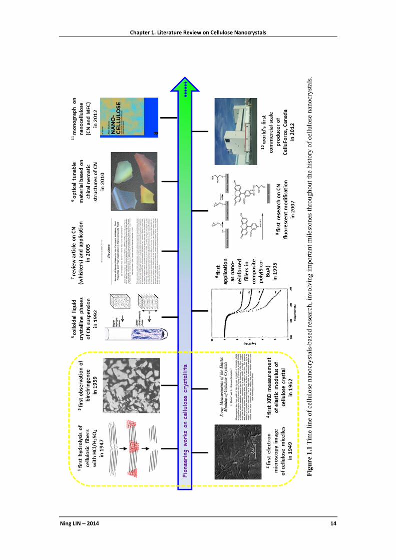
Chapter 1. Literature Review on Cellulose Nanocrystals
Ning LIN −−−− 2014 14
Figure
1.1
Tim
e line
of ce
llul
ose
nano
crys
tals
-bas
ed res
earc
h, in
volv
ing
impo
rtan
t mile
ston
es th
roug
hout
the
histor
y of
cel
lulo
se n
anoc
rystal
s.

Chapter 1. Literature Review on Cellulose Nanocrystals
Ning LIN −−−− 2014 15
1.1 Extraction and Production of Cellulose Nanocrystals
Cellulose microfibrils formed during biosynthesis are 2–20 nm in diameter, depending
on the source, and can be several micrometers in length. Each microfibril consists of
crystalline domains interspersed with disordered amorphous regions. The preparation
of cellulose nanocrystals involves a chemical hydrolysis process to dissolve amorphous
chains and release crystalline domains from cellulose fibers.
1.1.1 Extraction at Lab Scale
The extraction of cellulose nanocrystals by chemical hydrolysis is now an established
laboratory process and has been applied to a wide variety of raw materials. With diverse
hydrolyzing agents, cellulose nanocrystals possessing different surface groups and
surface chemistry can be prepared, as shown in Figure 1.2 (A). Hydrolysis with
hydrochloric acid preserves the hydroxyl groups of native cellulose but leads to less
stable aqueous suspensions.14 With the combination of TEMPO and hypochlorous
sodium, surface hydroxyl groups of cellulose nanocrystals can be selectively
transformed into carboxyl groups, which may be useful for subsequent modification.15
It was reported that ammonium persulfate [(NH4)2S2O8] can also be used as a
low-toxicity oxidant for the isolation of carboxylated cellulose nanocrystals.16
Moreover, an acid mixture composed of hydrochloric and an organic acid (acetic acid)
can be used to hydrolyze cellulose fibers inducing the hydrolysis and modification in
one single-step process,17 which offers the hydrophobic acetyl groups on the surface of
cellulose nanocrystals. Recently, some novel systems have been developed for the
isolation of cellulose nanocrystals, such as enzymatic hydrolysis,18 ionic liquid
hydrolysis,19 and gaseous acid hydrolysis.20
For most researches on cellulose nanocrystals, sulfuric acid hydrolysis has been more
extensively used for their preparation because of its high efficiency. When sulfuric acid
is used as the hydrolyzing agent, disordered or paracrystalline regions of cellulose
fibers are preferentially hydrolyzed, whereas crystalline regions that have a higher
resistance to acid attack remain intact. The general procedure for the preparation of

Chapter 1. Literature Review on Cellulose Nanocrystals
Ning LIN −−−− 2014 16
sulfuric acid-hydrolyzed cellulose nanocrystals is shown in Figure 1.2 (B). It should be
pointed out that during the hydrolysis, sulfuric acid can react with hydroxyl groups of
cellulose, which yields charged sulfate esters on the surface of nanocrystals and
promotes the dispersion of nanoparticles in water.21
Figure 1.2 (A) The different extraction methods of cellulose nanocrystals providing distinctive
surface chemistry: hydrochloric acid treatment provides hydroxyl, acetic acid provides acetyl,
TEMPO-mediated hypochlorite treatment provides carboxylic acid, and sulfuric acid treatment
provides sulfate esters as well as hydroxyl with desulfation treatment; (B) general procedure for the
preparation of cellulose nanocrystals with sulfuric acid hydrolysis.
During the extraction of cellulose nanocrystals with sulfuric acid hydrolysis,
Thielemans et al. detected the presence of adsorbed low molecular weight organic
compounds on the surface of nanocrystals, which can block the available reactive
sites.22 It was reported that a Soxhlet extraction treatment using ethanol was effective in
removing adsorbed species, and promoting improved reproducibility of subsequent
surface modification reactions.

Chapter 1. Literature Review on Cellulose Nanocrystals
Ning LIN −−−− 2014 17
1.1.2 Pilot and Commercial Production
Scale-up of cellulose nanocrystals for larger production is under way. Several
manufacturing facilities (as shown in Table 1.1) have or are being built that will
increase production to upwards of multiple tons per year. Alberta
Innovates-Technology Futures has constructed the world’s first pilot plant to produce
cellulose nanocrystals, with the production capacity of 100 kg per week. Domtar Corp.
and FPInnovations have created a joint venture (new organization named CelluForce)
to build a commercial-scale plant for the production of cellulose nanocrystals, in a
$40.8 million project. In January 2012, CellForce officially announced to open a plant
for the production of cellulose nanocrystals with a target production rate of 1 ton/day.
For the industrial production of cellulose nanocrystals, two issues should be
emphasized. One is the standardization for the procedure and conditions of cellulose
nanocrystals’ production, which is being constituted by TAPPI standard
recommendation.23 Another issue is the further development of practical end-uses for
cellulose nanocrystal-based materials, despite the fact that a large and growing body of
information on potential applications of cellulose nanocrystals has been identified and
demonstrated in the lab. It seems that many companies are interested by the potential of
cellulose nanocrystals to improve the quality of their products but face unknowns of
cost and sustainability of supply. At the same time, cellulose nanocrystals producers
remain mute on production costs and real marketing waiting for the potential uses. It is
a vicious circle.
Table 1.1 Industrial production of cellulose nanocrystals (CN) or carboxylated CN.24,25
Producer Country Product Estimated production capacity by 2015 FPInnovations Canada CN 10 kg/week (pilot production) Alberta Innovates- Technology Futures
Canada CN 100 kg/week (pilot production)
Bio Vision Technology Canada carboxylated CN 4 tons/year (commercial production) CelluForce/Domtar Canada CN 1 tons/day (commercial production) US Forest Service Forest Products Laboratory
USA CN, carboxylated CN
35-50 kg/day (pilot production)
Melodea Israel CN unknown Central Institute for Research on Cotton Technology (CIRCOT)
India CN, NFC, MFC unknown
Chapter 1. Literature Review on Cellulose Nanocrystals
Ning LIN −−−− 2014 18
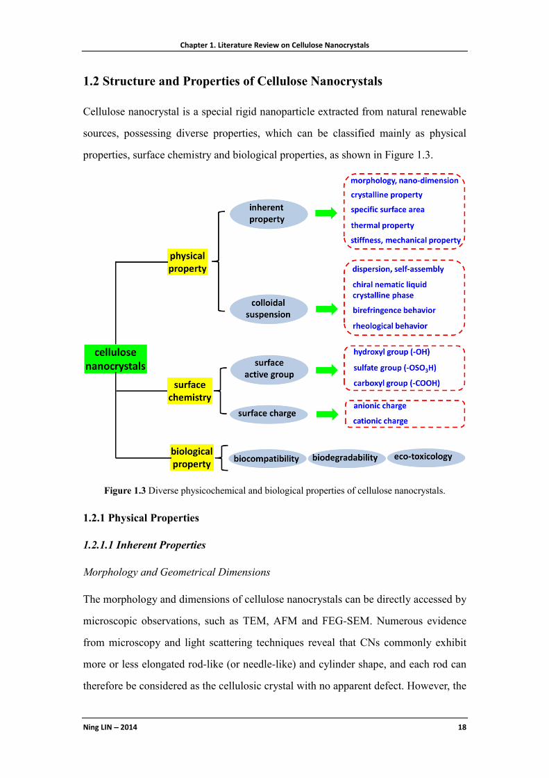
1.2 Structure and Properties of Cellulose Nanocrystals
Cellulose nanocrystal is a special rigid nanoparticle extracted from natural renewable
sources, possessing diverse properties, which can be classified mainly as physical
properties, surface chemistry and biological properties, as shown in Figure 1.3.
Figure 1.3 Diverse physicochemical and biological properties of cellulose nanocrystals.
1.2.1 Physical Properties
1.2.1.1 Inherent Properties
Morphology and Geometrical Dimensions
The morphology and dimensions of cellulose nanocrystals can be directly accessed by
microscopic observations, such as TEM, AFM and FEG-SEM. Numerous evidence
from microscopy and light scattering techniques reveal that CNs commonly exhibit
more or less elongated rod-like (or needle-like) and cylinder shape, and each rod can
therefore be considered as the cellulosic crystal with no apparent defect. However, the

Chapter 1. Literature Review on Cellulose Nanocrystals
Ning LIN −−−− 2014 19
specific nanocrystal cross-section is still in the disputes. According to the early reports,
cross-sections of cellulose microfibrils observed by TEM were found to be square or
rectangle, whereas their AFM topography showed a round profile.26 Recently, ellipse
cross-section for CN observed by AFM was proposed, and the effects of different
models of geometrical cross-sections on surface chemistry of cellulose nanocrystals
were investigated.27,28
If regarding cellulose nanocrystal as a cylinder with regularly round cross-section, the
parameters of CN physical dimension include the length (L), diameter (D) and aspect
ratio (L/D). The geometrical dimensions of CN are determined by several factors,
including the source of native cellulose and hydrolysis conditions. Nanocrystals
extracted from botanical sources (such as cotton, flax, ramie, sisal, etc.) commonly
have a length ranging from 100 nm to 700 nm, and a diameter about 5 nm to 30 nm.
Nanocrystals from tunicate and bacterial cellulose are several micrometers in length,
and 5 nm to 50 nm in diameter due to the highly crystalline property of these sources.
TEM images of CNs from four different sources are shown in Figure 1.4.
Figure 1.4 TEM images of dilute suspensions of cellulose nanocrystals from (a) ramie;29 (b)
cotton;30 (c) tunicate cellulose (Styela clava);31 (d) bacterial cellulose.32

Chapter 1. Literature Review on Cellulose Nanocrystals
Ning LIN −−−− 2014 20
Crystalline Properties
The crystalline structure of cellulose nanocrystals depends on the ordered packing of
cellulosic chains. During biosynthesis, van der Waals and intermolecular hydrogen
bonds between hydroxyl groups and oxygen atoms of adjacent molecules promote
parallel stacking of multiple cellulosic chains forming a highly ordered (crystalline)
structure. The hydrolysis conditions (acid concentration, temperature and duration)
significantly affect the degree of crystallinity (ratio between the mass of crystalline
domains and the total mass of nanocrystals) and crystalline dimensions of prepared CN.
Theoretically, the degree of crystallinity of CN can be 100%, but incomplete removal of
amorphous regions may result in a lower degree of crystallinity. The crystallinity index
of CNs is commonly reported as 54–90% depending on the source and hydrolysis
conditions.
The crystallinity index of CNs can be analyzed by different techniques including X-ray
diffraction (XRD), solid-state 13C NMR, and Raman spectroscopy. XRD height method
is the most popular and direct method to calculate the crystallinity index of CN, which
generally gives higher values than those of the peak area or peak deconvolution
method. The crystallinity index of CN can also be estimated from the spectrum of
cellulose samples acquired by solid-state 13C NMR as the peaks at 84 and 89 ppm are
assigned to the C4 atom for disordered and ordered cellulose, respectively. The
crystallinity index is then calculated as the ratio of the crystalline peak area (87–93
ppm) over the total area assigned to the C4 atom peak (80–93 ppm).33 Similarly in
Raman spectroscopy, the crystallinity index of CN can be estimated as the relative
intensity ratio of the Raman lines I1481cm and I1462cm, reflecting crystalline and
amorphous regions of cellulose I.34,35
Specific Surface Area
The specific surface area (Asp) of cellulose nanocrystal affects the determination of
surface hydroxyl group amount, which can be estimated from the average geometrical
dimensions and cross-section models for crystalline cellulose. Different cross-section
models for CN will provide distinct Asp values. For instance, with the square and round

Chapter 1. Literature Review on Cellulose Nanocrystals
Ning LIN −−−− 2014 21
cross-section models, Asp of CN depends only on the width (W) of nanoparticles;
whereas with the rectangle and ellipse cross-section models, Asp of CN is determined by
both the width (W) and the height (H) of the nanoparticles.
Thermal Properties
Thermal properties of cellulose nanocrystals, especially thermal stability, are of
importance for the processing and practical uses. Regardless the source of native
cellulose, sulfuric acid-hydrolyzed CNs commonly displays a significant reduction of
thermal stability in comparison with raw cellulose. This effect is ascribed to the
hydrolysis treatment, and the introduction of sulfate groups resulting from the use of
sulfuric acid for the preparation of CNs. Typically, the thermal degradation for sulfated
CN can be described as a two-step process, viz. low temperature process with thermal
degradation temperature (Td1) between 150 and 300 °C, and high temperature process
with thermal degradation temperature (Td2) between 300 and 450 °C. The low
temperature process involves the degradation of most accessible amorphous regions
which are highly sulfated. The high temperature process involves the degradation of
less accessible interior crystalline regions that are comparatively less sulfated. To
increase the thermal stability of sulfated CN, desulfation of nanoparticles with diluted
alkaline (NaOH) can be performed. A marked increase by 50−100 °C for Td1 compared
to untreated nanocrystals was reported.29,36
Stiffness, Modulus and Other Mechanical Properties
The elastic modulus of cellulose nanocrystal is an important physical property,
especially with respect to the ultimate aim of exploiting its full potential in composite
materials. Since 1930s, the elastic modulus of crystalline cellulose has been
investigated either by theoretical evaluations or by experimental measurements (wave
propagation, XRD, Raman spectroscopy, and AFM). The modulus of cellulose
microfibrils is expected to result from a mixing rule between the modulus of the
crystalline domains and the amorphous fraction. Therefore, it should be higher for more
crystalline cellulose nanocrystals. A broad range of values was reported, as shown in
Table 1.2, and it is generally accepted that the Young’s modulus of cellulose

Chapter 1. Literature Review on Cellulose Nanocrystals
Ning LIN −−−− 2014 22
nanocrystal should be in the range 100–200 GPa.
Table 1.2 Longitudinal (EL) and transverse (ET) moduli of crystalline cellulose.37
Material Method EL (GPa) ET (GPa) Ref.
cellulose I Calculation 77-121 [38] 56 [39]
Bleached ramie fibers (cellulose I) XRD 134 [40] Fortisan H fibers (cellulose II)
XRD 70 - 90 [41]
Cellulose I Calculation 172.9* [42] 70.8** 76 51-57 [43]
Cellobiose (two hydrogen bonds −cellulose I)
Calculation 136±6 [44]
Cellobiose (one hydrogen bond −cellulose II)
89±4
Ramie fibers (cellulose I) XRD 122-135 [45] Mercerized ramie fibers (cellulose II) 106-112 Cellulose I Calculation 167.5 11 [46] Cellulose II 162.1 50 Purified ramie fibers (cellulose I) XRD 138 [47] Polynosics (cellulose II) 88 Cellulose IIII 87 Cellulose IIIII 58 Cellulose IVI 75 Cellulose I Calculation 134-135 [48] Cellulose II 83
Cellulose II Calculation 155 24 - 51 [49] Cellulose Iβ Calculation 127.8 [50] Cellulose Iβ 115.2 Cellulose Iβ Raman 143 [51] Cellulose Iβ Calculation 124-155 [52] Cellulose Iβ Calculation 136-155*
114-117** [53]
Cellulose Iβ 116-149* 124-127**
Cellulose II 109-166* 101-106**
Cellulose Iβ Calculation 156 at 300K 117 at 500 K
[54]
Cellulose I Raman 57-105 [55] Ramie fibers (cellulose I) Inelastic X-ray
scattering 220 15 [56]
TEMPO-oxidized Cellulose Iβ AFM 145 [57] Acid hydrolyzed Cellulose Iβ 150 Wood AFM 18-50 [58] Disaccharide cellulose Iβ Calculation 85.2*/37.6** [59] Disaccharide cellulose Iβ 99.7*/33.0** Extended cellulose Iβ chains (10-40 glucoses)
126.0*/63.3**
Cellulose Iβ Calculation 139.5±3.5 28.8±2.9 [60] Cellulose Iβ Calculation 206 19 [61]
* with intramolecular hydrogen bondings ** without intramolecular hydrogen bondings

Chapter 1. Literature Review on Cellulose Nanocrystals
Ning LIN −−−− 2014 23
1.2.1.2 Properties of Colloidal Suspensions
Dispersion and Self-assembly in Solvents
The dispersion and self-aggregation states of cellulose nanocrystals in solvents will
significantly determine their properties and applications. Due to the high surface area
and hydrophilic nature (numerous hydroxyl groups), strong inter-particle hydrogen
bonding will inevitably induce the nanocrystals aggregation. This effect can be proved
by the weak dispersion and flocculating tendency of unstable HCl-hydrolyzed cellulose
nanocrystal aqueous suspensions. Although some solvents were reported to weaken the
interchain bonding in cellulose, such as DMF, with N atoms in its structure for the
formation of new hydrogen bonds (O–H⋅⋅⋅N) between hydroxyl groups and the N
atom,62 these solvents (particularly organic solvents) are very limited in terms of
practicality. In order to improve the dispersion of cellulose nanocrystals in water or
organic solvents, diverse surface treatments or modifications are performed based on
the strategy of converting surface properties and weakening interparticle hydrogen
bonds. One of the common method for the promotion of cellulose nanocrystals’
dispersion in water is the use of sulfuric acid as the hydrolyzing agent, which introduces
negatively charged sulfate groups (����� /�� ) on the surface of nanocrystals.
Attributed to the electrostatic repulsion between sulfate groups, sulfated cellulose
nanocrystals can perfectly dispersed in aqueous medium. With similar mechanism,
carboxylated cellulose nanocrystals can homogeneously dispersed in water via
electrostatic stability of surface carboxyl groups (���� /�� ). To facilitate the
dispersion of cellulose nanocrystals in organic solvents and enhance the compatibility
between nanocrystals and matrices, the surface chemical modification method is
generally used. It will be detailedly discussed in Section 1.3 (Surface modification of
cellulose nanocrystals) and Section 1.4 (Nano-reinforcing application of cellulose
nanocrystals in composites).
Chiral Nematic Liquid Crystalline Phase
It is known that any asymmetric rod-like particle (such as polymer micro-objects,
viruses, rod-like alumina, etc.) shows a liquid crystalline behavior and cellulose

Chapter 1. Literature Review on Cellulose Nanocrystals
Ning LIN −−−− 2014 24
nanocrystals are no exception. At low concentrations, suspensions of cellulose
nanocrystals are isotropic, that is, the rod-like nanoparticles are randomly oriented.
When a critical concentration is exceeded, the nanocrystals in the suspension align and
form a colloidal chiral-nematic phase. Over a certain concentration range the isotropic
and chiral-nematic phases coexist until finally, upon exceeding an upper critical
concentration, only the chiral-nematic phase is present.63 In fact, for sufficiently high
concentrations, cellulose crystallites have a helical twist along the main axis, which can
induce crystal suspensions to attain a helical twist normal to the main axis of the rod,
and thus organize into a chiral nematic phase or cholesteric phase of stacked planes
aligned along a perpendicular axis64 (as shown in Figure 1.5A). While the helix of
cellulose nanocrystals is always left-handed, reflecting the intrinsic chirality of
crystalline cellulose, the value of the pitch can vary greatly, from less than 1 to 50 mm
and beyond (as shown in Figure 1.5B, C). The pitch depends on the quality and
concentration of cellulose nanocrystals, but also temperature, and even ionic strength of
the solution.65 Many aspects of helix formation are still poorly understood, limiting our
ability to control it and to tune the pitch.66 It is worth noting that highly sulfonated
nanocrystals have different charge behavior than non-sulfonated hydroxyl surface
crystals and so can be expected to show different liquid crystalline behavior. For
instance, sulfuric acid and phosphoric acid derived nanocrystals give a chiral nematic
structure whereas hydrochloric acid derived nanocrystals with post-reaction
sulfonation give a birefringent glassy phase that shows a crosshatch pattern.67
When ordered colloidal phases of cellulose nanocrystals are dried slowly, the
chiral-nematic structure of the suspension can be preserved in solid films.68
Chiral-nematic phases have unusual optical properties, such as (a) reflection of light in
a Bragg-type manner, generating spectacular iridescent colors (as shown in Figure
1.5D), (b) high optical activity, that is, strong rotation of the plane of linearly polarized
light and (c) selective reflection of circularly polarized light of particular handedness
without changing the sense of circular polarization. The optical tunable nanomaterials
based on the chiral nematic liquid crystalline behavior of cellulose nanocrystals will be

Chapter 1. Literature Review on Cellulose Nanocrystals
Ning LIN −−−− 2014 25
further discussed in Section 1.5 (Advanced functional nanomaterials based on cellulose
nanocrystals).
Figure 1.5 (A) Schematic image of chiral nematic ordering present in cellulose nanocrystal
suspensions and illustration of half-helical pitch P/2;12 (B) Characteristic regularly spaced helix
lines in an aqueous suspension with 5 wt% cellulose nanocrystals derived from sulfuric acid
hydrolysis of wood pulp, observed in transmission. The distance between two lines is half-helical
pitch (P/2), and in this sample P≈13 μm;66 (C) SEM image of helix fracture surface for an actual
bulk cellulose nanocrystals film;69 (D) Illustration of photonic crystal Bragg reflection from a
cholesteric liquid crystal and the angular dependence of the color for the sample.66
Birefringence
Theoretically, a beam of unpolarized light entering certain anisotropic materials, such
as a crystal, will be divided into two beams, vibrating in different planes perpendicular
to each polarized rays. Cellulose nanocrystals display this property with a strong
absorption of one of the polarized rays, i.e. they possess two refractive indices. The
light emerging from such a material will be plane-polarized light, which is the property
of birefringence, or double refraction. The birefringence of cellulose nanocrystal
suspension results from two origins: (i) a structural form anisotropy of cellulose (∆n ~
0.05) and (ii) a flow anisotropy from alignment of nano-rods under flow operated
Chapter 1. Literature Review on Cellulose Nanocrystals
Ning LIN −−−− 2014 26
before observation. Birefringence of cellulose nanocrystal suspensions can be observed
with two linear polarizers crossed at 90° (cross-nicols or crossed-polars). Figure 1.6
shows photographs of birefringence domains for tunicate cellulose nanocrystal
suspensions with different concentrations in water (image A), freeze-dried and
re-dispersed CNs in water (image B), and dispersed CNs in water and diverse organic
solvents (image C). It should be pointed out that the presence of birefringence for
cellulose nanocrystal suspensions can be used as the criterion to assess the good or
weak dispersability of nanocrystals in solvents.70
Figure 1.6 Birefringence photographs viewed through crossed polarizers: (A) dispersions of
as-prepared H2SO4-hydrolyzed tunicate cellulose nanocrystals in water; (B) freeze-dried CNs
re-dispersed in water (concentrations of CNs are 0.1, 0.5, 1.0, 2.0, 3.0, 4.0, 5.0, 6.0, and 7.0 mg/mL
from left to right in images A and B); (C) dispersions of 5.0 mg/mL of as-prepared CNs in water,
freeze-dried and re-dispersed in water, DMF, DMSO, N-methyl pyrrolidone, formic acid, and
m-cresol from left to right in image C.70
Rheological Behavior
The hydrodynamics properties of cellulose nanocrystal suspensions are directly related
to the concentration, shear rate, size, length distribution, and temperature. In the dilute

Chapter 1. Literature Review on Cellulose Nanocrystals
Ning LIN −−−− 2014 27
regime, shear thinning behavior of nanocrystal suspensions can be observed, and
suspensions exhibit the effect of concentration dependence. At higher concentrations,
where cellulose nanocrystal suspensions became lyotropic, a typical behavior of liquid
crystalline is generally reported. With regard to the influence of shear rate on the
viscosity of nanocrystal suspension, three distinct regions can be observed. At low
shear rates, the viscosity of nanocrystal suspension decreases continuously, which is
supposed to correspond to a shear thinning behavior where the domains formed by
nanocrystals begin to flow. As the shear rate increases, the domains start to break up
and a semi-plateau region can be found in the flow curve, where the shear thinning is
less pronounced. At a critical rate, a further shear thinning occurred corresponding to
the alignment of individual nanocrystals in the flow direction.71 The aspect ratio is
another key parameter in determining the degree of shear-induced order and the
relaxation behavior after shear ceased. It was reported that the viscous suspensions
derived from long nanocrystals (280 nm) remained aligned for hours and even days
after shearing, while suspensions with shorter nanocrystals (180 nm) were found to
relax quickly.72 An attempt to correlate the intrinsic viscosity of cellulose nanocrystal
suspensions to the aspect ratio of constituting nanoparticles was also reported.73 Based
on the evaluation of phase behavior using a combination of low-magnification imaging
and hot-stage cross-polarized optical microscopy, it was found that both concentration
and temperature will significantly affect the microstructure and shear response of
cellulose nanocrystal suspensions.74 Recently, the effect of surface charges to
rheological behavior of cellulose nanocrystal suspensions was reported. Due to the
influence of surface charge, the degree of sulfation of cellulose nanocrystals has a
significant effect on the critical concentration at which transitions from isotropic to
liquid crystal and liquid crystal to gel occur.75
1.2.2 Surface Chemistry
1.2.2.1 Surface Active Groups and Reactivity
From a structural point of view, cellulose is a high molecular weight
homopolysaccharide composed of β-1,4-anhydro-D-glucopyranose units (Figure 1.7).

Chapter 1. Literature Review on Cellulose Nanocrystals
Ning LIN −−−− 2014 28
These units do not lie exactly in the plane with the structure, but rather they assume a
chair conformation with successive glucose residues rotated through an angle of 180°
about the molecular axis and hydroxyl groups in an equatorial position.76 The ability of
these hydroxyl groups to form hydrogen bonds plays a major role in the formation of
fibrillar and semicrystalline packing, which governs the important physical properties
of these highly cohesive materials.77 As indicated with blue dashed lines in Figure 1.7,
intramolecular hydrogen bonds occur primarily between the hydrogen borne by the OH
group of the C3 carbon and ring oxygen of the adjacent glucose unit (O5). The
intermolecular hydrogen bonds occur between the hydrogen of the OH−6 primary
hydroxyl and oxygen in position O3 in a cycle of a neighboring unit, as well as the
hydrogen of OH−2 and oxygen in position O6.
It is well known that the unidirectional parallel orientation of cellulose chains within
the elementary fibrils, occurring during biosynthesis and deposition, induces the
formation of crystals having one side face with hydroxyl pendant groups known as the
non-reducing end and another front face with reducing end groups bearing hemiacetal
functionality. Each cellulose chain possesses directional asymmetry with respect to the
termini of its molecular axis: one end has a hemiacetal group (green labeled groups in
Figure 1.7), a chemically reducing functionality, and the other has a pendant hydroxyl
group, the nominal non-reducing end (pink labeled groups in Figure 1.7).
Figure 1.7 Schematic representation of chemical structure and intra-, inter- molecular hydrogen
bonds in crystalline cellulose.

Chapter 1. Literature Review on Cellulose Nanocrystals
Ning LIN −−−− 2014 29
One of the most specific characteristics of cellulose is that each of its monomers bears
three hydroxyl groups, which endows cellulose nanocrystals a reactive surface covered
with numerous active hydroxyl groups. For each anhydroglucose unit, the reactivity of
hydroxyl groups on different positions is heterogeneous. The hydroxyl group at the 6
position acts as a primary alcohol whereas the hydroxyl groups in the 2 and 3 positions
behave as secondary alcohols. Indeed, the carbon atom which carries the hydroxyl
group in the 6 position is only attached to one alkyl group, while the carbons with the
hydroxyl groups in the 2 and 3 positions are joined directly to two alkyl groups, which
will induce steric effects derived from the supramolecular structure of cellulose and the
reacting agent.78 It has been reported that on the structure of cellulose, the hydroxyl
group at the 6 position can react ten times faster than the other OH groups, while the
reactivity of the hydroxyl group on the 2 position was found to be twice that of at the 3
position.79 However, regarding the surface reactivity of hydroxyl groups from
nanocellulose (such as CNC), the use of reactants or solvents may affect the reactivity
of hydroxyl groups from different positions. Recent studies reported the order of
reactivity for hydroxyl groups on CNC as nucleophiles with OH-C6 = OH-C2 > OH-C3
by etherification. The reactions between hydroxyl groups and reagents only occur or
are controlled on the surface of cellulose nanocrystals. Geometrical considerations
obtained from microscopic observations are generally used to determine the content of
active hydroxyl groups on the surface of nanocrystals. With the calculation according to
the Connely surface methodology, it was reported around 3.8 mmol/g available
hydroxyl groups for cotton cellulose nanocrystals with average width d = 4 nm and
average length L = 250 nm.63 Recently, total content of surface hydroxyl groups on
cellulose nanocrystals was reported to be affected by the cross-section shapes, such as
0.96 mmol/g for square or round cross-section model, 1.55 mmol/g for ellipse
cross-section model, and 2.65 mmol/g for rectangle cross-section model.27 However,
even for these surface cellulose chains, not all hydroxyl groups are accessible, since
some are oriented toward the inner of the nanoparticle. Indeed, due to different
orientation of some hydroxyl groups and high crystallinity of nanocrystals, as well as
large difference in reactivity of hydroxyl groups, it was assumed that the fraction of

Chapter 1. Literature Review on Cellulose Nanocrystals
Ning LIN −−−− 2014 30
hydroxyl groups available on the surface for the chemical modification was only 1/3 for
nanocrystals.80
The surface chemistry of cellulose nanocrystals, especially surface groups, is mainly
governed by the hydrolysis procedure for the extraction of nanocrystals. As discussed
previously (Figure 1.2A), besides hydroxyl groups (−��), diverse surface groups can
also be introduced on cellulose nanocrystals during different hydrolysis process,
including sulfate groups (−����� /�� ), carboxyl groups (−���� /�� ), and acetyl
groups (−�����). Recently, it is reported that with some mild reactions, surface
hydroxyl groups on cellulose nanocrystals can be simply converted as other active
groups, such as amino groups (−��),81 thiol group (−��).82
1.2.2.2 Surface charges
The dispersion stability of cellulose nanocrystals is dependent on the electrostatic
repulsion caused by surface charged groups. The surface hydroxyl groups remain intact
during hydrochloric acid hydrolysis, resulting in essentially no introduction of charged
groups. The amount of surface charge on cellulose nanocrystals prepared by
hydrochloric acid hydrolysis is therefore zero, which can be called as “charge-free”
nanocrystals (although traces of carboxyl groups up to 1.8 × 10⁻⁵ mol/g of cellulose
may be detected for nanocrystals from bleached wood pulp, as a result of oxidative
bleaching of the starting material).83 In contrast, surface sulfate groups are introduced
on cellulose nanocrystals during sulfuric acid hydrolysis via condensation esterification
(sulfation) between surface hydroxyls and an H2SO4 molecule, using another H2SO4
molecule as a condensation agent. The H2SO4−hydrolyzed nanocrystals, therefore, are
highly negatively charged, and form a well-dispersed aqueous colloidal suspension.
Surface charge amounts from sulfate groups of cellulose nanocrystals can be controlled
through the duration and temperature of H2SO4 hydrolysis. Phosphate group with
negative charges was also reported to be introduced on the surface of cellulose
nanocrystals, using a urea–phosphoric acid mixture for surface phosphorylation
reaction.84 Another negatively charged group, carboxyl group, can also be introduced
on the surface of cellulose nanocrystals with the acid hydrolysis and subsequent

Chapter 1. Literature Review on Cellulose Nanocrystals
Ning LIN −−−− 2014 31
TEMPO oxidation.
Besides the anionic surface charged groups, viz. negatively charged nanocrystals, the
preparation of positively charged cellulose nanocrystals by post-surface treatments was
also reported. Epoxypropyltrimethylammonium chloride,85,86 and quaternary
ammonium salts 87,88 were generally used as cationization agents for the preparation of
cationic cellulose nanocrystals. The type and amount of surface charged groups,
ζ-potential value, and dimensions of electrostatically stabilized cellulose nanocrystals
are summarized in Table 1.3.
Table 1.3 Surface charge amount and type, ζ-potentials, and dimensions for various electrically
stabilized cellulose nanocrystals. (Adapted from Ref. 89)
Types of
charged
group
Source
of CNs
Amount of surface
charge
ζ-
potential
(mV)
Dimensions of CN
(nm)
Ref.
In mol per
g of CN
In e
(nm-2)
Width Length
sulfate wood 8.4×10⁻⁵ — — 3.5 180±75 [83,90] a 4.7×10⁻⁵ — −32 6±1.5 145±25 [91] — — −51.5±0.8 8±1 90±10 [92]
cotton — 0.16 — 7 115 [67,93] 9.38–21.2×10⁻⁵
— — 7 177–390 [21]
— 0.41±0.05 −39±3 5–10 100–350 [94] tunicin 8.5×10⁻⁵ — — 20 2200 [95] b BC 5.0×10⁻⁶ 2.0×10⁻² — 10–50 1000–
2000 [96]
1.53×10⁻⁵ 5.1×10⁻² — 40±20 800±250 [97] 6±3×10⁻⁶ — — 7±17 855 [98]
ramie — 0.6 — 10.8±4.5 134±59 [99] carboxyl cotton 7.6×10⁻⁴ — — 23±7 250±91 [100]
cotton ~1.14×10⁻³ — — 5–10 100–200 [101] trance carboxyl
wood 1.8×10⁻⁵ — — 3.5 180±75 [84]
phosphate wood 3.0×10⁻⁵ — — 3.5 180±75 [84] c HTAP cotton 1.12×10⁻⁴ 0.26 +30±5 11±2 174±18 [85]
wood fiber
0.02–2.05×10⁻³
— +63 — — [86]
a The value is the sum of total anionic groups, including sulfate and carboxyl groups. b BC: bacterial
cellulose. c HTAP: 2-hydroxy-3-trimethyl-ammoniopropyl
1.2.3 Biological Properties
1.2.3.1 Eco-toxicology
Most studies reported non-toxicity of cellulose nanocrystals or toxicity similar to
Chapter 1. Literature Review on Cellulose Nanocrystals
Ning LIN −−−− 2014 32
microcrystalline cellulose and table salt. Kovacs et al. initially studied the inherent
eco-toxicology of cellulose nanocrystals with aquatic organisms.102 Rainbow trout
hepatocytes were selected as the model cells, and the toxicity monitoring program as
well as the in-depth toxicity assessment component was included in the toxicity testing
strategy. With the eco-toxicological characterization, cellulose nanocrystal was found
to have low toxicity potential and environmental risk, but showed no harm to aquatic
organisms at concentrations that could occur in receiving waters. In another report, the
cytotoxicity of cellulose nanocrystals against nine different cell lines was determined
by MTT and LDH assay, and no cytotoxic effects of cellulose nanocrystals against any
of these cell lines in the concentration range and exposure time studied (0~50 μg/mL
and 48 h)103 were reported. Similarly, cellulose nanocrystal was also found to have
non-toxic effect towards micro-organisms necessary for oxygen take-up.104 However,
recently it was reported that cellulose nanocrystals may induce some slight
dose-responsive cytotoxic and inflammatory effects on human lung cells, especially the
risk with inhalatory exposure under high concentrations of released cellulose
nanocrystal powders.105,106
It is well known that human body can only clear out impurity with the scale in
micrometer or above, while nanoparticles will penetrate into cells and eventually
remain in the system. Therefore, although studies conducted so far on cellulose
nanocrystals seem to indicate absence of toxicity, the research and systematic
assessment of eco-toxicology of cellulose nanocrystals are still incomplete and need
deeper investigation, especially for the effect of nanoparticle aggregation in body and
long-term in vivo data.
1.2.3.2 Biocompatibility
Biocompatibility is referred to as the capability of a prosthesis implanted in the body to
exist in harmony with tissue without causing deleterious changes, which is an essential
requirement for biomedical materials. It is a pity that direct study on the
biocompatibility of cellulose nanocrystals is rare. Even for the evaluation of the
biocompatibility of cellulose, the different studies are difficult to compare due to the

Chapter 1. Literature Review on Cellulose Nanocrystals
Ning LIN −−−− 2014 33
range of methodologies and sample preparations. According to the early reports,107,108
cellulose can be generally considered to be broadly biocompatible, invoking only
moderate (if any) foreign body responses in vivo. More recently, cellulose nanocrystals
or its composites were also reported to be used as bioscaffolds for cell cultivation
(which will be further introduced in Section 1.5.1.3). However, regardless the type of
cellulosic material (native cellulose, nanofibrillar cellulose, or cellulose nanocrystals),
it is sensible to judge the biocompatibility of such materials on a case-by-case basis.109
1.2.3.3 Biodegradability
In terms of degradation, cellulose may be considered as non-biodegradable in vivo or, at
best, slowly degradable, due to the lack of cellulase enzymes in animals. However, the
form (i.e., crystallinity, hydration and swelling) of cellulose may affect the degree of
degradation, absorption and immune response observed. Nonenzymatic, spontaneous
biodegradability of cellulose chains may perhaps account for slow breakdown of
unaltered cellulose within the human body, though this is admittedly conjecture and has
not been adequately studied.110 In an early in vivo study, Miyamoto et al. found that the
degradation of cellulose and cellulose derivatives in canine specimens depended
significantly on the cellulose crystalline form and chemical derivatization.107
Regenerated cellulose prepared by deacetylation of cellulose acetate (presumably the
highly crystalline cellulose II polymorph) did not measurably degrade over the course
of the 6-week experiment. Contrarily, however, up to 75% (w/w) of equivalent samples
of amorphous regenerated cellulose were degraded and absorbed over the same
experimental period.107 Another study reported that cellulose nanocrystals were
actually more biodegradable than their macroscopic counterparts in aqueous
environments.104 Recently, oxidized cellulose was rendered more vulnerable to
hydrolysis and therefore potentially degradable by the human body. Based on this
proposal, researchers attempted to enhance the biodegradability of cellulose through
oxidation, such as the report of improving bacterial cellulose degradability in vitro (in
water, phosphate buffered saline, and simulated body fluid) through periodate
oxidation.111 However, no in vivo result on the improvement of bacterial cellulose

Chapter 1. Literature Review on Cellulose Nanocrystals
Ning LIN −−−− 2014 34
biodegradability through oxidation treatment was reported, which needed further
investigation.
1.3 Surface Modification of Cellulose Nanocrystals
1.3.1 Approaches and Strategies for Surface Modification
1.3.1.1 Purpose and Challenge
Efficiency of nanofiller dispersion in a continuous matrix and adequacy of
nanofiller-matrix interfacial interactions are widely known to critically affect material
physical and mechanical properties. Hydroxyl groups present on cellulose and sulfate
ester units introduced during hydrolysis with sulfuric acid both contribute to the
hydrophilic character of cellulose nanocrystals. However, owing to the electrostatic
character and high ionic strength of CN, it is difficult to achieve a good dispersion level
of the nanoparticles in most common non-water organic solvents or non-polar
polymeric matrices. The reasons stem primarily from the inefficient electrostatic
repulsion and establishment of strong hydrogen bonds between nanocrystals, which
induce the rapid aggregation or flocculation of the colloidal suspension in highly
hydrophobic media.8
To prevent self-aggregation and promote efficient dispersion in non-aqueous media, the
surface of CN can be modified with hydrophobic compounds using covalent or
non-covalent coupling approaches. The purpose of the modification of CN consists in
reducing the surface energy and improving the degree of dispersion by transforming the
polar hydroxyl groups sitting at the surface of nanocrystals into moieties capable of
enhancing the interactions with non-polar polymers. In most cases the surface
modification of CNs can be conducted to (1) introduce stable negative or positive
electrostatic charges on the surface of nanocrystals to obtain better dispersion; (2) tune
the surface energy characteristics of nanocrystals to improve compatibility, especially
when used in conjunction with non-polar or hydrophobic matrices in nanocomposites;
and (3) tailor the nanoparticles to impart specific functional groups on nanocrystals for
advanced functional nanomaterials. The main challenge for surface modification of CN

Chapter 1. Literature Review on Cellulose Nanocrystals
Ning LIN −−−− 2014 35
is to choose a reagent and reaction medium that enable modification in such a way that
it only changes the surface of nanocrystals, while preserving the original morphology to
avoid any undesired bulk changes,112 polymorphic conversion and to maintain the
integrity of the crystal.76
1.3.1.2 Comparison of Approaches and Strategies
Surface modification of CN can be achieved by attaching small molecules or polymers
via covalent bonds or physical interactions. A wide variety of surface modification
techniques can be performed on the surface of CN, including physical absorption of
surfactant, chemical derivatization of coupling hydrophobic small molecules, or
chemical grafting of polymeric chains and advanced functional molecules (as shown in
Figure 1.8). These modification methods generally rely on the surface functionality
such as hydroxyl groups in native cellulose or on functionality derived from synthesis
of the nanoparticles as a ‘‘handle’’ for modification.113 Among these methods, a
convenient operation and procedure, i.e. physical absorption of surfactant or polymer
coating, is the simplest treatment in comparison with chemical modification. However,
this method was developed for improving dispersibility of CN in organic solvents and
is not expected to be effective in composite applications because of the lack of covalent
bonding between the hydroxyl groups from CNs and the groups from surfactants. In the
field of chemical modification, derivatization is the most effective approach to change
the surface properties of CNs, invoving the hydrophilicity, surface charge and density,
Figure 1.8 Diverse surface modifications on cellulose nanocrystals.

Chapter 1. Literature Review on Cellulose Nanocrystals
Ning LIN −−−− 2014 36
which can be carried out with the covalent conjunction between nanocrystals and
hydrophobic small molecules. In spite of easy achievement, it should be paid more
attention to the degree of derivatization on CNs to avoid the destruction of the substrate
if the reaction is pushed too far. Modification consisting of introducing functional
molecules on CN is a rather new field largely depending on its unique properties. Some
novel techniques such as “click chemistry” have been used in the functional
modification on CNs, which endows nanocrystals with advanced functions, such as
fluorescent labeling, gene delivery or photobactericidal effect.
Another chemical modification method, polymer grafting, can be realized with two
strategies, namely ‘grafting onto’ and ‘grafting from’. The ‘grafting onto’ approach
involves attachment of pre-synthesized polymer chains, carrying reactive end groups,
to the surface of CNs, whereas the ‘grafting from’ approach consists in mixing
nanocrystals with a monomer and an initiator agent to induce the polymerization of the
monomer from the nanoparticle surface. The schematic representation of diverse
approaches of modification of CNs is shown in Figure 1.9. For the first approach,
traditional ‘carboxylation–amidation reaction’ consisting in creating a covalent amide
bond between a primary amine-terminated polymer and carboxylated CNs by
COOH−NH2 coupling and ‘isocyanate-mediated reaction’ depending on the different
reactivity of isocyanate groups are widely used to graft polymeric chains onto the
surface of nanocrystals. The grafted polymers can be fully characterized before grafting
on the surface, offering the possibility of controlling the properties of the prepared
material. However, one can’t expect high grafting densities because of steric hindrance
and blocking of reactive sites by the already grafted polymer chains using the ‘grafting
onto’ method. ‘Grafting from’ has been proved to be a very effective approach to create
high grafting densities and well-controlled polymer structures on different kinds of
surfaces. Surface-initiated ring opening polymerization (ROP) and living radical
polymerization (LRP) are two methods that can be used for the strategy of ‘grafting
from’. In ROP, the hydroxyl groups on the surface of CNs act as initiator and the ratio
of monomer (such as ε-caprolactone or L-lactide) to initiating groups determines the DP

Chapter 1. Literature Review on Cellulose Nanocrystals
Ning LIN −−−− 2014 37
of the grafted polymer chains. For LRP modification, such as atom transfer radical
polymerization (ATRP) and single electron transfer-living radical polymerization
(SET-LR), two steps are involved, which are the initial formation of initiating sites for
LRP with the immobilized initiator on CNs, and the reaction of the initiator-modified
nanoparticles with a monomer to induce the polymerization. Because of the lower
viscosity of the medium and limitation of steric hindrance, this strategy has been
proved to be an effective way to create high grafting densities on the surface. However,
the drawback of this approach is the difficulty to precisely determine and characterize
the molecular weight of grafted polymers. To solve this problem, some studies recently
reported methods of cleaving the polymeric chains from the cellulose backbone, such
as dissolving PS-grafted CNs114 or PDMAEMA-grafted CNs115 in acidic condition with
acid hydrolysis or saponification treatment to cleave grafted PNiPAAm polymeric
chains from the surface of grafted CNs.116
Recently, some new strategies for the surface modification on CNs have been proposed,
such as grafting amine compounds or drug molecules on nanocrystals through the
change of glucose structure with periodate oxidation, while keeping the crystalline
integrity of nanocrystals.117,118
Figure 1.9 Comparison of different modifications of cellulose nanocrystals.
1.3.2 Adsorption of Anionic, Cationic and Nonionic Surfactants
In order to obtain non-flocculated dispersion of CNs in non-polar solvents, the easiest
approach is to coat the surface of nanocrystals with surfactants having polar heads and
long hydrophobic tails. With the simple mixing in solvent, non-covalent physical

Chapter 1. Literature Review on Cellulose Nanocrystals
Ning LIN −−−− 2014 38
absorption of surfactant on CNs will be a much more controllable and easy-operated
process than delicate chemical modification. Surfactants are usually amphiphilic
organic compounds, containing both water-insoluble (oil soluble) component for
hydrophobic groups (so-called tails) and water-soluble component for hydrophilic
groups (so-called heads). The hydrophilic end of the surfactant molecule may adhere on
the surface of CNs whereas the hydrophobic end may extend out providing a non-polar
surface and lowering the surface tension of the nanoparticles. With this process, the
ensuing surfactant-coated nanoparticles display reduced surface energy and improved
dispersibility or compatibility with non-polar organic media.
Since surfactant Beycostat NA (BNA) was applied as stabilizing agent to prepare stable
suspensions of CNs in nonpolar solvents,119 there were more studies focusing on the
influence and property of BNA-modified CNs. BNA is a commercial anionic
surfactant, and its chemical structure involves a phosphoric ester of polyoxyethylene
nonylphenyl ether. It was shown that the mixing ratio of surfactant and CNs should be
cautiously controlled. A low amount of surfactant is not sufficient to obtain a good
dispersion in organic solvents, whereas a too high amount of surfactant may induce
self-aggregation of nanoparticles during the mixing process. Meanwhile, using small
angle neutron scattering, it was observed that a surfactant layer 15 Å thick coated the
surface of CNs, which indicated the effective adsorption of BNA and folded
conformation of the surfactant molecule on the surface of nanocrystals.120 In further
studies, the properties of BNA-adsorbed CNs, such as morphology, self-assembling
and chiral nematic property, were investigated with various techniques including AFM,
TEM, and FESEM.121 Particularly, the chiral nematic ordering of BNA-adsorbed CNs
suspensions in cyclohexane with different concentrations was observed and
discussed.122 When using these surfactant-modified CNs as nanofillers to reinforce
polymeric matrix (such as PLA or PP), the presence of the surfactant on the surface of
nanocrystals was found to facilitate the dispersion of the nanofiller in the matrix and
improve the nucleation effect of the matrix for ensuing materials.123−125
Different from the hydrophilic-hydrophobic mechanism for the absorption of common

Chapter 1. Literature Review on Cellulose Nanocrystals
Ning LIN −−−− 2014 39
surfactants, cationic surfactant (such as DODA-Br) can be adsorbed on the surface of
CNs through strong electrostatic interactions between the negative charge of sulfate
groups on H2SO4-hydrolyzed nanocrystals and cationic charge of the surfactant. DODA
is a cationic surfactant that has been widely used in Langmuir films to investigate
adsorption and binding processes involving anionic polyelectrolytes at the air/water
interface. Rojas et al. applied cationic surfactant DODA to modify the surface of CNs,
and used this surfactant as the carrier to crosslink cellulose nanoparticles.126 With
Langmuir-Schaeffer (LS) technique, the precursor CNs-DODA complexes were
subsequently used to prepare monolayer films.
A nonionic surfactant, such as sorbitan monostearate, was also used to improve the
dispersion of CNs in organic solvents and to prevent their self-aggregation.127 From the
results of turbidity experiment performed on 0.3 and 0.6 wt% nanocrystals suspension
in THF, it was reported that the surfactant concentration determined the stability of
modified CNs in solvent. For high surfactant concentrations, the surfactant molecules
self-aggregate and probably do not adhere to the surface of nanocrystals, which
probably caused the weak stress transfer effect when using CNs as reinforcing filler.
The same surfactant was used to improve the dispersion of CNs in a polystyrene (PS)
matrix and to produce bead-free electrospun composite webs.128 In another study, as a
small surfactant molecule, tert-butanol, was used to modify the surface of CNs.129 It
was reported that tert-butanol was able to limit the aggregation of nanocrystals during
freeze-drying process, and promote more loose condition and homogeneous dispersion
in poly(lactic acid) (PLA) matrix.
1.3.3 Hydrophobic Groups from Chemical Derivatization
Because of their high surface area and hydrophilic nature, CNs tend to seriously
aggregate through hydrogen bonding, and can’t be easily dispersed in apolar solvents
and are poorly compatible with hydrophobic matrices. The inefficient reinforcement
results from poor dispersion induced by poor interfacial interaction between pristine
CNs and non-polar polymer matrices. Derivatization is one of the simplest and
immediate approach considering diverse chemical modifications. The chemical
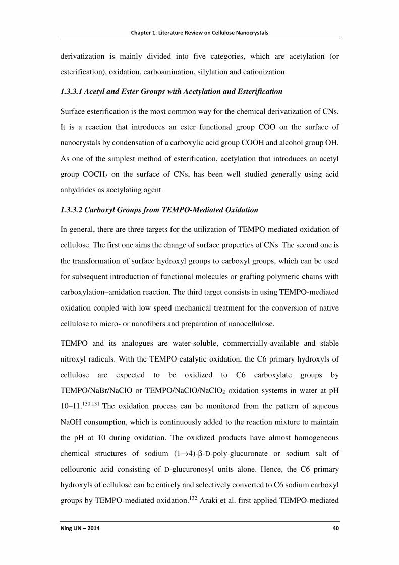
Chapter 1. Literature Review on Cellulose Nanocrystals
Ning LIN −−−− 2014 40
derivatization is mainly divided into five categories, which are acetylation (or
esterification), oxidation, carboamination, silylation and cationization.
1.3.3.1 Acetyl and Ester Groups with Acetylation and Esterification
Surface esterification is the most common way for the chemical derivatization of CNs.
It is a reaction that introduces an ester functional group COO on the surface of
nanocrystals by condensation of a carboxylic acid group COOH and alcohol group OH.
As one of the simplest method of esterification, acetylation that introduces an acetyl
group COCH3 on the surface of CNs, has been well studied generally using acid
anhydrides as acetylating agent.
1.3.3.2 Carboxyl Groups from TEMPO-Mediated Oxidation
In general, there are three targets for the utilization of TEMPO-mediated oxidation of
cellulose. The first one aims the change of surface properties of CNs. The second one is
the transformation of surface hydroxyl groups to carboxyl groups, which can be used
for subsequent introduction of functional molecules or grafting polymeric chains with
carboxylation–amidation reaction. The third target consists in using TEMPO-mediated
oxidation coupled with low speed mechanical treatment for the conversion of native
cellulose to micro- or nanofibers and preparation of nanocellulose.
TEMPO and its analogues are water-soluble, commercially-available and stable
nitroxyl radicals. With the TEMPO catalytic oxidation, the C6 primary hydroxyls of
cellulose are expected to be oxidized to C6 carboxylate groups by
TEMPO/NaBr/NaClO or TEMPO/NaClO/NaClO2 oxidation systems in water at pH
10–11.130,131 The oxidation process can be monitored from the pattern of aqueous
NaOH consumption, which is continuously added to the reaction mixture to maintain
the pH at 10 during oxidation. The oxidized products have almost homogeneous
chemical structures of sodium (1→4)-β-D-poly-glucuronate or sodium salt of
cellouronic acid consisting of D-glucuronosyl units alone. Hence, the C6 primary
hydroxyls of cellulose can be entirely and selectively converted to C6 sodium carboxyl
groups by TEMPO-mediated oxidation.132 Araki et al. first applied TEMPO-mediated
Chapter 1. Literature Review on Cellulose Nanocrystals
Ning LIN −−−− 2014 41
oxidation to introduce carboxyl groups and then graft PEG chains onto the surface of
CNs. It was a pity that this study only focused on the effect of grafting polymers, and
changes of dispersion state and fingerprint texture of oxidized CNs.101 Montanari et al.
systematically investigated the degree and size influence of the TEMPO-mediated
oxidation of cotton linter CNs.133 The degree of oxidation (DO) was determined by
conductometric titration as 0.24. During TEMPO-mediated oxidation, a decrease of the
crystal size occurred, and the introduction of negative charges on the surface of the
crystalline domains induced a better individualization and dispersion of the
crystallites,133 as shown in Figure 1.10a and a'. In another study, the maximum degree
of oxidation (DOmax) for CNs with TEMPO treatment was calculated as 0.095 (roughly
0.1) with a simplified rectangle model. With the change of NaClO/anydroglucose unit
ratio, the degree of oxidation of CNs varied from partial oxidation (DO=0.029) to
nearly complete oxidation (DO=0.097).136
Figure 1.10 (a and a') TEM images of cotton CNs and TEMPO-oxidized CNs;133 (b and b') TEM
images of sisal CNs and carboaminated CNs;134 (c and c') TEM images of cotton CNs and silylated
CNs dispersed in THF;135 (d and d') AFM height images of cotton CNs cationic modified CNs.85

Chapter 1. Literature Review on Cellulose Nanocrystals
Ning LIN −−−− 2014 42
1.3.3.3 Derivatization with Isocyanate Carboamination
Through the reaction between isocyano group and hydroxyl group, isocyanate
possessing a single isocyano group can be coupled onto the surface of CNs. The
chemical modification with isocyanate as coupling agent presents some advantages
such as (i) relatively high reaction rate; (ii) absence of secondary products, and (iii)
chemical stability of urethane moiety.137 Isocyanates used for the derivatization
modification of CNs were mainly phenyl isocyanate (PI), dimethylbenzyl isocyanate
(TMI) and n-octadecyl isocyanate (C18H37NCO). The former reagents (PI and TMI) are
isocyanates bearing a rigid phenyl in their chemical structure. The reactivity of
isocyanate with OH group is higher than that of anhydride and, on the other hand,
aromatic NCO group is more reactive than aliphatic one. It was reported that the
modification on CNs using long-chain n-octadecyl isocyanate with a degree of
substitution DS of 0.07 improved the dispersion of CNs in dichloromethane, as shown
in Figure 1.10b and b').80,134 Recently, a new isocyanate agent, UPy-NCO, was used for
the surface modification of CNs, which can provide active UPy motifs for further
reactions.138
1.3.3.4 Silyl Groups from Silylation
Silylation consists in the introduction of substituted silyl groups on the surface of
cellulose nanocrystals. The chemical structure of organosilanes (R1R2R3SiX) presents
two kinds of functional groups, namely hydrophobic aliphatic groups with different
lengths of carbon chains (R1R2R3) and functional groups (X) for the coupling with
hydroxyl groups of CNs. In the structure of most silylating agents used for the
derivatization of CNs, R1 and R3 are generally methoxy, and R2 varies from one carbon
chains to twelve carbon chains (CH3, i-C3H7, n-C4H9, n-C8H17, n-C12H25). X is a
hydrolyzable group able to react with active hydroxyl groups from CNs, typically
alkoxy, acyloxy, amine, or more commonly chlorine.
Trimethylsilylation was first applied for the silylation modification of CNs.32 The
topochemically trimethylsilylated derivatization of CNs reduced their hydrophilicity
and improved their dispersion in acetone. However, the degree and mechanism of

Chapter 1. Literature Review on Cellulose Nanocrystals
Ning LIN −−−− 2014 43
silylation of CNs were not investigated. Goussé et al. studied the process of silylation
modification of CNs and proposed a mechanism model to describe the influence of
silylation on CNs structure. Four alkyldimethylchlorosilane silylating agents
(IPDMSiCl, BDMSiCl, ODMSiCl, and DDMSiCl), with alkyl moieties ranging from
isopropyl to n-butyl, n-octyl and n-dodecyl, were used to prepare more stable silylated
CNs.139 It was shown that under low degree of silylation, just a few silyl groups are
distributed randomly on the surface of CNs, which provided them the structural
integrity but unchanged hydrophilic property. If too much silylating agent is used or too
long reaction duration is performed, excessive silylation breaks up the cellulose chains
and cause the solubilization of CNs in the reaction medium. Therefore, silylated
derivatization of CNs should be controlled in the moderate degree range to provide a
sufficient number of silyl groups, and confer a change to hydrophobic surface but not
induce the destruction of the original structure of CNs. With the silylating agent
DDMSiCl, Pei et al. conducted partial silylation of cotton CNs in toluene (as shown in
Figure 1.10c and c'), and used the modified CNs to reinforce PLA.135 The effect of
methacryloxypropyltrimethoxysilane (MPS) as silylating agent for the surface
modification of CNs was also investigated.140 Recently, A-174 as new silane coupling
agent for the surface silylation of CNs was reported. This special silylating agent can
provide vinyl groups on the surface of CNs acting as the initiator for successive free
radical polymerization.141
1.3.3.5 Cationic Groups from Cationization
A few studies were conducted on the surface cationic modification of CNs, to convert
surface anionic charges to cationic charges. Surface cationization of CNs with
2,3-epoxypropyltrimethylammonium chloride (EPTMAC) proceeds through the
nucleophilic addition of the alkali-activated cellulose hydroxyl groups to the epoxy
moiety of EPTMAC.85 The cationization process reversed the surface charge from
−39±3 mV before to +30±5 mV after treatment with EPTMAC, and led to a reduction
of the total surface charge density from 0.41 e nm-2 to 0.26 e nm-2. The modification
also promoted the formation of stable dispersions and cationized CNs were more
Chapter 1. Literature Review on Cellulose Nanocrystals
Ning LIN −−−− 2014 44
spreadable than pristine CNs on flat mica surfaces, as shown from AFM images in
Figure 1.10d and d'. Glycidyltrimethylammonium chloride (GTMAC) and quaternary
ammonium salts were also used as cationic agents for surface cationization of CNs.86,87
Different from the common electrostatic adsorption method, a chemical grafting
method was proposed to introduce cationic poly(ethylene imine) (PEI) polymers on the
surface of CNs. The synthesis of PEI-grafted CNs included three steps, which were
surface modification of CNs using tosyl chloride, then cationic ring opening grafting
polymerization of 2-ethyl-2-oxazoline monomer with tosylated CNs as macroinitiators,
and finally production of cationic PEI-modified CNs with the hydrolysis treatment in
acidic condition.142
1.3.4 Polymeric Chains with Physical Adsorption or Chemical Grafting
The practical application of CNs faced many obstacles mainly related to poor
dispersibility in common nonpolar solvents and interfacial incompatibility between the
hydrophobic components and hydrophilic CNs. To prevent the aggregation and
improve the compatibility of CNs in diverse matrices, polymer chain grafting seems to
be of interest because the grafted polymer chains can act as compatibilizer, particularly
when they are of the same nature as the matrix, which can further improve the
interfacial adhesion and maximize the compatibility between both components. On the
other hand, grafting special polymers on CNs may endow these natural nanoparticles
advanced functional application. Polymer grafting can be accomplished by utilizing
different approaches such as “grafting onto” or “grafting from” strategies. Polymer
adsorption can also promote interfacial interactions with a polymeric matrix.
1.3.4.1 Hydrophilic Polymer–PEG and PEO
Cellulose nanocrystals are hydrophilic and therefore have limited ability to disperse in
hydrophobic solvents and polymer matrices.143 To restrain the coagulation of
nanoparticles in apolar solvents, the method of grafting or adsorption of hydrophilic
polymers on the surface of CNs was proposed. This modification involves the steric
colloidal stabilization of CN suspension, which results from the hindrance of direct
contact between nanoparticles and therefore inhibition of suspension coagulation. A

Chapter 1. Literature Review on Cellulose Nanocrystals
Ning LIN −−−− 2014 45
promising candidate is poly(ethylene glycol) (PEG), a water–soluble polymer
consisting of hydrophilic and flexible long chains. Four synthetic routes were proposed
to introduce PEG (or PEO) on the surface of CNs, which were mainly based on the
strategy of “grafting onto” and physical interaction, as shown in Figure 1.11. However,
one of the drawbacks of the “grafting onto” approach is the increase of the viscosity of
the reaction medium especially for polymeric chains with high molecular weight (Mn).
Therefore, the Mns of grafted PEG onto CNs in these systems were lower than 2500
g/mol. In 2001, Araki et al. first reported the steric stabilization of CN suspensions by
PEG grafting using a carboxylation–amidation procedure.101 By grafting PEG in a ratio
of 0.2–0.3 g/g of cellulose on CNs, the modified CNs showed drastically enhanced
dispersion stability and ability to redisperse into either water or chloroform from the
freeze–dried state. However, due to uncompleted reaction between surface carboxyl
groups and amino groups (only 20–30% carboxyl consumption in the amidation step),
the improved stability effect induced by the modification could not exactly be proved.
For these reasons, Kloser and Gray proposed an alkaline epoxide ring-opening method
for the surface grafting of PEG onto CNs.144 Desulfation of sulfuric acid-prepared
Figure 1.11 Different methods of introducing PEG chains on the surface of cellulose nanocrystals.

Chapter 1. Literature Review on Cellulose Nanocrystals
Ning LIN −−−− 2014 46
nanocrystals were carried out and the nanoparticles were modified with α-epoxy,
ω-methoxy-terminated PEG under alkaline conditions. Aqueous dispersions of
PEG-grafted CNs were stable over time and exhibited similar phase behavior as the
initial electrostatically stabilized CN suspensions, with the onset of chiral nematic
phase separation occurring at 5 wt%.
Physical interaction of PEG with cellulose in water was performed to introduce high
molecular weight PEG (polyoxyethylene-PEO) on the surface of CNs. Different from
grafting PEG of low molecular weigtht, adsorbed long PEO chains wrapped the
nanoparticles and promoted the interactions between CNs and apolar matrix, which
improved the dispersibility and thermal stability of CNs.145
1.3.4.2 Polyester–PCL and PLA
Aliphatic polyesters constitute the most versatile group of degradable polymers, which
are often biocompatible and hydrophobic with low glass transition temperature. In the
family of polyester, poly(caprolactone) (PCL) and poly (lactic acid) (PLA) are the most
common materials, which can be traditionally synthesized by ring-opening
polymerization (ROP) of cyclic ε-caprolactone and L-lactide monomers, respectively.
Therefore, grafting of PCL and PLA polymeric chains (especially PCL) on the surface
of CNs was widely investigated.
In the initial stage, several studies mainly focused on grafting PCL chains onto CNs
with isocyanate-mediated reaction. This chemical modification is based on the different
reactivity of the two isocyanate groups from 2,4-TDI, the isocyanate at the 4-position
being 7 times more reactive than the isocyanate at the 2-position.146 Grafting PCL onto
the surface of CNs involves a three-step process. The first step requires the reaction of
PCL on isocyanate group of phenylisocyanate. The second step involves the reaction of
PCL, now protected, with 4-position isocyanate group of 2,4-TDI. During the third
step, unreacted 2-position isocyanate group of 2,4-TDI is then reacted with the surface
hydroxyl groups of CNs to graft PCL chains onto the nanoparticles. PCL chains with
various molecular weights (such as 10000, 42500, 80000 and 2000 g/mol) were grafted
onto CNs for the reinforcement of co-continuous nanocomposites or electrospinned

Chapter 1. Literature Review on Cellulose Nanocrystals
Ning LIN −−−− 2014 47
nanofibers.147,148 It was shown that the grafting efficiency decreased with the length of
polymeric chains, and there was stronger reinforcing effect for long chains
PCL-modified CNs to nanocomposites resulting fromth presence of chain
entanglements and co-crystallization.
Because of the high achievable grafting density, and possibility of chain length control,
the “grafting from” strategy is generally privileged aiming in grafting PCL chains on
the surface of CNs in recent studies. Regarding this modification, the hydroxyl groups
on the surface of nanocrystals act as initiator to induce the polymerization of cyclic
ε-caprolactone (CL) monomer and the ratio of monomer to initiating groups determines
the DP of the grafted polymer chains. The grafting reaction was performed under
predetermined conditions and stopped by adding a few drops of dilute hydrochloric
acid solution. The non-grafted PCL homopolymer can be removed by solubilization–
centrifugation or Soxhlet extraction. Surface grafting of hydrophobic PCL moieties on
CNs can facilitate the dispersibility of nanoparticles in hydrophobic systems. Habibi et
al. and Goffin et al. applied Sn(Oct)2-catalyzed ROP grafting of PCL on CNs to
reinforce a PCL matrix using casting-evaporation29 and extrusion.149 In another study,
Lin et al. performed microwave irradiation to assist PCL surface ROP on CNs, and
prepared filaceous PCL-grafted CNs150 that were used to reinforce PLA matrix.
Reaction conditions and grafting efficiency reported in the literature for the preparation
of PCL-grafted CNs are summarized in Table 1.4. Citric acid was also used as a benign
alternative to metal catalyst in surface-initiated ROP for grafting PCL on CNs. With
“green” citric acid as catalyst, grafting efficiency of PCL on the surface of CNs was up
to 58 wt %, about 4 times higher than for earlier reported PCL-grafted CNs using
common catalyst of Sn(Oct)2.151 Carlsson et al. reported the monitoring of CN surface
grafting with PCL by Quartz Crystal Microbalance, which provided a tool to regulate
the polymerization reaction.152
Recently, two studies investigated the reproducibility and interfacial molecular
modeling of the PCL grafting on the surface of CNs. It was reported that the process of
Soxhlet extraction using ethanol before modification can effectively remove the

Chapter 1. Literature Review on Cellulose Nanocrystals
Ning LIN −−−− 2014 48
adsorbed low molecular weight organic compounds produced during the hydrolysis,
which may block the reactive sites of CNs and cause the variable surface
composition.22 Molecular dynamics simulation was used to estimate the adhesion based
on surface energies with different amounts of grafted caprolactone. The modifications
on nanocrystals lead to an increased work of adhesion between the surface and its
surrounding medium. Hydrogen bonds were formed extensively between the grafted
caprolactone and the surrounding medium.153
Table 1.4 Reactive conditions and grafting efficiency for the preparation of poly(caprolactone)
(PCL)-grafted CNs (L= length, W= width, T= thick, D= diameter; GE%: Grafting efficiency).
Source of cellulose
Dimensions of CNs (nm)
Method Catalyst/cou-pling agent
Solvent Condition GE% Ref.
ramie L=150-250 D=6-8
grafting onto 2,4-TDI toluene 70-80 °C, 7 days
[147]
ramie L=100-250 D=3-10
grafting onto 2,4-TDI toluene 75 °C, 24 h
30% [148]
ramie L=150-250 D=6-8
grafting from Sn(Oct)2 toluene 95 °C, 24 h
85% [29, 149]
cotton linter
L=200-300 D=10-20
microwave -grafting from
Sn(Oct)2 several minutes
85% [150]
cotton wool
grafting from citric acid 120 °C, 24 h
58% [22, 151]
Another biodegradable polyester, namely PLA, was chemically grafted on the surface
of CNs using the “grafting from” strategy with Sn(Oct)2–catalyzed ROP of L-lactide
initiated from the hydroxyl groups available on the CNs surface.154 The grafting
reaction was performed in toluene at 80 °C for 24 h, and grafting efficiency of PLA
chains was determined by gravimetry as 83%. The effect of covalent bounding of PLA
chains on CNs was investigated by dispersion experiment in organic solvent
(chloroform) in comparison with ungrafted CNs and CNs with physically adsorbed
PLA chains. It was shown that hydrophobic PLA chains grafted on CN surface were
highly solvated maintaining the nanoparticles in more stable and homogeneous
suspension in the apolar solvent. Another study investigated the “grafting from”
strategy for high molecular weight PLA chains with partial pre-acetylation of surface
hydroxyl groups to control the grafting density, while maintaining sufficient
interparticle contact for efficient stress transfer within a percolating network.155

Chapter 1. Literature Review on Cellulose Nanocrystals
Ning LIN −−−− 2014 49
1.3.4.3 Polymethacrylates
Originating from the attributes of “livingness”, control, and robustness from living
radical polymerization (LRP), the techniques of surface-initiated ATRP (SI-ATRP) and
SET-LRP (SI-SET-LRP) were developed to “graft from” polymer chains or brushes on
the surface of CNs. It was proved to be a versatile technique to synthesize well-defined
polymers with high surface grafting density and low polydispersity on the surface of
CNs. Copper-mediated LRP is generally chosen for its versatility with respect to
monomer choice and ease of synthesis. Generally, two steps are involved in the
chemical modification of grafting polymeric chains on CNs with LRP techniques, as
shown in Figure 1.12. The first step is the initial formation of initiating sites for LRP, in
which an initiator is immobilized on the nanoparticles, and macromolecular initiator
(commonly used as CNs-Br) can be obtained. It should be pointed out that
2-bromoisobutyryl bromide (BriB) is always chosen for the esterification of
nanocrystals, and provides the initiating sites. To prevent the destruction of the
crystalline structure of CNs, this reaction should be performed at low temperature
(sometimes with ice bath), which can dissipate superfluous heat. The second step
involves the reaction of the initiator-modified nanoparticles with monomers (C=C
alkene molecules) to induce the polymerization.
Figure 1.12 Synthetic routes of grafting polyolefin chains on CNs with the technique of ATRP.
Early reports on using SI-ATRP to graft polyolefin polymeric chains on CNs surface
were published by Zhang et al. concerning polystyrene (PS),114 poly [2-(N,N-dimethyl

Chapter 1. Literature Review on Cellulose Nanocrystals
Ning LIN −−−− 2014 50
amino) ethyl methacrylate] (PDMAEMA),115 and poly{6-[4-(4-methoxyphenylazo)
phenoxy] hexyl methacrylate} (PMMAZO).156 All modifications were performed on
the investigation of CNs extracted from filter paper, and the focus of these studies
emphasized on liquid crystal phase behavior and fingerprint texture of modified CNs. It
was a pity that the amount of bromine in the initiator-modified CNs was very low (only
0.6 wt%) in these studies, which restricted the subsequent grafting reaction. On the
other hand, the issue of grafting polymeric chains with different lengths, which implied
the controllable feature of ATRP, was not investigated. By altering the extent of initiator
surface modification through polymerization control, Morandi et al. produced
PS-grafted CNs with varying grafting densities and different polymer brush lengths.157
Various grafting densities of PS on the surface of CNs were realized by controlling the
final content of initiating sites under different conditions (duration and temperature of
BriB/CNs reactions). They further reported a method of grafting a photosensitive
moiety bearing initiating sites onto the surface of CNs, followed by grafting PS chains
with ATRP. This new pathway for the synthesis of PS-grafted CNs provided a
possibility to degraft the PS chains using UV-irradiation, and exactly investigated the
grafted polymer brush length and molecular weights.158 Walther et al. reported an
effective method of chemical vapor deposition (CVD) and continued esterification to
improve the initiator density on CN surface.159 The simple CVD pretreatment of BriB
for CNs was conducted for 24 h at room temperature (5 wt% bromine content), which
promoted the subsequent esterification to obtain more initiating sites (15 wt% bromine
content). Whereafter, poly(acrylic acid) (PAA) polyelectrolyte chains were grafted
from CNs via Cu-mediated surface initiated-ATRP of tert-butyl acrylate (tBA) and
subsequent hydrolysis. Via SI-SET-LRP technique at room temperature,116 CNs
derived from ramie fiber can be grafted with thermoresponsive
poly(N-isopropylacrylamide) (PNiPAAm) brushes. In their further study, based on
these grafted copolymers, an ultrathin and temperature-responsive material was
prepared without the need of anchoring polymeric matrix.160
With the method of HCl hydrolysis or saponification, grafted polyolefin chains can be

Chapter 1. Literature Review on Cellulose Nanocrystals
Ning LIN −−−− 2014 51

Chapter 1. Literature Review on Cellulose Nanocrystals
Ning LIN −−−− 2014 52
cleaved from the surface of CNs, and the properties of grafted polymers, such as
molecular weight (Mn) and polydispersity, can be carefully investigated by gel
permeation chromatography (GPC) or size exclusion chromatography (SEC). As
shown in Table 1.5, diverse monomers, reactive conditions and properties of grafted
polyolefin chains on CNs are summarized.
Despite the advantages of controllability and high grafting density of surface-initiated
LRP, there are some shortcomings for this technique, which are the inconvenience of
experimental operation (with three freeze-pump-thaw or vacuum-nitrogen cycles) and
rigorous reactive conditions of anaerobic circumstance. Some studies exploited other
approaches to graft polyolefin on the surface of CNs. Harrisson et al. grafted PS and
poly(t-butyl acrylate) (PtBA) chains on CNs via the carbodiimide-mediated amidation
reaction between the terminal amine groups from polyolefin polymers and carboxylic
acid groups on the surface of oxidized CNs.100 PS or PtBA polymers were first
synthesized on a chemical of N-(2-(2-Bromoisobutyryl) ethoxy) ethyl phthalimide,
which provided the terminal amine groups through subsequent deprotection reaction.
Whereafter, the hydrophobic polymer was grafted onto the surface of oxidized CNs
with the amidation reaction. The polymer-grafted nanocrystals can be readily dispersed
in nonpolar solvents such as acetone.
1.3.4.4 Block Copolymer
Besides the introduction of hydrophilic polymers (such as PEG) and hydrophobic
polymers (such as polyolefin), block copolymers including both hydrophilic and
hydrophobic segments can be used to modify the surface of CNs. A kind of
saccharide-based amphiphilic triblock copolymer, xyloglucan oligosaccharide-poly
(ethylene glycol)-polystyrene (XGO-co-PEG-co-PS), was used for the surface
modification of CNs.161 The triblock copolymer was pre-synthesized and physically
adsorbed on CNs in the DMF/H2O (1:100 v/v) solvent. The weight fraction of adsorbed
copolymer on CNs was determined as 45% by TGA. The modified CNs with adsorbed
triblock copolymer were easily redispersed in organic solvent (toluene), and chiral
nematic self-ordering of modified CNs was observed in nonpolar solvent. In another
Chapter 1. Literature Review on Cellulose Nanocrystals
Ning LIN −−−− 2014 53
study, Azzam et al. introduced PPG-co-PEG copolymers on the surface of CNs using
the “grafting onto” approach.162 The coupling reaction was performed between the
carboxyl groups from oxidized CNs and amine-terminated groups from Jeffamine
copolymers either in water or in DMF. It was shown that in DMF, the low molecular
weight copolymer can be grafted onto CNs with a high grafting density (DS=0.1049).
Individual nanocrystal was decorated by a shell of polymer chains, which induced the
steric stabilization, surface-active, and thermoreversible aggregation of modified
nanoparticles. However, due to the limitation of “grafting onto” strategy, only
copolymers of low Mn (1000 or 2070 g/mol) can be used in this chemical modification.
1.3.4.5 Other Hydrophobic Polymer
Polypropylene (PP) is a common hydrophobic polymer, which is classicaly used as the
representative apolar matrix. The comparison of the reinforcing effect of CN grafted
with maleated PP and CN dispersed with the surfactant Beycostat NA (BNA) into
atactic PP as the matrix was reported.163 It was shown that the PP-grafted CNs tended to
aggregate in the matrix, and reduced the ductility of ensuing nanocomposites, whereas
surfactant-coated CNs showed good dispersion in the matrix (transparent films), and
improved ductility. Via the carboxylation–amidation procedure, polypropylene glycol
(PPG, Mn=600) hydrophobic chains can also be grafted onto the surface of CNs with
the covalent reaction between carboxylated CNs and amine-terminated PPG.164 The
approach involving direct ‘grafting onto’ modification for the introduction of
hydrophobic polymers on CNs seems to be inefficient, which may be attributed to the
weak interaction resulting from the compulsive conjunction between hydrophilic
nanocrystals and hydrophobic long chains.
1.3.5 Advanced Functional Groups
Recently, more and more studies have led to the emergence of diverse potential special
applications of CNs as advanced functional nanomaterials, which are closely associated
with their modification using common derivatization or polymer grafting through the
introduction of advanced functional groups .63 The properties and applications of these
functional nanomaterials based on CNs will be systematically discussed in Section 1.5,

Chapter 1. Literature Review on Cellulose Nanocrystals
Ning LIN −−−− 2014 54
and this section will just focus on the modification reaction and process.
1.3.5.1 Fluorescent and Dye Molecules
Through covalent conjunction with nanoparticles, fluorophores can be used to label
these nanoparticles not only for fluorescence bioassay and bioimaging applications but
also for the investigation of bioeffects and safety of nanoparticles inside cells or human
being. For the first time, Dong and Roman introduced a fluorescent molecule (FITC) on
the surface of CNs via a three-step approach.11 First, the surface of the nanocrystal was
decorated with epoxy functional groups via the reaction with epichlorohydrin. Then,
the epoxy ring was opened with ammonium hydroxide to introduce primary amino
groups on CNs. Finally, the primary amino group was reacted with the isothiocyanate
group of FITC, in which covalent association between CNs and FITC was realized via
the formed thiourea groups. It was shown that unlabeled suspension was colorless and
slightly opaque, whereas FITC-labeled CNs suspension appeared clear and yellow.
Meanwhile, according to the results of UV/vis spectroscopy, FITC-labeled
nanocrystals showed absorption maxima of both the dianionic (490 nm) and the anionic
(453 and 472 nm) form of FITC, whereas unlabeled CNs did not show any absorption
peaks in the wavelength range 200 to 600 nm. In another study, with similar method,
two fluorescent molecules (FITC and RBITC), were introduced on the surface of CNs,
which was used to investigate the effects of cellular uptake and cytotoxicity for
different fluorescent CNs.165 The grafting of fluorescent terpyridine molecules166,167
and pyrene molecules168 on the surface of CNs was also reported.
Ryley and Thielemans developed two synthetic approaches to introduce two
fluorescent or dye molecules on CNs for the preparation of dual fluorescent labeled
CNs.169 The dual florescent labeled CNs (FITC and RBITC labeled CNs) and two dual
dyes labeled CNs (FAM-SE and TAMRA-SE labeled CNs as well as OG-SE and
TAMRA-SE labeled CNs) both exhibited pH sensing in McIlvaine buffers with varying
pH conditions.
Chapter 1. Literature Review on Cellulose Nanocrystals
Ning LIN −−−− 2014 55
1.3.5.2 Amino Acid and DNA
As natural rigid nanocrystals, CNs are expected to be good candidates for the
construction of biocompatible and biologically active nanomaterials, such as drug
carrier and gene delivery. The anchoring of amino acid or DNA molecules on the
surface of CNs allows the creation of a binding site to which drugs or targeting
molecules can be attached. The chemical modification of L-leucine on CNs was
performed through a two-step process involving the reaction between Fmoc-protected
L-leucine and thereafter the removal of Fmoc-protecting group. The first step consisted
in the reaction between CNs and Fmoc–L-leucine using DMAP as the catalyst and
EDC·HCl as the coupling agent. The removal of the protecting Fmoc group was
performed by stirring Fmoc–L-leucine CNs in piperidine/DMF solution.170 Mangalam
et al. developed the synthetic route of grafting different single-stranded
oligonucleotides with an amino modifier on CNs, and subsequent duplexing
complementary oligonucleotides formed with individual modified CNs via the
molecular recognition ability of the oligomeric base pairs.171 The grafting reaction of
amino-modified ssDNA on CNs was realized using the carboxylation–amidation
procedure. Two kinds of oligonucleotide-modified CNs, both strand A and strand B
modified CNs, were mixed together in water for the preparation of duplexed
DNA-modified CNs. It was shown that complementary strands of DNA bonded to
separate populations of CNs hybridized under suitable conditions to form bonded
structures.
1.3.5.3 Photobactericidal Porphyrin Molecule
With the benefits of biocompatibility and biodegradability of CNs, these kind of natural
nanoparticles have been more studied for biomedical material applications. Feese et al.
developed surface-modified CNs with cationic porphyrin via the Cu(I)-catalyzed
Huisgen−Meldal−Sharpless 1,3-dipolar cycloaddition “click reaction” occurring
between azide groups on the cellulosic surface and porphyrinic alkynes.172,173 During
the reaction, about 50% of all azide groups on the surface of modified CNs were
substituted by porphyrin molecules. Covalent attachment of porphyrin molecule to the

Chapter 1. Literature Review on Cellulose Nanocrystals
Ning LIN −−−− 2014 56
surface of CNs will potentially contribute to the longer lasting or permanent
antimicrobial property, minimizing the leaching of the biocidal agent into the
surrounding environment.
1.3.5.4 Self-Crosslinking of Nanocrystals
Contrarily to the traditional application of CNs as the reinforcing filler, some studies
apply CNs themselves to be the matrix for the construction of self-crosslinked
materials. Goetz et al. first developed the self-crosslinking of CNs extracted from MCC
with poly(methyl vinyl ether comaleic acid) (PMVEMA) and PEG as the crosslinking
agents by reacting varying amounts of CNs (0–100%).174,175 The crosslinking of CNs
with PMVEMA and PEG was anticipated to occur via an esterification reaction among
the hydroxyl groups of cellulose, terminal hydroxyl groups of PEG and the carboxylic
acid groups of PMVEMA. The crosslinking between three components trapped the
nanocrystals in the crosslinked network, preventing aggregation and subsequently
producing CNs self-crosslinked nanomaterials through intramolecular and interchain
reactions.
In another study, a three-step route of click chemistry was utilized for the synthesis and
formation of cellulose nanoplatelet gels with the regular self-crosslinking of CNs.176
Initially the primary hydroxyl groups on the surface of CNs were selectively activated
by converting them to carboxylic acids using TEMPO-mediated oxidation. Further
reactions were carried out via carbodiimide-mediated formation of an amide linkage
between precursors carrying the amine groups and the carboxylic acid groups on the
surface of oxidized CNs. Finally, with two kinds of modified CNs containing the azide
derivative or alkyne derivative on their surface, the click chemistry reaction of
Cu(I)-catalyzed Huisgen 1,3-dipolar cycloaddition between the azide and the alkyne
was performed on CNs, bringing together the nanocrystalline materials in a unique
regularly packed arrangement demonstrating a degree of molecular control for creating
self-crosslinked CNs nanomaterials.
Inspired by the water-enhanced mechanical gradient character of the squid beak, Fox et
al. recently reported a mechanical gradient nanocomposite made from modified

Chapter 1. Literature Review on Cellulose Nanocrystals
Ning LIN −−−− 2014 57
tunicate CNs by nanocrystals−nanocrystals crosslinking in PVA matrix. CNs were first
modified by the introduction of allyl moieties on the surface. With the addition of small
molecule tetra-thiol cross-linker and radical photoinitiator, modified CNs
self-crosslinked under exposure to UV irradiation via photoinduced thiol−ene
chemistry reaction. With the control of UV exposure duration, the degree of
crosslinking of the nanocrystals can be regulated, and fabricated mechanical gradient
nanocomposites.177
1.4 Nano-Reinforcement of Cellulose Nanocrystals in Plastic
Composites
The use of cellulose nanocrystals as reinforcing additive (or named as nanofiller) is
their most widely studied application. They have the potential to significantly enhance
the mechanical performance of plastic composites. As shown in Table 1.6, more than
half of recent review publications on cellulose nanocrystals during past three years
focused on or mentioned their application in composites. Various instances will be only
summarized but not repeatedly expounded in this section and the emphasis will be put
on the discussion of enhancing effect and mechanism.
1.4.1 Processing Techniques
Four common strategies, viz. casting-evaporation, melt-compounding, electrospinning,
and layer-by-layer assembly, have been reported in the literature to homogeneously mix
cellulose nanocrystals with a polymeric matrix, as shown in Figure 1.13. The earliest
study on the effect of processing approach to properties of cellulose nanocrystals-based
composites was reported by Hajji et al.193 Composites prepared by casting-evaporation
method showed higher mechanical performance than melt-compounding method,
which was probably attributed to less structural damage and preservation of
nanocrystals’ orientation in matrix during casting-evaporation.
Because of the good dispersion level of sulfated cellulose nanocrystals (H2SO4
hydrolysis) in water, it is simple to process nanocomposites by casting and water
evaporation using either water-soluble polymers or polymer aqueous dispersions

Chapter 1. Literature Review on Cellulose Nanocrystals
Ning LIN −−−− 2014 58
Tab
le 1
.6 R
evie
w p
ublic
atio
ns o
n ce
llulo
se n
anoc
ryst
als
duri
ng p
ast t
hree
yea
rs (
2012
−20
14).
Tit
le
Jo
urn
al
Yea
r R
ef.
Pro
cess
ing
of p
olym
er n
anoc
ompo
site
s re
info
rced
with
cel
lulo
se n
anoc
ryst
als:
a c
halle
nge
Int.
Pol
ym. P
roc.
20
12
[178
] G
reen
com
posi
tes
from
sus
tain
able
cel
lulo
se n
anof
ibri
ls: a
rev
iew
C
arbo
hydr
. Po
lym
. 20
12
[179
]
Cel
lulo
se n
anoc
ryst
als
and
mic
rofi
brill
ated
cel
lulo
se a
s bu
ildin
g bl
ocks
for
the
des
ign
of h
iera
rchi
cal
func
tiona
l m
ater
ials
J.
Mat
er. C
hem
. 20
12
[180
]
Pre
para
tion,
pro
pert
ies
and
appl
icat
ions
of
poly
sacc
hari
de n
anoc
ryst
als
in a
dvan
ced
func
tiona
l nan
omat
eria
ls: a
rev
iew
N
anos
cale
20
12
[63]
R
evie
w o
f ce
llulo
se n
anoc
ryst
als
pate
nts:
pre
para
tion,
com
posi
tes
and
gene
ral a
pplic
atio
ns
Rec
ent P
at.
Nan
otec
hnol
. 20
12
[181
]
App
licat
ions
of
fun
ctio
naliz
ed a
nd n
anop
artic
le-m
odif
ied
nan
ocry
stal
line
cellu
lose
T
rend
s B
iote
chno
l. 20
12
[182
]
Pro
duct
ion
of n
anoc
ryst
allin
e ce
llulo
se f
rom
lign
ocel
lulo
sic
biom
ass:
Tec
hnol
ogy
and
appl
icat
ions
C
arbo
hydr
. Po
lym
. 20
13
[183
]
Cel
lulo
se r
einf
orce
d po
lym
er c
ompo
site
s an
d na
noco
mpo
site
s: a
cri
tical
rev
iew
C
ellu
lose
20
13
[184
] R
einf
orce
d pl
astic
s an
d ae
roge
ls b
y na
nocr
ysta
lline
cel
lulo
se
J. N
anop
art.
Res
. 20
13
[185
] St
ate
of th
e ar
t man
ufac
turi
ng a
nd e
ngin
eeri
ng o
f na
noce
llulo
se: a
rev
iew
of
avai
labl
e da
ta a
nd in
dust
rial
app
licat
ions
J.
Bio
mat
er.
Nan
obio
tech
nol.
2013
[1
86]
Ass
essi
ng n
anoc
ellu
lose
dev
elop
men
ts u
sing
sci
ence
and
tech
nolo
gy in
dica
tors
M
ater
. Res
. 20
13
[187
] N
anoc
ellu
lose
: a n
ew a
gele
ss b
iona
nom
ater
ial
Mat
er. T
oday
20
13
[188
] B
acte
rial
cel
lulo
se s
caff
olds
and
cel
lulo
se n
anow
hisk
ers
for
tissu
e en
gine
erin
g N
anom
edic
ine
2013
[1
09]
Stre
ngth
and
bar
rier
enh
ance
men
ts o
f co
mpo
site
s an
d pa
ckag
ing
boar
ds b
y na
noce
llulo
ses
– a
liter
atur
e re
view
N
ord.
Pul
p Pa
p.
Res
. J.
2013
[1
89]
Nan
ocel
lulo
se p
aten
ts t
rend
s: a
com
preh
ensi
ve r
evie
w o
n pa
tent
s on
cel
lulo
se n
anoc
ryst
als,
mic
rofi
brill
ated
and
ba
cter
ial c
ellu
lose
R
ecen
t Pat
. N
anot
echn
ol.
2013
[1
90]
Ele
ctro
stat
ic o
r st
eric
? –
prep
arat
ions
and
cha
ract
eriz
atio
ns o
f w
ell-
disp
erse
d sy
stem
s co
ntai
ning
rod
-lik
e na
now
hisk
ers
of c
ryst
allin
e po
lysa
ccha
ride
s So
ft M
atte
r 20
13
[89]
Sust
aina
ble
nano
mat
eria
ls d
eriv
ed f
rom
pol
ysac
char
ides
and
am
phip
hilic
com
poun
ds
Soft
Mat
ter
2013
[1
91]
Key
adv
ance
s in
the
chem
ical
mod
ific
atio
n of
nan
ocel
lulo
ses
Che
m. S
oc. R
ev.
2014
[1
92]
Cel
lulo
se n
anoc
ryst
al-b
ased
mat
eria
ls:
from
liq
uid
crys
tal
self
-ass
embl
y an
d gl
ass
form
atio
n to
mul
tifun
ctio
nal
thin
fi
lms
NPG
Asi
a M
ater
. 20
14
[66]
Chapter 1. Literature Review on Cellulose Nanocrystals
Ning LIN −−−− 2014 59
(latex) as matrix. For the processing from water-insoluble polymers, the
casting-evaporation approach can be performed by involving a solvent exchange
procedure, in which the aqueous suspension of cellulose nanocrystals is solvent
exchanged in the proper liquid medium for the dissolution of the polymer by several
successive centrifugation and redispersion operations, using sonication after each
solvent exchange step to avoid aggregation.178 This mode of casting-evaporation
processing allows the preservation of the individualization state of the nanocrystals
resulting from their colloidal dispersion in solvents, and therefore keeps the maximum
nanoreinforcing effect for the composites. Indeed, during liquid evaporation, strong
interactions between nanoparticles can settle and promote the formation of a rigid
percolating network (which will be discussed in the following section). During slow
solvent evaporation, because of Brownian motions in the suspension or solution (whose
viscosity remains low, up to the end of the process when the latex particle or polymer
concentration becomes very high), the rearrangement of the nanoparticles is possible.
There is enough time for the interaction and connection among cellulose nanocrystals
to form a percolating network, which is the basis of their reinforcing effect for
composites.194,195
The wet processing approach, such as casting-evaporation, has been extensively used in
various reports for the preparation of cellulose nanocrystal-based composites.
However, this technique is both non-industrial and non-economic, and also limits the
number of polymer matrices that can be used in association with cellulose nanocrystals.
In view of the realization of large-scale production of cellulose nanocrystals (as
discussed in Section 1.1.2), more industrial composite processing techniques should be
introduced for the preparation of cellulose nanocrystal-based composites, e.g.
melt-compounding techniques, such as extrusion or injection molding. The processing
through the melt-compounding technique is commonly regarded as a “green”
(solvent-free), and industrially and economically viable procedure. However, due to the
issues of inherent incompatibility and thermal stability, this processing method is
infrequently employed for the preparation of cellulose nanocrystal-based composites.
Chapter 1. Literature Review on Cellulose Nanocrystals
Ning LIN −−−− 2014 60
In fact, the hydrophilic nature of cellulose causes irreversible agglomeration of the
nanoparticles during drying and aggregation in non-polar matrices, and the sulfate
groups resulting from H2SO4 hydrolysis seriously induce the reduction of the thermal
stability of nanocrystals.188
Figure 1.13 Four common processing techniques for the preparation of cellulose nanocrystal
(CN)-based composites.
Electrospinning is another alternative processing approach to mix cellulose
nanocrystals and polymer matrix, which can be used to prepare composite fibers with
diameters ranging from several microns down to 100 nm. In this process, a polymer
solution is positively charged to high voltage to produce the submicron scale fibers
from an orifice to a collector. Therefore, to find suitable solvents to dissolve cellulose
and to process it in the melt state are the key points for this processing technique.196
Unfortunately, only a few studies reported the preparation of cellulose

Chapter 1. Literature Review on Cellulose Nanocrystals
Ning LIN −−−− 2014 61
nanocrystal-based electrospinning composite fibers, which shows the remaining
challenge of this processing approach.
Layer-by-layer assembly (LBL) is a technique by which thin films or nanoparticles of
appositely charged layers are deposited. In a common procedure of LBL assembly,
positive or negative charged species are sequentially adsorbed by alternatively dipping
into the solutions. The excess solution after each adsorption step is rinsed with solvent
and thus a thin layer of charged species on the surface ready for the next adsorption step
is obtained. There are many obvious advantages of LBL technique, such as simple
manipulation, controllable thickness, and high concentration of cellulose nanocrystals
in multilayer films. However, the matrix materials used in this approach must be
polycation or polyanion with positive or negative charges (corresponding to anionic
sulfated CNs, or cationic modified CNs), as few reported for chitosan, PDDA, PHA,
PEI, DODA,63 which limits the wide application of LBL technique.
1.4.2 Pristine Cellulose Nanocrystal Reinforced Plastic Composites
Cellulose nanocrystal-based composites have been studied extensively because of the
promising reinforcing effect of these nanoparticles for numerous polymeric matrices. A
spectacular improvement of the shear modulus by more than two orders of magnitude
was observed in cellulose nanocrystal-reinforced poly(styrene-co-butyl acrylate) latex
nanocomposites.10 Since then, this rod-like nanoparticle was attempted to be
incorporated into various polymers, as shown in Figure 1.14. Broadly, the polymeric
matrices used for the preparation of cellulose nanocrystal-reinforced plastic composites
can be divided into two parts, biodegradable polymers and non-biodegradable
polymers. Natural polymers (including cellulose derivatives, starch, natural rubber
(NR) etc.) and various biopolymers (such as PHA, PLA, PCL etc.) are commonly used
as biodegradable polymeric matrices to prepare cellulose nanocrystals-reinforced
“bionanocomposites”. In the family of non-degradable polymers, polymers derived
from petrochemical sources are used as matrix for cellulose nanocrystal-reinforced
nanocomposites, such as PP, PS, PE etc.
Considering the most used processing approach “casting-evaporation”, aqueous and

Chapter 1. Literature Review on Cellulose Nanocrystals
Ning LIN −−−− 2014 62
organic-solvent systems are two divisions for the preparation of cellulose
nanocrystal-based plastic composites. The incorporation of cellulose nanocrystals into
aqueous polymer system is relatively straight forward because of the good dispersion of
nanocrystals in water. Casting and drying to remove the water result in successful
incorporation of cellulose nanocrystals into the polymer matrices. The use of water
over organic solvents promotes the use of cellulose nanocrystals in greener production
process. However, as discussed before, proper dispersion of cellulose nanocrystals in
polymer systems using organic solvents is difficult due to the tendency of nanocrystals
to aggregate in these solvents. Despite several reports on the use of DMF, it is still a
trouble to find suitable organic solvents for both dispersing cellulose nanocrystals and
solubilizing most of hydrophobic polymers. Therefore, in many organic-solvent
systems, solvent-exchange procedure is performed to convert the well dispersed
cellulose nanocrystals from water to organic solvent (such as ethanol or acetone), and
then introduce the suspension into the desired organic polymer solution to prepare
composites. The resulting mixture of polymer and cellulose nanocrystals in the organic
solvent can be processed in a manner similar to regular polymer solutions, including
solution casting and evaporative drying to form thin films, or melting extrusion.185
Figure 1.14 Various polymeric matrices used for the preparation of cellulose
nanocrystal-reinforcing plastic composites. (SF: silk fibroin; SP: soy protein; NR: natural rubber;
PHA: poly(hydroxy alkanoate); PHB: poly(hydroxyl butyrate); PLA: poly(lactic acid); PCL:

Chapter 1. Literature Review on Cellulose Nanocrystals
Ning LIN −−−− 2014 63
polycaprolactone; PBS: poly(butylene succinate); PFA: poly(furfuryl alcohol); PEO:
polyoxyethylene; PVA: poly(vinyl alcohol); PVAc: poly(vinyl acetate); WPU: waterborne
polyurethane; PU: polyurethane; PP: polypropylene; PS: polystyrene; PR: phenolic resin; PMMA:
poly(methyl methacrylate); PVC: poly(vinyl chloride); LDPE: low-density polyethylene)
Generally, when using pristine cellulose nanocrystals in composites, most studies
report similar tendency of improved mechanical stiffness, corresponding to the increase
of strength and elastic modulus (especially for elastic modulus), but at the cost of
elasticity. Mechanical reinforced mechanism will be discussed in the following section.
1.4.3 Modified Cellulose Nanocrystal Reinforced Plastic Composites
In order to promote their dispersion and improve the compatibility of cellulose
nanocrystals in non-polar or hydrophobic matrices, the surface characteristics of
cellulose nanoparticles can be tuned by surface chemical modification (such as
derivatization and grafting) or physical modification (such as coating with surfactant
and macromolecules). Various strategies and instances of surface modification of
cellulose nanocrystals have been introduced before, so emphasis of this section will be
paid on the effects of surface modification to the performance of prepared composites.
Table 1.7 summarizes diverse systems of modified cellulose nanocrystal reinforced
polymeric matrices for plastic composites.
Surface chemical modification of cellulose nanocrystals has a dual effect on the
mechanical properties of ensuing composites. The decoration of nanoparticles with
grafted moieties obviously restricts the possibility of inter-particle interactions and then
the establishment of a percolating network within the continuous medium. Therefore,
the outstanding properties resulting from this phenomenon are definitely lost. However,
depending on the matrix and processing conditions, it can allow a better dispersion and
then promote the compatibility of the filler with the matrix, which will induce higher
mechanical performance to be reached compared to the aggregated state of
nanoparticles. Specifically, the approach of chemical derivatization can be easily
performed by covalently introducing hydrophobic small molecules or groups, such as
acetylation, esterification, silylation etc. Derivatization is the most effective mean to

Chapter 1. Literature Review on Cellulose Nanocrystals
Ning LIN −−−− 2014 64
change the surface properties of cellulose nanocrystals, such as hydrophilicity, surface
energy, surface charge and density, which can significantly improve the dispersion of
nanocrystals in organic solvents and hydrophobic polymers. Another approach,
polymer grafting, is specially beneficial to the enhancement of compatibility between
cellulose nanocrystals and matrix, which derives from interfacial interactions between
grafted polymer chains and polymeric matrix.
Table 1.7 Modified cellulose nanocrystals reinforcing various matrices for plastic composites.
Modification Matrix Filler content Processing Ref.
Ph
ysi
cal
mod
ific
ati
on
surfactant coating (sorbitan monostearate)
PS 0−6 wt% casting-evaporation [127]
surfactant coating (BeycostatA B09)
PLA 0−5 wt% melt-extrusion [124]
surfactant coating (BeycostatA B09)
PLA 0−5 wt% casting-evaporation [197]
surfactant coating (BNA) PP 0−6 wt% casting-evaporation [125] surfactant coating (sorbitan monostearate)
PS 0−9 wt% electrospinning [128]
polyaniline coating PU 0−15 wt% casting-evaporation [198] PEO adsorption LDPE 0−9 wt% melt-extrusion [145] polyamide 6 coating PA 0−3 wt% melt-extrusion [199]
Ch
emic
al
mo
dif
ica
tio
n
silylation CAB 0−10 wt% casting-evaporation [32] silylation PLA 0−2 wt% casting-evaporation [135] silylation PLA 0−3 wt% melt-extrusion [140] acetylation PLA 0−10 wt% casting-evaporation [200] acetylation CAB 0−7 wt% casting-evaporation [201] acetylation PU 0−25 wt% casting-evaporation [202] carbamination PCL 0−12 wt% casting-evaporation [80,
203] carboxylation protein 0−5 wt% electrospinning [204] oxidation fibrin 0−50 wt% casting-evaporation [205] esterification PHB 0−7 wt% casting-evaporation [206] esterification CAB 0−0.9 wt% casting-evaporation [207] grafting onto aliphatic chain LDPE 0−15 wt% melt-extrusion [99] grafting onto aliphatic chain PLA 0−15 wt% melt-extrusion [208] grafting onto PCL PCL 0−50 wt% casting-evaporation [147] grafting onto PCL PCL 0−7.5 wt% electrospinning [148] grafting onto maleated-PP PP 0−6 wt% casting-evaporation [125] grafting onto PHBV PHBV 0−30 wt% casting-evaporation [209] in situ grafting onto PU PU 0−5 wt% casting-evaporation [210] in situ grafting onto chitosan chitosan 0−1.9 wt% casting-evaporation [211] grafting from PCL PCL 0−8 wt% melt-extrusion [149] grafting from PCL PCL 0−40 wt% casting-evaporation [29] grafting from PCL PLA 0−12 wt% casting-evaporation [150] grafting from PLA PLA 0−8 wt% melt-extrusion [154] carbamination; or grafting from PLA
PLA/NR 0−5 wt% melt-extrusion; or casting-evaporation
[212,213]
grafting onto PEG and adsorbing PEO
LDPE 0−20 wt% melt-extrusion [214]

Chapter 1. Literature Review on Cellulose Nanocrystals
Ning LIN −−−− 2014 65
Non-covalent modification, viz. physical interactions or adsorption of molecules onto
the surface of cellulose nanocrystals, is a simpler approach than chemical modification
to regulate the properties of cellulose nanocrystals. As introduced before, agents for
physical modification of cellulose nanocrystals include diverse surfactants and high
molecular weight polymers or copolymers. It is a pity that in order to tune the surface
property, the amount of surfactant or macromolecules required to cover cellulose
nanocrystals’ surface is high, which may undermine the properties of prepared
composites. In addition, specific physical interaction and mechanism between cellulose
nanocrystals and different adsorbed agents need to be further investigated, especially
adsorbing concentration and strength.
Although there have been intensive studies on surface modification of cellulose
nanocrystals to reinforce all kinds of polymeric matrices (as shown in Table 1.7), no
available industrial modification for the conversion of surface property of cellulose
nanocrystals to adapt their practical application in composites is reported. Regardless
of physical or chemical modification, each modification method has its limitations. In
future, for the purpose of developing cellulose nanocrystal-based composites to replace
petrochemical plastics, novel surface modification is required, with simple treatment or
procedure, high-efficiency property regulation, optimized performance of composites
and industrial production.
1.4.4 Mechanisms for Performance Enhancement
1.4.4.1 Mechanical Properties Enhancement
Besides the processing approaches discussed before, another two factors are also
expected to determine the reinforcing capability of cellulose nanocrystals, which are
compatibility between nanocrystals and matrix (interfacial mechanism), and
dimensions of cellulose nanocrystals (percolating mechanism). Interfacial mechanism
refers to the interactions between nanoparticles and matrix, while percolating
mechanism only depends on the hydrogen bonds among nanoparticles. The
filler/matrix interactions can be tuned by appropriate surface modification treatment of
the nanoparticles. This strategy is classically used in composite science to

Chapter 1. Literature Review on Cellulose Nanocrystals
Ning LIN −−−− 2014 66
compatibilize the reinforcing phase with the matrix for optimal performance. The
challenge consists therefore in promoting the homogeneous dispersion of cellulose
nanocrystals and avoiding agglomeration during processing, thus requiring eventually
favorable filler/matrix interactions, and at the same time promoting filler/filler
interactions to allow the beneficial formation of a percolating network of nanoparticles.
These two requirements are simply conflicting and no suitable strategy has been
proposed so far.
Cellulose Nanocrystal/Matrix Interfacial Interactions
Strong interfacial interactions are often associated with the existence of an interphase.
By definition, the interphase exists from some points in the fillers where the local
properties begin to change from the filler bulk properties, through the actual interface
into the matrix where the local properties again equal the bulk properties. The usual
question is to quantify the magnitude of the interactions between filler and polymer
chains and the distance away from the surface where the force field may be still in
effect. If significant specific interactions between polymer and filler exist, this will tend
to immobilize a layer of polymer around each solid particle. This immobilized polymer
layer contributes to the effective filler volume fraction in the compound.215
The filler/matrix interaction can be altered by different means. It obviously depends on
the chemical structure of the matrix. Polar matrices tend to strongly interact with the
cellulosic surface whereas apolar matrices weakly interact. The polarity of
nanoparticles can be tuned by a surface chemical modification allowing favorable
interacions with a non-polar matrix. The microstructure of the interfacial zone in the
vicinity of the reinforcing phase can also be locally altered by the presence of the filler
and affects the mechanical performance of the material.215
Rigid Percolating Network
The use of the percolation model to describe and predict the mechanical behavior of
cellulose nanocrystal-based composites suggests the formation of a rigid network of
nanocrystals, which should be responsible for the unusual reinforcing effect observed at
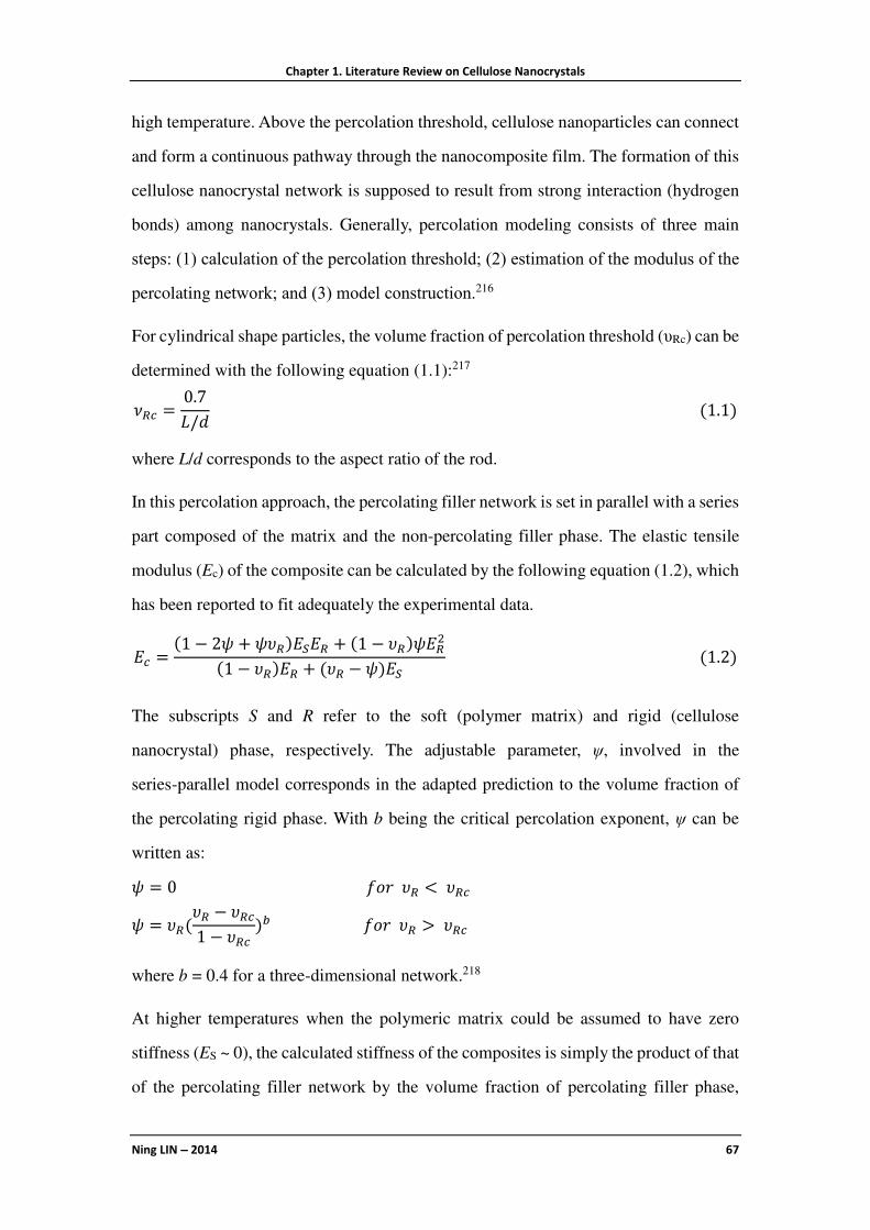
Chapter 1. Literature Review on Cellulose Nanocrystals
Ning LIN −−−− 2014 67
high temperature. Above the percolation threshold, cellulose nanoparticles can connect
and form a continuous pathway through the nanocomposite film. The formation of this
cellulose nanocrystal network is supposed to result from strong interaction (hydrogen
bonds) among nanocrystals. Generally, percolation modeling consists of three main
steps: (1) calculation of the percolation threshold; (2) estimation of the modulus of the
percolating network; and (3) model construction.216
For cylindrical shape particles, the volume fraction of percolation threshold (υRc) can be
determined with the following equation (1.1):217
�� =0.7
�/�(1.1)
where L/d corresponds to the aspect ratio of the rod.
In this percolation approach, the percolating filler network is set in parallel with a series
part composed of the matrix and the non-percolating filler phase. The elastic tensile
modulus (Ec) of the composite can be calculated by the following equation (1.2), which
has been reported to fit adequately the experimental data.
�� =(1 − 2� + ���)���� + (1 − ��)���
(1 − ��)�� + (�� − �)��(1.2)
The subscripts S and R refer to the soft (polymer matrix) and rigid (cellulose
nanocrystal) phase, respectively. The adjustable parameter, ψ, involved in the
series-parallel model corresponds in the adapted prediction to the volume fraction of
the percolating rigid phase. With b being the critical percolation exponent, ψ can be
written as:
� = 0� !�� <���
� = ��(�� − ���
1 − ���)#� !�� >���
where b = 0.4 for a three-dimensional network.218
At higher temperatures when the polymeric matrix could be assumed to have zero
stiffness (ES ~ 0), the calculated stiffness of the composites is simply the product of that
of the percolating filler network by the volume fraction of percolating filler phase,

Chapter 1. Literature Review on Cellulose Nanocrystals
Ning LIN −−−− 2014 68
which can be calculated according to the following equation (1.3):
�� = ���(1.3�
1.4.4.2 Thermal Properties Improvement
For most nanocomposite systems reinforced with cellulose nanocrystals, the addition of
the nanoparticles did not affect appreciably the glass transition temperature (Tg) and
melting/crystallization temperature (Tm) of the host polymer. This observation is quite
surprising considering the highly reactive nature and high specific area of
polysaccharide nanocrystals. Nevertheless, it can be well understood for hydrophobic
polymers because of the poor filler/matrix adhesion. The effect of cellulose
nanocrystals on the thermal stability (Td) of nanocomposites depends obviously on the
initial thermal stability of the neat polymer. Improved thermal stability of the
composites results from favorable interactions with the cellulosic filler and the
confinement effect of the filler to the polymeric matrix.219
1.4.4.3 Barrier Properties Enhancement
Most studies of cellulose nanocrystal-based composites focus on their mechanical
properties. However, there is an increasing interest in their barrier properties due to
increased tortuosity provided by nanoparticles (as shown in Figure 1.15). Indeed,
because of their small size, the surface-to-volume ratio of the nanoparticles is
significantly greater than that of microparticles. Moreover, the low permeability of
cellulose can be enhanced by the highly crystalline nature of cellulose nanocrystals and
ability to form a dense percolating network (Figure 1.15). Provided that strong
Figure 1.15 Barrier effects of cellulose nanocrystals in plastic composites.

Chapter 1. Literature Review on Cellulose Nanocrystals
Ning LIN −−−− 2014 69
particle-polymer molecular interactions exist, the smaller particles have a greater
ability to bond to the surrounding polymer material, thereby reducing the chain
segmental mobility and thus the penetrant diffusivity.220
1.5 Advanced Functional Materials Based on Cellulose Nanocrystals
Based on the special properties and construction strategies of CNs in functional
materials, this section will comprehensively discuss the emerging applications on four
sections, including surface properties-induced functional materials; unique
nano-reinforcing effects in functional materials; optical materials derived from chiral
nematic ordering; and special films and systems ascribed to barrier properties.
1.5.1 Surface Characteristics Induced Functional Materials
Even the most rigorous researcher will be attracted by diverse and interesting surface
characteristics of cellulose nanocrystals. First of all, cellulose nanocrystals possess a
reactive surface covered with numerous active hydroxyl groups (−OH), which provide
the possibility of modification via a chemical reaction strategy. Designing the chemical
grafting routes, various functional molecules or polymeric chains (such as fluorescent
molecules, DNA, peptide etc.) can be introduced on the surface of nanocrystals, with
the target of tailoring physicochemical properties and contributing novel functions. In
addition, surface hydroxyl groups of cellulose nanocrystals can be used as anchoring
and stabilizing sites, which provides a preferential template or scaffold for these natural
nanocrystals during the synthesis of various inorganic nanoparticles. On the other hand,
negatively charged surface (sulfated groups −OSO3H) of CNs resulting from
H2SO4-hydrolysis induces the electrostatic stabilization of the nanoparticles in aqueous
medium, and also provides the feasibility to perform electrostatic adsorption or
grafting. From the reactions by surface charges, CNs can be constructed as
high-efficiency adsorbing materials and emulsion nano-stabilizer. Finally, as
nanoscaled particles, CNs exhibited promising bioactivity and biocompatibility in
cellular cultivation and high surface area for application in water decontamination.

Chapter 1. Literature Review on Cellulose Nanocrystals
Ning LIN −−−− 2014 70
1.5.1.1 Active Groups
Importing Functional Groups or Molecules
Since the first report on the fluorescently labeling of CNs with FITC molecule,11 more
and more researchers investigated the fluorescent modification on the surface of CNs.
Modification on CNs with fluorescently labeling enables potential use of nanocrystals
in the field of biomedical applications, such as optical bioimaging, biosensor,
bio-probe, photodynamic therapy (PDT), together with diverse fluorescence
techniques, such as spectrofluorometry, fluorescence microscopy, and flow cytometry.
Besides the most common FITC reagent, many fluorescent molecules have been
attempted to covalently attach on the surface of CNs, such as RBITC,165 pyrene dyes,168
terpyridine and its derivatives,167,221 tryptophan-based peptides,222 PSE,223 FAM-SE,
TAMRA-SE, OG-SE,224 and PEI-chlorin p6 derivatives.225 However, most studies
focused on the pathway of introducing fluorescent molecules on CNs, but rarely
covered the in vivo or even clinic research. Consequently, there is a long way for the
practical application of fluorescent cellulose nanocrystals in biomedical materials.
Available hydroxyl groups and negative charges on the surface of CNs provide the
possibility of enzyme/protein immobilization on the basis of chemical conjunction and
electrostatic adsorption. Regarding the chemical conjunction approach, some studies
directly immobilized enzyme/protein on CNs with chemical grafting, such as
immobilization of lysozyme on amino-glycine-CNs with carbodiimide-activation
coupling reaction;226 and peroxidase on CNs with the activation of cyanogen bromide
treatment.227 Another approach consists in functionalizing with smaller nanoparticles
(generally gold nanoparticles Au), and then realizing the immobilization of
enzyme/protein on CNs with the aid of inorganic nanoparticles. Luong et al.
investigated Au/CNs as a catalytic platform for enzyme immobilization, which
exhibited significant biocatalytic activity and preservation of original activity. The
recovered specific activities were ∼70% and 95% for CGTase and alcohol oxidase
enzymes, respectively.228 More complicated carrier based on CNs/PEI/Au was
developed to immobilize glucose oxidase enzyme.229 Mahmoud et al. developed a

Chapter 1. Literature Review on Cellulose Nanocrystals
Ning LIN −−−− 2014 71
special nanocomposite consisting of magnetite nanoparticles (Fe3O4) and Au
nanoparticles embedded on CNs used as a magnetic support for the covalent
conjugation of papain and facilitated recovery of this immobilized enzyme.230 The
conjugated material retained high enzyme activity and good stability and reusability.
Based on the similar strategy of enzyme/protein immobilization, labeled DNA or
enzyme was immobilized on CNs as a probe, and this material can be used for the
identification or recognition of target DNA sequence, enzyme molecules, and as a
platform for immunoassays and diagnostics.231,232 Edwards et al. reported a
colorimetric approach to the detection of human neutrophil elastase (HNE) using
peptide conjugated cotton CNs.233
As mentioned in Section 1.3.5, some reports recently focus on the functional
modification of CNs for potential biological application, such as surface grafting of
amino acid molecules to offer biologically active building blocks on CNs;170 surface
grafting duplexing complementary DNA oligonucleotides to produce DNA-based
biocompatible materials;171 ferrocene-decorated CNs potentially to be used in
biosensors and bioelectrochemical assemblies;234 surface grafting imidazolium salt
([MPIM][Br]) for ion exchange systems or catalysts;169 and introduced cationic
porphyrin groups for the development of photobactericidal materials.172,173
Template for Synthesizing Inorganic Nanoparticles
The strategy of natural nanocrystals as templates for synthesizing inorganic
nanoparticles breaks new ground to make optimum use of inorganic materials, such as
rare noble metal resources, in association with the most abundant and renewable
bioresources. As a promising template in the synthesis of inorganic nanoparticles,
cellulose nanocrystals always play the role of reducing agent, structure-directing agent
and stabilizer during the reactions. Using rod-like CN suspensions as the template,
diverse inorganic nanoparticles have been prepared, such as mesoporous silica (Si,
SiO2, SiC) nanoparticles;235−239 calcium carbonate (CaCO3) nanoparticles;240 silver
(Ag) nanoparticles;231,241,242 gold (Au) nanoparticles;243,244 nickel (Ni) nanocrystals;245
platinum (Pt) nanoparticles;246,247 selenium (Se) nanoparticles;248 palladium (Pd)
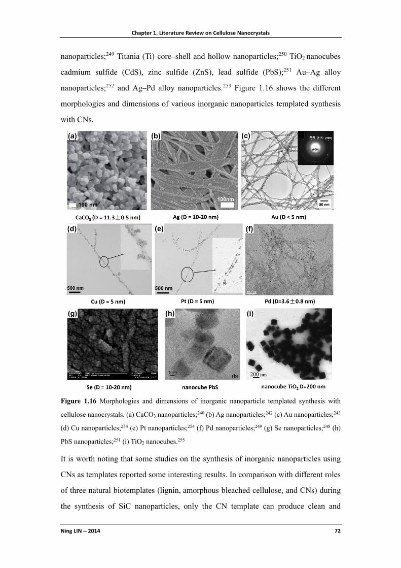
Chapter 1. Literature Review on Cellulose Nanocrystals
Ning LIN −−−− 2014 72
nanoparticles;249 Titania (Ti) core–shell and hollow nanoparticles;250 TiO2 nanocubes
cadmium sulfide (CdS), zinc sulfide (ZnS), lead sulfide (PbS);251 Au–Ag alloy
nanoparticles;252 and Ag–Pd alloy nanoparticles.253 Figure 1.16 shows the different
morphologies and dimensions of various inorganic nanoparticles templated synthesis
with CNs.
Figure 1.16 Morphologies and dimensions of inorganic nanoparticle templated synthesis with
cellulose nanocrystals. (a) CaCO3 nanoparticles;240 (b) Ag nanoparticles;242 (c) Au nanoparticles;243
(d) Cu nanoparticles;254 (e) Pt nanoparticles;254 (f) Pd nanoparticles;249 (g) Se nanoparticles;248 (h)
PbS nanoparticles;251 (i) TiO2 nanocubes.255
It is worth noting that some studies on the synthesis of inorganic nanoparticles using
CNs as templates reported some interesting results. In comparison with different roles
of three natural biotemplates (lignin, amorphous bleached cellulose, and CNs) during
the synthesis of SiC nanoparticles, only the CN template can produce clean and

Chapter 1. Literature Review on Cellulose Nanocrystals
Ning LIN −−−− 2014 73
uniform SiC nanowires (70 nm in diameter, > 100 µm in length) without the camelback
structure.239 Padalkar et al. reported the effects of the concentration of surfactant, salt
solution, reaction time, and pH of the salt solution to the average size of synthesized
inorganic nanoparticles (Ag, Cu, Au, and Pt) and coverage of CNs.254 Monodisperse
palladium (Pd) nanoparticle synthesized from colloidal CNs showed excellent active
catalyst effect on the Heck coupling of styrene and iodobenzene, which proved the
possibility of using CNs as an efficient support for catalysis.249 The introduction of CNs
that acted as a morphology-inducer and coordinating agent results in acceleration of the
crystal growth of TiO2 particles and promotes the formation of nanocube
morphology.255 The synthesis of porous TiO2 nanoclusters with different shapes,248
flower-like TiO2 nanocrystals256 using CNs as templates, and investigation of
photocatalytic activity of TiO2/CNs257 were also reported.
Based on the template synthesis of inorganic nanoparticles with CNs, some
organic/inorganic hybrid nanomaterials were reported. Especially for the combination
of Ag nanoparticles and CNs, ensuing materials exhibited the antimicrobial property
from nano-Ag, and enhanced mechanical performance from CNs.258,259
1.5.1.2 Surface Charges and Hydrophilicity
Cellulose nanocrystals are generally prepared by sulfuric acid hydrolysis inducing the
formation of sulfate ester groups (−OSO3−/H+) on the surface of the nanoparticles via an
esterification process. These charged moieties, which density increases with the
severity (acid concentration, time, temperature) of the treatment, cause the formation of
a negative electrostatic layer covering the nanocrystals and promote the high stability
of the H2SO4-prepared nanocrystal aqueous suspensions. Meanwhile, surface negative
charge provides the accessibility to electrostatic adsorption and conjunction with
cationic groups or molecules. With the negative charges on the surface of CNs, some
structural materials (such as layer-by-layer (LBL) assembling nanomaterials) and
functional materials (such as emulsion nano-stabilizer and adsorbed or permselective
materials) can be developed.

Chapter 1. Literature Review on Cellulose Nanocrystals
Ning LIN −−−− 2014 74
High-Efficiency Adsorption and Permselective Membrane
Owing to the negatively charged surface of CNs, direct application should be the
adsorption against positively charged molecules. Based on this strategy, Hsiao and
co-workers developed some efficient adsorbing membranes from cellulose nanofibers
and nanocrystals.260,261 Generally, CNs were anchored on the matrix surface, forming a
crosslinked nanostructured mesh with very high surface-to-volume ratio and a
negatively charged surface. The mean pore size and pore size distribution of this system
could be adjusted by the loading of CNs, where the resulting membrane not only
possessed good mechanical properties but also high surface charge density. Prepared
CN-based membrane exhibited 16 times higher adsorption capacity against a positively
charged dye over a commercial nitrocellulose-based membrane.262
The negatively charged sulfate groups on the surface of CNs can also be used to
develop permselective membranes, which are attributed to the inhibition of negatively
charged species but admission of positively charged species. A suspension of cotton
CNs (1.5 wt%) was used to form membranes at glassy carbon electrodes via the
interparticle hydrogen bonding between negatively charged nanocrystals with the
simple drop-coating procedure. This nanostructured membrane possesses the
permselective function to substances with different charge properties. It was shown that
positively charged ruthenium hexamine [Ru(NH3)63+] was significantly adsorbed by the
membrane, with a large permeability (P) value of 216%, whereas the negatively
charged potassium hexachloroiridate [IrCl63−] was almost excluded by the membrane,
with a P value of only 33.9%.263
Emulsion Nano-stabilizer
Research effort on the development of sustainable, biodegradable, and nontoxic solid
particle emulsion stabilizers has not stopped since several decades. Capron and
co-workers firstly introduced CNs in emulsion and investigated their effects to stabilize
the emulsion. Cellulose nanocrystal was observed to promote monodispersed oil
(hexadecane) droplets around 4 mm in diameter in water stable for several months,
which proved the promising ability of CNs to stabilize the hexadecane–water

Chapter 1. Literature Review on Cellulose Nanocrystals
Ning LIN −−−− 2014 75
interface.264 Subsequent study indicated that neutral CNs dispersed in water can
stabilize oil/water interfaces and produce Pickering emulsions with outstanding
stability, whereas sulfated nanocrystals did not show this interfacial property.265
Surface charge density of CNs can be modulated by various postsulfation/desulfation
treatments. Regardless the crystalline origin, electrostatic interaction played the major
role in the control of the interface between oil and water. Cellulose nanocrystals with a
surface charge density above 0.03 e/nm2 were not able to efficiently stabilize at the
oil/water interface, whereas a decreasing surface charge density led to stable emulsions.
Recently, Capron and Cathala investigated the formation of a gel structure from
oil-in-water high internal phase emulsions by colloidal network of CNs. They found
that high internal phase emulsions are only possible when a Pickering emulsion is
formed, suggesting that a reorganization of CNs at the oil/water interface occurs as the
volume of the internal phase and the interfacial area increases.266 Zoppe et al. compared
different nano-stabilized effects of CNs before and after polymer brushes grafting.
Water-continuous Pickering emulsions produced by thermo-responsive PNiPAAm
brushes grafted onto CNs can be stable during the time of observation for 4 months,
whereas unmodified nanocrystals were unable to stabilize heptane-in-water
emulsions.267
1.5.1.3 Nanoscale and High Surface Area
Because of the properties of porosity, biocompatibility and right mechanical properties
similar to natural tissue, CN-based biomaterials can provide a cell-friendly
environment to encourage cells attachment and proliferation as a special tissue
bioscaffold. Furthermore, derived from the nanoscale dimensions, cellulose
nanocrystals have high surface area (several hundred m2/g), which is useful and
efficient to adsorb organic molecules and other nanoparticles for water purification.
Surface Cell Cultivation
Regarding CN-based media for the cultivation of cells, most of the studies applied
conventional suspensions of unmodified or fluorescent-modified nanocrystals as the
environment for cells. From insect cells Sf9 and Hamster lung fibroblast V79, to human
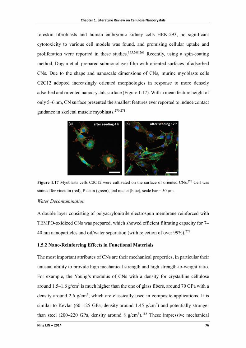
Chapter 1. Literature Review on Cellulose Nanocrystals
Ning LIN −−−− 2014 76
foreskin fibroblasts and human embryonic kidney cells HEK-293, no significant
cytotoxicity to various cell models was found, and promising cellular uptake and
proliferation were reported in these studies.165,268,269 Recently, using a spin-coating
method, Dugan et al. prepared submonolayer film with oriented surfaces of adsorbed
CNs. Due to the shape and nanoscale dimensions of CNs, murine myoblasts cells
C2C12 adopted increasingly oriented morphologies in response to more densely
adsorbed and oriented nanocrystals surface (Figure 1.17). With a mean feature height of
only 5–6 nm, CN surface presented the smallest features ever reported to induce contact
guidance in skeletal muscle myoblasts.270,271
Figure 1.17 Myoblasts cells C2C12 were cultivated on the surface of oriented CNs.270 Cell was
stained for vinculin (red), F-actin (green), and nuclei (blue), scale bar = 50 µm.
Water Decontamination
A double layer consisting of polyacrylonitrile electrospun membrane reinforced with
TEMPO-oxidized CNs was prepared, which showed efficient filtrating capacity for 7–
40 nm nanoparticles and oil/water separation (with rejection of over 99%).272
1.5.2 Nano-Reinforcing Effects in Functional Materials
The most important attributes of CNs are their mechanical properties, in particular their
unusual ability to provide high mechanical strength and high strength-to-weight ratio.
For example, the Young’s modulus of CNs with a density for crystalline cellulose
around 1.5–1.6 g/cm3 is much higher than the one of glass fibers, around 70 GPa with a
density around 2.6 g/cm3, which are classically used in composite applications. It is
similar to Kevlar (60–125 GPa, density around 1.45 g/cm3) and potentially stronger
than steel (200–220 GPa, density around 8 g/cm3).188 These impressive mechanical

Chapter 1. Literature Review on Cellulose Nanocrystals
Ning LIN −−−− 2014 77
properties make cellulose nanocrystals ideal candidates for the processing of reinforced
nanocomposites (as discussed in Section 1.4) and development of functionally
mechanical materials.
1.5.2.1 Soft Matter
Hydrogels and Gels
Hydrogels are defined as three-dimensional polymer networks swollen by trapping
large amounts of solvent (usually water), and widely used in the fields of food,
biomaterials, agriculture etc. Hydrogels from rod-like CNs are usually divided in two
types. One consists in the direct preparation from CN suspension; another consists in
composite hydrogels prepared from CN reinforced matrices. Cellulose nanocrystals can
form gels by themselves under some conditions resulting from the strong hydrogen
bonding interactions between nanoparticles. Aqueous suspensions with 0.4 wt% CNs
extracted from softwood flour can form a transparent hydrogel with unique rigid and
uniform morphology, and the elastic solid-like behavior of hydrogel was dependent on
the mechanical agitation.273 In addition, by involving a solvent exchange process, CNs
were proved to be able to form stable macroscopic organogels in glycerol,274 ethanol275
or acetone.276 More recently, Way et al. reported the different gelation behaviors of
surface modified CNs with either carboxylic acid (−COOH) or amine (−NH2). At low
pH, amine-modified nanocrystals formed flown suspension because of the electrostatic
repulsions of the ammonium moieties inhibiting aggregation of the nanoparticles.
However, a transition to hydrogels was observed at higher pH where the
amine-modified nanocrystals are neutral and the attractive forces based on hydrogen
bonding dominate. The opposite behavior was observed for carboxylated CNs, which
were dispersible in water at high pH and form stable hydrogels in an acidic
environment.81
On the basis of the crosslinking method, composite hydrogels can be divided into
chemical gels and physical gels. Physical gels are formed by molecular self-assembly
through ionic or hydrogen bonds, while chemical gels are formed by covalent bonds.
The introduction of rigid CNs in polymeric matrices to prepare physical hydrogel

Chapter 1. Literature Review on Cellulose Nanocrystals
Ning LIN −−−− 2014 78
nanomaterials generally improves the structural stability and enhances the elastic
modulus. To date, it was reported that CNs have been used in various synthetic
polymeric matrices for the preparation of gel nanomaterials, such as PEG,277 PVA,278,279
PHEMA,280,281 poly(NIPAM-co-BMA),282 starch-g-poly(sodium acrylate),283
PAM,141,284as well as natural polymeric matrices, for instance regenerated cellulose,285
agarose,286−288 and cyclodextrin.289,290
On the other hand, cellulose nanocrystal-based hydrogels can also be obtained by
chemical crosslinking method, such as in situ crosslinking between CNs and
poly(MVE-co-MA)–PEG,174,175 gelation,291 PAA,292 and PNiPAAm293 as matrices.
Sponge, Foam, Aerogel and Tissue-Engineering Nanoscaffold
Sponge is a material full of pores and channels allowing water or other molecules to
circulate through them. With a similar structure, foam is a substance that is formed by
trapping pockets of gas in a liquid or solid. Aerogel is a synthetic porous ultralight
material derived from the gel, in which the liquid component of the gel has been
replaced by gas. As nanomaterials with high porosity and specific surface area as well
as low density, sponge, foam and aerogel materials generally can be produced by
extracting the liquid component using freeze-drying or supercritical CO2 drying. These
treatments allow the liquid to be slowly dried off without causing the solid matrix to
collapse through capillary action, as happen with conventional evaporation. Table 1.8
shows diverse CN-based sponge, foam and aerogel materials prepared with different
techniques.
Table 1.8 Diverse cellulose nanocrystals-based sponge, foam, aerogel and nanoscaffold materials.
Sources Matrices Materials Techniques Ref.
cotton alginate sponge ionic crosslinking/freeze-casting [294] cotton starch/PVA sponge freezing/thawing cycles [295] polyurethane foam evaporation under high vacuum [296] MCC xylans foam freeze-casting [297] softwood pulp PVA foam freeze-casting [298] ramie fiber arabinoxylan aerogel freeze-casting [299] cotton wool aerogel supercritical CO2 drying [300] tunicate clay/PVA aerogel freeze-casting [301] cotton MAH-g-PLA scaffold electrospining [302]

Chapter 1. Literature Review on Cellulose Nanocrystals
Ning LIN −−−− 2014 79
Tissue-engineered porous nanoscaffold is a kind of novel biomedical nanomaterial
providing the necessary template and physical support to guide the differentiation and
proliferation of cells into targeted functional tissues or organs. Blaker et al. developed a
technique of combining the ice-microsphere template with thermally induced phase
separation lining the pore walls with bacterial CNs to produce in situ porous PLA/CN
scaffolds. The resultant nanoscaffolds showed porous morphology with controlled
porosities up to 97% of spherical interconnected pore walls lined by CNs, which can be
more suitable for the expansion and direct interaction with cells in three-dimensional
culture.303 Electrospun fibrous nanoscaffolds reinforced with CNs in
MAH-grafted-PLA matrix with improved interfacial adhesion were also reported.302
1.5.2.2 Special Mechanical Materials
Special mechanical materials based on CNs are mainly referred as mechanically
switchable, stimuli-responsive materials, such as mechanically adaptive materials and
mechanically gradient materials. As inspired by biological creatures such as sea
cucumbers, which have the ability to reversibly change the stiffness of their dermis,
three-dimensional percolating networks based on rigid CNs were introduced in various
polymeric matrices, such as poly(EO–EPI) copolymer (1:1), PVA and
poly(butylmethacrylate) (PBMA). The first generation of percolating CN materials was
based on poly(EO–EPI) and PVA matrix.304 In the rubbery zone, the tensile storage
moduli (E′c) of the dry matrix materials at 25 °C increased by over two orders of
magnitude from 3.7 MPa (neat matrix) to about 800 MPa for a nanocrystal content of
19% (v/v). However, upon exposure to water, these CN/matrix nanomaterials exhibit a
pronounced and reversible modulus reduction (40-fold), from 800 to 20 MPa, for the
materials with the same nanocrystal content of 19% (v/v) at room temperature. The
second generation of mechanically adaptive nanomaterials was on the basis of PVA or
PBMA matrices, which are dynamically sensitive to the change of thermal transition
[ambient (ca. 23 °C) to physiological (37 °C)].305−307 For instance, below the
glass-transition temperature (Tg), the tensile storage moduli of dry nanomaterials
increased only 2–2.5 fold when adding 16.5% (v/v) nanocrystals, while E′ increased

Chapter 1. Literature Review on Cellulose Nanocrystals
Ning LIN −−−− 2014 80
1000 fold above Tg. At the same time, when being exposed to physiological conditions
(immersion in artificial cerebrospinal fluid, 37 °C) these nanomaterials displayed a
significant decrease of E′ from several GPa to several MPa. Recently, introduction of
cotton CNs in rubbery polyurethane matrix was also reported, to produce mechanically
adaptive nanocomposites, which can change their mechanical properties upon exposure
to water and displayed a water-activated shape-memory effect.308
The theoretical model for the mechanically adaptive nanomaterial mechanism has been
proposed, as shown in Figure 1.18.63 The mechanical properties of these
chemoresponsive materials can selectively and reversibly be controlled through the
formation and decoupling of a three-dimensional network of well-individualized rigid
nanocrystals in response to specific chemical triggers. In the ‘‘on’’ state, strong
hydrogen bonds between rigid, percolating nanocrystals maximize stress transfer and
induce the reinforcement of the nanomaterials. In contrast, with the environmental
stimuli, such as exposure to water or the increase of temperature, the hydrogen bonding
interactions among nanocrystals are disrupted and dissociated from each other, which
are switched ‘‘off’’ by the introduction of the chemical regulators.
Figure 1.18 Schematic representation of the architecture and switching mechanism in the
mechanically adaptive nanomaterials.63
Recently, inspired by the water-enhanced mechanical gradient character of the squid
beak, Fox et al. reported mechanically gradient nanocomposites made from CNs where
the degree of crosslinking can be controlled along the length of the materials. With the
reaction between modified CNs and photoinitiator, different crosslinking degrees of
nanocrystals were controlled by UV irradiation. Under varied states (dry or wet) and
temperatures, different lengths and regions of the film exhibited different mechanical

Chapter 1. Literature Review on Cellulose Nanocrystals
Ning LIN −−−− 2014 81
properties, with an increase in irradiation time consistent with the increased
crosslinking. A dramatic increase in the mechanical contrast was observed for the wet
film (E′stiff/E′soft > 5) over the dry film either below or above Tg (E′stiff/E′soft of ca. 1.1
and 1.5, respectively).177
1.5.2.3 Self-Healable and Shape-Memory Materials
Named as “self-healable materials” and “shape-memory materials”, these two kinds of
materials are repairable or revertible after being damaged or changed in shape, which
are attractive for many applications because the features can improve the reliability,
functionality, and lifetime of a broad range of products. The self-healable ability of
materials is generally realized with the division and re-conjunction of supramolecular
structure in materials under an external stimulus, such as ultraviolet radiation or heating
energy. Biyani et al. reported light-healable nanocomposites based on a telechelic
poly(ethylene-co-butylene) that was functionalized with hydrogen-bonding
ureidopyrimidone (UPy) and CNs decorated with the same binding motif.138 When
these materials were exposed to ultraviolet radiation, the UPy motifs were excited and
the absorbed energy was converted into heat, which caused the conjunction of the
hydrogen-bonding motifs and restoration of the material. As a result, deliberately
introduced defects can be healed quickly and efficiently and exhibited high strength and
stiffness from the reinforcement provided by CNs. As shown in Figure 1.19, the
nanocomposite samples containing 10 wt% CNs were found to heal considerably faster,
requiring less than 20 s of UV exposure (image a). In another study, a supramolecular
polymer blend, formed via π−π interactions between a π-electron rich pyrenyl
end-capped oligomer and a chain-folding oligomer containing pairs of π-electron poor
naphthalene-diimide units, has been reinforced with CNs (from 1.25 to 20.0 wt%) to
afford a healable nanocomposite material. All the nanocomposites can reheal upon
exposure to elevated temperatures. The best combination of healing efficiency and
mechanical properties was obtained with the 7.5 wt% CNs nanocomposite which
exhibited a tensile modulus enhanced by as much as a factor of 20 over the matrix
material alone and could be fully rehealed at 85 °C within 30 min.31

Chapter 1. Literature Review on Cellulose Nanocrystals
Ning LIN −−−− 2014 82
With the regulation of reversible formation and disruption of CN percolation network,
elastomeric polyurethane nanocomposites containing CNs (from 1.1 to 29.6 wt%) can
be prepared, which are rapidly switchable shape-memory materials activated by water.
The CN percolation network reformed after drying to provide a fixing force for the
temporary shape. The entropy elasticity of polyurethane matrix then enabled rapid
shape recovery when percolation network was disrupted again during wetting.309,310
Figure 1.19 (a) Optical microscope images of deliberately damaged ureidopyrimidone-CNs
materials (with 10 wt% nanocrystal content) before, during, and after exposure to UV light
(320−390 nm, 350 mW/cm2, 20 s). (b) Atomic force micrographs of ureidopyrimidone-CNs
materials (with 10 wt% nanocrystal content) in the deliberately damaged (1), partially healed (2),
and completely healed (3) state (color range; black = 7 µm, white = 0 µm).138
1.5.2.4 Polymeric Electrolytes and Batteries
The incorporation of CNs into ion-conducting solid polymer electrolytes allows
combination of both mechanical reinforcement of a separator and high ionic
conductivity medium. Dufresne et al. have investigated a series of PEO-based polymer
electrolytes reinforced with CNs, which displayed high ionic conductivity as well as
electro-chemical, thermal, and mechanical stability. Solid polymer electrolytes with
CNs were generally prepared in two steps: the first step consisting in the preparation of
a polymeric matrix–nanocrystal mixture; and the second step involving the introduction
of the lithium salt, such as [lithium trifluoromethyl sulfonyl imide (LiTFSI) or LiClO4]
into the swelled nanocrystal-reinforced polymer nanomaterial in a concentrated
acetonitrile salt solution.311−313 Recently, electrospinning and hot-pressing technique

Chapter 1. Literature Review on Cellulose Nanocrystals
Ning LIN −−−− 2014 83
without solvent was also reported to prepare composite polymer electrolytes reinforced
with CNs.314
Generally, the addition of rigid CNs in polymer electrolytes leads to high mechanical
performance with a high increase in the storage modulus at higher temperatures, and a
slight decrease of ionic conductivity. For instance, by adding 10 wt% tunicin
nanocrystals in PEO composite materials, the gain in storage modulus exceeded by far
a factor of 100 with respect to the unfilled polymer electrolyte; while the values of ionic
conductivity still remained higher than 10-4 S/cm above 60 °C (melting point of
PEO).313,315 To further enhance the performance of polymer electrolytes, different
solvents (water or organic medium) for the dispersion of nanocrystals, crosslinking
agent,316 plasticizer,317 or different polymeric matrices318,319 were chosen to develop
and investigate the effects in the solid polymer electrolyte system.
Recently, cellulose nanocrystal was reported to be used as optically transparent
substrate for the preparation of recyclable organic solar cells. The solar cells fabricated
on CNs substrates displayed good rectification in the dark and reached a power
conversion efficiency of 2.7%. Meanwhile, this solar cell can be easily separated and
recycled into its major components using low-energy processes at room temperature.320
1.5.2.5 Semi-Conducting Material
Based on the combination of anionic surface charged CNs and positively charged
π-conjugated polymers, functional semi-conducting material can be prepared. Stable
suspension of CNs and PANI-PPE derivative mixture can be used to produce the
semi-conducting film by solution-casting treatment. Further investigations on electrical
conductivity, photoluminescence, and mechanical properties revealed that this
semi-conducting material synergistically combined the electronic characteristics from
the conjugated polymers with the outstanding mechanical reinforcement from CNs.321
In another study, the introduction of CNs in PEDOT/PSS-based PS conductive polymer
matrix extremely reduced the electrical percolation threshold 5-fold of ensuing
nanocomposites. It was reported that the percolation threshold of PEDOT:PSS in
PEDOT:PSS/PS blends, being 2.2 wt%, was lowered to 0.4 wt% by adding 0.8 wt% of

Chapter 1. Literature Review on Cellulose Nanocrystals
Ning LIN −−−− 2014 84
nonconducting CNs. Such a low percolation threshold of PEDOT:PSS was attributed to
the templating effect of CNs, which adsorbed the conductive polymers on the surface
and covered the electrical percolation with the network formed by CNs.322
1.5.3 Optical Materials Derived From Liquid Crystalline
Owing to their anisotropic rod-like morphology, cellulose nanoparticles have a
lyotropic liquid crystalline behavior in suspensions, which is a phase transition from an
isotropic liquid to an ordered liquid crystal when changing the concentration.69 Indeed,
above a critical concentration, a chiral nematic phase forms. Under certain conditions,
the suspension may be slowly evaporated to obtain semi-translucent films that maintain
the chiral nematic liquid crystal order formed in the suspension. These films exhibit
iridescence reflecting polarized light in a narrow wavelength range determined by the
chiral nematic pitch and the refractive index of the film. Derived from the liquid
crystalline property, tunable optical nanomaterials can be prepared based on rod-like
CNs with two strategies. The first strategy is the simple slow evaporation for CN
suspension to preserve the chiral nematic ordering phase. Another strategy is the
combination of liquid crystalline CNs and inorganic matrix to produce optical
materials.
Ultrasound treatment was found to increase the chiral nematic pitch in suspension and
red-shift the reflection wavelength of CN films as the applied energy increased.323
Figure 1.20 shows solid films cast from aliquots of 2.8 wt% CN suspensions prepared
from bleached softwood kraft pulp and sonicated with increasing (left to right) energy
inputs. The energy was measured in J/g of CNs. The films exhibit reflected iridescence
with colors ranging from blue-violet to red. By combining sonication and electrolyte
addition the reflective properties of the film can be predictably tuned. The effects of
sonicating a CNs suspension were shown to be cumulative and permanent. Moreover,
suspensions sonicated with different energy inputs can be mixed to prepare films
having a reflection band intermediate between those obtained from the individual
suspensions. Recently, differential heating rates of aqueous CNs suspension during film
casting were investigated.65 Placing materials of different temperatures beneath an

Chapter 1. Literature Review on Cellulose Nanocrystals
Ning LIN −−−− 2014 85
evaporating CNs suspension resulted in watermark-like patterns of different reflection
wavelength incorporated within the final film structure. The patterned areas are of
different thickness and different chiral nematic pitch than the surrounding film; heating
results in thicker areas of longer pitch. Thermal pattern creation in CNs films is
proposed to be caused by differences in evaporation rates and thermal motion in the
areas of the suspension. These CN-based optical materials with tunable iridescence
were proposed to have the potential for covert encryption as an anti-counterfeiting
measure and an intrinsic level of covert encryption since the reflection left-circularly
polarized light.324
Figure 1.20 Cellulose nanocrystal films produced from suspensions treated with increasing applied
ultrasonic energy (0, 250, 700, 1800, and 7200 J/g of nanocrystals) from left to right. Viewing is
normal to the film surface under diffuse lighting. Samples of CNs suspension were sonicated using
a Sonics vibra-cell 130 W 20 kHz ultrasonic processor with a 6 mm diameter probe: typically, 15
mL of a 2–3 wt% CNs suspension was placed in a 50 mL plastic tube and sonicated at 60% of the
maximum power.323
MacLachlan M. J. and his team developed a series of photonic, mesoporous, inorganic
(silica) optical materials which were a cast of a chiral nematic liquid crystal formed
from CNs. Inspired by the brilliant iridescent colors of beetle exoskeletons, their first
study consisted in preparing the free-standing mesoporous silica films with tunable
chiral nematic structures from CNs.12,325 As shown in Figure 1.21a, the peak reflected
wavelength of the biomimetic optical nanomaterials can be varied across the entire
visible spectrum and into the near-infrared through simple changes under synthetic
conditions. Figure 1.21b shows photographs of the different colors of mesoporous
silica nanomaterials. With increasing CN contents from samples S1 to S4, the
mesoporous films exhibited blue, green, yellow and red color, respectively. The colors
in these films can be attributed to the chiral nematic pore structure arising from CNs

Chapter 1. Literature Review on Cellulose Nanocrystals
Ning LIN −−−− 2014 86
present in the materials. Moreover, the addition of a drop of water will cause the wet
part of the film to become completely transparent, as shown in Figure 1.21d. With the
elimination of water (or other common solvents) upon drying, these optical
nanomaterials can fully regain their iridescence, which results in a reversible decrease
of the circular dichroism signal to 30 millidegrees (Figure 1.21c). Furthermore, some
mesoporous silica and carbon films decorated with CNs and metal nanoparticles
through calcination and carbonization were developed.326−328 Recently, the preparation
of responsive photonic hydrogels from self-assembly of CNs and hydrogel precursors
was reported. Because of the long-range chiral nematic order from CN component, this
kind of hydrogel showed iridescence and rapidly respondence to various stimuli.329
Although the mechanism of CN optical materials is commonly recognized as the chiral
nematic liquid crystal ordering from nanocrystal suspension, another explanation is
also proposed. Picard et al. observed a solid multilamellar structure for CN suspension
after evaporation, with each lamella made of roughly parallel rows of bundles.
Consequently, they explained the iridescence of CN optical films as a multilamellar
structure inducing light interference, not a helical cholesteric liquid crystal.330
Figure 1.21 Optical characterization of CNs/silica composite films. (a) Transmission spectra; (b)
photographs showing the different colors of mesoporous films S1 to S4 with gradual increase of
nanocrystal contents; (c) circular dichroism spectra of a green mesoporous film before (green curve)
and after (black curve) infiltration with water; and (d) photograph of the transparent green film after
the addition of a drop of water.12

Chapter 1. Literature Review on Cellulose Nanocrystals
Ning LIN −−−− 2014 87
1.5.4 Special Films and Systems Ascribed to Barrier Properties
1.5.4.1 Drug DeliveryBarrier for Drug Molecules
Despite an extended history of use in tableting, there is still considerable continuing
research into the use of cellulose with other types (nanocellulose) in advanced
drug-loaded systems whereby the rate of tablet disintegration as special excipients, or
prolonged drug release as novel drug carriers. As a drug delivery excipient, Burt et al.
investigated the capability of pure cellulose nanocrystals to bind the water soluble
antibiotics (tetracycline and doxorubicin), and the potential of cationic-CNs to bind
nonionized hydrophobic anticancer agents (docetaxel, paclitaxel, and etoposide).331
Besides direct use as excipient, CNs can also be used as co-stabilizer to improve the
physicochemical and flow properties of polymeric excipients. Acrylic beads prepared
via emulsion polymerization using CNs as co-stabilizer were proved to be a suitable
excipient. The presence of CNs affected positively the size and size distribution of the
bead excipient, which formed the stable structure together with low flow time and
reduced cotangent of angle.332
The biomaterial application of cellulose nanocrystals in drug delivery can be mainly
divided into two aspects, viz. microspheres (or microparticles),333 and hydrogels (or
gels).289,290,334 Table 1.9 summarizes various drug carrier systems based on CNs. It was
reported that solid carriers formed from CNs and different matrices spatially trapped
drug molecules, and imparted the regulation of drug release. Lin et al. developed a
pH-sensitive CN/sodium alginate microsphere-based controlled release system for drug
delivery. The presence of CNs in alginate-based microspheres showed more consistent
swelling patterns, higher encapsulation efficiency, and promising sustained release
profiles of the drug.333 With similar strategy, another work developed a polyelectrolyte
macroion complex via the conjunction between cationic chitosan and anionic CNs,
which has the potential as the multiparticulate oral drug delivery, but unfortunately
without drug loading or drug release results.285
Recently, some researchers attempted to directly attach drug molecules on CNs, which
were performed using covalent coupling between modified CNs and drug molecules.

Chapter 1. Literature Review on Cellulose Nanocrystals
Ning LIN −−−− 2014 88
Tab
le 1
.9 D
rug
carr
ier
syst
ems
base
d on
cel
lulo
se n
anoc
ryst
als.
Sou
rce
Matr
ix
Carr
ier
Mod
el d
rug
Rel
ease
tim
e a
nd
med
ium
M
ech
an
ism
mo
del
R
ef.
cotto
n so
dium
alg
inat
e m
icro
sphe
re
theo
phyl
line
16 h
in p
H=
7.4
PBS
solu
tion
Ritg
er-P
eppa
s eq
uatio
n [3
33]
cotto
n cy
clod
extr
in/P
luro
nic
hydr
ogel
D
OX
6.
5 d
in w
ater
R
itger
-Pep
pas
equa
tion
[290
] co
tton
cycl
odex
trin
/Plu
roni
c hy
drog
el
BSA
20
h in
pH
=7.
4 PB
S so
lutio
n —
[2
89]
MC
C
PL
A
fibe
r C
olu
mbia
Blu
e 48
h in
wat
er
Hig
uchi
equ
atio
n [3
37]
cotto
n pu
lp
rege
nera
ted
cellu
lose
ge
l B
SA
48 h
in s
imul
ated
bod
y fl
uid
Fick
ian
diff
usio
n la
w
[334
] eu
caly
ptus
w
ood
pulp
EA
; MM
A; B
MA
be
ad
prop
rano
lol
hydr
ochl
orid
e 12
h in
pH
=6.
8 PB
S so
lutio
n —
[3
32]
com
mer
cial
so
ftw
ood
CTA
B
susp
ensi
on
DO
X;
TET;
etop
osid
e;
pacl
itax
el; d
ocet
axel
1-
4 d
in P
BS
solu
tion
—
[331
]

Chapter 1. Literature Review on Cellulose Nanocrystals
Ning LIN −−−− 2014 89
With a series of oxidation, reductive-amination and esterification reactions in aqueous
media, a novel CN-based delivery system attached to the syringyl alcohol linker
through a γ-aminobutyric acid spacer molecule can be produced, on which small model
amine drugs (e.g. phenylpropanolamine) can be covalently connected.118 Similarly,
with the binding of bi-functional fusion protein on CNs, hydrophobic solid drug
nanoparticles can be adsorbed by the packed protein film on nanocrystals, and improve
the long-term stability of drugs under physiological condition.335
1.5.4.2 Barrier NanocompositesBarrier for Water and Oxygen
Cellulose is a hydrophilic polymer and it obviously absorbs water when immersed in
liquid water or conditioned in moist atmosphere. However, the water vapor
permeability is decreased when the cellulose fibers are disintegrated to the nanoscale
level, such as cellulose nanocrystals.336 Moreover, the sensitivity to moisture of the
nanoparticles can be tuned via pre-treatment before homogenization or post-treatment
(polymer impregnation, or chemical grafting).214 The gas permeability is also reduced
in dry atmospheres when decreasing the size of the cellulosic particles because of the
crystalline and dense structure of the nanoparticle film. Whatever the treatment or the
experimental conditions used to produce nanocellulose (including CNs and MFC), it is
seen as a new biomaterial for the creation of a good barrier for extending food
shelf-life, and also improves food quality for antioxidants and antimicrobials.
1.5.5 Other Functional Applications
Cellulose nanocrystal (1, 2, 3 wt%) was used to improve the performance of PVA as a
wood adhesive. With the measurement of block shear tests, wood failure value
increased from 59% (pure PVA) to 84% for adhesive with 1 wt% CNs and finally to
97% for adhesive with 3 wt% CNs. Fluctuations in properties of PVA as a wood
adhesive and a polymer at different CN loadings can be attributed to the effect of
homogeneous dispersion of nanocrystals to the change of PVA structure.338

Chapter 1. Literature Review on Cellulose Nanocrystals
Ning LIN −−−− 2014 90
1.6 Conclusions
The study of cellulose nanocrystals is a fascinating research field that has attracted
significant interest during last twenty years, largely attributed to the potential of
cellulose nanocrystals to be used as a renewable and rigid nanomaterial. In light of this
review chapter, specific research topics on cellulose nanocrystals generally can be
classified as four parts, including ‘evolution of preparation’, ‘exploration of property’,
‘revolution of modification’, and ‘development of application’.
In spite of the fact that there has been industrial production of cellulose nanocrystals in
some companies, more high-efficiency and ‘green’ technique is demanded; and
standardization for the procedure and control for the properties of produced cellulose
nanocrystals are absolutely necessary. Investigation on special properties of cellulose
nanocrystals is the prerequisite and fundament for their potential applications. The
discovery of new property and exploration of further mechanism will extend the
research on cellulose nanocrystals. The presence of a large number of chemical
functionalities within their structure provides a unique platform for significant surface
modification on cellulose nanocrystals. Through surface modification, researchers can
voluntarily transform the surface properties of cellulose nanocrystals, or impart
functional molecules on cellulose nanocrystals for novel properties and applications.
Further attention, focusing on the development of innovative modification approach,
will be put on the simplicity, cost reduction, and potential functions for the growth of
this topic. Many institutes all over the world are dedicated to the application of
cellulose nanocrystals, especially on the plastic composites and functional materials.
However, due to various reasons, only the statement of “on the edge of breakthrough”
for practical application of cellulose nanocrystals can be given until now.
Undoubtedly, the production of cellulose nanocrystals and their nanomaterials is an
emerging area of nanotechnology and materials science. However, it is also clear that
there are still many challenges to overcome for diverse areas of cellulose nanocrystals
research and the development of a practical product.

Chapter 1. Literature Review on Cellulose Nanocrystals
Ning LIN −−−− 2014 91
1.7 References
1. Payen, A. Mémoire sur la composition du tissu propre des plantes et du ligneux. C. R. Hebd.
Seances Acad. Sci. 1838, 7, 1052–1056.
2. Wertz, J.-L.; Bédué, O.; Mercier, J. P. Cellulose science and technology. EPFL Press,
Lausanne, Switzerland, 2010.
3. Nägeli, C. Die Stärkekörner. In pflanzenphysiologische untersuchungen, Nägeli, C.; Cramer,
C. Eds. Friedrich Schulthess, Zürich, Switzerland, 1858.
4. Hull, A. W. A new method of X-ray crystal analysis. Phys. Rev. 1917, 10, 661–696.
5. Nikerson, R. F.; Habrle, J.-A. Cellulose intercrystalline structure study by hydrolytic methods.
Ind. Eng. Chem. 1947, 39, 1507–1512.
6. Ranby, B. G. Aqueous colloidal solutions of cellulose micelles. Acta Chemica Scandinavica
1949, 3, 649–650.
7. Ranby, B. G.; Ribi, E. Ueber den feinbau der zellulose. Experientia 1950, 6, 12–14.
8. Marchessault, R. H.; Morehead, F. F.; Walter, N. M. Liquid crystal systems from fibrillar
polysaccharides. Nature 1959, 184, 632–633.
9. Revol, J. F.; Bradford, H.; Giasson, J.; Marchessault, R. H.; Gray, D. G. Helicoidal
self-ordering of cellulose microfibrils in aqueous suspension. Int. J. Biol. Macromol. 1992, 14,
170–172.
10. Favier, V.; Canova, G. R.; Cavaillé, J. Y.; Chanzy, H.; Dufresne, A.; Gauthier, C.
Nanocomposite materials from latex and cellulose whiskers. Polym. Adv. Technol. 1995, 6,
351–355.
11. Dong, S.; Roman, M. Fluorescently labeled cellulose nanocrystals for bioimaging applications.
J. Am. Chem. Soc. 2007, 129, 13810–13811.
12. Shopsowitz, K. E.; Qi, H.; Hamad, W. Y.; MacLachlan, M. J. Free-standing mesoporous silica
films with tunable chiral nematic structures. Nature 2010, 468, 422–426.
13. http://www.nrcan.gc.ca/media-room/news-release/2012/1915
14. Beck-Candanedo, S.; Roman, M.; Gray, D. G. Effect of reaction conditions on the properties
and behavior of wood cellulose nanocrystal suspensions. Biomacromolecules 2005, 6, 1048–
1054.
15. Hirota, M.; Tamura, N.; Saito, T.; Isogai, A. Water dispersion of cellulose II nanocrystals
prepared by TEMPO-mediated oxidation of mercerized cellulose at pH 4.8. Cellulose 2010,
17, 279–288.
16. Leung, A. C. W.; Hrapovic, S.; Lam, E.; Liu, Y.; Male, K. B.; Mahmoud, K. A.; Luong, J. H. T.
Characteristics and properties of carboxylated cellulose nanocrystals prepared from a novel
one-step procedure. Small 2011, 7, 302–305.
17. Braun, B.; Dorgan, J. R. Single-step method for the isolation and surface functionalization of
cellulosic nanowhiskers. Biomacromolecules 2009, 10, 334–341.
18. Filson, P. B.; Dawson-Andoha, B. E.; Schwegler-Berry, D. Enzymatic-mediated production of
cellulose nanocrystals from recycled pulp. Green Chem. 2009, 11, 1808–1814.
19. Man, Z.; Muhammad, N.; Bustam, A. S. M. A.; Kumar, M. V.; Rafiq, S. Preparation of
cellulose nanocrystals using an ionic liquid. J. Polym. Environ. 2011, 19, 726–731.
20. Kontturi, E.; Meriluoto, A.; Nuopponen, M. Processing for preparing micro- and
nanocrystalline cellulose. 2011, Patent WO2011/114005.

Chapter 1. Literature Review on Cellulose Nanocrystals
Ning LIN −−−− 2014 92
21. Dong, X. M.; Revol, J.-F.; Gray, D. G. Effect of microcrystallite preparation conditions on the
formation of colloid crystals of cellulose. Cellulose 1998, 5, 19–32.
22. Labet, M.; Thielemans, W. Improving the reproducibility of chemical reactions on the surface
of cellulose nanocrystals: ROP of ε-caprolactone as a case study. Cellulose 2011, 18, 607–617.
23. TAPPI, Workshop on international standards for nanocellulose. Arlington, USA, June 9th,
2011.
24. The global market for nanocellulose to 2017; Futures Markets Inc., Edinburgh, Lothian
EH74NA, United Kingdom, 2012, pp. 1–66.
25. Dufresne, A.; Thomas, S.; Pothan, L. A. Biopolymer nanocomposites: Processing, properties
and applications. John Wiley & Sons, Inc., 2013, USA, pp. 461–508.
26. Hanley, S. J.; Giasson, J.; Revol, J.-F.; Gray, D. G. Atomic force microscopy of cellulose
microfibrils: comparison with transmission electron microscopy. Polymer 1992, 33, 4639–
4642.
27. Lin, N.; Dufresne, A. Surface Chemistry, Morphological Analysis and Properties of Cellulose
Nanocrystals with Gradiented Sulfation Degrees. Nanoscale 2014, 6, 5384–5393.
28. Fraschini, C.; Chauve, G.; Le Berre, J.-F.; Ellis, S.; Méthot, M.; O’Connor, B.; Bouchard, J.
Critical discussion of light scattering and microscopy techniques for CNC particle sizing.
Nord. Pulp Pap. Res. J. 2014, 29, 31–40.
29. Habibi, Y.; Goffin, A.-L.; Schiltz, N.; Duquesne, E.; Dubois, P. Bionanocomposites based on
poly(ε-caprolactone)-grafted cellulose nanocrystals by ring-opening polymerization. J. Mater.
Chem. 2008, 18, 5002–5010.
30. Fleming, K.; Gray, D.; Prasannan, S.; Matthews, S. Cellulose crystallites: a new and robust
liquid crystalline medium for the measurement of residual dipolar couplings. J. Am. Chem.
Soc. 2000, 122, 5224–5225.
31. Fox, J.; Wie, J. J.; Greenland, B. W.; Burattini, S.; Hayes, W.; Colquhoun, H. M.; Mackay, M.
E.; Rowan, S. J. High-strength, healable, supramolecular polymer nanocomposites. J. Am.
Chem. Soc. 2012, 134, 5362−5368.
32. Grunert, M.; Winter, W. T. Nanocomposites of cellulose acetate butyrate reinforced with
cellulose nanocrystals. J. Polym. Environ. 2002, 10, 27–30.
33. Newman, R. H. Homogeneity in cellulose crystallinity between samples of Pinus radiata wood.
Holzforschung 2004, 58, 91–96.
34. Schenzel, K.; Fischer, S.; Brendler, E. New method for determining the degree of cellulose I
crystallinity by means of FT Raman spectroscopy. Cellulose 2005, 12, 223–231.
35. Chunilall, V.; Bush, T. Application of FT-Raman spectroscopy to study changes in cellulose I
crystallinity through the dissolving pulp process. Proceedings of the 16th International
symposium on Wood, Fibre and Pulping Chemistry, 8th–10th June 2011, pp. 472–477.
36. Roman, M.; Winter, W. T. Effect of sulfate groups from sulfuric acid hydrolysis on the thermal
degradation behavior of bacterial cellulose. Biomacromolecules 2004, 5, 1671–1677.
37. Dufresne, A. Nanocellulose. From nature to high performance tailored materials. Walter de
Gruyter GmbH, Berlin/Boston, 2012, pp. 30–31.
38. Meyer, K.; Lotmar, W. Sur l'élasticité de la cellulose. Helv. Chim. Acta 1936, 19, 68–86.
39. Treloar, L. R. G. Calculations of elastic moduli of polymer crystals: III. Cellulose. Polymer
1960, 1, 290–303.

Chapter 1. Literature Review on Cellulose Nanocrystals
Ning LIN −−−− 2014 93
40. Sakurada, I.; Nukushina, Y.; Ito, T. Experimental determination of the elastic modulus of
crystalline regions oriented polymers. J. Polym. Sci. 1962, 57, 651–660.
41. Mann, J.; Roldan-Gonzales, L. X-ray measurements of the elastic modulus of cellulose
crystals. Polymer 1962, 3, 549–553.
42. Tashiro, K.; Kobayashi, M. Calculation of crystallite modulus of native cellulose. Polym. Bull.
1985, 14, 213–218.
43. Jaswon, M. A.; Gillis, P. P.; Mark, R. E. The elastic constants of crystalline native cellulose.
Proc. Roy. Soc. 1968, 306, 389–412.
44. Kroon-batenburg, L. M. J.; Kroon, J.; Northolt, M. G. Chain modulus and intermolecular
hydrogen bonding in native and regenerated cellulose fibers. Polym. Commun. 1986, 27, 290–
292.
45. Matsuo, M.; Sawatari, C.; Iwai, Y.; Ozaki, F. Effect of orientation distribution and crystallinity
on the measurement by X-ray diffraction of the crystal lattice moduli of cellulose I and II.
Macromolecules 1990, 23, 3266–3275.
46. Tashiro, K.; Kobayashi, M. Theoretical evaluation of three-dimensional elastic constants of
native and regenerated celluloses: role of hydrogen bonds. Polymer 1991, 32, 1516–1526.
47. Nishino, T.; Takano, K.; Nakamae, K. Elastic modulus of the crystalline regions of cellulose
polymorphs. J. Polym. Sci. B: Polym. Phys. 1995, 33, 1647–1651.
48. Reiling, S.; Brickmann, J. Theoretical investigations on the structure and physical properties of
cellulose. Macromol. Theory Simul. 1995, 4, 725–743.
49. Ganster, J.; Blackwell, J. NpH-MD-simulations of the elastic moduli of cellulose II at room
temperature. J. Mol. Model. 1996, 2, 278–285.
50. Neyertz, S.; Pizzi, A.; Merlin, A.; Maigret, B.; Borwn, D.; Deglise, X. A new all-atom force
field for crystalline cellulose I. J. Appl. Polym. Sci. 2000, 78, 1939–1946.
51. Šturcova, A.; Davies, G. R.; Eichhorn, S. J. Elastic modulus and stress-transfer properties of
tunicate cellulose whiskers. Biomacromolecules 2005, 6, 1055–1061.
52. Tanaka, F.; Iwata, T. Estimation of the elastic modulus of cellulose crystal by molecular
mechanics simulation. Cellulose 2006, 13, 509–517.
53. Eichhorn, S. J.; Davies, G. R. Modelling the crystalline deformation of native and regenerated
cellulose. Cellulose 2006, 13, 291–307.
54. Bergenstråhle, M.; Berglund, L. A.; Mazeau, K. Thermal response in crystalline I cellulose: A
molecular dynamics study. J. Phys. Chem. B 2007, 111, 9138–9145.
55. Rusli, R.; Eichhorn, S. J. Determination of the stiffness of cellulose nanowhiskers and the
fiber-matrix interface in a nanocomposite using Raman spectroscopy. Appl. Phys. Lett. 2008,
93, 033111.
56. Diddens, I.; Murphy, B.; Krisch, M.; Müller, M. Anisotropic elastic properties of cellulose
measured using inelastic X-ray scattering. Macromolecules 2008, 41, 9755–9759.
57. Iwamoto, S.; Kai, W.; Isogai, A.; Iwata, T. Elastic modulus of single cellulose microfibrils
from tunicate measured by atomic force microscopy. Biomacromolecules 2009, 10, 2571–
2576.
58. Lahiji, R. R.; Xu, X.; Reifenberger, R.; Raman, A.; Rudie, A.; Moon, R. J. Atomic force
microscopy characterization of cellulose nanocrystals. Langmuir 2010, 26, 4480–4488.
59. Cintrón, M. S.; Johnson, G. P.; French, A. D. Young’s modulus calculations for cellulose Ia by
MM3 and quantum mechanics. Cellulose 2011, 18, 505–516.

Chapter 1. Literature Review on Cellulose Nanocrystals
Ning LIN −−−− 2014 94
60. Wu, X.; Moon, R. J.; Martini, A. Crystalline cellulose elastic modulus predicted by atomistic
models of uniform deformation and nanoscale indentation. Cellulose 2013, 20, 43–55.
61. Dri, F. L.; Hector Jr., L. G.; Moon, R. J.; Zavattieri, P. D. Anisotropy of the elastic properties of
crystalline cellulose Iβ from first principles density functional theory with Van der Waals
interactions. Cellulose 2013, 20, 2703–2718.
62. Wu, Q.; Henriksson, M.; Liu, X.; Berglund, L. A. A high strength nanocomposite based on
microcrystalline cellulose and polyurethane. Biomacromolecules 2007, 8, 3687–3692.
63. Lin, N.; Huang, J.; Dufresne, A. Preparation, properties and applications of polysaccharide
nanocrystals in advanced functional nanomaterials: a review. Nanoscale 2012, 4, 3274–3294.
64. Shopsowitz, K. E.; Qi, H.; Hamad, W. Y.; MacLachlan, M. J. Free-standing mesoporous silica
films with tunable chiral nematic structures. Nature 2010, 468, 422–426.
65. Beck, S.; Bouchard, J.; Chauve, G.; Berry, R. Controlled production of patterns in iridescent
solid films of cellulose nanocrystals. Cellulose 2013, 20, 1401–1411.
66. Lagerwall, J. P. F.; Schütz, C.; Salajkova, M.; Noh, J.; Park, J. H.; Scalia, G.; Bergström, L.
Cellulose nanocrystal-based materials: from liquid crystal self-assembly and glass formation to
multifunctional thin films. NPG Asia Mater. 2014, 6, e80.
67. Fleming, K.; Gray, D. G.; Matthews, S. Cellulose crystallites. Chem.–Eur. J. 2001, 7, 1831–
1835.
68. Roman, M.; Gray, D. G. Parabolic focal conics in self-assembled solid films of cellulose
nanocrystals. Langmuir 2005, 21, 5555–5561.
69. Majoinen, J.; Kontturi, E.; Ikkala, O.; Gray, D. G. SEM imaging of chiral nematic films cast
from cellulose nanocrystal suspensions. Cellulose 2012, 19, 1599–1605.
70. van den Berg, O.; Capadona, J. R.; Weder, C. Preparation of homogeneous dispersions of
tunicate cellulose whiskers in organic solvents. Biomacromolecules 2007, 8, 1353–1357.
71. Bercea, M.; Navard, P. Shear dynamics of aqueous suspensions of cellulose whiskers.
Macromolecules 2000, 33, 6011–6016.
72. Orts, W. J.; Godbout, L.; Marchessault, R. H.; Revol, J.-F. Enhanced ordering of liquid
crystalline suspensions of cellulose microfibrils: a small angle neutron scattering study.
Macromolecules 1998, 31, 5717–5725.
73. Boluk, Y.; Lahiji, R.; Zhaod, L.; McDermott, M. T. Suspension viscosities and shape parameter
of cellulose nanocrystals (CNC). Colloid. Surface A. 2011, 377, 297–303.
74. Ureña-Benavides, E. E.; Ao, G.; Davis, V. A.; Kitchens, C. L. Rheology and phase behavior of
lyotropic cellulose nanocrystal suspensions. Macromolecules 2011, 44, 8990–8998.
75. Shafiei-Sabet, S.; Hamad, W. Y.; Hatzikiriakos, S. G. Influence of degree of sulfation on the
rheology of cellulose nanocrystal suspensions. Rheol. Acta. 2013, 52, 741–751.
76. Habibi, Y.; Lucia, L. A.; Rojas, O. J. Cellulose nanocrystals: chemistry, self-assembly, and
applications. Chem. Rev. 2010, 110, 3479–3500.
77. French, A. D.; Bertoniere, N. R.; Brown, R. M.; Chanzy, H.; Gray, D.; Hattori, K.;
Kirk-Othmer G. W. Encyclopedia of chemical technology. ed. Seidel, A. John Wiley & Sons,
Inc., 5th edn, 2004, 5, pp. 360–394.
78. Wakelyn, P. J. Handbook of fiber chemistry. ed. Levin, M.; Pearce, E. M. Marcel Dekker, New
York, 1998, pp. 642–654.
79. Hebeish, A.; Guthrie, J. T. The chemistry and technology of cellulosic copolymers.
Springer-Verlag, Berlin, 1981.

Chapter 1. Literature Review on Cellulose Nanocrystals
Ning LIN −−−− 2014 95
80. Siqueira, G.; Bras, J.; Dufresne, A. Cellulose whiskers versus microfibrils: Influence of the
nature of the nanoparticle and its surface functionalization on the thermal and mechanical
properties of nanocomposites. Biomacromolecules 2009, 10, 425–432.
81. Way, A. E.; Hsu, L.; Shanmuganathan, K.; Weder, C.; Rowan, S. J. pH-Responsive cellulose
nanocrystal gels and nanocomposites. ACS Macro Lett. 2012, 1, 1001−1006.
82. Lokanathan, A. R.; Nykänen, A.; Seitsonen, J.; Johansson, L.-S.; Campbell, J.; Rojas, O. J.;
Ikkala, O.; Laine, J. Cilia-mimetic hairy surfaces based on end-immobilized nanocellulose
colloidal rods. Biomacromolecules 2013, 14, 2807–2813.
83. Araki, J.; Wada, M.; Kuga, S.; Okano, T. Flow properties of microcrystalline cellulose
suspension prepared by acid treatment of native cellulose. Colloid. Surface A. 1998, 142, 75–
82.
84. Araki, J.; Wada, M.; Kuga, S.; Okano, T. Hydrocolloids: physical chemistry and industrial
application of gels, polysaccharides, and proteins, ed. Nishinari, K. Elsevier, Amsterdam,
2000, 1, pp. 283–288.
85. Hasani, M.; Cranston, E. D.; Westman, G.; Gray, D. G. Cationic surface functionalization of
cellulose nanocrystals. Soft Matter 2008, 4, 2238–2244.
86. Zaman, M.; Xiao, H.; Chibante, F.; Ni, Y. Synthesis and characterization of cationically
modified nanocrystalline cellulose. Carbohydr. Polym. 2012, 89, 163–170.
87. Salajková, M.; Berglund, L. A.; Zhou, Q. Hydrophobic cellulose nanocrystals modified with
quaternary ammonium salts. J. Mater. Chem. 2012, 22, 19798–19805.
88. Cao, X.; Wang, X.; Ding, B.; Yu, J.; Sun, G. Novel spider-web-like nanoporous networks
based on jute cellulose nanowhiskers. Carbohydr. Polym. 2013, 92, 2041–2047.
89. Araki, J. Electrostatic or steric? – preparations and characterizations of well-dispersed systems
containing rod-like nanowhiskers of crystalline polysaccharides. Soft Matter 2013, 9, 4125–
4141.
90. Araki, J.; Wada, M.; Kuga, S.; Okano, T. Influence of surface charge on viscosity behavior of
cellulose microcrystal suspension. J. Wood Sci. 1999, 45, 258–261.
91. de Mesquita, J. P.; Donnici, C. L.; Pereira, F. V. Biobased nanocomposites from layer-by-layer
assembly of cellulose nanowhiskers with chitosan. Biomacromolecules 2010, 11, 473–480.
92. Boluk, Y.; Zhao, L.; Incani, V. Dispersions of nanocrystalline cellulose in aqueous polymer
solutions: structure formation of colloidal rods. Langmuir 2012, 28, 6114–6123.
93. Dong, X. M.; Kimura, T.; Revol, J.-F.; Gray, D. G. Effects of ionic strength on the
isotropic−chiral nematic phase transition of suspensions of cellulose crystallites. Langmuir
1996, 12, 2076–2082.
94. Cranston, E. D.; Gray, D. G.; Rutland, M. W. Direct surface force measurements of
polyelectrolyte multilayer films containing nanocrystalline cellulose. Langmuir 2010, 26,
17190–17197.
95. Rusli, R.; Shanmuganathan, K.; Rowan, S. J.; Weder, C.; Eichhorn, S. J. Stress transfer in
cellulose nanowhisker composites—influence of whisker aspect ratio and surface charge.
Biomacromolecules 2011, 12, 1363–1369.
96. Araki, J.; Kuga, S. Effect of trace electrolyte on liquid crystal type of cellulose microcrystals.
Langmuir 2001, 17, 4493–4496.

Chapter 1. Literature Review on Cellulose Nanocrystals
Ning LIN −−−− 2014 96
97. Hirai, A.; Inui, O.; Horii, F.; Tsuji, M. Phase separation behavior in aqueous suspensions of
bacterial cellulose nanocrystals prepared by sulfuric acid treatment. Langmuir 2009, 25, 497–
502.
98. Winter, H. T.; Cerclier, C.; Delorme, N.; Bizot, H.; Quemener, B.; Cathala, B. Improved
colloidal stability of bacterial cellulose nanocrystal suspensions for the elaboration of
spin-coated cellulose-based model surfaces. Biomacromolecules 2010, 11, 3144–3151.
99. de Menezes, A. J.; Siqueira, G.; Curvelo, A. A. S.; Dufresne, A. Extrusion and characterization
of functionalized cellulose whiskers reinforced polyethylene nanocomposites. Polymer 2009,
50, 4552–4563.
100. Harrisson, S.; Drisko, G. L.; Malmström, E.; Hult, A.; Wooley, K. L. Hybrid rigid/soft and
biologic/synthetic materials: polymers grafted onto cellulose microcrystals.
Biomacromolecules 2011, 12, 1214–1223.
101. Araki, J.; Wada, M.; Kuga, S. Steric stabilization of a cellulose microcrystal suspension by
poly(ethylene glycol) grafting. Langmuir 2001, 17, 21–27.
102. Kovacs, T.; Naish, V.; O’Connor, B.; Blaise, C.; Gagné, F.; Hall, L.; Trudeau, V.; Martel, P. An
ecotoxicological characterization of nanocrystalline cellulose (NCC). Nanotoxicology 2010, 4,
255–270.
103. Dong, S.; Hiran, A. A.; Colacino, K. R.; Lee, Y. W.; Roman, M. Cytotoxicity and cellular
uptake of cellulose nanocrystals. Nano Life 2012, 2, 1241006 (11 pages).
104. Kümmerer, K.; Menz, J.; Schubert, T.; Thielemans, W. Biodegradability of organic
nanoparticles in the aqueous environment. Chemosphere 2011, 82, 1387–1392.
105. Clift, M. J. D.; Foster, E. J.; Vanhecke, D.; Studer, D.; Wick, P.; Gehr, P.; Rothen-Rutishauser,
B.; Weder, C. Investigating the interaction of cellulose nanofibers derived from cotton with a
sophisticated 3D human lung cell coculture. Biomacromolecules 2011, 12, 3666–3673.
106. Endes, C.; Müller, S.; Schmid, O.; Vanhecke, D.; Foster, E. J.; Petri-Fink, A.;
Rothen-Rutishauser, B.; Weder, C.; Clift, M. J. D. Risk assessment of released cellulose
nanocrystals – mimicking inhalatory exposure. J. Physics: Conference Series 2013, 429,
012008 (10 Pages).
107. Miyamoto, T.; Takahashi, S.; Ito, H.; Inagaki, H.; Noishiki, Y. Tissue biocompatibility of
cellulose and its derivatives. J. Biomed. Mater. Res. 1989, 23, 125–133.
108. Märtson, M.; Viljanto, J.; Hurme, T.; Laippala, P.; Saukko, P. Is cellulose sponge degradable or
stable as implantation material? An in vivo subcutaneous study in the rat. Biomaterials 1999,
20, 1989–1995.
109. Dugan, J. M.; Gough, J. E.; Eichhorn, S. J. Bacterial cellulose scaffolds and cellulose
nanowhiskers for tissue engineering. Nanomedicine 2013, 8, 287–298.
110. Czaja, W. K.; Young, D. J.; Kawecki, M.; Browm, R. M. The future prospects of microbial
cellulose in biomedical applications. Biomacromolecules 2007, 8, 1–12.
111. Li, J.; Wan, Y. Z.; Li, L. F.; Liang, H.; Wang, J. H. Preparation and characterization of
2,3-dialdehyde bacterial cellulose for potential biodegradable tissue engineering scaffolds.
Mater. Sci. Eng. C 2009, 29, 1635–1642.
112. Klemm, D.; Kramer, F.; Moritz, S.; Lindström, T.; Ankerfors, M.; Gray, D.; Dorris, A.
Nanocelluloses: a new family of nature-based materials. Angew. Chem. Int. Ed. 2011, 50,
5438–5466.
Chapter 1. Literature Review on Cellulose Nanocrystals
Ning LIN −−−− 2014 97
113. Moon, R. J.; Martini, A.; Nairn, J.; Simonsen, J.; Youngblood, J. Cellulose nanomaterials
review: structure, properties and nanocomposites. Chem. Soc. Rev. 2011, 40, 3941–3994.
114. Yi, J.; Xu, Q.; Zhang, X.; Zhang, H. Chiral-nematic self-ordering of rodlike cellulose
nanocrystals grafted with poly(styrene) in both thermotropic and lyotropic states. Polymer
2008, 49, 4406–4412.
115. Yi, J.; Xu, Q.; Zhang, X.; Zhang, H. Temperature-induced chiral nematic phase changes of
suspensions of poly(N,N-dimethylaminoethyl methacrylate)-grafted cellulose nanocrystals.
Cellulose 2009, 16, 989–997.
116. Zoppe, J. O.; Habibi, Y.; Rojas, O. J.; Venditti, R. A.; Johansson, L.-S.; Efimenko, K.;
Österberg, M.; Laine, J. Poly(N-isopropylacrylamide) brushes grafted from cellulose
nanocrystals via surface-initiated single-electron transfer living radical polymerization.
Biomacromolecules 2010, 11, 2683–2691.
117. Dash, R.; Elder, T.; Ragauskas, A. J. Grafting of model primary amine compounds to cellulose
nanowhiskers through periodate oxidation. Cellulose 2012, 19, 2069–2079.
118. Dash, R.; Ragauskas, A. J. Synthesis of a novel cellulose nanowhisker-based drug delivery
system. RSC Adv. 2012, 2, 3403–3409.
119. Heux, L.; Chauve, G.; Bonini, C. Nonflocculating and chiral-nematic self-ordering of
cellulose microcrystals suspensions in nonpolar solvents. Langmuir 2000, 16, 8210–8212.
120. Bonini, C.; Heux, L.; Cavaillé, J.-Y.; Lindner, P.; Dewhurst, C.; Terech, P. Rodlike cellulose
whiskers coated with surfactant: a small-angle neutron scattering characterization. Langmuir
2002, 18, 3311–3314.
121. Kvien, I., Tanem, B. S.; Oksman, K. Characterization of cellulose whiskers and their
nanocomposites by atomic force and electron microscopy. Biomacromolecules 2005, 6, 3160–
3165.
122. Elazzouzi-Hafraoui, S.; Putaux, J.-L.; Heux, L. Self-assembling and chiral nematic properties
of organophilic cellulose nanocrystals. J. Phys. Chem. B 2009, 113, 11069–11075.
123. Bondeson, D.; Oksman, K. Dispersion and characteristics of surfactant modified cellulose
whiskers nanocomposites. Compos. Interfaces 2007, 14, 617–630.
124. Fortunati, E.; Armentano, I.; Zhou, Q.; Iannoni, A.; Saino, E.; Visai, L.; Berglund, L. A.;
Kenny, J. M. Multifunctional bionanocomposite films of poly(lactic acid), cellulose
nanocrystals and silver nanoparticles. Carbohydr. Polym. 2012, 87, 1596–1605.
125. Ljungberg, N.; Cavaillé, J.-Y.; Heux, L. Nanocomposites of isotactic polypropylene
reinforced with rod-like cellulose whiskers. Polymer 2006, 47, 6285–6292.
126. Habibi, Y.; Hoeger, I.; Kelley, S. S.; Rojas, O. J. Development of Langmuir-Schaeffer
cellulose nanocrystal monolayers and their interfacial behaviors. Langmuir 2010, 26, 990–
1001.
127. Kim, J.; Montero, G.; Habibi, Y.; Hinestroza, J. P.; Genzer, J.; Argyropoulos, D. S.; Rojas, O.
J. Dispersion of cellulose crystallites by nonionic surfactants in a hydrophobic polymer matrix.
Polym. Eng. Sci. 2009, 49, 2054–2061.
128. Rojas, O. J.; Montero, G. A.; Habibi, Y. Electrospun nanocomposites from polystyrene loaded
with cellulose nanowhiskers. J. Appl. Polym. Sci. 2009, 113, 927–935.
129. Petersson, L.; Kvien, I.; Oksman, K. Structure and thermal properties of poly(lactic
acid)/cellulose whiskers nanocomposite materials. Compos. Sci. Technol. 2007, 67, 2535–
2544.

Chapter 1. Literature Review on Cellulose Nanocrystals
Ning LIN −−−− 2014 98
130. Hirota, M.; Tamura, N.; Saito, T.; Isogai, A. Oxidation of regenerated cellulose with NaClO2
catalyzed by TEMPO and NaClO under acid-neutral conditions. Carbohydr. Polym. 2009, 78,
330–335.
131. Fujisawa, S.; Isogai T.; Isogai, A. Temperature and pH stability of cellouronic acid. Cellulose
2010, 17, 607–615.
132. Isogai, A.; Saito, T.; Fukuzumi, H. TEMPO-oxidized cellulose nanofibers. Nanoscale 2011, 3,
71–85.
133. Montanari, S.; Roumani, M.; Heux, L.; Vignon, M. R. Topochemistry of carboxylated
cellulose nanocrystals resulting from TEMPO-mediated oxidation. Macromolecules 2005, 38,
1665–1671.
134. Siqueira, G.; Bras, J.; Dufresne, A. New process of chemical grafting of cellulose
nanoparticles with a long chain isocyanate. Langmuir 2010, 26, 402–411.
135. Pei, A.; Zhou, Q.; Berglund, L. A. Functionalized cellulose nanocrystals as biobased
nucleation agents in poly(L-lactide) (PLLA)–Crystallization and mechanical property effects.
Compos. Sci. Technol. 2010, 70, 815–821.
136. Habibi, Y. ; Chanzy, H. ; Vignon, M. R. TEMPO-mediated surface oxidation of cellulose
whiskers. Cellulose 2006, 13, 679–687.
137. Gandini, A.; Belgacem, M. N. Chemical modification of wood. In monomers, polymers and
composites from renewable resources, 1st ed.; Elsevier: Great Britain, 2008, 419–432.
138. Biyani, M. V.; Foster, E. J.; Weder, C. Light-healable supramolecular nanocomposites based
on modified cellulose nanocrystals. ACS Macro Lett. 2013, 2, 236−240.
139. Goussé, C.; Chanzy, H.; Excoffier, G.; Soubeyrand, L.; Fleury, E. Stable suspensions of
partially silylated cellulose whiskers dispersed in organic solvents. Polymer 2002, 43, 2645–
2651.
140. Raquez, J.-M.; Murena, Y.; Goffin, A.-L.; Habibi, Y.; Ruelle, B.; DeBuyl, F.; Dubois, P.
Surface-modification of cellulose nanowhiskers and their use as nanoreinforcers into
polylactide: A sustainably-integrated approach. Compos. Sci. Technol. 2012, 72, 544–549.
141. Yang, J.; Han, C.-R.; Duan, J.-F.; Ma, M.-G.; Zhang, X.-M.; Xu, F.; Sun, R.-C. Synthesis and
characterization of mechanically flexible and tough cellulose nanocrystals–polyacrylamide
nanocomposite hydrogels. Cellulose 2013, 20, 227–237.
142. Tehrani, A. D.; Neysi, E. Surface modification of cellulose nanowhisker throughout graft
polymerization of 2-ethyl-2-oxazoline. Carbohydr. Polym. 2013, 97, 98–104.
143. de Souza Lima, M. M.; Borsali, R. Rodlike cellulose microcrystals: structure, properties, and
applications. Macromol. Rapid Commun. 2004, 25, 771–787.
144. Kloser, E.; Gray, D. G. Surface grafting of cellulose nanocrystals with poly(ethylene oxide) in
aqueous media. Langmuir 2010, 26, 13450–13456.
145. Azouz, K. B.; Ramires, E. C.; den Fonteyne, W. V.; El Kissi, N.; Dufresne, A. Simple method
for the melt extrusion of a cellulose nanocrystal reinforced hydrophobic polymer. ACS Macro
Lett. 2012, 1, 236−240.
146. Belgacem, M. N.; Quillerou, J.; Gandini, A. Urethanes and polyurethanes bearing furan
moieties—3. Synthesis, characterization and comparative kinetics of the formation of
diurethanes. Eur. Polym. J. 1993, 29, 1217−1224.
147. Habibi, Y.; Dufresne, A. Highly filled bionanocomposites from functionalized polysaccharide
nanocrystals. Biomacromolecules 2008, 9, 1974–1980.

Chapter 1. Literature Review on Cellulose Nanocrystals
Ning LIN −−−− 2014 99
148. Zoppe, J. O.; Peresin, M. S.; Habibi, Y.; Venditti, R. A.; Rojas, O. J. Reinforcing
poly(ε-caprolactone) nanofibers with cellulose nanocrystals. ACS Appl. Mater. Interfaces
2009, 9, 1996–2004.
149. Goffin, A.-L.; Raquez, J.-M.; Duquesne, E.; Siqueira, G.; Habibi, Y.; Dufresne, A.; Dubois, P.
Poly(ε-caprolactone) based nanocomposites reinforced by surface-grafted cellulose
nanowhiskers via extrusion processing: Morphology, rheology, and thermo-mechanical
properties. Polymer 2011, 52, 1532–1538.
150. Lin, N.; Chen, G.; Huang, J.; Dufresne, A.; Chang, P. R. Effects of polymer-grafted natural
nanocrystals on the structure and mechanical properties of poly(lactic acid): A case of cellulose
whisker-graft-polycaprolactone. J. Appl. Polym. Sci. 2009, 113, 3417–3425.
151. Labet, M.; Thielemans, W. Citric acid as a benign alternative to metal catalysts for the
production of cellulose-grafted-polycaprolactone copolymers. Polym. Chem. 2012, 3, 679–
684.
152. Carlsson, L.; Utsel, S.; Wågberg, L.; Malmström, E.; Carlmark, A. Surface-initiated
ring-opening polymerization from cellulose model surfaces monitored by a Quartz Crystal
Microbalance. Soft Matter 2012, 8, 512–517.
153. Bergenstråhle, M.; Mazeau, K.; Berglund, L. A. Molecular modeling of interfaces between
cellulose crystals and surrounding molecules: Effects of caprolactone surface grafting. Eur.
Polym. J. 2008, 44, 3662–3669.
154. Goffin, A.-L.; Raquez, J.-M.; Duquesne, E.; Siqueira, G.; Habibi, Y.; Dufresne, A.; Dubois, P.
From interfacial ring-opening polymerization to melt processing of cellulose
nanowhisker-filled polylactide-based nanocomposites. Biomacromolecules 2011, 12,
2456-2465.
155. Braun, B.; Dorgan, J. R.; Hollingsworth, L. O. Supra-molecular ecobionanocomposites based
on polylactide and cellulosic nanowhiskers: synthesis and properties. Biomacromolecules
2012, 13, 2013−2019.
156. Xu, Q.; Yi, J.; Zhang, X.; Zhang, H. A novel amphotropic polymer based on cellulose
nanocrystals grafted with azo polymers. Eur. Polym. J. 2008, 44, 2830–2837.
157. Morandi, G.; Heath, L.; Thielemans, W. Cellulose nanocrystals grafted with polystyrene
chains through surface-initiated atom transfer radical polymerization (SI-ATRP). Langmuir
2009, 25, 8280–8286.
158. Morandi, G.; Thielemans, W. Synthesis of cellulose nanocrystals bearing photocleavable
grafts by ATRP. Polym. Chem. 2012, 3, 1402–1407.
159. Majoinen, J.; Walther, A.; McKee, J. R.; Kontturi, E.; Aseyev, V.; Malho, J. M.; Ruokolainen,
J.; Ikkala, O. Polyelectrolyte brushes grafted from cellulose nanocrystals using Cu-mediated
surface-initiated controlled radical polymerization. Biomacromolecules 2011, 12, 2997–3006.
160. Zoppe, J. O.; Österberg, M.; Venditti, R. A.; Laine, J.; Rojas, O. J. Surface interaction forces of
cellulose nanocrystals grafted with thermoresponsive polymer brushes. Biomacromolecules
2011, 12, 2788–2796.
161. Zhou, Q.; Brumer, H.; Teeri, T. T. Self-organization of cellulose nanocrystals adsorbed with
xyloglucan oligosaccharide-poly(ethylene glycol)-polystyrene triblock copolymer.
Macromolecules 2009, 42, 5430–5432.

Chapter 1. Literature Review on Cellulose Nanocrystals
Ning LIN −−−− 2014 100
162. Azzam, F.; Heux, L.; Putaux, J.-L.; Jean, B. Preparation by grafting onto, characterization, and
properties of thermally responsive polymer-decorated cellulose nanocrystals.
Biomacromolecules 2010, 11, 3652–3659.
163. Ljungberg, N. ; Bonini, C. ; Bortolussi, F. ; Boisson, C. ; Heux, L. ; Cavaillé J. Y. New
nanocomposite materials reinforced with cellulose whiskers in atactic polypropylene: Effect of
surface and dispersion characteristics. Biomacromolecules 2005, 6, 2732-2739.
164. Peng, B.; Han, X.; Liu, H.; Berry, R. C.; Tam, K. C. Interactions between surfactants and
polymer-grafted nanocrystalline cellulose. Colloid. Surface. A 2013, 421, 142–149.
165. Mahmoud, K. A.; Mena, J. A.; Male, K. B.; Hrapovic, S.; Kamen, A.; Luong, J. H. T. Effect of
surface charge on the cellular uptake and cytotoxicity of fluorescent labeled cellulose
nanocrystals. ACS Appl. Mater. Interfaces 2010, 2, 2924–2932.
166. Hassan, M. L.; Moorefield, C. M.; Elbatal, H. S.; Newkome, G. R. New
metallo-supramolecular terpyridine-modified cellulose functional nanomaterials. J.
Macromol. Sci. A 2012, 49, 298–305.
167. Hassan, M. L.; Moorefield, C. M.; Elbatal, H. S.; Newkome, G. R.; Modarelli, D. A.; Romano,
N. C. Fluorescent cellulose nanocrystals via supramolecular assembly of terpyridine-modified
cellulose nanocrystals and terpyridine-modified perylene. Mater. Sci. Eng. B 2012, 177, 350–
358.
168. Zhang, L.; Li, Q.; Zhou, J.; Zhang, L. Synthesis and photophysical behavior of pyrene-bearing
cellulose nanocrystals for Fe3+ sensing. Macromol. Chem. Phys. 2012, 213, 1612−1617.
169. Eyley, S.; Thielemans, W. Imidazolium grafted cellulose nanocrystals for ion exchange
applications. Chem. Commun. 2011, 47, 4177–4179.
170. Cateto, C. A.; Ragauskas, A. Amino acid modified cellulose whiskers. RSC Adv. 2011, 1,
1695–1697.
171. Mangalam, A. P.; Simonsen, J.; Benight, A. S. Cellulose/DNA hybrid nanomaterials.
Biomacromolecules 2009, 10, 497–504.
172. Feese, E.; Sadeghifar, H.; Gracz, H. S.; Argyropoulos, D. S.; Ghiladi, R. A. Photobactericidal
porphyrin-cellulose nanocrystals: synthesis, characterization, and antimicrobial properties.
Biomacromolecules 2011, 12, 3528–3539.
173. Carpenter, B. L.; Feese, E.; Sadeghifar, H.; Argyropoulos, D. S.; Ghiladi, R. A.
Porphyrin-cellulose nanocrystals: A photobactericidal material that exhibits broad spectrum
antimicrobial activity. J. Photochem. Photobiol. 2012, 88, 527–536.
174. Goetz, L.; Mathew, A.; Oksman, K.; Gatenholm, P.; Ragauskas, A. J. A novel nanocomposite
film prepared from crosslinked cellulosic whiskers. Carbohydr. Polym. 2009, 75, 85–89.
175. Goetz, L.; Foston, M.; Mathew, A. P.; Oksman, K.; Ragauskas, A. J. Poly(methyl vinyl
ether-co-maleic acid)-polyethylene glycol nanocomposites cross-linked in situ with cellulose
nanowhiskers. Biomacromolecules 2010, 11, 2660–2666.
176. Filpponen, I.; Argyropoulos, D. S. Regular linking of cellulose nanocrystals via click
chemistry: synthesis and formation of cellulose nanoplatelet gels. Biomacromolecules 2010,
11, 1060–1066.
177. Fox, J. D.; Capadona, J. R.; Marasco, P. D.; Rowan, S. J. Bioinspired water-enhanced
mechanical gradient nanocomposite films that mimic the architecture and properties of the
squid beak. J. Am. Chem. Soc. 2013, 135, 5167−5174.

Chapter 1. Literature Review on Cellulose Nanocrystals
Ning LIN −−−− 2014 101
178. Dufresne, A. Processing of polymer nanocomposites reinforced with cellulose nanocrystals: a
challenge. Int. Polym. Proc. 2012, 27, 557–564.
179. Abdul Khalil, H. P. S.; Bhat, A. H.; Ireana Yusra, A. F. Green composites from sustainable
cellulose nanofibrils: a review. Carbohydr. Polym. 2012, 87, 963–979.
180. Tingaut, P.; Zimmermann, T.; Sèbe, G. Cellulose nanocrystals and microfibrillated cellulose as
building blocks for the design of hierarchical functional materials. J. Mater. Chem. 2012, 22,
20105–20111.
181. Durán, N.; Lemes, A. P.; Seabra, A. B. Review of cellulose nanocrystals patents: preparation,
composites and general applications. Recent Pat. Nanotech. 2012, 6, 16–28.
182. Lam, E.; Male, K. B.; Chong, J. H.; Leung, A. C. W.; Luong, J. H. T. Applications of
functionalized and nanoparticle-modified nanocrystalline cellulose. Trends Biotechnol. 2012,
30, 283–290.
183. Brinchi, L.; Cotana, F.; Fortunati, E.; Kenny, J. M. Production of nanocrystalline cellulose
from lignocellulosic biomass: Technology and applications. Carbohydr. Polym. 2013, 94, 154–
169.
184. Miao, C.; Hamad, W. Y. Cellulose reinforced polymer composites and nanocomposites: a
critical review. Cellulose 2013, 20, 2221–2262.
185. Leung, A. C. W.; Lam, E.; Chong, J.; Hrapovic, S.; Luong, J. H. T. Reinforced plastics and
aerogels by nanocrystalline cellulose. J. Nanopart. Res. 2013, 15, 1636. (24 pages)
186. Rebouillat, S.; Pla, F. State of the art manufacturing and engineering of nanocellulose: a
review of available data and industrial applications. J. Biomater. Nanobiotechnol. 2013, 4,
165–188.
187. Milanez, D. H.; do Amaral, R. M.; de Faria, L. I. L.; Gregolin, J. A. R. Assessing
nanocellulose developments using science and technology indicators. Mater. Res. 2013, 16,
635–641.
188. Dufresne, A. Nanocellulose: a new ageless bionanomaterial. Mater. Today 2013, 16, 220–227.
189. Paunonen, S. Strength and barrier enhancements of composites and packaging boards by
nanocelluloses – a literature review. Nord. Pulp Pap. Res. J. 2013, 28, 165–181.
190. Charreau, H.; Foresti, M. L.; Vázquez, A. Nanocellulose patents trends: a comprehensive
review on patents on cellulose nanocrystals, microfibrillated and bacterial cellulose. Recent
Pat. Nanotechnol. 2013, 7, 56–80.
191. Akhlaghi, S. P.; Peng, B.; Yao, Z.; Tam, K. C. Sustainable nanomaterials derived from
polysaccharides and amphiphilic compounds. Soft Matter 2013, 9, 7905–7918.
192. Habibi, Y. Key advances in the chemical modification of nanocelluloses. Chem. Soc. Rev.
2014, 43, 1519–1542.
193. Hajji, P.; Cavaille, J. Y.; Favier, V.; Gauthier, C.; Vigier, G. Tensile behavior of
nanocomposites from latex and cellulose whiskers. Polym. Compos. 1996, 17, 612–619.
194. Dufresne, A. Comparing the mechanical properties of high performances polymer
nanocomposites from biological sources. J Nanosci. Nanotechnol. 2006, 6, 322–330.
195. Dufresne, A. Polysaccharide nanocrystal reinforced nanocomposites. Can. J. Chem. 2008, 86,
484–494.
196. Dufresne, A. Nanocellulose. From nature to high performance tailored materials. Walter de
Gruyter GmbH, Berlin/Boston, 2012, pp. 261−262.

Chapter 1. Literature Review on Cellulose Nanocrystals
Ning LIN −−−− 2014 102
197. Fortunati, E.; Peltzer, M.; Armentano, I.; Torre, L.; Jiménez, A.; Kenny, J. M. Effects of
modified cellulose nanocrystals on the barrier and migration properties of PLA
nano-biocomposites. Carbohydr. Polym. 2012, 90, 948–956.
198. Auad, M. L.; Richardson, T.; Orts, W. J.; SMedeiros, E.; Mattoso, L. H. C.; Mosiewicki, M.
A.; Marcovich, N. E.; Aranguren, M. I. Polyaniline-modified cellulose nanofibrils as
reinforcement of a smart polyurethane. Polym. Int. 2011, 60, 743–750.
199. Corrêa, A. C.; de Morais Teixeira, E.; Carmona, V. B.; Teodoro, K. B. R.; Ribeiro, C.;
Mattoso, L. H. C.; Marconcini, J. M. Obtaining nanocomposites of polyamide 6 and cellulose
whiskers via extrusion and injection molding. Cellulose 2014, 21, 311–322.
200. Lin, N.; Huang, J.; Chang, P. R.; Feng, J.; Yu, J. Surface acetylation of cellulose nanocrystal
and its reinforcing function in poly(lactic acid). Carbohydr. Polym. 2011, 83, 1834–1842.
201. Yang, Z.-Y.; Wang, W.-J.; Shao, Z.-Q.; Zhu, H.-D.; Li, Y.-H.; Wang, F.-J. The transparency
and mechanical properties of cellulose acetate nanocomposites using cellulose nanowhiskers
as fillers. Cellulose 2013, 20, 159–168.
202. Lin, S.; Huang, J.; Chang, P. R.; Wei, S.; Xu, Y.; Zhang, Q. Structure and mechanical
properties of new biomass-based nanocomposite: Castor oil-based polyurethane reinforced
with acetylated cellulose nanocrystal. Carbohydr. Polym. 2013, 95, 91–99.
203. Follain, N.; Belbekhouche, S.; Bras, J.; Siqueira, G.; Marais, S.; Dufresne, A. Water transport
properties of bio-nanocomposites reinforced by Luffa cylindrica cellulose nanocrystals. J.
Membrane Sci. 2013, 427, 218–229.
204. Wang, Y.; Chen, L. Cellulose nanowhiskers and fiber alignment greatly improve mechanical
properties of electrospun prolamin protein fibers. ACS Appl. Mater. Interfaces 2014, 6, 1709–
1718.
205. Brown, E. E.; Hu, D.; Lail, N. A.; Zhang, X. Potential of nanocrystalline cellulose−fibrin
nanocomposites for artificial vascular graft applications. Biomacromolecules 2013, 14,
1063−1071.
206. Zhang, R.; Zhu, C.; Shan, X.; Xia, J.; Zhu, Q.; Hu, Y. Study on the
poly(3-hydroxybutyrate-co-4-hydroxybutyrate)-based nanocomposites reinforced by surface
modified nanocrystalline cellulose. J. Appl. Polym. Sci. 2013, 130, 2015–2022.
207. Blachechen, L. S.; de Mesquita, J. P.; de Paula, E. L.; Pereira, F. V.; Petri, D. F. S. Interplay of
colloidal stability of cellulose nanocrystals and their dispersibility in cellulose acetate butyrate
matrix. Cellulose 2013, 20, 1329–1342.
208. Espino-Pérez, E.; Bras, J.; Ducruet, V.; Guinault, A.; Dufresne, A.; Domenek, S. Influence of
chemical surface modification of cellulose nanowhiskers on thermal, mechanical, and barrier
properties of poly(lactide) based bionanocomposites. Eur. Polym. J. 2013, 49, 3144–3154.
209. Yu, H.-Y.; Qin, Z.-Y.; Yan, C.-F.; Yao, J.-M. Green nanocomposites based on functionalized
cellulose nanocrystals: a study on the relationship between interfacial interaction and property
enhancement. ACS Sustainable Chem. Eng. DOI: 10.1021/sc400499g
210. Park, S. H.; Oh, K. W.; Kim, S. H. Reinforcement effect of cellulose nanowhisker on
bio-based polyurethane. Compos. Sci. Technol. 2013, 86, 82–88.
211. de Mesquita, J. P.; Donnici, C. L.; Teixeira, I. F.; Pereira, F. V. Bio-based nanocomposites
obtained through covalent linkage between chitosan and cellulose nanocrystals. Carbohydr.
Polym. 2012, 90, 210–217.

Chapter 1. Literature Review on Cellulose Nanocrystals
Ning LIN −−−− 2014 103
212. Bitinis, N.; Verdejo, R.; Bras, J.; Fortunati, E.; Kenny, J. M.; Torre, L.; López-Manchado, M.
A. Poly(lactic acid)/natural rubber/cellulose nanocrystal bionanocomposites Part I. Processing
and morphology. Carbohydr. Polym. 2013, 96, 611–620.
213. Bitinis, N.; Fortunati, E.; Verdejo, R.; Bras, J.; Kenny, J. M.; Torre, L.; López-Manchado, M.
A. Poly(lactic acid)/natural rubber/cellulose nanocrystal bionanocomposites. Part II:
Properties evaluation. Carbohydr. Polym. 2013, 96, 621–627.
214. Lin, N.; Dufresne, A. Physical and/or chemical compatibilization of extruded cellulose
nanocrystal reinforced polystyrene nanocomposites. Macromolecules 2013, 46, 5570−5583.
215. Dufresne, A. Nanocellulose: From nature to high performance tailored materials. Walter de
Gruyter GmbH, Berlin/Boston, 2012, pp. 339–340.
216. Dufresne, A. Nanocellulose: From nature to high performance tailored materials. Walter de
Gruyter GmbH, Berlin/Boston, 2012, pp. 327–333.
217. Favier, V.; Dendievel, R.; Canova, G.; Cavaillé, J. Y.; Gilormini, P. Simulation and modeling
of three-dimensional percolating structures: case of a latex matrix reinforced by a network of
cellulose fibers. Acta Mater. 1997, 45, 1557-1565.
218. Stauffer, D. Introduction to Percolation Theory (Taylor and Francis, London and
Philadelphia), 1985.
219. Dufresne, A. Nanocellulose: From nature to high performance tailored materials. Walter de
Gruyter GmbH, Berlin/Boston, 2012, pp. 277–320.
220. Dufresne, A. Nanocellulose: From nature to high performance tailored materials. Walter de
Gruyter GmbH, Berlin/Boston, 2012, pp. 373–409.
221. Hassan, M. L.; Moorefield, C. M.; Elbatal, H. S.; Newkomeb, G. R. New
metallo-supramolecular terpyridine-modified cellulose functional nanomaterials. J.
Macromol. Sci. A 2012, 49, 298–305.
222. Barazzouk, S.; Daneault, C. Tryptophan-based peptides grafted onto oxidized nanocellulose.
Cellulose 2012, 19, 481–493.
223. Yang, Q.; Pan, X. A facile approach for fabricating fluorescent cellulose. J. Appl. Polym. Sci.
2010, 117, 3639–3644.
224. Nielsen, L. J.; Eyley, S.; Thielemans, W.; Aylott, J. W. Dual fluorescent labelling of cellulose
nanocrystals for pH sensing. Chem. Commun. 2010, 46, 8929–8931.
225. Drogat, N.; Granet, R.; Morvan, C. L.; Bégaud-Grimaud, G.; Krausz, P.; Sol, V.
Chlorin-PEI-labeled cellulose nanocrystals: synthesis, characterization and potential
application in PDT. Bioorg. Med. Chem. Lett. 2012, 22, 3648–3652.
226. Edwards, J. V.; Prevost, N.; Condon, B.; French, A.; Wu, Q. Immobilization of
lysozyme-cellulose amide-linked conjugates on cellulose I and II cotton nanocrystalline
preparations. Cellulose 2012, 19, 495–506.
227. Yang, R.; Tan, H.; Wei, F.; Wang, S. Peroxidase conjugate of cellulose nanocrystals for the
removal of chlorinated phenolic compounds in aqueous solution. Biotechnology 2008, 7, 233–
241.
228. Mahmoud, K. A.; Male, K. B.; Hrapovic, S.; Luong, J. H. T. Cellulose nanocrystal/gold
nanoparticle composite as a matrix for enzyme immobilization. ACS Appl. Mater. Interfaces
2009, 1, 1383–1386.
229. Incani, V.; Danumah, C.; Boluk, Y. Nanocomposites of nanocrystalline cellulose for enzyme
immobilization. Cellulose 2013, 20, 191–200.

Chapter 1. Literature Review on Cellulose Nanocrystals
Ning LIN −−−− 2014 104
230. Mahmoud, K. A.; Lam, E.; Hrapovic, S.; Luong, J. H. T. Preparation of well-dispersed
gold/magnetite nanoparticles embedded on cellulose nanocrystals for efficient immobilization
of papain enzyme. ACS Appl. Mater. Interfaces 2013, 5, 4978−4985.
231. Liu, H.; Wang, D.; Shang, S.; Song, Z. Preparation of silver nanoparticles on cellulose
nanocrystals and the application in electrochemical detection of DNA hybridization. Cellulose
2011, 18, 67–74.
232. Cerclier, C.; Guyomard-Lack, A.; Moreau, C.; Cousin, F.; Beury, N.; Bonnin, E.; Jean, B.;
Cathala, B. Coloured semi-reflective thin films for biomass-hydrolyzing enzyme detection.
Adv. Mater. 2011, 23, 3791–3795.
233. Edwards, J. V.; Prevost, N.; Sethumadhavan, K.; Ullah, A.; Condon, B. Peptide conjugated
cellulose nanocrystals with sensitive human neutrophil elastase sensor activity. Cellulose 2013,
20, 1223–1235.
234. Eyley, S.; Shariki, S.; Dale, S. E. C.; Bending, S.; Marken, F.; Thielemans, W.
Ferrocene−decorated nanocrystalline cellulose with charge carrier mobility. Langmuir 2012,
28, 6514−6519.
235. Dujardin, E.; Blaseby M.; Mann, S. Synthesis of mesoporous silica by sol–gel mineralisation
of cellulose nanorod nematic suspensions. J. Mater. Chem. 2003, 13, 696–699.
236. Scheel, H.; Zollfrank, C.; Greil, P. Luminescent silica nanotubes and nanowires: Preparation
from cellulose whisker templates and investigation of irradiation-induced luminescence. J.
Mater. Res. 2009, 24, 1709–1715.
237. Gruber, S.; Gottschlich, A.; Scheel, H.; Aresipathi, C.; Bannat, I.; Zollfrank, C.; Wark, M.
Molecular and supramolecular templating of silica-based nanotubes and introduction of metal
nanowires. Phys. Status Solidi B 2010, 247, 2401–2411.
238. Gruber, S.; Taylor, R. N. K.; Scheel, H.; Greil P.; Zollfrank, C. Cellulose-biotemplated silica
nanowires coated with a dense gold nanoparticle layer. Mater. Chem. Phys. 2011, 129, 19–22.
239. Shin, Y.; Exarhos, G. J. Conversion of cellulose materials into nanostructured ceramics by
biomineralization. Cellulose 2007, 14, 269–279.
240. Gebauer, D.; Oliynyk, V.; Salajkova, M.; Sort, J.; Zhou, Q.; Bergström, L.; Salazar-Alvarez,
G. A transparent hybrid of nanocrystalline cellulose and amorphous calcium carbonate
nanoparticles. Nanoscale 2011, 3, 3563–3566.
241. Drogat, N.; Granet, R.; Sol, V.; Memmi, A.; Saad, N.; Koerkamp, C. K.; Bressollier, P.;
Krausz, P. Antimicrobial silver nanoparticles generated on cellulose nanocrystals. J. Nanopart.
Res. 2011, 13, 1557–1562.
242. Ifuku, S.; Tsuji, M.; Morimoto, M.; Saimoto, H.; Yano, H. Synthesis of silver nanoparticles
templated by TEMPO-mediated oxidized bacterial cellulose nanofibers. Biomacromolecules
2009, 10, 2714–2717.
243. Koga, H.; Tokunaga, E.; Hidaka, M.; Umemura, Y.; Saito, T.; Isogai, A.; Kitaoka, T.
Topochemical synthesis and catalysis of metal nanoparticles exposed on crystalline cellulose
nanofibers. Chem. Commun. 2010, 46, 8567–8569.
244. Lam, E.; Hrapovic, S.; Majid, E.; Chong, J. H.; Luong, J. H. T. Catalysis using gold
nanoparticles decorated on nanocrystalline cellulose. Nanoscale 2012, 4, 997–1002.
245. Shin, Y.; Bae, I.-T.; Arey, B. W.; Exarhos, G. J. Simple preparation and stabilization of nickel
nanocrystals on cellulose nanocrystal. Mater. Lett. 2007, 61, 3215–3217.
Chapter 1. Literature Review on Cellulose Nanocrystals
Ning LIN −−−− 2014 105
246. Benaissi, K.; Johnson, L.; Walsh, D. A.; Thielemans, W. Synthesis of platinum nanoparticles
using cellulosic reducing agents. Green Chem. 2010, 12, 220–222.
247. Johnson, L.; Thielemans, W.; Walsh, D. A. Synthesis of carbon-supported Pt nanoparticle
electrocatalysts using nanocrystalline cellulose as reducing agent. Green Chem. 2011, 13,
1686–1693.
248. Shin, Y.; Blackwood, J. M.; Bae, I.-T.; Arey, B. W.; Exarhos, G. J. Synthesis and stabilization
of selenium nanoparticles on cellulose nanocrystal. Mater. Lett. 2007, 61, 4297–4300.
249. Cirtiu, C. M.; Dunlop-Brière, A. F.; Moores, A. Cellulose nanocrystallites as an efficient
support for nanoparticles of palladium: application for catalytic hydrogenation and Heck
coupling under mild conditions. Green Chem. 2011, 13, 288–291.
250. Nelson, K.; Deng, Y. The shape dependence of core–shell and hollow titania nanoparticles on
coating thickness during layer-by-layer and sol–gel synthesis. Nanotechnology 2006, 17,
3219–3225.
251. Padalkar, S.; Capadona, J. R.; Rowan, S. J.; Weder, C.; Moon, R. J.; Stanciu, L. A.
Self-assembly and alignment of semiconductor nanoparticles on cellulose nanocrystals. J.
Mater. Sci. 2011, 46, 5672–5679.
252. Shin, Y.; Bae, I.-T.; Arey, B. W.; Exarhos, G. J. Facile stabilization of gold-silver alloy
nanoparticles on cellulose nanocrystal . J. Phys. Chem. C 2008, 112, 4844–4848.
253. Liu, H.; Wang, D.; Shang, S.; Song, Z. Synthesis and characterization of Ag–Pd alloy
nanoparticles/carboxylated cellulose nanocrystals nanocomposites. Carbohydr. Polym. 2011,
83, 38–43.
254. Padalkar, S.; Capadona, J. R.; Rowan, S. J.; Weder, C.; Won, Y.-H.; Stanciu, L. A.; Moon, R. J.
Natural biopolymers: novel templates for the synthesis of nanostructures. Langmuir 2010, 26,
8497–8502.
255. Zhou, Y.; Ding, E.-Y.; Li, W.-D. Synthesis of TiO2 nanocubes induced by cellulose
nanocrystal (CNC) at low temperature. Mater. Lett. 2007, 61, 5050–5052.
256. Peng, X.; Ding, E. Low-temperature synthesis of flower-like TiO2 nanocrystals. Micro Nano
Lett. 2011, 6, 998–1001.
257. Liu, S.; Tao, D.; Bai, H.; Liu, X. Cellulose-nanowhisker-templated synthesis of titanium
dioxide/cellulose nanomaterials with promising photocatalytic abilities. J. Appl. Polym. Sci.
2012, 126, E281–E289.
258. Xiong, R.; Lu, C.; Zhang, W.; Zhou, Z.; Zhang, X. Facile synthesis of tunable silver
nanostructures for antibacterial application using cellulose nanocrystals. Carbohydr. Polym.
2013, 95, 214–219.
259. Liu, H.; Song, J.; Shang, S.; Song, Z.; Wang, D. Cellulose nanocrystal/silver nanoparticle
composites as bifunctional nanofillers within waterborne polyurethane. ACS Appl. Mater.
Interfaces 2012, 4, 2413−2419.
260. Ma, H.; Burger, C.; Hsiao, B. S.; Chu, B. Ultrafine polysaccharide nanofibrous membranes for
water purification. Biomacromolecules 2011, 12, 970–976.
261. Ma, H.; Hsiao, B. S.; Chu, B. Ultrafine cellulose nanofibers as efficient adsorbents for
removal of UO22+ in water. ACS Macro Lett. 2012, 1, 213−216.
262. Ma, H.; Burger, C.; Hsiao, B. S.; Chu, B. Nanofibrous microfiltration membrane based on
cellulose nanowhiskers. Biomacromolecules 2012, 13, 180−186.

Chapter 1. Literature Review on Cellulose Nanocrystals
Ning LIN −−−− 2014 106
263. Thielemans, W.; Warbey, C. R.; Walsh, D. A. Permselective nanostructured membranes based
on cellulose nanowhiskers. Green Chem. 2009, 11, 531–537.
264. Kalashnikova, I.; Bizot, H.; Cathala, B.; Capron I. New pickering emulsions stabilized by
bacterial cellulose nanocrystals. Langmuir 2011, 27, 7471–7479.
265. Kalashnikova, I.; Bizot, H.; Cathala, B.; Capron I. Modulation of cellulose nanocrystals
amphiphilic properties to stabilize oil/water interface. Biomacromolecules 2012, 13, 267−275.
266. Capron, I.; Cathala, B. Surfactant-free high internal phase emulsions stabilized by cellulose
nanocrystals. Biomacromolecules 2013, 14, 291−296.
267. Zoppe, J. O.; Venditti, R. A.; Rojas, O. J. Pickering emulsions stabilized by cellulose
nanocrystals grafted with thermo-responsive polymer brushes. J. Colloid Interface Sci. 2012,
369, 202–209.
268. Liebert, T.; Kostag, M.; Wotschadlo, J.; Heinze, T. Stable cellulose nanospheres for cellular
uptake. Macromol. Biosci. 2011, 11, 1387–1392.
269. Male, K. B.; Leung, A. C. W.; Montes, J.; Kamen, A.; Luong, J. H. T. Probing inhibitory
effects of nanocrystalline cellulose: inhibition versus surface charge. Nanoscale 2012, 4,
1373–1379.
270. Dugan, J. M.; Gough, J. E.; Eichhorn, S. J. Directing the morphology and differentiation of
skeletal muscle cells using oriented cellulose nanowhiskers. Biomacromolecules 2010, 11,
2498–2504.
271. Dugan, J. M.; Collins, R. F.; Gough, J. E.; Eichhorn, S. J. Oriented surfaces of adsorbed
cellulose nanowhiskers promote skeletal muscle myogenesis. Acta Biomaterialia 2013, 9,
4707–4715.
272. Cao, X.; Huang, M.; Ding, B.; Yu, J.; Sun, G. Robust polyacrylonitrile nanofibrous membrane
reinforced with jute cellulose nanowhiskers for water purification. Desalination 2013, 316,
120–126.
273. Gong, G.; Mathew, A. P.; Oksman, K. Strong aqueous gels of cellulose nanofibers and
nanowhiskers isolated from softwood flour. Tappi J. 2011, 2, 7–14.
274. Dorris, A.; Gray, D. G. Gelation of cellulose nanocrystal suspensions in glycerol. Cellulose
2012, 19, 687–694.
275. Siqueira, G.; Mathew, A. P.; Oksman, K. Processing of cellulose nanowhiskers/ cellulose
acetate butyrate nanocomposites using sol–gel process to facilitate dispersion. Compos. Sci.
Technol. 2011, 71, 1886–1892.
276. Capadona, J. R.; Shanmuganathan, K.; Triftschuh, S.; Seidel, S.; Rowan, S. J.; Weder, C.
Polymer nanocomposites with nanowhiskers isolated from microcrystalline cellulose.
Biomacromolecules 2009, 10, 712–716.
277. Yang, J.; Han, C.-R.; Duan, J.-F.; Xu, F.; Sun, R.-C. Mechanical and viscoelastic properties of
cellulose nanocrystals reinforced poly(ethylene glycol) nanocomposite hydrogels. ACS Appl.
Mater. Interfaces 2013, 5, 3199−3207.
278. Abitbol, T.; Johnstone, T.; Quinn, T. M.; Gray, D. G. Reinforcement with cellulose
nanocrystals of poly(vinyl alcohol) hydrogels prepared by cyclic freezing and thawing. Soft
Matter 2011, 7, 2373–2379.
279. Mihranyan, A. Viscoelastic properties of cross-linked polyvinyl alcohol and surface- oxidized
cellulose whisker hydrogels. Cellulose 2013, 20, 1369–1376.

Chapter 1. Literature Review on Cellulose Nanocrystals
Ning LIN −−−− 2014 107
280. Zhou, C.; Wu, Q.; Yue, Y.; Zhang, Q. Application of rod-shaped cellulose nanocrystals in
polyacrylamide hydrogels. J. Colloid Interface Sci. 2011, 353, 116–123.
281. Zhou, C.; Wu, Q.; Zhang, Q. Dynamic rheology studies of in situ polymerization process of
polyacrylamide–cellulose nanocrystal composite hydrogels. Colloid Polym. Sci. 2011, 289,
247–255.
282. Wu, L.; Zhou, H.; Sun, H.-J.; Zhao, Y.; Yang, X.; Cheng, S. Z. D.; Yang, G. Thermoresponsive
bacterial cellulose whisker/poly(NIPAM-co-BMA) nanogel complexes: synthesis,
characterization, and biological evaluation. Biomacromolecules 2013, 14, 1078−1084.
283. Spagnol, C.; Rodrigues, F. H. A.; Pereira, A. G. B.; Fajardo, A. R.; Rubira, A. F.; Muniz, E. C.
Superabsorbent hydrogel nanocomposites based on starch-g-poly(sodium acrylate) matrix
filled with cellulose nanowhiskers. Cellulose 2012, 19, 1225–1237.
284. Karaaslan, M. A.; Tshabalala, M. A.; Yelle, D. J.; Buschle-Diller, G. Nanoreinforced
biocompatible hydrogels from wood hemicelluloses and cellulose whiskers. Carbohydr.
Polym. 2011, 86, 192–201.
285. Wang, H.; Roman, M. Formation and properties of chitosan/cellulose nanocrystal
polyelectrolyte macroion complexes for drug delivery applications. Biomacromolecules 2011,
12, 1585–1593.
286. Bica, C. I. D.; Borsali, R.; Geissler, E.; Rochas, C. Dynamics of cellulose whiskers in agarose
gels. 1. polarized dynamic light scattering. Macromolecules 2001, 34, 5275–5279.
287. Bica, C. I. D.; Borsali, R.; Rochas, C.; Geissler, E. Dynamics of cellulose whiskers spatially
trapped in agarose hydrogels. Macromolecules 2006, 39, 3622–3627.
288. Osorio-Madrazo, A.; Eder, M.; Rueggeberg, M.; Pandey, J. K.; Harrington, M. J.; Nishiyama,
Y.; Putaux, J.-L.; Rochas, C.; Burgert, I. Reorientation of cellulose nanowhiskers in agarose
hydrogels under tensile loading. Biomacromolecules 2012, 13, 850−856.
289. Zhang, X.; Huang, J.; Chang, P. R.; Li, J.; Chen, Y.; Wang, D.; Yu, J.; Chen, J. Structure and
properties of polysaccharide nanocrystal-doped supramolecular hydrogels based on
Cyclodextrin inclusion. Polymer 2010, 51, 4398–4407.
290. Lin, N.; Dufresne, A. Supramolecular hydrogels from in situ host−guest inclusion between
chemically modified cellulose nanocrystals and cyclodextrin. Biomacromolecules 2013, 14,
871−880.
291. Dash, R.; Foston, M.; Ragauskas, A. J. Improving the mechanical and thermal properties of
gelatin hydrogels cross-linked by cellulose nanowhiskers. Carbohydr. Polym. 2013, 91, 638–
645.
292. Yang, J.; Han, C.-R.; Duan, J.-F.; Ma, M.-G.; Zhang, X.-M.; Xu, F.; Sun, R.-C.; Xie, X.-M.
Studies on the properties and formation mechanism of flexible nanocomposite hydrogels from
cellulose nanocrystals and poly(acrylic acid). J. Mater. Chem. 2012, 22, 22467–22480.
293. Cha, R.; He, Z.; Ni, Yong. Preparation and characterization of thermal/pH-sensitive hydrogel
from carboxylated nanocrystalline cellulose. Carbohydr. Polym. 2012, 88, 713–718.
294. Lin, N.; Bruzzese, C.; Dufresne, A. TEMPO-oxidized nanocellulose participating as
crosslinking aid for alginate-based sponges. ACS Appl. Mater. Interfaces 2012, 4, 4948−4959.
295. Wang, Y.; Chang, C.; Zhang, L. Effects of freezing/thawingcycles and cellulose nanowhiskers
on structure and properties of biocompatible starch/PVA sponges. Macromol. Mater. Eng.
2010, 295, 137–145.

Chapter 1. Literature Review on Cellulose Nanocrystals
Ning LIN −−−− 2014 108
296. Li, Y.; Ragauskas, A. J. Ethanol organosolv lignin-based rigid polyurethane foam reinforced
with cellulose nanowhiskers. RSC Adv. 2012, 2, 3347–3351.
297. Köhnke, T.; Lin, A.; Elder, T.; Theliander, H.; Ragauskas, A. J. Nanoreinforced xylan–
cellulose composite foams by freeze-casting. Green Chem. 2012, 14, 1864–1869.
298. Dash, R.; Li, Y.; Ragauskas, A. J. Cellulose nanowhisker foams by freeze casting. Carbohydr.
Polym. 2012, 88, 789–792.
299. Paës, G.; Chabbert, B. Characterization of arabinoxylan/cellulose nanocrystals gels to
investigate fluorescent probes mobility in bioinspired models of plant secondary cell wall.
Biomacromolecules 2012, 13, 206−214.
300. Heath, L.; Thielemans, W. Cellulose nanowhisker aerogels. Green Chem. 2010, 12, 1448–
1453.
301. Gawryla, M. D.; van den Berg, O.; Weder, C.; Schiraldi, D. A. Clay aerogel/cellulose whisker
nanocomposites: a nanoscale wattle and daub. J. Mater. Chem. 2009, 19, 2118–2124.
302. Zhou, C.; Shi, Q.; Guo, W.; Terrell, L.; Qureshi, A. T.; Hayes, D. J.; Wu, Q. Electrospun
bio-nanocomposite scaffolds for bone tissue engineering by cellulose nanocrystals reinforcing
maleic anhydride grafted PLA. ACS Appl. Mater. Interfaces 2013, 5, 3847−3854.
303. Blaker, J. J.; Lee, K.-Y.; Mantalaris, A.; Bismarck, A. Ice-microsphere templating to produce
highly porous nanocomposite PLA matrix scaffolds with pores selectively lined by bacterial
cellulose nano-whiskers. Compos. Sci. Technol. 2010, 70, 1879–1888.
304. Capadona, J. R.; Shanmuganathan, K.; Tyler, D. J.; Rowan, S. J.; Weder, C.
Stimuli-responsive polymer nanocomposites inspired by the sea cucumber dermis. Science
2008, 319, 1370–1374.
305. Shanmuganathan, K.; Capadona, J. R.; Rowan, S. J.; Weder, C. Bio-inspired
mechanically-adaptive nanocomposites derived from cotton cellulose whiskers. J. Mater.
Chem. 2010, 20, 180–186.
306. Shanmuganathan, K.; Capadona, J. R.; Rowan, S. J.; Weder, C. Stimuli-responsive
mechanically adaptive polymer nanocomposites. ACS Appl. Mater. Interfaces 2010, 2, 165–
174.
307. Rusli, R.; Shanmuganathan, K.; Rowan, S. J.; Weder, C.; Eichhorn, S. J. Stress-transfer in
anisotropic and environmentally adaptive cellulose whisker nanocomposites.
Biomacromolecules 2010, 11, 762–768.
308. Mendez, J.; Annamalai, P. K.; Eichhorn, S. J.; Rusli, R.; Rowan, S. J.; Foster, E. J.; Weder, C.
Bioinspired mechanically adaptive polymer nanocomposites with water-activated
shape-memory effect. Macromolecules 2011, 44, 6827–6835.
309. Luo, H.; Hu, J.; Zhu, Y. Path-dependent and selective multi-shape recovery of a
polyurethane/cellulose-whisker nanocomposite. Mater. Lett. 2012, 89, 172–175.
310. Zhu, Y.; Hu, J.; Luo, H.; Young, R. J.; Deng, L.; Zhang, S.; Fan, Y.; Ye, G. Rapidly switchable
water-sensitive shape-memory cellulose/elastomer nano-composites. Soft Matter 2012, 8,
2509–2517.
311. Alloin, F.; Azizi Samir, M. A. S.; Cavaillé, J.-Y.; Dufresne, A.; Paillet, M.; Sanchez, J.-Y.
French patent FR0207746, 2002.
312. Alloin, F.; D’Aprea, A.; Kissi, N. E.; Dufresne, A.; Bossard, F. Nanocomposite polymer
electrolyte based on whisker or microfibrils polyoxyethylene nanocomposites. Electrochim.
Acta 2010, 55, 5186–5194.

Chapter 1. Literature Review on Cellulose Nanocrystals
Ning LIN −−−− 2014 109
313. Azizi Samir, M. A. S.; Alloin, F.; Gorecki, W.; Sanchez, J.-Y.; Dufresne, A. Nanocomposite
polymer electrolytes based on poly(oxyethylene) and cellulose nanocrystals. J. Phys. Chem. B
2004, 108, 10845–10852.
314. Lalia, B. S.; Samad, Y. A.; Hashaikeh, R. Nanocrystalline cellulose-reinforced composite mats
for lithium-ion batteries: electrochemical and thermomechanical performance. J. Solid State
Electrochem. 2013, 17, 575–581.
315. Azizi Samir, M. A. S.; Chazeau, L.; Alloin, F.; Cavaillé, J.-Y.; Dufresne, A.; Sanchez, J.-Y.
POE-based nanocomposite polymer electrolytes reinforced with cellulose whiskers.
Electrochim. Acta 2005, 50, 3897–3903.
316. Azizi Samir, M. A. S.; Alloin, F.; Sanchez, J.-Y.; Dufresne, A. Cross-linked nanocomposite
polymer electrolytes reinforced with cellulose whiskers. Macromolecules 2004, 37, 4839–
4844.
317. Azizi Samir, M. A. S.; Mateos, A. M.; Alloin, F.; Sanchez, J.-Y.; Dufresne, A. Plasticized
nanocomposite polymer electrolytes based on poly(oxyethylene) and cellulose whiskers.
Electrochim. Acta 2004, 49, 4667–4677.
318. Schroers, M.; Kokil, A.; Weder, C. Solid polymer electrolytes based on nanocomposites of
ethylene oxide–epichlorohydrin copolymers and cellulose whiskers. J. Appl. Polym. Sci. 2004,
93, 2883–2888.
319. Angulakshmi, N.; Thomas, S.; Nahm, K. S.; Stephan, A. M.; Elizabeth, R. N. Electrochemical
and mechanical properties of nanochitin-incorporated PVDF-HFP-based polymer electrolytes
for lithium batteries. Ionics 2011, 17, 407–414.
320. Zhou, Y.; Fuentes-Hernandez, C.; Khan, T. M.; Liu, J.-C.; Hsu, J.; Shim, J. W.; Dindar, A.;
Youngblood, J. P.; Moon, R. J.; Kippelen, B. Recyclable organic solar cells on cellulose
nanocrystal substrates. Sci. Rep. 2013, 3, 1536 (5 pages).
321. van den Berg, O.; Schroeter, M.; Capadona, J. R.; Weder, C. Nanocomposites based on
cellulose whiskers and (semi)conducting conjugated polymers. J. Mater. Chem. 2007, 17,
2746–2753.
322. Tkalya, E.; Ghislandi, M.; Thielemans, W.; van der Schoot, P.; de With, G.; Koning, C.
Cellulose nanowhiskers templating in conductive polymer nanocomposites reduces electrical
percolation threshold 5-fold. ACS Macro Lett. 2013, 2, 157−163.
323. Beck, S.; Bouchard, J.; Berry R. Controlling the reflection wavelength of iridescent solid films
of nanocrystalline cellulose. Biomacromolecules 2011, 12, 167–172.
324. Zhang, Y. P.; Chodavarapu, V. P.; Kirk, A. G.; Andrews, M. P. Nanocrystalline cellulose for
covert optical encryption. J. Nanophotonics 2012, 6, 063516.
325. Shopsowitz, K. E.; Hamad, W. Y.; MacLachlan, M. J. Flexible and iridescent chiral nematic
mesoporous organosilica films. J. Am. Chem. Soc. 2012, 134, 867−870.
326. Shopsowitz, K. E.; Hamad, W. Y.; MacLachlan, M. J. Chiral nematic mesoporous carbon
derived from nanocrystalline cellulose. Angew. Chem. Int. Ed. 2011, 50, 10991−10995.
327. Shopsowitz, K. E.; Kelly, J. A.; Hamad, W. Y.; MacLachlan, M. J. Biopolymer templated glass
with a twist: controlling the chirality, porosity, and photonic properties of silica with cellulose
nanocrystals. Adv. Funct. Mater. 2014, 24, 327–338.
328. Kelly, J. A.; Shopsowitz, K. E.; Ahn, J. M.; Hamad, W. Y.; MacLachlan, M. J. Chiral nematic
stained glass: controlling the optical properties of nanocrystalline cellulose-templated
materials. Langmuir 2012, 28, 17256−17262.

Chapter 1. Literature Review on Cellulose Nanocrystals
Ning LIN −−−− 2014 110
329. Kelly, J. A.; Shukaliak, A. M.; Cheung, C. C. Y.; Shopsowitz, K. E.; Hamad, W. Y.;
MacLachlan, M. J. Responsive photonic hydrogels based on nanocrystalline cellulose. Angew.
Chem. Int. Ed. 2013, 52, 8912−8916.
330. Picard, G.; Simon, D.; Kadiri, Y.; LeBreux, J. D.; Ghozayel, F. Cellulose nanocrystal
iridescence: a new model. Langmuir 2012, 28, 14799−14807.
331. Jackson, J. K.; Letchford, K.; Wasserman, B. Z.; Ye, L.; hamad, W. Y.; Burt, H. M. The use of
nanocrystalline cellulose for the binding and controlled release of drugs. Int. J. Nanomed.
2011, 6, 321–330.
332. Villanova, J. C. O.; Ayres, E.; Carvalho, S. M.; Patrício, P. S.; Pereira, F. V.; Oréfice, R. L.
Pharmaceutical acrylic beads obtained by suspension polymerization containing cellulose
nanowhiskers as excipient for drug delivery. Eur. J. Pharm. Sci. 2011, 42, 406–415.
333. Lin, N.; Huang, J.; Chang, P. R.; Feng, L.; Yu, J. Effect of polysaccharide nanocrystals on
structure, properties, and drug release kinetics of alginate-based microspheres. Colloid.
Surface. B 2011, 85, 270–279.
334. Wang, Y.; Chen, L. Impacts of nanowhisker on formation kinetics and properties of
all-cellulose composite gels. Carbohydr. Polym. 2011, 83, 1937–1946.
335. Varjonen, S.; Laaksonen, P.; Paananen, A.; Valo, H.; Hähl, H.; Laaksonen, T.; Linder, M. B.
Self-assembly of cellulose nanofibrils by genetically engineered fusion proteins. Soft Matter
2011, 7, 2402–2411.
336. Minellia, M.; Baschettia, M. G.; Doghieria, F.; Ankerforsb, M.; Lindströmb, T.; Siróc, I.;
Plackett, D. Investigation of mass transport properties of microfibrillated cellulose (MFC)
films. J. Membrane Sci. 2010, 358, 67–75.
337. Xiang, C.; Taylor, A. G.; Hinestroza, J. P.; Frey, M. W. Controlled release of nonionic
compounds from poly(lactic acid)/cellulose nanocrystal nanocomposite fibers. J. Appl. Polym.
Sci. 2013, 127, 79–86.
338. Kaboorani, A.; Riedl, B.; Blanchet, P.; Fellin, M.; Hosseinaei, O.; Wang, S. Nanocrystalline
cellulose (NCC): A renewable nano-material for polyvinyl acetate (PVA) adhesive. Eur.
Polym. J. 2012, 48, 1829–1837.

Chapter 2. Surface Chemistry and Properties of CN with Gradiented Sulfation Degrees
Ning LIN −−−− 2014 111
CHAPTER 2.
Property of Cellulose Nanocrystals: Surface Sulfate Groups
“Surface Chemistry, Morphological Analysis and Properties of Cellulose
Nanocrystals with Gradiented Sulfation Degrees”

Chapter 2. Surface Chemistry and Properties of CN with Gradiented Sulfation Degrees
Ning LIN −−−− 2014 112

Chapter 2. Surface Chemistry and Properties of CN with Gradiented Sulfation Degrees
Ning LIN −−−− 2014 113
Chapter 2. Surface Chemistry, Morphological Analysis and Properties of
Cellulose Nanocrystals with Gradiented Sulfation Degrees
ENGLISH ABSTRACT ............................................................................................................ 114
RESUME FRANÇAIS ............................................................................................................ 117
2.1 Introduction .................................................................................................................. 123
2.2 Experimental Methodology ....................................................................................... 126
2.2.1 Materials ................................................................................................................... 126
2.2.2 Extraction of Cellulose Nanocrystals via Sulfuric Acid Hydrolysis ............................ 126
2.2.3 Surface Postsulfation of H2SO4-hydrolyzed Cellulose Nanocrystals ......................... 126
2.2.4 Surface Desulfation of H2SO4-hydrolyzed Cellulose Nanocrystals............................ 127
2.2.5 Characterization and Analyses ................................................................................. 127
2.3 Spectroscopic Proofs for Gradient Sulfation of Cellulose Nanocrystals ............ 130
2.3.1 Fourier Transform Infrared Spectroscopy (FTIR) ...................................................... 130
2.3.2 X-ray photoelectron spectroscopy (XPS) .................................................................. 132
2.4 Morphological Observation and Dimensions ......................................................... 134
2.5 Morphological Models and Surface Chemistry of Cellulose Nanocrystals ........ 137
2.6 Effect of Sulfate Groups on the Properties of Cellulose Nanocrystals ............... 142
2.6.1 Crystalline Structure and Crystalline Dimensions .................................................... 143
2.6.2 ζ-potential of Aqueous Suspensions ........................................................................ 145
2.6.3 Thermal Stability ....................................................................................................... 145
2.6.4 Birefringence of Aqueous Suspensions .................................................................... 147
2.7 Conclusions ................................................................................................................... 148
2.8 References .................................................................................................................... 148
Chapter 2. Surface Chemistry and Properties of CN with Gradiented Sulfation Degrees
Ning LIN −−−− 2014 114
ENGLISH ABSTRACT
Typical procedure currently employed for the extraction of cellulose nanocrystals
consists of subjecting pure cellulosic material to strong acid hydrolysis under strictly
controlled conditions of temperature, agitation, and duration. The nature of the acid
is extremely important for the surface properties of prepared cellulose nanocrystals.
Sulfuric acid is the most extensively used acid for the production of cellulose
nanocrystals; and in most commonly reported protocol, the hydrolyzing conditions
include ca. 65 wt% H2SO4−concentration with ratios of acid to cellulose of 8.75–17.5
mL/g as well as temperature of 45 °C for 30–90 min. When sulfuric acid is used as
hydrolyzing agent, sulfation of cellulose nanocrystals occurs during the course of
H2SO4−catalyzed hydrolysis through the esterification reaction between surface
hydroxyl groups borne by cellulose nanoparticles and H2SO4 molecules. This process
provides charged surfaces bearing sulfate esters (−�����) and releases sulfated
cellulose nanocrystals.
It is commonly regarded that the esterification level of cellulose nanocrystals, viz.
sulfate group contents, depends highly on several factors, such as hydrolysis duration,
temperature, and H2SO4 concentration. Therefore, sulfate group contents on
cellulose nanocrystals should increase with acid concentration, acid-to-cellulose ratio,
and hydrolysis duration,1 because of the durative aspect of the esterification reaction.
However, there is also report on the minor effect of H2SO4−hydrolysis conditions to
the surface charge and sulfur content of cellulose nanocrystal, which gave the
contrary conclusion of “sulfur content of the cellulose may be controlled by factors
other than hydrolysis conditions.”2 Inconsistent results from different studies indicate
that it is not so easy to precisely control or determine the amount of sulfate groups
on cellulose nanocrystals. For the prepared cellulose nanocrystals, regardless the
surface sulfate group content, removal or addition of sulfate groups can be achieved
by different post-treatment methods, such as desulfation and postsulfation. For the
former treatment, solvolytic desulfation (with pyridine),3 acid desulfation (with HCl)4
and alkaline desulfation (with NaOH)5 have been reported to remove (or “wash”)

Chapter 2. Surface Chemistry and Properties of CN with Gradiented Sulfation Degrees
Ning LIN −−−− 2014 115
most of sulfate groups from the surface of cellulose nanocrystals. To introduce
additional sulfate esters, postsulfation with H2SO4 solution of cellulose nanocrystals
is commonly performed,6 but the degree of postsulfation of the nanocrystals should
be carefully controlled (no destruction of the crystalline structure).
The presence of sulfate groups on the surface of cellulose nanocrystals is expected to
significantly affect various properties of nanocrystals. The most promising influence
of sulfate groups to cellulose nanocrystals is the promotion of nanoparticle
dispersion in water derived from the electrostatic repulsion, which induces the
desired formation of stable colloidal suspensions of cellulose nanocrystals.
Meanwhile, the introduction of charged sulfate esters makes the formation of
negative layer covering nanocrystals, which can be used for electrostatic adsorption
or layer-by-layer assembly. It is widely reported that sulfate groups cause a sharp
decrease in degradation temperature for cellulose nanocrystals even at low levels.1
However, a recent study reported a slight increase of the degradation temperature
for higher levels of sulfated cellulose nanocrystals.7 Another influence of sulfate
groups is the transformation of rheological properties of cellulose nanocrystal
suspensions, including the change of viscosity,8 and transitions from isotropic to
liquid crystal and liquid crystal to gel.9 Recently, a study further revealed an
interesting effect of surface sulfate groups on cellulose nanocrystals to material
adhesion. It was reported that cellulose nanocrystals bearing sulfate groups yielded
adhesion strength to PDMA gel materials, whereas HCl−hydrolyzed cellulose
nanocrystals bearing only hydroxyl groups were useless as a glue.10
In fact, direct influence of sulfate groups to cellulose nanocrystals is the change of
surface chemistry, viz. the reduction of available hydroxyl groups. Especially for
studies intending to subsequent chemical modification, the issue of accurate content
of hydroxyl and sulfate groups (surface degree of substitution of available or total
hydroxyl groups to sulfate groups) is necessary and important. It should be pointed
out that the determination of the surface −����� degree of substitution is on the
basis of dimension measurement and specific surface area analysis of cellulose

Chapter 2. Surface Chemistry and Properties of CN with Gradiented Sulfation Degrees
Ning LIN −−−− 2014 116
nanocrystals. Different characterization techniques can offer distinct results for the
dimensions of cellulose nanocrystals (such as AFM and TEM). Moreover, with
different methods and experiments for the calculation, the data for specific surface
area of cellulose nanocrystals are dramatically varied, such as reported values of 22.9
m2/g,11 170 m2/g,12 and 533 m2/g.13 Despite the fact that several studies attempted
to calculate the proportion of sulfate and hydroxyl groups on the surface of cellulose
nanocrystals after H2SO4 hydrolysis, inconsistent conclusions were provided due to
the reasons mentioned before.11,14 The study introduced in this chapter is a new
attempt to analyze the effect of sulfate groups to surface chemistry, and physical
properties of cellulose nanocrystals according to theoretical calculations. As shown in
the schematic diagram, two series of cellulose nanocrystal samples (postsulfated and
desulfated nanocrystals) behaving gradient degrees of sulfate groups were prepared
through the regulation of reactant ratios and reactive conditions. Through the
comparison of four cross-section models (square, round, rectangle, ellipse), different
effects of sulfate groups to surface chemistry and physical properties (crystallinity,
zeta-potential, thermal stability, and birefringence behavior) of cellulose nanocrystals
were further discussed and validated.
Schematic diagram for the preparation of cellulose nanocrystals with gradient
sulfation degrees, and surface chemistry affected by different cross-section models.

Chapter 2. Surface Chemistry and Properties of CN with Gradiented Sulfation Degrees
Ning LIN −−−− 2014 117
RESUME FRANÇAIS
La méthode classique actuellement employée pour l'extraction de nanocristaux de
cellulose consiste à soumettre un matériau cellulosique pur à une hydrolyse acide
forte dans des conditions strictement contrôlées de température, d'agitation et de
durée. La nature de l'acide est extrêmement importante pour les propriétés de
surface des nanocristaux de cellulose ainsi préparés. L'acide sulfurique est l'acide le
plus largement utilisé pour la production de nanocristaux de cellulose; et le
protocole le plus fréquemment mentionné implique les conditions d’hydrolyse
suivantes : environ 65% en poids de H2SO4, avec des rapports acide/cellulose de 8,75
à 17,5 ml/g, ainsi qu'une température de 45 °C et une durée d’hydrolyse de 30 à 90
min. Lorsque l'acide sulfurique est utilisé comme agent d'hydrolyse, une sulfatation
des nanocristaux de cellulose se produit au cours du traitement d’hydrolyse catalysé
par H2SO4 à travers une réaction d'estérification entre les groupements hydroxyles de
surface portés par les nanoparticules de cellulose et les molécules de H2SO4. Ce
processus produit des surfaces chargées portant des esters de sulfate (−�����) et la
libération de nanocristaux de cellulose sulfatés.
On considère généralement que le degré d'estérification des nanocristaux de
cellulose, à savoir le taux de groupements sulfate, est fortement lié à plusieurs
facteurs, tels que la durée d'hydrolyse, la température et la concentration en H2SO4.
Par conséquent, le taux de groupements sulfate sur les nanocristaux de cellulose
devrait augmenter avec la concentration de l'acide, le rapport acide/cellulose et la
durée de l'hydrolyse,1 en raison de l’aspect duratif de la réaction d'estérification.
Cependant, certaines études montrent l'effet limité des conditions d'hydrolyse avec
H2SO4 sur la charge de surface et la teneur en soufre des nanocristaux de cellulose,
ce qui conduit à la conclusion contraire que "la teneur en soufre de la cellulose peut
être contrôlée par des facteurs autres que les conditions d'hydrolyse".2 Ces résultats
contradictoires émanant de différentes études indiquent qu’il n'est pas si facile de
contrôler avec précision ou de déterminer la quantité de groupements sulfates des
nanocristaux de cellulose. Pour les nanocristaux de cellulose ainsi préparés, quelle

Chapter 2. Surface Chemistry and Properties of CN with Gradiented Sulfation Degrees
Ning LIN −−−− 2014 118
que soit la teneur en groupements sulfates de surface, la suppression ou l’addition
de groupements sulfates peuvent être obtenus par différentes méthodes de
post-traitement, telles que la désulfatation et la postsulfation. Pour le premier
traitement, il a été montré que la désulfatation solvolytique (avec de la pyridine),3 la
désulfatation acide (HCl)4 et la désulfatation alcaline (avec NaOH)5 permettait
d’éliminer (ou de "laver") la plupart des groupements sulfates de la surface des
nanocristaux de cellulose. Pour introduire des sulfates d’ester supplémentaires, la
postsulfation avec une solution de H2SO4 des nanocristaux de cellulose est
couramment réalisée,6 mais le degré de postsulfation des nanocristaux doit être
contrôlé avec soin (pour éviter la destruction de la structure cristalline).
On peut s’attendre à ce que la présence de groupements sulfates à la surface des
nanocristaux de cellulose affecte de manière significative différentes propriétés des
nanocristaux. L'influence la plus intéressante des groupements sulfates sur les
nanocristaux de cellulose est de promouvoir leur dispersion dans l'eau directement
liée à la répulsion électrostatique, ce qui induit la formation souhaitable de
suspensions colloïdales stables de nanocristaux de cellulose. Par ailleurs,
l'introduction d'esters de sulfate chargés permet la formation d’une enveloppe
négative autour des nanocristaux, qui peut être utilisée pour l'adsorption
électrostatique ou assemblage couche par couche. Il est bien connu que les
groupements sulfates provoquent une forte diminution de la température de
dégradation des nanocristaux de cellulose, même à faible taux.1 Cependant , une
étude récente a montré une légère augmentation de la température de dégradation
pour des niveaux plus élevés de nanocristaux de cellulose sulfatés.7 Un autre effet
des groupements sulfates est l’altération des propriétés rhéologiques des
suspensions de nanocristaux de cellulose, incluant notamment le changement de
viscosité,8 et les transitions isotrope à cristal liquide et cristal liquide à gel.9
Récemment, une étude a révélé en outre un effet intéressant des groupements
sulfates de surface des nanocristaux de cellulose en termes d’adhérence. Il a été
montré que les nanocristaux de cellulose portant des groupements sulfates

Chapter 2. Surface Chemistry and Properties of CN with Gradiented Sulfation Degrees
Ning LIN −−−− 2014 119
permettent une forte adhérence à des matériaux tels que des gels de PDMA , alors
que les nanocristaux de cellulose hydrolysés par HCl portant seulement des
groupements hydroxyles se sont avérés inutilisables comme colle.10
En fait, l'influence directe des groupements sulfates sur les nanocristaux de cellulose
est l’altération de la chimie de surface, à savoir la réduction des groupements
hydroxyles disponibles. Notamment, pour les études impliquant une modification
chimique ultérieure, la question de la teneur exacte en groupements hydroxyles et
sulfates (degré de substitution surfacique des groupements hydroxyles totaux ou
disponibles par des groupements sulfates) est nécessaire et importante. Il convient
de souligner que la détermination du degré de substitution des −����� de surface
est basée sur la mesure de la dimension et de l'analyse de surface spécifique des
nanocristaux de cellulose. Des techniques de caractérisation différentes peuvent
conduire à des résultats distincts pour les dimensions des nanocristaux de cellulose
(par exemple AFM et TEM). De plus, avec ces différentes méthodes et techniques de
calcul, les données de surface spécifique des nanocristaux de cellulose peuvent
varier considérablement, et des valeurs telles que 22,9 m2/g ,11 170 m2/g,12 et 533
m2/g13 ont été reportées. Bien que plusieurs études aient tenté de calculer les
proportions de groupements sulfates et hydroxyles à la surface des nanocristaux de
cellulose après hydrolyse à H2SO4, des conclusions contradictoires ont été reportées
pour les raisons mentionnées auparavant.11,14 L'étude présentée dans ce chapitre est
une nouvelle tentative d’analyse de l’influence des groupements sulfates sur la
chimie de surface des nanocristaux de cellulose et leurs propriétés physiques selon
des calculs théoriques. Comme le montre le "schéma de principe", deux séries
d’échantillons de nanocristaux de cellulose (nanocristaux postsulfatés et désulfatés)
présentant des gradients de degré de groupements sulfates ont été préparés en
régulant les rapports des réactifs et les conditions de réaction. En comparant quatre
modèles pour la section (carré, rond, rectangle, ellipse), les différents effets des
groupements sulfates sur la chimie de surface et les propriétés physiques

Chapter 2. Surface Chemistry and Properties of CN with Gradiented Sulfation Degrees
Ning LIN −−−− 2014 120
(cristallinité, potentiel zêta, stabilité thermique et biréfringence) des nanocristaux de
cellulose sont en outre discutés et validés.
Schéma de principe de la préparation des nanocristaux de cellulose avec un
gradient de degré de sulfatation et chimie de surface en fonction des différents
modèles de section transversale.
References:
1. Roman, M.; Winter, W. T. Effect of sulfate groups from sulfuric acid hydrolysis on
the thermal degradation behavior of bacterial cellulose. Biomacromolecules
2004, 5, 1671–1677.
2. Beck-Candanedo, S.; Roman, M.; Gray, D. G. Effect of reaction conditions on the
properties and behavior of wood cellulose nanocrystal suspensions.
Biomacromolecules 2005, 6, 1048–1054.
3. Jiang, F.; Esker, A. R.; Roman, M. Acid-catalyzed and solvolytic desulfation of
H2SO4-hydrolyzed cellulose nanocrystals. Langmuir 2010, 26, 17919–17925.
4. Kalashnikova, I.; Bizot, H.; Cathala, B.; Capron, I. Modulation of cellulose
nanocrystals amphiphilic properties to stabilize oil/water interface.
Biomacromolecules 2012, 13, 267−275.
5. Kloser, E.; Gray, D. G. Surface grafting of cellulose nanocrystals with
poly(ethylene oxide) in aqueous media. Langmuir 2010, 26, 13450–13456.

Chapter 2. Surface Chemistry and Properties of CN with Gradiented Sulfation Degrees
Ning LIN −−−− 2014 121
6. Araki, J.; Wada, M.; Kuga, S.; Okano, T. Birefringent glassy phase of a cellulose
microcrystal suspension. Langmuir 2000, 16, 2413–2415.
7. Voronova, M. I.; Surov, O. V.; Zakharov, A. G. Nanocrystalline cellulose with
various contents of sulfate groups. Carbohydr. Polym. 2013, 98, 465–469.
8. Araki, J.; Wada, M.; Kuga, S. Influence of surface charge on viscosity behavior of
cellulose microcrystal suspension. J. Wood. Sci. 1999, 45, 258–261.
9. Shafiei-Sabet, S.; Hamad, W. Y.; Hatzikiriakos, S. G. Influence of degree of
sulfation on the rheology of cellulose nanocrystal suspensions. Rheol. Acta. 2013,
52, 741–751.
10. Rose, S.; Prevoteau, A.; Elzière, P.; Hourdet, D.; Marcellan, A.; Leibler, L.
Nanoparticle solutions as adhesives for gels and biological tissues. Nature 2014,
505, 382–385.
11. Gu, J.; Catchmark, J. M.; Kaiser, E. Q.; Archibald, D. D. Quantification of cellulose
nanowhiskers sulfate esterification levels. Carbohydr. Polym. 2013, 92, 1809–
1816.
12. Dufresne, A. Dynamic mechanical analysis of the interphase in bacterial
polyester/cellulose whiskers natural composites. Compos. Interfaces 2000, 7, 53–
67.
13. Siqueira, G.; Bras, J.; Dufresne, A. New process of chemical grafting of cellulose
nanoparticles with a long chain isocyanate. Langmuir 2010, 26, 402–411.
14. Abitbol, T.; Kloser, E.; Gray, D. G. Estimation of the surface sulfur content of
cellulose nanocrystals prepared by sulfuric acid hydrolysis. Cellulose 2013, 20,
785–794.
The work in this chapter is originated from:
Ning Lin and Alain Dufresne, Surface chemistry, morphological analysis and
properties of cellulose nanocrystals with gradiented sulfation degrees. Nanoscale
2014, 6, 5384–5393.

Chapter 2. Surface Chemistry and Properties of CN with Gradiented Sulfation Degrees
Ning LIN −−−− 2014 122
Chapter 2. Surface Chemistry, Morphological Analysis and
Properties of Cellulose Nanocrystals with Gradiented Sulfation
Degrees
Ning Lin and Alain Dufresne
Grenoble Institute of Technology (Grenoble INP), The International School of Paper, Print Media
and Biomaterials (Pagora), CS10065, 38402 Saint Martin d′Hères Cedex, France
ABSTRACT
The process of sulfuric acid-hydrolysis of cellulose fibers for the preparation of
cellulose nanocrystals (CNs) includes an esterification reaction between acid and
cellulose molecules, which induce the covalent coupling of sulfate groups on the
surface of prepared CNs. Negatively charged sulfate groups play an important role in
both surface chemistry and physical properties of CNs. This study explored the
strategy of introducing a gradient of sulfate groups on the surface of CNs, and further
investigated the effect of the degree of sulfation on surface chemistry, morphology
and dimension, and physical properties of different CN samples. Based on the
discussion of its surface chemistry, the selection of different cross-section models was
reported to significantly affect the calculation of the degree of substitution of sulfate
groups on CNs. A new ellipse cross-section model was proposed on the basis of AFM
observation. The effect of sulfate groups on crystal properties and thermal stability
was discussed and validated, and birefringence behavior of nanocrystal suspensions
was observed.
KEYWORDS
Cellulose Nanocrystals, Gradient Sulfation, Surface Chemistry, Morphological
Analysis, Physical Property

Chapter 2. Surface Chemistry and Properties of CN with Gradiented Sulfation Degrees
Ning LIN −−−− 2014 123
2.1 Introduction
Despite being the most available natural polymer on earth, it is only quite recently
that cellulose has gained prominence as a nanostructured material, in the form of
cellulose nanocrystals (CNs) and nanofibrillated cellulose (NFC).1 Cellulose
nanocrystal is being developed for use over a broad range of applications, even if a
high number of unknowns remain at date. After intensive studies, it was shown that
the potential of CN focused on its mechanical properties as nano-reinforcing phase
and advanced functional applications based on its special properties. The sound
markets impacted by CNs include composites, electronics (flexible circuits), energy
(flexible battery, such as Li-ion battery and solar panels), packaging, coatings,
detergents, adhesives, construction, pulp and paper, inks and printing, filtration,
medicine and life science (scaffolds in tissue engineering, artificial skin and cartilage,
wound healing, and vessel substitutes), optical devices (including reflective properties
for security papers and UV or IR reflective barriers), rheological modifiers, and
cosmetics.1,2 Properties of CNs, related materials, and potential foreshadowed
applications have been extensively reviewed, and some recent review articles include
preparation technologies,3 properties,4 nanocomposites,5,6 and functional materials.7−9
Common preparation of CN consists in an acid-induced destructuring process, during
which the heterogeneous acid hydrolysis involves the diffusion of acid molecules into
cellulose fibers, and following cleavage of glycosidic bonds. Different strong acids
have been shown to successfully degrade non-crystalline (amorphous) regions of
cellulose fibers to release crystalline cellulosic nanoparticles, such as sulfuric,
hydrochloric, phosphoric, hydrobromic, nitric acids, and a mixture composed of
hydrochloric and organic acids.10 Nevertheless, the hydrolysis treatment with sulfuric
acid to prepare CNs has been extensively investigated and appears to be the most
effective method. One of the main reason for using sulfuric acid as a hydrolyzing
agent is that if CNs are prepared using hydrochloric acid, their ability to disperse in
solvents is limited and the suspension is unstable tending to flocculate. Indeed, during
hydrolysis, sulfuric acid reacts with the surface hydroxyl groups via an esterification

Chapter 2. Surface Chemistry and Properties of CN with Gradiented Sulfation Degrees
Ning LIN −−−− 2014 124
process allowing the grafting of anionic sulfate ester groups (−�����).11 These sulfate
groups are randomly distributed on the surface of the cellulosic nanoparticle. The
presence of these negatively charged sulfate ester groups induces the formation of a
negative electrostatic layer covering the surface of nanocrystals and promotes their
dispersion in solvents. The high stability of sulfuric acid-hydrolyzed CNs results
therefore from an electrostatic repulsion between individual nanoparticles.
Besides the promotion of high stability of nanocrystals in solvents, surface −�����
groups with negative charges also provide CNs the accessibility for diverse functional
applications via electrostatic adsorption, such as layer-by-layer (LBL) assembling
nanomaterials from anionic nanocrystals and cationic matrix;12,13 electrochemical
nanocomposites with the process of electrochemical co-deposition;14 microfiltration
membrane with high adsorption capacity against positively charged dye;15
permselective membranes from exclusion or adsorption for different charged
molecules;16 and electrostatic adsorption of enzymes (cellulase) upon enzymatic
hydrolysis of substrates.17 Recently, it was reported that surface sulfate groups and
charge density of CNs even play a significant role in the control of water/oil interface
for emulsion stability.18
The objectives of this study are to discuss the surface chemistry of sulfated cellulose
nanocrystals, and further investigate the morphology and properties of nanocrystals
with different contents of sulfate groups. Using postsulfation and desulfation
treatments, a series of CNs (nine) samples behaving gradient degrees of sulfate groups
were prepared through the regulation of reactant ratios and reactive conditions.
Chlorosulfonic acid was used as sulfating agent for the postsulfation of CNs, which
can provide higher levels of surface sulfation, and avoid the drawbacks of deep
degradation of nanocrystals induced by strong sulfuric acid. Figure 2.1 shows the
strategy and mechanism of introducing gradient sulfate groups on the surface of CNs.
In addition, surface chemistry analysis for sulfated CN is a significant research point
because of its crucial influence to subsequent surface modifications, especially the
determination of the surface degree of substitution (DS) of diverse grafted groups or

Chapter 2. Surface Chemistry and Properties of CN with Gradiented Sulfation Degrees
Ning LIN −−−− 2014 125
molecules on nanocrystals. Several studies reported different DS values ranging from
1/10 to 1/3 or 1/2 of free hydroxyl groups replaced by sulfate groups for 65 wt%
H2SO4−hydrolyzed CNs.19,20 In this study, based on the comparison of four
cross-section models (square, round, rectangle, ellipse), different effects to surface
chemistry of CNs were analyzed and discussed. Moreover, surface sulfate groups
should significantly affect physical properties of CNs or their suspensions, such as
crystal properties, zeta-potential, thermal properties, and birefringence behavior. It
should be noted that despite that some studies have reported the influence of the
degree of sulfation on the thermal degradation of CNs, inconsistent conclusions were
provided. For instance, in an earlier study, it was believed that the sulfate groups
borne by CNs caused a significant decrease in degradation temperature, even at low
levels.21 However, a recent study reported a slight increase of the degradation
temperature for higher levels of sulfated groups on CNs.22 In this study, besides the
thermal degradation, various effects of sulfate groups to the physical properties of
CNs will be further investigated and validated.
Figure 2.1 Strategy of gradient introducing sulfate groups on the surface of H2SO4-hydrolyzed
cellulose nanocrystals.
Chapter 2. Surface Chemistry and Properties of CN with Gradiented Sulfation Degrees
Ning LIN −−−− 2014 126
2.2 Experimental Methodology
2.2.1 Materials
Native cotton fibers were obtained from Whatman filter paper. Sulfuric acid (H2SO4,
98%), chlorosulfonic acid (ClSO3H), dimethylformamide (DMF), sodium hydroxide
(NaOH) and other reagents were purchased from Sigma−Aldrich, and used without
further treatment.
2.2.2 Extraction of Cellulose Nanocrystals via Sulfuric Acid Hydrolysis
Cellulose nanocrystals (CNs) were prepared by H2SO4 hydrolysis of native cotton
fiber, according to our previous protocol.23 Cotton fiber was milled with a laboratory
milling device to obtain a fine particulate substance. Acid hydrolysis was performed
at 45 °C with 65 wt% H2SO4 (preheated) for 60 min under mechanical stirring (25.0 g
fibers for 500 mL solution). The suspension was diluted with ice cubes to stop the
reaction and washed until neutrality by successive centrifugations at 10,000 rpm
(rotation per minute) for 10 min each step and dialyzed against distilled water for
three days. Upon exhaustive dialysis treatment, free acid molecules were removed.
CN suspension was dispersed by ultrasonic treatment using a Branson sonifier for
three 5-min cycles (with cooling as necessary to prevent overheating). Finally, the
released CN powder was obtained by freeze-drying.
2.2.3 Surface Postsulfation of H2SO4-hydrolyzed Cellulose Nanocrystals
Surface postsulfation was realized using chlorosulfonic acid (ClSO3H) as sulfation
agent to obtain sulfated cellulose with different degrees of substitution.24,25 About 3.0
g freeze-dried CN, with 18.53 mmol equivalent anydroglucose unit (AGU), was
suspended in 100 mL anhydrous DMF and ultrasonically dispersed with a Branson
Sonifier for 5 min. The sulfation agent, a complex mixture of ClSO3H and DMF, was
prepared by slowly adding ClSO3H into 50 mL anhydrous DMF with constant stirring
and cooling in ice bath. (Caution! Chlorosulfonic acid is a strong corrosive and
volatile chemical; thus, the mixing treatment should be handled in the fume hood with
precaution and proper safety protection.) Sulfation of CN was initiated by gradually
Chapter 2. Surface Chemistry and Properties of CN with Gradiented Sulfation Degrees
Ning LIN −−−− 2014 127
adding ClSO3H into the prepared nanocrystal suspension, with the regulation of
reactant ratios between ClSO3H and AGU ranging from 0.5 to 2.0 mol/mol, keeping
vigorous stirring for 0.5–2.0 h at room temperature. The reaction was terminated by
adding ethanol. The suspension was washed with anhydrous alcohol and exchanged to
water, centrifuged and dialyzed against deionized water for three days. Finally, the
products of sulfated CNs were obtained by freeze-drying treatment.
2.2.4 Surface Desulfation of H2SO4-hydrolyzed Cellulose Nanocrystals
It was reported that two different treatments can be performed for the desulfation of
CNs, viz. solvolytic desulfation,26 and alkaline desulfation.27,28 In order to realize the
desulfation gradient, alkaline (NaOH) desulfation approach was used to remove
surface sulfate groups on CNs in this work. Different concentrations of alkaline
solution (ranging from 0.5 to 2.0 mol/L) were added to the CN aqueous suspension,
with the control of maximum alkaline concentration at 2 mol/L. In a typical procedure,
100 mL aqueous CN suspension (resulting CN concentration about 2 wt%) was mixed
with 50 mL NaOH solution with given concentration, and the mixture was stirred at
60 °C for 1–3 h. During the reaction, cellulose nanoparticles in suspension gradually
aggregated and settled in the bottom because of the removal of surface sulfate groups.
With centrifugation and dialysis treatments, the obtained suspension was purified, and
finally the desulfated CN powder was prepared by freeze-drying treatment.
On the basis of postsulfation and desulfation experiments, CN samples possessing
different contents of surface sulfate groups were named as CN-(0 to 8), as shown in
the strategy depicted in Figure 1. In here, CN-0 represents the initial CN product
hydrolyzed from 65 wt% sulfuric acid without further modification; CN-(1 to 4) are
the further sulfated nanocrystals, and CN-(5 to 8) are desulfated nanocrystals obtained
with different reactant ratios and reaction durations.
2.2.5 Characterization and Analyses
Fourier Transform Infrared Spectroscopy (FTIR): Infrared spectra were recorded at
room temperature on a FTIR Perkin-Elmer Spectrum One spectrometer to

Chapter 2. Surface Chemistry and Properties of CN with Gradiented Sulfation Degrees
Ning LIN −−−− 2014 128
characterize the surface postsulfation and desulfation of CNs. All freeze-dried
samples were prepared as KBr pellets (1 wt% CN powder in anhydrous KBr), and
analyzed using a spectral width ranging from 4000 to 400 cm−1 with a 2 cm−1
resolution and an accumulation of 32 scans.
X-ray Photoelectron Spectroscopy (XPS): Surface postsulfation and desulfation of
CNs (samples CN-0; CN-4; CN-8) were further analyzed by XPS following the signal
of sulfur element (S) and sodium element (Na). The S/C and Na/C content ratios were
calculated according to Eq. (2.1) from the XPS results. The experiments were carried
out using an XR3E2 apparatus (Vacuum Generators, U.K.) equipped with an
unmonochromated Mg KR X-ray source (1253.6 eV) and operated at 15 kV under a
current of 20 mA. Samples were prepared as pastilles (10 mm diameter) and
characterized in an ultrahigh vacuum chamber (10−8 mbar) with electron collection by
a hemispherical analyzer at a 90° angle.
� �⁄ =�
�
��(2.1)
Elemental Analysis: Elemental analysis was used to determine the total content of
sulfur element in CN samples, and was performed at Analysis Central Service of the
Centre National de la Recherche Scientifique (Vernaison, France). Before analysis,
freeze-dried samples were further treated under vacuum at room temperature for 8 h.
The sulfur, carbon, and oxygen element contents were measured for postsulfated and
desulfated CNs.
Morphology and Dimensions: The morphology of CN samples was observed using
atomic force microscopy (AFM). Suspensions of approximately 0.01 wt%
nanocrystals were dispersed in water with ultrasonic dispersion for 30 min and then
deposited on the mica substrate. The substrate loaded with nanoparticles was imaged
in tapping mode with a Nanoscope IIIa microscope from Veeco Instruments. Silicon
cantilevers were used to perform the imaging at a frequency of 264−339 kHz and a
typical radius of curvature of 10−15 nm. The CN dimensions, including length (L),
width (W), and height (H), were measured by Nanoscope software. Over 100 rod-like

Chapter 2. Surface Chemistry and Properties of CN with Gradiented Sulfation Degrees
Ning LIN −−−− 2014 129
nanocrystals were statistically analyzed to determine the average length, width, height
and distribution.
Surface Charge: The ζ-potential of CNs in water (0.01 wt%) was determined by a
zeta-meter (Zetasizer DTS0230 Malvern Instruments). The instrument measures
electrophoretic light scattering of a 35 mW solid state laser beam at a 660 nm
wavelength, and all reported values were an average of 5 measurements with
confidence intervals of 95%.
Crystal Properties: The crystallinity index and crystalline dimensions in different
planes of CNs were measured by X-ray diffraction analysis, in order to investigate the
effect of surface sulfate groups to crystalline properties. The X-ray diffraction patterns
were recorded on a Philips PW 1720 X-ray generator operated at 45 kV and 40 mA
with Cu Kα radiation (λ = 0.154 nm) in the range 2θ = 5−50° for samples using a
fixed time mode with a step interval of 0.066°. The crystallinity index (χc) of native
cotton and CN was calculated according to Segal equation:
�� =���� − ���
����× 100%(2.2)
In Eq. (2.2), I200 is the overall intensity of the peak at 2θ about 22.7°, and Iam
represents the intensity of the baseline at 2θ about 18.0°.
Crystalline dimensions of different planes for CNs samples were calculated according
to Scherrer equation:
�� ! =0.9#
$%/�'()*(2.3)
In Eq. (2.3), Bhkl is the average crystalline width of a specific plane; λ represents the
wavelength of incident X-rays (λ = 0.15418 nm); θ is the center of the peak; and β1/2
(in radius) represents the full width at half maximum (FWHM) of the reflection peak.
Thermal Degradation: To investigate the effect of sulfate groups with different
degrees of substitution on the thermal stability of CNs and native cotton,
thermogravimetric analysis (TGA) was performed using a thermal analyzer

Chapter 2. Surface Chemistry and Properties of CN with Gradiented Sulfation Degrees
Ning LIN −−−− 2014 130
Perkin-Elmer TGA-6 under air flow. Samples of ca. 10 mg were heated from 20 to
600 °C at a heating rate of 10 °C/min.
Birefringence Behavior of Suspensions: The suspension of lyophilized CNs was
transferred to a 5 mL glass vial, and ultrasonicated for 30 min. The concentration of
nanocrystals in suspension was controlled as about 0.5 wt%, which was used to
observe the dispersion and birefringence between two crossed polarizers due to
macroscopic anisotropy of rod-like nanoparticles. The presence of birefringence was
used as the criterion of good dispersability or somewhat aggregation for nanoparticles
in aqueous suspensions.29
2.3 Spectroscopy Proofs for Gradient Sulfation of Cellulose
Nanocrystals
2.3.1 Fourier Transform Infrared Spectroscopy (FTIR)
The main challenge for the chemical functionalization of cellulose nanocrystals (CNs)
is to conduct a reaction taking place on the surface of nanocrystals, while preserving
the original morphology and structure to avoid any polymorphic conversion and to
maintain the integrity of the crystal.10 In this study, to realize the further surface
sulfation of CNs, the duration and reactant ratio between sulfated agent and CN
(ClSO3H:AGU) are critical conditions. The addition of ClSO3H was regulated as 0.5,
1.0, 1.5 or maximum 2.0 ratios according to the AGU of cellulose, and the reactions
were performed for 0.5, 1.0, 1.5 and 2.0 h, respectively. Under these conditions, the
desired achievement of sufficient substitution of hydroxyl groups to sulfated groups
and preservation of original crystalline structure and geometrical morphology of
nanocrystals can be realized, which will be proved by following characterizations and
results. In fact, higher reaction ratios (such as 4:1 for ClSO3H:AGU) or longer
durations (such as 6.0 h) were also attempted in our experiments. However, under
these conditions, the turbid suspensions containing nanocrystals was converted to a
translucent solution, and a very small amount of gray particles after centrifugation
was recovered. It indicated that under high sulfated agent concentrations and

Chapter 2. Surface Chemistry and Properties of CN with Gradiented Sulfation Degrees
Ning LIN −−−− 2014 131
excessive sulfation reactions, the crystalline and chemical structures of CNs may be
seriously destroyed and degraded as monosaccharide, and even induced carbonization.
On the other hand, for the desulfation reaction the alkaline content should be carefully
controlled in order to avoid the transformation of cellulose type I to type II. In our
experiments, the surface sulfated moieties are removed under mild alkaline (NaOH)
conditions, that is maximum concentration of alkaline was only 2 mol/L (≤ 8 wt%).
The yields of postsulfated and desulfated nanocrystals are shown in Table 2.1, which
are in the range of 75−87 wt%.
Table 2.1 Yields of modified CN samples after surface postsulfation and desulfation treatments;
sulfur content %S (expressed as elemental sulfur by weight content) measured by elemental
analysis; surface hydroxyl group (–OH) and sulfate group (–�����), contents, and degree of
substitution (DS) for various CN samples.
Samples Yield Elemental analysis Surface –OH Surface –����
� ,��--./
% %S %C %O mmol/g mmol/g %
CN-0 0.67 42.31 50.75
1.55
0.21 13.45
CN-1 86.21 0.77 42.21 50.64 0.24 15.51
CN-2 83.45 0.91 42.25 50.29 0.28 18.28
CN-3 82.76 1.09 42.16 50.40 0.34 21.94
CN-4 79.31 1.31 40.72 50.25 0.41 26.32
CN-5 87.93 0.53 42.01 51.46 0.17 10.68
CN-6 84.48 0.46 42.17 50.78 0.14 9.27
CN-7 81.03 0.29 42.47 50.97 0.09 5.83
CN-8 75.86 0.18 42.49 51.22 0.06 3.62
To prove the reliability of the postsulfation and desulfation treatments for CNs used in
this study, spectroscopic characterization was performed for all samples. From FTIR
spectra (as shown in Figure 2.2), the peak located at 1033 cm-1 was followed, which
was attributed to the stretching vibration of sulfated groups (−SO3).30 In comparison
with pristine nanocrystals (CN-0), the magnitude of the peak gradually increased for
postsulfated nanocrystals (solid lines in Figure 2.2), whereas it gradually decreased
for desulfated nanocrystals (dot lines in Figure 2.2). These changes indicated the
implementation of introducing gradient contents of sulfate groups on the surface of
CN samples.
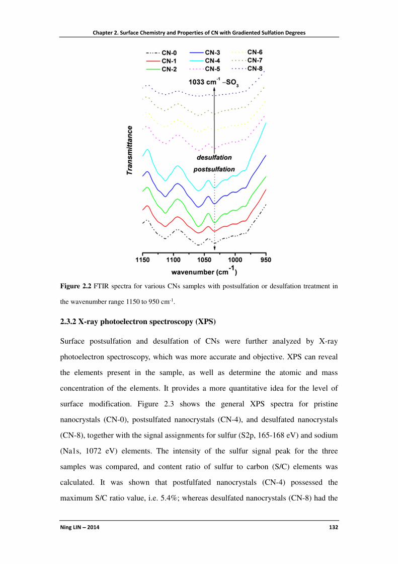
Chapter 2. Surface Chemistry and Properties of CN with Gradiented Sulfation Degrees
Ning LIN −−−− 2014 132
Figure 2.2 FTIR spectra for various CNs samples with postsulfation or desulfation treatment in
the wavenumber range 1150 to 950 cm-1.
2.3.2 X-ray photoelectron spectroscopy (XPS)
Surface postsulfation and desulfation of CNs were further analyzed by X-ray
photoelectron spectroscopy, which was more accurate and objective. XPS can reveal
the elements present in the sample, as well as determine the atomic and mass
concentration of the elements. It provides a more quantitative idea for the level of
surface modification. Figure 2.3 shows the general XPS spectra for pristine
nanocrystals (CN-0), postsulfated nanocrystals (CN-4), and desulfated nanocrystals
(CN-8), together with the signal assignments for sulfur (S2p, 165-168 eV) and sodium
(Na1s, 1072 eV) elements. The intensity of the sulfur signal peak for the three
samples was compared, and content ratio of sulfur to carbon (S/C) elements was
calculated. It was shown that postfulfated nanocrystals (CN-4) possessed the
maximum S/C ratio value, i.e. 5.4%; whereas desulfated nanocrystals (CN-8) had the

Chapter 2. Surface Chemistry and Properties of CN with Gradiented Sulfation Degrees
Ning LIN −−−− 2014 133
lowest sulfur content, with the S/C ratio of 0.25%. Regarding the sodium element,
only CN-8 nanocrystal sample showed the signal of sodium with a Na/C ratio of 4.5%,
which was a further proof for the desulfation treatment of CNs.
Figure 2.3 General XPS spectra for pristine cellulose nanocrystals (CN-0), postsulfated cellulose
nanocrystals (CN-4), and desulfated cellulose nanocrystals (CN-8) with signal assignments
(sodium Na1s, sulfur S2p).
The weight contents of sulfur (S), carbon (C), and oxygen (O) elements in the various
CNs samples were measured by elemental analysis, and the data are shown in Table
2.1. The sulfur element content in pristine nanocrystals (CN-0) was about 0.67%,
which was close to the value reported in the previous report.19 Regarding the changes

Chapter 2. Surface Chemistry and Properties of CN with Gradiented Sulfation Degrees
Ning LIN −−−− 2014 134
in sulfur content for postsulfated and desulfated nanocrystals, a logical tendency can
be observed. In contrast to CN-0, the sulfur content for postsulfated nanocrystals
CN-(1 to 4) and desulfated nanocrystals CN-(5 to 8) gradually increased up to a
maximum value of 1.31%, and decreased to a minimum value of 0.18%, respectively.
In fact, it was reported that during the biosynthesis process, trace amounts of sulfur
remained in plant tissues, which can explain the presence of slight sulfur for CN-8
sample.20
2.4 Morphological Observation and Dimensions
Before discussion on CN surface chemistry, the morphology of nanocrystals should
be checked, and the geometrical dimensions should be defined. AFM was used to
Figure 2.4 AFM images for pristine cellulose nanocrystals (CN-0), postsulfated cellulose
nanocrystals (CN-4), and desulfated cellulose nanocrystals (CN-8) at different observation scales.

Chapter 2. Surface Chemistry and Properties of CN with Gradiented Sulfation Degrees
Ning LIN −−−− 2014 135
observe the morphology and measure the dimensions of the nanoparticles. Figure 2.4
shows the amplitude, height and phase images from AFM at different observation
scales (10×10 µm; 3.3×3.3 µm; 1.1×1.1 µm). It was shown that all CN samples
exhibited the typical and expected rod-like or needle-like morphology, with a length
of about 100−300 nm, and width of about 10−30 nm. Interestingly, by comparing the
three series images, it seemed that the dimensions of postsulfated nanocrystals CN-4
were more homogeneous, which may be associated to the enhanced electrostatic
repulsion among nanoparticles in aqueous suspension.
Further statistical analysis of nanocrystal dimensions was performed by Nanoscope
software. As representative samples, statistical analysis for dimensions and
distribution determined from AFM images for samples CN-0, CN-4 and CN-8 are
shown in Figure 2.5 and Figure 2.6. Through the statistic measurements with over 100
individual nanocrystals from AFM images (mainly focusing on the images with the
scale of 3.3×3.3 µm and 1.1×1.1 µm), the average length (L), width (W) and height (H)
of CNs were about 150 nm, 22 nm, and 5 nm, respectively. Specifically, pristine
nanocrystals CN-0 showed a length of 169.9±86.8 nm, width of 23.0±8.9 nm, and
height of 5.2±1.9 nm; whereas postsulfated nanocrystals CN-4 exhibited a length of
152.0±61.6 nm, width of 22.7±8.7 nm, and height of 5.0±1.3 nm; and desulfated
nanocrystals CN-8 behaved a length of 148.6±68.1 nm, width of 21.8±8.9 nm, and
height of 5.0±1.5 nm. From the dimension changes, it was shown that the average
length gradually decreased for modified nanocrystals because of the reduction of large
scale nanocrystals. As shown in Figure 2.5, CN-0 nanocrystals with a length higher
than 220 nm disappeared in CN-4 and CN-8 samples, which can be attributed to the
further removal of amorphous region during the postsulfation and desulfation
treatments. Geometrical dimension data for all nine CN samples are summarized in
Table 2.2. The nanoscaled dimension and distribution of cotton CNs demonstrate that
these nanoparticles have the promising potential to be used as rigid nano-reinforcing
filler for composites.

Chapter 2. Surface Chemistry and Properties of CN with Gradiented Sulfation Degrees
Ning LIN −−−− 2014 136
Figure 2.5 Size statistics for length (L) and width (W) from AFM images for cellulose nanocrystal
samples (CN-0, CN-4 and CN-8); red dashed lines are Gaussian distribution fitting line according
to the statistical data.
Figure 2.6 Size statistics of height (H) for cellulose nanocrystal samples (CN-0, CN-4 and CN-8)
with the analysis of NanoScope software.

Chapter 2. Surface Chemistry and Properties of CN with Gradiented Sulfation Degrees
Ning LIN −−−− 2014 137
Table 2.2 Geometrical dimensions of various CN samples from the results of AFM.
Sample Geometrical dimension (nm)
Sample Geometrical dimension (nm)
Length Width Height Length Width Height
CN-0 169.9±86.8 23.0±8.9 5.2±1.9
CN-1 159.5±67.4 22.3±10.4 5.3±2.0 CN-5 157.6±71.3 23.6±10.1 5.1±1.7
CN-2 154.1±57.9 21.7±7.9 5.1±1.8 CN-6 150.6±64.8 21.9±8.7 5.1±1.6
CN-3 154.6±69.2 22.3±9.7 5.2±1.5 CN-7 151.6±69.5 22.6±9.4 5.1±1.4
CN-4 152.0±61.6 22.7±8.7 5.0±1.3 CN-8 148.6±68.1 21.8±8.9 5.0±1.5
2.5 Morphological Models and Surface Chemistry of Cellulose
Nanocrystals
The degree of substitution (DS) of surface hydroxyl groups (–OH) replaced by sulfate
groups (−�����) can be calculated according to Eq. (2.4). In this equation, 2�--./
represents the mole fraction of sulfate groups, which can be calculated according to
Eq. (2.5) from the results of elemental analysis. The amount of surface (–OH) on CNs
[23456��7(�-8)] can be deduced from some fundamental models (as shown in Figure
2.7 a, b, c), and calculated from Eq. (2.6).
,� =2�--./
23456��7(�-8)(2.4)
2�--./ = 2 =:
;
=:�<�%
;
(2.5)
23456��7(�>?) = � ∙ �ABA�!(�<)(2.6)
According to previous reports,20,31 the representative elementary crystallite region of
cotton CN was proposed (model b), which exhibits a square or parallelogram section
with 10 × 10 glucose units based on the d-spacing of (110), (11D0), and (200) planes
and its size is roughly 5.3 nm × 6.6 nm. Crystalline dimensions for the three planes
can be obtained from XRD experiments. (Further discussion on the effect of sulfate
groups on crystalline properties of CNs will be provided in next part.) Taking
hydroxyl groups and length of cellobiose unit into consideration (model c), 36 glucan
chains are located on the surface of the crystal, which correspond to 120 available

Chapter 2. Surface Chemistry and Properties of CN with Gradiented Sulfation Degrees
Ning LIN −−−− 2014 138
hydroxyl groups. Consequently, the number of hydroxyls per unit surface area (N0) on
cotton cellulose nanocrystal can be calculated as 8.13 × 10⁻³ mmol/m2 according to
Eq. (2.7).
� =120���E�
F�2�%%� G 2�%%H���2.7�
In Eq. (2.7),
�%%� � 6.6 � 1.032:�
�%%H� � 5.3 � 1.032:�
F � 6.02 � 10����J(KLMN(O)2P:QRN�
Figure 2.7 Schematic models of (a) an individual rod-like cellulose nanocrystal and dimensions
(cuboid shape with square section; cylinder shape with round section; cuboid shape with
rectangular section; cylinder shape with ellipsoid section); (b) an idealized elementary crystallite

Chapter 2. Surface Chemistry and Properties of CN with Gradiented Sulfation Degrees
Ning LIN −−−− 2014 139
region for cotton cellulose nanocrystal from the analysis of XRD (one black bar represents a
glucan chain, and green bars are surface available glucan chains); (c) chemical conformation and
dimensions of one single cellobiose unit.
Consequently, to determine the amount of surface –OH on CNs, the key point is to
calculate the value of the total specific surface area [�ABA�!��<�], which can be
calculated from Eq. (2.8). In this equation, S�7!!4!B37 is the density of cellulose (1.50
g/cm3); �3TUV!7��<� and W3TUV!7��<� represent the surface area and volume of a
single nanocrystal, respectively.
�ABA�!��<� �:�<
S�7!!4!B37∙�3TUV!7��<�
W3TUV!7��<��2.8�
Interestingly, with different cross-section models, the results of �ABA�!��<� will be
completely different. Generally, cellulose nanocrystal can be considered as a kind of
rod-like or needle-like nanoparticle, so the simplest cross-section model is cuboid or
cylinder with the square or round bottom, as shown in model (a). However, in this
study, according to AFM observations, the width and height of CNs are different
(W=22 nm, and H=5 nm), which indicates that the cuboid or cylinder model with a
rectangular or ellipsoidal cross-section for the nanocrystal may be more reasonable.
If choosing the cuboid model (square section) for CN,
�3TUV!7��<� � 4YZ
W3TUV!7��<� � Y�Z
So, in Eq. (2.8):
�ABA�!��<� �:�<
S�7!!4!B37∙4
Y�2.8 � 1�
If choosing the cylinder model (round section) for CN,
�3TUV!7��<� � πYZ
W3TUV!7��<� � πY�Z/4

Chapter 2. Surface Chemistry and Properties of CN with Gradiented Sulfation Degrees
Ning LIN −−−− 2014 140
So, in Eq. (2.8):
�ABA�!��<� �:�<
S�7!!4!B37∙4
Y(2.8 − 2)
If choosing the cuboid model (rectangular section) for CN,
�3TUV!7(�<) = 2(Y +E)Z
W3TUV!7(�<) = YEZ
So, in Eq. (2.8):
�ABA�!(�<) =:�<
S�7!!4!B37∙2(Y +E)
YE(2.8 − 3)
If choosing the cylinder model (ellipsoid section) for CN,
�3TUV!7(�<) = [2πE + 4(Y −E)\Z
W3TUV!7(�<) = πZEY
So, in Eq. (2.8):
�ABA�!(�<) =:�<
S�7!!4!B37∙2πE + 4(Y − E)
πYE(2.8 − 4)
It is observed that the values for �ABA�!(�<)using square and round section models
show the same results (Eq. 2.8−1 and 2.8−2), depending on the width (W) of the
nanocrystal only; whereas the values for �ABA�!(�<)using rectangular and ellipsoid
section models depend on both the width (W) and height (H) (Eq. 2.8−3 and 2.8−4).
Table 2.3 Total specific surface area [�ABA�!(�<)], amount of sulfate groups [2�--./] and surface
hydroxyl groups [23456��7(�-8)], and degree of sulfation substitution (DS) for CN-0 calculated
from different cross-section models.
Model �ABA�!(�<) Surface –OH Surface –����
� ,��--./
m2 mmol/g mmol/g %
Square/Round 117.5 0.96
0.21
21.88
Ellipse 191.2 1.55 13.45
Rectangle 325.4 2.65 7.90

Chapter 2. Surface Chemistry and Properties of CN with Gradiented Sulfation Degrees
Ning LIN −−−− 2014 141
Data for total specific surface area [�ABA�!��<�], amount of 2�--./ and23456��7��-8�,
and degree of substitution (DS) of sulfate groups for CN-0 were calculated based on
the different cross-section models, and are summarized in Table 2.3. It is shown that if
square or round models are chosen for cotton CNs, more than 20% of surface
hydroxyl groups have been substituted by sulfate groups during the H2SO4−
esterification hydrolysis, which indicates that most of active hydroxyl groups located
on C-6 have been replaced. Therefore, from these two models, desulfation treatment
for H2SO4−hydrolyzed CNs is necessary before chemical modification. However, if
choosing rectangle model for CNs, only 7.90% (only about 1/3 of the value for square
or round models) surface hydroxyl groups is substituted during the H2SO4−hydrolysis,
and a large amount of active hydroxyl groups have been remained.
In this study, ellipse model for the CNs cross-section is also proposed, and the DS
value of sulfate groups for nine CN samples is shown in Table 2.1. Specifically, if
ellipse model is chosen, about 13% of the surface hydroxyl groups are substituted by
sulfate groups during hydrolysis of pristine nanocrystal CN-0. The maximum DS
value is about 26% for CN-4, which indicates that 1/4 of the hydroxyl groups on the
CN surface can be replaced by sulfate groups during the postsulfation treatment.
The proposal of ellipse model for cotton CNs is based on AFM observation and
presumption of cross-section shape. Figure 2.8 presents the orthography image of
CNs. However, due to the small scale of height dimension (about 5 nm), it was
difficult to directly observe and determine the end shape of nanocrystals. With the
enlargements of height to ten, thirty, and fifty times (× z axis), some interesting
changes appeared on AFM images. As indicated by the green and blue circles in
Figure 2.8, it seems that after enlargement, the shape of CN cross-section transformed
to “mountain” with a peak on the top. If the nanocrystal cross-section is rectangular,
the size from top to bottom should remain invariable during the height enlargement;
whereas ellipsoid cross-section was suitable for this change. Although the ellipse
cross-section model should be further validated by other observation techniques,32 it
is a new choice to be used for the analysis of CN surface chemistry.

Chapter 2. Surface Chemistry and Properties of CN with Gradiented Sulfation Degrees
Ning LIN −−−− 2014 142
Figure 2.8 AFM image analysis for the observation of the cross-section shape for cellulose
nanocrystals (CN-4).
2.6 Effect of Sulfate Groups on the Properties of Cellulose
Nanocrystals
Besides the surface chemistry discussed above, different sulfate group contents on the
surface of CNs should also significantly affect their physical properties, such as
crystalline structure and thermal stability for inherent nanocrystal, and birefringence
behavior and rheological behavior for nanocrystal suspensions. Some studies have
reported the influence of sulfate groups on the rheological behavior of CN
suspensions. For instance, it was reported that the introduction of more sulfate groups
on the surface of CNs drastically reduced the viscosity of the suspension and removed
the time dependence.33 Recently, it was found that the degree of sulfation of CNs has
a significant effect on the critical concentration at which transitions from isotropic to
liquid crystal and liquid crystal to gel occur.34 It is a pity that some inconsistent

Chapter 2. Surface Chemistry and Properties of CN with Gradiented Sulfation Degrees
Ning LIN −−−− 2014 143
conclusions on the effect of sulfate groups were given in different studies, such as
thermal stability of sulfated CNs as mentioned in the Introduction Section. In this
study, important physical properties of CNs possibly affected by the sulfate group
content have been further investigated. Because of the wide range and detailed sulfate
group contents (nine samples from minimum to maximum contents), the global trend
analysis on properties for sulfated CNs would be more reasonable.
2.6.1 Crystalline Structure and Crystalline Dimensions
Crystal properties are among the most important physical properties of CNs, which
can reflect the integrity of the original structure of the nanocrystals. The degree of
postsulfation should be carefully controlled to react only on the surface of CNs, in
order to maximally introduce sulfate groups and avoid the further structural
destruction. On the other hand, due to the use of alkaline solution for desulfation
treatment, the polymorphic and chemical transformation of cellulose (such as type I
for cotton cellulose in this study) must be prevented. XRD is the common technique
for the investigation of crystalline structure of cellulose. The X-ray diffraction
patterns for the various CNs with different surface sulfate group contents are shown in
Figure 2.9, and the values of the crystallinity index (χc) and crystalline dimensions are
summarized in Table 2.4. The similar patterns and intensity preservation of diffraction
peaks for all samples indicated that the postsulfation and desulfation treatments did
not destroy or convert the inherent crystalline structure of nanocrystals. Indeed, the
characteristic diffraction peaks at 2θ angles around 14.9°, 16.5°, 20.5°, 22.7°, 34.5°
assigned to the typical reflection planes of cellulose I, 11D0, 110, 012, 200, 004,
respectively, were preserved.35 The crystallinity index (χc) of CNs was calculated
according to the Segal method (Eq. 2),36 which exhibited a high crystallinity (about 88%
for χc) and maintenance of the crystalline structure. From the calculation according to
Scherrer equation (Eq. 2.3), the crystalline dimensions of different planes (����, �%%H�
and �%%�) for CNs samples can be obtained. As shown in Table 2.4, the sizes of 200,
11D0, and 110 planes for cotton CNs were about 6.8 nm, 5.3 nm, and 6.6 nm, which
was consistent with the parallelogram elementary crystallite model proposed in Figure

Chapter 2. Surface Chemistry and Properties of CN with Gradiented Sulfation Degrees
Ning LIN −−−− 2014 144
2.7b. On the basis of XRD results, it can be stated that surface sulfate group content
does not obviously affect or has less influence to the crystal properties of CNs.
Figure 2.9 X-ray diffraction patterns for various cellulose nanocrystals with different surface
sulfate group contents.
Table 2.4 Values of crystallinity index (χc), crystalline dimensions (����, �%%H� and �%%�), and
ζ-potentials for various CN samples from the results of XRD and zeta-potential analysis.
Sample χc
Crystalline dimension
(nm) ζ-potential
% ���� �%%H� �%%� mV
CN-0 88.6 6.8 5.3 6.6
−40.4±1.7
CN-1 88.1 6.7 5.3 6.5 −47.8±1.9
CN-2 88.2 6.8 5.3 6.6 −54.8±2.2
CN-3 88.5 6.8 5.3 6.5 −61.7±2.4
CN-4 88.0 6.7 5.3 6.7 −66.1±1.5
CN-5 87.1 6.7 5.2 6.7 −32.8±2.3
CN-6 88.0 6.8 5.2 6.7 −27.6±1.4
CN-7 87.5 6.7 5.3 6.6 −17.9±1.1
CN-8 88.2 6.8 5.3 6.7 −7.4±0.8

Chapter 2. Surface Chemistry and Properties of CN with Gradiented Sulfation Degrees
Ning LIN −−−− 2014 145
2.6.2 ζζζζ-potential of Aqueous Suspensions
Owing to the presence of sulfate groups on their surface, rod-like CNs changed to
negatively charged nanoparticles. In the colloidal system of sulfated CNs, the
dispersion medium and the stationary layer of fluid attached to the dispersed
nanoparticles will provide the potential difference, which is named zeta potential
(ζ-potential). The value of zeta potential is related to the stability of the colloidal
dispersion, and indicates the degree of repulsion between adjacent, similarly charged
particles in a suspension. The effect of sulfate groups on ζ-potential of CNs is
presented in Table 2.4. With the increase of sulfate group content, sulfated CNs
possessed a more negative surface, and exhibited lower ζ-potential values. Especially
for postsulfated nanocrystals sample CN-4, the value of ζ-potential reached −66.1±1.5
mV, which indicated the strong repulsive force between nanoparticles, and therefore
the promotion of its dispersion in suspension (as proved by AFM). It is worth noting
that desulfation treatment for sulfated nanocrystals was also effective, which can be
reflected by the significant increase of ζ-potential for desulfated CN samples.
2.6.3 Thermal Stability
Thermal stability is another important physical property for CNs, which plays a
critical role in the preparation of melt processed CN-reinforced composites. As
mentioned in the Introduction Section, the effect of sulfate groups on the surface of
CNs to thermal stability led to inconsistent conclusions from different reports.
Therefore, thermal degradation behavior of sulfated CNs was further investigated by
thermogravimetric analysis in this study, which is shown in Figure 2.10. Interestingly,
in comparison with all nanocrystal samples, native cotton exhibited the highest
thermal stability for the first phase of thermal decomposition (Td1). Because of the
hydrolysis and presence of sulfate groups, the values of Td1 for all postsulfated
nanocrystals sharply decreased to about 150 °C compared with native cotton. By
determining Td1 for postsulfated CNs themselves (event a in Figure 2.10), it appears
that more sulfate groups on nanocrystals induced a lowering of Td1, such as the
decrement (∆t1) of 12.3 °C from CN-1 to CN-4 samples. In contrast to postsulfated

Chapter 2. Surface Chemistry and Properties of CN with Gradiented Sulfation Degrees
Ning LIN −−−− 2014 146
nanocrystals, the values of Td1 for desulfated CNs generally enhanced about 100 °C
attributed to the removal of sulfate groups and presence of sodium on nanocrystals.
From the analysis of Td1 for desulfated CNs themselves (event b in Figure 2.10),
similar conclusion can be obtained, i.e. less sulfate groups on nanocrystals resulted in
higher Td1, such as the increment (∆t2) of 13.8 °C from CN-5 to CN-8 samples. It
should be pointed out that according to our investigation, besides sulfate groups, the
introduction of sodium on the surface of CNs may be another factor that significantly
affects their thermal property. Furthermore, from the TGA thermograms in Figure 8,
thermal degradation behavior of cellulose for the second phase of thermal
decomposition (Td2) can also be observed. Differently, all nanocrystal samples
possessed higher Td2 values than native cotton (about 65 °C), which may be
associated to the higher crystallinity for nanocrystal samples.
Figure 2.10 Effect of surface sulfate groups on the thermal stability of cellulose nanocrystals
(TGA thermograms).

Chapter 2. Surface Chemistry and Properties of CN with Gradiented Sulfation Degrees
Ning LIN −−−− 2014 147
2.6.4 Birefringence of Aqueous Suspensions
In 1959, the birefringence character of acid-treated cellulose and chitin crystallites
was first reported,37 which was the behavior through strong adsorption of one of
polarized rays to possess two refractive indices. Birefringence of CN suspensions can
be observed with two linear polarizers crossed at 90° (cross-nicols or crossed-polars),
and gives a quick indication of the successful preparation of well-dispersed CN
suspensions. Figure 2.11 shows the photographs of aqueous suspensions of various
CNs with postsulfation and desulfation treatments. The presence of negative charges
(−�����) on CN surface induced an electrostatic repulsion between nanoparticles, and
the suspensions exhibited the formation of birefringent domains. Because of the stable
dispersion and flow anisotropy from alignment of nano-rods under flow state, the
birefringence character for postsulfated nanocrystal suspensions was maintained. With
the desulfation and progressive removal of sulfate groups, nanoparticles in
suspensions tended to aggregate, which induced the reduction and weakening of
birefringent domains. Especially for strongly desulfated nanocrystals, samples CN-7
and CN-8, the birefringence behavior of suspensions was difficult to be observed.
Figure 2.11 Photographs of aqueous suspensions of various cellulose nanocrystals viewed through
cross polarizers (0.5 wt%).
Chapter 2. Surface Chemistry and Properties of CN with Gradiented Sulfation Degrees
Ning LIN −−−− 2014 148
2.7 Conclusions
The objective of this study was to investigate how surface sulfate groups affect the
surface chemistry and physical properties of cellulose nanocrystals. Through the
regulation of reactant ratios and reactive conditions for postsulfation and desulfation,
gradient contents of sulfate groups can be introduced on the surface of CNs. Based on
the discussion of nanocrystal’s surface chemistry, the influence of different
cross-section models for the calculation of the degree of substitution of the sulfate
groups was investigated, and a new ellipsoid model for cotton CNs was proposed on
the basis of AFM observation. Finally, the effects of sulfate groups on diverse
properties of CNs and their suspensions have been discussed, and some issues with
inconsistent conclusions from previous reports were further validated. Future studies
will focus on the electrostatic interactions between negative charges from sulfated
CNs and positive charges from cationic polymers or molecules. With the detection
and measurement of electrostatic interactions, it is expected to reveal the strength and
mechanism of this special interaction from coupling charges and therefore advance
the application of CN electrostatic grafting.
2.8 References
1. Dufresne, A. Nanocellulose. From nature to high performance tailored materials;
Walter de Gruyter GmbH: Berlin and Boston, MA; 2012.
2. The global market for nanocellulose to 2017; Futures Markets Inc.: Edinburgh,
Lothian EH74NA, United Kingdom, 2012.
3. Brinchi, L.; Cotana, F.; Fortunati, E.; Kenny, J. M. Production of nanocrystalline
cellulose from lignocellulosic biomass: technology and applications. Carbohydr.
Polym. 2013, 94, 154–169.
4. Araki, J. Electrostatic or steric? – preparations and characterizations of
well-dispersed systems containing rod-like nanowhiskers of crystalline
polysaccharides. Soft Matter 2013, 9, 4125–4141.
5. Dufresne, A. Nanocellulose: a new ageless bionanomaterial. Mater. Today 2013,

Chapter 2. Surface Chemistry and Properties of CN with Gradiented Sulfation Degrees
Ning LIN −−−− 2014 149
16, 220–227.
6. Leung, A. C. W.; Lam, E.; Chong, J.; Hrapovic, S.; Luong, J. H. T. Reinforced
plastics and aerogels by nanocrystalline cellulose. J. Nanopart. Res. 2013, 15,
1636 (24 pages).
7. Lin, N.; Huang, J.; Dufresne, A. Preparation, properties and applications of
polysaccharide nanocrystals in advanced functional nanomaterials: a review.
Nanoscale 2012, 4, 3274–3294.
8. Tingaut, P.; Zimmermann, T.; Sèbe, G. Cellulose nanocrystals and
microfibrillated cellulose as building blocks for the design of hierarchical
functional materials. J. Mater. Chem. 2012, 22, 20105–20111.
9. Lam, E.; Male, K. B.; Chong, J. H.; Leung, A. C.W.; Luong, J. H. T.
Applications of functionalized and nanoparticle-modified nanocrystalline
cellulose. Trends Biotechnol. 2012, 30, 283–290.
10. Habibi, Y.; Lucia, L. A.; Rojas, O. J. Cellulose nanocrystals: chemistry,
self-assembly, and applications. Chem. Rev. 2010, 110, 3479–3500.
11. Rånby, B. G. Aqueous colloidal solutions of cellulose micelles. Acta Chem.
Scand. 1949, 3, 649–650.
12. Cranston, E. D.; Gray, D. G.; Rutland, M. W. Direct surface force measurements
of polyelectrolyte multilayer films containing nanocrystalline cellulose. Langmuir
2010, 26, 17190–17197.
13. Moreau, C.; Beury, N.; Delorme, N.; Cathala, B. Tuning the architecture of
cellulose nanocrystal–poly(allylamine hydrochloride) multilayered thin films:
Influence of dipping parameters. Langmuir 2012, 28, 10425−10436.
14. Liew, S. Y.; Thielemans, W.; Walsh, D. A. Electrochemical capacitance of
nanocomposite polypyrrole/cellulose films. J. Phys. Chem. C 2010, 114, 17926–
17933.
15. Ma, H.; Burger, C.; Hsiao, B. S.; Chu, B. Nanofibrous microfiltration membrane
based on cellulose nanowhiskers. Biomacromolecules 2012, 13, 180−186.
16. Thielemans, W.; Warbey, C. R.; Walsh, D. A. Permselective nanostructured
membranes based on cellulose nanowhiskers. Green Chem. 2009, 11, 531–537.

Chapter 2. Surface Chemistry and Properties of CN with Gradiented Sulfation Degrees
Ning LIN −−−− 2014 150
17. Jiang, F.; Kittle, J. D.; Tan, X.; Esker, A. R.; Roman, M. Effects of sulfate groups
on the adsorption and activity of cellulases on cellulose substrates. Langmuir
2013, 29, 3280−3291.
18. Kalashnikova, I.; Bizot, H.; Cathala, B.; Capron, I. Modulation of cellulose
nanocrystals amphiphilic properties to stabilize oil/water interface.
Biomacromolecules 2012, 13, 267−275.
19. Abitbol, T.; Kloser, E.; Gray, D. G. Estimation of the surface sulfur content of
cellulose nanocrystals prepared by sulfuric acid hydrolysis. Cellulose 2013, 20,
785–794.
20. Gu, J.; Catchmark, J. M.; Kaiser, E. Q.; Archibald, D. D. Quantification of
cellulose nanowhiskers sulfate esterification levels. Carbohydr. Polym. 2013, 92,
1809–1816.
21. Roman, M.; Winter, W. T. Effect of sulfate groups from sulfuric acid hydrolysis
on the thermal degradation behavior of bacterial cellulose. Biomacromolecules
2004, 5, 1671–1677.
22. Voronova, M. I.; Surov, O. V.; Zakharov, A. G. Nanocrystalline cellulose with
various contents of sulfate groups. Carbohydr. Polym. 2013, 98, 465–469.
23. Lin, N.; Dufresne, A. Physical and/or chemical compatibilization of extruded
cellulose nanocrystal reinforced polystyrene nanocomposites. Macromolecules
2013, 46, 5570−5583.
24. Zhang, K.; Brendler, E.; Geissler, A.; Fischer, S. Synthesis and spectroscopic
analysis of cellulose sulfates with regulable total degrees of substitution and
sulfation patterns via 13C NMR and FT Raman spectroscopy. Polymer 2011, 52,
26–32.
25. Wang, Z.-M.; Li, L.; Xiao, K.-J.; Wu, J.-Y. Homogeneous sulfation of bagasse
cellulose in an ionic liquid and anticoagulation activity. Bioresource Technol.
2009, 100, 1687–1690.
26. Jiang, F.; Esker, A. R.; Roman, M. Acid-catalyzed and solvolytic desulfation of
H2SO4-hydrolyzed cellulose nanocrystals. Langmuir 2010, 26, 17919–17925.
27. Kloser, E.; Gray, D. G. Surface grafting of cellulose nanocrystals with

Chapter 2. Surface Chemistry and Properties of CN with Gradiented Sulfation Degrees
Ning LIN −−−− 2014 151
poly(ethylene oxide) in aqueous media. Langmuir 2010, 26, 13450–13456.
28. Hasani, M.; Cranston, E. D.; Westman, G.; Gray, D. G. Cationic surface
functionalization of cellulose nanocrystals. Soft Matter 2008, 4, 2238–2244.
29. van den Berg, O.; Capadona, J. R.; Weder, C. Preparation of homogeneous
dispersions of tunicate cellulose whiskers in organic solvents. Biomacromolecules
2007, 8, 1353–1357.
30. Suganuma, S.; Nakajima, K.; Kitano, M.; Yamaguchi, D.; Kato, H.; Hayashi, S.;
Hara, M. Hydrolysis of cellulose by amorphous carbon bearing SO3H, COOH,
and OH groups. J. Am. Chem. Soc. 2008, 130, 12787–12793.
31. Elazzouzi-Hafraoui, S.; Nishiyama, Y.; Putaux, J.-L.; Heux, L.; Dubreuil, F.;
Rochas, C. The shape and size distribution of crystalline nanoparticles prepared
by acid hydrolysis of native cellulose. Biomacromolecules 2008, 9, 57–65.
32. Majoinen, J.; Haataja, J. S.; Appelhans, D.; Lederer, A.; Olszewska, A.; Seitsonen,
J.; Aseyev, V.; Kontturi, E.; Rosilo, H.; Österberg, M.; Houbenov, N.; Ikkala, O.
Supracolloidal multivalent interactions and wrapping of dendronized
glycopolymers on native cellulose nanocrystals. J. Am. Chem. Soc. 2014, 136,
866−869.
33. Araki, J.; Wada, M.; Kuga, S. Influence of surface charge on viscosity behavior
of cellulose microcrystal suspension. J. Wood. Sci. 1999, 45, 258–261.
34. Shafiei-Sabet, S.; Hamad, W. Y.; Hatzikiriakos, S. G. Influence of degree of
sulfation on the rheology of cellulose nanocrystal suspensions. Rheol. Acta. 2013,
52, 741–751.
35. Lin, N.; Bruzzese, C.; Dufresne, A. TEMPO-Oxidized nanocellulose participating
as crosslinking aid for alginate-based sponges. ACS Appl. Mater. Interfaces 2012,
4, 4948−4959.
36. Segal, L.; Creely, J. J.; Martin, A. E.; Conrad, C. M. An empirical method for
estimating the degree of crystallinity of native cellulose using the X-ray
diffractometer. Text. Res. J. 1959, 29, 786−794.
37. Marchessault, R. H.; Morehead, F. F.; Walter, N. M. Liquid crystal systems from
fibrillar polysaccharides Nature 1959, 184, 632−633.

Chapter 2. Surface Chemistry and Properties of CN with Gradiented Sulfation Degrees
Ning LIN −−−− 2014 152

Chapter 3. Physical and/or Chemical Compatibilization of Extruded CN Reinforced PS Nanocomposites
Ning LIN −−−− 2014 153
CHAPTER 3.
Application of Cellulose Nanocrystals: Extruded Nanocomposites
“Physical and/or Chemical Compatibilization of Extruded Cellulose
Nanocrystal Reinforced Polystyrene Nanocomposites”

Chapter 3. Physical and/or Chemical Compatibilization of Extruded CN Reinforced PS Nanocomposites
Ning LIN −−−− 2014 154

Chapter 3. Physical and/or Chemical Compatibilization of Extruded CN Reinforced PS Nanocomposites
Ning LIN −−−− 2014 155
Chapter 3. Physical and/or Chemical Compatibilization of Extruded
Cellulose Nanocrystal Reinforced Polystyrene Nanocomposites
ENGLISH ABSTRACT ............................................................................................................. 156
RESUME FRANÇAIS ............................................................................................................. 159
3.1 Introduction ................................................................................................................... 165
3.2 Experimental Methodology ........................................................................................ 167
3.2.1 Materials .................................................................................................................... 167
3.2.2 Extraction of Cellulose Nanocrystals from Cotton Fiber ........................................... 168
3.2.3 Carboxylation of Cellulose Nanocrystals ................................................................... 168
3.2.4 Chemical Grafting of PEG5000 on Cellulose Nanocrystals ........................................ 169
3.2.5 Physical Adsorption of PEO5M on Pristine and Modified Nanocrystals ................... 169
3.2.6 Preparation of Nanocomposites from CN and Polystyrene by Extrusion ................. 170
3.2.7 Characterization and Analyses .................................................................................. 171
3.3 Surface Modification and Properties of Cellulose Nanocrystals .......................... 175
3.3.1 Surface Grafting of PEG on Cellulose Nanocrystals ................................................... 175
3.3.2 Crystalline Structure .................................................................................................. 178
3.3.3 Morphology and Surface Adsorption ........................................................................ 179
3.3.4 Thermal Stability ........................................................................................................ 180
3.3.5 Surface Hydrophilicity................................................................................................ 181
3.3.6 Rheological Behavior of Modified Cellulose Nanocrystals in Suspension ................. 183
3.4 Structure and Properties of Extruded Nanocomposites ........................................ 184
3.4.1 Appearance Observation and Light Transmittance ................................................... 184
3.4.2 Dispersion and Microstructure of Nanocrystals in Composites ................................ 187
3.4.3 Thermal Properties .................................................................................................... 190
3.4.4 Mechanical Properties ............................................................................................... 192
3.4.5 Barrier Properties ...................................................................................................... 194
3.5 Conclusions .................................................................................................................... 196
3.6 References ..................................................................................................................... 197

Chapter 3. Physical and/or Chemical Compatibilization of Extruded CN Reinforced PS Nanocomposites
Ning LIN −−−− 2014 156
ENGLISH ABSTRACT
Since the first report in 1995,1 cellulose nanocrystals have been widely used as the
strength enhancing additive to improve the mechanical performance of a wide range
of plastic composites because of their impressive mechanical properties. Commonly,
the values of strength and modulus for cellulose nanocrystals are recognized as being
in the range of 10 GPa and 100−160 GPa, respectively, which are comparable to
Kevlar. Recently, based on the atomic structure model of cellulose in tandem with
quantum mechanics, the computed Young’s modulus of cellulose nanocrystal was
predicted as 206 GPa,2 which showed that this special nanoparticle even possess the
stiffness of steel and strongly favored its usage in the development of new composite
materials. In fact, according to the statistical analysis of Future Markets Inc, about
half demand (46%) of nanocellulose (including cellulose nanocrystals and
microfibrillated cellulose) by end user market focuses on its composite application.3
For the development of cellulose nanocrystal-based composites, two fundamental
issues should be emphasized, which are the composite processing method and the
compatibility between the nanoparticles and the matrix. Basically, four main
methods are generally employed for the processing of cellulose nanocrystal-based
composites, which are casting-evaporation, melt-compounding, electrospinning, and
layer-by-layer assembly.4 Particularly, the techniques of casting-evaporation and
melt-compounding are commonly used for the preparation of cellulose
nanocrystal-based composites because of their easy manipulation. Specifically,
processing by the casting-evaporation method allows the preservation of
individualization state of the nanoparticles resulting from their colloidal dispersion in
liquid medium. However, this technique is non-industrial, non-economic, and shows
some limitation regarding the choice of the polymeric matrix. In industry,
melt-compounding techniques, such as extrusion or injection molding, are commonly
used to process thermoplastic polymers because they are “green” (solvent-free),
effective and economic processing methods. However, due to the extensive use of
sulfuric acid hydrolysis (with high efficiency and promotion of colloidal stability) in

Chapter 3. Physical and/or Chemical Compatibilization of Extruded CN Reinforced PS Nanocomposites
Ning LIN −−−− 2014 157
the extraction and production of cellulose nanocrystals, additional surface sulfate
groups result in the weakness of their thermal stability (as discussed in the previous
work Chapter 2), and limit the thermal processing temperature of cellulose
nanocrystal-based composites. Another issue is the inherent incompatibility between
cellulose nanoparticles and non-polar molecules (polymeric matrix). During the
thermal processing, irreversible self-aggregation of cellulose nanocrystals ascribed to
their hydrophilic nature causes weak interfacial adhesion (even microphase
separation) between nanoparticles and matrix, and finally induces inferior
performances of composites. All of these issues have mainly restricted the practical
utilization of melt-compounding to process cellulose nanocrystal-based composites.
In order to improve the thermal stability and promote the compatibility of cellulose
nanocrystals with non-polar polymeric matrices during the extrusion process, a novel
strategy involving a double-polymer-layer protection and physical and/or chemical
compatibilization of cellulose nanocrystals was proposed as shown in the “Schematic
diagram”. In this work, cotton cellulose nanocrystals (CNs) were prepared by sulfuric
acid hydrolysis. The chemical compatibilization of cellulose nanocrystals (CN-g-PEG)
was realized by surface grafting of low molecular weight poly(ethylene glycol)
(PEG5000) through chemical carboxylation–amidation reaction. The physical
compatibilization of cellulose nanocrystals (CN/PEO) was performed by surface
interaction with high molecular weight polyoxyethylene (PEO5M) through physical
blending. Meanwhile, because of possible entanglements and interactions between
both polymeric chains (PEG and PEO with the same chemical structure),
two-layer-modified cellulose nanocrystals (CN-g-PEG/PEO) can be developed, which
can induce double-polymer-layer protection and both covalent and non-covalent
compatibilization of cellulose nanocrystals in polymer nanocomposites. It was shown
that the different surface modification strategies brought cellulose nanocrystals
distinct thermal behaviors during extrusion processing. Both CN-g-PEG and
CN-g-PEG/PEO nanocrystals showed improved thermal stability, while CN and
CN/PEO seriously degraded and carbonized during extrusion. The comparison of

Chapter 3. Physical and/or Chemical Compatibilization of Extruded CN Reinforced PS Nanocomposites
Ning LIN −−−− 2014 158
adsorbing PEO capacity between CN and CN-g-PEG was also discussed and it was
proved that CN-g-PEG can adsorb about 60 wt% (corresponding to CN-g-PEG weight)
PEO chains, but CN can’t or can only adsorb a small amount of PEO chains.
Then, four series of extruded nanocomposites were prepared with various loading
levels of neat or modified cellulose nanocrystals using polystyrene (PS) as polymeric
matrix. Interestingly, only double-polymer-layer modified nanocrystals reinforcing
composites (CN-PEG/PEO-PS) were translucent and gave a good appearance. The
microstructural analysis of extruded composites indicated that two-layer polymers
covering the surface of cellulose nanocrystals can not only protect the nanoparticles
from thermal degradation but also facilitate the interactions between nanocrystals
and PS matrix. The investigation of mechanical properties, thermal properties, and
water vapor barrier performances of extruded composites further proved the
different effects of chemical and/or physical compatibilization of modified cellulose
nanocrystals.
Schematic diagram of extruded cellulose nanocrystals (CN) reinforced polystyrene
(PS) nanocomposites with physical and/or chemical compatibilization

Chapter 3. Physical and/or Chemical Compatibilization of Extruded CN Reinforced PS Nanocomposites
Ning LIN −−−− 2014 159
RESUME FRANÇAIS
Depuis la première étude en 1995,1 les nanocristaux de cellulose ont été largement
utilisés comme additif de renfort pour améliorer les performances mécaniques d’un
large éventail de composites à base de plastique en raison de leurs remarquables
propriétés mécaniques. Généralement, les valeurs communément admises pour la
résistance et le module des nanocristaux de cellulose sont de l’ordre de 10 GPa et
100-160 GPa, respectivement, valeurs qui sont comparables à celles du Kevlar.
Récemment, sur la base du modèle de la structure atomique de la cellulose couplé
avec la mécanique quantique, le module d’Young du nanocristal de cellulose a été
estimé à 206 GPa,2 ce qui montre que cette nanoparticule spéciale présente même
une rigidité similaire à celle de l'acier et a fortement favorisé son utilisation dans le
développement de nouveaux matériaux composites. De fait, selon l'analyse
statistique de Future markets Inc, environ la moitié de la demande (46%) pour la
nanocellulose (incluant nanocristaux de cellulose et cellulose microfibrillée) par les
utilisateurs finaux se concentre sur son application composite.3
Le développement des composites à base de nanocristaux cellulosiques est
cependant tributaire de deux questions fondamentales qui méritent d'être soulignés,
à savoir la méthode de mise en forme du composite et la compatibilité entre les
nanoparticules et la matrice. Fondamentalement, quatre méthodes principales sont
généralement utilisées pour la mise en œuvre des matériaux composites à base de
nanocristaux de cellulose, qui sont la coulée-évaporation, le mélange à l’état fondu,
l’électrofilage et l’assemblage couche par couche.4 En particulier, les techniques de
coulée-évaporation et de mélange à l'état fondu sont couramment utilisés pour la
préparation de matériaux composites à base de nanocristaux de cellulose en raison
de leur manipulation facile. Plus spécifiquement, la mise en forme par le procédé de
coulée-évaporation permet de préserver l’état d’individualisation des nanoparticules
résultant de leur dispersion colloïdale dans un milieu liquide. Cependant, cette
technique est non-industrielle, non économique et montre certaines limitations en
ce qui concerne le choix de la matrice polymère. Dans l'industrie, les techniques de

Chapter 3. Physical and/or Chemical Compatibilization of Extruded CN Reinforced PS Nanocomposites
Ning LIN −−−− 2014 160
mise en forme à l'état fondu, telles que l'extrusion ou le moulage par injection, sont
couramment utilisées pour mettre en œuvre les polymères thermoplastiques parce
que ce sont des procédés "verts" (sans solvant), efficaces et économiques. Toutefois,
en raison de l'utilisation très répandue de l'hydrolyse à l'acide sulfurique (avec une
bonne efficacité et améliorant la stabilité colloïdale) pour l'extraction et la production
de nanocristaux de cellulose, des groupes sulfate de surface additionnels se forment
ce qui conduit à une réduction de leur stabilité thermique (comme décrit dans
l’étude précédente chapitre 2) et limite la température de mise en œuvre thermique
des matériaux composites à base de nanocristaux de cellulose. Un autre problème
est l'incompatibilité inhérente entre les nanoparticules de cellulose et des molécules
non-polaires (matrice polymère). Pendant la transformation thermique,
l’auto-agrégation irréversible des nanocristaux de cellulose attribuée à leur nature
hydrophile entraîne une faible adhérence interfaciale (et même une micro-séparation
de phase) entre les nanoparticules et la matrice, et induit au final de piètres
performances aux composites. Tous ces aspects ont essentiellement restreint
l'utilisation pratique de la transformation à l’état fondu pour mettre en œuvre les
composites à base de nanocristaux cellulosiques.Afin d'améliorer la stabilité
thermique et de promouvoir la compatibilité des nanocristaux de cellulose avec des
matrices polymères non polaires au cours du processus d'extrusion, une nouvelle
stratégie impliquant une double couche polymère physique et/ou chimique
protectrice et compatibilisante des nanocristaux de cellulose a été proposée comme
illustré dans le "schéma de principe". Dans cette étude, les nanocristaux de cellulose
de coton (CNs) ont été préparés par hydrolyse à l'acide sulfurique. La
compatibilisation chimique des nanocristaux de cellulose (CN-g-PEG) a été réalisée
par greffage en surface de poly (éthylène glycol) (PEG5000) de faible poids
moléculaire par réaction chimique de carboxylation-amidation. La compatibilisation
physique des nanocristaux de cellulose (CN/PEO) a été réalisée par interaction de
surface avec un polyoxyéthylène (PEO5M) de poids moléculaire élevé par mélange
physique. Parallèlement, en raison de l'enchevêtrement et des interactions possibles
entre les deux chaînes polymères (PEG et PEO avec la même structure chimique), des

Chapter 3. Physical and/or Chemical Compatibilization of Extruded CN Reinforced PS Nanocomposites
Ning LIN −−−− 2014 161
nanocristaux de cellulose modifiés avec deux couches (CN-g-PEG/PEO) ont pu être
préparés, ce qui peut induire une double-couche polymère de protection et
compatibilisation à la fois covalente et non covalente des nanocristaux de cellulose
dans les nanocomposites polymères. Nous avons observé que les différentes
stratégies de modification de la surface des nanocristaux de cellulose conduisent à
des comportements thermiques distincts au cours de la transformation par extrusion.
Les nanocristaux CN-g-PEG et CN-g-PEG/PEO ont montré une stabilité thermique
améliorée, alors que les nanocristaux CN et CN/PEO sont sérieusement dégradés et
carbonisés lors de l'extrusion. La capacité d'adsorption du PEO par les nanocristaux
CN et CN -g-PEG a également été discutée et il a été montré que le CN-g-PEG peut
adsorber environ 60% (par rapport à la masse de CN-g-PEG) de chaînes de PEO,
tandis que les nanocristaux CN ne peuvent pas ou ne peuvent adsorber qu’en faible
quantité les chaînes de PEO.
Schéma de principe de la préparation des nanocomposites extrudés à base de
polystyrène (PS) renforcé par des nanocristaux de cellulose (CN) compatibilisés
physiquement et/ou chimiquement
Par la suite, quatre séries de nanocomposites extrudés ont été préparés avec
différents taux de nanocristaux de cellulose vierges ou modifiés en utilisant du
polystyrène (PS) comme matrice polymère. Il est intéressant de noter que seuls les
composites renforcés avec des nanocristaux de cellulose modifiés avec une double
couche polymère (CN-PEG/PEO-PS) étaient translucides et avaient un bon aspect.

Chapter 3. Physical and/or Chemical Compatibilization of Extruded CN Reinforced PS Nanocomposites
Ning LIN −−−− 2014 162
L'analyse microstructurale des matériaux composites extrudés a montré que les deux
couches polymères en surface des nanocristaux de cellulose permettent non
seulement de protéger les nanoparticules de la dégradation thermique, mais
également de faciliter les interactions entre les nanocristaux et la matrice PS. L'étude
des propriétés mécaniques, des propriétés thermiques et des performances de
barrière à la vapeur d'eau des composites extrudés ont souligné les différents effets
de la compatibilisation chimique et/ou physique des nanocristaux de cellulose
modifiés.
References:
1. Favier, V.; Canova, G. R.; Cavaillé, J. Y.; Chanzy, H.; Dufresne, A.; Gauthier, C.
Nanocomposite materials from latex and cellulose whiskers. Polym. Adv. Technol.
1995, 6, 351–355.
2. Dri, F. L.; Hector Jr., L. G.; Moon, R. J.; Zavattieri, P. D. Anisotropy of the elastic
properties of crystalline cellulose Iβ from first principles density functional theory
with Van der Waals interactions. Cellulose 2013, 20, 2703–2718.
3. The global market for nanocellulose to 2017; Futures Markets Inc., Edinburgh,
Lothian EH74NA, United Kingdom, 2012, pp. 1–66.
4. Dufresne, A. “Nanocellulose. From nature to high performance tailored
materials”. 2012 by Walter de Gruyter GmbH, Berlin/Boston. pp. 235−275.
The work in this chapter is originated from:
Ning Lin and Alain Dufresne, Physical and/or chemical compatibilization of
extruded cellulose nanocrystal reinforced polystyrene nanocomposites.
Macromolecules 2013, 46, 5570−5583.

Chapter 3. Physical and/or Chemical Compatibilization of Extruded CN Reinforced PS Nanocomposites
Ning LIN −−−− 2014 163
Chapter 3. Physical and/or Chemical Compatibilization of
Extruded Cellulose Nanocrystal Reinforced Polystyrene
Nanocomposites
Ning Lin and Alain Dufresne
The International School of Paper, Print Media and Biomaterials (Pagora), Grenoble Institute of
Technology (Grenoble INP), CS10065, 38402 Saint Martin d′Hères Cedex, France
ABSTRACT
Impressive mechanical properties and reinforcing capability make cellulose
nanocrystal (CN) a promising candidate as biomass nanofiller for the development of
polymer-based nanocomposites. With the recent announcement of large-scale CN
production, the use of industrial processing techniques for the preparation of
CN-reinforced nanocomposites, such as extrusion, is highly required. However, low
thermal stability of sulfuric acid-prepared CN limits the processing since most
polymeric matrices are processed at temperatures close to 200 °C or above. It has
been proved that surface interactions of polymers on CN as compatibilizer, such as
hydrophilic polyoxyethylene (PEO), can improve its thermal stability due to the
shielding and wrapping of PEO. However, the weak combination between CN and
PEO allows the free movement of surface polymer, which can induce the
self-aggregation of CN and microphase separation in composites especially during
melt processing. Using carboxylation−amidation reaction, short poly(ethylene glycol)
(PEG) chains can be first grafted on the surface of the nanocrystals, and immobilize
long PEO chains on modified nanocrystals through physical adsorption and
entanglement. Two polymeric layers should further improve the thermal stability of
CNs, and surface polymeric chains should provide significant dispersibility and
compatibilization for extruded nanocomposites. Rheological analysis showed better
Chapter 3. Physical and/or Chemical Compatibilization of Extruded CN Reinforced PS Nanocomposites
Ning LIN −−−− 2014 164
PEO adsorption for PEG-grafted nanocrystals than pristine CN. Results from AFM
and SEM revealed homogeneous dispersion and good compatibility of modified
nanocrystals in PS matrix. Finally, the thermal, mechanical, and barrier properties of
ensuing nanocomposites have been investigated to study the effect of physically
and/or chemically modified nanocrystals.
KEYWORDS
Cellulose Nanocrystals, Polystyrene, Compatibilization, Extrusion, Nanocomposites

Chapter 3. Physical and/or Chemical Compatibilization of Extruded CN Reinforced PS Nanocomposites
Ning LIN −−−− 2014 165
3.1 Introduction
Using controlled strong acid hydrolysis, needle-like or rod-like nanocrystals can be
produced by breaking down cellulose fibers and isolating the crystalline domains.
With their impressive mechanical properties, such as longitudinal modulus close to
150 GPa, high aspect ratio (10−70) and surface area (few 100 m2/g), cellulose
nanocrystals (CNs) have proven to be an ideal candidate as reinforcing bionanofiller
for polymeric matrices,1−4 since the first demonstration in 1995.5 The mass production
of CNs, which should increase upward of multiple tons per day in the future, gives a
strong boost for their application in composites at industrial scale. According to the
forecasting of Future Markets Inc., the production of nanocellulose (including CNs
and nanofibrillated cellulose) will increase by 1000% in the next two years, and will
likely increase by further 500% at least by 2017.6
Apart from their large-scale industrial production, the processing of CN-based
nanocomposites is another crucial issue since it determines their usage properties for
practical applications. Two challenges deserve more attention during the processing of
nanocomposites: (1) the balance between compounding method and dispersion and (2)
the compatibility of nanocrystals in polymeric matrices. Wet processing methods such
as casting/evaporation, using polymer solution or polymer dispersion (latex) in liquid
medium are commonly used in most studies of composites reinforced with CNs.7,8
The main advantage of this technique is the preservation of the dispersion state of the
nanoparticles in the liquid medium. However, the range of available polymeric
matrices is restricted, and this processing technique is both nonindustrial and
noneconomic. Melting-compounding techniques, such as extrusion or injection
molding, are probably the most convenient processing techniques for the industrial
production of CN-based nanocomposites, because of being a green process together
with large-scale production and economical availability. Extrusion is a high volume
manufacturing process in which polymeric solid material is transported by a screw
and progressively melted to form a continuous profile by passing through a die.
However, in most scientific studies, this conventional processing technique is

Chapter 3. Physical and/or Chemical Compatibilization of Extruded CN Reinforced PS Nanocomposites
Ning LIN −−−− 2014 166
infrequently employed for the preparation of CN-reinforced polymer nanocomposites.
This is ascribed to low thermal stability of cellulose and inherent incompatibility
between cellulose and most synthetic polymers issues.9 The hydrophilic nature of
cellulose causes irreversible agglomeration during drying and incompatibility with
nonpolar matrices because of the formation of additional hydrogen bonds between
nanoparticles. Moreover, because of the introduction of sulfate groups resulting from
common acid hydrolysis procedure involving sulfuric acid, CNs present inferior
thermal stability with degradation temperature lower than 150 °C. However, most
polymeric matrices from petrochemical synthesis are melt and processed at 200 °C or
above,10 which requires higher thermal stability for CNs.
On the basis of previous studies for different nanocomposite systems reinforced with
CNs, three strategies are commonly used in the processing of CN based
nanocomposites by melt compounding, in order to enhance the compatibility between
hydrophilic nanoparticles and hydrophobic matrices.9 If a polar polymer is used as
matrix, there is no a priori need to compatibilize the filler with the matrix material.11,12
For nonpolar polymeric matrices, surface functionalization, such as grafting of
hydrophobic moieties or chemical derivatization, can be conducted to tune the
compatibility between nanoparticles and matrix.13−17 Finally, adsorption of surfactants
or macromolecules on the surface of CNs was also attempted to be used as processing
aids to compatibilize cellulose nanoparticles and matrix.18−22
The challenge of melt processing for CN is to find the suitable treatment to both
improve the thermal stability and avoid the issues of inherent incompatibility between
cellulose and most polymeric matrices. In a previous work, we have demonstrated that
physical interaction of polyoxyethylene (PEO) chains on the surface of CNs can
improve dispersibility and thermal stability of nanocrystals during the melt processing
of low-density polyethylene (LDPE) based nanocomposites.18 The improved thermal
stability of PEO-adsorbed CNs was ascribed to a protection role of wrapped PEO
chains that hides the surface sulfate groups of nanocrystals. On the other hand, it has
been proved that chemical functionalization on the surface of CNs is a most effective

Chapter 3. Physical and/or Chemical Compatibilization of Extruded CN Reinforced PS Nanocomposites
Ning LIN −−−− 2014 167
approach to avoid irreversible agglomeration during drying, and enhance the
compatibility between nanoparticles and nonpolar matrices.13,23 In this work, surface
modification through physical interaction and/or chemical grafting was performed on
cellulose nanocrystals, which produces three different types of modified nanocrystals,
viz. CN/PEO, CN-g-PEG, and CN-g-PEG/PEO. Together with pristine CN, the
reinforcing effect and interfacial mechanisms of four types of nanocrystals extruded
with polystyrene were investigated. The PEG-covalent and PEO-noncovalent
compatilization treatments affect the thermal stability of CN and rheological
properties in different ways. Moreover, with the combination of strategies of grafting
PEG and adsorbing PEO, novel modified nanocrystals, CN-g-PEG/PEO, can be
prepared bearing two protective layers for CNs in order to adapt to the conditions of
high temperature extrusion. It is supposed that with the common
carboxylation−amidation reaction, the short grafted PEG chains (molecular weight
5000 g/mol) will evenly cover the surface of nanocrystals, which provide the first
polymeric layer on nanocrystals. Because of the same chemical structure and
hydrophilic property, long PEO chains (molecular weight 5×10⁶ g/mol) can be tightly
coupled on CN-g-PEG nanoparticles through entanglement and physical adsorption.
Meanwhile, the immobilization of PEO5M by grafted PEG will restrict the free
self-aggregation of the polymer during the processing by extrusion. As the second
polymeric layer, PEO5M will play the shielding effect to surface sulfate groups of
nanocrystals, and also act as a polymer compatibilizer between rigid nanofiller and
polymeric matrix.
3.2 Experimental Methodology
3.2.1 Materials
Native cotton fibers were obtained from Whatman filter paper. Sodium hypochlorite
(NaClO) solution was reagent grade and the available chlorine was 10−15%.
Methoxypoly(ethylene glycol) amine (MPEG−NH2) with an average molecular
weight (Mw) 5000 g/mol was used in the grafting reaction with an amine loading

Chapter 3. Physical and/or Chemical Compatibilization of Extruded CN Reinforced PS Nanocomposites
Ning LIN −−−− 2014 168
content ≥ 0.17 mmol/g. Polyoxyethylene (PEO5M) with Mw = 5×10⁶ g/mol was used
for the physical compatibilization. Polystyrene (PS) with a density 1.047 g/mL and
Mw = 2.8×10⁵ g/mol was chosen as the matrix for the processing of nanocomposites.
TEMPO (C9H18ON, 98%), sodium bromide (NaBr), hydrochloric acid (HCl, 33%),
sulfuric acid (H2SO4, 98%), N-(3-Dime-thylaminopropyl)-Ń-ethylcarbodiimide
hydrochloride (EDC), N-hydroxysuccinimide (NHS) and other reagents were used
without further treatment. All the chemicals and materials were purchased from
Sigma−Aldrich.
3.2.2 Extraction of Cellulose Nanocrystals from Cotton Fiber
Cellulose nanocrystals (CNs) were prepared by H2SO4 hydrolysis of native cotton
fiber, according to our previous literature.24 The fiber was milled with a laboratory
milling device to obtain a fine particulate substance and extracted in 2 wt% NaOH
solution (50.0 g of fibers for 2 L solution) for 12 h at room temperature and then
filtered and rinsed with distilled water. Acid hydrolysis was performed at 45 °C with
65 wt% H2SO4 (preheated), for 45 min under mechanical stirring (50.0 g of fibers for
1 L acid solution). Amorphous or paracrystalline regions of cellulose were
preferentially hydrolyzed, whereas crystalline regions that have higher resistance to
acid attack remained intact. The suspension was diluted with ice cubes to stop the
reaction and washed until neutrality by successive centrifugations at 10,000 rpm
(rotation per minute) for 10 min each step, and dialyzed against distilled water for five
days. After dialysis, the CN dispersion was completed by ultrasonic treatment using a
Branson sonifier, and finally the released CN powder was obtained by freeze-drying.
3.2.3 Carboxylation of Cellulose Nanocrystals
TEMPO-mediated oxidation of CN was performed according to the procedure
described in the literature.25,26 About 20.0 g of CN (123.530 mmol anydroglucose unit
AGU) was suspended in distilled water (1.5 L) and treated by ultrasonic dispersion for
10 min. TEMPO (0.579 g) and NaBr (6.353 g) were dissolved in another 100 mL
distilled water and added dropwise to the nanocrystal suspension. A certain amount of
12 wt% NaClO solution was added slowly to the suspension to start the oxidizing

Chapter 3. Physical and/or Chemical Compatibilization of Extruded CN Reinforced PS Nanocomposites
Ning LIN −−−− 2014 169
reaction. The pH of the mixture was maintained at 10.0 by adding 0.5 M NaOH, while
stirring the suspension for 4 h at room temperature. After oxidation, the reaction was
quenched by adding ethanol (ca. 20 mL), and 0.5 M HCl was slowly added to the
suspension to adjust the pH to 6.5−7.0. The oxidized cellulose was converted from
sodium carboxylate groups (−COONa) to free carboxyl groups (−COOH). The
suspension was washed thoroughly with water and then dialyzed for 24 h. Finally, by
freeze-drying, the oxidized CN (OCN) powder was obtained.
3.2.4 Chemical Grafting of PEG5000 on Cellulose Nanocrystals
Grafting of PEG5000 onto nanocrystals was performed with the
carboxylation−amidation reaction creating a covalent amide bond between a primary
amine-terminated polymer and carboxylated CN by COOH−NH2 coupling.27 It was
reported that during this reaction, the addition of NHS to EDC and amidation at
neutral pH was critical to avoid the formation of the detrimental stable N-acyl urea
instead of the desired amide product.28 The grafting reaction was achieved in aqueous
media with the addition of 1.2 mmol −NH2/1 mmol −COOH. The pH of the
suspension was adjusted to 7.5, and maintained throughout the reaction by adding 0.5
M HCl or 0.5 M NaOH solutions. Then, the solution containing EDC and NHS was
added in the CN suspension, both 1.5 times equivalent to the carboxyl content. The
reaction was carried out for24 h at50 °C, and finally coagulated by adding an excess
of ethanol. After the reaction, the sample was dialyzed thoroughly against distilled
water to remove excess reagents and free MPEG−NH2.
3.2.5 Physical Adsorption of PEO5M on Pristine and Modified Nanocrystals
The weight ratio of PEO5M and nanocrystals was controlled as 35/65 during the
process of physical adsorption. The typical procedure was taken from our previous
paper as reference:18 PEO solutions were prepared by adding 1.25 wt% of the polymer
in distilled water. The solution was protected against light by an aluminum foil and
weakly stirred at 500 rpm for 4 days at room temperature. The desired amount of
pristine (CN) or grafted nanocrystal (CN-g-PEG) aqueous suspension was added to
the PEO solution. The proper amount of distilled water was then added to reach a

Chapter 3. Physical and/or Chemical Compatibilization of Extruded CN Reinforced PS Nanocomposites
Ning LIN −−−− 2014 170
final PEO concentration in water of 1 wt%, and the mixture was stirred for 5 h.
Finally, the suspension was freeze-dried to obtain the solid products.
3.2.6 Preparation of Nanocomposites from Cellulose and Polystyrene by
Extrusion
Nanocomposites from unmodified and modified cotton nanocrystals reinforced PS
matrix were prepared by twin-screw extrusion. The lyophilizate of nanocrystals and
PS pellets were introduced in the mixing chamber of a twin-screw DSM Micro 15
compounder with the following mixing conditions: 200 °C at 150 rpm for 10 min.
Extrusion was carried out with a slit die of 0.6 mm in width and 1 cm in length. Four
kinds of nanocrystals, namely pristine nanocrystals, PEO5M-physically adsorbed
nanocrystals, PEG5000-chemically grafted, and nanocrystals with both chemical
PEG5000 grafting and physical PEO5M adsorption, were used to enhance the
performance of PS nanocomposites, which were marked as CN−PS, CN/PEO−PS,
CN-g-PEG−PS and CN-g-PEG/PEO−PS, respectively. Extruded films prepared from
pure polystyrene were coded as PS−F. The compositions and labels for
nanocomposites with different nanocrystal contents are shown in Table 3.1.
Table 3.1 Composition of the various extruded nanocomposites.
[PEG-grafting efficiency for CN-g-PEG from elemental analysis: 21.77 wt%; CN (or CN-g-PEG) :
PEO5M = 65 : 35 (w/w)]
Nanocomposite PS CN PEO PEG Nanocomposite PS CN PEO PEG
PS-F 100 0 0 0 PS-F 100 0 0 0
CN-PS-2 98.0 2.0 0 0 CN/PEO-PS-2 96.9 2.0 1.1 0
CN-PS-5 95.0 5.0 0 0 CN/PEO-PS-5 92.3 5.0 2.7 0
CN-PS-8 92.0 8.0 0 0 CN/PEO-PS-8 90.7 8.0 4.3 0
CN-PS-12 88.0 12.0 0 0 CN/PEO-PS-12 81.5 12.0 6.5 0
CN-PS-15 85.0 15.0 0 0 CN/PEO-PS-15 76.9 15.0 8.1 0
CN-g-PEG-PS-2 97.4 2.0 0 0.6 CN-g-PEG/PEO-PS-2 96.0 2.0 1.4 0.6
CN-g-PEG-PS-5 93.6 5.0 0 1.4 CN-g-PEG/PEO-PS-5 90.2 5.0 3.4 1.4
CN-g-PEG-PS-8 89.8 8.0 0 2.2 CN-g-PEG/PEO-PS-8 84.3 8.0 5.5 2.2
CN-g-PEG-PS-12 84.7 12.0 0 3.3 CN-g-PEG/PEO-PS-12 76.5 12.0 8.2 3.3
CN-g-PEG-PS-15 80.8 15.0 0 4.2 CN-g-PEG/PEO-PS-15 70.5 15.0 10.3 4.2
CN-g-PEG-PS-20 74.4 20.0 0 5.6 CN-g-PEG/PEO-PS-20 60.6 20.0 13.8 5.6

Chapter 3. Physical and/or Chemical Compatibilization of Extruded CN Reinforced PS Nanocomposites
Ning LIN −−−− 2014 171
3.2.7 Characterization and Analyses
Fourier Transform Infrared Spectroscopy (FTIR): Infrared spectra were recorded at
room temperature on a FTIR Perkin-Elmer Spectrum One spectrometer to
characterize the surface modification of nanocrystals. All freeze-dried samples were
prepared as KBr pellets (1 wt% in anhydrous KBr), and analyzed using a spectral
width ranging from 4000 to 400 cm−1 with a 2 cm−1 resolution and an accumulation of
32 scans.
X-ray Photoelectron Spectroscopy (XPS): The chemical modification through PEG
chains grafting on the surface of nanocrystals was further analyzed by XPS. The
experiments were carried out using an XR3E2 apparatus (Vacuum Generators, U.K.)
equipped with an unmonochromated Mg KR X-ray source (1253.6 eV) and operated
at 15 kV under a current of 20 mA. Samples were placed in an ultrahigh vacuum
chamber (10−8 mbar) with electron collection by a hemispherical analyzer at a 90°
angle. Signal decomposition was done using Spectrum NT, and the overall spectrum
was shifted to ensure that the C−C/C−H contribution to the C 1s signal occurred at
285.0 kV.
Elemental Analysis: Elemental analysis was used to determine the PEG grafting
efficiency on nanocrystals, which was performed at Analysis Central Service of the
Centre National de la Recherche Scientifique (Vernaison, France). The carbon,
oxygen, hydrogen and nitrogen element contents for both CN−COONa and
CN-g-PEG samples were measured. PEG-grafting efficiency (GE %) was calculated
according to eq. 3.1:
��%��� + 1 − ��%���� = ������� 3.1�
where C is the relative carbon content of the sample. The precision of the
measurement is considered to be 0.3% for C and H elements and 0.5% for O element.
X-ray Diffffffffraction (XRD) Analysis: To investigate the effect of PEG grafting and PEO
adsorption on cellulose nanocrystals, X-ray measurements were carried out on pristine
nanocrystals, pure PEO, and modified nanocrystals. The X-ray diffraction patterns

Chapter 3. Physical and/or Chemical Compatibilization of Extruded CN Reinforced PS Nanocomposites
Ning LIN −−−− 2014 172
were recorded on a Philips PW 1720 X-ray generator operated at 30 kV and 20 mA
with Cu Kα radiation (λ = 0.154 nm) in the range 2θ = 5−50° for samples using a
fixed time mode with a step interval of 0.02°.
Atomic Force Microscopy (AFM): The morphology of CN, CN−COONa, and
CN-g-PEG was observed with AFM. The suspensions of approximately 0.01 wt%
were dispersed in water with ultrasonic dispersion for 30 min and then deposited on
the mica substrate. The suspension of CN/PEO or CN-g-PEG/PEO complex was
magnetically stirred for 1 h, and then deposited on the mica substrate. The substrate
loaded with nanoparticles was imaged in tapping mode with a Nanoscope IIIa
microscope from Veeco Instruments. Silicon cantilevers, with a typical radius of
curvature of 10−15 nm, were used to perform the imaging at a frequency of 264−339
kHz.
The surface morphology of extruded nanocomposite (CN-g-PEG/PEO−PS-20) was
investigated by AFM under QNM mode analysis. The film sample was placed on the
surface of steel substrate, which was scanned at a frequency < 1 Hz, and changing the
rate according to the scan size. In order to observe the morphology of modified
nanocrystals in extruded composites, CN-g-PEG/PEO−PS-20 film was dissolved in
tetrahydrofuran (THF), and exchanged with acetone to remove most of PS chains. The
acetone suspension containing CN-g-PEG/PEO nanoparticles was dropped on the
mica substrate, and imaged in tapping mode.
Thermogravimetric Analysis (TGA): The contents of grafted PEG and absorbed PEO
on the surface of nanocrystals were determined by thermogravimetric analysis using a
thermal analyzer Perkin-Elmer TGA-6 under nitrogen flow. Freeze-dried samples of
ca. 10 mg were heated from 30 to 500 °C at a heating rate of 10 °C/min.
Contact Angle Measurements: The surface hydrophilicity of cellulose before and
after modification was investigated using contact angle measurement, which was
performed at room temperature using a sessile drop contact angle system at Attension
Theta contact angle meter. Freeze-dried nanocrystals were compacted under a
pressure of 10 MPa with a KBr press to obtain samples with smooth surfaces. A small

Chapter 3. Physical and/or Chemical Compatibilization of Extruded CN Reinforced PS Nanocomposites
Ning LIN −−−− 2014 173
drop of water (ca. 5 µL) was deposited on the surface of the sample, while 20 pictures
were taken over a period of 10 s. Attension Theta Software was used to calculate the
contact angle.
Rheological Behavior of CN or CN-g-PEG and PEO Mixtures: To investigate the
capacity of CN-g-PEG and CN to adsorb PEO5M chains, the viscosity of various
suspensions containing nanoparticles and PEO5M chains was analyzed with a
rotational rheometer, Rheolyst Physica MCR-301. The cone and plate geometry, with
a 50 mm diameter plate and an angle of 1°, was used. To prevent solvent evaporation
during measurements, the geometry was enclosed in a solvent trap which saturates the
atmosphere. The lower pan (sample chamber) was equipped with a Peltier
thermoelectric device that insures a controlled temperature, fixed at 20 °C ± 0.1 °C.
Viscosity of the suspension was recorded applying a flow cycle consisting in a
continuously increasing shear rate ramp from 0.05 to 10 s−1 for 4 min. All rheological
tests were performed in triplicate.
Transmittance of Visible Light: Transmittance of nanocomposite films was measured
with a Shimadzu UV 2401-(PC) UV−vis spectrophotometer. The film samples were
cut at 40 mm × 35 mm, and analyzed with the UV spectrophotometer within a
wavelength range 200−800 nm. The transmittance spectra were acquired using air as
background. The resolution of the spectrophotometer was 1.5 nm and the photometric
accuracy was ± 0.01 in absorption.
Scanning Electron Microscopy (SEM): The microstructure and homogeneity of
extruded nanocomposites were observed using SEM on a Quanta 200 FEI device
(Everhart−Thornley detector). Before observation, the samples were cryofractured
and gold-coated. The cross sections of nanocomposites were observed under an
acceleration voltage of 10 kV.
Mechanical Properties: The mechanical properties of nanocomposites were
investigated through dynamic mechanical analysis (DMA) measurement using a
RSA3 (TA Instruments, USA) equipment working in the tensile mode. The strain
amplitude was fixed at 0.05%, well below the limit of the linear viscoelastic regime.

Chapter 3. Physical and/or Chemical Compatibilization of Extruded CN Reinforced PS Nanocomposites
Ning LIN −−−− 2014 174
The samples were thin rectangular strips with dimensions of about 10 × 10 × 0.1 mm3.
Measurement of the storage tensile modulus (E’) was performed in isochronal
condition (1 Hz), and the temperature was varied from − 60 to + 150 °C at a heating
rate of 5 °C/min.
Thermal Properties: The thermal behavior of nanocomposites was investigated by
both differential scanning calorimetery (DSC) and thermogravimetric analysis (TGA).
The glass transition temperature of nanocomposites was measured with a
Perkin-Elmer DSC instrument using aluminum pans. The samples were scanned from
25 to 250 °C at a heating rate of 10 °C/min. The thermal degradation of
nanocomposites was studied with a thermal analyzer Perkin-Elmer TGA-6 instrument
under nitrogen flow in the temperature range from 30 to 600 °C at a heating rate of
10 °C/min.
Barrier Properties: Direct water vapor permeability (WVP) was determined from the
slope of the weight gain versus time curves at 25 °C. The extruded films were
sandwiched between the aluminum top (open O-ring) and bottom parts of a
specifically designed permeability cell with screws containing 5.0 g of anhydrous
calcium chloride (CaCl2) to generate 0% relative humidity (RH). A Viton rubber
O-ring was placed between the nanocomposite film and the bottom part of the cell to
enhance sealability. Then, the cells were placed in the desired environment of 50%
RH at 25 °C, and the water vapor weight gain through the film was monitored as a
function of time. Water weight gain was calculated as the total cell weight gain minus
the gain through the sealing. All the tests were performed in duplicate, and WVP
value for each nanocomposite was calculated as the average according to eq. 3.2:
WVP g/m ∙ S ∙ Pa� =∆# ∙ $
% ∙ ∆& ∙ ∆' 3.2�
where ∆m is the mass increase (g) of the CaCl2 sample, A is the area of the film (4 ×
10−4 m2), and ∆t is the exposure time in the chamber. The thickness of the film is e
and ∆P (1425.36 Pa) is the partial water vapor pressure difference across the film
specimen corresponding to 50% RH at 25 °C.

Chapter 3. Physical and/or Chemical Compatibilization of Extruded CN Reinforced PS Nanocomposites
Ning LIN −−−− 2014 175
3.3 Surface Modification and Properties of Cellulose Nanocrystals
3.3.1 Surface Grafting of PEG on Cellulose Nanocrystals
Chemical grafting of PEG chains on the surface of cellulose nanocrystals was
investigated by FTIR spectroscopy, as shown in Figure 3.1. Although the peaks for
CN-g-PEG located at 2889, 1252, and 852 cm−1 were overlapped with cellulose
component, the increase of intensity for these peaks still can be observed by
comparing the spectra for CN-g-PEG and CN, which was ascribed to the coupling of
PEG on nanocrystals. More evident changes for modified nanocrystals were presented
in the spectrum range 1800−1500 cm−1. Two additional peaks appeared at 1562 and
1705 cm−1 on the spectrum for PEG-grafted nanocrystals, corresponding to the
stretching vibrations of amide groups and carboxyl groups, respectively.27 These
characteristic peaks were not visible on the spectrum for pure MPEG−NH2. Moreover,
it should be pointed out that the appearance of a stretching band at 1612 cm−1 on the
spectrum for carboxylated (ungrafted) nanocrystals (CN−COONa) was assigned to
salt forms of carboxylic acid, as reported in our previous work.24
Figure 3.1 FTIR spectra for MPEG−NH2, CN, CN−COONa, and CN-g-PEG.
Further verification and grafting efficiency for CN surface modification were realized
by XPS and elemental analysis. As shown in Figure 3.2, on the general XPS spectra,

Chapter 3. Physical and/or Chemical Compatibilization of Extruded CN Reinforced PS Nanocomposites
Ning LIN −−−− 2014 176
there were new signals corresponding to nitrogen (N 1s: 402 eV) and sodium (Na 1s:
1072 eV) assigned to the surface chemistry of CN-g-PEG and CN−COONa samples
in comparison with CN. On the other hand, high resolution C 1s spectra were resolved
into several components (Table 3.2) which were assigned to C−C/C−H (285 eV, the
binding energy scale was changed according to this value as starting position), C−N
(286.2 eV), C−O (286.5 eV), O−C−O/C=O (288.05 eV), O−C=O (289.0 eV). It was
clear that in comparison with pristine CN and CN−COONa samples, only the
spectrum of CN-g-PEG showed evident C−N peak, which corresponded to the
covalent coupling from carbodiimide grafting reaction. Furthermore, from the ratio
Figure 3.2 General XPS spectra for CN, CN−COONa, and CN-g-PEG with signal assignments;
XPS decomposition of the C 1s signal into its constituent contributions for CN, CN−COONa, and
CN-g-PEG.

Chapter 3. Physical and/or Chemical Compatibilization of Extruded CN Reinforced PS Nanocomposites
Ning LIN −−−− 2014 177
change of different compositions of carbon signal, the proportion of C4 (C=O) and C5
(O−C=O) for CN−COONa sample increased obviously, which indicated the adequate
surface modification of nanocrystals from hydroxyl groups to carboxyl groups. The
grafting efficiency (GE %) of PEG chains for CN-g-PEG sample was determined
from the results of elemental analysis reported in Table 3.2. Because of only surface
carboxylation of cellulose nanocrystals, pristine CN and CN−COONa samples have
similar chemical compositions, which exhibited small change of element contents (C,
O, and H) for carboxyl nanocrystals. However, the significant difference in the
elemental composition for pure cellulose and poly(ethylene glycol) provides the
possibility to compare the elemental change before and after chemical grafting. The
nitrogen content (0.43) for CN-g-PEG samples provided the critical result to prove the
successful grafting reaction. Considering instrumental error and adsorption of
moisture for hydrophilic materials during the delivery and manipulation, the values of
grafting efficiency were confirmed mainly based on the data from carbon element, and
calculated according to eq. 3.1 as 21.77 wt%, which was nearly in agreement with
previous report (ca. 20−30 wt%).27
Table 3.2 Surface functional group composition as obtained from the decomposition of the C1s
signal; carbon, oxygen, hydrogen and nitrogen elements content from elemental analysis.
Samples C1 C2 C3 C4 C5
C-C/C-H C-N C-O O-C-O/C=O O-C=O
BE (eV) 285 286.2 286.5+0.1 288.05+0.05 289.0+0.1
CN 22.4 0.0 60.1 15.6 1.9
CN-COONa 7.9 0.0 17.5 58.4 16.2
CN-g-PEG 15.9 12.2 57.3 13.7 0.9
Samples
Carbon Oxygen Hydrogen Nitrogen GE a
% % % % %
MPEG-NH2 55.37 36.31 9.29 0.10 ―
CN 44.44 49.38 6.17 ― ―
CN-COONa 44.13 48.80 6.13 ― ―
CN-g-PEG 46.82 45.52 6.57 0.43 21.77
a GE% is the grafting efficiency of PEG on nanocrystals according to the calculation using
Equation (3.1).

Chapter 3. Physical and/or Chemical Compatibilization of Extruded CN Reinforced PS Nanocomposites
Ning LIN −−−− 2014 178
3.3.2 Crystalline Structure
The changes in nanocrystal crystallinity induced by chemical grafting and/or physical
adsorption were investigated by XRD analysis (Figure 3.3). Pure MPEG−NH2 and
PEO5M showed the same crystalline features at 2θ = 19.2°, 22.5°−23.5°. However,
these characteristic peaks cannot be observed on the pattern for CN-g-PEG, which
was attributed to the amorphous state of grafted PEG chains, and limited surface
crystallization from relatively short chain length.27,29 The quite similar patterns for
pristine CN, carboxylated CN−COONa, and grafted CN-g-PEG indicated the original
integrity of cellulose crystal after chemical modification. In fact, from the calculation
according to the Segal method,30 the crystallinity index (Ic) of cellulose was about
90.67% for CN, 89.42% for CN−COONa, and 89.86% for CN-g-PEG. The main peak
at 2θ = 22.5° (002) for cellulose shifted to 22.7°, and became broader. Meanwhile,
new crystalline peaks at 2θ = 19.2°, 26.2°, and 26.9° ascribed to PEG and/or PEO
chains appeared on the patterns of these two complexes.
Figure 3.3 X-ray diffraction patterns for pure MPEG−NH2, PEO5M, CN; CN−COONa,
CN-g-PEG; and CN-g-PEG/PEO, CN/PEO complexes.

Chapter 3. Physical and/or Chemical Compatibilization of Extruded CN Reinforced PS Nanocomposites
Ning LIN −−−− 2014 179
3.3.3 Morphology and Surface Adsorption
The morphology of pristine CN, chemically modified nanocrystals (CN−COONa and
CN-g-PEG), and physically adsorbed complexes (CN/PEO and CN-g-PEG/PEO)
were observed using AFM as shown in Figure 3.4. Pristine CN presented a typical
rod-like morphology with a length of 100−300 nm and a diameter of 5−20 nm (image
A). After carboxylation, CN−COONa also maintained the rod-like shape, and
exhibited improved dispersibility in water (image B). Because of the electrostatic
repulsion between grafted chains, CN-g-PEG showed much better dispersibility in
water than unmodified nanocrystals (image C). By comparing the images for
Figure 3.4 AFM images for (A) CN, (B) CN−COONa, (C) CN-g-PEG, (D−F) CN-g-PEG/PEO,
and (G−I) CN/PEO.

Chapter 3. Physical and/or Chemical Compatibilization of Extruded CN Reinforced PS Nanocomposites
Ning LIN −−−− 2014 180
CN-g-PEG/PEO and CN/PEO, some interesting observations can be found. Because
of surface adsorption of high molecular weight polymer (PEO5M), these two samples
tended to form a membrane on the substrate. For the complex with homogeneous
adsorption and dispersion of nanoparticles, the images for the membrane are regular
and ordered, just as CN-g-PEG/PEO in images D and E. Further observation at higher
resolution (image F) indicated that nanocrystals embedded in the complex with the
surrounding PEG and PEO chains. However, for the CN/PEO complex, a serious
aggregation appeared (images G and H), which may include aggregated nanoparticles
and/or PEO. In any case, the adsorption of PEO on pristine CN showed irregular
arrangement and ineffective organization. With observation at higher resolution, it
even appeared some isolated nanocrystals without any immobilization of PEO chain
(image I). It was supposed that CN-g-PEG can adsorb PEO more efficiently, and
regularly arrange these polymeric chains on the surface of CN-g-PEG; while pristine
CN can only immobilize few amount of PEO, and many more polymeric chains
should be free and away from nanoparticles. Three additional experiments, TGA for
thermal properties, contact angle measurement for surface hydrophilicity, and
rheological behavior of the suspensions, were performed to support this supposition.
3.3.4 Thermal Stability
The thermal degradation of nanocrystals before and after chemical and/or physical
modification is shown in Figure 3.5. It is known that the presence of sulfate groups on
the surface of CN induces the reduction of its thermal stability. This reduction of
thermal stability depends on the density of sulfate groups and then hydrolysis
conditions. Neat H2SO4-prepared CN starts to degrade from 150 °C (Figure 3.5).
When PEO is adsorbed on the surface of CN, the degradation temperature of CN/PEO
was slightly improved to about 160 °C. Moreover, a two-step degradation process was
observed for CN/PEO. The low temperature process starting from 160 °C is most
probably ascribed to the degradation of cellulose, and the high temperature process
starting at 300 °C is possibly attributed to the degradation of PEO. These results
revealed the limited adsorption and weak interaction between pristine nanocrystals

Chapter 3. Physical and/or Chemical Compatibilization of Extruded CN Reinforced PS Nanocomposites
Ning LIN −−−− 2014 181
and PEO chains. With the surface grafting of PEG, CN-g-PEG showed a significant
enhancement of the degradation temperature, which can be attributed to the uniform
shield of sulfate groups induced by the coating with the grafted PEG layer. Regarding
the CN-g-PEG/PEO complex, the thermal degradation was further improved, which
presented only 5% weight loss at the temperature of 320 °C. This improvement can be
explained by the covalent protection provided by the PEG layer, and the close
connection and interaction between PEG and PEO chains.
Figure 3.5 TGA thermograms for pure MPEG−NH2, PEO5M; pristine CN, grafted CN-g-PEG;
and CN/PEO and CN-g-PEG/PEO complexes.
3.3.5 Surface Hydrophilicity
The influence of chemical grafting and physical adsorption on the surface
hydrophilicity of cellulosic nanoparticles was further investigated by contact angle
measurement, as shown in Figure 3.6. The contact angle of the water drop for pristine
CN decreased sharply with spreading in 5 s, which resulted from the swelling
property of cellulose by water.31 For PEG-grafted nanocrystals (CN-g-PEG), the
contact angle kept a similar reduction with time as CN due to the gradual spread of
the drop on the PEG layer. In spite of similar chemical structure, the dynamic contact
angle for pure PEO5M remained roughly constant around 50°, whereas the one for

Chapter 3. Physical and/or Chemical Compatibilization of Extruded CN Reinforced PS Nanocomposites
Ning LIN −−−− 2014 182
pure MPEG−NH2 gradually decreased from 39.6° to 21.8° over 20 s, which indicated
the significant effect of the molecular weight of poly(ethylene glycol) polymers to
surface hydrophilicity. Interestingly, the value of the contact angle for
CN-g-PEG/PEO (67.7°) was even higher than that of pure PEO5M, which reflected
the tight combination resulting from entanglements between PEG and PEO chains. On
the contrary, because of the weak immobilization and uneven coverage of PEO on
pristine nanocrystals (CN/PEO), the water drop was prone to penetrate through the
Figure 3.6 Time-dependence of contact angle for one drop of water on sheet samples, and
inserted photographs for the water drop on the sheet samples after 0, 2, 5, and 20 s. (Zero time is
the moment of drop deposition.)

Chapter 3. Physical and/or Chemical Compatibilization of Extruded CN Reinforced PS Nanocomposites
Ning LIN −−−− 2014 183
PEO layer and directly contact the surface of cellulose, which induced the obvious
reduction of the contact angle. The difference and change of surface hydrophilicy
together with the thermal degradation behavior for CN-g-PEG/PEO and CN/PEO
complexes indicated the immobilization of PEO chains from grafted PEG chains.
3.3.6 Rheological Behavior of Modified Cellulose Nanocrystals in Suspension
Direct study of the capability of pristine CN and CN-g-PEG to absorb PEO polymeric
chains can be investigated by studying the rheological behavior of nanocrystal
suspensions in the presence of PEO. For this study, suspensions with a constant
nanocrystal concentration and gradual increase of PEO content as shown in Table 3.3
were prepared and their viscosity vs shear rate was determined.
Table 3.3 Composition, adsorption ratio, and viscosity for CN-g-PEG/PEO (or CN/PEO)
suspensions.
CN-g-PEG
(or CN) H2O PEO5M PEO%
PEO:CN-g-PEG
(or PEO:CN) a
Viscosity b
(CN-g-PEG/PEO)
Viscosity b
(CN/PEO)
g g g wt% w/w Pa⋅s Pa⋅s
0.10 5.38
0 0 0 16.16 0.31
0.015 0.28 0.15 24.93 0.17
0.030 0.56 0.30 19.37 0.24
0.045 0.84 0.45 14.61 1.09
0.054 1.00 0.54 18.39 5.60
0.060 1.12 0.60 27.98 7.51
0.070 1.30 0.70 63.43 25.85
0.085 1.58 0.85 95.47 198.80
a The ratio of PEO:CN-g-PEG (or PEO:CN) reflects the capacity of 1.0 g CN-g-PEG (or CN) to
adsorb PEO5M chains. b Steady shear viscosity of CN-g-PEG/PEO (or CN/PEO) suspension
measured for a shear rate of 0.3 s‑1.
As shown in Figure 3.7 (A), the suspensions with low PEO content (S-CN/PEO-1 and
S-CN/PEO-2) presented a similar behavior as the neat CN suspension, which
indicated the capability of surface adsorbing 0.15−0.30 g of PEO for 1.0 g of pristine
CN. This result was nearly in accordance with our previous report of optimal ratio for
PEO-adsorbed CN (20 wt% PEO/80 wt% CN).18 The addition of higher PEO contents
in the CN suspension releases free polymer chains, and induced the increase of the

Chapter 3. Physical and/or Chemical Compatibilization of Extruded CN Reinforced PS Nanocomposites
Ning LIN −−−− 2014 184
viscosity. For PEG-grafted nanocrystals (CN-g-PEG), a maximum of 0.60 g of PEO
polymer (S-CN-g-PEG/PEO-5) can be adsorbed. Grafted nanocrystals display roughly
a two times higher PEO-adsorbing capability than pristine CN [Figure 3.7 (B)]. The
comparison of the rheological behavior between PEO-adsorbed CN-g-PEG and CN
suspension proved a more effective adsorption and immobilization of PEO chains on
nanocrystals probably because of entanglements between grafted PEG chains and
PEO chains. It is worth noting that in this work the ratio of nanocrystals and absorbed
PEO5M was controlled as 65/35 (w/w) to ensure the adsorption of all the PEO on
CN-g-PEG, which was close to the capability of surface adsorbing 0.54 g of PEO for
1.0 g of nanocrystals.
Figure 3.7 Viscosity vs shear rate for suspensions: (A) PEO5M-adsorbed CN; (B)
PEO5M-adsorbed CN-g-PEG with a continuously increase of the shear rate from 0.05 to 10 s−1 for
4 min at 20 °C. The labels in figure correspond to the gradual increase of PEO5M content in
suspensions as shown in Table 3.3.
3.4 Structure and Properties of Extruded Nanocomposites
3.4.1 Appearance Observation and Light Transmittance
Pristine nanocrystals as well as physically and/or chemically modified nanocrystals,
viz. CN, CN/PEO, CN-g-PEG, and CN-g-PEG/PEO, were extruded with a PS matrix
for the preparation of nanocomposites. The microstructure and interfacial mechanisms
were investigated and discussed on the basis of the properties and comparison of the
different series of extruded nanocomposites. First, Figure 3.8 shows the appearance of

Chapter 3. Physical and/or Chemical Compatibilization of Extruded CN Reinforced PS Nanocomposites
Ning LIN −−−− 2014 185
extruded nanocomposites with the four nanohybrids. The addition of pristine CN
progressively darkens the nanocomposites extruded at 200 °C. Despite the
homogeneous dispersion of dark dots, the color of nanocomposites gradually
darkened with the increase of CN content, which was ascribed to the low thermal
degradation temperature of cellulose and further reduction of thermal stability from
sulfate groups during the extraction of nanocrystals. Especially, the nanocomposite
with 15 wt% pristine CN (CN−PS-15) was completely dark and opaque. Regarding
the incorporation of PEO5M-adsorbed nanocrystals (CN/PEO), although the darkness
of nanocomposites containing low nanofiller contents was less pronounced, a serious
degradation of cellulose still occurred for extruded nanocomposites with higher
CN/PEO contents (12 and 15 wt%). It was reported that the adsorbed polymeric PEO
layer can play an effect of coating and shielding for surface sulfate groups on
nanocrystals.18 However, it is worth noting that in this previous work extrusion was
performed at 160 °C using low density polyethylene as matrix. In the present work,
extrusion was carried out at 200 °C. Moreover, when adding higher PEO-adsorbed
nanocrystal contents, the physical protection provided by the PEO layer may be lost
because of the tendency to aggregation of hydrophilic PEO in hydrophobic PS matrix.
The problem of lower thermal stability of CNs can be improved considerably by
surface chemical modification. As shown in Figure 3.8, CN-g-PEG−PS based
nanocomposites exhibited transparent or translucent appearance, even when
introducing high nanofiller contents (20 wt%), which can be attributed to the covalent
and close conjunction between PEG chains and nanocrystal. However, due to the
shortness of PEG chains, the whole surface of the nanocrystal may not be fully
covered by grafted chains, which caused the inhomogeneous appearance of extruded
nanocomposites. The most promising materials were nanocomposites reinforced with
nanocrystals subjected to both physical and chemical modification, i.e.,
CN-g-PEG/PEO−PS. All extruded nanocomposites were transparent, and presented
homogeneous dispersion of the nanofiller. In comparison with CN/PEO and
CN-g-PEG, both a covalently grafted-PEG layer, together with homogeneous
PEO-adsorbed layer resulting from tight entanglement and attraction between

Chapter 3. Physical and/or Chemical Compatibilization of Extruded CN Reinforced PS Nanocomposites
Ning LIN −−−− 2014 186
compatible PEG and PEO chains were present. The immobilization of long PEO
chains on nanocrystals with grafted PEG chains restricted the tendency of PEO
self-aggregation, and promoted the protective effects from two polymeric layers.
Figure 3.8 Pictures for extruded nanocomposites filled with pristine CN, CN/PEO, CN-g-PEG,
and CN-g-PEG/PEO nanohybrids.
The light transmittance ratio of extruded nanocomposites was measured using UV
spectrophotometer, as shown in Figure 3.9. It shows that the addition of nanoparticles
in PS matrix induced a decrease of light transmittance, especially for composites
containing high filler contents. However, this effect was completely different for the
four nanohybrids. The introduction of CN or CN/PEO caused a sharp decrease of the
light transmittance for PS based nanocomposite films, which presented transmittance
ratios as low as 5.75% and 13.9% (maximum visible light at 700 nm) for CN−PS-15
and CN/PEO-15 nanocomposites. For nanocomposites loaded with chemically
modified nanocrystals, such as CN-g-PEG−PS-15, the transmittance ratio at light
wavelength of 700 nm was improved to 32.5%. However, with the addition of higher
CN-g-PEG contents (20 wt%), the transmittance ratio of the nanocomposite decreased
sharply. The most interesting result was obtained for CN-g-PEG/PEO−PS
nanocomposites. Despite the gradual reduction of the transmittance ratio, when
increasing the nanofiller content, all extruded nanocomposites maintained the high

Chapter 3. Physical and/or Chemical Compatibilization of Extruded CN Reinforced PS Nanocomposites
Ning LIN −−−− 2014 187
transmittance to some extent, such as all series materials behaving a transmittance
ratio of visible light > 40%.
Figure 3.9 UV transmittance spectra for CN−PS, CN/PEO−PS, CN-g-PEG−PS, and
CN-g-PEG/PEO−PS nanocomposites.
3.4.2 Dispersion and Microstructure of Nanocrystals in Composites
The surface of CN-g-PEG/PEO−PS-20 nanocomposite and morphology of
nanohybrids inside the extruded composite were observed by AFM with QNM and
Tapping modes. As shown in Figure 3.10 (A), the surface image of
CN-g-PEG/PEO−PS-20 nanocomposite exhibited uniform and regular aspect, which
should indicate good compatibility and miscibility of all components in the matrix.
The nanoparticles were extracted from the extruded composite by dissolution of the
material in THF and exchange to acetone for the removal of the PS matrix. The
morphology of the extracted nanoparticles was observed [Figure 3.10 (B)]. It was

Chapter 3. Physical and/or Chemical Compatibilization of Extruded CN Reinforced PS Nanocomposites
Ning LIN −−−− 2014 188
observed that hydrophilic CN-g-PEG/PEO complexes aggregated upon drying, but
kept their original rod-like shape and dimensions.
Figure 3.10 AFM images for extruded nanocomposite CN-g-PEG/PEO−PS-20 (A) and suspension
of CN-g-PEG/PEO−PS-20 nanocomposite dissolved in THF and exchanged with acetone (B).
Further investigation of the microstructure of extruded nanocomposites was
performed by observing their cryofractured cross-section morphology by SEM. As
shown in Figure 3.11, compared to neat PS−F (image A), some nanoscaled dark dots
were observed for CN−PS-2 nanocomposite (image B). Much more dots appeared in
the cross-section morphology of CN−PS-15 nanocomposite (as indicated by yellow
arrow and circle in image C), which were attributed to the carbonization and
degradation of nanocrystals during extrusion. For PEO-adsorbed nanocrystals, due to
the coverage and filling of PEO chains on the surface of nanocrystals, no more dark
dots were observed as shown for CN/PEO−PS-2 and CN/PEO−PS-15 (images D and
E). However, serious inhomogeneity and microphase separation existed in these
nanocomposites, especially for high CN/PEO contents, such as the evident hole in the
cross-section of CN/PEO-15 (as indicated by red arrow in image E). Regarding the
nanocomposites filled with CN-g-PEG (images F and G) and CN-g-PEG/PEO
(images H and I), a similar cross-section morphology as the one observed for PS−F
was reported, which indicated the good dispersion level and miscibility of these two
nanofillers within the matrix. It is worth noting that although having the same PEO

Chapter 3. Physical and/or Chemical Compatibilization of Extruded CN Reinforced PS Nanocomposites
Ning LIN −−−− 2014 189
content, no microphase separation was observed for CN-g-PEG/PEO−PS-15 (image I)
contrarily to CN/PEO−PS-15 (image E). It is ascribed to favorable interactions
between adsorbed PEO and grafted PS chains in the nanocomposite.
Figure 3.11 SEM images of the cross-section for extruded PS nanocomposites with low (2 wt%)
and high nanohybrid content (15 wt%): (A) PS−F; (B) CN−PS-2, (C) CN−PS-15; (D)
CN/PEO−PS-2; (E) CN/PEO−PS-15; (F) CN-g-PEG−PS-2; (G) CN-g-PEG−PS-15;
(H)CN-g-PEG/PEO−PS-2; (I) CN-g-PEG/PEO−PS-15.

Chapter 3. Physical and/or Chemical Compatibilization of Extruded CN Reinforced PS Nanocomposites
Ning LIN −−−− 2014 190
3.4.3 Thermal Properties
The thermal properties of extruded nanocomposites, such as glass transition
temperature (Tg) of polystyrene component, melting temperature (Tm) of PEO
component, and degradation temperature (Td), were analyzed using DSC and TGA. As
shown in DSC thermograms in Figure 3.12, the introduction of pristine CN or
Figure 3.12 DSC thermograms for CN−PS, CN/PEO−PS, CN-g-PEG−PS, and
CN-g-PEG/PEO−PS nanocomposites.

Chapter 3. Physical and/or Chemical Compatibilization of Extruded CN Reinforced PS Nanocomposites
Ning LIN −−−− 2014 191
CN/PEO in PS matrix caused a slightly gradual decrease of Tg-PS. It is ascribed to
inherent incompatibility between cellulose and polystyrene, and ensuing weak
interfacial interactions between the nanofiller and the matrix. It indicates that the
simple addition of hydrophilic CN and physical PEO adsorption on nanocrystals
cannot allow homogeneous dispersion in the hydrophobic PS matrix. On the contrary,
for CN-g-PEG and CN-g-PEG/PEO reinforced nanocomposites Tg-PS remains stable
or slightly increases upon nanofiller addition. It is an indication that chemical grafting
improves the compatibility of the nanoparticles with the PS matrix. Furthermore, a
melting endotherm was visible for highly filled PEO-adsorbed CN (CN/PEO and
CN-g-PEG/PEO) nanocomposites. It is obviously ascribed to the melting of the PEO
phase (Tm-PEO). The same PEO was used for the two series of extruded
nanocomposites, but it appears for lower CN contents for PEG-grafted nanoparticles.
The chemical grafting improves the ability of adsorbed PEO chains to crystallize
possibly because of cocrystallization between grafted and adsorbed polymeric chains.
Moreover, Tm-PEO was located around 55−56 °C for CN/PEO−PS and slightly higher
than 60 °C for CN-g-PEG/PEO−PS. This significant increase of Tm-PEO for
CN-g-PEG/PEO−PS nanocomposites reflects bigger crystal size for PEO.
TGA experiments were performed to evaluate the thermal stability for extruded
nanocomposites loaded with low (2 wt%), moderate (8 wt%), and high (15 wt%)
nanofiller contents, as shown in Figure 3.13. Compared to neat PS−F material, the
initial degradation temperature (Td1) corresponding to 5% weight loss for all extruded
nanocomposites was increased; especially for CN/PEO−PS-15 and
CN-g-PEG/PEO−PS-15 nanocomposites for which the increment in Td1 value was
higher than 30 °C. This effect is probably ascribed to favorable interactions between
adsorbed PEO chains and PS matrix chains. Complete thermal degradation of
polystyrene occurred around 415 °C, as shown in the thermogram for PS−F. However,
for extruded nanocomposites this temperature was shifted to about 430 °C. At this
temperature the char content was 7%, 5%, 4%, and only 2% for CN−PS-15,
CN/PEO−PS-15, CN-g-PEG−PS-15, and CN-g-PEG/PEO−PS-15 nanocomposites,

Chapter 3. Physical and/or Chemical Compatibilization of Extruded CN Reinforced PS Nanocomposites
Ning LIN −−−− 2014 192
respectively. The lowest char content for CN-g-PEG/PEO−PS nanocomposites
indicates higher compatibility between CN-g-PEG/PEO nanofiller and PS matrix,
which promote the integrated architecture of ensuing nanocomposites.
Figure 3.13 TGA for extruded nanocomposites with low (2 wt%), moderate (8 wt%), and high (15
wt%) nanohybrid contents.
3.4.4 Mechanical Properties
The mechanical properties of the four nanocomposite series were investigated by
dynamic mechanical analysis (DMA). The evolution of the storage tensile modulus as
a function of temperature was determined (results not shown). For the unfilled PS
matrix a classical behavior for an amorphous polymer was observed with two plateaus
corresponding to the glassy and rubbery state of the polymer and a sharp modulus
drop associated with the glass transition temperature (Tg). The glassy modulus of PS
was not significantly affected when adding nanocrystals regardless their
physical/chemical modification. This is attributed to the fact that in this temperature
range the difference between the elastic modulus of CN and that of the glassy PS

Chapter 3. Physical and/or Chemical Compatibilization of Extruded CN Reinforced PS Nanocomposites
Ning LIN −−−− 2014 193
matrix was not high enough to easily appreciate a reinforcing effect for such low filler
contents.32 However, above Tg, the reinforcing effect of nanocrystals became apparent
(ECN ≫ EPS), and the modulus of nanocomposites was higher than for the neat PS
material.
The evolution of the storage tensile modulus at Tg + 10 °C (389 K) as a function of
nanocrystal content is plotted in Figure 3.14. For pristine CN (black symbols), a
negligible improvement of the stiffness of the material was observed. This could be
ascribed to the low thermal stability of this series which resulted in nanocomposites
reinforced with degraded CN. However, the maintenance of the storage modulus for
CN−PS nanocomposites indicates the possible incomplete thermal degradation of
nanocrystals during processing at 200 °C. For nanocomposites filled with CN/PEO
(red symbols), the modulus first slightly increased upon nanofiller addition, and
suddenly dropped for CN/PEO contents higher than 10 wt%. Even if the thermal
degradation of the nanofiller is limited by adsorbed PEO, this coating restricts the
interactions between nanoparticles and then weakens the possible formation of a
percolating cellulose network within the polymeric matrix. Moreover, indirect
interactions between the nanoparticles are expected to establish through a soft melt
PEO interphase. Interestingly, the PEG-grafted CN (CN-g-PEG and CN-g-PEG/PEO
nanofillers) provided a higher reinforcing effect to the PS matrix even if it was
reported that the chemical modification on polysaccharide nanocrystals induces the
partial destruction of the three-dimensional network of nanoparticles, which was
unfavorable to the enhancement of mechanical properties.33 As shown in Figure 3.14,
below the percolation threshold (wRc = 7.49 wt%, calculated from the critical volume
fraction (υRc) of the rigid phase at the percolation threshold),34 the modulus of
nanocomposites enhanced gradually. This is obviously ascribed to higher thermal
stability and better dispersion within the polymeric matrix of grafted CN. It could also
indicate that chemical grafting did not consume all hydroxyl groups on the surface of
nanocrystals, which ensured the formation of weak hydrogen bonding between the
nanocrystals. However, above the percolation threshold the modulus of

Chapter 3. Physical and/or Chemical Compatibilization of Extruded CN Reinforced PS Nanocomposites
Ning LIN −−−− 2014 194
CN-g-PEG−PS nanocomposites continued to gradually increase while it stabilized for
CN-g-PEG/PEO−PS nanocomposites. It could be ascribed to adsorbed-PEO chains on
the CN surface that may weaken the interactions among remaining free hydroxyl
groups, and block the further nanocrystal network architecture.
Figure 3.14 Storage modulus at Tg + 10 °C (389 K) and fitted curves for extruded nanocomposites
vs nanocrystal content.
3.4.5 Barrier Properties
Generally, the introduction of nanocellulose in a polymeric continuous phase
improves the barrier performance of nanocomposites, because of the tortuous path
induced to the diffusion and permeability of moisture or gas molecules.35 The effect of
adding CN, CN/PEO, CN-g-PEG, and CN-g-PEG/PEO nanohybrids on the water
vapor permeation (WVP) for extruded nanocomposites is shown in Figure 3.15. With
the addition of pristine CN, the WVP value increased remarkably by nearly three to
six times compared to neat PS−F material. In fact, during the processing by extrusion
at high temperature, pristine CN without any protection inevitably degraded and
carbonized, which may leave interstices between nanoparticles and matrix for ensuing
nanocomposites (as indicated in Figure 3.11 by SEM observation). These interstices

Chapter 3. Physical and/or Chemical Compatibilization of Extruded CN Reinforced PS Nanocomposites
Ning LIN −−−− 2014 195
provided the space and pathway for the travel of moisture, and ultimately deteriorated
the water vapor barrier properties of CN−PS nanocomposites. When PEO-adsorbed
nanocrystals CN/PEO were used, the possible interstices were filled with PEO chains,
and it revived the nanobarrier effect of rigid nanocrystals, which can be proved by the
decrease of WVP for nanocomposites containing low CN/PEO contents (< 10 wt%).
However, as mentioned before, the incompatibility and microphase separation
between superfluous PEO (for 12 and 15 wt% nanofiller contents) and PS in
nanocomposites caused the aggregation of hydrophilic PEO chains, which may
promote the moisture sorption and increase WVP value again. A similar tendency was
obtained for the WVP of CN-g-PEG−PS nanocomposites. As shown in Figure 3.15,
the most promising nanocomposites based on their superior water vapor barrier
properties were CN-g-PEG/PEO−PS materials, which commonly exhibited a
reduction of 35% of WVP in comparison with neat PS−F. For CN-g-PEG/PEO−PS
nanocomposites loaded with various nanofiller contents, the chemical grafting and
physical adsorption on nanocrystals enhanced the thermal stability and facilitated the
homogeneous dispersion and compatibility between modified nanocrystals and matrix.
It should be pointed out that entanglements between grafted PEG chains and adsorbed
Figure 3.15 Effect of addition of CN, CN/PEO, CN-g-PEG, and CN-g-PEG/PEO nanohybrids on
water vapor permeation (WVP) for extruded nanocomposites.

Chapter 3. Physical and/or Chemical Compatibilization of Extruded CN Reinforced PS Nanocomposites
Ning LIN −−−− 2014 196
PEO should inhibit the self-aggregation of hydrophilic polymer; and at the same time
long PEO chains served as “bridges” promoting the compatibility between modified
nanocrystals and matrix. The well dispersed nanoparticles can create a tortuous
pathway and block the effective travel for the diffusion of vapor through the
composites matrix. Moreover, increased crystallinity of the PEO phase for
CN-g-PEG/PEO−PS nanocomposites as shown from DSC analysis probably also
facilitates this effect.
3.5 Conclusions
A strategy involving two poly(ethylene glycol)/polyoxyethylene (PEG/PEO) layers on
the surface of cellulose nanocrystals using chemical grafting and physical adsorption
was investigated. These modifications did not affect the crystallinity of the
nanoparticles and improved their thermal stability. Four kinds of nanocrystals, i.e.
pristine nanocrystal (CN), PEO-adsorbed nanocrystal (CN/PEO), PEG-grafted
nanocrystal (CN-g-PEG), and both PEG-grafted and PEO-adsorbed nanocrystal
(CN-g-PEG/PEO), were extruded with a hydrophobic polymeric matrix (PS) to
prepare melt processed nanocomposites. The basic idea is consistent with the
possibility of entanglements and cocrystallization between short PEG chains and long
PEO chains, which can closely wrap and protect the surface of the nanocrystals.
Meanwhile, the chemical and physical compatibilization imparted by PEG and PEO
layers promoted the interfacial interactions between cellulosic nanoparticles and
apolar matrix. Preliminary results showed the possibility of processing modified
nanocrystals (CN-g-PEG/PEO) at high temperature (200 °C) avoiding the degradation
of the nanoparticle, and providing a good dispersion and compatibility between
modified nanocrystals and matrix. Furthermore, the surface modifications of
nanocrystals did not block totally the interactions between the nanoparticles, and kept
at least partially the three-dimensional network architecture, which is beneficial for
the improvement of mechanical properties of extruded nanocomposites. With this
strategy, it is expected to overcome an important challenge aimed at melt processing
nanocellulose based nanocomposites as industrial products. However, further

Chapter 3. Physical and/or Chemical Compatibilization of Extruded CN Reinforced PS Nanocomposites
Ning LIN −−−− 2014 197
investigation is necessary to fully understand the interactions between grafted PEG
and adsorbed PEO, such as the chain length ratio (1/10 for PEG/PEO was used in this
case), interphase thickness of PEG and PEO layers in extruded nanocomposites.
Moreover, a simpler method for the introduction of the first PEG layer on cellulose
nanocrystals to replace the chemical grafting reaction, such as electrostatic grafting, is
another important issue to drive the practical application of this strategy. All these
studies are in progress and will be present in future work.
3.6 References
1. Klemm, D.; Kramer, F.; Moritz, S.; Lindström, T.; Ankerfors, M.; Gray, D.; Dorri,
A. Nanocelluloses: A new family of nature-based materials. Angew. Chem. Int.
Edit. 2011, 50, 5438–5466.
2. Habibi, Y.; Lucia, L. A.; Rojas, O. J. Cellulose nanocrystals: Chemistry,
self-assembly, and applications. Chem. Rev. 2010, 110, 3479–3500.
3. Moon, R. J.; Martini, A.; Nairn, J.; Simonsen, J.; Youngblood, J. Cellulose
nanomaterials review: structure, properties and nanocomposites. Chem. Soc. Rev.
2011, 40, 3941–3994.
4. Lin, N.; Huang, J.; Dufresne, A. Preparation, properties and applications of
polysaccharide nanocrystals in advanced functional nanomaterials: a review.
Nanoscale 2012, 4, 3274–3294.
5. Favier, V.; Canova, G. R.; Cavaillé, J. Y.; Chanzy, H.; Dufresne, A.; Gauthier, C.
Nanocomposite materials from latex and cellulose whiskers. Polym. Adv. Technol.
1995, 6, 351–355.
6. The global market for nanocellulose to 2017; Futures Markets Inc., Edinburgh,
Lothian EH74NA, United Kingdom, 2012.
7. Dagnon, K. L.; Shanmuganathan, K.; Weder, C.; Rowan, S. J. Water-triggered
modulus changes of cellulose nanofiber nanocomposites with hydrophobic
polymer matrices. Macromolecules 2012, 45, 4707−4715.
8. Fox, J. D.; Capadona, J. R.; Marasco, P. D.; Rowan, S. J. Bioinspired
water-enhanced mechanical gradient nanocomposite films that mimic the
Chapter 3. Physical and/or Chemical Compatibilization of Extruded CN Reinforced PS Nanocomposites
Ning LIN −−−− 2014 198
architecture and properties of the squid beak. J. Am. Chem. Soc. 2013, 135, 5167–
5174.
9. Dufresne, A. “Nanocellulose. From nature to high performance tailored
materials”. 2012 by Walter de Gruyter GmbH, Berlin/Boston. pp. 252−260.
10. Bellayer, S.; Gilman, J. W.; Eidelman, N.; Bourbigot, S.; Flambard, X.; Fox, D.
M.; De Long, H. C.; Trulove, P. C. Preparation of homogeneously dispersed
multiwalled carbon nanotube/polystyrene nanocomposites via melt extrusion
using trialkyl imidazolium compatibilizer. Adv. Funct. Mater. 2005, 5, 910–916.
11. Alloin, F.; D’Aprea, A.; Dufresne, A.; El Kissi, N.; Bossard, F. Poly(oxyethylene)
and ramie whiskers based nanocomposites: influence of processing: extrusion and
casting/evaporation. Cellulose 2011, 18, 957–973.
12. Orts, W. J.; Shey, J.; Imam, S. H.; Glenn, G. M.; Guttman, M. E.; Revol, J.-F.
Application of cellulose microfibrils in polymer nanocomposites. J. Polym. Env.
2005, 13, 301–306.
13. Goffin, A.-L.; Raquez, J.-M.; Duquesne, E.; Siqueira, G.; Habibi, Y.; Dufresne, A.;
Dubois, P. From interfacial ring-opening polymerization to melt processing of
cellulose nanowhisker-filled polylactide-based nanocomposites.
Biomacromolecules 2011, 12, 2456–2465.
14. Goffin, A.-L.; Raquez, J.-M.; Duquesne, E.; Siqueira, G.; Habibi, Y.; Dufresne, A.;
Dubois, P. Poly(ε-caprolactone) based nanocomposites reinforced by
surface-grafted cellulose nanowhiskers via extrusion processing: Morphology,
rheology, and thermo-mechanical properties. Polymer 2011, 52, 1532–1538.
15. de Menezes, A. J.; Siqueira, G.; Curvelo, A. A. S.; Dufresne, A. Extrusion and
characterization of functionalized cellulose whiskers reinforced polyethylene
nanocomposites. Polymer 2009, 50, 4552–4563.
16. Raquez, J.-M.; Murena, Y.; Goffin, A.-L.; Habibi, Y.; Ruelle, B.; DeBuyl, F.;
Dubois, P. Surface-modification of cellulose nanowhiskers and their use as
nanoreinforcers into polylactide: A sustainably-integrated approach. Compos. Sci.
Technol. 2012, 72, 544–549.
17. Xu, S. H.; Gu, J.; Luo, Y. F.; Jia, D. M. Effects of partial replacement of silica

Chapter 3. Physical and/or Chemical Compatibilization of Extruded CN Reinforced PS Nanocomposites
Ning LIN −−−− 2014 199
with surface modified nanocrystalline cellulose on properties of natural rubber
nanocomposites. eXPRESS Polym. Lett. 2012, 6, 14–25.
18. Azouz, K. B.; Ramires, E. C.; Van den Fonteyne, W.; El Kissi, N.; Dufresne, A.
Simple method for the melt extrusion of a cellulose nanocrystal reinforced
hydrophobic polymer. ACS Macro Lett. 2012, 1, 236−240.
19. Fortunati, E.; Armentano, I.; Zhou, Q.; Iannoni, A.; Saino, E.; Visai, L.; Berglund,
L. A.; Kenny, J. M. Multifunctional bionanocomposite films of poly(lactic acid),
cellulose nanocrystals and silver nanoparticles. Carbohydr. Polym. 2012, 87,
1596–1605.
20. Zhou, Q.; Brumer, H.; Teeri, T. T. Self-organization of cellulose nanocrystals
adsorbed with xyloglucan oligosaccharide−poly(ethylene glycol)−polystyrene
triblock copolymer. Macromolecules 2009, 42, 5430–5432.
21. Oksman, K.; Mathew, A. P.; Bondeson, D.; Kvien, I. Manufacturing process of
cellulose whiskers/polylactic acid nanocomposites. Compos. Sci. Technol. 2006,
66, 2776–2784.
22. Bondeson, D.; Oksman, K. Polylactic acid/cellulose whisker nanocomposites
modified by polyvinyl alcohol. Composites Part A 2007, 38, 2486–2492.
23. Lin, N.; Chen, G.; Huang, J.; Dufresne, A.; Chang, P. R. Effects of
polymer-grafted natural nanocrystals on the structure and mechanical properties
of poly(lactic acid): A case of cellulose whisker-graft-polycaprolactone. J. Appl.
Polym. Sci. 2009, 113, 3417–3425.
24. Lin, N.; Bruzzese, C.; Dufresne, A. TEMPO-Oxidized nanocellulose participating
as crosslinking aid for alginate-based sponges. ACS Appl. Mater. Interfaces 2012,
4, 4948−4959.
25. Montanari, S.; Roumani, M.; Heux, L.; Vignon, M. R. Topochemistry of
carboxylated cellulose nanocrystals resulting from TEMPO-mediated oxidation.
Macromolecules 2005, 38, 1665−1671.
26. Habibi, Y.; Chanzy, H.; Vignon, M. R. TEMPO-Mediated surface oxidation of
cellulose whiskers. Cellulose 2006, 13, 679–687.
27. Araki, J.; Wada, M.; Kuga, S. Steric Stabilization of a cellulose microcrystal
Chapter 3. Physical and/or Chemical Compatibilization of Extruded CN Reinforced PS Nanocomposites
Ning LIN −−−− 2014 200
suspension by poly(ethylene glycol) grafting. Langmuir 2001, 17, 21–27.
28. Follain, N.; Marais, M.-F.; Montanari, S.; Vignon, M. R. Coupling onto surface
carboxylated cellulose nanocrystals. Polymer 2010, 51, 5332–5344.
29. Thielemans, W.; Naceur Belgacem, M.; Dufresne, A. Starch nanocrystals with
large chain surface modifications. Langmuir 2006, 22, 4804–4810.
30. Segal, L.; Creely, J. J.; Martin, A. E. Jr.; Conrad, C. M. An empirical method for
estimating the degree of crystallinity of native cellulose using the X-ray
diffractometer. Text. Res. J. 1959, 29, 786–794.
31. Lin, N.; Huang, J.; Chang, P. R.; Feng, J.; Yu J. Surface acetylation of cellulose
nanocrystal and its reinforcing function in poly(lactic acid). Carbohydr. Polym.
2011, 83, 1834–1842.
32. Loos, M.R.; Manas-Zloczower I. Micromechanical models for carbon nanotube
and cellulose nanowhisker reinforced composites. Polym. Eng. Sci. 2013, 53,
882–887.
33. Nair, K.G.; Dufresne, A. Crab shell chitin whiskers reinforced natural rubber
nanocomposites. 3. Effect of chemical modification of chitin whiskers.
Biomacromolecules 2003, 4, 1835–1842.
34. Dufresne, A.; Cavaille, J.-Y.; Helbert, W. Thermoplastic nanocomposites filled
with wheat straw cellulose whiskers. Part II: Effect of processing and modeling.
Polym. Compos. 1997, 18, 198–210.
35. Lavoine, N.; Desloges, I.; Dufresne, A.; Bras, J. Microfibrillated cellulose–its
barrier properties and applications in cellulosic materials: A review. Carbohyd.
Polym. 2012, 90, 735−764.

Chapter 4. TEMPO-Oxidized Nanocellulose Participating as Crosslinking Aid for Alginate-Based Sponges
Ning LIN −−−− 2014 201
CHAPTER 4.
Application of Cellulose Nanocrystals: Biocomposite Sponges
“TEMPO-Oxidized Nanocellulose Participating as Crosslinking Aid for
Alginate-Based Sponges”

Chapter 4. TEMPO-Oxidized Nanocellulose Participating as Crosslinking Aid for Alginate-Based Sponges
Ning LIN −−−− 2014 202

Chapter 4. TEMPO-Oxidized Nanocellulose Participating as Crosslinking Aid for Alginate-Based Sponges
Ning LIN −−−− 2014 203
Chapter 4. TEMPO-Oxidized Nanocellulose Participating as
Crosslinking Aid for Alginate-Based Sponges
ENGLISH ABSTRACT .......................................................................................................... 204
RESUME FRANÇAIS .......................................................................................................... 207
4.1 Introduction ................................................................................................................ 213
4.2 Experimental Methodology ..................................................................................... 215
4.2.1 Materials ................................................................................................................. 215
4.2.2 Extraction of Cellulose Nanocrystals (CNs) ............................................................. 215
4.2.3 Preparation of Microfibrillated Cellulose (MFC) .................................................... 216
4.2.4 TEMPO-Mediated Oxidation of Cellulose Nanocrystals (OCN) .............................. 216
4.2.5 TEMPO-Mediated Oxidation of Microfibrillated Cellulose (OMFC) ....................... 217
4.2.6 Preparation of Alginate/Nanocellulose Crosslinked Sponges ................................ 218
4.2.7 Characterization and Analyses ............................................................................... 218
4.3 TEMPO-Mediated Oxidation of Nanocellulose .................................................... 222
4.3.1 Fourier Transform Infrared Spectroscopy (FTIR) .................................................... 222
4.3.2 Determination of the Degree of Oxidation (DO) .................................................... 224
4.3.3 Morphology and Dimensions of Nanocellulose ..................................................... 224
4.3.4 Crystalline Integrity ................................................................................................ 226
4.4 Structure and Properties of Crosslinked Sponges ................................................ 227
4.4.1 Crosslinking and Crystalline Properties .................................................................. 227
4.4.2 Microstructure ........................................................................................................ 229
4.4.3 Porosity, Water Absorption, and Structural Stability ............................................. 230
4.4.4 Mechanical Stability................................................................................................ 232
4.4.5 Thermal Stability ..................................................................................................... 234
4.5 Roles and Mechanisms of Nanocelluloses in Alginate-Based Sponges ............ 236
4.6 Conclusions ................................................................................................................. 238
4.7 References .................................................................................................................. 239

Chapter 4. TEMPO-Oxidized Nanocellulose Participating as Crosslinking Aid for Alginate-Based Sponges
Ning LIN −−−− 2014 204
ENGLISH ABSTRACT
As far as compatibility is concerned, the most promising strategy for the
construction of nanocomposites is the utilization of filler and matrix bearing similar
chemical or physical properties and providing strong interfacial adhesion between
both components. For cellulose nanocrystal-based composites, hydrophobic
moieties are classically introduced on the surface of nanoparticles through chemical
or physical modification, which converts the surface of nanocrystals to a polarity
close to the one of the matrix, and hence enhances the filler/matrix interfacial
interactions. Further improved interactions can be obtained by chemically or
physically introduced polymeric chains on the surface of nanoparticles to promote
entanglements between grafted polymer chains and matrix, and possible
co-crystallization when using semicrystalline polymers. However, simpler and
significant approach for the improvement of the compatibility can also be realized
via direct conjunction between the filler and the matrix, such as taking advantage of
surface hydroxyl groups of cellulose nanocrystals to graft the waterborne
polyurethane prepolymer for the co-construction of nanocomposites1 or in situ
crosslinking cellulose nanocrystals with the matrix for the fabrication of hydrogels.2,3
Sodium alginate is a linear and polyanionic copolymer extracted mainly from marine
algae, which can be well dissolved in water to form a high-viscosity sol. The most
important property of aqueous sodium alginate is the sol–gel transformation with
inner sphere ion–alginate complex formation during physical crosslinking with
polyvalent cations. Generally, divalent cations can produce a crosslinked “egg-box”
assembly resulting from the binding of residing sodium ions in the polyguluronate (G)
segments. The chelation at the G-residue of the alginate molecules results in ionic
interaction between the guluronic acid groups while the van der Waal forces
between alginate segments assist the formation of a three-dimensional gel network.4
Sodium alginate can be made into materials with various forms such as sponges,
films and microspheres, depending on the crosslinking process and drying techniques.
Especially, for alginate-based sponges, it was reported that the porous sponge from

Chapter 4. TEMPO-Oxidized Nanocellulose Participating as Crosslinking Aid for Alginate-Based Sponges
Ning LIN −−−− 2014 205
alginate matrix can produce a promising cell delivery system for the cultivation of
fibroblast;5 silver sulfadiazine-loaded alginate sponges showed good wound healing
effect on Wistar rat.6 The present interest in alginate for sponge materials involves an
understanding of the advantages of alginate and the development of strategies
targeted at improving the structural and performance stability of the materials.
Since the first report of TEMPO-mediated oxidation of polysaccharides,7 this
method opened a new field of efficient and selective conversion chemistry of
alcoholic hydroxyl groups to aldehydes, ketones and carboxyl groups under mild
conditions. Generally, because of the catalytic oxidation of TEMPO, the C6 primary
hydroxyls of cellulose are expected to be oxidized to C6 carboxylate groups by
TEMPO/NaBr/NaClO oxidation system in water at pH 10–11.8 Different effects of
TEMPO-oxidization can be observed depending on the type of nanocellulose,
namely cellulose nanocrystals (CN) and microfibrillated cellulose (MFC). Different
from highly crystalline and rigid CN, MFC consists of both individual and aggregated
nanofibrils made of alternating crystalline and amorphous cellulose domains, which
appears as flexible and long cellulose filaments. In the case of TEMPO-oxidized
cellulose nanocrystals, carboxyl groups provide negative charges to the nanocrystals
(similar effect as sulfate groups), and also promote the individualization of
nanoparticles in aqueous suspension. On the other hand, TEMPO-oxidation can be
used to extract microfibrillated cellulose with “spontaneous” oxidation and
disintegration under mild conditions of overcharged amorphous domain of native
cellulose fibers. Interestingly, regardless the type of nanocellulose, after
TEMPO-mediated oxidation, the chemical structure of carboxylated cellulose, with
the replacement of hydroxyl group to carboxyl group, is quite similar to that of
sodium alginate, both possessing pyranoid rings with hydroxyl and carboxyl groups.
Based on our previous study (microspheres obtained from the mixture of cellulose
nanocrystals and alginate),9 inspired from the similar structure of carboxylated
cellulose and sodium alginate, the idea of TEMPO-oxidized nanocellulose
participating as crosslinking aid for the construction of alginate-based sponges was

Chapter 4. TEMPO-Oxidized Nanocellulose Participating as Crosslinking Aid for Alginate-Based Sponges
Ning LIN −−−− 2014 206
proposed and shown in the “Schematic diagram”. In this study, with the new
TEMPO-induced carboxyl groups borne by nanocellulose, oxidized nanocellulose
can participate in the construction of crosslinked network and play a fundamental
role on the structural and mechanical stability of alginate-based sponges. Two types
of oxidized nanocellulose, viz. carboxylated cellulose nanocrystals (OCN) and
carboxylated microfibrillated cellulose (OMFC), were introduced in alginate sponges,
and unmodified cellulose nanocrystals and microfibrillated cellulose with hydroxyl
groups were used as the comparison. The porosity (P), water absorption (WA),
water retention (WR), mechanical properties, and thermal properties of sponges
were investigated. Sponges reinforced with OMFC display better performance in
terms of porosity, water absorption and water retention, with the highest values of
Pmax (97.4±0.8%), WAmax (1468.4±27.1%), and WRmax (846.7±19.5%) for 50 wt%
OMFC loading level. However, sponges reinforced with OCN showed significantly
improved mechanical performance. The different performances of sponges
indicated the special role and possibility of improvement of the properties for both
types of oxidized nanocellulose, in which OCN played more a nano-reinforcing effect
to promote the mechanical stability, while OMFC facilitated the structural stability
much better. A model of semi-interpenetrating polymer network (SIPN) composed
of OMFC and alginate was proposed based on the observation and discussion of the
microstructure of crosslinked sponges.
Schematic diagram of TEMPO-induced carboxyl groups on nanocellulose
participating in the construction of crosslinked network with “egg-box” multimer
for alginate-based sponge.

Chapter 4. TEMPO-Oxidized Nanocellulose Participating as Crosslinking Aid for Alginate-Based Sponges
Ning LIN −−−− 2014 207
RESUME FRANÇAIS
En termes de compatibilité, la stratégie la plus prometteuse pour l’élaboration de
nanocomposites consiste à utiliser une charge et une matrice présentant des
propriétés chimiques ou physiques similaires et offrant ainsi une forte adhésion
interfaciale entre les deux composants. Pour les composites à base de nanocristaux
de cellulose, la stratégie classique consiste à introduire, par modification chimique
ou physique, des segments hydrophobes à la surface des nanocristaux, qui
permettent ainsi d’atteindre à la surface des nanocristaux un niveau de polarité
proche de celui de la matrice et par conséquent d’améliorer les interactions
interfaciales charge/matrice. Des interactions plus importantes peuvent être
obtenues en introduisant par voie chimique ou physique des chaînes polymères à la
surface des nanoparticules, favorisant ainsi l'enchevêtrement entre les chaînes de
polymère greffé et celles de la matrice, et éventuellement la co-cristallisation si des
polymères semicristallins sont utilisés. Cependant, une approche plus simple et
importante pour l'amélioration de la compatibilité consiste à établir une liaison
directe entre la charge et la matrice en profitant des groupements hydroxyles de
surface des nanocristaux pour greffer le prépolymère de polyuréthane en phase
aqueuse pour la co-construction des nanocomposites1 ou la réticulation in situ des
nanocristaux de cellulose avec la matrice pour la fabrication d'hydrogels.2,3
L'alginate de sodium est un copolymère linéaire et polyanionique essentiellement
extrait d'algues marines qui se dissout facilement dans l'eau pour former une
solution colloïdale de forte viscosité. La propriété la plus importante de l'alginate de
sodium aqueux est la transformation sol-gel impliquant la formation d’un complexe
ion-alginate lors de la réticulation physique avec des cations polyvalents. En général,
les cations divalents peuvent conduire à la formation d’un assemblage réticulé de
type "boite à œufs" résultant de la fixation des ions sodium par les segments
polyguluronate (G). La chélation par le résidu G des molécules d'alginate résulte
d’une interaction ionique entre les groupements acide guluronique tandis que les
forces de van der Waals entre les segments alginate participent à la formation d'un

Chapter 4. TEMPO-Oxidized Nanocellulose Participating as Crosslinking Aid for Alginate-Based Sponges
Ning LIN −−−− 2014 208
réseau tridimensionnel sous forme de gel.4 L'alginate de sodium peut être
transformé en matériaux de formes diverses, telles que des éponges, des films et
des microsphères, en fonction du procédé de réticulation et des techniques de
séchage. Notamment, pour les éponges à base d'alginate, il a été montré que
l'éponge poreuse à matrice d'alginate pouvait produire un système prometteur de
relargage de cellules pour la culture de fibroblastes;5 les éponges d'alginate
chargées de sulfadiazine d’argent ont démontré un bon effet cicatrisant sur le rat
Wistar.6 L'intérêt actuel pour l'alginate sous forme d’éponge nécessite une
compréhension des avantages de l'alginate et le développement de stratégies visant
à améliorer la stabilité structurelle et la stabilité de la performance de ces
matériaux.
Depuis la première étude d'oxydation assistée par TEMPO des polysaccharides,7 ce
procédé a ouvert un nouveau domaine de chimie de conversion efficace et sélectif
des groupements hydroxyles en groupements aldéhydes, cétones et carboxyles en
conditions douces. En général, en raison de l'oxydation catalytique de TEMPO, on
s’attend à ce que les hydroxyles primaires C6 de la cellulose soient oxydés en
groupements carboxylates C6 par le système d’oxydation TEMPO/NaBr/NaClO dans
l'eau à pH 10-11.8 On observe différents effets de l’oxydation TEMPO selon le type
de nanocellulose, à savoir nanocristaux de cellulose (CN) et cellulose microfibrillée
(MFC). A la différence des CN très cristallins et rigides, la MFC consiste en
nanofibrilles, à la fois individuelles et agrégées, composées d’une alternance de
domaines cristallins et amorphes de cellulose. La MFC se présente sous la forme de
filaments flexibles et longs. Dans le cas des nanocristaux de cellulose oxydés TEMPO,
les groupements carboxyles apportent des charges négatives aux nanocristaux (effet
similaire à celui des groupements sulfates) et favorisent également
l'individualisation des nanoparticules en suspension aqueuse. Par ailleurs,
l’oxydation TEMPO peut être utilisée pour extraire la cellulose microfibrillée par un
processus d’oxydation "spontanée" et désintégration dans des conditions douces
des domaines amorphes fortement chargés des fibres de cellulose natives. Il est

Chapter 4. TEMPO-Oxidized Nanocellulose Participating as Crosslinking Aid for Alginate-Based Sponges
Ning LIN −−−− 2014 209
intéressant de noter que quel que soit le type de nanocellulose, la structure
chimique de la cellulose carboxylée après oxydation assistée par TEMPO, impliquant
le remplacement du groupe hydroxyle par un groupement carboxyle, est très
similaire à celle de l'alginate de sodium, les deux composés possédant des cycles
pyranoses avec des groupements hydroxyles et carboxyles. Sur la base de notre
étude précédente (microsphères obtenues à partir du mélange de nanocellulose et
d’alginate)9 et de la similarité de structure de la cellulose carboxylée et de l'alginate
de sodium, nous avons proposé l'idée de l’élaboration d’éponges à base d’alginate
en utilisant la nanocellulose oxydée TEMPO comme soutien à la réticulation. Cette
stratégie est illustrée dans le "schéma de principe". Pour cette étude, grâce aux
groupements carboxyles de la nanocellulose résultant du traitement TEMPO, la
nanocellulose oxydée peut participer à la construction d'un réseau réticulé et jouer
un rôle fondamental sur la stabilité structurelle et mécanique des éponges à base
d'alginate. Deux types de nanocellulose oxydée, à savoir nanocristaux de cellulose
carboxylés (OCN) et cellulose microfibrillée carboxylée (OMFC), ont été introduits
dans les éponges d’alginate. Des nanocristaux de cellulose et de la cellulose
microfibrillée non modifiés avec des groupements hydroxyles ont également été
utilisés pour comparaison. La porosité (P), l'absorption d'eau (WA), la rétention
d'eau (WR), les propriétés mécaniques et les propriétés thermiques des éponges
ont été étudiées. Les éponges renforcées avec OMFC présentent les meilleures
performances en termes de porosité, d'absorption d'eau et de rétention d'eau avec
les plus hautes valeurs de Pmax (97,4 ± 0,8%), WAmax (1468,4 ± 27,1%), et WRmax
(846,7 ± 19,5%) pour un taux de charge en OMFC de 50% en poids. Cependant, les
éponges renforcées avec OCN présentent des performances mécaniques nettement
améliorées, avec la valeur la plus élevée de résistance à la compression (126,5 kPa
sous une contrainte de compression de 70%) pour un taux de charge en OCN de 10%
en poids par rapport à l'éponge d’alginate pure. Les différentes performances des
éponges montrent le rôle particulier et la possibilité d'amélioration des propriétés
pour les deux types de nanocellulose oxydée, pour lesquelles OCN apporte plutôt
un effet de nano-renforcement en favorisant la stabilité mécanique, tandis que

Chapter 4. TEMPO-Oxidized Nanocellulose Participating as Crosslinking Aid for Alginate-Based Sponges
Ning LIN −−−− 2014 210
OMFC améliore de manière plus effective la stabilité structurelle. Grâce à une
analyse plus approfondie, un modèle de réseau de polymères semi-interpénétrés
(SIPN) composé d’OMFC et d'alginate a été proposé sur la base des observations et
de la discussion de la microstructure des éponges réticulées.
Schéma de principe de la participation des groupements carboxyles de la
nanocellulose résultant du traitement TEMPO à la construction du réseau réticulé
avec le multimère "boite à œufs" pour l’éponge à base d’alginate.
References:
1. Cao, X.; Habibi, Y.; Lucia, L. A. One-pot polymerization, surface grafting, and
processing of waterborne polyurethane-cellulose nanocrystal nanocomposites.
J. Mater. Chem. 2009, 19, 7137–7145.
2. Goetz, L.; Foston, M.; Mathew, A. P.; Oksman, K.; Ragauskas, A. J. Poly(methyl
vinyl ether-co-maleic acid)-polyethylene glycol nanocomposites cross-linked in
situ with cellulose nanowhiskers. Biomacromolecules 2010, 11, 2660–2666.
3. Dash, R.; Foston, M.; Ragauskas, A. J. Improving the mechanical and thermal
properties of gelatin hydrogels cross-linked by cellulose nanowhiskers.
Carbohydr. Polym. 2013, 91, 638–645.
4. Goh, C. H.; Heng, P. S.; Chan, L. Alginates as a useful natural polymer for
microencapsulation and therapeutic applications. Carbohydr. Polym. 2012, 88,
1–12.

Chapter 4. TEMPO-Oxidized Nanocellulose Participating as Crosslinking Aid for Alginate-Based Sponges
Ning LIN −−−− 2014 211
5. Shapiro, L.; Cohen, S. Novel alginate sponges for cell culture and transplantation.
Biomaterials 1997, 18, 583–590.
6. Choi, Y. S.; Hong, S. R.; Lee, Y. M.; Song, K.W.; Park, M. H.; Nam, Y. S. Study on
gelatin-containing artificial skin: I. Preparation and characteristics of novel
gelatin–alginate sponge. Biomaterials 1999, 20, 409–417.
7. de Nooy, A. E. J.; Besemer, A. C.; van Bekkum, H. Highly selective nitroxyl
radical-mediated oxidation of primary alcohol groups in water-soluble glucans.
Carbohydr. Res. 1995, 269, 89–98.
8. Isogai, A.; Saito, T.; Fukuzumi, H. TEMPO-oxidized cellulose nanofibers.
Nanoscale 2011, 3, 71–85.
9. Lin, N.; Huang, J.; Chang, P. R.; Feng, L.; Yu, J. Effect of polysaccharide
nanocrystals on structure, properties, and drug release kinetics of
alginate-based microspheres. Colloid. Surface. B 2011, 85, 270–279.
The work in this chapter is originated from:
Ning Lin, Cécile Bruzzese, and Alain Dufresne, TEMPO-Oxidized nanocellulose
participating as crosslinking aid for alginate-based sponges. ACS Appl. Mater.
Interfaces 2012, 4, 4948−4959.

Chapter 4. TEMPO-Oxidized Nanocellulose Participating as Crosslinking Aid for Alginate-Based Sponges
Ning LIN −−−− 2014 212
Chapter 4. TEMPO-Oxidized Nanocellulose Participating as
Crosslinking Aid for Alginate-Based Sponges
Ning Lin, Cécile Bruzzese, and Alain Dufresne
Grenoble Institute of Technology (Grenoble INP), The International School of Paper, Print
Media, and Biomaterials (Pagora), BP65, 38402 Saint Martin d′Hères Cedex, France
ABSTRACT
Crosslinked polysaccharide sponges have been prepared by freeze-drying of
amorphous alginate−oxidized nanocellulose in the presence of a Ca2+ ionic
crosslinking agent. The new carboxyl groups on the surface of nanocellulose induced
by the chemical oxidization provided the possibility of participating in the
construction of an alginate-based sponge’s structure and played a fundamental role in
the structural and mechanical stability of ensuing sponges. Furthermore, enhanced
mechanical strength induced by oxidized cellulose nanocrystals and the formation of
a semi-interpenetrating polymer network from oxidized microfibrillated cellulose
were reported. Together with the facile and ionic crosslinking process, the ultrahigh
porosity, promising water absorption and retention, as well as the improved
compression strength of the crosslinked sponges should significantly extend the use
of this soft material in diverse practical applications.
KEYWORDS
Cellulose Nanocrystals, Microfibrillated Cellulose, Alginate, Crosslinked Sponge

Chapter 4. TEMPO-Oxidized Nanocellulose Participating as Crosslinking Aid for Alginate-Based Sponges
Ning LIN −−−− 2014 213
4.1 Introduction
Sponges are attractive soft materials defined as dispersions of gas (usually air) in a
solid matrix. They have become the focus of material science due to their
applications in the fields of food, pharmaceuticals, agriculture, personal care
products, and even electronics. Particularly, as matrices for the construction of
diverse biomaterials, sponges are also widely studied as controlled drug delivery
systems, wound dressings, and tissue engineering scaffolds. Derived from natural
algae, alginate has been proved to be a promising candidate for the fabricating of
porous sponges from the gelation-freezing method, which exhibit the desirable
advantages of being biocompatible, non-toxic, relatively economical and
biodegradable under normal conditions.1 Alginate is a linear, unbranched
polysaccharide composed of α-1,4-linked β-D-mannuronic acid (M-block) and
α-L-guluronic acid (G-block), forming stable gels in the presence of divalent cations
such as calcium, due to the ionic interaction between cation and the carboxyl
functional group through the formation of “egg-box”-calcium linked junctions.
However, owing to weak mechanical performance, unstable thermal properties, and
uncontrollable structure degradation, the practical application of neat alginate sponge
is limited. To solve these problems, intensive research has been performed to prepare
composite sponges based on alginate, such as the addition of chitosan,2−9
dextran,10−12 gelatin,13 collagen,14,15 keratin,16 silk fibroin,17,18 bacterial cellulose,19
starch,20 hydroxyapatite,21 calcium phosphate,22 and poly(ethylene glycol).23 It is a
pity that most of these works only emphasized on the enhancement of the
mechanical performance of sponges, but realized at the expense of biocompatibility
(even adding an organic crosslinking agent). The key issue for developing
alginate-based sponge is how to closely connect the additive and alginate component
and meanwhile preserve the structural coherence of the sponge.
Cellulose is the most abundant natural biopolymer, an almost inexhaustible raw
material (extracted from wood, hemp, cotton, etc.) and a useful source of renewable
material, which has been used for more than 150 years in a wide variety of

Chapter 4. TEMPO-Oxidized Nanocellulose Participating as Crosslinking Aid for Alginate-Based Sponges
Ning LIN −−−− 2014 214
applications. Cellulose nanocrystal (CN) is the rigid crystalline component of natural
cellulose obtained from the removal of amorphous regions through hydrolysis with
acid, enzyme, or mechanical separation. As a biobased reinforcing nanofiller, CNs
have attracted significant interest during the last 20 years because of their numerous
advantages, and have been widely used to enhance the properties of various host
matrices in the preparation of nanocomposites.24−27 Furthermore, the potential of
CNs has also been proved for special functional nanomaterials.28,29 For example,
alginate-based microspheres containing CNs have been designed as a biomedical
material to transport drug molecules, which showed improved encapsulation
efficiency and promising sustainable drug release profiles.30 More recently,
microfibrillated cellulose (MFC), another nanomaterial obtained from natural
cellulose (such as wood or plant fibers), has also shown promising improvement on
mechanical properties and thermal stability of polymer matrices,31,32 such as
poly(vinyl alcohol),33 polypyrrole,34 poly(caprolactone),35 poly(lactic acid),36−38
epoxy resin,39 cellulose acetate,40 chitosan,41,42 and amylopectin.43 In comparison
with CN, MFC exhibits both amorphous and crystalline parts and presents a web-like
structure with a higher aspect ratio. In a previous work, an attempt to introduce CN
and MFC in alginate capsules that were subsequently extruded with a commercial
biopolymer for developing thermoplastic nanocomposite materials was reported.44
In this study, we attempted a novel approach to design native alginate/cellulose
crosslinked sponges by preparing well-dispersed and structure-participating
TEMPO-mediated oxidized cellulose nanocrystals (OCN) and TEMPO-oxidized
microfibrillated cellulose (OMFC) with divalent calcium ions (Ca2+) crosslinking
process. Both cellulose and alginate belong to the polysaccharide family, and their
similar chemical structure should provide good compatibility for the ensuing
crosslinked sponges. Furthermore, with the new TEMPO-induced carboxyl groups
on cellulose, oxidized nanocellulose can participate in the construction of the
crosslinked network for alginate-based sponge and play a fundamental role of
structural and mechanical stability of the sponges. Particularly, with the ionic

Chapter 4. TEMPO-Oxidized Nanocellulose Participating as Crosslinking Aid for Alginate-Based Sponges
Ning LIN −−−− 2014 215
crosslinking of even diffusion-penetration process, as well as the fact that all the
components (alginate, cellulose, and water as solvent) originate from nature,
biodegradable and nontoxic biosponges can be obtained. Successful
TEMPO-oxidized modification of CN and MFC was confirmed by Fourier transform
infrared spectroscopy (FTIR) and conductimetric analysis. Based on the analysis of
the mechanical and thermal properties, porosity and water absorption, and structure
and morphology of the crosslinked sponges, three comparisons have been
investigated, namely pristine cellulose and oxidized cellulose, cellulose nanocrystals
and microfibrillated cellulose, and neat alginate sponge and sponges containing
different cellulose contents. Finally, the roles of different oxidized nanocelluloses
and construction mechanism in the three-dimensional network structure of the
sponge were discussed.
4.2 Experimental Methodology
4.2.1 Materials
Native cotton fibers were obtained from Whatman filter paper. Sodium hypochlorite
(NaClO) solution from Sigma−Aldrich was reagent grade and the available chlorine
was 10−15%. 2,2,6,6-tetramethyl-piperidinyl-1-oxyl (TEMPO, C9H18ON, 98%),
sodium bromide (NaBr), hydrochloric acid (HCl, 37%), sulfuric acid (H2SO4, 98%),
and anhydrous ethanol were also obtained from Sigma-Aldrich. Sodium alginate,
sodium hydroxide (NaOH), and calcium chloride (CaCl2) of laboratory grade were
purchased from Carl Roth and used without further treatment.
4.2.2 Extraction of Cellulose Nanocrystals (CNs)
Cellulose nanocrystals (CNs) were prepared from cotton fiber, according to previous
literature.45 The fiber was milled with a laboratory milling device to obtain a fine
particulate substance and extracted in 2 wt% aqueous NaOH solution (12.5 g fibers
for 500 mL solution) for 12 h at room temperature and then filtered and rinsed with
distilled water. Acid hydrolysis was performed at 45 °C with 65 wt % H2SO4
(preheated), for 45 min under mechanical stirring (12.5 g fibers for 250 mL solution).

Chapter 4. TEMPO-Oxidized Nanocellulose Participating as Crosslinking Aid for Alginate-Based Sponges
Ning LIN −−−− 2014 216
Amorphous or paracrystalline regions of cellulose were preferentially hydrolyzed,
whereas crystalline regions that have higher resistance to acid attack remained
intact.46 The suspension was diluted with ice cubes to stop the reaction and washed
until neutrality by successive centrifugations at 10,000 rpm for 10 min each step and
dialyzed against distilled water for 5 days. After dialysis, the CN dispersion was
completed by ultrasonic treatment using a Branson sonifier, and finally, the released
CN powder was obtained by freeze-drying.
4.2.3 Preparation of Microfibrillated Cellulose (MFC)
Microfibrillated cellulose (MFC) was prepared by a high-pressure homogenization
process. First, cotton fibers were treated with an alkali aqueous solution (2 wt %
NaOH) for 24 h at room temperature under mechanical stirring to remove xylem.47
After filtration and rinsing with distilled water several times, the suspension of
cotton fibers was pumped through a microfluidizer processor (Model M-110 EH-30).
The slurry was passed through the valves that applied high pressure. Size reduction
of products occurred into an interaction chamber (IXC) using cells of different sizes.
After that, the suspension was sonicated with a Branson sonifier 4 times, each time
for 5 min. Finally, the suspension was freeze-dried for the loose MFC sample.
4.2.4 TEMPO-Mediated Oxidation of Cellulose Nanocrystals (OCN)
TEMPO-mediated oxidation of CN was performed according to a procedure
described in the literature.48,49 About 0.5 g CN was suspended in distilled water (50
mL) and treated by ultrasonic dispersion for 15 min. TEMPO (14.75 mg, 0.094
mmol) and NaBr (162 mg, 1.57 mmol) were dissolved in another 50 mL distilled
water and were added dropwise to the CN suspension. A certain amount of 12 wt%
NaClO (3.0 g, 4.84 mmol) solution was added slowly to the suspension to start the
oxidizing reaction. The pH condition of the mixture was maintained at 10 by adding
0.5 M NaOH, while stirring the suspension for 3 h at room temperature. After
oxidation, the reaction was quenched by the addition of ethanol (ca. 1 mL), and the
pH was adjusted to 7 with 0.5 M HCl. The suspension of oxidized CN (OCN) was
washed thoroughly with water three times and then freeze-dried to obtain a powder.

Chapter 4. TEMPO-Oxidized Nanocellulose Participating as Crosslinking Aid for Alginate-Based Sponges
Ning LIN −−−− 2014 217
4.2.5 TEMPO-Mediated Oxidation of Microfibrillated Cellulose (OMFC)
Oxidation of microfibrillated cellulose (OMFC) was performed with the method of
TEMPO-mediated oxidation coupled with low speed mechanical treatment.50 This
method used TEMPO radical as a catalyst with the primary oxidant such as
hypochlorite to oxidize the hydroxyl groups on the surface of cellulose. Meanwhile,
the oxidation helped to degrade the material such that low speed mechanical
treatment fibrillates the cellulose fibers to prepare OMFC.24 Cotton cellulose fiber
suspension was homogenized for 30 min, and the pH was regulated at 10 with NaOH
solution (0.5 M). Then, the solution of TEMPO and NaBr was added, and the pH of
the suspension was maintained at 10. The oxidation process started when the NaClO
solution (12 wt%) was added dropwise and stirred continuously at room temperature
at 500 rpm for 3 h. After reaction, the OMFC suspension was washed with water 3
times and homogenized again with an Ultra turrax for 30 min. Finally, a solid OMFC
sample was obtained by freeze-drying. During the preparation of OMFC, it is worth
noting that OMFC suspension should be cooled with an ice bath to avoid thermal
degradation resulting from the high-speed shearing during the homogenization
treatment.
Figure 4.1 Scheme of preparation for alginate-based crosslinked sponge.

Chapter 4. TEMPO-Oxidized Nanocellulose Participating as Crosslinking Aid for Alginate-Based Sponges
Ning LIN −−−− 2014 218
4.2.6 Preparation of Alginate/Nanocellulose Crosslinked Sponges
Cellulose (CN, OCN, MFC, OMFC), either oxidized or unmodified, was introduced
in the alginate solution for the preparation of bio-crosslinked sponges. The detailed
procedure is shown in Figure 4.1. First, homogeneous suspensions (3 wt%) of
cellulose were prepared using ultrasonic treatment for 15 min in distilled water.
Meanwhile, sodium alginate (SA) was dissolved in water (3 wt%) and mildly stirred
for 2 h to obtain a homogeneous solution. Then, nanocellulose suspensions were
carefully added in the as-prepared SA solution under mechanical stirring until a
homogeneous dispersion was obtained. It should be pointed out that the viscous
property of alginate solution facilitated the dispersion and stability of
micro/nano-scale cellulose. The alginate/nanocellulose mixture was cast in Petri dish
plates and freeze-dried at −80 °C for 24 h. Whereafter, the freeze-dried mixture was
immerged in 5 wt% CaCl2 aqueous solution and left under quiescent conditions for
12 h to allow the Ca2+−induced crosslinking process with diffusion−penetration
reaction. After the process of ionic crosslinking, the mixture was abundantly rinsed
with distilled water to remove unbounded Ca2+ ions and freeze-dried again to
prepare the crosslinked sponge. The weight ratios and compositions of the various
alginate/nanocellulose crosslinked sponges are summarized in Table 4.1.
4.2.7 Characterization and Analyses
Fourier Transform Infrared Spectroscopy (FTIR): Infrared spectra were recorded
at room temperature on a FTIR Perkin-Elmer Spectrum One spectrometer to
characterize nanocellulose powders and alginate/nanocellulose sponges. Freeze-dried
unmodified and oxidized nanocellulose powders were analyzed as KBr pellets (1 wt%
in anhydrous KBr). Herein, for clear observation, oxidized nanocellulose powders
(OCN and OMFC) were converted to their acid forms by ion exchange to displace
the carboxyl absorption band toward higher wavelengths. 1 M HCl was slowly
added to the oxidized nanocellulose (cellulose−COONa) suspension under magnetic
stirring for 30 min. Then, the suspension was washed with 0.01 M HCl and distilled
water at least 5 times. The sodium carboxylate groups (−COONa) on oxidized nano-

Chapter 4. TEMPO-Oxidized Nanocellulose Participating as Crosslinking Aid for Alginate-Based Sponges
Ning LIN −−−− 2014 219
Tab
le 4
.1 C
ompo
siti
on, p
oros
ity
(P),
wat
er a
bsor
ptio
n (W
A)
and
wat
er r
eten
tion
(W
R)
of v
ario
us a
lgin
ate/
nano
cell
ulos
e sp
onge
s.
Sam
ple
Cel
lulo
se/S
A
wei
ght r
atio
Cel
lulo
se s
uspe
nsio
n S
A s
olut
ion
P
WA
W
R
Cel
lulo
se (
g)
Wat
er (
g)
SA
(g)
W
ater
(g)
%
%
%
SA
-F
0/10
0 0
0.0
0.60
20
.0
78.8
+0.
6 83
5.3+
14.7
22
1.1+
9.6
SA
/CN
-10
10/9
0 0.
06
2.0
0.54
18
.0
87.3
+0.
7 10
40.4
+21
.6
569.
0+11
.5
SA
/CN
-30
30/7
0 0.
18
6.0
0.42
14
.0
82.7
+0.
4 97
2.6+
16.5
48
4.5+
10.3
SA
/CN
-50
50/5
0 0.
30
10.0
0.
30
10.0
81
.0+
0.5
793.
0+11
.8
340.
9+6.
4
SA
/OC
N-1
0 10
/90
0.06
2.
0 0.
54
18.0
89
.3+
0.6
1087
.1+
18.6
53
2.6+
9.2
SA
/OC
N-3
0 30
/70
0.18
6.
0 0.
42
14.0
85
.2+
0.6
1021
.9+
14.3
50
1.4+
11.7
SA
/OC
N-5
0 50
/50
0.30
10
.0
0.30
10
.0
81.3
+0.
9 98
7.2+
16.1
44
2.7+
7.9
SA
/MF
C-1
0 10
/90
0.06
2.
0 0.
54
18.0
88
.6+
0.7
1132
.9+
22.4
67
9.9+
10.9
SA
/MF
C-3
0 30
/70
0.18
6.
0 0.
42
14.0
91
.3+
0.4
1137
.7+
27.3
57
9.4+
13.7
SA
/MF
C-5
0 50
/50
0.30
10
.0
0.30
10
.0
90.0
+0.
4 11
72.8
+25
.6
543.
2+13
.2
SA
/OM
FC
-10
10/9
0 0.
06
2.0
0.54
18
.0
94.2
+0.
7 13
23.8
+20
.5
734.
6+17
.1
SA
/OM
FC
-30
30/7
0 0.
18
6.0
0.42
14
.0
96.5
+0.
8 13
98.5
+29
.8
808.
1+14
.3
SA
/OM
FC
-50
50/5
0 0.
30
10.0
0.
30
10.0
97
.4+
0.8
1468
.4+
27.1
84
6.7+
19.5

Chapter 4. TEMPO-Oxidized Nanocellulose Participating as Crosslinking Aid for Alginate-Based Sponges
Ning LIN −−−− 2014 220
cellulose were converted to free carboxyl groups (−COOH).51 All the powder
samples were characterized by FTIR using a spectral width ranging from 4000 to
400 cm−1, and all the sponge samples were recorded in the 4000−600 cm−1 range,
with a 2 cm−1 resolution and an accumulation of 20 scans.
Conductimetry: The carboxyl content of oxidized nanocellulose was determined by
conductimetric titration.52 Samples (50 mg) were suspended into 15 mL of 0.01 M
hydrochloric acid solutions. When a stable suspension was obtained, it was titrated
with 0.01 M NaOH. The titration curves presented a remarkable reproducibility and
the degree of oxidation (DO) or total amount of carboxyl groups was calculated
from:
�� =162 × � × � � − ��
� − 36 × � × � � − ���4.1�
where C is the NaOH concentration (mol/L), V1 and V2 are the amount of NaOH as
shown in conductimetric titration curves, and w (g) is the weight of the dried sample.
Atomic Force Microscopy (AFM) and Nanosizer: Four samples of CN, MFC, OCN,
and OMFC suspensions of approximately 0.01 wt% in water were sonicated for 15
min and then deposited on the mica substrate. The substrate loaded with the sample
was imaged in tapping mode with a Nanoscope IIIa microscope from Veeco
Instruments. Silicon cantilevers with a typical radius of curvature of 10−15 nm were
used to perform the imaging at a frequency of 264−339 kHz. In addition, the average
length and dimension distribution of CN, MFC, OCN, and OMFC were determined
with a commercial VASCO particle size analyzer (Zetasizer NanoZS, Malvern,
France). The measurement was performed in the range from 10 to 600 nm for CN
and OCN and 110 to 3000 nm for MFC and OMFC.
X-ray Diffffffffraction (XRD) Analysis: X-ray measurements were carried out for
unmodified and oxidized nanocellulose powders. Sponge samples were cut as square
pieces (1.5×1.5 cm) and pressed into a flattened shape. The X-ray diffraction patterns
were recorded on a Philips PW 1720 X-ray generator operated at 30 kV and 20 mA
with Cu Kα radiation (λ = 0.154 nm) in the range 2θ = 3−70° for cellulose powders

Chapter 4. TEMPO-Oxidized Nanocellulose Participating as Crosslinking Aid for Alginate-Based Sponges
Ning LIN −−−− 2014 221
and 2θ = 5−50° for sponges using a fixed time mode with a step interval of 0.02°.
Morphological Characterization of Sponges: The microstructure of the sponges was
observed using environmental scanning electron microscopy (ESEM) on a Quanta
200 FEI device (Everhart-Thornley Detector) at high voltage (10 kV). Before
observation, samples were placed onto the sample holder and coated with gold.
Porosity of Sponges: The average porosity was determined by a fluid replacement
method.14 Ethanol was chosen as the displacement liquid because it penetrates easily
into the pores and did not induce shrinkage or swelling. In brief, the geometrical
volume (Vs) of the sponge samples was calculated by measuring diameter and height,
and the pore volume (Vp) was measured by ethanol displacement method. The
pre-weighed sponge (Wo) was immersed in absolute ethanol at room temperature and
then placed in a desiccator under a reduced pressure for 5 min to remove air bubbles.
After wiping gently with a filter paper to remove surface ethanol, samples were
weighed immediately as We. The porosity of the sponge was calculated according to
the following equation:
� = �
�× 100 =
�� −��
�� �× 100%�4.2�
where ρe represents the density of ethanol (0.789 g/cm3). An average value of five
replicates for each sample was taken.
Water Absorption of Sponges: The water sorption of sponges was determined
gravimetrically. The freeze-dried sponge sample was cut into a 2.0 × 1.5 cm piece
and placed in water for 30 min. Subsequently, the wet sponge was removed and
weighed immediately after absorbing excess water from the surface with a filter
paper. The water absorption rate (WA) was calculated with the following formula:
� =!" −!#
!#
× 100%�4.3�
where Mw (g) and Md (g) are the weight of the sponge soaked in water and initial dry
weight, respectively. An average value of five replicates for each sample was taken.
Water Retention of Sponges: To measure the water retention rate, sponge sample

Chapter 4. TEMPO-Oxidized Nanocellulose Participating as Crosslinking Aid for Alginate-Based Sponges
Ning LIN −−−− 2014 222
soaked with water was carefully removed, placed in a centrifuge tube and
centrifugated at 3,500 rpm for 3 min. The water retention rate (WR) was calculated
with the following formula:
�$ =!% −!#
!#
× 100%�4.4�
where Mh (g) is the weight of the sponge after centrifugation, and Md (g) is the initial
dry weight. An average value of five replicates for each sample was taken.
Reuse Evaluation: Reusable property is defined as the repetitive absorbing ability of
sponge sample after drawing off water. The soaked sponge sample was dried to
constant weight, and then, the water absorption test was repeated. The values of
water absorption rate during the process of absorbing−discharging water for 10 times
were measured.
Mechanical Performance: The mechanical properties of all sponge samples
(compression test) were investigated using a RSA3 (DMA, TA Instruments)
dynamical mechanical analyzer fitted with a 100 N load cell. For compression
testing, the sponge samples were prepared as cylinders 1.5 cm in diameter and
compressed to 80% of their original thickness with a constant crosshead speed of
0.005 mm/s at room temperature. Mechanical testing results were averaged on five
replicates.
Thermal Properties: The thermal behavior of sponge samples was investigated
using a Perkin-Elmer DSC 7 differential scanning calorimeter (DSC) instrument
using aluminum pans. The samples were scanned from 20 to 250 °C at a heating rate
of 10 °C/min. In addition, a thermal analyzer Perkin-Elmer TGA-6 was used for the
thermogravimetric analysis under nitrogen flow. Sponge samples of ca. 10 mg were
heated from 20 to 600 °C at a heating rate of 10 °C/min.
4.3 TEMPO-Mediated Oxidation of Nanocellulose
4.3.1 Fourier Transform Infrared Spectroscopy (FTIR)
Figure 4.2 presents the FTIR spectra for unmodified CN and MFC,

Chapter 4. TEMPO-Oxidized Nanocellulose Participating as Crosslinking Aid for Alginate-Based Sponges
Ning LIN −−−− 2014 223
TEMPO-modified OCN and OMFC, acidic form OCN-COOH and OMFC−COOH
obtained by ion exchange, as well as neat sodium alginate powder (SAP). Upon
oxidization of the surface hydroxyl groups, some changes in the spectrum of
modified nanocellulose can be observed. The most important change is the
appearance of the carboxyl groups (C=O) stretching band at 1612 cm−1 (OCN) or
1619 cm−1 (OMFC) attributed to sodium carboxylate groups (−COONa), similar to
the structure of sodium alginate. Meanwhile, to eliminate the interference with the
absorbed water band (1640 cm−1) and clearly observe the changes resulting from the
modification, HCl treatment of oxidized nanocellulose was performed to largely
converted the carboxylate−COONa structure to acid−COOH, as shown in Figure 4.2,
with a band characteristic of free carboxyl groups at 1735 cm−1 (acidic form
Figure 4.2 FTIR spectra for unmodified cellulose nanocrystals (CN), oxidized cellulose
nanocrystals (OCN), acidic form of oxidized cellulose nanocrystals (OCN−COOH); and
unmodified microfibrillated cellulose (MFC), oxidized microfibrillated cellulose (OMFC),
acidic form of oxidized microfibrillated cellulose (OMFC−COOH) as well as neat sodium
alginate powder (SAP).

Chapter 4. TEMPO-Oxidized Nanocellulose Participating as Crosslinking Aid for Alginate-Based Sponges
Ning LIN −−−− 2014 224
−COOH). On the other hand, the slight reduction of the bands related to hydrogen
bonds stretching vibration of −OH groups around 3400 cm−1 and to stretching
vibrations of C−H at 2900 cm−1 can also be observed. Based on this analysis, the
success of the chemical oxidization of cellulose nanocrystals and microfibrillated
cellulose was clearly verified by FTIR.
4.3.2 Determination of the Degree of Oxidation (DO)
The degree of oxidation (DO) or total amount of carboxyl groups can be determined
by conductimetric titration. Typical titration curves are shown in Figure 4.3.
According to the literature,48 the maximum degree of TEMPO-oxidation (DOmax) of
nanocellulose was roughly close to 0.10. Based on the data from titration curves and
using eq. 4.1, the values of DO(OCN) and DO(OMFC) were 0.092 and 0.095,
respectively, which indicated that almost all the surface accessible hydroxyl groups
of nanocellulose have been oxidized. These results were consistent with the
experimental expectation of slightly excess reaction ratio.
Figure 4.3 Conductimetric titration curves for oxidized cellulose nanocrystals (OCN) and
oxidized microfibrillated cellulose (OMFC).
4.3.3 Morphology and Dimensions of Nanocellulose
AFM together with a nanosizer were used to investigate the changes of morphology

Chapter 4. TEMPO-Oxidized Nanocellulose Participating as Crosslinking Aid for Alginate-Based Sponges
Ning LIN −−−− 2014 225
and dimensions of nanocellulose before and after oxidization. As shown in Figure
4.4, unmodified CN exhibited a typical rod-like morphology with a length of
100−300 nm and a diameter of 10−30 nm, which was in accordance with our
previous report.53 After modification, the average length only slightly decreased
from 220 nm (CN) to 204 nm (OCN), and also maintained the rod-like shape. On the
other hand, the histogram in Figure 4.4 showed that most MFC presented a relatively
uniform size with length of 1.0−1.2 µm and entangled and self-aggregated
morphology due to the long chains in AFM image. Similarly to OCN, the
TEMPO-mediated oxidation of MFC induced the presence of blurry outline and
slight decrease of the dimensions. From the results of the nanosizer and AFM, the
preservation of original morphologies and geometrical dimensions of nanocellulose
was proved.
Figure 4.4 Distribution histograms for the length and AFM images (inserted) for CN, OCN,
MFC, and OMFC.

Chapter 4. TEMPO-Oxidized Nanocellulose Participating as Crosslinking Aid for Alginate-Based Sponges
Ning LIN −−−− 2014 226
4.3.4 Crystalline Integrity
The main challenge when chemically modifying the surface of nanocellulose is to
conduct the process in such a way that it only changes the surface, while preserving
the original morphology to avoid any polymorphic conversion and to maintain the
integrity of the crystal.26 Therefore, the impact of oxidation on the crystalline
structure of CN and MFC was further investigated with XRD analysis. As shown in
Figure 4.5, in spite of a slight decrease of the intensities, after the modification the
main diffraction characteristics of OCN and OMFC from cellulose were still clearly
presented, such as the diffraction peaks at 2θ angles around 14.8° (110), 16.4° (11&0),
and 22.6° (200 ). Meanwhile, the crystallinity index (Ic) of cellulose can be
calculated according to the Segal method,54 which showed values of 90.3% for CN,
88.5% for OCN, 82.4% for MFC, and 82.6% for OMFC. Based on this analysis, it
appears that upon chemical modification the original crystalline structure of CN and
MFC was preserved. It is worth noting that in comparison with cellulose, the XRD
pattern of alginate powder did not exhibit any distinct crystalline characteristic,
which indicated the mainly amorphous structure of alginate.
Figure 4.5 X-ray diffraction patterns for CN, OCN, MFC, OMFC, as well as alginate sodium
powder (SAP).

Chapter 4. TEMPO-Oxidized Nanocellulose Participating as Crosslinking Aid for Alginate-Based Sponges
Ning LIN −−−− 2014 227
4.4 Structure and Properties of Crosslinked Sponges
4.4.1 Crosslinking and Crystalline Properties
The intermolecular interactions between alginate and nanocellulose components in
the sponges, especially the ionic interaction after crosslinking, have been confirmed
by FTIR analysis, as shown in Figure 4.6. The characteristic peak of alginate is
located at 1602 cm−1, corresponding to the carbonyl bond (−C=O). The peaks at
1426 and 1020 cm−1 in both alginate and cellulose were attributed to carboxyl
−COO− and −C−O−C stretching bands, respectively. However, the apparent changes
in spectra were observed from the spectrum of SA/OMFC sponge (Figure 4.6B). The
typical alginate adsorption bands (−C=O and −COO−) shifted toward higher
wavelengths, from 1602 to 1612 cm−1 and from 1426 to 1435 cm−1, as the OMFC
content in sponge increased. These shifts to higher wavelengths could be attributed
to the carboxylic groups on oxidized nanocellulose linking adjacent alginate
molecules to form the crosslinked network. There was no evident shift of these two
bands in the spectra of alginate/unmodified nanocellulose [both SA/CN (Figure 4.6A)
Figure 4.6 FTIR spectra for SA/CN, SA/OCN, SA/MFC, and SA/OMFC sponges with various
cellulose contents and neat SA-F sponge.

Chapter 4. TEMPO-Oxidized Nanocellulose Participating as Crosslinking Aid for Alginate-Based Sponges
Ning LIN −−−− 2014 228
and SA/MFC] sponges. All these changes suggested the evidence of ionic
interactions and molecular compatibility between alginate and oxidized
nanocellulose.
Figure 4.7 XRD patterns for SA/CN, SA/OCN, SA/MFC, and SA/OMFC sponges with various
cellulose contents and neat SA-F sponge.
The X-ray diffraction (XRD) patterns of crosslinked sponges can reflect the
crystalline structure of these sponges and provide correlations of nanocellulose
within alginate component. As shown in Figure 4.7, neat alginate sponge (SA-F)
showed an amorphous structure. However, the alginate/nanocellulose sponges
gradually presented the crystalline characteristic of cellulose with the increase of
cellulose content, such as the diffraction peaks at 14.8°, 16.4°, and particularly 22.6°.
This result is consistent with another study on crystalline properties of cellulose
nanocrystal−alginate nanocomposite fibers by wide-angle X-ray diffraction (WAXD)
analysis.55 Moreover, for the sponges with high nanocellulose content (50 wt%), a
slightly reduced intensity of characteristic peaks of cellulose was observed upon
oxidation. It can be attributed to the effect of ionic crosslinking of oxidized
nanocellulose, leading to the close conjunction between oxidized nanocellulose and

Chapter 4. TEMPO-Oxidized Nanocellulose Participating as Crosslinking Aid for Alginate-Based Sponges
Ning LIN −−−− 2014 229
amorphous alginate in sponges. In addition, among the patterns of all sponges,
SA/CN-50 and SA/MFC-50 showed the strongest diffraction peaks of cellulose,
which may indicate some self-aggregation of nanocellulose in alginate sponges and
result in the changes of these two sponges.
4.4.2 Microstructure
The cross-section morphologies of the sponges observed by scanning electron
microscopy (SEM) are shown in Figure 4.8. The neat alginate sponge (SA-F)
exhibited a random stratified structure with a porous morphology [Figure 4.8 (a)],
which was in accordance with other reports.3,12 When cellulose nanocrystals (CN or
OCN) were introduced in alginate, the self-aggregation of nanocrystals appeared in
the sponges for high nanocrystal content (50 wt%). Nevertheless, due to the similar
chemical structures between cellulose and alginate, CN and OCN still can be
compatible with alginate, and no obvious agglomeration or separation at the
interface was observed [Figure 4.8 (b) and (c)]. Slightly different from cellulose
nanocrystals, sponges containing MFC (SA/MFC-50) showed an irregular network
structure with smaller pores [Figure 4.8 (d)] in comparison with neat alginate sponge.
Figure 4.8 SEM images of the cross-section morphology of crosslinked sponges: (a) SA-F, (b)
SA/CN-50, (c) SA/OCN-50, (d) SA/MFC-50, and (e) SA/OMFC-50.

Chapter 4. TEMPO-Oxidized Nanocellulose Participating as Crosslinking Aid for Alginate-Based Sponges
Ning LIN −−−− 2014 230
The most interesting morphology was obtained for the sponge with 50 wt% OMFC
(SA/OMFC-50), which exhibited a highly porous structure from interconnected
pores with size in the range of 100−300 µm, as shown in Figure 8e. The
high-magnification morphology indicated that a homogeneous combination between
oxidized microfibrillated cellulose and alginate component was reached. Meanwhile,
there was no apparent or serious self-aggregation or microphase separation in the
sponge in spite of the high OMFC content. The introduction of OMFC, as the
essential structural component, constituted the regular internal three-dimensional
network of crosslinked sponge together with the alginate.
4.4.3 Porosity, Water Absorption, and Structural Stability
Highly porous and interconnected pore structures are required for alginate-based
sponges to ensure water absorption and mechanical strength, especially as
biomedical materials, such as tissue engineering scaffolds. It is worth noting that the
freeze-drying method, which can prevent the destruction of the porous structure
during the removal of water, was adopted in the preparation of crosslinked sponges.
The porosity (P), water absorption (WA), and water retention (WR) values for the
various alginate/cellulose sponges are summarized in Table 4.1. The structural
stability was evaluated from the reuse effects of the sponges, as shown in Figure 4.9.
In general, compared with the porosity of neat alginate sponge (SA-F, 78.8%), the
porosity of all the alginate/nanocellulose sponges was improved above 80%,
especially reaching a maximum of 97.4% for the SA/OMFC-50 sponge. For the
sponges filled with CNs, for both oxidized and unmodified nanocrystals, the increase
in porosity was more remarkable for the sponges with low nanocrystal contents,
which may be attributed to the homogeneous dispersion of nanocrystals in
alginate-based sponges. On the other hand, with higher length, MFC and OMFC
generally presented considerable improvement in the porosity of sponges in
comparison with nanocrystals. Especially for SA/OMFC sponges, all of them
possessed ultrahigh porosity (> 94.0%). It was presumed that the high porosity of
SA/OMFC sponges originated from the internal network microstructure resulting

Chapter 4. TEMPO-Oxidized Nanocellulose Participating as Crosslinking Aid for Alginate-Based Sponges
Ning LIN −−−− 2014 231
from the interconnection between alginate and OMFC chains.
Water absorption (WA) and water retention (WR) are very important properties for
sponge materials, which are highly dependent on their inherent structure and
morphology. The interconnected pores of sponges will provide indispensable
channels and roomage to “reserve” the liquid molecules. As for the sponges
composed of regular pore arrangement and “strong” network connection, water
molecules will readily pass through and fully permeate the entire sponge. Evidently,
in comparison with neat alginate sponge (only 835.3% for SA-F), SA/OMFC
crosslinked sponges held more open and ordered pores as well as solid internal
structure (from the SEM observation), which exhibited considerable water
absorption (> 1300%). At the same time, the uniform and solid porous structure of
crosslinked sponge will “lock” the water molecules and prevent the easy runoff of
water. Comparing the water retention between neat alginate and SA/OMFC sponges,
after the centrifugation treatment, WR of SA-F sponge sharply decreased to 321.1%,
whereas WR of SA/OMFC-50 sponge remained at a level higher than 800%. It
should be noted that, similarly to porosity results, no improvement in water
absorption and retention for alginate sponges filled with CN was observed. This may
be attributed to the simple effect from rigid nanocrystals to the sponges of enhancing
the mechanical strength, but not essential to the crosslinked network.
Used as a daily material, the evaluation of reusable effect of sponges is critical for
their practical application. Figure 4.9 shows the results, for various
alginate/nanocellulose sponges in distilled water, of water absorption during the
process of absorbing−discharging water for 10 times. After reusing for only four or
five times, the water absorption of neat alginate sponge (SA-F) decreased sharply
and was finally reduced by 59% for the tenth time. This can be attributed to the
structure disruption of these fragile sponges during the process of repetitive drying
and discharging water, and the open pores in the original structure might collapse or
conglutinate. In fact, after reusing for 10 times, the shrinkage and collapse of the
internal structure were reflected by the change of external shape appearing in neat

Chapter 4. TEMPO-Oxidized Nanocellulose Participating as Crosslinking Aid for Alginate-Based Sponges
Ning LIN −−−− 2014 232
alginate sponge, as shown in the inserted image (a) in Figure 4.9. However, the reuse
effects of SA/OMFC crosslinked sponges were much better than for the SA-F
sponge, which kept water absorption at a fairly high level. For instance, after
absorbing−discharging water for 10 times, the water absorption of SA/OMFC-50
sponge only decreased by 18%, and also, its shape integrity was well preserved
(inserted image (b) in Figure 4.9).
Figure 4.9 Reusable properties for various alginate/nanocellulose crosslinked sponges.
4.4.4 Mechanical Stability
As mentioned, neat alginate sponge is a kind of brittle material with low mechanical
performance. On the contrary, sponges containing oxidized microfibrillated cellulose
(such as SA/OMFC-30) possessed higher mechanical stability, exhibiting excellent
flexibility and strength, as shown in Figure 4.1 and Figure 4.10.
The compressive stress−strain curves for various sponges are shown in Figure 4.10.
Mechanical testing of the Ca2+ crosslinked sponges under compression indicated that
alginate-based sponges filled with (both unmodified and oxidized) cellulose

Chapter 4. TEMPO-Oxidized Nanocellulose Participating as Crosslinking Aid for Alginate-Based Sponges
Ning LIN −−−− 2014 233
nanocrystals were generally stronger and more robust than neat alginate. The highest
mechanical performance was obtained for SA/OCN-10. For compressive strain of
30%, 50%, and 70%, the compressive strength of SA/OCN-10 increased by factors
of 2.78, 2.80, and 2.94, respectively, in comparison with SA-F. It is also worth noting
that, compared to pristine nanocrystals, SA/OCN-10 with oxidized nanocrystals
exhibited higher mechanical strength than SA/CN-10, which can be attributed to the
OCN-participating crosslinking and good conjunction between OCN and alginate
during the crosslinking process. However, if superfluous nanocrystals were
introduced in sponges, especially for unmodified nanocrystals, this reinforcing effect
shaded-off because of the self-aggregation of nanoparticles (such as SA/CN-50).
These changes in mechanical properties are consistent with the observed
microstructures from SEM and XRD. On the other hand, OMFC also showed
considerable improvement of the mechanical properties compared to alginate-based
sponges. For compressive strain of 30%, 50%, and 70%, the compressive strength of
SA/OMFC-30 increased by factors of 2.03, 1.79, and 1.68, respectively, compared to
SA-F. In comparison with the mechanical performance of sponges reinforced with
Figure 4.10 Compressive stress−strain curves for various alginate/nanocellulose crosslinked
sponges.

Chapter 4. TEMPO-Oxidized Nanocellulose Participating as Crosslinking Aid for Alginate-Based Sponges
Ning LIN −−−− 2014 234
both nanocelluloses, it should be pointed out that rigid nanocrystals exhibited a
better nano-reinforcing effect, which can be attributed to their capability of
effectively enduring and transferring the stress, resulting in higher mechanical
strength especially under large compressive strain (such as 70%).
4.4.5 Thermal Stability
As reported in previous studies,56 alginate undergoes a two-step thermal degradation
process, with a first-step thermal degradation temperature (Td1) in the range
106−190 °C and the second-step (Td2) in the range 219−261 °C. Figure 4.11 shows
DSC thermograms for a series of alginate/nanocellulose sponges. As shown in
Figure 4.11 (A), the introduction of CN in alginate caused the decrease of the
first-stage thermal degradation temperature (Td1) of the sponges, which can be
attributed to the influence of rigid nanocrystals. Especially for high CN contents, the
Td1 of SA/CN-50 sponge was reduced by 15.8 °C in comparison with that of neat
alginate sponge (SA-F). Similarly, the addition of OCN induced the decrease of
sponges’ Td1. Interestingly, as shown in Figure 4.11 (B), sponges filled with OMFC
Figure 4.11 DSC thermograms for SA/CN, SA/OCN, SA/MFC, and SA/OMFC sponges with
various nanocellulose contents and neat SA-F sponge.

Chapter 4. TEMPO-Oxidized Nanocellulose Participating as Crosslinking Aid for Alginate-Based Sponges
Ning LIN −−−− 2014 235
displayed the tendency to gradually increase Td1 values. All the first-stage thermal
degradation temperatures of sponges were higher than 144 °C, especially for
SA/OMFC-50 sponge (Td1 = 161.9 °C), which was 14.3 °C higher than that for the
SA-F sponge.
Thermogravimetric analysis (TGA) was performed to evaluate the presence and
evolution of the second-stage (Td2) thermal degradation process in crosslinked
alginate-based sponges, as shown in Figure 4.12. The thermograms of neat alginate
(SA-F) sponge showed the second-stage degradation at a temperature of 222.0 °C.
The degradation temperatures (Td2) of sponges filled with 50 wt% CN and OCN
were close to the Td2 of SA-F. However, the value of Td2 for the sponge filled with
OMFC (SA/OMFC-50) shifted toward higher temperatures (237.4 °C), which was
15.4 °C higher than that for SA-F. Moreover, in comparison with the neat alginate
sponge, the total weight loss for SA/OMFC-50 was 51.8% (i.e. lower than 58.3% for
SA-F). The considerable increase in both the first-stage and second-stage thermal
degradation temperatures observed from DSC and TGA experiments indicated a
better thermal stability for crosslinked sponges with oxidized nanocellulose
(especially for OMFC).
Figure 4.12 TGA thermograms for SA-F, SA/CN-50, SA/OCN-50, SA/MFC-50, and
SA/OMFC-50 sponges.

Chapter 4. TEMPO-Oxidized Nanocellulose Participating as Crosslinking Aid for Alginate-Based Sponges
Ning LIN −−−− 2014 236
4.5 Roles and Mechanisms of Difffffffferent Nanocelluloses in
Alginate-Based Sponges
The role of nanocellulose in alginate-based sponges can be discussed according to
three different effects, namely, (i) by comparing pristine and oxidized nanocellulose,
(ii) depending on the nanocellulose content, and (iii) by comparing cellulose
nanocrystals and microfibrillated cellulose. First, the surface chemical modification
induced by oxidization of nanocellulose is beneficial to enhance the effect of
nanocellulose in sponges, as reflected through the higher strength of SA/OCN-10
compared to SA/CN-10 and higher water absorption of SA/OMFC-10 compared to
SA/MFC-10. This effect can be attributed to the capability of participating ionic
crosslinking of oxidized nanocellulose during the structural formation of
alginate-based sponges, as shown in Figure 4.13 (a) and (b). Although it was difficult
to observe this physical interaction directly, this effect of oxidized
nanocellulose-participating crosslinking was reflected by the results of FTIR, XRD,
and the changes of structural, mechanical, and thermal properties.
Despite the fact that oxidized nanocellulose-participating crosslinking should
facilitate the close conjunction with alginate component, the content of nanocellulose
is another parameter affecting the performance of sponges. From SEM observation
and XRD results, it seemed that low nanocellulose contents can be homogeneously
dispersed, while superfluous nanocellulose (50 wt%) induced self-aggregation in
sponges, which may even cause the microphase separation between nanocellulose
and alginate in the system. The influence of nanocellulose content was reflected in
the performance of resultant sponges, especially for the sponges with high contents
of unmodified nanocellulose, such as the weak water absorption of SA/CN-50 and
lower compressive strength of SA/MFC-50 in comparison with neat alginate sponge
(SA-F).
It is probably more interesting to compare the role of oxidized nanocrystalline and
microfibrillated cellulose. As shown in Figure 4.13 (d), benefiting from the new

Chapter 4. TEMPO-Oxidized Nanocellulose Participating as Crosslinking Aid for Alginate-Based Sponges
Ning LIN −−−− 2014 237
carboxyl groups and the advantage of long filaments, OMFC can not only participate
to the crosslinking but also can be involved with alginate in the structural
construction of the sponge, which can form a semi-interpenetrating polymer network
(SIPN) in ensuing alginate-based sponges. Derived from the effect of
OMFC-participating network structure, SA/OMFC sponges presented ultrahigh
porosity, even pore interconnection, and good structural stability, which is
particularly favorable for water absorption and retention. On the other hand,
Figure 4.13 Schematic representation of the structure of egg-box junction zones in
TEMPO-mediated oxidation of cellulose/alginate/calcium system: (a) coordination of Ca2+ in a
cavity created by a pair of guluronate sequences along alginate chains; (b) laterally associated
egg-box multimer in composite sponge; (c) oxidized cellulose nanocrystals (OCN) in the
cross-linking of alginate sponge; (d) construction of the semi-interpenetrating polymer network
(SIPN) composed of oxidized microfibrillated cellulose (OMFC) and alginate. The black solid
circles in panel a represent the oxygen atoms possibly involved in the coordination with Ca2+.

Chapter 4. TEMPO-Oxidized Nanocellulose Participating as Crosslinking Aid for Alginate-Based Sponges
Ning LIN −−−− 2014 238
possessing a rigid and rod-like nanostructure, oxidized nanocrystals are more
suitable to enhance the mechanical strength of sponges. As schematized in Figure
4.13 (c), rigid OCN participated in the crosslinking and served as the coupling points
with alginate, which is beneficial to the “strong” reinforced framework of sponges.
However, compared to OMFC, it is difficult with OCN to build the network structure
in sponges, which leads to the weak improvement of porosity and water absorption.
The results from FTIR, with the obvious shifts to higher wavelengths for SA/OMFC
sponges and only slight shift for SA/OCN sponges despite the high OCN content (50
wt%), also proved the presumption.
4.6 Conclusions
In this work, we designed oxidized nanocellulose-participating crosslinking sponges
from two oxidized nanocelluloses and reported remarkable enhanced mechanical
strength with oxidized cellulose nanocrystals and promising improved porosity,
water absorption, and structural stability with oxidized microfibrillated cellulose.
Rigid oxidized cellulose nanocrystals served as the coupling points, and fully played
the nanoreinforcement effect for alginate-based sponges, whereas oxidized
microfibrillated cellulose can join in the structural construction and form the
semi-interpenetrating polymer network for alginate-based sponges. With the
improvement of porosity and water absorption as well as high mechanical strength,
these sponges have potential applications as kinetic energy absorbers, thermal and
acoustic insulating materials, reinforcing platforms, and scaffolds for
nanocomposites, etc. Inspired by the different effects of the two oxidized
nanocelluloses, we will attempt to incorporate both oxidized nanocrystalline and
microfibrillated cellulose in alginate-based materials, and develop application of the
ensuing sponges in the field of biomedical materials, such as drug carriers for wound
dressing.

Chapter 4. TEMPO-Oxidized Nanocellulose Participating as Crosslinking Aid for Alginate-Based Sponges
Ning LIN −−−− 2014 239
4.7 References
1. Augst, A. D.; Kong, H. J.; Mooney, D. J. Alginate hydrogels as biomaterials.
Macromol. Biosci. 2006, 6, 623–633.
2. Hyland, L. L.; Taraban, M. B.; Hammouda, B.; Yu, Y. B. Mutually reinforced
multicomponent polysaccharide networks. Biopolymers 2011, 95, 840–851.
3. Yuan, N.-Y.; Lin, Y-A.; Ho, M.-H.; Wang, D.-M.; Lai, J.-Y.; Hsieh, H.-J. Effects
of the cooling mode on the structure and strength of porous scaffolds made of
chitosan, alginate, and carboxymethyl cellulose by the freeze-gelation method.
Carbohyd. Polym. 2009, 78, 349–356.
4. Seo, S.-J.; Kim, I.-Y.; Choi, Y.-J.; Akaike, T.; Cho, C.-S. Enhanced liver
functions of hepatocytes cocultured with NIH 3T3 in the alginate/galactosylated
chitosan scaffold. Biomaterials 2006, 27, 1487–1495.
5. Öztürk, E.; Ağalar, C.; Keçeci, K.; Denkbaş, E. B. Preparation and
characterization of ciprofloxacin-loaded alginate/chitosan sponge as a wound
dressing material. J. Appl. Polym. Sci. 2006, 101, 1602–1609.
6. Li, Z.; Ramay, H. R.; Hauch, K. D.; Xiao, D.; Zhang, M. Chitosan–alginate
hybrid scaffolds for bone tissue engineering. Biomaterials 2005, 26, 3919–3928.
7. Ho, M.-H.; Kuo, P.-Y.; Hsieh, H.-J.; Hsien, T.-Y.; Hou, L.-T.; Lai, J.-Y.; Wang,
D.-M. Preparation of porous scaffolds by using freeze-extraction and
freeze-gelation methods. Biomaterials 2004, 25, 129–138.
8. Lai, H. L.; Abu’Khalil, A.; Craig, D. Q. M. The preparation and characterisation
of drug-loaded alginate and chitosan sponges. Int. J. Pharmaceut. 2003, 251,
175–181.
9. Chung, T. W.; Yang, J.; Akaike, T.; Cho, K. Y.; Nah, J. W.; Kim, S. I.; Cho, C. S.
Preparation of alginate/galactosylated chitosan scaffold for hepatocyte
attachment. Biomaterials 2002, 23, 2827–2834.
10. Khan, F.; Khanam, A.; Parihar, M. S.; Bilgainya, R.; Rai, K.; Khan, F.
Dissipative convective structures and nanoparticles encapsulation in
Cu/alginate/dextran composite hydrogels and sponges. Carbohyd. Polym. 2011,

Chapter 4. TEMPO-Oxidized Nanocellulose Participating as Crosslinking Aid for Alginate-Based Sponges
Ning LIN −−−− 2014 240
83, 586–590.
11. Bilgainya, R.; Khan, F.; Mann, S. Spontaneous patterning and nanoparticle
encapsulation in carboxymethylcellulose/alginate/dextran hydrogels and
sponges. Mat. Sci. Eng. C-Mater. 2010, 30, 352–356.
12. Khan, F.; Walsh, D.; Patil, A. J.; Perriman, A. W.; Mann, S. Self-organized
structural hierarchy in mixed polysaccharide sponges. Soft Matter 2009, 5,
3081–3085.
13. Choi, Y. S.; Hong, S. R.; Lee, Y. M.; Song, K. W.; Park, M. H.; Nam, Y. S. Study
on gelatin-containing artificial skin: I. Preparation and characteristics of novel
gelatin-alginate sponge. Biomaterials 1999, 20, 409–417.
14. Sang, L.; Luo, D.; Xu, S.; Wang, X.; Li, X. Fabrication and evaluation of
biomimetic scaffolds by using collagen–alginate fibrillar gels for potential tissue
engineering applications. Mat. Sci. Eng. C-Mater. 2011, 31, 262–271.
15. Sakai, S.; Masuhara, H.; Yamada, Y.; Ono, T.; Ijima, H.; Kawakami, K.
Transition of mechanical property of porous alginate scaffold with cells during
culture period. J. Biosci. Bioeng. 2005, 100, 127–129.
16. Hamasaki, S.; Tachibana, A.; Tada, D.; Yamauchi, K.; Tanabe, T. Fabrication of
highly porous keratin sponges by freeze-drying in the presence of calcium
alginate beads. Mat. Sci. Eng. C-Mater. 2008, 28, 1250–1254.
17. Roh, D.-H.; Kang, S.-Y.; Kim, J.-Y.; Kwon, Y.-B.; Kweon, H. Y.; Lee, K.-G.;
Park, Y.-H.; Baek, R.-M.; Heo, C.-Y.; Choe, J.; Lee, J.-H. Wound healing effect
of silk fibroin/alginate-blended sponge in full thickness skin defect of rat. J.
Mater. Sci.-Mater. M. 2006, 17, 547–552.
18. Lee, K.-G.; Kweon, H. Y.; Yeo, J.-H.; Woo, S.-O.; Lee, J.-H.; Park, Y. H.
Structural and physical properties of silk fibroin/alginate blend sponges. J. Appl.
Polym. Sci. 2004, 93, 2174–2179.
19. Chiaoprakobkij, N.; Sanchavanakit, N.; Subbalekha, K.; Pavasant, P.;
Phisalaphong, M. Characterization and biocompatibility of bacterial
cellulose/alginate composite sponges with human keratinocytes and gingival
fibroblasts. Carbohyd. Polym. 2011, 85, 548–553.

Chapter 4. TEMPO-Oxidized Nanocellulose Participating as Crosslinking Aid for Alginate-Based Sponges
Ning LIN −−−− 2014 241
20. Nussinovitch, A.; Corradini, M. G.; Normand, M. D.; Peleg, M. Effect of starch,
sucrose and their combinations on the mechanical and acoustic properties of
freeze-dried alginate gels. Food Res. Int. 2001, 34, 871–878.
21. Lin, H.-R.; Yeh, Y.-J. Porous alginate/hydroxyapatite composite scaffolds for
bone tissue engineering: Preparation, characterization, and in vitro studies. J.
Biomed. Mater. Res. B 2004, 71B, 52–65.
22. Despang, F.; Börner, A.; Dittrich, R.; Tomandl, G.; Pompe1, W.; Gelinsky, M.
Alginate/calcium phosphate scaffolds with oriented, tube-like pores.
Materialwiss. Werkst. 2005, 36, 761–767.
23. Hrynyk, M.; Martins-Green, M.; Barron, A. E.; Neufeld, R. J. Alginate-PEG
sponge architecture and role in the design of insulin release dressings.
Biomacromolecules 2012, 13, 1478−1485.
24. Moon, R. J.; Martini, A.; Nairn, J.; Simonsen, J.; Youngblood, J. Cellulose
nanomaterials review: structure, properties and nanocomposites. Chem. Soc. Rev.
2011, 40, 3941–3994.
25. Klemm, D.; Kramer, F.; Moritz, S.; Lindström, T.; Ankerfors, M.; Gray, D.;
Dorri, A. Nanocelluloses: A new family of nature-based materials. Angew. Chem.
Int. Edit. 2011, 50, 5438–5466.
26. Habibi, Y.; Lucia, L. A.; Rojas, O. J. Cellulose nanocrystals: Chemistry,
self-assembly, and applications. Chem. Rev. 2010, 110, 3479–3500.
27. Azizi Samir, M. A. S.; Alloin, F.; Dufresne, A. Review of recent research into
cellulosic whiskers, their properties and their application in nanocomposite field.
Biomacromolecules 2005, 6, 612–626.
28. Lin, N.; Huang, J.; Dufresne, A. Preparation, properties and applications of
polysaccharide nanocrystals in advanced functional nanomaterials: a review.
Nanoscale 2012, 4, 3274–3294.
29. Lam, E.; Male, K. B.; Chong, J. H.; Leung, A. C. W.; Luong J. H. T.
Applications of functionalized and nanoparticle-modified nanocrystalline
cellulose. Trends in Biotechnology 2012, 30, 283–290.
30. Lin, N.; Huang, J.; Chang, P. R.; Feng, L.; Yu, J. Effect of polysaccharide

Chapter 4. TEMPO-Oxidized Nanocellulose Participating as Crosslinking Aid for Alginate-Based Sponges
Ning LIN −−−− 2014 242
nanocrystals on structure, properties, and drug release kinetics of alginate-based
microspheres. Colloid. Surface. B 2011, 85, 270–279.
31. Siró, I.; Plackett, D. Microfibrillated cellulose and new nanocomposite materials:
A review. Cellulose 2010, 17, 459–494.
32. Lavoine, N.; Desloges, I.; Dufresne, A.; Bras, J. Microfibrillated cellulose–its
barrier properties and applications in cellulosic materials: A review. Carbohyd.
Polym. 2012, 90, 735−764.
33. Lu, J.; Wang, T.; Drzal, L. T. Preparation and properties of microfibrillated
cellulose polyvinyl alcohol composite materials. Compos. Part A-Appl. S. 2008,
39, 738–746.
34. Nyström, G. ; Mihranyan, A.; Razaq, A.; Lindström, T.; Nyholm, L.; Strømme,
M. A nanocellulose polypyrrole composite based on microfibrillated cellulose
from wood. J. Phys. Chem. B 2010, 114, 4178–4182.
35. Lönnberg, H.; Larsson, K.; Lindström, T.; Hult, A.; Malmström, E. Synthesis of
polycaprolactone-grafted microfibrillated cellulose for use in novel
bionanocomposites–influence of the graft length on the mechanical properties.
ACS Appl. Mater. Inter. 2011, 3, 1426–1433.
36. Bulota, M.; Tanpichai, S.; Hughes, M.; Eichhorn, S. J. Micromechanics of
TEMPO-oxidized fibrillated cellulose composites. ACS Appl. Mater. Inter. 2012,
4, 331−337.
37. Tingaut, P.; Zimmermann, T.; Lopez-Suevos, F. Synthesis and characterization
of bionanocomposites with tunable properties from poly(lactic acid) and
acetylated microfibrillated cellulose. Biomacromolecules 2010, 11, 454–464.
38. Suryanegara, L.; Nakagaito, A. N.; Yano, H. The effect of crystallization of PLA
on the thermal and mechanical properties of microfibrillated cellulose-reinforced
PLA composites. Compos. Sci. Technol. 2009, 69, 1187–1192.
39. Lu, J.; Askeland, P.; Drzal, L. T. Surface modification of microfibrillated
cellulose for epoxy composite applications. Polymer 2008, 49, 1285–1296.
40. Lu, J.; Drzal, L. T. Microfibrillated cellulose/cellulose acetate composites: Effect
of surface treatment. J. Polym. Sci. Pol. Phys. 2010, 48, 153–161.

Chapter 4. TEMPO-Oxidized Nanocellulose Participating as Crosslinking Aid for Alginate-Based Sponges
Ning LIN −−−− 2014 243
41. Hassan, M. L.; Hassan, E. A.; Oksman, K. N. Effect of pretreatment of bagasse
fibers on the properties of chitosan/microfibrillated cellulose nanocomposites. J.
Mater. Sci. 2011, 46, 1732–1740.
42. Nordqvist, D.; Idermark, J.; Hedenqvist, M. S. Enhancement of the wet
properties of transparent chitosan-acetic-acid-salt films using microfibrillated
cellulose. Biomacromolecules 2007, 8, 2398–2403.
43. Plackett, D.; Anturi, H.; Hedenqvist, M.; Ankerfors, M.; Gällstedt, M.;
Lindström, T.; Siró, I. Physical properties and morphology of films prepared
from microfibrillated cellulose and microfibrillated cellulose in combination
with amylopectin. J. Appl. Polym. Sci. 2010, 117, 3601–3609.
44. Lemahieu, L.; Bras, J.; Tiquet, P.; Augier, S.; Dufresne A. Extrusion of
nanocellulose-reinforced nanocomposites using the dispersed nano-objects
protective encapsulation (DOPE) process. Macromol. Mater. Eng. 2011, 296,
984–991.
45. Azouz, K. B.; Ramires, E. C.; Van den Fonteyne, W.; Kissi, N. E.; Dufresne, A.
Simple method for the melt extrusion of a cellulose nanocrystal reinforced
hydrophobic polymer. ACS Macro Lett. 2012, 1, 236−240.
46. Ruiz, M. M.; Cavaillé, J. Y.; Dufresne, A.; Gérard, J. F.; Graillat, C. Processing
and characterization of new thermoset nanocomposites based on cellulose
whiskers. Compos. Interface. 2000, 7, 117–131.
47. Okubo, K.; Fujii, T.; Thostenson, E. T. Multi-scale hybrid biocomposite:
Processing and mechanical characterization of bamboo fiber reinforced PLA
with microfibrillated cellulose. Compos. Part A-Appl. S. 2009, 40, 469–475.
48. Habibi, Y.; Chanzy, H.; Vignon, M. R. TEMPO-mediated surface oxidation of
cellulose whiskers. Cellulose 2006, 13, 679–687.
49. Isogai, A.; Saito, T.; Fujisawa, S. Nanoscale 2011, 3, 71–85.
50. Saito, T.; Nishiyama, Y.; Putaux, J. L.; Vignon M.; Isogai, A. Homogeneous
suspensions of individualized microfibrils from TEMPO-catalyzed oxidation of
native cellulose. Biomacromolecules 2006, 7, 1687–1691.
51. Okita, Y.; Fujisawa, S.; Saito, T.; Isogai, A. TEMPO-Oxidized cellulose

Chapter 4. TEMPO-Oxidized Nanocellulose Participating as Crosslinking Aid for Alginate-Based Sponges
Ning LIN −−−− 2014 244
nanofibrils dispersed in organic solvents. Biomacromolecules 2011, 12, 518–
522.
52. da Silva Perez, D.; Montanari, S.; Vignon M. R. TEMPO-Mediated oxidation of
cellulose III. Biomacromolecules 2003, 4, 1417–1425.
53. Lin, N.; Huang, J.; Chang, P. R.; Feng, J.; Yu, J. Surface acetylation of cellulose
nanocrystal and its reinforcing function in poly(lactic acid). Carbohyd. Polym.
2011, 83, 1834–1842.
54. Segal, L.; Creely, J. J.; Martin, A. E. Jr.; Conrad, C. M. An empirical method for
estimating the degree of crystallinity of native cellulose using the X-ray
diffractometer. Text. Res. J. 1959, 29, 786–794.
55. Ureña-Benavides, E. E.; Kitchens, C. L. Wide-angle X-ray diffraction of
cellulose nanocrystal−alginate nanocomposite fibers. Macromolecules 2011, 44,
3478–3484.
56. Siddaramaiah; Mruthyunjaya Swamy, T. M.; Ramaraj, B.; Lee, J. H. Sodium
alginate and its blends with starch: Thermal and morphological properties. J.
Appl. Polym. Sci. 2008, 109, 4075−4081.

Chapter 5. Supramolecular Hydrogels from In Situ Host−Guest Inclusion between Modified CN and cyclodextrin
Ning LIN −−−− 2014 245
CHAPTER 5.
Application of Cellulose Nanocrystals: Supramolecular Hydrogels
“Supramolecular Hydrogels from In Situ Host−Guest Inclusion between
Chemically Modified Cellulose Nanocrystals and Cyclodextrin”

Chapter 5. Supramolecular Hydrogels from In Situ Host−Guest Inclusion between Modified CN and cyclodextrin
Ning LIN −−−− 2014 246

Chapter 5. Supramolecular Hydrogels from In Situ Host−Guest Inclusion between Modified CN and cyclodextrin
Ning LIN −−−− 2014 247
Chapter 5. Supramolecular Hydrogels from In Situ Host−Guest Inclusion
between Chemically Modified Cellulose Nanocrystals and Cyclodextrin
ENGLISH ABSTRACT ............................................................................................................ 248
RESUME FRANÇAIS ............................................................................................................ 251
5.1 Introduction .................................................................................................................. 257
5.2 Experimental Methodology ....................................................................................... 259
5.2.1 Materials ................................................................................................................... 259
5.2.2 Extraction of Cellulose Nanocrystals (CN) ................................................................ 260
5.2.3 Synthesis of β-Cyclodextrin-Grafted Cellulose Nanocrystals [(β)CD-g-CN (1)] ......... 260
5.2.4 Inclusion Complex of (β)CD-g-CN/Pluronic Polymers (2) ......................................... 261
5.2.5 Supramolecular Hydrogels via In Situ Inclusion [(β)CD-g-CN/Pluronic] -[α(CD)/
Pluronic] (3) ....................................................................................................................... 262
5.2.6 Characterization and Analyses ................................................................................. 263
5.3 Chemical Modification of Cellulose Nanocrystals.................................................. 267
5.3.1 Chemical Grafting of β-CD on the Surface ................................................................ 267
5.3.2 Inclusion of Pluronic Polymers in Grafted β-CD ....................................................... 269
5.4 Structure and Properties of Supramolecular Hydrogels ....................................... 271
5.4.1 Crystalline Structure of Inclusive Complexes ........................................................... 271
5.4.2 Morphology and Dimensions of CN and Hydrogels.................................................. 274
5.4.3 Rheological Behavior of Hydrogels ........................................................................... 275
5.4.4 pH-Sensitivity of Hydrogels ...................................................................................... 278
5.5 Drug Release and Mechanism Study ........................................................................ 279
5.6 Conclusions ................................................................................................................... 281
5.7 References .................................................................................................................... 282
Chapter 5. Supramolecular Hydrogels from In Situ Host−Guest Inclusion between Modified CN and cyclodextrin
Ning LIN −−−− 2014 248
ENGLISH ABSTRACT
Hydrogels are defined as three-dimensional polymer network materials able to
entrap large amounts of solvent (usually water), and widely used in the fields of food,
cosmetics, biomaterials, agriculture etc. On the one hand, rod-like cellulose
nanocrystals (CN) can form hydrogels by themselves under some conditions resulting
from the strong hydrogen bonding interactions between nanoparticles. It was
reported that aqueous suspensions with 0.4 wt% CN extracted from softwood flour
can form a transparent hydrogel with unique rigid and uniform morphology.1 Surface
modified tunicate CN with either carboxylic acid (−COOH) or amine (−NH2) in water
can also form hydrogels, and exhibit pH-sensitivity.2 Moreover, by involving a solvent
exchange process, stable organogels based on CN can be prepared by dispersing the
nanoparticles in glycerol,3 ethanol,4 or acetone.5 On the other hand, composite
hydrogels based on CN can be developed by physical blending or chemical
crosslinking. Physical hydrogels based on CN are fabricated by direct introduction of
the nanoparticles in the hydrogel matrix with ionic or hydrogen bonding interactions,
while chemical hydrogels based on CN are formed by the crosslinking of the
nanoparticles and hydrogel matrix therefore involving additional covalent bonding
interactions. Generally, by adding rigid CN in appropriate concentrations, the
structural stability and elastic modulus of composite hydrogels can be improved and
enhanced.6 Although diverse materials (from synthetic polymeric matrices, e.g.
poly(NIPAM-co-BMA)7 to natural polymeric matrices, e.g. agarose8) have been used
as matrix for the preparation of CN-based hydrogels, the development of novel
hydrogel systems based on CN and its further applications still attract much more
attention, especially for chemically crosslinked hydrogels.
Cyclodextrin (CD), a family of macrocyclic oligosaccharides linked by α-1, 4 glycosidic
bonds, has been extensively studied in diverse fields since its discovery by Villiers in
1891.9 Among them, α-, β-, and γ-cyclodextrins are the most common members,
which are composed of 6, 7, and 8 glucose units with different scales, respectively.
The hydrophobic cavity of CD gives it inclusion capacity with a variety of compounds

Chapter 5. Supramolecular Hydrogels from In Situ Host−Guest Inclusion between Modified CN and cyclodextrin
Ning LIN −−−− 2014 249
ranging from small molecule, ions, proteins, oligonucleotides, and selected polymers.
Since the first report on the formation of hydrogels through the complexes inclusion
with high molecular weight PEG and α-CD,10 threading CD units onto polymer chains
to form polypseudorotaxanes as physical crosslinkers has become the most
extensively studied approach for engineering supramolecular hydrogels based on
CD.11 The physicochemical properties, biocompatibility, biodegradability, and
sensitivity of CD-based supramolecular hydrogels can be elegantly tailored by
carefully selecting appropriate guest polymers for assembling, and further regulate
various functions for the hydrogels, such as temperature-, photon-, chemical-, redox-,
or pH-sensitivities. It should be pointed out that since CD-based hydrogels are
formed by supramolecular self-assembly in an aqueous solution without the use of
chemical crosslinkers, in situ loading of drug payload can be achieved at room
temperature, which is especially beneficial to biomacromolecular drugs. Therefore,
CD-based hydrogels derived from complexes inclusion have been extensively
investigated and used as injectable drug delivery systems.12
The combination of cellulose nanocrystals and cyclodextrin-based hydrogels can be
realized through simple doping and blending for the formation of physical hydrogels,
which has been reported previously.13 In this study, based on the inclusion of high
molecular weight triblock copolymer and α-CD, composite hydrogels were fabricated
by the introduction of pristine CN. However, it was reported that the concentration
of CN in all composite hydrogels must be lower than 2.5 wt%, and only the hydrogel
with loading level of 0.5 wt% CN gave the highest storage modulus. In spite of no
further explanation on this issue in this study, it can be presumed that pristine CN
seriously self-aggregated during hydrogel preparation due to the hydrophilicity of
cellulose, which restricted the loading levels to low values and limited the effect of
CN in hydrogels.
The present work is an attempt to design a novel chemical hydrogel with the
construction of participating cellulose nanocrystals and cyclodextrin via the
“host-guest” inclusion method from selective grafted chains and different types of

Chapter 5. Supramolecular Hydrogels from In Situ Host−Guest Inclusion between Modified CN and cyclodextrin
Ning LIN −−−− 2014 250
cyclodextrins. The key factor of this strategy emphasized on the utilization of a block
copolymer (polymeric segments with distinct properties) and CD (with different sizes
and recognition) for the selective inclusions, as shown in the “Schematic diagram”. By
means of grafted β-CD as target sites, Pluronic polymers have been introduced on
the surface of modified CN with the first inclusion between β-CD and hydrophobic
segment of Pluronic polymer [poly(propylene glycol), PPG]. Pluronic polymers were
semi-chemically immobilized on the surface of CN, which provided the possibility of
the second inclusion through uncovered poly(ethylene glycol) segments on CN and
α-CD. Attributed to the effect of in situ crosslinking, modified cellulose nanocrystals
can strongly interact with CD, which will restrict their aggregation and induce the
construction of supramolecular hydrogels. The highest concentration of CN in this
hydrogel system can reach more than 10 wt%, and with the introduction of high
loading levels of modified nanocrystals (>5 wt%), a significant enhancement of the
structural and thermal stability of in situ hydrogels can be achieved. Further in vitro
drug release study indicated that these supramolecular hydrogels can be promising
drug carrier, which exhibited prolonged drug release behavior for doxorubicin with
special release kinetics.
Schematic diagram of the synthesis pathway for the preparation of supramolecular
hydrogels from in situ inclusion between cellulose nanocrystals (CN) and
cyclodextrin (CD).

Chapter 5. Supramolecular Hydrogels from In Situ Host−Guest Inclusion between Modified CN and cyclodextrin
Ning LIN −−−− 2014 251
RESUME FRANÇAIS
Les hydrogels sont par définition des matériaux constitués d’un réseau polymère
tridimensionnel capable de piéger de grandes quantités de solvant (habituellement
de l'eau), et largement utilisés dans les domaines de l'alimentaire, de la cosmétique,
des biomatériaux, de l'agriculture, etc. D'une part les nanocristaux de cellulose (CN)
sous forme de bâtonnets peuvent former des hydrogels par eux-mêmes sous
certaines conditions grâce aux fortes interactions de type liaison hydrogène entre
nanoparticules. Il a été montré que des suspensions aqueuses contenant 0,4% en
poids de CN extraits de farine de résineux pouvaient former un hydrogel transparent
avec une morphologie rigide et uniforme unique.1 Des CN de tunicier modifiés en
surface avec de l'acide carboxylique (-COOH) ou une amine (-NH2) dans l'eau
peuvent également former des hydrogels et présenter une sensibilité au pH.2 En
outre, par un processus d'échange de solvants, des organogels stables à base de CN
peuvent être préparés en dispersant les nanoparticules dans du glycérol,3 de
l'éthanol4 ou de l’acétone.5 D'autre part, les hydrogels composites à base de CN
peuvent être préparés par mélange physique ou réticulation chimique. Les hydrogels
physiques à base de CN sont obtenus par introduction directe des nanoparticules
dans la matrice d'hydrogel par des interactions ioniques ou liaison hydrogène, tandis
que les hydrogels chimiques à base de CN sont formés par la réticulation des
nanoparticules et de la matrice d'hydrogel impliquant donc des interactions
supplémentaires de type liaison covalente. En général, en ajoutant des nanocristaux
de cellulose rigides en concentrations appropriées, la stabilité structurelle et le
module élastique des hydrogels composites peuvent être améliorés et renforcés.6
Bien que divers matériaux (à partir de matrices polymères synthétiques, par exemple
le poly(NIPAM-co-BMA)7 ou de matrices polymères naturels , par exemple l’agarose8)
ont été utilisés comme matrice pour la préparation d'hydrogels à base de CN, le
développement de nouveaux systèmes hydrogel à base de CN et leurs applications
ultérieures attirent toujours l'attention des chercheurs, en particulier pour les
hydrogels réticulés chimiquement.

Chapter 5. Supramolecular Hydrogels from In Situ Host−Guest Inclusion between Modified CN and cyclodextrin
Ning LIN −−−− 2014 252
Les cyclodextrines (CD), une famille d'oligosaccharides macrocycliques liés par des
liaisons glycosidiques de type α-1, 4, ont été largement étudiées dans divers
domaines depuis leur découverte par Villiers en 1891.9 Dans cette famille de
composés, les cyclodextrines α, β et γ sont les membres les plus courants, qui sont
composées de 6, 7 et 8 unités glucose, respectivement, avec des échelles différentes.
La cavité hydrophobe de la CD lui confère une capacité d'inclusion de divers
composés, comme des petites molécules, des ions, des protéines, des
oligonucléotides et certains polymères. Depuis la première étude sur la formation
d'hydrogels à partir de complexes d'inclusion avec des PEG de poids moléculaire
élevé et de α-CD,10 l’inclusion d’unités CD sur des chaînes polymères pour former des
polypseudorotaxanes comme agent de réticulation physique est devenu l’approche la
plus largement étudiée pour les hydrogels supramoléculaires industriels à base de CD
ou de ses dérivés.11 Les propriétés physico-chimiques, la biocompatibilité, la
biodégradabilité, et la sensibilité des hydrogels supramoléculaires à base de CD
peuvent être élégamment adaptées en sélectionnant soigneusement les polymères
hôtes appropriés pour l'assemblage et ajuster davantage les différentes fonctions des
hydrogels, telles que la sensibilité à la température, aux photons, aux produits
chimiques, au potentiel redox ou au pH. Il convient de souligner que puisque les
hydrogels à base de CD sont formés par auto-assemblage supramoléculaire en
solution aqueuse sans l'utilisation d'agents de réticulation chimiques, le chargement
in situ de principes actifs peut être réalisé à température ambiante, ce qui est
particulièrement bénéfique pour les médicaments biomacromoléculaires. Par
conséquent, les hydrogels à base de CD provenant de complexes d'inclusion ont été
largement étudiés et utilisés comme systèmes de relargage de médicaments
injectables.12
L’association de nanocristaux de cellulose et d'hydrogels à base de cyclodextrine peut
être réalisée par simple dopage et mélange conduisant à la formation d'hydrogels
physiques, comme reporté précédemment.13 Dans cette étude, des hydrogels
composites ont été fabriqués par inclusion de copolymères séquencés triblocs de

Chapter 5. Supramolecular Hydrogels from In Situ Host−Guest Inclusion between Modified CN and cyclodextrin
Ning LIN −−−− 2014 253
poids moléculaire élevé et de α-CD et par introduction de CN vierges. Cependant,
cette étude souligne que la concentration en CN dans tous les hydrogels composites
doit être inférieure à 2,5% en poids et que l'hydrogel avec un taux de charge en CN
de 0,5% en poids donnait le module de conservation le plus élevé. Malgré l’absence
d’explication sur ce sujet dans cette étude, on peut supposer que les CN vierges
s’auto-agrègent fortement lors de la préparation de l'hydrogel en raison de
l'hydrophilie de la cellulose, ce qui limite le taux de charge à des valeurs faibles et
limite l'effet des CN dans les hydrogels.
Dans notre étude, nous avons tenté de concevoir un nouvel hydrogel chimique à
partir d’une stratégie visant à associer dans l’élaboration de l’hydrogel les
nanocristaux de cellulose et la cyclodextrine, eux-mêmes associés par la méthode
"hôte-invité" à partir de chaînes greffées sélectivement et différents types de
cyclodextrines. Le facteur clé de cette stratégie a consisté en l'utilisation d'un
copolymère séquencé (segments polymères avec des propriétés distinctes) et de CD
(avec différentes tailles et reconnaissance) pour des inclusions sélectives comme
indiqué dans le "Schéma de principe". En utilisant des β-CD comme sites cibles, les
polymères Pluronic ont été introduits à la surface du CN modifié par une première
inclusion entre la β-CD et le segment hydrophobe du polymère Pluronic
[poly(propylène glycol), PPG]. Les polymères Pluronic ont été ainsi chimiquement
semi-immobilisés à la surface des CN, ce qui a permis la seconde inclusion entre les
segments libres du poly (éthylène glycol) à la surface des CN et la α-CD. Par un effet
de réticulation in situ, les nanocristaux de cellulose modifiés ont pu interagir
fortement avec la CD, ce qui a permis de limiter leur agrégation et conduire à la
formation d'hydrogels supramoléculaires. La concentration en CN dans cet hydrogel
peut être supérieure à 10% en poids, et grâce à l’ajout de taux élevés de nanocristaux
modifiés (> 5% en poids), une amélioration significative de la stabilité structurelle et
thermique des hydrogels in situ a pu être obtenue. En outre, une étude de relargage
de médicament in vitro a montré que ces hydrogels supramoléculaires peuvent être
des vecteurs de médicaments prometteurs, présentant un comportement de

Chapter 5. Supramolecular Hydrogels from In Situ Host−Guest Inclusion between Modified CN and cyclodextrin
Ning LIN −−−− 2014 254
libération de médicament prolongée pour la doxorubicine avec une cinétique de
relargage spéciale.
Schéma de principe de la voie de synthèse pour la préparation d’hydrogels
supramoléculaires par inclusion in situ entre des nanocristaux de cellulose (CN) et
la cyclodextrine (CD).
References:
1. Gong, G.; Mathew, A. P.; Oksman, K. Strong aqueous gels of cellulose nanofibers
and nanowhiskers isolated from softwood floor. Tappi J. 2011, 2, 7–14.
2. Way, A. E.; Hsu, L.; Shanmuganathan, K.; Weder, C.; Rowan, S. J. pH-responsive
cellulose nanocrystal gels and nanocomposites. ACS Macro Lett. 2012, 1,
1001−1006.
3. Dorris, A.; Gray, D. G. Gelation of cellulose nanocrystal suspensions in glycerol.
Cellulose 2012, 19, 687–694.
4. Siqueira, G.; Mathew, A. P.; Oksman, K. Processing of cellulose nanowhiskers/
cellulose acetate butyrate nanocomposites using sol–gel process to facilitate
dispersion. Compos. Sci. Technol. 2011, 71, 1886–1892.
5. Capadona, J. R.; Shanmuganathan, K.; Triftschuh, S.; Seidel, S.; Rowan, S. J.;
Weder, C. Polymer nanocomposites with nanowhiskers isolated from
microcrystalline cellulose. Biomacromolecules 2009, 10, 712–716.

Chapter 5. Supramolecular Hydrogels from In Situ Host−Guest Inclusion between Modified CN and cyclodextrin
Ning LIN −−−− 2014 255
6. Lin, N.; Huang, J.; Dufresne, A. Preparation, properties and applications of
polysaccharide nanocrystals in advanced functional nanomaterials: a review.
Nanoscale 2012, 4, 3274–3294.
7. Wu, L.; Zhou, H.; Sun, H.-J.; Zhao, Y.; Yang, X.; Cheng, S. Z. D.; Yang, G.
Thermoresponsive bacterial cellulose whisker/poly(NIPAM-co-BMA) nanogel
complexes: synthesis, characterization, and biological evaluation.
Biomacromolecules 2013, 14, 1078−1084.
8. Osorio-Madrazo, A.; Eder, M.; Rueggeberg, M.; Pandey, J. K.; Harrington, M. J.;
Nishiyama, Y.; Putaux, J.-L.; Rochas, C.; Burgert, I. Reorientation of cellulose
nanowhiskers in agarose hydrogels under tensile loading. Biomacromolecules
2012, 13, 850−856.
9. Villiers, A. Sur la fermentation de la fécule par l'action du ferment butyrique. C. R.
Acad. Sci. 1891, 112, 536–538.
10. Li, J.; Harada, A.; Kamachi, M. Sol–gel transition during inclusion complex
formation between α-cyclodextrin and high molecular weight poly(ethylene
glycol)s in aqueous solution. Polym. J. 1994, 26, 1019–1026.
11. Zhang, J.; Ma, P. X. Cyclodextrin-based supramolecular systems for drug delivery:
Recent progress and future perspective. Adv. Drug Deliv. Rev. 2013, 65, 1215–
1233.
12. Concheiro, A.; Alvarez-Lorenzo, C. Chemically cross-linked and grafted
cyclodextrin hydrogels: From nanostructures to drug-eluting medical devices.
Adv. Drug Deliv. Rev. 2013, 65, 1188–1203.
13. Zhang, X.; Huang, J.; Chang, P. R.; Li, J.; Chen, Y.; Wang, D.; Yu, J.; Chen, J.
Structure and properties of polysaccharide nanocrystal-doped supramolecular
hydrogels based on cyclodextrin inclusion. Polymer 2010, 51, 4398–4407.
The work in this chapter is originated from:
Ning Lin and Alain Dufresne, Supramolecular hydrogels from in situ host−guest
inclusion between chemically modified cellulose nanocrystals and cyclodextrin.
Biomacromolecules 2013, 14, 871−880.
Chapter 5. Supramolecular Hydrogels from In Situ Host−Guest Inclusion between Modified CN and cyclodextrin
Ning LIN −−−− 2014 256
Chapter 5. Supramolecular Hydrogels from In Situ Host−Guest
Inclusion between Chemically Modified Cellulose Nanocrystals
and Cyclodextrin
Ning Lin and Alain Dufresne
Grenoble Institute of Technology (Grenoble INP), The International School of Paper, Print Media
and Biomaterials (Pagora), CS10065, 38402 Saint Martin d′Hères Cedex, France
ABSTRACT
When grafted β-cyclodextrin is used as targeting sites, Pluronic polymers have been
introduced on the surface of cellulose nanocrystals by means of inclusion interaction
between β-cyclodextrin and hydrophobic segment of the polymer. Because of the
steric stabilization effect, surface poly(ethylene glycol) chains facilitate the dispersion
and compatibility of nanocrystals, which also enhance the loading levels of
nanocrystals in the hydrogel system. Meanwhile, uncovered poly(ethylene glycol)
segments render the participating inclusion of α-cyclodextrin for the architecture of in
situ hydrogels. Surface grafting and inclusion reactions were proved by solid 13 C
NMR and FTIR. Grafting efficiency of β-cyclodextrin and inclusion efficiency of
Pluronic on the surface of nanocrystals were confirmed by UV spectroscopy and
elemental analysis. A significant enhancement of the structural and thermal stability
of in situ hydrogels with high loading levels of modified nanocrystals (> 5.77 wt%)
was observed by rheological analysis. Further study reveals the performance and
behavior of hydrogels under a different pH environment. Finally, in situ hydrogels
were used as drug carrier for in vitro release of doxorubicin and exhibit the behavior
of prolonged drug release with special release kinetics.
KEYWORDS
Cellulose Nanocrystals, Cyclodextrin, Host−Guest Inclusion, Hydrogels

Chapter 5. Supramolecular Hydrogels from In Situ Host−Guest Inclusion between Modified CN and cyclodextrin
Ning LIN −−−− 2014 257
5.1 Introduction
Cellulose nanocrystal (CN) has attracted a great deal of interest in material science
due to its appealing intrinsic properties including nanodimensions, high surface area,
unique morphology, low density, and mechanical strength, as well as availability,
renewability, and biodegradability.1 Abundant hydroxyl groups on the surface of CN
are suitable for physical adsorption or diverse chemical modifications including
esterification, etherification, oxidation, silylation, and polymer grafting.2−4 Modified
nanocrystals with alterative surface properties were not only widely used as nanofiller
to enhance various matrices through the development of composites,5,6 but also for the
exploration of advanced functional nanomaterials.7,8 Recently, much more attention
focused on the application of CN in the field of biomaterials, such as fluorescent
labels for bioimaging,9,10 labeled carriers for DNA detection11 and photodynamic
therapy,12 enzyme immobilization,13,14 photobactericidal material,15,16 biomedical
scaffold,17,18 and hydrogels.19,20 Hydrogel materials have shown wide potential
applications in the biomaterial field, such as drug delivery, because of their safety,
biocompatibility and biodegradability. Among hydrogel systems, hydrogels based on
supramolecular self-assembly between cyclodextrin (CD) and polymeric chains are
most widely studied, and have promising application prospects. Particularly,
supramolecular hydrogels formed with the host−guest inclusion between polymer and
CD molecules, exhibit reversible thixotropism, making them suitable for syringeable
drug delivery application.21 Although CN has attracted wide attention in the
utilization as physical reinforcing filler in various hydrogel matrices, such as
cyclodextrin,22 agarose,23,24 poly(vinyl alcohol),25 poly(N-isopropyl acrylamide),26
and poly(2-hydroxyethylmethacrylate),27 to the best of our knowledge, only one
example of pristine CN in cyclodextrin-based hydrogel has been reported.22 However,
in this study, due to the strong hydrophilicity and easy self-assembling property of
this highly crystalline nanoparticle, the loading level of CN in cyclodextrin hydrogel
was quite low (< 2.5 wt%).22 Recently, it was shown that a weak reinforcing
percolating network from surface-modified CN can be formed in aqueous gels.28 If

Chapter 5. Supramolecular Hydrogels from In Situ Host−Guest Inclusion between Modified CN and cyclodextrin
Ning LIN −−−− 2014 258
only introducing CN with extremely low contents, it is believed that the rigid network
formed by the linkage of these nanoparticles cannot be obtained, and even weaken the
promising nanoreinforcing effect for ensuing materials.
Herein, with the strategy of surface chemical modification of CN with Pluronic
polymers (poloxamer), the loading level of CN was elevated, and the dispersion of the
nanoparticles was improved. As shown in Figure 5.1 (A), β-cyclodextrin (β-CD) was
first covalently grafted on the surface of CN using epichlorohydrin (EPI) as coupling
agent, which is a typical strategy for the synthesis of cyclodextrin derivatives.
Afterward, Pluronic polymer was immobilized on modified CN by inclusion of the
hydrophobic polymeric segments in grafted β-CD [Figure 5.1 (B)]. This process was
performed with the assistant of ultrasonic treatment to induce the inclusion. Pluronic
polymers used here were triblock copolymers with different molecular weights
(Pluronic F68 or F108), both bearing hydrophobic poly(propylene glycol) (PPG) and
hydrophilic poly(ethylene glycol) (PEG) segments (PEG-b-PPG-b-PEG). The
inclusion of the hydrophobic PPG chain segments in the inner cavity of β-CD allows
closely connecting the polymer and the nanoparticle, contrary to surface physical
adsorption of polymers on CN. Finally, with the in situ “host−guest” inclusion of
uncovered PEG chain segments in smaller size α-cyclodextrin (α-CD) molecules, the
architecture of supramolecular hydrogel based on modified CN can be formed [Figure
5.1 (C)]. This work is an attempt to design a novel hydrogel with the construction of
participating CN in hydrogels via the interactions between compatible grafted chains
and polymeric matrix. The key factor of this strategy emphasized on the use of block
copolymer and cyclodextrin with different sizes and selected inclusive interactions.
Pluronic polymers were semichemically immobilized on the surface of CN, which
provided the possibility of good dispersion, and in situ inclusion of nanocrystals in the
hydrogel system. Moreover, the similar structure of CN and CD, both from
polysaccharide, preserved the biocompatibility and biodegradability of ensuing
materials, which endowed a “green” note to the hydrogel with promising mechanical
property, thermal stability, and sustained drug release behavior. These supramolecular

Chapter 5. Supramolecular Hydrogels from In Situ Host−Guest Inclusion between Modified CN and cyclodextrin
Ning LIN −−−− 2014 259
hydrogels formed with rigid nanocrystals can promote the potential application of
nanocellulose in the field of biomedical technology and nanoscience.
Figure 5.1 Synthesis pathway for (A) β-cyclodextrin grafting on cellulose nanocrystal
(β)CD-g-CN; (B) inclusion complex of (β)CD-g-CN and Pluronic polymers; (C) supramolecular
hydrogels from in situ inclusion between (β)CD-g-CN/Pluronic and α-CD: (a) hydrogel
CN-CD/F68-2, (b) hydrogel CN-CD/F108-2, (c) water, (d) drug-loaded hydrogel
CN-CD/F108-2-Dox.
5.2 Experimental Methodology
5.2.1 Materials
Native cotton fibers were obtained from Whatman filter paper. α-Cyclodextrin (α-CD),
β-Cyclodextrin (β-CD), Pluronic polymer F68 (PEG76-b-PPG30-b-PEG76, Mn = 8400),
phenolphthalein, sodium carbonate (Na2CO3), and Pluronic polymer F108
(PEG133-b-PPG50-b-PEG133, Mn = 14600) were reagent grade from Sigma−Aldrich.
Epichlorohydrin (99%) was obtained from Acros Organics. Sodium hydroxide (NaOH)
and sulfuric acid (H2SO4, 98%) of laboratory grade were purchased from Carl-Roth

Chapter 5. Supramolecular Hydrogels from In Situ Host−Guest Inclusion between Modified CN and cyclodextrin
Ning LIN −−−− 2014 260
and used without further treatment. Doxorubicin hydrochloride (Dox·HCl) was
chosen as the model drug and purchased from Sigma−Aldrich.
5.2.2 Extraction of Cellulose Nanocrystals (CN)
Cellulose nanocrystals (CN) were prepared by H2SO4 hydrolysis of native cotton fiber,
according to our previous literature.29 The fiber was milled with a laboratory milling
device to obtain a fine particulate substance and extracted in 2 wt% aqueous NaOH
solution (12.5 g fibers for 500 mL solution) for 12 h at room temperature and then
filtered and rinsed with distilled water. Acid hydrolysis was performed at 45 °C with
65 wt% H2SO4 (preheated) for 45 min under mechanical stirring (12.5 g fibers for 250
mL solution). Amorphous or paracrystalline regions of cellulose were preferentially
hydrolyzed, whereas crystalline regions that have higher resistance to acid attack
remained intact. The suspension was diluted with ice cubes to stop the reaction and
washed until neutrality by successive centrifugations at 10,000 rpm (rotation per
minute) for 10 min each step and dialyzed against distilled water for five days. After
dialysis, the CN dispersion was completed by ultrasonic treatment using a Branson
sonifier, and finally, the released CN powder was obtained by freeze-drying.
5.2.3 Synthesis of β-Cyclodextrin-Grafted Cellulose Nanocrystals [(β)CD-g-CN
(1)]
The grafting reaction of β-cyclodextrin on the surface of cellulose nanocrystals was
accomplished using epichlorohydrin (EPI) as coupling agent through a one-step
procedure [Figure 5.1 (A)].30 First, 4.0 g NaOH was dissolved in 20 mL of distilled
water as the aqueous NaOH solution. β-CD (10.351 g, 9.12 mmol) was dissolved in
the prepared NaOH solution. At the same time, 1.0 g CN was dispersed in another 20
mL of distilled water as homogeneous suspension, added in the β-CD alkaline
solution, and stabilized for 30 min. The desired amount of EPI (5.96 mL, 76 mmol)
was thus added into the reaction medium, and the reaction was allowed to proceed at
40 °C for 6 h. It should be pointed out that the EPI/CD molar ratio was controlled at
about 8.3 (below 10) to avoid self-crosslinking of β-CD. Meanwhile, the effective
NaOH solution concentration was controlled at about 16 wt% to facilitate the grafting

Chapter 5. Supramolecular Hydrogels from In Situ Host−Guest Inclusion between Modified CN and cyclodextrin
Ning LIN −−−− 2014 261
reaction.30 After modification, the suspension was centrifuged at 10,000 rpm for 15
min and washed with distilled water repeatedly until neutrality to remove both
ungrafted β-CD and residual NaOH. Finally, the grafted product [(β)CD-g-CN (1)]
was obtained as powder by freeze-drying for 2 days.
The grafting efficiency of β-CD on the surface of CN was confirmed with the
methods of weight measurement and photometric titration. Three grafting experiments
were repeated for weight measurement. After grafting β-CD, the weights of products
(β)CD-g-CN were 1.1579, 1.1684, and 1.1368 g, corresponding to a grafting
efficiency of 15.79, 16.84, and 13.68 wt%, respectively. According to the results of
weight measurement, the average β-CD grafting efficiency was 15.44 wt%. Further
accurate measurement from photometric titration was also used to investigate the
β-CD grafting efficiency (GE%) for (β)CD-g-CN.
5.2.4 Inclusion Complex of (β)CD-g-CN/Pluronic Polymers (2)
Theoretically, due to the dimensions as well as hydrophilic and hydrophobic property
of β-CD and Pluronic polymers, one grafted β-CD should host one guest Pluronic
chain for the inclusion. In this work, expecting for complete inclusion, the molar ratio
between grafted β-CD and Pluronic polymer was regulated as 1:2. The protocol for
the preparation of inclusion complex [(β)CD-g-CN/Pluronic (2)] was as follow: 500
mg powdered (β)CD-g-CN (74.4 µmol grafted β-CD according to the 16.89 wt%
grafting efficiency from photometric titration analysis) was dispersed in 10 mL of
distilled water under mechanical stirring for 12 h and then ultrasonicated for 30 min.
Meanwhile, 1.25 g (148.8 µmol) Pluronic F68 (PEG76-b-PPG30-b-PEG76) was
dissolved in another 40 mL of distilled water. It should be pointed out that because of
the hydrophobic interactions among PPG chains, Pluronic polymers may
self-associate in water to form micelles.31 To avoid it, a dilute aqueous solution of
Pluronic polymer (45.7 mg/mL) was used for the inclusion between (β)CD-g-CN and
Pluronic polymers. The Pluronic solution was protected against light by an aluminum
foil and weakly stirred for 12 h at room temperature. Then, the aqueous suspension
containing (β)CD-g-CN was added dropwise into the Pluronic solution and the

Chapter 5. Supramolecular Hydrogels from In Situ Host−Guest Inclusion between Modified CN and cyclodextrin
Ning LIN −−−− 2014 262
mixture was submitted to mild ultrasonic assistant treatment for 20 min and
mechanical stirring for 72 h at room temperature. During the reaction, the
hydrophobic PPG chains can insert into the hydrophobic cavity of β-CD and form the
stable inclusion complex resulting from the physical crosslinking interaction.32 As for
Pluronic F108 (PEG133-b-PPG50-b-PEG133), due to different molecular weight, 2.173 g
F108 polymer was mixed with 500 mg (β)CD-g-CN suspension.
The suspension of inclusion complex was centrifugated at 10,000 rpm for 10 min to
collect the precipitate and the supernatant. The powdered (β)CD-g-CN/Pluronic
precipitate was washed with distilled water and the supernatant was analyzed with
FTIR tracing method. Because of the hydrophilic property of PEG chains, free
Pluronic polymer dissolves in water, but inclusive Pluronic polymer forms stable
suspensions. This process of successive solubilization-centrifugation was performed
repeatedly until the disappearance of the characteristics bands of Pluronic from the
result of FTIR. The content of inclusive Pluronic was confirmed by elemental
analysis.
5.2.5 Supramolecular Hydrogels via In Situ Inclusion [(β)CD-g-CN/Pluronic]
-[α(CD)/ Pluronic] (3)
The architecture of in situ supramolecular hydrogel was realized with the “host−guest”
inclusion between α-CD, Pluronic polymer, and modified CN. The preparation
method of in situ hydrogels CN-CD/F68 (3) was adapted from previous literature.33,34
The powdered (β)CD-g-CN/F68 was dispersed in 3.5 mL of water, with specific F68
polymer content according to the inclusion efficiency from the result of elemental
analysis. Another controlled weight of pure F68 was added in the suspension of
modified CN under mild stirring at 4 °C until a stable suspension was obtained. It was
reported that the viscosity of Pluronic aqueous solution remains unchanged under
such temperature conditions.33 Another saturated solution of α-CD (1.5 mL water)
was added in the mixture of modified CN and pure F68, in which the concentrations
of Pluronic F68 and α-CD were 13 and 9.7 w/v, respectively. The mixture was
ultrasonicated for 10 min and then conditioned overnight at room temperature for
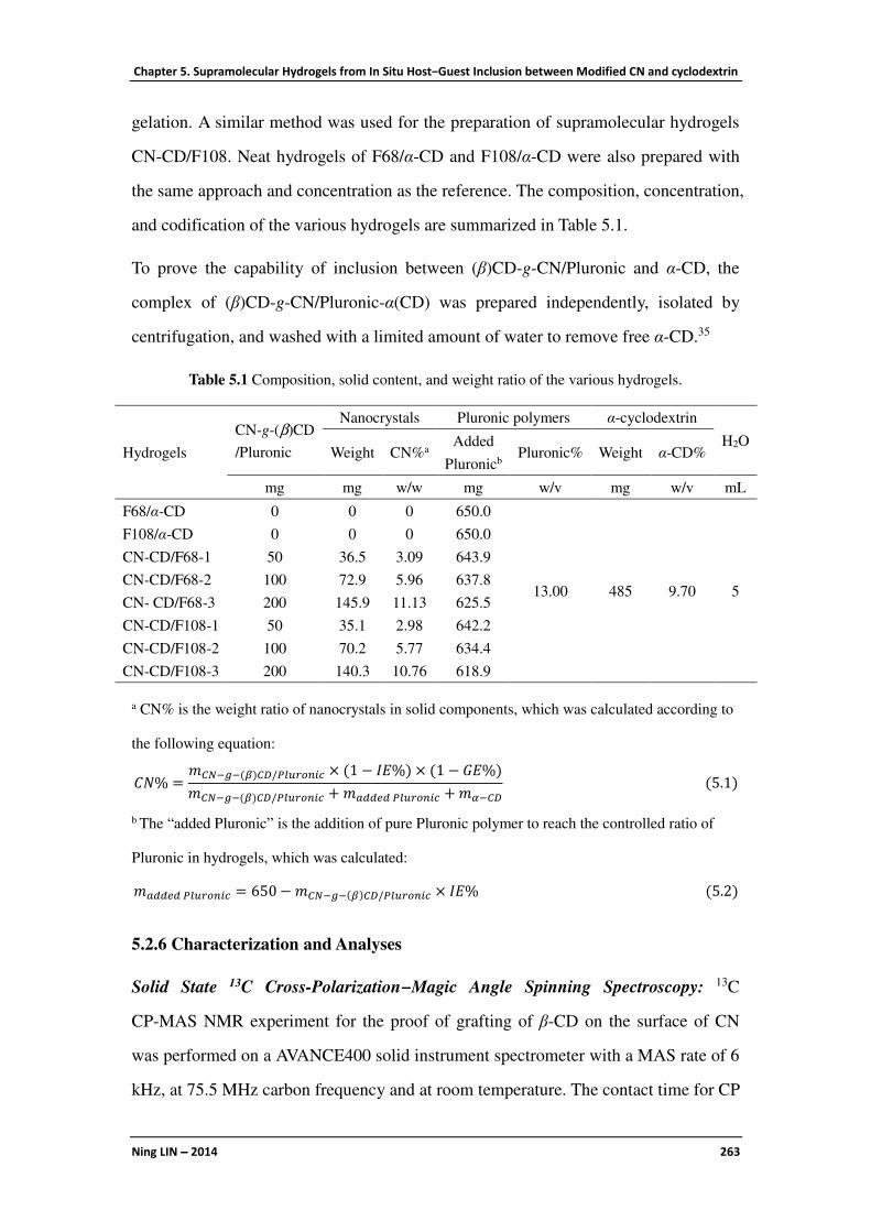
Chapter 5. Supramolecular Hydrogels from In Situ Host−Guest Inclusion between Modified CN and cyclodextrin
Ning LIN −−−− 2014 263
gelation. A similar method was used for the preparation of supramolecular hydrogels
CN-CD/F108. Neat hydrogels of F68/α-CD and F108/α-CD were also prepared with
the same approach and concentration as the reference. The composition, concentration,
and codification of the various hydrogels are summarized in Table 5.1.
To prove the capability of inclusion between (β)CD-g-CN/Pluronic and α-CD, the
complex of (β)CD-g-CN/Pluronic-α(CD) was prepared independently, isolated by
centrifugation, and washed with a limited amount of water to remove free α-CD.35
Table 5.1 Composition, solid content, and weight ratio of the various hydrogels.
Hydrogels
CN-g-(β)CD
/Pluronic
Nanocrystals Pluronic polymers α-cyclodextrin
H2O Weight CN%a
Added
Pluronicb Pluronic% Weight α-CD%
mg mg w/w mg w/v mg w/v mL
F68/α-CD 0 0 0 650.0
13.00 485 9.70 5
F108/α-CD 0 0 0 650.0
CN-CD/F68-1 50 36.5 3.09 643.9
CN-CD/F68-2 100 72.9 5.96 637.8
CN- CD/F68-3 200 145.9 11.13 625.5
CN-CD/F108-1 50 35.1 2.98 642.2
CN-CD/F108-2 100 70.2 5.77 634.4
CN-CD/F108-3 200 140.3 10.76 618.9
a CN% is the weight ratio of nanocrystals in solid components, which was calculated according to
the following equation:
��% =����(�)�/�������� × (1 − ��%) × (1 − ��%)
����(�)�/�������� +�!""#"�������� +�%��(5.1)
b The “added Pluronic” is the addition of pure Pluronic polymer to reach the controlled ratio of
Pluronic in hydrogels, which was calculated:
�!""#"�������� = 650−����(�)�/�������� × ��%(5.2)
5.2.6 Characterization and Analyses
Solid State 13C Cross-Polarization−Magic Angle Spinning Spectroscopy: 13C
CP-MAS NMR experiment for the proof of grafting of β-CD on the surface of CN
was performed on a AVANCE400 solid instrument spectrometer with a MAS rate of 6
kHz, at 75.5 MHz carbon frequency and at room temperature. The contact time for CP

Chapter 5. Supramolecular Hydrogels from In Situ Host−Guest Inclusion between Modified CN and cyclodextrin
Ning LIN −−−− 2014 264
was 1 ms with a proton 90 pulse of 5.5 µs and decoupling power of 45 kHz. The delay
time after the acquisition of the FID signal was 2 s. The chemical shifts were
calibrated with the hexamethylbenzene standard methyl resonance at 17.3 ppm.
UV−vis Spectroscopy: The content of grafted β-CD in (β)CD-g-CN (I) was analyzed
with a Shimadzu UV 2401-(PC) UV−vis spectrophotometer. Phenolphthalein (phe)
was used as indicator to detect the change of absorbency. First, together with sodium
carbonate solution, pure phe solution, phe solution containing 50 mg unmodified CN,
and phe solution containing 50 mg (β)CD-g-CN were well dispersed and stabilized
for 20 h. The phe absorption experiment was performed on the UV spectrophotometer
using 1 cm thick quartz cuvette with wavelengths ranging from 450 to 650 nm to
confirm the characteristic absorbed wavelength (λmax) of phe solution.
The standard curve for the weight of β-CD and absorbency of phe solution was
quantified with the photometric titration method. Two mL phe solution and 2 mL
sodium carbonate solution were added in #1 volumetric flask (25 mL), and set the
volume to the mark with distilled water. β-CD solutions of 1, 2, 3, 4, and 5mL were
added in #2, #3, #4, #5, and #6 volumetric flask (25 mL), respectively, and then 10
mL of distilled water was added in each flask. After that, both 2 mL of phe solution
and 2 mL of sodium carbonate solution were added in each flask (except #1), and
finally the volume was set to the mark with distilled water. All solutions were
submitted to absorption analysis at the wavelength of 552 nm (λmax), and the standard
curve and equation from the weight of β-CD (W, mg) and absorbency of phe (A) was
determined.
Similar experiments were carried out for the measurement of grafted β-CD content.
Five samples of 5, 10, 20, 30, and 40 mg (β)CD-g-CN in 10 mL of distilled water
were added in another #7, #8, #9, #10, and #11 volumetric flask (25 mL), respectively,
and then both 2 mL of phe solution and 2 mL of sodium carbonate solution were
added. All mixtures were set the volume to the mark of volumetric flask with distilled
water and stabilized for 20 h. The absorbency at the wavelength of 552 nm was
recorded and corresponded to the weight content of grafted β-CD according to the

Chapter 5. Supramolecular Hydrogels from In Situ Host−Guest Inclusion between Modified CN and cyclodextrin
Ning LIN −−−− 2014 265
standard equation. Finally, the β-CD grafting efficiency of CN-g-(β)CD (I) can be
calculated.
Fourier Transform Infrared Spectroscopy (FTIR): Infrared spectra were recorded at
room temperature on a FTIR Perkin-Elmer Spectrum One spectrometer to
characterize CN, (β)CD-g-CN, (β)CD-g-CN/Pluronic, and β-CD. Freeze-dried
powders were analyzed as KBr pellets (1 wt % in anhydrous KBr) using a spectral
width ranging from 4000 to 400 cm−1 with a 2 cm−1 resolution and an accumulation of
20 scans. On the other hand, during the preparation of inclusion complex
(β)CD-g-CN/Pluronic, the supernatants containing free Pluronic polymers were
collected from the process of successive solubilization-centrifugation. The
supernatant was carefully dropped on the surface of neat KBr compressing tablet, and
water was vaporized at room temperature for 12 h. The KBr tablet together with
Pluronic polymers on the surface were also characterized by FTIR.
Elemental Analysis: Elemental analysis was performed at Analysis Central Service of
the Centre National de la Recherche Scientifique (Vernaison, France). The carbon,
oxygen, and hydrogen element contents for both (β)CD-g-CN/F68 and
(β)CD-g-CN/F108 samples were measured. The results from elemental analysis were
used to determine the content of inclusive Pluronic polymers for (β)CD-g-CN/
Pluronic (IE%) according to eq. 5.3:
��%��������� + (1 − ��%)����(�)� = ����(�)�/��������(5.3)
where C is the relative carbon content in the sample; IE% is the inclusion efficiency
for inclusive Pluronic polymers for grafted β-CD. The precision of the measurement
is considered to be 0.3% for C and H elements and 0.5% for O element.
X-ray Diffffffffraction Analysis (XRD): XRD was used to prove the possibility of
inclusion interactions between (β)CD-g-CN/Pluronic and α-CD, which was recorded
on a Philips PW 1720 X-ray generator operated at 30 kV and 20 mA with Cu Kα
radiation (λ = 0.154 nm) in a range of 2θ = 3−50° using a fixed time mode with a step
interval of 0.02°.

Chapter 5. Supramolecular Hydrogels from In Situ Host−Guest Inclusion between Modified CN and cyclodextrin
Ning LIN −−−− 2014 266
Transmission Electron (TEM) and Optical Microscopy: The dimensions and
morphology of pristine CN were observed by TEM. Drops of 0.001 wt% cellulose
suspensions were deposited on glow discharged carbon-coated TEM grids. The
specimens were then negatively stained with 2% uranyl acetate. After complete
drying under room conditions, specimens were observed using a Philips CM200
electron microscope operating at 80 kV.
The morphology of hydrogel crystals was directly observed by optical microscopy.
The hydrogel was dropped on the glass slide, and formed after standing a period of
time in aqueous solution. Excess water was removed carefully with the absorption by
the edge of filter paper, and the morphology was observed after the complete dryness.
Rheological Analysis: Viscoelastic behavior of hydrogels was evaluated on a
Rheolyst Physica MCR-301 rheometer. Viscosity of the hydrogel was recorded
applying a flow cycle consisting in a continuously increasing shear rate ramp at 20 °C
from 0.05 to 10 s−1 for 4 min. The storage (G′) and the loss (G″) moduli were
recorded under the shear stress of 100 Pa in the range of 0.5−100 rad/s angular
frequency interval using a cone−plate geometry (diameter 20 mm, angle 1°). The
dependence of G′ and G″ on temperature was evaluated at 20 rad/s and 50 Pa in the
range of 20−70 °C.
The influence of pH conditions on the property of hydrogel was investigated to
determine the loss (G″) and storage (G′) moduli with different pH ranging from1 to 11.
The adjustment of pH conditions was achieved by the addition of HCl and NH4OH,
which was a negligible amount. Tests were conducted in triplicate at a fixed frequency
of 1 Hz with 50 Pa shear stress at 20 °C.
In Vitro Release of Doxorubicin·HCl from Hydrogels: Doxorubicin·HCl (1.0 mg)
was dissolved in 2 mL of distilled water and then (β)CD-g-CN/Pluronic and
controlled amount of Pluronic was added. The mixture was kept at 4 °C overnight in
order to obtain a homogeneous suspension. Afterward, α-CD was added to induce in
situ host−guest inclusion and form stable hydrogel. The study of in vitro drug release
was performed in water at 37 °C. The hydrogel was placed in a test tube with 20 mL

Chapter 5. Supramolecular Hydrogels from In Situ Host−Guest Inclusion between Modified CN and cyclodextrin
Ning LIN −−−− 2014 267
of water and then incubated in a shaking water bath. Solution of 3 mL aliquots
containing released drug was withdrawn from the testing tube periodically. The
volume of solution in the tube was kept constant by adding another 3 mL of water
after each sampling. The cumulative release ratio of doxorubicin·HCl from hydrogels
was measured by UV/vis spectrophotometer (Shimadzu UV 2401-(PC)) at the
absorbency of 481 nm. The experiment for in vitro drug release was performed in
triplicate.
5.3 Chemical Modification of Cellulose Nanocrystals
5.3.1 Chemical Grafting of β-CD on the Surface
Figure 5.2 13C (CP/MAS) solid-state NMR spectra of CN, β-CD, and (β)CD-g-CN.

Chapter 5. Supramolecular Hydrogels from In Situ Host−Guest Inclusion between Modified CN and cyclodextrin
Ning LIN −−−− 2014 268
The 13C CP-MAS spectra of CN, β-CD, and (β)CD-g-CN are shown in Figure 5.2.
The chemical shifts of carbon atoms from original CN were assigned to C1 (105 ppm),
C4 (88 ppm), C2, 3, 5 (71−75 ppm), and C6 (65 ppm). Possessing the same glucose
structure units, the characteristic chemical shifts related to C2′, 3′, 5′ for β-CD
superimpose with cellulose. However, because of different monomer unit quantity and
molecular conformations, the presence of β-CD can be determined by the position
shifts of C1′ (102 ppm), C4′ (81 ppm), and C6′ (64 ppm). As shown in the spectrum
of (β)CD-g-CN, all carbon characteristics from both CN and β-CD can be identified,
which indicates the surface grafting of β-CD and integrity of CN.
Figure 5.3 (A) UV absorption spectra for pure phenolphthalein solution (phe), phe solution
treated with pristine CN (CN/phe), and phe solution treated with β-CD grafted CN
((β)CD-g-CN/phe); (B) standard curve and equation from β-CD content and phe absorbency; (C)
β-CD grafting efficiency from (β)CD-g-CN samples with different quantities.
Under pH > 10 conditions, phenolphthalein (phe) aqueous solution shows red color,
which darkens when increasing phe concentration. It was reported that the
hydrophobic group of phe molecule can insert into the hydrophobic cavity of β-CD,
which reduce the free phe concentration in solution, and finally cause the decline of
absorbency from light color solution. As shown in the UV−vis spectra of Figure 5.3
(A), the impact of pristine CN was negligible on the phe molecule, which showed tiny
absorption and a slight decrease of absorbency in comparison with neat phe solution.
However, the introduction of (β)CD-g-CN caused a sharp reduction of absorbency,
which indicates that the hydrophobic cavity of β-CD includes plentiful phe molecules.

Chapter 5. Supramolecular Hydrogels from In Situ Host−Guest Inclusion between Modified CN and cyclodextrin
Ning LIN −−−− 2014 269
Based on Beer−Lambert law, within a range of controlled concentration, the content
of grafted β-CD on the surface of CN can be detected according to the absorbency of
alkaline phe solution. The absorbency of phenolphthalein (A) showed a negative
correlation with the concentration of grafted β-CD (Cβ‑CD, mg/mL), which was in
agreement with the weight of grafted β-CD (Wβ‑CD, mg) because of the same volume
of solution (25 mL) used in the experiment. The standard curve and equation between
A and Wβ‑CD are shown in Figure 5.3 (B). Different quantities of (β)CD-g-CN (5, 10,
20, 30, 40 mg) were added in the phe solution, and the content of grafted β-CD was
calculated from absorbency according to the equation reported in Figure 5.3 (B). As
shown in Figure 5.3 (C), the average value of β-CD grafting efficiency (GE%) was
determined as 16.89 wt% using this photometric titration method, which is almost in
agreement with the result of weight measurement (15.44 wt%).
5.3.2 Inclusion of Pluronic Polymers in Grafted β-CD
Although possessing similar glucose unit structure, the grafting and inclusion reaction
between CN, β-CD and Pluronic polymers can be investigated by the change of peak
intensity for specific bond stretching bands. Figure 5.4 (A, B) shows the FTIR spectra
of CN before and after grafting or inclusion reaction. With the surface grafting of
β-CD on CN, the intensity of three peaks located at 1163, 1113, and 1059 cm−1
decreased, which are ascribed to antisymmetric bridge oxygen stretching,
antisymmetric in-phase ring stretching and C−O stretching for cellulose, respectively.
Furthermore, another C−O stretching band at 1031 cm−1 broadened after surface
grafting. These results are in agreement with other reports for cyclodextrin-EPI
cellulose.30 By comparing the spectra for (β)CD-g-CN and (β)CD-g-CN/Pluronic, it is
interesting to note that the intensity of the peaks located at 1031 and 898 cm−1 slightly
increased, which can be attributed to the emerging C−O groups and antisymmetric
out-of-phase stretching carbon from the inclusive PEG-PPG-PEG polymer. However,
it was very important to confirm the inclusion reaction between grafted β-CD and
Pluronic polymers and prove the complete removal of free and surface physically
adsorbed Pluronic polymers on nanocrystals. An experiment of FTIR tracing was

Chapter 5. Supramolecular Hydrogels from In Situ Host−Guest Inclusion between Modified CN and cyclodextrin
Ning LIN −−−− 2014 270
designed to investigate the effect of the process of successive solubilization-
centrifugation during the preparation of inclusion complex ((β)CD-g-CN/Pluronic).
As shown in Figure 5.4 (C), after the post-treatment of purification for three times, the
spectra for the supernatant (F3) from washed (β)CD-g-CN/Pluronic did not exhibited
any characteristics of Pluronic polymers, which proved the availability of free
Pluronic removal using this method. On the other hand, the physical mixture of
Figure 5.4 FTIR spectra for (A, B) CN before and after chemical modifications: (a) β-CD, (b)
CN, (c) (β)CD-g-CN, (d) (β)CD-g-CN/F68, (e) (β)CD-g-CN/F108; (C) pure Pluronic polymers
and supernatants from the preparation of (β)CD-g-CN/Pluronic complexes with the treatment of
successive solubilization-centrifugation (F1 and F3 represent the supernatant after the treatment
for one and three times); (D) comparisons between pristine CN, CN/F108 with physically
adsorbed Pluronic F108, and CN(R-Pluronic) from the purification of CN/Pluronic with the
removal of adsorbed Pluronic using successive solubilization-centrifugation method.

Chapter 5. Supramolecular Hydrogels from In Situ Host−Guest Inclusion between Modified CN and cyclodextrin
Ning LIN −−−− 2014 271
CN/Pluronic was also prepared using the same concentration and treated with the
same purification treatment. By comparing the spectra of CN(R-Pluronic) and pure
CN, it was observed that they were nearly similar [as shown in Figure 5.4 (D)], which
indicates that the purification treatment can also effectively remove the polymer
physically adsorbed on the surface of nanocrystals. It should be pointed out that,
because the main body of (β)CD-g-CN is cellulose (only 16.89 wt% grafted β-CD),
the possible physical adsorption of Pluronic was supposed to mainly occur on the
surface of CN. Consequently, the above comparative experiments were carried out on
CN to prove the effect of purification.
The content or inclusion efficiency (IE%) of Pluronic polymer on the surface of
(β)CD-g-CN/Pluronic complexes was determined by elemental analysis. As shown in
Table 5.2, with the similar glucose unit structure (C6H10O5), CN and β-CD have the
same theoretical values for the three element contents (C, O, and H), resulting in
small change of element contents for the grafted nanocrystals [(β)CD-g-CN]. On the
other hand, although presenting different molecular weights and polymer segments,
there is a little difference in theoretical values for element contents between the two
Pluronic polymers. Regarding experimental values of these original materials,
because of the instrumental error and influence of hydrophily of cellulose and β-CD,
some variance of the experimental values was inevitable for CN and β-CD, which was
mainly attributed to the adsorption of moisture (H2O) during the delivery and
experiment. Therefore, the values of inclusion efficiency were confirmed mainly
based on the data from carbon element, and calculated according to eq. 5.3 As shown
in Table 5.2, the inclusion efficiency (IE%) of Pluronic polymers for (β)CD-g-CN/F68
and (β)CD-g-CN/F108 complexes were 12.25 and 15.57 wt%, respectively.
5.4 Structure and Properties of Supramolecular Hydrogels
5.4.1 Crystalline Structure of Inclusive Complexes
The inclusion of polymeric chains in cyclodextrin can also be investigated with XRD
analysis. The crystalline diffraction patterns of original materials and various

Chapter 5. Supramolecular Hydrogels from In Situ Host−Guest Inclusion between Modified CN and cyclodextrin
Ning LIN −−−− 2014 272
inclusive complexes are presented in Figure 5.5. As shown in Figure 5.5 (A), the
characteristic diffraction peaks of cellulose, β-CD and α-CD, on one hand, and
Pluronic polymers on the other hand were assigned to the 2θ angles at 22.6, 12.9, 14.3,
19.1, and 23.2°, respectively. The sharp diffraction peak for inclusive complexes
located at about 20.0° strongly supports the channel-type crystalline structure of the
obtained polyrotaxanes [Figure 5.5 (B)], which indicated that cyclodextrin underwent
a solid-state phase transformation as it formed an inclusion compound with polymeric
chains.36 Pluronic F108 with longer polymeric chains can include more α-CD
molecules than short Pluronic F68, which is reflected through the higher intensity for
(β)CD-g-CN/F108-α(CD) complex at 20.0°. On the other hand, by comparing the
diffraction patterns of (β)CD-g-CN/Pluronic and (β)CD-g-CN/Pluronic-α(CD)
complexes, the crystalline feature of α-CD appeared with the emergence of a weak
peak at 14.3°. In general, the inclusive complexes or hydrogels of cyclodextrin and
polymers do not present the obvious crystalline features of cyclodextrin. However, in
this case, the inclusion between α-CD and PEG chains occurred at the surface of CN,
Figure 5.5 X-ray diffraction patterns of (A) original materials: CN, β-CD, α-CD, Pluronic F68,
and F108; (B, C) inclusive complexes of (a) (β)CD-g-CN/F68, (b) (β)CD-g-CN/F108, (c)
(β)CD-g-CN/F68-α(CD), (d) (β)CD-g-CN/F108-α(CD).
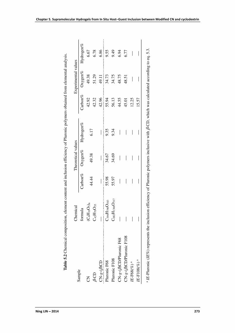
Chapter 5. Supramolecular Hydrogels from In Situ Host−Guest Inclusion between Modified CN and cyclodextrin
Ning LIN −−−− 2014 273
Tab
le 5
.2 C
hem
ical
com
posi
tion,
elem
ent
conte
nt
and i
ncl
usi
on e
ffic
iency o
f P
luro
nic
po
lym
ers
ob
tain
ed f
rom
ele
men
tal
anal
ysi
s.
Sam
ple
C
hem
ical
form
ula
Theo
reti
cal
val
ues
Ex
per
imen
tal
val
ues
Car
bon%
O
xygen
%
Hydro
gen
%
Car
bo
n%
O
xyg
en%
H
yd
rog
en%
CN
(C
6H
10O
5) n
44.4
4
49.3
8
6.1
7
42
.92
49
.38
6.6
7
β-C
D
C4
2H
70O
35
42
.32
51
.29
6.7
8
CN
-g-(
β)C
D
42
.96
49
.11
6.8
6
Plu
ronic
F68
C
39
4H
79
0O
18
3
55.9
8
34.6
7
9.3
5
5
5.9
4
34
.73
9.5
5
Plu
ronic
F108
C6
82H
13
66O
31
7
55.9
7
34.6
9
9.3
4
56
.13
34
.75
9.4
9
CN
-g-(
β)C
D/P
luro
nic
F68
4
4.5
5
48
.75
6.9
4
CN
-g-(
β)C
D/P
luro
nic
F108
45
.01
48
.51
6.7
7
IE-F
68(%
) a
1
2.2
5
IE-F
108(%
) a
15
.57
a IE
-Plu
ronic
(IE
%)
repre
sents
the
incl
usi
on e
ffic
iency o
f P
luro
nic
poly
mer
s in
clusi
ve
wit
h β
-CD
, w
hic
h w
as c
alcu
late
d a
cco
rdin
g t
o e
q.
5.3
.

Chapter 5. Supramolecular Hydrogels from In Situ Host−Guest Inclusion between Modified CN and cyclodextrin
Ning LIN −−−− 2014 274
which may induce the arrangement of inclusive chains (PEG/α-CD), and cause the
slight exhibition of α-CD crystalline feature. This phenomenon also indicates the
capability of inclusion between (β)CD-g-CN/Pluronic and α-CD, which ensured the
future in situ formation of hydrogels. In addition, the crystalline character of cellulose
was covered in these patterns, which induced the broadening of the peak observed
between 18 and 25°. By magnifying the patterns, the traces of crystalline cellulose can
be observed with a ill-defined peak located at about 22.6°, as shown in Figure 5.5 (C).
5.4.2 Morphology and Dimensions of CN and Hydrogels
TEM was used to observe the morphology and dimensions of cellulose nanocrystals,
as shown in Figure 5.6. Nanocrystals exhibited a typical rod-like morphology with a
length of 200−400 nm and a diameter of 10−20 nm with an aspect ratio (L/d) of about
18. This result was in accordance with our previous report for the morphological
observation of cellulose nanocrystals using atomic force microscopy (AFM).29
Generally, during the process of observation, the inclusion complex of α-CD/PEG
hydrogel recrystallizes in water and forms crystals of regular shapes with facets on
microscope grid at room temperature.35 As shown in Figure 5.7, both in situ hydrogels
derived from Pluronic F68 and F108 exhibited the regular appearance with
rectangular morphology in the size range of about 10−20 µm long and 2−4 µm wide.
Figure 5.6 TEM micrographs of cellulose nanocrystals.
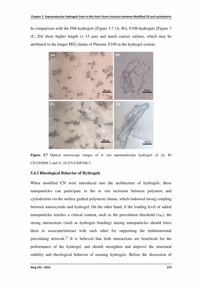
Chapter 5. Supramolecular Hydrogels from In Situ Host−Guest Inclusion between Modified CN and cyclodextrin
Ning LIN −−−− 2014 275
In comparison with the F68-hydrogels [Figure 5.7 (A, B)], F108-hydrogels [Figure 7
(C, D)] show higher length (> 15 µm) and much coarser surface, which may be
attributed to the longer PEG chains of Pluronic F108 in the hydrogel system.
Figure 5.7 Optical microscopy images of in situ supramolecular hydrogels of (A, B)
CN-CD/F68-2 and (C, D) CN-CD/F108-2.
5.4.3 Rheological Behavior of Hydrogels
When modified CN were introduced into the architecture of hydrogels, these
nanoparticles can participate to the in situ inclusion between polymers and
cyclodextrin via the surface grafted polymeric chains, which endowed strong coupling
between nanocrystals and hydrogel. On the other hand, if the loading level of added
nanoparticles reaches a critical content, such as the percolation threshold (νRc), the
strong interactions (such as hydrogen bonding) among nanoparticles should force
them to associate/interact with each other for supporting the tridimensional
percolating network.37 It is believed that both interactions are beneficial for the
performance of the hydrogel, and should strengthen and improve the structural
stability and rheological behavior of ensuing hydrogels. Before the discussion of

Chapter 5. Supramolecular Hydrogels from In Situ Host−Guest Inclusion between Modified CN and cyclodextrin
Ning LIN −−−− 2014 276
rheological properties of in situ hydrogels, the value of νRc can be calculated with the
dependence upon the aspect ratio (L/d) of rod-like CN according to eq. 5.2:
-.� =0.7
0 1⁄(5.4)
The νRc value is therefore inversely proportional to the aspect ratio of CN. According
to the observations from TEM, the value of νRc in this case was close to 3.89 vol%
(6.09 wt% with the density of cotton cellulose 1.565 g/mg).
The rheological behavior of hydrogels was investigated on the basis of three aspects,
including change of viscosity as a function of the shear rate, dependence of storage
moduli (G′) and loss moduli (G″) as a function of angular frequency and temperature.
When subjected to flow experiments, all hydrogels exhibited the physical nature of
gelation and were found to be thixotropic and reversible, as shown in Figure 5.8 (A,
A′). Even for in situ hydrogel with the highest initial viscosity (CN-CD/F108-3), the
viscosity of the hydrogel greatly reduced when increasing the shear rate. This
property makes the hydrogels syringeable, which is useful and important for drug
delivery systems. Two different effects on the rheological behavior of in situ
hydrogels as a function of angular frequency were observed depending on the CN
content, as shown in Figure 5.8 (B, B′). With the addition of only 50 mg modified CN,
the improvement of the mechanical properties of the hydrogel was unobvious
(CN-CD/Pluronic-1) in comparison with the neat hydrogel (Pluronic/α-CD). However,
when the loading level increased above 100 mg, the moduli of both F68- and
F108-hydrogels were enhanced significantly. Meanwhile, with the introduction of
enough modified CN, the in situ hydrogels can maintain better structural stability in
contrast to the sharp decrease of moduli observed for neat hydrogels under high
angular frequency. The dependence of the hydrogel moduli as a function of
temperature is shown in Figure 5.8 (C, C′). The effect of introducing modified CN in
hydrogels was more evident and induced the promising thermal stability for in situ
hydrogels contrary to the strong variation induced by sol−gel transition for neat
hydrogels. It should be pointed out that the F108-hydrogels commonly presented

Chapter 5. Supramolecular Hydrogels from In Situ Host−Guest Inclusion between Modified CN and cyclodextrin
Ning LIN −−−− 2014 277
better mechanical properties and thermal stability than F68-hydrogels, which may be
attributed to the longer polymeric chains with more inclusive cyclodextrin molecules
for Pluronic F108. In addition, based on the rheological behavior of in situ hydrogels,
it was interesting that the mechanical properties of CN-CD/Pluronic-2 were similar to
that of CN-CD/Pluronic-3, which indicates that the introduction of CN in the hydrogel
Figure 5.8 Rheological behavior of hydrogels: (A, A′) viscosity vs shear rate; (B, B′) storage
modulus (G′, filled symbols) and loss modulus (G″, open symbols) vs angular frequency; (C, C′)
G′ (filled symbols) and G″ (open symbols) vs temperature at a fixed frequency of 20 rad/s and 50
Pa shear stress in the range of 20−70 °C.

Chapter 5. Supramolecular Hydrogels from In Situ Host−Guest Inclusion between Modified CN and cyclodextrin
Ning LIN −−−− 2014 278
CN-CD/Pluronic-2 may be a critical point for the support of some special structure.
According to the results of elemental analysis reported in Table 5.1, the true contents
of CN in hydrogels CN-CD/F68-2 and CN-CD/F108-2 were 5.96 and 5.77 wt%,
respectively, which were very close to the previously calculated percolation threshold
(νRc, 6.09 wt%). It was believed that cellulose nanoparticles with adequate loading
level could form the percolating network in the architecture of hydrogels and properly
improved the performance of in situ hydrogels.
5.4.4 pH-Sensitivity of Hydrogels
The influence of different pH values on the properties (G′ and G″) of hydrogels was
investigated under acidic, neutral, and alkaline conditions. As shown in Figure 5.9 (A),
under acidic conditions, in situ hydrogels CN-CD/F68-2 and CN-CD/F68-3 kept high
moduli and stability, which was about 5 times higher than for neat hydrogels. The
maximum values of G′ for all hydrogels appeared in neutral condition (pH = 7).
However, when increasing the pH value to alkaline environment, the moduli for all
hydrogels dropped sharply, which may be attributed to the higher solubility and
Figure 5.9 Evolution of G′ (filled symbols) and G″ (open symbols) for various hydrogels under
different pH conditions at a fixed frequency of 1 Hz with 50 Pa shear stress at 20 °C.

Chapter 5. Supramolecular Hydrogels from In Situ Host−Guest Inclusion between Modified CN and cyclodextrin
Ning LIN −−−− 2014 279
dissolution of cyclodextrin in alkaline condition. Similar results can also be observed
for the CN-CD/F108 hydrogel system [Figure 5.9 (B)]. However, it was interesting
that, different from other hydrogels, hydrogel CN-CD/F108-3 preserved a storage
moduli (G′) at high levels and seemed to possess the ability of alkaline resistance. A
further study will focus on this interesting phenomenon.
5.5 Drug Release and Mechanism Study
Doxorubicin is an anticancer drug, which is widely used in the resistant of solid
tumors, including breast cancer, ovarian carcinoma, transitional cell bladder
carcinoma, and thyroid carcinoma. However, the non-negligible side effect, primarily
the cardiotoxicity resulting from high concentration of drug in blood, demands ideal
carrier for this drug for effective drug controlled release.38 As shown in Figure 5.10
(A), in situ hydrogels formed with participating modified CN (CN-CD/F68-2-Dox
and CN-CD/F108-2-Dox) exhibited promising controlled release behavior with
sustained release for about one week, whereas neat hydrogels (F68/α-CD and
F108/α-CD) displayed an initial burst release and shortened release period (only 2.5
days). The prolonged drug release for in situ hydrogels upon modified CN
introduction was supposed to result from two factors. As reported in our previous
literature,39 rigid CN has been proven to induce physical “obstruction effect” in an
alginate-based microsphere for controlled drug release property. On the other hand, in
this study, if the loading level of nanocrystals reaches the percolation threshold,
inducing a network structure with linkage among nanoparticles, it may provide
another “locking effect” to delay the diffusion of doxorubicin molecules (as shown in
Figure 10B). Both effects from modified nanocrystals led to the sustained drug release
of in situ hydrogels. It is worth noting that when comparing the hydrogels formed
with different Pluronic chains, there was no significant relation between the length of
chains used in the hydrogel and the drug release behavior. However, the final
cumulative release ratio depended on the length of used Pluronic for hydrogels, which
indicates the lower final cumulative release for hydrogels with longer Pluronic chains.

Chapter 5. Supramolecular Hydrogels from In Situ Host−Guest Inclusion between Modified CN and cyclodextrin
Ning LIN −−−− 2014 280
Figure 5.10 (A) In vitro release profiles of doxorubicin·HCl for hydrogels with different
compositions; (B) possible “locking effect” from the linkage between nanoparticles.
To prove the different release mechanisms for hydrogels, Ritger-Peppas equation was
used to analyze the results of drug release:
4 = 56� (ln4 = 59 + : ln 6)(5.5�
where Q is the amount of drug released at time t; k is a constant associated with the
structural and geometrical characteristics of dosage form; and n is the release
(diffusion) exponent, which depends on the release mechanism. It was reported that n
values approximating to 0.50 indicate a Fickian diffusion release, whereas the n value
in the range of 0.50 to 0.85 represents both diffusion-controlled release and
non-Fickian diffusion release (anomalous transport).40 Estimated parameters and fitted
equations for the various hydrogels with different compositions are shown in Table

Chapter 5. Supramolecular Hydrogels from In Situ Host−Guest Inclusion between Modified CN and cyclodextrin
Ning LIN −−−− 2014 281
5.3. It was shown that neat hydrogels (Pluronic/α-CD-Dox) underwent the common
Fickian diffusion, but in situ hydrogels (CN/CD-Pluronic-Dox) exhibited an
anomalous transport release mechanism. These results revealed the different effects
and changes of in situ hydrogels with the introduction of modified nanocrystals
compared to neat hydrogels.
Table 5.3 Estimated parameters and drug release mechanism for drug release of hydrogels
according to Ritger-Peppas equation.
Sample Equation k n R a Mechanism
F68/α-CD-Dox InQ = 2.387 + 0.465Int 10.885 0.465 0.890 Fickian diffusion
F108/α-CD-Dox InQ = 2.170 + 0.498Int 8.760 0.498 0.897 Fickian diffusion
CN-CD/F68-2-Dox InQ = 0.393 + 0.771Int 1.482 0.771 0.931 anomalous transport
CN-CD/F108-2-Dox InQ = 0.003 + 0.832Int 1.003 0.832 0.945 anomalous transport
a R is the regression coefficient for the fitted curves according to Ritger-Peppas equation.
5.6 Conclusions
This work was an attempt to use chemical modification methods to promote the
compatibility and enhance the loading levels of cellulose nanocrystals in hydrogel
materials. Meanwhile, uncovered polymeric chains render the participating inclusion
of α-cyclodextrin for the architecture of in situ hydrogels. Grafting efficiency of
β-cyclodextrin on the surface of nanocrystals was confirmed by UV spectroscopy as
16.89 wt%, and inclusion efficiency of Pluronic F68 and F108 polymers were
measured by elemental analysis as 12.25 and 15.57 wt%, respectively. With the
introduction of high loading levels of modified nanocrystals (> 5 wt%), a significant
enhancement of structural and thermal stability of in situ hydrogels was observed
from rheological analysis. A further study revealed the performance of hydrogels as
drug carrier for in vitro release of doxorubicin and exhibited the behavior of
prolonged drug release with special release kinetics, which was attributed to the
“obstruction effect” and “locking effect”. Based on their performances and properties,
these in situ hydrogels as syringeable drug delivery provide a scientific example for
the application of biomass nanocrystals in the field of biomedical materials.

Chapter 5. Supramolecular Hydrogels from In Situ Host−Guest Inclusion between Modified CN and cyclodextrin
Ning LIN −−−− 2014 282
5.7 References
1. Azizi Samir, M. A. S.; Alloin, F.; Dufresne, A. Review of recent research into
cellulosic whiskers, their properties and their application in nanocomposite field.
Biomacromolecules 2005, 6, 612–626.
2. Habibi, Y.; Lucia, A. L.; Rojas, O. J. Cellulose nanocrystals: Chemistry,
self-assembly, and applications. Chem. Rev. 2010, 110, 3479–3500.
3. Klemm, D.; Kramer, F.; Moritz, S.; Lindström, T.; Ankerfors, M.; Gray, D.;
Dorris, A. Nanocelluloses: A new family of nature-based materials. Angew. Chem.
Int. Ed. 2011, 50, 5438–5466.
4. Moon, R. J.; Martini, A.; Nairn, J.; Simonsen, J.; Youngblood, J. Cellulose
nanomaterials review: structure, properties and nanocomposites. Chem. Soc. Rev.
2011, 40, 3941–3994.
5. Dufresne, A. Processing of polymer nanocomposites reinforced with
polysaccharide nanocrystals. Molecules 2010, 15, 4111–4128.
6. Eichhorn, S. J.; Dufresne, A.; Aranguren, M.; Marcovich, N. E.; Capadona, J. R.;
Rowan, S. J.; Weder, C.; Thielemans, W.; Roman, M.; Renneckar, S.; Gindl, W.;
Veigel, S.; Keckes, J.; Yano, H.; Abe, K.; Nogi, M.; Nakagaito, A. N.; Mangalam,
A.; Simonsen, J.; Benight, A. S.; Bismarck, A.; Berglund, L. A.; Peijs, T. Review:
current international research into cellulose nanofibres and nanocomposites. J.
Mater. Sci. 2010, 45, 1–33.
7. Lin, N.; Huang, J.; Dufresne, A. Preparation, properties and applications of
polysaccharide nanocrystals in advanced functional nanomaterials: a review.
Nanoscale 2012, 4, 3274–3294.
8. Lam, E.; Male, K. B.; Chong, J. H.; Leung, A. C. W.; Luong, J. H. T. Applications
of functionalized and nanoparticle-modified nanocrystalline cellulose. Trends
Biotechnol. 2012, 30, 283–290.
9. Dong, S.; Roman, M. Fluorescently labeled cellulose nanocrystals for bioimaging
applications. J. Am. Chem. Soc. 2007, 129, 13810–13811.
10. Mahmoud, K. A.; Mena, J. A.; Male, K. B.; Hrapovic, S.; Kamen A.; Luong, J. H.
Chapter 5. Supramolecular Hydrogels from In Situ Host−Guest Inclusion between Modified CN and cyclodextrin
Ning LIN −−−− 2014 283
T. Effect of surface charge on the cellular uptake and cytotoxicity of fluorescent
labeled cellulose nanocrystals. ACS Appl. Mater. Interfaces 2010, 2, 2924–2932.
11. Liu, H.; Wang, D.; Song, Z.; Shang, S. Preparation of silver nanoparticles on
cellulose nanocrystals and the application in electrochemical detection of DNA
hybridization. Cellulose 2011, 18, 67–74.
12. Drogat, N.; Granet, R.; Le Morvan, C.; Bégaud-Grimaud, G.; Krausz, P.; Sol, V.
Chlorin-PEI-labeled cellulose nanocrystals: Synthesis, characterization and
potential application in PDT. Bioorg. Med. Chem. Lett. 2012, 22, 3648–3652.
13. Mahmoud, K. A.; Male, K. B.; Hrapovic, S.; Luong, J. H. T. Cellulose
nanocrystal/gold nanoparticle composite as a matrix for enzyme immobilization.
ACS Appl. Mater. Interfaces 2009, 1, 1383–1386.
14. Edwards, J. V.; Prevost, N. T.; Condon, B.; French, A.; Wu, Q. Immobilization of
lysozyme-cellulose amide-linked conjugates on cellulose I and II cotton
nanocrystalline preparations. Cellulose 2012, 19, 495–506.
15. Feese, E.; Sadeghifar, H.; Gracz, H. S.; Argyropoulos, D. S.; Ghiladi, R. A.
Photobactericidal porphyrin-cellulose nanocrystals: Synthesis, characterization,
and antimicrobial properties. Biomacromolecules 2011, 12, 3528–3539.
16. Carpenter, B. L.; Feese, E.; Sadeghifar, H.; Argyropoulos, D. S.; Ghiladi, R. A.
Porphyrin-cellulose nanocrystals: A photobactericidal material that exhibits broad
spectrum antimicrobial activity. Photochem. Photobiol. 2012, 88, 527–536.
17. Wang, Y.; Chang, C.; Zhang, L. Effects of freezing/thawing cycles and cellulose
nanowhiskers on structure and properties of biocompatible starch/PVA sponges.
Macromol. Mater. Eng. 2010, 295, 137–145.
18. Gatenholm, P.; Klemm, D. Bacterial nanocellulose as a renewable material for
biomedical applications. MRS Bulletin 2010, 35, 208–213.
19. Chang, C.; Zhang, L. Cellulose-based hydrogels: Present status and application
prospects. Carbohydr. Polym. 2011, 84, 40–53.
20. Sannino, A.; Demitri, C.; Madaghiele, M. Biodegradable cellulose-based
hydrogels: design and applications. Materials 2009, 2, 353–373.
21. Li, J. Self-assembled supramolecular hydrogels based on polymer–cyclodextrin

Chapter 5. Supramolecular Hydrogels from In Situ Host−Guest Inclusion between Modified CN and cyclodextrin
Ning LIN −−−− 2014 284
inclusion complexes for drug delivery. NPG Asia Mater. 2010, 2, 112–118.
22. Zhang, X.; Huang, J.; Chang, P. R.; Li, J.; Chen, Y.; Wang, D.; Yu, J.; Chen, J.
Structure and properties of polysaccharide nanocrystal-doped supramolecular
hydrogels based on cyclodextrin inclusion. Polymer 2010, 51, 4398–4407.
23. Osorio-Madrazo, A.; Eder, M.; Rueggeberg, M.; Pandey, J. K.; Harrington, M. J.;
Nishiyama, Y.; Putaux, J.-L.; Rochas, C.; Burgert, I. Reorientation of cellulose
nanowhiskers in agarose hydrogels under tensile loading. Biomacromolecules
2012, 13, 850−856.
24. Bica, C. I. D.; Borsali, R.; Rochas, C.; Geissler, E. Dynamics of cellulose
whiskers spatially trapped in agarose hydrogels. Macromolecules 2006, 39,
3622−3627.
25. Abitbol, T.; Johnstone, T.; Quinn, T. M.; Gray, D. G. Reinforcement with
cellulose nanocrystals of poly(vinyl alcohol) hydrogels prepared by cyclic
freezing and thawing. Soft Matter 2011, 7, 2373–2379.
26. Cha, R.; He, Z.; Ni, Y. Preparation and characterization of thermal/pH-sensitive
hydrogel from carboxylated nanocrystalline cellulose. Carbohydr. Polym. 2012,
88, 713–718.
27. Karaaslan, M. A.; Tshabalala, M. A.; Yelle, D. J.; Buschle-Diller, G.
Nanoreinforced biocompatible hydrogels from wood hemicelluloses and cellulose
whiskers. Carbohydr. Polym. 2011, 86, 192–201.
28. Way, A. E.; Hsu, L.; Shanmuganathan, K.; Weder, C.; Rowan, S. J.
pH-Responsive cellulose nanocrystal gels and nanocomposites. ACS Macro Lett.
2012, 1, 1001−1006.
29. Lin, N.; Bruzzese, C.; Dufresne, A. TEMPO-Oxidized nanocellulose participating
as crosslinking aid for alginate-based sponges. ACS Appl. Mater. Interfaces 2012,
4, 4948−4959.
30. Zhao, Q.; Wang, S.; Cheng, X.; Yam, R. C. M.; Kong, D.; Li, R. K. Y. Surface
Modification of cellulose fiber via supramolecular assembly of biodegradable
polyesters by the aid of host−guest inclusion complexation. Biomacromolecules
2010, 11, 1364–1369.

Chapter 5. Supramolecular Hydrogels from In Situ Host−Guest Inclusion between Modified CN and cyclodextrin
Ning LIN −−−− 2014 285
31. Mortensen, K.; Pedersen, J. S. Structural study on the micelle formation of
poly(ethylene oxide)-poly(propylene oxide)-poly(ethylene oxide) triblock
copolymer in aqueous solution. Macromolecules 1993, 26, 805–812.
32. Tsai, C.-C.; Leng, S.; Jeong, K.-U.; Van Horn, R. M.; Wang, C.-L.; Zhang, W.-B.;
Graham, M. J.; Huang, J.; Ho, R.-M.; Chen, Y.; Lotz, B.; Cheng, S. Z. D.
Supramolecular structure of β-cyclodextrin and poly(ethylene
oxide)-block-poly(propylene oxide)-block-poly(ethylene oxide) inclusion
complexes Macromolecules 2010, 43, 9454–9461.
33. Li, J.; Li, X.; Zhou, Z.; Ni, X.; Leong, K. W. Formation of supramolecular
hydrogels induced by inclusion complexation between Pluronics and
α-cyclodextrin. Macromolecules 2001, 34, 7236–7237.
34. Simões, S. M. N.; Veiga, F.; Torres-Labandeira, J. J.; Ribeiro, A. C. F.;
Sandez-Macho, M. I.; Concheiro, A.; Alvarez-Lorenzo, C. Syringeable Pluronic–
α-cyclodextrin supramolecular gels for sustained delivery of vancomycin. Eur. J.
Pharm. Biopharm. 2012, 80, 103–112.
35. He, L.; Huang, J.; Chen, Y.; Xu, X.; Liu, L. Inclusion interaction of highly
densely PEO grafted polymer brush and α-cyclodextrin. Macromolecules 2005,
38, 3845–3851.
36. Peet, J.; Rusa, C. C.; Hunt, M. A.; Tonelli, A. E.; Balik, C. M. Solid-state
complexation of poly(ethylene glycol) with α−cyclodextrin. Macromolecules
2005, 38, 537–541.
37. Dufresne, A. Polysaccharide nanocrystal reinforced nanocomposites. Can. J.
Chem. 2008, 86, 484–494.
38. Xun, W.; Wu, D.-Q.; Li, Z.-Y.; Wang, H.-Y.; Huang, F.-W.; Cheng, S.-X.; Zhang,
X.-Z.; Zhuo R.-X. Peptide-functionalized thermo-sensitive hydrogels for
sustained drug delivery. Macromol. Biosci. 2009, 9, 1219–1226.
39. Lin, N.; Huang, J.; Chang, P. R.; Feng L.; Yu, J. Effect of polysaccharide
nanocrystals on structure, properties, and drug release kinetics of alginate-based
microspheres. Colloid. Surface. B 2011, 85, 270–279.

Chapter 5. Supramolecular Hydrogels from In Situ Host−Guest Inclusion between Modified CN and cyclodextrin
Ning LIN −−−− 2014 286
40. Ritger, P. L.; Peppas, N. A. A simple equation for description of solute release II.
Fickian and anomalous release from swellable devices. J. Control. Release 1987,
5, 37–42.

Chapter 6. General Conclusions and Perspectives for Future Research
Ning LIN −−−− 2014 287
CHAPTER 6.
General Conclusions and Perspectives for Future Research

Chapter 6. General Conclusions and Perspectives for Future Research
Ning LIN −−−− 2014 288
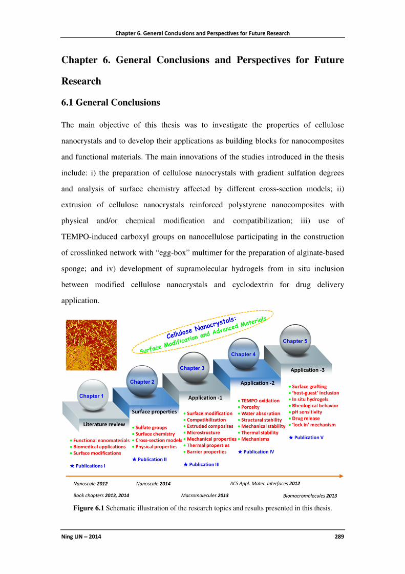
Chapter 6. General Conclusions and Perspectives for Future Research
Ning LIN −−−− 2014 289
Chapter 6. General Conclusions and Perspectives for Future
Research
6.1 General Conclusions
The main objective of this thesis was to investigate the properties of cellulose
nanocrystals and to develop their applications as building blocks for nanocomposites
and functional materials. The main innovations of the studies introduced in the thesis
include: i) the preparation of cellulose nanocrystals with gradient sulfation degrees
and analysis of surface chemistry affected by different cross-section models; ii)
extrusion of cellulose nanocrystals reinforced polystyrene nanocomposites with
physical and/or chemical modification and compatibilization; iii) use of
TEMPO-induced carboxyl groups on nanocellulose participating in the construction
of crosslinked network with “egg-box” multimer for the preparation of alginate-based
sponge; and iv) development of supramolecular hydrogels from in situ inclusion
between modified cellulose nanocrystals and cyclodextrin for drug delivery
application.
Figure 6.1 Schematic illustration of the research topics and results presented in this thesis.

Chapter 6. General Conclusions and Perspectives for Future Research
Ning LIN −−−− 2014 290
Revolving around the properties and applications of cellulose nanocrystals, diverse
modifications have been performed on cellulose nanocrystals in order to further
investigate their surface properties and tailor their applicability for various advanced
materials. General research topics for each study are summarized in Figure 6.1.
In chapter 1, the state of the art on research on the topic of cellulose nanocrystals (also
including cellulose whiskers and nanocrystalline cellulose) was reviewed, and
significant studies were specially introduced on the basis of four parts, viz.
preparation and production; physical, chemical and biological properties; surface
modification; as well as traditional and advanced applications. In view of my PhD
studies, the emphasis of literature review in this chapter was put on the physical and
chemical properties, surface modification, and functional nanomaterials.
The research work introduced in chapter 2 originated from the following questions:
“how many active hydroxyl groups have been replaced by sulfate groups during
H2SO4 hydrolysis; and how surface sulfate groups affect the surface chemistry and
physical properties of cellulose nanocrystals?” To answer these questions, gradient
contents of sulfate groups were introduced on the surface of cellulose nanocrystals
through the regulation of reactant ratios and reactive conditions for postsulfation and
desulfation. Based on the analysis of surface chemistry for gradient modified
nanocrystals, it was proved that the choice of different cross-section models will
significantly affect the calculation of the degree of substitution of sulfate groups on
cellulose nanocrystals. Taking cotton cellulose nanocrystals as example, if square or
round model was chosen for nanocrystals, more than 20% of surface hydroxyl groups
have been substituted by sulfate groups during the H2SO4−esterification hydrolysis
(65 wt% acid concentration, 45 °C), which indicated that most of the active hydroxyl
groups located on C-6 have been replaced during the extraction. If ellipse model was
chosen, about 13% of the surface hydroxyl groups were replaced by sulfate groups
during hydrolysis. However, if choosing rectangle model for cellulose nanocrystals,
only 7.9% of the surface hydroxyl groups was substituted during the
H2SO4−hydrolysis (only about 1/3 of the value for square or round model), and a large

Chapter 6. General Conclusions and Perspectives for Future Research
Ning LIN −−−− 2014 291
amount of active hydroxyl groups have been remained. Moreover, the effect of sulfate
groups on various physical properties of cellulose nanocrystals and their suspensions
have been discussed. The parallelogram elementary crystallite model for cotton
cellulose nanocrystals was further validated by XRD analysis, and determination of
negative charges induced by the presence of sulfate groups on cellulose nanocrystals
by ζ-potential measurement. The influence of sulfate groups on the thermal stability
of cellulose nanocrystals was investigated, which indicated that more sulfate groups
on nanocrystals induced a lowering of the temperature corresponding to the first phase
of thermal decomposition (Td1), and higher crystallinity for nanocrystals resulting in
higher second phase thermal decomposition temperature (Td2). From the observation
of birefringence of aqueous suspensions, it was concluded that the dispersion and
formation of birefringent domains can be affected by the content of surface sulfate
groups on nanocrystals.
Chapter 3 was a study on the preparation of extruded composites in order to settle the
thermal stability issue of cellulose nanocrystals during melting-coumpounding of
polymer nanocomposites. A strategy involving two poly(ethylene
glycol)/polyoxyethylene (PEG/PEO) layers on the surface of cellulose nanocrystals
using chemical grafting and physical interactions was proposed. Four kinds of
nanocrystals, i.e. pristine nanocrystal, PEO-adsorbed nanocrystal, PEG-grafted
nanocrystal, and both PEG-grafted and PEO-adsorbed nanocrystal, were extruded
with a hydrophobic polymeric matrix (PS) to prepare nanocomposites. The basic idea
was consistent with the possibility of entanglements and co-crystallization between
short PEG chains and long PEO chains, which can closely wrap and protect the
surface of the nanocrystals. Meanwhile, the chemical and physical compatibilization
imparted by PEG and PEO layers promoted the interfacial interactions between
cellulosic nanoparticles and apolar matrix. The possibility of processing modified
nanocrystals (CN-g-PEG/PEO) at high temperature (200 °C) avoiding the degradation
of the nanoparticle, and providing a good dispersion and compatibility between
modified nanocrystals and matrix was shown. Furthermore, the nanocrystal surface

Chapter 6. General Conclusions and Perspectives for Future Research
Ning LIN −−−− 2014 292
modifications did not block totally the interactions between the nanoparticles, and
kept at least partially the three-dimensional network architecture, which was
beneficial for the improvement of mechanical properties of extruded nanocomposites.
Indeed, from the results of TGA, double-layer modified cellulose nanocrystals
showed a significant enhancement of degradation temperature (about 120 °C increase
in comparison with pristine cellulose nanocrystals), which can be attributed to the
uniform shield of sulfate groups induced by the coating with the grafted PEG chains
and protection of adsorbed PEO layer. Extruded nanocomposites filled with
double-layer modified cellulose nanocrystals exhibited transparent appearance and
high transmittance ratios. Meanwhile, improved mechanical and water vapor barrier
performances were observed on these extruded nanocomposites. With the strategy
proposed in this study, it is expected to overcome an important challenge aimed at
melt processing cellulose nanocrystal-based composites for industrial production.
With the purpose of improving weak performance (mechanical and structural stability)
of neat alginate sponges, oxidized nanocellulose-participating crosslinked sponges
were designed and investigated. This topic was discussed in chapter 4. Two types of
nanocellulose extracted from cotton were used in this study, viz. cellulose
nanocrystals with ‘rigid’ rod-like morphology and microfibrillated cellulose with ‘soft’
filament morphology. After TEMPO-selective oxidation treatment, modified
nanocellulose possessed the capability of participating to ionic crosslinking during the
structural formation of alginate-based sponges. Interestingly, different types of
oxidized nanocellulose provided distinct enhancing effects for sponges. Remarkable
reinforced mechanical strength was reported with oxidized cellulose nanocrystals
whereas promising improved porosity, water absorption, and structural stability were
observed for oxidized microfibrillated cellulose. More specifically, compared with the
porosity (78.8%), water absorption (835.3%) and water retention (221.1%) of neat
alginate sponge, crosslinked sponge filled with 50 wt% oxidized microfibrillated
cellulose possessed ultrahigh porosity (97.4%), water absorption (1468.4%) and water
retention (846.7%). Even after reusing for 10 times, this kind of crosslinked sponges

Chapter 6. General Conclusions and Perspectives for Future Research
Ning LIN −−−− 2014 293
filled with oxidized microfibrillated cellulose can maintain high levels of water
absorption. However, oxidized cellulose nanocrystals have a better impact on the
reinforcement of mechanical properties of sponges. For compressive strains of 30%,
50%, and 70%, the compressive strength of crosslinked sponges containing 10 wt%
oxidized cellulose nanocrystals increased by factors of 2.78, 2.80, and 2.94, in
comparison with neat alginate sponge, respectively. It was supposed that rigid
oxidized cellulose nanocrystals served as the coupling points, and fully played the
nano-reinforcement effect for alginate-based sponges, whereas oxidized
microfibrillated cellulose can join in the structural construction and form the
semi-interpenetrating polymer network for alginate-based sponges.
Inspired from the study of physical blending of pristine polysaccharide nanocrystals
and cyclodextrin, an attempt of using chemical modification methods to promote the
compatibility and enhance the cellulose nanocrystal loading levels in
cyclodextrin-based hydrogel materials was performed and reported in chapter 5.
β-cyclodextrin was first covalently grafted on the surface of cellulose nanocrystals
using epichlorohydrin as coupling agent. Then, Pluronic polymer was immobilized on
modified cellulose nanocrystals by inclusion of the hydrophobic polymeric segments
in grafted β-cyclodextrin. Pluronic polymers used in this study were tri-block
copolymers with different molecular weights (Pluronic F68 or F108), both bearing
hydrophobic PPG and hydrophilic PEG segments (PEG-b-PPG-b-PEG). The
inclusion of the hydrophobic PPG chain segments in the inner cavity of β-CD allowed
the close connection of chains and nanoparticles. Finally, through the ‘host-guest’
inclusion interactions between modified cellulose nanocrystals, Pluronic polymers,
and different size of cyclodextrins, in situ hydrogels were prepared and developed as
novel drug carrier. With the introduction of high loading levels of modified
nanocrystals (> 5 wt%), a significant enhancement of the structural and thermal
stability for in situ hydrogels was observed from rheological analysis. Meanwhile,
these in situ hydrogels bearing modified cellulose nanocrystals showed special
pH-sensitivity. A further study revealed the performance of in situ hydrogels as drug

Chapter 6. General Conclusions and Perspectives for Future Research
Ning LIN −−−− 2014 294
carrier for in vitro release of doxorubicin, which showed promising controlled release
behavior with sustained release for about one week whereas neat hydrogels displayed
an initial burst release and shortened release period (only 2.5 days). The prolonged
drug release behavior for in situ hydrogels was analyzed by release kinetics, and was
supposed to be attributed to the “obstruction effect” and “locking effect” from
modified cellulose nanocrystals. Based on their performances and properties, these in
situ hydrogels as syringeable drug delivery provide a scientific example for the
application of biomass nanocrystals in the field of biomedical materials.
6.2 Possible Research Points for Further Study
Table 6.1 Future research points for the studies presented in this thesis.
Studies in this thesis Future research points
Surface chemistry and properties
of CN with gradient sulfation
degrees
• Investigation of electrostatic interactions between negative
charges from sulfated CN and positive charges from cationic
polymers or molecules
• Measurement of strength and mechanism of electrostatic
interactions from coupling charges
• Strategy for surface electrostatic grafting on CN
Physical and/or chemical
compatibilization of extruded CN
reinforced PS nanocomposites
• Further investigation on interactions between grafted PEG
and adsorbed PEO
• Effect of PEG/PEO chain lengths ratio to compatibility
• Measurement of interphase thickness of PEG and PEO
layers in extruded nanocomposites
• Simpler method for the replacement of PEG chemical
grafting on the surface of CN
• Investigation of other polymers or copolymers
TEMPO-Oxidized nanocellulose
participating as crosslinking aid
for alginate-based sponges
• Direct proofs for participating crosslinking of oxidized
nanocellulose, alginate and Ca2+
• Attempt of incorporating both oxidized CN and oxidized
MFC in sponges for synergistic enhancements
• Development of ensuing sponges in biomedical
applications, such as drug-loaded wound dressing
Supramolecular hydrogels from in
situ host−guest inclusion between
modified CN and cyclodextrin
• Characterization of ‘obstruction effect” and “locking
effect’ of CN-based drug delivery systems
• Presence of CNs (with surface hydroxyl groups and/or
sulfate groups) to structure and activity of drug molecules
• Development of multi-membrane hydrogel systems for
novel drug release behavior

Chapter 6. General Conclusions and Perspectives for Future Research
Ning LIN −−−− 2014 295
Following the studies introduced in this thesis, some research topics can be carried out
in future works. Table 6.1 shows some points originated from the studies developed in
each chapter. Specifically, for the study on “gradient sulfate groups on cellulose
nanocrystals” reported in chapter 2, future research can focus on the electrostatic
interactions between negative charges from sulfated nanocrystals and positive charges
from cationic polymers or molecules. With the detection and measurement of
electrostatic interactions, it is expected to reveal the strength and mechanism of this
special interaction from coupling charges and therefore advance the application of
cellulose nanocrystal electrostatic grafting. Figure 6.2 provides the research strategy
of polymer electrostatic grafting onto cellulose nanocrystals and proposes the original
questions of “Is it true for more sulfate groups, more adsorbed polymeric chains and
stronger electrostatic interactions?”
Figure 6.2 Strategy of polymer electrostatic grafting onto cellulose nanocrystals.
Regarding the study on “extruded cellulose nanocrystal-based composites” reported in
chapter 3, further investigation is necessary to fully understand the interactions
between grafted PEG and adsorbed PEO, such as the effect of chain length ratio (1/10
for PEG/PEO was used in this case), and interphase thickness of PEG and PEO layers
in extruded nanocomposites. A careful control of the amount of decorating PEG/PEO
chains is also desirable to monitor the possibility of hydrogen bonding between

Chapter 6. General Conclusions and Perspectives for Future Research
Ning LIN −−−− 2014 296
nanoparticles. Moreover, a simpler method for the introduction of the first PEG layer
on cellulose nanocrystals would be to use electrostatic grafting instead of the chemical
grafting reaction. It is another important issue to drive the practical application of this
strategy.
With respect to the study on “crosslinked alginate sponges” reported in chapter 4 and
inspired from the different effects of two oxidized nanocelluloses, it will be interesting
to attempt the idea of incorporating both oxidized nanocrystalline and microfibrillated
cellulose in alginate-based materials, and develop applications of the ensuing sponges
in the field of biomedical materials, such as drug carriers for wound dressing or bio
scaffolds.
Regarding the study on “supramolecular hydrogels from cellulose nanocrystals and
cyclodextrin” reported in chapter 5, further investigation on the effect of surface
properties of cellulose nanocrystals to the structure and bioactivity of drug molecules
can significantly promote their application in novel biomedical nanomaterials. In
addition, taking advantage of the ‘obstruction effect’ and ‘locking effect’ from
cellulose nanocrystals, it is promising to develop complexing drug delivery systems
for novel drug release behaviors, such as multi-membrane hydrogel systems. In the
following one year, some ideas will be carried out during my post-doctoral research.
6.3 Perspectives and Challenges in Future
It can be evidenced that cellulose nanocrystal is a unique and magic material.
Cellulose nanocrystal can be used in all kinds of fields, but it is still a rather new
material to us. There are still many unknown properties or undiscovered applications
for cellulose nanocrystals. Despite being the most available and common natural
polymer on earth, it is only recently that the prominence of this nanostructured
material was recognized. Now we stand on the edge of breakthrough of cellulose
nanocrystal research, and maybe it is the right corner for future materials science.
It is impossible to predict which fields will become the practical market for cellulose
nanocrystals in future. Now, more and more researchers are attracted by this material,

Chapter 6. General Conclusions and Perspectives for Future Research
Ning LIN −−−− 2014 297
and in the near future cellulose nanocrystals may be possibly used for the
development of ultimate body armor, super-flexible screens, high-efficiency filters,
bendable batteries, ultra-absorbent aerogels, cars and computers, bounteous biofuel,
cosmetics, money currency etc. And now they are not just illusion. As few examples,
IBM is reported to use cellulose nanocrystals to create components for computers, and
the US army is considering cellulose nanocrystals to make lightweight body armor
and ballistic glass.
Generally, the exploration of suitable approaches for cellulose nanocrystal in diverse
types of materials is everlasting challenge for its future study. On the topic of
production, the issues of cost reduction, properties control, and sustainability of
supply are the challenges for cellulose nanocrystals. Promising commercial cellulose
nanocrystals should be the products with both low cost and controllable properties
(such as controlled dimensions, surface properties etc.). Regarding the application of
cellulose nanocrystals in composites and functional materials, how to regulate or
balance the compatibility and the formation of rigid network, and how to characterize
the interactions between cellulose nanocrystals and diverse molecules, are the future
challenges. In order to improve the compatibility between nanoparticles and
polymeric matrices, surface modifications are commonly performed on cellulose
nanocrystals. However, these surface treatments always limit the interactions among
nanoparticles, which induce the weak formation of rigid network and inferior
nano-reinforcing effects from cellulose nanocrystals. On the other hand, derived from
intramolecules or surface groups, it is very difficult to directly observe or detect the
physical interactions between cellulose nanocrystals and diverse molecules in various
functional materials (such as interactions between cellulose nanocrystals and drug
molecules, calcium crosslinking in this dissertation). Finally, the preservation of
nano-reinforcing effect of cellulose nanocrystals in different conditions is another
challenge, such as keeping high mechanical or barrier properties in wet atmosphere
for prepared nanomaterials.
Cellulose nanocrystal is a precious gift from Nature, which deserves future study!
Chapter 6. General Conclusions and Perspectives for Future Research
Ning LIN −−−− 2014 298
Résumé en Français
Ning LIN −−−− 2014 299
Résumé en Français
Introduction générale
Le nanocristal de cellulose est l’élément constitutif structurel primaire des plantes et
peut être extrait de manière économique du bois et des fibres végétales, ou de la
tunicine et de la cellulose bactérienne. L’hydrolyse des régions amorphes de la
cellulose native permet de libérer les nanocristaux de cellulose – domaines
cellulosiques cristallins, sous forme de bâtonnets rigides de 5–20 nm de diamètre et
quelques dizaines à quelques centaines de nanomètres de longueur. Le
développement des technologies d’extraction et de l’application des nanocristaux de
cellulose a été activement poursuivi par divers groupes, notamment aux USA, au
Canada et en Europe.1 En tant que nanomatériau abondant et renouvelable, le
nanocristal de cellulose pourrait être utilisé pour fabriquer et développer une large
gamme de produits à haute valeur ajoutée, comme (i) des matériaux biomédicaux
pour les produits pharmaceutiques et l'administration de médicaments, le
remplacement de l'os et la réparation des dents, etc., (ii ) des matériaux composites
avancés (fibres filées et textiles à haute résistance, polymères renforcés et
bioplastiques novateurs; emballages "intelligents", etc., (iii) des additifs pour
l'alimentation, les cosmétiques, les revêtements, les peintures, les adhésifs, les
pigments et encres; (iv) des composants structurels pour la fabrication du papier, la
construction, l'aérospatiale et les transports, (v) des matériaux optiquement
commutables et irisés .2
Depuis la première étude sur l'utilisation des nanocristaux de cellulose comme phase
de renfort dans des nanocomposites il y a environ 20 ans, une quantité importante
de littérature a été consacrée à la recherche sur les nanocristaux de cellulose.
Différentes terminologies ont été utilisées dans la littérature pour désigner ces
nanoparticules cellulosiques hautement cristallines, comme "nanocristaux de
cellulose", "cellulose nanocristalline", "nanotrichites de cellulose" (ou simplement

Résumé en Français
Ning LIN −−−− 2014 300
"trichites"), "microcristaux de cellulose en bâtonnet", "cristallites de cellulose" et
"micelles de cellulose" (dans la littérature la plus ancienne).
En 2011, certaines normes internationales sur la nanocellulose ont été proposées par
la Division Nanotechnologie Internationale de TAPPI, et la nomenclature
"nanocristaux de cellulose" a été recommandée comme désignation générale.3
Notamment, au cours des dix dernières années, l’étude des nanocristaux de cellulose
est devenu un sujet chaud et d'actualité partout dans le monde. De plus en plus de
chercheurs se consacrent à l'étude de ce merveilleux nanomatériau, et chaque année
de nombreuses conférences académiques sont proposées pour stimuler le
développement des nanocristaux de cellulose, comme par exemple au cours de
l'année 2013, "TAPPI International Conference on Nanotechnology for Renewable
Materials", "245th ACS National Meeting-Divisions of Composites from Natural
Resources", "EPNOE 2013 International Polysaccharide Conference", etc.
Figure 1. Dendrogramme de la classification et des sujets de recherche sur les nanocristaux de
cellulose.

Résumé en Français
Ning LIN −−−− 2014 301
Du point de vue de la nanotechnologie et de la science des matériaux, les sujets de
recherche sur les nanocristaux de cellulose peuvent être divisés en quatre domaines,
à savoir la préparation, les propriétés, la modification, et les applications (incluant les
composites et les matériaux fonctionnels) comme le montre la figure 1. En ce qui
concerne la préparation des nanocristaux de cellulose, différentes approches pour
l’extraction des nanocristaux de cellulose à l'échelle du laboratoire sont
fréquemment reportées. Cependant, la technique idéale d’un procédé "vert",
"simple" et à "bas cout" pour la production à grande échelle de nanocristaux de
cellulose est toujours en cours de recherche. L'étude des propriétés des nanocristaux
de cellulose inclue leur propriétés physiques, de chimie de surface et biologiques.
L’objectif de la modification de surface des nanocristaux de cellulose est la régulation
des propriétés de surface ou l’apport de nouvelles molécules fonctionnelles pour
diverses applications. Pour l'application traditionnelle comme nano-renfort des
nanocristaux de cellulose, quatre approches de mise en œuvre sont principalement
appliquées pour préparer des composites à base de nanocristaux cellulosiques, viz.
Evaporation, fusion-mélange, électrofilage, et assemblage couche à couche.
L'émergence des matériaux fonctionnels liée à certaines propriétés uniques des
nanocristaux de cellulose attire de plus en plus l’attention des chercheurs, en
particulier au cours des cinq dernières années, comme les matériaux optiquement
ajustables, les matériaux mécaniquement adaptatifs et divers matériaux biomédicaux
à base de nanocristaux de cellulose .
La Figure 2 (A) indique le nombre d’articles de recherche publiés sur les nanocristaux
de cellulose au cours des dix dernières années selon le système ISI Web of
Knowledge. De manière générale, l'augmentation du nombre de publications sur les
nanocristaux de cellulose est plus marquée sur la période 2004-2013, ce qui induit le
changement brusque de quelques articles en 2004 à plus de 180 articles en 2013.
Plus précisément, au cours de ces années, un saut dans l'augmentation continue du
nombre d’articles sur l'étude des nanocristaux de cellulose est apparu depuis 2008.
L’analyse des thèmes de recherche des études sur les nanocristaux de cellulose en

Résumé en Français
Ning LIN −−−− 2014 302
2013 est plus intéressante et indique les points les plus ciblés dans cette recherche.
Comme le montre la Figure 2 (B), en 2013, l'étude sur l'application des nanocristaux
de cellulose (incluant les composites et les matériaux fonctionnels) est sans doute le
principal intérêt de recherche et représente environ 60% des publications. On
constate qu'il y est moins d’études sur l'extraction seule des nanocristaux de
cellulose, mais plus d’études sur le développement de procédés différents pour
l’obtention des nanocristaux de cellulose ayant des propriétés de surface contrôlées,
telles que l’extraction de nanocristaux de cellulose thermostables par hydrolyse à
l'acide phosphorique,4 la synthèse de nanocristaux de cellulose carboxylés guidée par
le spectroscopie Raman avec du persulfate d'ammonium,5 les nanocristaux de
cellulose de taille contrôlée par hydrolyse acide composée6 etc. On observe
également une diminution du nombre d'études sur la modification de surface seule
des nanocristaux de cellulose. Dans les études sur les nouvelles modifications de
surface, le but spécifique de la modification est toujours impliqué, en particulier la
modification fonctionnelle pour contribuer à un rôle additionnel ou l’effet des
nanocristaux de cellulose. Ces changements reflètent le retour rationnel des études
Figure 2. (A) Evolution du nombre d’articles de recherche sur les nanocristaux de cellulose au
cours des dix dernières années (2004-2013) selon le système ISI Web of Knowledge; (B)
Proportions des différents thèmes de recherche abordés dans les publications de l'année 2013.
(Dans les statistiques sur les proportions des différents sujets pour les publications en 2013, les
articles de synthèse sont exclus, et certaines publications peuvent impliquer deux thèmes de
recherche.)

Résumé en Français
Ning LIN −−−− 2014 303
sur le sujet, qui exigent des modifications plus raisonnées et utiles des nanocristaux
de cellulose, comme l’intégration de nanoparticules d’or/magnétiques à la surface du
nanocristal de cellulose pour une immobilisation efficace,7 la conjugaison d’amino-
trisulfonate tétraphénylporphyrine sur les nanocristaux de cellulose pour des
propriétés de liaison particulières et la production de singulets oxygène8 etc. Il est
intéressant de voir qu’au cours de la dernière année, de nombreuses publications
mettent l’accent sur l’étude ou la discussion des propriétés des nanocristaux de
cellulose, comme la distribution d’orientation des nanocristaux de cellulose par
anisotropie diamagnétique,9 l’effet des groupements sulfates sur l’adsorption
d’enzyme type cellulases,10 la prédiction de la rigidité des nanocristaux de cellulose
avec des modèles de mécanique quantique,11 les comportements d'auto-assemblage
de nanocristaux de cellulose bactérienne et de tunicier12 etc. Bien que les
mécanismes de certaines études doivent être vérifiés, l’exploration fondamentale des
propriétés du nanocristal de cellulose est si importante qu'elle favorise
considérablement son application. En ce qui concerne les applications des
nanocristaux de cellulose, l'étude de ceux-ci comme phase de nano-renfort pour
préparer divers composites est encore l'un des sujets de recherche commun.
Toutefois, pour répondre aux exigences des produits industriels, de nombreuses
études mettent en œuvre les composites à base de nanocristaux de cellulose avec
une approche de mélange à l’état fondu. Les matériaux fonctionnels à base de
nanocristaux de cellulose ont été un sujet attractif en 2013 (40% des études
d'application), en raison de l'innovation engendrée par divers matériaux à haute
valeur ajoutée avec des propriétés nouvelles ou améliorées, telles que les
nanocomposites à gradient mécanique avec des nanocristaux de cellulose réticulés,13
les cellules solaires organiques recyclables sur substrats de nanocristaux de
cellulose,14 les hydrogels sensibles photoniques à base de nanocristaux de
cellulose,15 la culture de myogénèse de squelette musculaire sur des nanocristaux de
cellulose16 etc. Bien que certaines recherches ne soient basées que sur des études
préliminaires, il y a un grand potentiel sur ce sujet pour l'application des nanocristaux
de cellulose.

Résumé en Français
Ning LIN −−−− 2014 304
Le nanocristal de cellulose est à la fois un ancien et un nouveau matériau. Il s'agit
d'un matériau si ancien depuis sa première extraction en 1947, et il est également si
nouveau en raison de son importance récente au cours seulement des 20 dernières
années. Cette thèse est le résumé de mes recherches au cours des trois dernières
années sur ce bionanomatériau sans âge et la structure de la thèse est illustrée sur la
Figure 3. Le chapitre 1 est une revue de la littérature sur les nanocristaux de cellulose.
Dans ce chapitre, quatre domaines de la recherche sur les nanocristaux de cellulose
(préparation, propriétés, modification, et applications) sont introduits en détail à
travers l'analyse et la comparaison de publications sélectionnées. Le chapitre 2 est un
travail de recherche sur les propriétés des nanocristaux de cellulose. Dans ce chapitre,
l'effet d’un gradient de groupements sulfates portés par les nanocristaux de cellulose
sur la chimie de surface, la morphologie et les propriétés physiques est étudié et
discuté. Les chapitres 3, 4 et 5 sont des travaux de recherche sur certaines
applications des nanocristaux de cellulose, à savoir la préparation de
nanocomposites par extrusion, d’éponges biocomposites et d’hydrogels
supramoléculaires. Dans le chapitre 3, une nouvelle stratégie impliquant une
protection polymère double-couche et une compatibilisation physique et/ou
chimique des nanocristaux de cellulose est proposée pour à la fois améliorer la
stabilité thermique et promouvoir la compatibilité des nanocristaux de cellulose avec
des matrices polymères non polaires au cours du processus d'extrusion. Dans le
chapitre 4, avec l'idée de la participation en tant qu’aide à la réticulation de
nanocellulose oxydée TEMPO pour la construction des matériaux (deux types de
nanocellulose sont utilisés, nanocristaux de cellulose et cellulose microfibrillée), des
éponges à base d'alginate sont développées avec une meilleure stabilité mécanique
ou stabilité structurelle. Des hydrogels supramoléculaires résultant d’une inclusion
hôte-invité in situ entre nanocristaux de cellulose chimiquement modifiés et
cyclodextrine sont étudiés dans le chapitre 5. Dans cette étude, la combinaison de
nanocristaux de cellulose et de cyclodextrine dans les hydrogels est réalisée à partir
d’une conception intelligente et l'application des hydrogels préparés pour le
relargage de médicaments est en outre analysée et discutée. Le dernier chapitre

Résumé en Français
Ning LIN −−−− 2014 305
(chapitre 6) regroupe les conclusions de cette thèse et les perspectives pour des
recherches futures sur les nanocristaux de cellulose.
Figure 3. Structure et organisation de cette thèse.
Conclusions générales
L'objectif principal de cette thèse était d'étudier les propriétés des nanocristaux de
cellulose et de développer leurs applications comme blocs de construction pour des
nanocomposites et des matériaux fonctionnels. Les principales innovations des
études présentées dans la thèse comprennent: i) la préparation de nanocristaux de
cellulose avec des gradients de degré de sulfatation et l'analyse de la chimie de
surface pour différents modèles de section; ii) l'extrusion de nanocomposites à base
de polystyrène renforcé par des nanocristaux de cellulose en utilisant une méthode
de modification physique et/ou chimique et de compatibilisation, iii) l'utilisation de
groupements carboxyles induits par TEMPO sur la nanocellulose participant à la
construction du réseau réticulé type "boite à œufs" avec un multimère pour la
préparation d'une éponge à base d'alginate, et iv) le développement d'hydrogels

Résumé en Français
Ning LIN −−−− 2014 306
supramoléculaires par inclusion in situ de nanocristaux de cellulose modifiés et de
cyclodextrine pour une application d'administration de médicament.
Figure 4 Représentation schématique des sujets de recherche et des résultats présentés dans
cette thèse.
Centrées autour des propriétés et applications des nanocristaux de cellulose,
diverses modifications ont été effectuées sur des nanocristaux de cellulose de coton
afin d'approfondir leurs propriétés de surface et d'adapter leur applicabilité à
différents matériaux avancés. Les thèmes généraux de recherche pour chaque étude
sont résumés dans la figure 6.1.
Dans le chapitre 1, l'état de l'art de la recherche sur le thème des nanocristaux de
cellulose (incluant également les whiskers de cellulose et la cellulose nanocristalline)
a été examiné, et des études importantes ont été spécialement introduites sur la
base de quatre parties, à savoir la préparation et la production; les propriétés
physiques, chimiques et biologiques; la modification de surface; ainsi que des
applications traditionnelles et plus avancées. Compte tenu de mes études de
doctorat, au cours de la revue de la littérature présentée dans ce chapitre, l'accent a

Résumé en Français
Ning LIN −−−− 2014 307
été mis sur les propriétés physiques et chimiques, la modification de surface et les
nanomatériaux fonctionnels.
Le travail de recherche présenté dans le chapitre 2 repose sur les questions suivantes:
"combien de groupements hydroxyles actifs ont été remplacés par des groupements
sulfates pendant l’hydrolyse avec H2SO4, et comment les groupements sulfate de
surface affectent la chimie de surface et les propriétés physiques des nanocristaux de
cellulose?" Pour répondre à ces questions, des gradients de groupements sulfates
ont été introduits à la surface des nanocristaux de cellulose en régulant les rapports
des réactifs et les conditions de réaction pour la postsulfation et la désulfatation. Sur
la base de l'analyse de la chimie de surface des nanocristaux modifiés, il a été prouvé
que le choix de différents modèles de section transversale affecte de manière
significative le calcul du degré de substitution des groupements sulfates des
nanocristaux de cellulose. En considérant des nanocristaux de cellulose de coton
comme exemple, si le modèle carré ou rond est choisi pour les nanocristaux, plus de
20% des groupements hydroxyles de surface sont substitués par des groupements
sulfates au cours de l’hydrolyse-estérification par H2SO4 (concentration en acide de
65% en poids, 45°C), ce qui indique que la plupart des groupements hydroxyle actifs
situés en C6 ont été remplacés au cours de l'extraction. Si le modèle d’ellipse est
choisi, environ 13 % des groupements hydroxyles de surface sont remplacés par des
groupements sulfates au cours de l'hydrolyse. Toutefois, en choisissant le modèle
rectangle pour les nanocristaux de cellulose, seulement 7,9 % des groupements
hydroxyles de surface sont substitué au cours de l’hydrolyse avec H2SO4 (seulement
environ 1/3 de la valeur correspondant au modèle carré ou rond), et une grande
quantité de groupements hydroxyles actifs subsistent. En outre, l’effet des
groupements sulfates sur les différentes propriétés physiques des nanocristaux de
cellulose et de leurs suspensions a été discuté. De plus, le modèle parallélogramme
de la cristallite élémentaire des nanocristaux de cellulose de coton a été validé par
analyse de diffraction des rayons X et par la détermination des charges négatives
induites par la présence des groupements sulfates sur les nanocristaux de cellulose

Résumé en Français
Ning LIN −−−− 2014 308
par mesure du potentiel ζ. L’influence des groupements sulfates sur la stabilité
thermique des nanocristaux de cellulose a été étudiée, et cette étude a révélé
qu’une quantité croissante de groupements sulfates sur les nanocristaux induit un
abaissement de la température correspondant à la première phase de
décomposition thermique (Td1), et une cristallinité plus élevée pour les nanocristaux
se traduisant par une température plus élevée de la deuxième phase de
décomposition thermique (Td2). A partir de l’observation de la biréfringence des
suspensions aqueuses, il a été conclu que la dispersion et la formation de domaines
biréfringents peuvent être affectées par la teneur en groupements sulfates de la
surface des nanocristaux.
Le chapitre 3 est une étude sur la préparation de composites extrudés visant à
résoudre les problèmes de stabilité thermique des nanocristaux de cellulose au cours
de la mise en forme à l’état fondu des nanocomposites polymères. Une stratégie
impliquant deux couches de poly(éthylène glycol)/polyoxyéthylène (PEG/PEO) à la
surface des nanocristaux de cellulose grâce à un greffage chimique et des
interactions physiques a été proposé. Quatre types de nanocristaux, à savoir des
nanocristaux vierges, des nanocristaux adsorbés avec du PEO, des nanocristaux
greffés avec du PEG, et des nanocristaux à la fois greffés avec du PEG et adsorbés
avec du PEO, ont été extrudés avec une matrice polymère hydrophobe (PS) pour
préparer des nanocomposites. L'idée de base s’appuyait sur la possibilité
d'enchevêtrements et de co-cristallisation entre les chaînes de PEG courtes et les
longues chaînes de PEO, pouvant étroitement envelopper et protéger la surface des
nanocristaux. Parallèlement, la compatibilisation chimique et physique conférée par
les couches de PEG et de PEO a favorisé les interactions interfaciales entre les
nanoparticules de cellulose et la matrice apolaire. La possibilité de mettre en forme
les nanocristaux modifiés (CN-g-PEG/PEO) à haute température (200°C) en évitant la
dégradation de la nanoparticule et en offrant une bonne dispersion et une bonne
compatibilité entre les nanocristaux modifiés et la matrice a été montrée. En outre,
les modifications de surface des nanocristaux n’ont pas bloqué totalement les

Résumé en Français
Ning LIN −−−− 2014 309
interactions entre les nanoparticules et ont maintenu au moins partiellement
l'architecture de réseau en trois dimensions, qui a été bénéfique pour l'amélioration
des propriétés mécaniques des nanocomposites extrudés. En effet, les résultats
d’ATG ont montré que les nanocristaux de cellulose modifiés par une double couche
présentent une amélioration significative de la température de dégradation (environ
120°C d'augmentation par rapport aux nanocristaux de cellulose vierges), qui peut
être attribuée à la protection uniforme des groupements sulfates induite par
l’enrobage avec les chaînes de PEG greffées et par la protection de la couche de PEO
adsorbé. Les nanocomposites extrudés et chargés avec des nanocristaux de cellulose
modifiés avec une double couche présentaient un aspect transparent et des taux de
transmittance élevés. Parallèlement, des performances mécaniques et barrières à la
vapeur d'eau améliorées ont été observées pour ces nanocomposites extrudés. La
stratégie proposée dans cette étude devrait permettre de surmonter un défi
important visant à mettre en forme à l’état fondu des composites à base de
nanocristaux de cellulose pour une production industrielle.
Dans l’optique d'améliorer la faible performance (stabilité mécanique et structurelle)
des éponges d'alginate pur, des éponges réticulées avec le soutien de nanocellulose
oxydée ont été élaborées et étudiées. Ce sujet a été abordé dans le chapitre 4. Deux
types de nanocellulose extraite de coton ont été utilisés dans cette étude, à savoir
des nanocristaux de cellulose avec une morphologie en bâtonnet "rigide" et de la
cellulose microfibrillée avec une morphologie en longs filaments "souples". Après le
traitement d'oxydation sélectif TEMPO, la nanocellulose modifiée présentait la
capacité de participer à une réticulation ionique lors de la formation structurale des
éponges à base d'alginate. Il est intéressant de noter que les différents types de
nanocellulose oxydée apportent des améliorations distinctes aux éponges. Une
résistance mécanique remarquablement renforcée a été rapportée pour les
nanocristaux de cellulose oxydés tandis que des améliorations prometteuses de la
porosité, de l'absorption d'eau, et de la stabilité structurelle ont été observées pour
la cellulose microfibrillée oxydée. Plus spécifiquement, par rapport à la porosité

Résumé en Français
Ning LIN −−−− 2014 310
(78,8 %), l'absorption d'eau (835,3 %) et la rétention d'eau (221,1 %) de l’éponge
d’alginate pur, une éponge réticulée et chargée avec 50% en poids de cellulose
microfibrillée oxydée possédait une porosité (97,4 %), une absorption d'eau
(1468,4 %) et une rétention d'eau (846,7 %) très importantes. Même après 10
utilisations, ce type d'éponges réticulées et chargées avec la cellulose microfibrillée
oxydée peuvent maintenir des niveaux élevés d'absorption d'eau. Cependant, les
nanocristaux de cellulose oxydés ont un meilleur impact sur le renforcement des
propriétés mécaniques des éponges. Pour des contraintes de compression de 30 %,
50% et 70 %, la résistance à la compression des éponges réticulées contenant 10 %
en poids de nanocristaux de cellulose oxydés ont augmenté d’un facteur 2,78, 2,80 et
2,94, en comparaison avec l’éponge d'alginate pur, respectivement. Il a été supposé
que les nanocristaux de cellulose oxydés rigides servaient de points de couplage et
remplissaient totalement l'effet de nano-renfort pour les éponges à base d'alginate,
tandis que la cellulose microfibrillée oxydée pouvait se joindre à la construction
structurale et former le réseau polymère semi-interpénétré des éponges à base
d'alginate.
Inspiré par l'étude du mélange physique de nanocristaux polysaccharides vierges et
de cyclodextrine, une tentative d'utiliser des méthodes de modification chimique
pour promouvoir la compatibilité et améliorer les taux de charge en nanocristaux de
cellulose dans des matériaux hydrogels à base de cyclodextrine a été réalisée et
rapportée dans le chapitre 5. La β-cyclodextrine a tout d’abord été greffée de
manière covalente à la surface des nanocristaux de cellulose à l'aide de
l'épichlorhydrine comme agent de couplage. Ensuite, le polymère Pluronic a été
immobilisé sur les nanocristaux de cellulose modifiés par inclusion des segments
polymères hydrophobes dans la β-cyclodextrine greffée. Les polymères Pluronic
utilisés dans cette étude étaient des copolymères triblocs avec des masses
moléculaires différentes (Pluronic F68 ou F108), portant à la fois des segments PPG
hydrophobes et des segments PEG hydrophiles (PEG-b-PPG-b-PEG). L'inclusion des
segments hydrophobes de la chaîne de PPG dans la cavité interne de la β-CD a

Résumé en Français
Ning LIN −−−− 2014 311
permis une connexion intime des chaînes et des nanoparticules. Enfin, par des
interactions d’inclusion de type "hôte-invité" entre les nanocristaux de cellulose
modifiés, les polymères Pluronic et des cyclodextrines de différente tailles, des
hydrogels ont été préparés in situ et utilisés comme nouveau vecteur de
médicaments. Grâce à l'introduction de taux de charge élevés de nanocristaux
modifiés (> à 5% en poids), une amélioration notable de la stabilité structurelle et
thermique des hydrogels in situ a été observée à partir de l'analyse rhéologique.
Parallèlement, ces hydrogels formés in situ et incluant des nanocristaux de cellulose
modifiés ont montré une sensibilité au pH particulière. Une étude ultérieure a révélé
la performance des hydrogels formés in situ comme vecteur de médicament pour la
libération in vitro de la doxorubicine, avec un comportement prometteur de
libération contrôlée montrant une libération prolongée pendant environ une
semaine alors que les hydrogels purs affichent une libération initiale rapide et une
période de libération courte (seulement 2,5 jours). Le comportement de libération
prolongée de médicament des hydrogels formés in situ a été analysé grâce à une
cinétique de libération et a été attribué à "l'effet d'obstruction" et "l’effet de
blocage" des nanocristaux de cellulose modifiés. A partir de leurs performances et de
leurs propriétés, ces hydrogels injectables formés in situ fournissent un exemple
scientifique pour l'application des nanocristaux issus de la biomasse dans le domaine
des matériaux biomédicaux.
Perspectives pour la recherche future
Nous constatons que le nanocristal de cellulose est un matériau unique et magique.
Le nanocristal de cellulose peut être utilisé dans toutes sortes de domaines, mais il
reste un matériau relativement nouveau pour nous. Il subsiste encore beaucoup de
propriétés ou d’applications inconnues des nanocristaux de cellulose. En dépit d'être
le polymère naturel le plus disponible et répandu sur terre, ce n'est que récemment
que l'importance de ce matériau nanostructuré a été reconnue. Nous sommes
maintenant à l’orée de la percée de la recherche sur le nanocristal de cellulose et
c’est peut-être une opportunité pour la science des matériaux du futur.

Résumé en Français
Ning LIN −−−− 2014 312
Tableau 1 Activités de recherche future pour les études présentées dans cette thèse.
Etudes de cette thèse Activités de recherche future
Chimie de surface et propriétés
des NC avec des gradients de
degré de sulfatation
• Etude des interactions électrostatiques entre les charges
négatives des NC sulfatés et les charges positives de
polymères ou molécules cationiques
• Mesure de la force et du mécanisme des interactions
électrostatiques des charges de couplage
• Stratégie de greffage électrostatique de surface des
Compatibilisation physique et/ou
chimique de nanocomposites
extrudés PS renforcé par des NC
• Etude approfondie des interactions entre le PEG greffé et
le PEO adsorbé
• Effet du rapport des longueurs de chaine PEG/PEO sur la
compatibilité
• Mesure de l’épaisseur de l’interphase des couches de PEG
et PEO dans les composites extrudés
• Méthode plus simple pour remplacer le greffage chimique
du PEG à la surface des NC
• Etude d’autres polymères ou copolymères
Nanocellulose oxydée TEMPO
assistant la réticulation des
éponges à base d’alginate
• Preuves directes de l’assistance à la réticulation de la
nanocellulose oxydée, de l’alginate et de Ca2+
• Tentative d’incorporation simultanée de NC oxydé et de
MFC oxydée dans les éponges pour des améliorations
synergiques
• Développement de ces éponges dans des applications
biomédicales, comme des pansements médicamenteux
actifs
Hydrogels supramoléculaires pour
inclusion hôte-invité in situ entre
NC modifié et cyclodextrine
• Caractérisation de ‘l’effet d’obstruction” et de “l’effet de
blocage’ des systèmes de relargage de médicament à base
de NC
• Présence des NCs (avec des groupements hydroxyles
et/ou sulfates de surface) sur la structure et l’activité des
médicaments
• Développement de systèmes hydrogels multi-membranes
pour de nouveaux comportements de libération de
médicaments
A partir des études présentées dans cette thèse, certains sujets de recherche
peuvent être traités dans des travaux futurs. Le tableau 6.1 présente quelques points
issus des études développées dans chaque chapitre. Plus précisément, pour l'étude
sur les "gradients de groupements sulfates sur les nanocristaux de cellulose"
présentée dans le chapitre 2, la recherche future peut se concentrer sur les
interactions électrostatiques entre les charges négatives des nanocristaux sulfatés et

Résumé en Français
Ning LIN −−−− 2014 313
les charges positives de polymères ou molécules cationiques. En détectant et
mesurant les interactions électrostatiques, on pourrait accéder à la force et au
mécanisme de cette interaction particulière à partir des charges de couplage et donc
favoriser l'application du greffage électrostatique du nanocristal de cellulose. La
Figure 6.2 montre la stratégie de recherche du greffage électrostatique de polymère
sur des nanocristaux de cellulose et propose les questions initiales de l’étude: "Est-il
possible que la multiplication des groupements sulfates entraine un nombre plus
important de chaînes polymères adsorbées et des interactions électrostatiques plus
fortes?"
Figure 5 Stratégie du greffage électrostatique de polymère sur les nanocristaux de cellulose.
En ce qui concerne l'étude sur les "composites extrudés à base de nanocristaux de
cellulose" présentée dans le chapitre 3, un examen plus approfondi est nécessaire
pour bien comprendre les interactions entre PEG greffé et PEO adsorbé, comme le
rapport des longueurs de chaîne (un rapport PEG/PEO de 1/10 a été utilisé dans ce
cas) et l'épaisseur de l'interphase des couches de PEG et PEO dans les
nanocomposites extrudés. Un contrôle minutieux de la quantité de chaînes PEG/PEO
en surface est également souhaitable pour pouvoir contrôler la possibilité de liaison
hydrogène entre les nanoparticules. De plus, un procédé plus simple pour
l’introduction de la première couche de PEG sur les nanocristaux de cellulose serait

Résumé en Français
Ning LIN −−−− 2014 314
d'utiliser le greffage électrostatique au lieu de la réaction de greffage chimique. C’est
un point important pour promouvoir l'application pratique de cette stratégie.
Pour l'étude sur les "éponges réticulées d'alginate" présentée dans le chapitre 4 et
inspirée des différences d’effet de deux nanocelluloses oxydées, il sera intéressant
d’essayer d'intégrer à la fois la cellulose nanocristalline et la cellulose microfibrillée
oxydées dans les matériaux à base d'alginate et de développer des applications de
ces éponges dans le domaine des matériaux biomédicaux comme vecteurs de
médicaments pour les pansements ou des bio supports.
Pour ce qui est de l'étude sur les "hydrogels supramoléculaires de nanocristaux de
cellulose et cyclodextrine" reportée dans le chapitre 5, un examen plus approfondi
de l'effet des propriétés de surface des nanocristaux de cellulose sur la structure et
l'activité biologique des molécules actives pourrait promouvoir leur application pour
de nouveaux nanomatériaux biomédicaux. En outre, en profitant de l'effet
"d’obstruction" et de "l’effet de verrouillage" des nanocristaux de cellulose, il serait
intéressant de développer des systèmes de relargage complexant de médicaments
pour de nouveaux comportements de libération de médicament, tels que des
systèmes hydrogel multi-membranes. Dans l'année à venir, quelques idées seront
développées au cours de ma recherche post-doctorale.
Il est impossible de prédire quel domaine va devenir le marché pratique pour les
nanocristaux de cellulose dans l'avenir. Actuellement, de plus en plus de chercheurs
de tous les types de domaines sont attirés par ce produit et dans le proche futur les
nanocristaux de cellulose pourraient être utilisés pour le développement de
blindages corporels, d’écrans ultra-flexibles, de filtres à haute efficacité, de batteries
flexibles, d’ultra-aérogels absorbants, de voitures et d’ordinateurs, de biocarburants,
etc. Il ne s’agit pas seulement d’illusions. A titre d’exemple, IBM prévoit d’utiliser des
nanocristaux de cellulose pour créer des composants d’ordinateurs et l'armée
américaine envisage l’utilisation des nanocristaux de cellulose pour des protections
légères et des gilets pare-balles. Le nanocristal de cellulose est un don précieux de la
nature!

Résumé en Français
Ning LIN −−−− 2014 315
Références:
1. Leung, A. C. W.; Lam, E.; Chong, J.; Hrapovic, S.; Luong, J. H. T. Reinforced plastics
and aerogels by nanocrystalline cellulose. J. Nanopart. Res. 2013, 15, 1636 (24
pages).
2. From the introduction of the product of nanocrystalline cellulose in the company
of CelluForce. http://celluforce.com/en/product_applications.php
3. TAPPI People Resources Solutions, “Roadmap for the development of
international standards for nanocellulose”. Published at October 24th, 2011.
http://www.tappinano.org/pdf/RoadmapforNanocelluloseStandards.pdf
4. Espinosa, S. C.; Kuhnt, T.; Foster, E. J.; Weder, C. Isolation of thermally stable
cellulose nanocrystals by phosphoric acid hydrolysis. Biomacromolecules 2013,
14, 1223−1230.
5. Lam, E.; Leung, A. C. W.; Liu, Y.; Majid, E.; Hrapovic, S.; Male, K. B.; Luong, J. H. T.
Green strategy guided by raman spectroscopy for the synthesis of ammonium
carboxylated nanocrystalline cellulose and the recovery of byproducts. ACS
Sustainable Chem. Eng. 2013, 1, 278−283.
6. Baek, C.; Hanif, Z.; Cho, S.-W.; Kim, D.-I.; Um, S. H. Shape control of cellulose
nanocrystals via compositional acid hydrolysis. J. Biomed. Nanotechnol. 2013, 9,
1293–1298.
7. Mahmoud, K. A.; Lam, E.; Hrapovic, S.; Luong, J. H. T. Preparation of
well-dispersed gold/magnetite nanoparticles embedded on cellulose
nanocrystals for efficient immobilization of papain enzyme. ACS Appl. Mater.
Interfaces 2013, 5, 4978−4985.
8. Chauhan, P.; Hadad, C.; Sartorelli, A.; Zarattini, M.; Herreros-López, A.; Mba, M.;
Maggini, M.; Prato, M.; Carofiglio, T. Nanocrystalline cellulose–porphyrin hybrids:
synthesis, supramolecular properties, and singlet-oxygen production. Chem.
Commun. 2013, 49, 8525−8527.

Résumé en Français
Ning LIN −−−− 2014 316
9. Song, G.; Kimura, F.; Kimura, T.; Piao, G. Orientational distribution of cellulose
nanocrystals in a cellulose whisker as studied by diamagnetic anisotropy.
Macromolecules 2013, 46, 8957−8963.
10. Jiang, F.; Kittle, J. D.; Tan, X.; Esker, A. R.; Roman, M. Effects of sulfate groups on
the adsorption and activity of cellulases on cellulose substrates. Langmuir 2013,
29, 3280−3291.
11. Dri, F. L.; Hector Jr., L. G.; Moon, R. J.; Zavattieri, P. D. Anisotropy of the elastic
properties of crystalline cellulose Iβ from first principles density functional theory
with Van der Waals interactions. Cellulose 2013, 20, 2703–2718.
12. Khandelwal, M.; Windle, A. H. Self-assembly of bacterial and tunicate cellulose
nanowhiskers. Polymer 2013, 54, 5199–5206.
13. Fox, J. D.; Capadona, J. R.; Marasco, P. D.; Rowan, S. J. Bioinspired
water-enhanced mechanical gradient nanocomposite films that mimic the
architecture and properties of the squid beak. J. Am. Chem. Soc. 2013, 135,
5167−5174.
14. Zhou, Y.; Fuentes-Hernandez, C.; Khan, T. M.; Liu, J.-C.; Hsu, J.; Shim, J. W.; Dindar,
A.; Youngblood, J. P.; Moon, R. J.; Kippelen, B. Recyclable organic solar cells on
cellulose nanocrystal substrates. Sci. Rep. 2013, 3, 1536 (5 pages).
15. Kelly, J. A.; Shukaliak, A. M.; Cheung, C. C. Y.; Shopsowitz, K. E.; Hamad, W. Y.;
MacLachlan, M. J. Responsive photonic hydrogels based on nanocrystalline
cellulose. Angew. Chem. Int. Ed. 2013, 52, 8912−8916.
16. Dugan, J. M.; Collins, R. F.; Gough, J. E.; Eichhorn, S. J. Oriented surfaces of
adsorbed cellulose nanowhiskers promote skeletal muscle myogenesis. Acta
Biomaterialia 2013, 9, 4707–4715.

Appendix and Abbreviations
Ning LIN −−−− 2014 317
Appendix and Abbreviations
A-174 γ-Methacryloxypropyl trimethoxy silane
AFM Atomic force microscope
BDMSiCl n-Butyldimethylchlorosilane
BMA Butylmetacrylate
BSA Bovine serum albumin
CAB Cellulose acetate butyrate
CGTase Cyclodextrin glycosyl transferase
CN Cellulose nanocrystal
CTAB Cetyl trimethylammonium bromide
DDMSiCl n-Dodecyldimethylchlorosilane
DMAP 4-Dimethylaminopyridine
DMF N,N′-Dimethylformamide
DODA Dioctadecyldimethylammonium
DOX Doxorubicin hydrochloride
EA Ethyl acrylate
EDC·HCl N-(3-Dimethylaminopropyl)-N′-ethylcarbodiimide
hydrochloride
EPTMAC 2,3-Epoxypropyltrimethylammonium chloride
FAM-SE Fluorophores 5-(and-6)-carboxyfluorescein succinimidyl
ester
FEG-SEM Field-emission gun scanning electron microscope
FITC Fluorescein isothiocyanate
GTMAC Glycidyltrimethylammonium chloride
IPDMSiCl Isopropyldimethylchlorosilane
LDH Lactate dehydrogenase
MAH Maleic anhydride
MCC Microcrystalline Cellulose
MFC Microfibrillated cellulose

Appendix and Abbreviations
Ning LIN −−−− 2014 318
MMA methylmethacrylate
MPS Methacryloxypropyltrimethoxysilane
MTT 3-(4,5-Dimethylthiazol-2-yl)-2,5-diphenyltetrazolium
bromide
NMR Nuclear magnetic resonance
ODMSiCl n-Octyldimethylchlorosilane
OG-SE Oregon Green 488 carboxylic acid, succinimidyl ester
PA Polyamide
PAA Poly(acrylic acid)
PAM Polyacrylamide
PBMA Poly(butylmethacrylate)
PANI−PPE Polyaniline−Poly(p-phenylene ethynylene)
PDDA Poly(diallyldimethylammonium chloride)
PDMA Poly(dimethylacrylamide)
PDMAEMA Poly [2-(N,N-dimethyl amino) ethyl methacrylate]
PEDOT/PSS Poly(3,4-ethylenedioxythiophene)/Poly(styrene sulfonate)
PEG Poly(ethylene glycol)
PEI Poly(ethylene imine)
PHA Poly(allylamine hydrochloride)
PHBV Poly(3-hydroxybutyrate-co-3-hydroxyvalerate)
PHEMA Poly(2-hydroxyethylmethacrylate)
PI Phenyl isocyanate
PLA Poly(lactic acid)
PMMAZO Poly{6-[4-(4-methoxyphenylazo) phenoxy] hexyl
methacrylate}
PMVEMA Poly(methyl vinyl ether comaleic acid)
Poly(EO–EPI) Poly(ethyleneoxide–epichlorohydrin)
Poly(MVE-co-MA)
–PEG
Poly(methyl vinyl ether-co-maleic acid)–polyethylene
glycol

Appendix and Abbreviations
Ning LIN −−−− 2014 319
PNiPAAm Poly(N-isopropylacrylamide)
Poly(NIPAM-co-BMA) Poly(2-hydroxyethylmethacrylate)
PS Polystyrene
PSE 1-Pyrenebutyric acid N-hydroxy succinimide ester
PVA Poly(vinyl alcohol)
RBITC Rotary Bangalore IT Corridor
TAMRA-SE Fluorophore 5-(and-6)-carboxytetramethylrhodamine
succinimidyl ester
TEM Transmission electron microscope
TEMPO 2,2,6,6-Tetramethyl-piperidinyl-1-oxyl
TET Tetracycline hydrochloride
TDI Toluene diisocyanate
TMI Isopropenyl-α,α'-dimethylbenzyl isocyanate
UPy Ureidopyrimidone

Appendix and Abbreviations
Ning LIN −−−− 2014 320

Résumé
Ce travail porte sur les propriétés des nanocristaux de cellulose, leur modification de
surface et le développement de matériaux avancés. Diverses approches sont utilisées
sur ces substrats nanométriques visant à modifier leurs propriétés de surface et
étendre leur utilisation dans des applications très sophistiquées, telles que la
postsulfation et la désulfatation, le greffage et l'adsorption de polymères, l’oxydation
sélective, le greffage moléculaire et l'inclusion "hôte-invité". Sur la base de
modifications de surface, l'analyse des propriétés (pour différents taux de
groupements sulfates) et divers nanomatériaux dérivés des nanocristaux de cellulose
sont étudiés et préparés, notamment des nanocristaux sulfatés à différents taux, des
nanocomposites extrudés, des éponges biocomposites et des hydrogels
supramoléculaires. L'effet d’un gradient de groupements sulfates sur la chimie de
surface, la morphologie et les propriétés physiques des nanocristaux de cellulose
est discuté et notamment quatre modèles de section transversale sont comparés
pour la détermination de la mesure du degré de substitution surfacique des
nanocristaux de cellulose. Une stratégie nouvelle de protection impliquant une
double couche polymère et la compatibilisation physique et/ou chimique des
nanocristaux de cellulose est proposée afin de promouvoir à la fois la stabilité
thermique des nanoparticules et la compatibilité des nanocristaux avec des matrices
polymères non polaires au cours de la mise en forme par extrusion. En participant à
la réticulation pour la construction de matériaux avancés, des nanocristaux de
cellulose sélectivement oxydés (et de la cellulose microfibrillée oxydée pour
comparaison) sont introduits dans de l'alginate pour développer des éponges
biocomposites présentant une meilleure stabilité mécanique et une meilleure
stabilité structurelle. Grâce à la conception intelligente par inclusion 'hôte-invité' in
situ entre des nanocristaux de cellulose chimiquement modifiés et la cyclodextrine,
deux polysaccharides hydrophiles sont combinés dans des hydrogels
supramoléculaires pour l'administration de médicaments. En un mot, cette thèse
contribue à l’avancée des nanocristaux de cellulose dans les domaines de l'analyse
des propriétés et le développement des applications.
Mots-clés: nanocristaux de cellulose, modification de surface, nanocomposites,
éponges, hydrogels, gradient de sulfatation, compatibilisation, inclusion hôte-invité

Abstract
The present work focuses on the properties of cellulose nanocrystals, their surface
modification and development of advanced materials. Diverse approaches are
employed on these nanoscaled substrates aiming to modify their surface properties
and extend their use in highly sophisticated applications, such as postsulfation and
desulfation, polymer grafting and adsorption, selective oxidation, molecular grafting,
and ‘host-guest’ inclusion. On the basis of surface modifications, properties analysis
(for different sulfate group contents) and various nanomaterials derived from
cellulose nanocrystals are investigated and prepared, including gradient sulfated
nanocrystals, extruded nanocomposites, biocomposite sponges, and supramolecular
hydrogels. The effect of gradient degrees of sulfate groups on cellulose nanocrystals
to surface chemistry, morphology and physical properties are discussed, particularly
four cross-section models are compared for the determination of the surface degree
of substitution on cellulose nanocrystals. A novel strategy involving a
double-polymer-layer shield and physical and/or chemical compatibilization of
cellulose nanocrystals is proposed, in order to realize both improvement of thermal
stability and promotion of compatibility for nanocrystals with non-polar polymeric
matrices during processing by melt-extrusion. With the idea of participating as
crosslinking aid for the construction of advanced materials, selectively oxidized
cellulose nanocrystals (with oxidized microfibrillated cellulose as comparison) are
introduced in alginate for the development of biocomposite sponges with improved
mechanical stability or structural stability. Through the smart design of in situ
‘host−guest’ inclusion between chemically modified cellulose nanocrystals and
cyclodextrin, two hydrophilic polysaccharides are combined in supramolecular
hydrogels for use as drug delivery. In a word, this dissertation contributes to the
advances of cellulose nanocrystals in the topics of property analysis and application
development.
Keywords: cellulose nanocrystals, surface modification, nanocomposites, sponges,
hydrogels, gradient sulfation, compatibilization, host−guest inclusion, extrusion
Recommended
![Modification of the spontaneous emission of CdTe nanocrystals in TiO[sub 2] inverted opals](https://img.pdfslide.net/doc/110x75/6335cc7d64d291d2a3029fe4/modification-of-the-spontaneous-emission-of-cdte-nanocrystals-in-tiosub-2-inverted.jpg)


