
of June 13, 2013.This information is current as
Natural AntibodiesTerminal Differentiation and Production of CpG Drives Human Transitional B Cells to
Isabella Quinti and Rita CarsettiSeganti, Roberta Zuntini, Simona Ferrari, Maria Cagliuso,Manuela Rosado, Alessandro Plebani, Cinzia Auriti, Giulio Federica Capolunghi, Simona Cascioli, Ezio Giorda, Maria
http://www.jimmunol.org/content/180/2/8002008; 180:800-808; ;J Immunol
Referenceshttp://www.jimmunol.org/content/180/2/800.full#ref-list-1
, 22 of which you can access for free at: cites 53 articlesThis article
Subscriptionshttp://jimmunol.org/subscriptions
is online at: The Journal of ImmunologyInformation about subscribing to
Permissionshttp://www.aai.org/ji/copyright.htmlSubmit copyright permission requests at:
Email Alertshttp://jimmunol.org/cgi/alerts/etocReceive free email-alerts when new articles cite this article. Sign up at:
Print ISSN: 0022-1767 Online ISSN: 1550-6606. Immunologists All rights reserved.Copyright © 2008 by The American Association of9650 Rockville Pike, Bethesda, MD 20814-3994.The American Association of Immunologists, Inc.,
is published twice each month byThe Journal of Immunology
by guest on June 13, 2013http://w
ww
.jimm
unol.org/D
ownloaded from

CpG Drives Human Transitional B Cells to TerminalDifferentiation and Production of Natural Antibodies
Federica Capolunghi,1* Simona Cascioli,* Ezio Giorda,* Maria Manuela Rosado,*Alessandro Plebani,‡ Cinzia Auriti,† Giulio Seganti,† Roberta Zuntini,§ Simona Ferrari,§
Maria Cagliuso,¶ Isabella Quinti,¶ and Rita Carsetti*
The receptor TLR9, recognizing unmethylated bacterial DNA (CpG), is expressed by B cells and plays a role in the maintenanceof serological memory. Little is known about the response of B cells stimulated with CpG alone, without additional cytokines. Inthis study, we show for the first time the phenotypic modification, changes in gene expression, and functional events downstreamto TLR9 stimulation in human B cell subsets. In addition, we demonstrate that upon CpG stimulation, IgM memory B cellsdifferentiate into plasma cells producing IgM Abs directed against the capsular polysaccharides of Streptococcus pneumoniae. Thisnovel finding proves that IgM memory is the B cell compartment responsible for the defense against encapsulated bacteria. Wealso show that cord blood transitional B cells, corresponding to new bone marrow emigrants, respond to CpG. Upon TLR9engagement, they de novo express AID and Blimp-1, genes necessary for hypersomatic mutation, class-switch recombination, andplasma cell differentiation and produce Abs with anti-pneumococcal specificity. Transitional B cells, isolated from cord blood, havenot been exposed to pneumococcus in vivo. In addition, it is known that Ag binding through the BCR causes apoptotic cell deathat this stage of development. Therefore, the ability of transitional B cells to sense bacterial DNA through TLR9 represents a toolto rapidly build up the repertoire of natural Abs necessary for our first-line defense at birth. The Journal of Immunology, 2008,180: 800–808.
T he function of B cells is to secrete Abs. Serum Igs includelow- and high-affinity Abs of different isotypes, eitherproduced without intentional immunization (natural Abs)
or resulting from antigenic challenge (adaptive Abs). Natural Absare mostly of IgM isotype and, thanks to the combination of low-affinity and high-avidity, represent an efficient first-line defenseagainst invading pathogens (1). Adaptive Abs, instead, are the finalproducts of the complex response to Ag occurring in the germinalcenters, through somatic mutations, affinity selection, and class-switch recombination. They specifically and exclusively recognizetheir target pathogen and have the function to clear the infectionand to prevent its recurrence. Serum Abs represent our serologicalmemory. A constant level of Abs is indispensable to ensure pro-tection against infections. It has been proposed that serologicalmemory is maintained by two independent mechanisms: specificand aspecific recall (2). Antigenic stimulation through recurrentinfections or booster immunization is responsible for the specificrecall and results in the increase of Abs with a single specificity.Continuous stimulation of TLR9 by small concentration of bacte-rial DNA is thought to be one of the mechanisms of aspecific
recall, inducing the production of Abs of all isotypes and all spec-ificities. TLRs were first identified in Drosophila, where they playa determinant role in embryogenesis and host defense against in-vading fungal and bacterial pathogens (3). In mammalians there isno evidence that TLRs are involved in body organization, but theyare extremely important for the recognition of pathogen-associatedmolecular patterns, such as components of bacterial outer mem-brane (4, 5). Activation of TLRs initiates an intracellular signalingcascade resulting in the nuclear translocation of NF-�B transcrip-tion factor, known to induce the production of a variety of inflam-matory mediators and cytokines and in the phagocytosis of theinvading pathogen (6). TLRs are expressed by the cells of theinnate immune system and are indispensable for pathogen recog-nition and clearance. TLRs, however, also play an important rolein the adaptive immunity: they mediate the maturation of dendriticcells (DC)2 and are also expressed by T and B cells (7). AlteredTLRs expression or function may play a role in human disease,affecting the susceptibility to infection or sepsis (8, 9).
TLR9 recognizes CpG motif in unmethylated bacterial DNA(10), although another ligand was recently suggested for this re-ceptor (11). In humans, expression of TLR9 is restricted to B cells(12) and plasmacytoid DC (13). Both in vivo and in vitro CpGoligodeoxynucleotides can be used to stimulate TLR9 (14). CpGhas been demonstrated to stimulate human B cells in several ex-perimental conditions, that were based on the use of different com-binations of ILs (IL-2, IL-10, IL-15) (15, 16), vitamins (vitamin A)(17), growth factors (BAFF) (16), ligands (CD40L) (16, 17), oranti-Ig in the presence of plasmacytoid DCs (18). Also, mouse B
*Laboratory of B-cell Development Research Center and †Department of Medical andSurgical Neonatology, Children Hospital “Bambino Gesu,” Rome, Italy; ‡Departmentof Pediatrics and Institute of Molecular Medicine “Angelo Nocivelli,” University ofBrescia, Brescia, Italy; §Medical Genetics Laboratory, S. Orsola-Malpighi UniversityHospital, Bologna, Italy; and ¶Division of Allergy and Clinical Immunology, Depart-ment of Clinical Medicine, University of Rome “La Sapienza,” Rome, Italy
Received for publication January 25, 2007. Accepted for publication October29, 2007.
The costs of publication of this article were defrayed in part by the payment of pagecharges. This article must therefore be hereby marked advertisement in accordancewith 18 U.S.C. Section 1734 solely to indicate this fact.1 Address correspondence and reprint requests to Dr. Federica Capolunghi, Labora-tory of B-cell Development Research Center, Children Hospital “Bambino Gesu,”Piazza S. Onofrio 4, 00165 Rome, Italy. E-mail address: [email protected]
2 Abbreviations used in this paper: DC, dendritic cell; PnPS, pneumococcal polysac-charide; AID, activation-induced cytidine deaminase; Blimp, B lymphocyte-inducedmaturation protein.
Copyright © 2008 by The American Association of Immunologists, Inc. 0022-1767/08/$2.00
The Journal of Immunology
www.jimmunol.org
by guest on June 13, 2013http://w
ww
.jimm
unol.org/D
ownloaded from

cells, especially marginal zone B cells, express activation markersand produce Abs in response to CpG (19).
In the human peripheral blood, about half of total B cells, ex-pressing the memory marker CD27 and carrying somatic muta-tions, are thought to be memory B cells (20, 21). It is assumed thatmemory B cells are the final product of germinal center reactions,produce high-affinity Abs, and have the function to protect fromreinfection. It has been, however, demonstrated that a subset ofmemory B cells, the IgM memory, can be detected in the absenceof germinal centers, require the spleen for their generation, andfunction and produce natural Abs, including those against Strep-tococcus pneumoniae. This lead to the hypothesis that IgM mem-ory B cells may represent a separate lineage of development, spe-cialized for the first-line protection against infections (22–25).
We have shown that the appearance of IgM memory B cells isdevelopmentally regulated: they are absent in the cord blood andbecome detectable in the peripheral blood during the first year oflife. In infants the absence of IgM memory B cells is associated tothe lack of anticapsular pneumococcal polysaccharides (anti-PnPS) IgM in the serum, inability to mount a protective Ab re-sponse to polysaccharide vaccines, and increased susceptibility toS. pneumoniae infection (22, 26).
It has been shown that human memory B cells proliferate andproduce Abs in response to CpG in vitro (27). In this study, wedemonstrate that IgM memory B cells produce Abs directedagainst S. pneumoniae upon TLR9 engagement, thereby explain-ing the correlation between IgM memory B cells and protectionagainst pneumococcal infection (22, 23, 26) at the molecular andcellular level.
We also demonstrate that transitional B cells, corresponding tothe most immature B cell type in the peripheral blood (23), respondto TLR9 stimulation, first acquiring the phenotype of IgM memoryB cells and then terminally differentiating into plasma cells thatproduce antibacterial Abs.
Stimulation of innate receptor, therefore, may regulate the finaldifferentiation and function of human transitional B and ensure thefirst-line protection against infection.
Materials and MethodsCell isolation
Blood samples were collected from the peripheral vein of adult volunteersand from placental cord of normal, full-term neonates from uncomplicatedpregnancies following normal vaginal delivery or caesarian section. In-formed consensus was obtained from adult donors. Blood from healthydonors and asplenic patient was obtained from the Bambino Gesu Chil-dren Hospital (Rome, Italy) and placental cord blood from the Casa diCura Santa Famiglia (Rome, Italy). Studies have been reviewed andapproved by the ethics committee of these hospitals. Human PBMCswere isolated by Ficoll-Paque Plus (Amersham Biosciences) density-gradient centrifugation.
B cells were purified by negative selection with anti-CD2, anti-CD3,anti-CD14 mAbs (BD Biosciences), using Dynabeads M-450 goat anti-Mouse IgG (Dynal Biotech). To identify B cell subsets, cells were stainedwith the appropriate combinations of fluorochrome-conjugated Abs toCD10, CD22, CD24, CD27, CD38, CD138 (BD Biosciences), IgA, IgG,IgM (Jackson ImmunoResearch Laboratories) and then separated by cellsorting (FACSVantage; BD Biosciences). All analysis were performed ona FACSCalibur (BD Biosciences) interfaced to a Macintosh CellQuestcomputer program. Gated events (50,000) on living cells were analyzed foreach sample.
Proliferation assay
Sorted cells were labeled with at the final concentration of 0.1 �g/mlCMFDA (5-chloromethylfluorescein diacetate, CellTracker; MolecularProbes) and cultured at 2–3 � 105 cells per well in 96-well plates incomplete RPMI 1640 (InvivoGen) supplemented with 10% FBS (HyCloneLaboratories). Human CpG oligodeoxynucleotides (Hycult Biotechnology)was used at the optimal concentration of 2.5 �g/ml. Control GpC (Invivo-
Gen) was used at the same concentration. Cell proliferation was measuredon day 5 by FACSCalibur flow cytometer (BD Biosciences).
Immunofluorescence analysis
Cells were spotted on poly-L-lysine-coated glass slides and fixed at roomtemperature for 10 min in 2% paraformaldehyde. Cells were permeabilizedfor 10 min in 0.1% Triton X-100 and blocked for 1 h in PBS with 0.5%BSA. After three washes in PBS, cells were incubated 1 h at with anti-IgMFITC at 1/10 dilution (BD Biosciences). After incubation the cells werewashed five times with PBS. To stain DNA, Hoechst dye (Sigma-Aldrich)was added for the last 2 min at a final concentration of 10 �g/ml. Cellswere washed extensively in PBS and slides were mounted in 50% glycerolin PBS.
ELISA immunoassays
Secreted Igs were detected at day 7 by ELISA. Briefly, 96-well plates(Corning) were coated overnight with purified goat anti-human IgA plusIgG plus IgM (Jackson ImmunoResearch Laboratories). After washingwith PBS/0.05% Tween and blocking with PBS/gelatin 1%, plates wereincubated for 1 h with the supernatants of the cultured cells. After washing,plates were incubated for 1 h with peroxidase-conjugated goat anti-humaneither IgA or IgG or IgM Abs (Jackson ImmunoResearch Laboratories). Theassay was developed with o-phenylenediamine tablets (Sigma-Aldrich) as achromogenic substrate.
ELISA for the quantization of the serotype-specific anti-PnPS IgM, aspreviously described (26), was performed according to the protocol estab-lished by the Vaccines, Immunization, and Biologicals Department of theWorld Health Organization (Geneva, Switzerland).
Real-time PCR and TaqMan analysis
Total RNA was extracted using RNeasy Mini kit (Qiagen). cDNA wassynthesized with High Capacity cDNA Archive kit (Applied Biosystems).Quantitative real-time PCR was performed on an ABI PRISM 7700 se-quence detector (Applied Biosystems) and TaqMan reagents (Applied Bio-systems), according to the manufacturer’s instructions. The sequences ofprobe and primers used for TLR9 were (probe) ACGATGCCTTCGTGGTCTTCGACAAA, (forward) GGACCTCTGGTACTGCTTCCA, (re-verse) AAGCTCGTTGTACACCCAGTCT. Probes and primers for TLR9were purchased from Applied Biosystems. For quantitation of TLR9 ex-pression, GAPDH was used as endogenous control. Relative quantitationdata were performed using the comparative method. Results are expressedrelative to PBMC with the threshold cycle ��Ct method as described bythe manufacturer’s instructions.
Real-time PCR analysis for activation-induced cytidine deaminase(AID) and B lymphocyte-induced maturation protein 1 (Blimp-1) in 7-daycultured cells was performed running triplicates of equal amount of seriallydiluted cDNA (dilution 1/3) for each sample. Quantitative data were ob-tained normalizing the threshold cycle value of the target genes in theCD27bright plasma cells population to the threshold cycle value of theCD27� pool (1 � lowest dilution).
Real-time PCR analysis for AID and Blimp-1 in 48-h cultured cells wasperformed with iCycler iQ detection system (Bio-Rad). TaqMan GeneExpression assays for AICDA (AID) (Assay ID Hs00221068_m1) and forPRDM1 (Blimp-1) (Assay ID Hs00153357_m1) were purchased from Ap-plied Biosystems. AICDA or PRDM1 gene expression levels were quanti-fied using �-actin as endogenous control. �-actin primers and probe werepurchased from Applied Biosystems. Results are expressed relative to un-treated cells using the ��Ct comparative method.
ResultsTLR9 stimulation generates plasma cells from memory B cellsand prolongs the survival of mature-naive B cells
The two largest populations of B cells in the peripheral blood,mature-naive and memory B cells, can be identified by the expres-sion of CD24 and CD27 (Fig. 1A). CD24�, CD27�, IgM�, IgD�
mature-naive B cells (Fig. 1A, ma) correspond to the circulatingfraction of the B cells found in the lymphoid follicle, which to-gether with T cells and DC, are the major actors of the adaptiveimmune response. In healthy adults 30–50% of the B cells areCD24brightCD27� memory B cells (Fig. 1A, me). This populationincludes IgM memory B cells, expressing IgM and IgD, andswitched memory B cells, carrying Igs of other isotypes (Fig. 1A).
801The Journal of Immunology
by guest on June 13, 2013http://w
ww
.jimm
unol.org/D
ownloaded from

To study the functional effect of TLR9 signaling, we culturedsorted mature-naive and memory B cells with CpG. Cells werelabeled with the CellTracker reagent CMFDA and analyzed after 5days. At this time most memory B cells in the control wells hadundergone apoptosis (data not shown) and few had survived (Fig.1B, untreated). In contrast, in the cultures containing CpG, 50% ofthe memory B cells up-regulated CD27 expression and proliferated(Fig. 1B, in R2). In seven independent experiments with sortedmemory B cells from different donors, we found that the frequencyof CD27bright cells after CpG was 42.9% with SD 8.9%. The restof the memory B cells showed a prolonged survival (Fig. 1B, inR1). In mature-naive B cells the stimulation of TLR9 also im-proved survival, with a 10-fold higher cell recovery after culturewith CpG. Only 3% of mature-naive B cells proliferated (Fig. 1B,in R2). Experiment performed in seven different donors confirmedthe low frequency of proliferating cells (mean 2.16%, SD 1.3%).These results indicate that CpG alone directly stimulates bothmemory and mature-naive B cells. The response, however, isdifferent in the two populations and is not identical for each cell ina given subset: half of the memory pool proliferated (CD27bright)and the other half was rescued from death (CD27�). In the mature-naive subset the exposure to CpG prolonged the lifespan of allcells and induced proliferation in a minute fraction. Control GpC
FIGURE 1. TLR9 drives memory B cells to terminal differentiation. A, Dot plots show PBMCs from a healthy donor depleted of CD2�, CD3�, andCD14� cells. Cells were stained before sorting with anti-CD27, anti-CD24, anti-IgM, and anti-IgD to identify B cell subsets. Mature-naive B cells (ma)gated in CD24�CD27� cells are IgM� and IgD�. Memory B cells (me) are gated in CD24�CD27� cells. The staining with anti-IgM and anti IgD identifiesswitched memory B cells as IgM� and IgD� and IgM memory B cells as IgM� and IgDdull. Number at gated region indicates the percentage of switchedmemory (25%) and IgM memory (75%) in me. For sorting we used a strategy that left the BCR untouched. Mature-naive B cells (ma) were identified asCD24�CD27� and memory B cells (me) as CD24�CD27�. Purity of the sorted populations was 99%. B, Proliferation assay. Sorted memory (top) andmature-naive (bottom) B cells stained with CMFDA were cultured for 5 days with or without CpG. Region 2 (R2) includes CD27bright proliferating cellsand Region 1 (R1) CD27� nonproliferating cells in stimulated cells. Data shown are representative of seven independent experiments. C, Histogram plotsshow the expression of CD24, CD38 (top), and CD138 (bottom) in untreated and CpG-stimulated memory B cells. Untreated cells (gray line histogram),CpG-stimulated cells (dotted line histogram) gated in R1, and stimulated cells (black line histogram) contained in R2 are represented. By immunofluo-rescence we show sorted CD27� (R1) and CD27bright (R2) cells. IgM is shown in green and the DNA staining (Hoechst dye) in blue. All images wereacquired at a magnification �100.
FIGURE 2. Abs secretion in CpG stimulated B cell subsets. ELISAshows histogram for IgM, IgA, and IgG Ab concentrations in the super-natants of 7 days cultures with or without CpG, containing the indicatedsubpopulations. Symbols represent the response of memory B cells of threedonors (numbered as D1, D2, and D3) and IgM memory and mature B cellsof three different donors (D4, D5, and D6). Black bar represents the aver-age cell numbers.
802 CpG PROMOTES DIFFERENTIATION OF TRANSITIONAL B CELLS
by guest on June 13, 2013http://w
ww
.jimm
unol.org/D
ownloaded from
did not have any significant effect in memory and mature-naive Bcells (data not shown).
As the high expression of CD27 has been correlated with thecommitment to the plasma cell lineage (28), we decided to analyzethe expression of CD24, CD38, and CD138 in untreated or CpG-stimulated memory B cells (Fig. 1C). The high expression ofCD24, typical of circulating memory B cells, remained stable inuntreated cultures (Fig. 1C, gray line histogram), was lost in theproliferating CD27bright population (Fig. 1C, black line histogramcorresponding to the cells in R2), and decreased in the rest of thememory B cells exposed to CpG (dotted line histogram corre-sponding to the cells in R1). In contrast, CD38 was absent in R1for cells both exposed and unexposed to CpG and was high inCD27bright cells (R2). Moreover, CD27bright cells (R2) specificallyacquired the expression of the plasma cell marker CD138 (synde-can-1). We next sorted CD27� cells and CD27bright cells fromstimulated memory to check intracellular Igs and morphology byimmunofluorescence (Fig. 1C). CD27bright cells were larger and
showed high amounts of intracellular IgM compared withCD27� cells. According to the presence of switched memory Bcells in the memory pool, some of the CD27� cells were IgM�.Thus, CD27brightCD138�, derived from memory B cells afterTLR9 stimulation, acquired the phenotype of plasma cells,whereas CD27�CD138� cells retained the phenotype and mor-phology of memory B cells. To assess the function of plasmacells generated through TLR9 stimulation, we measured Ab se-cretion in the culture supernatants (Fig. 2). After 7 days inculture, memory B cells produced IgM, IgG, and IgA. SortedIgM memory B cells consistently produced IgM and smallamounts of IgG. IgM was almost undetectable in the superna-tant of mature-naive B cells.
TLR9 stimulation induces the differentiation of transitionalB cells
As mentioned, most of the B cells in the peripheral blood belongto the mature-naive and memory pools. In the adult, however,
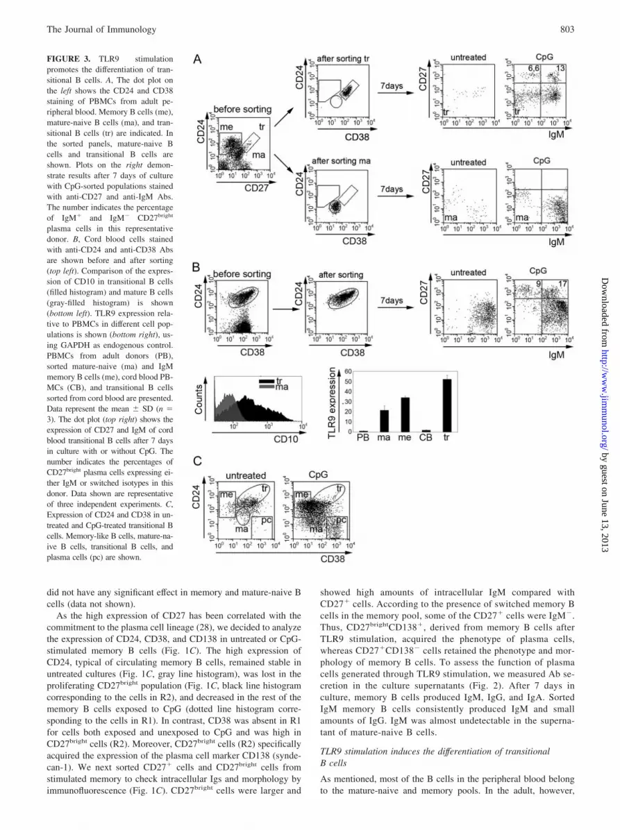
FIGURE 3. TLR9 stimulationpromotes the differentiation of tran-sitional B cells. A, The dot plot onthe left shows the CD24 and CD38staining of PBMCs from adult pe-ripheral blood. Memory B cells (me),mature-naive B cells (ma), and tran-sitional B cells (tr) are indicated. Inthe sorted panels, mature-naive Bcells and transitional B cells areshown. Plots on the right demon-strate results after 7 days of culturewith CpG-sorted populations stainedwith anti-CD27 and anti-IgM Abs.The number indicates the percentageof IgM� and IgM� CD27bright
plasma cells in this representativedonor. B, Cord blood cells stainedwith anti-CD24 and anti-CD38 Absare shown before and after sorting(top left). Comparison of the expres-sion of CD10 in transitional B cells(filled histogram) and mature B cells(gray-filled histogram) is shown(bottom left). TLR9 expression rela-tive to PBMCs in different cell pop-ulations is shown (bottom right), us-ing GAPDH as endogenous control.PBMCs from adult donors (PB),sorted mature-naive (ma) and IgMmemory B cells (me), cord blood PB-MCs (CB), and transitional B cellssorted from cord blood are presented.Data represent the mean � SD (n �3). The dot plot (top right) shows theexpression of CD27 and IgM of cordblood transitional B cells after 7 daysin culture with or without CpG. Thenumber indicates the percentages ofCD27bright plasma cells expressing ei-ther IgM or switched isotypes in thisdonor. Data shown are representativeof three independent experiments. C,Expression of CD24 and CD38 in un-treated and CpG-treated transitional Bcells. Memory-like B cells, mature-na-ive B cells, transitional B cells, andplasma cells (pc) are shown.
803The Journal of Immunology
by guest on June 13, 2013http://w
ww
.jimm
unol.org/D
ownloaded from

1–2% of peripheral B cells are transitional B cells, recently gen-erated in the bone marrow (23). Transitional B cells cannot bedistinguished from mature-naive B cells using the CD24 and CD27markers. Transitional B cells can be identified by adding the CD38marker. Transitional B cells are CD24brightCD38brightCD27�, ma-ture-naive B cells are CD24�CD38�CD27�, and memory B cellscan be identified by the lack of CD38 and the expression of CD24and CD27 (CD24brightCD38�CD27�) (23). We sorted transitionalB cells and a pure mature-naive population from the peripheralblood of the same healthy donor (Fig. 3A). Both populations co-expressed IgM and IgD and lacked switched isotypes (data notshown). After 7 days in culture with CpG, mature-naive B cells,depleted of transitional B cells, did not proliferate (data not shown)and did not generate plasma cells (Fig. 3A, ma). In contrast, uponCpG stimulation, 19.6% of the transitional B cells differentiatedinto CD27bright plasma cells, expressing not only IgM (13%) butalso other isotypes (6.6%) (Fig. 3A, tr). The presence of switchedisotypes was unexpected because all transitional B cells coex-pressed IgM and IgD at the beginning of the culture. This exper-iment was reproduced with sorted transitional B cells from twoadditional donors. The frequency of plasma cells was 27.1%(IgM� (16%), IgM� (11.1%)) and 20% (IgM� (13%), IgM�
(7%)) in these cases.To confirm these data, we used cord blood as a richer source of
transitional B cells (29, 30). In this tissue all B cells have theCD24brightCD38bright transitional phenotype and the majority ofthem expressed CD10 (Fig. 3B). To determine whether TLR9 isexpressed at this stage of B cell development we conducted real-time PCR on isolated B cell subsets. Transitional B cells expressedhigher levels of TLR9 compared with IgM memory and mature Bcells (Fig. 3B). Upon TLR9 stimulation, similarly to adult transi-tional B cells, 26% of cord blood transitional B cells proliferated(data not shown) and differentiated into CD27bright plasma cells(25.5 � 2.3%, with three different donors, n � 3) expressingIgM (19.2 � 2.9%, n � 3) or switched isotypes (6.3 � 2.3%, n �3) (Fig. 3B). The relative expression of CD24 and CD38 dem-onstrated that the phenotype of most transitional B cells hadchanged after CpG stimulation: a large part (41.3 � 4.2%, n �3) of the cells acquired the phenotype of memory B cellsCD24brightCD38�CD27� (Fig. 3, B and C, me). A small pop-ulation of transitional B cells (21.9 � 5.2%, n � 3) becamephenotypically identical with mature-naive B cells (Fig. 3C,ma). Similarly to those generated from adult memory B cells,cord blood-derived plasma cells expressed high levels of CD38and lacked CD24 (Fig. 3C, pc). IgM� and IgM� plasma cellsdeveloped from cord blood only by day 7–9 of CpG stimulation,whereas IgM memory-like CD27� B cells were detectable byday 5 (data not shown).
Cord blood-derived plasma cells were functional. In the su-pernatants of CpG-stimulated cultures we detected both IgM(15.2 � 5 �g/ml) and IgG (2.5 � 1.7 �g/ml) Abs, but never IgA(Fig. 4A).
IgM memory and transitional B cells produceanti-polysaccharide IgM Abs
We have recently shown a correlation between the presence ofIgM memory B cells and the protection from lower respiratorytract infections caused by encapsulated bacteria (22, 26). To pro-vide the direct evidence of the functional role of IgM memory Bcells against S. pneumoniae, we measured the levels of anti-PnPSserotype 14 IgM Abs in CpG-stimulated cultures of sorted IgMmemory cells. We choose PnPS serotype 14 because this type is acommon in Europe and specific Abs can be detected in the serumof most healthy donors (26).
We found that 300,000 IgM memory B cells stimulated withCpG secreted 38 � 9 ng/ml anti-PnPs IgM Abs. Mature-naive Bcells did not secrete anti-PnPS IgM Abs (Fig. 4B). Therefore, therepertoire of IgM memory B cells, isolated from adult peripheralblood contains the anti-PnPS 14 specificity. We calculated thatanti-PnPS serotype 14 IgM produced after polyclonal CpG stim-ulation corresponds to �0.2% of total IgM detected in the super-natant of IgM memory B cells.
Anti-PnPS IgM were also detectable, at a lower concentration(7.7 � 3.2 ng/ml), in the supernatants of cord blood transitional Bcells (Fig. 4B). In this case they were equivalent to 0.08% of totalIgM produced by cord blood B cells. Thus, Abs with the samespecificity are produced after CpG stimulation not only by natu-rally occurring IgM memory B cells, but also by transitional Bcells.
TLR9 stimulation induces the expression of AID
In humans, anti-polysaccharide Abs generated through a thymus-independent mechanism carry somatic mutations (31). This para-dox occurs because somatic hypermutation is thought to happenonly in the germinal center in response to thymus-dependent Ags(32). Recently the identification of the AID has shed light on themechanism of somatic hypermutation and class-switch recombi-nation. AID has been demonstrated to be absolutely required forthe induction of somatic hypermutation and class-switch recombi-nation at the Ig loci (33, 34). Both these events are indispensableprerequisites for the production of high-affinity Abs of differentisotypes.
The finding that anti-polysaccharide IgM and Abs of differentisotypes were always detectable in the supernatants of IgM mem-ory and transitional B cells lead us to hypothesize that the stimu-lation with CpG may induce AID expression.
FIGURE 4. Cord blood-derived transitional B cells secrete Abs afterTLR9 stimulation. A, ELISA shows the concentrations of IgM, IgA, andIgG Abs produced at day 7 of culture by untreated or CpG-stimulated cordblood transitional B cells. Data are the mean � SD (n � 3). B, We showthe concentrations of anti-PnPS Abs (serotype 14) produced at day 7 bymature-naive B cells (ma), peripheral IgM memory B cells (IgM me), andcord blood transitional B cells (tr CB). Data are the mean � SD (n � 3).
804 CpG PROMOTES DIFFERENTIATION OF TRANSITIONAL B CELLS
by guest on June 13, 2013http://w
ww
.jimm
unol.org/D
ownloaded from

We extracted RNA from mature-naive and memory B cellsstimulated with CpG for 48 h, and we analyzed AID expression byreal-time PCR. Results were normalized using �-actin as endog-enous control and were plotted as relative values to AID expres-sion in untreated mature-naive B cells (Fig. 5A, left). We foundthat the basal level of AID mRNA was higher in memory B cellsthan in mature-naive B cells. After stimulation with CpG, AIDmRNA slightly increased in mature-naive, but was down-regulatedin memory B cells. To further explore the effect of TLR9 stimu-lation on B cell development, we analyzed the expression ofBlimp-1, a transcription factor expressed in plasma cell-committedB lymphocytes, functioning as a master regulator of terminal B celldifferentiation. After 48 h of treatment, we found a stronger in-
duction of Blimp-1 mRNA in CpG-treated memory compared withmature-naive B cells (Fig. 5A, right).
After 5–7 days of culture with CpG, half of the memory pooldifferentiates into plasma cells and the other half maintains thememory phenotype (Fig. 1). These two populations are supposedto express inversely proportional levels of AID and Blimp-1 (35).We sorted, based on the expression of CD27, IgM memory B cellsand plasma cells after 7 days of culture with CpG. Real-time PCRwas performed on isolated CD27� and CD27bright cells (Fig. 5B,respectively, IgM memory and plasma cells). Because we werecomparing a proliferating population (terminally developing intoplasma cells) to a nonproliferating pool, we could not use anyhousekeeping gene as endogenous control (36–39). For this reason
FIGURE 5. Differential gene regulation in the memory pool after CpG stimulation. A, AID and Blimp-1 expression was measured by real-time PCR.Sorted mature-naive and IgM memory B cells were cultured for 48 h with or without CpG. Results are expressed relative to AID or Blimp-1 expressionin untreated mature-naive B cells; �-actin was used as endogenous control. Results are representative of three or more independent experiments. Data showmean � SD from triplicate values. B, IgM memory B cells were stimulated for an additional 5 days with CpG to allow the differentiation into CD27bright
plasma cells and then sorted for the expression of CD27. AID and Blimp-1 mRNA levels were analyzed comparing CD27bright plasma cells to CD27� IgMmemory B cells. Histograms show the expression levels for each gene in serially diluted samples (1/3 dilutions). Results are expressed relative to lowestdilution of memory B cells (lowest � 1). Results represent three or more independent experiments. Data show mean � SD from triplicate values.
FIGURE 6. AID and Blimp-1 are induced uponTLR9 engagement in transitional B cells. AID andBlimp-1 expression in transitional B cells was measuredby real-time PCR. A, Transitional B cells (tr) sortedfrom cord blood, were cultured for 48 h with or withoutCpG. B, After an additional 5 days in culture with CpG,transitional B cells were sorted based on the expressionof CD27. AID and Blimp-1 mRNA levels were ana-lyzed comparing CD27bright cells (R2) and CD27� cells(R1) to the transitional pool at time 0 (tr time 0). Resultsshow the expression levels for each gene in serially di-luted samples and were expressed relative to the tran-sitional pool at time 0. Results represent three or moreindependent experiments. Data show mean � SD fromtriplicate values.
805The Journal of Immunology
by guest on June 13, 2013http://w
ww
.jimm
unol.org/D
ownloaded from

we run in parallel the same amount of total cDNA for each sortedpopulations (see Materials and Methods). We observed that AIDexpression was higher in IgM memory B cells than in plasma cells.On the contrary, Blimp-1 was exclusively expressed in plasmacells (Fig. 5B). Similar experiments were performed on transitionalB cells. After 48 h of stimulation AID mRNA was induced intransitional B cells sorted from both adult peripheral blood (datanot shown) and cord blood (Fig. 6A). De novo expression ofBlimp-1 was detectable at this time (Fig. 6A), thus indicating thatcommitment to plasma cells terminal differentiation had initiatedin culture. To distinguish the expression profile of CD27bright
(plasma cells) and CD27� (IgM memory-like) cells, we sortedthese two populations developed in vitro after 7 days of culturewith CpG. AID mRNA was present in both populations, whereasBlimp-1 was dramatically increased only in the proliferating poolcontaining the plasma cells (Fig. 6B).
Transitional B cells of asplenic individuals produceanti-PnPS IgM
Asplenic individuals lack IgM memory B cells in the peripheralblood, but have a normal frequency of switched memory B cells(22). We show that upon TLR9 engagement transitional B cellsdevelop into a cell type functionally equivalent to IgM memory Bcells. Based on these observations, we questioned whether thespleen plays a role in regulating the TLR9-dependent differentia-tion of transitional B cells. We found that in the absence of thespleen, the frequency of circulating transitional B cells is higherthan in healthy controls (Fig. 7A). This finding is very clear inchildhood when the bone marrow output is higher, and less evidentin the adult.
We stimulated with CpG transitional B cells isolated from a2-mo-old asplenic child. After 7 days of culture, transitional Bcells differentiated into CD27bright plasma cells and produced anti-PnPS IgM (Fig. 7, C and D).
DiscussionOver 5 years ago, Bernasconi et al. (2) formulated the hypothesisthat serological memory, representing the individual own antigenic
experience, may be preserved not only by antigenic recall, but alsothrough aspecific recall. They demonstrated that the combinationof CpG and IL-15 was sufficient to induce Ab secretion by memoryB cells and suggested that this mechanism may contribute to main-tain the diversity and concentration of serum Igs.
Our aim was to measure the intrinsic ability of B cells to senseand react to TLR signals, excluding the modulating and steeringaction of exogenous cytokines. Therefore, we performed our ex-periments on sorted B cell populations and never added solublegrowth or differentiation factors. In this experimental setting, themeasured effects depend on the response of B cells to the initialCpG signal. This response may include the production of cyto-kines, such as IL-10 and IL-6, generating and maintaining a Bcell-intrinsic cytokine-loop.
In this study, we demonstrate that TLR9 stimulation not onlyinduces the differentiation of plasma cells from memory B cells,but also has important effects at the mature-naive and transitionalstage. The stimulation with the CpG prolongs the lifespan of ma-ture-naive B cells in vitro, suggesting that the size of the human Bcell compartment may be regulated by pathogen exposure. AnotherTLR ligand, LPS, has a similar function in the mouse (40). Theaddition of a second signal, derived by the BCR and/or IL-10,changes the response of mature B cells.
In humans, the development of transitional B cells strictly de-pends on the signaling function of the BCR. Genetic mutations ofcomponents of the BCR signaling machinery arrest development atthe pro- or pre-B cell stage in the bone marrow and result in theablation of all peripheral B cell pools. The comparison of humangenetic diseases with the murine models often demonstrates amore permissive regulation of mouse B cell development (41).
Human transitional B cells are thought to be the precursors of ma-ture-naive B cells, which in turn differentiate into switched and IgMmemory B cells. Switched memory B cells originate from the germi-nal center reaction, after somatic hypermutation, affinity selection, andclass switching. This result has been definitely proven by the study ofpatients with primary immunodeficiency caused by genetic mutationsof CD40L and CD40, AID, and uracyl N-glycosylase (42, 43).
FIGURE 7. Transitional B cells give rise to IgMplasma cells producing anti-PnPS IgM in the absence ofthe spleen. A, PBMCs isolated from a 7-year-old as-plenic child and from an age-matched healthy control(HC) were analyzed for the expression of CD24 andCD38. Transitional B cells (tr) were identified asCD24brightCD38bright (oval gate). Percentage representstransitional B cells vs total B cells. B, PBMCs stainedwith anti-CD27 and anti-CD22 (left column). Mature-naive (ma) and memory B cells (me) were gated, re-spectively, on CD22�CD27� and CD22�CD27� cells.Gated memory B cells stained for IgM and IgD (rightcolumn) are also shown. IgM memory B cells (IgM) areabsent in the asplenic child, whereas switched memorycompartment B cells (switch) are still present. C, Phe-notypic analysis of sorted transitional B cells from a2-mo-old asplenic infant after 9 days of culture with orwithout CpG. The number indicates the percentages ofCD27bright IgM� and CD27bright IgM� plasma cellsshown. D, ELISA presents anti-PnPS serotype 14 se-cretion in the supernatants cultured for 5, 7, and 9 days.
806 CpG PROMOTES DIFFERENTIATION OF TRANSITIONAL B CELLS
by guest on June 13, 2013http://w
ww
.jimm
unol.org/D
ownloaded from

The origin of IgM memory B cells is less clear and less studied.It was originally assumed that they also derive from the germinalcenters but they can be detected also in patients unable to builtgerminal centers (19). In addition, two other observations supportthe hypothesis that IgM memory B cells belong to a lineage ofdevelopment different from switched memory B cells. First, IgMmemory B cells are generated in the spleen during the first 2 yearsof life, but not in the lymph nodes (22). Secondly, they are re-sponsible for the first-line protection against encapsulated bacterialinfection, a task that cannot be fulfilled by switched memory Bcells (22, 26).
In this study, we demonstrate that IgM memory-like B cells canbe generated in vitro from transitional B cells after TLR9 stimu-lation. Transitional B cells, exposed to CpG, change their pheno-type and function. After 5 days of culture about half of them be-come phenotypically identical with IgM memory B cells anddifferentiate into plasma cells secreting IgM and a small amount ofIgG in the following 2–4 days (Fig. 4).
In the mouse model it has been recently demonstrated that TLRstimulation induces plasma cell differentiation, Ab production, andAID expression by splenic transitional B cells (44). The inductionof AID (Fig. 6) by TLR9 triggering in human transitional B cellsis a novel finding. The function of AID in somatic mutation mayexplain the production of IgM Abs with anti-polysaccharide spec-ificity (32). In contrast, the role of AID in class-switch recombi-nation may justify the production of IgG in our experimental sys-tem (Fig. 4). Abs of IgA or IgE isotypes were never found incultures of IgM memory and transitional B cells. Whereas it hasbeen reported before that CpG inhibits class switching to IgE (45),it was surprising to find no IgA. This observation could be ex-plained either by the requirement of mucosal factors favoringswitching to IgA or by the different origin of IgA plasma cells.
In our in vitro model, the stimulus driving the differentiation oftransitional B cells is initiated by the interaction of TLR9 with itsligand CpG and, therefore, does not involve a defined Ag or a BCRof selected specificity. It is known that Ag encounter through theBCR causes the apoptotic cell death of transitional B cells, repre-senting an important mechanism of tolerance, with the function ofremoving unwanted and dangerous specificities from the pool ofmore mature naive B cells (46–48). Moreover, cord blood transi-tional B cells are generated and circulate in a sterile environmentbecause bacterial colonization occurs hours after birth (49). Thus,the Abs produced by transitional B cells are natural Abs, generatedwithout intentional immunization (1). Natural Abs constitute aninnate repertoire able to recognize the bacteria, which always col-onize humans and can therefore be encountered immediately afterbirth. It remains to be demonstrated whether this repertoire com-prises not only germline but also mutated Igs, as suggested by thefact that anti-PnPS IgM always contains somatic mutations (31,32). The composition of the Ig repertoire may reflect the selectiveforce of pathogens on the host ready-to-use pool of Abs during theco-evolution of host and infectious agents (50).
In vivo transitional B cells are collected in the spleen. Thisorgan is absolutely required for the presence of IgM memory Bcells. Patients with congenital asplenia lack IgM memory B cellpool (22), do not produce anti-PnPS IgM, and have a high risk ofinvasive pneumococcal infection (51). As transitional B cells froman asplenic infant produce anti-PnPS IgM upon TLR9 stimulation,they have the potential to give rise to IgM memory B cells andplasma cells. The absence of IgM memory B cells in asplenicindividuals may be explained by the function of the spleen asunique site for their development. Here the slow blood flow andthe filtering function of macrophages (52) may favor the encounterof transitional B cells with bacterial DNA and possibly other bac-
terial products initiating the further maturation to IgM memory andplasma cells. In the spleen IgM memory B cells are localized in themarginal zone, admixed with canonical memory B cells (53).
Our findings demonstrate a novel role for TLR9 in human Bcells intertwining the innate and adaptive immunity and suggestingonce more that the so-called IgM memory B cells are differentfrom canonical memory B cells. We propose that they could bebetter defined as “natural memory,” taking into consideration theiralready demonstrated independence from the germinal centers,their production of natural Abs and the role of innate TLR9 in theirgeneration, survival, differentiation, and function.
AcknowledgmentsWe thank Prof. Gian Franco Bottazzo for enthusiastic support, Prof. Al-berto Ugazio for helpful discussions and scientific contributions, Prof.Claudio Sette for ideas, experience, and suggestions, and Dr. AnnalisaPantosti for invaluable knowledge of everything about encapsulated bac-teria and more. We especially thank Matilde Sinibaldi and Pamela Biellifor support.
DisclosuresThe authors have no financial conflict of interest.
References1. Ochsenbein, A. F., T. Fehr, C. Lutz, M. Suter, F. Brombacher, H. Hengartner, and
R. M. Zinkernagel. 1999. Control of early viral and bacterial distribution anddisease by natural antibodies. Science 286: 2156–2159.
2. Bernasconi, N. L., E. Traggiai, and A. Lanzavecchia. 2002. Maintainance ofserological memory by policlonal activation of human memory B cells. Science298: 2199–2202.
3. Lemaitre, B., E. Nicolas, L. Michaut, J. M. Reichart, and J. A. Hoffmann. 1996.The dorsoventral regulatory gene cassette spatzle/toll/cactus controls the potentantifungal response in Drosophila adults. Cell 86: 973–983.
4. Akira, S., K. Takeda, and T. Kaisho. 2001. Toll-like receptors: critical proteinslinking innate and acquired immunity. Nat. Immunol. 2: 675–680.
5. Medzhitov, R. 2001. Toll-like receptors and innate immunity. Nat. Rev. Immunol.1: 135–145.
6. Akira, S., and S. Takeda. 2004. Toll-like receptor signalling. Nat. Rev. Immunol.4: 499–511.
7. Iwasaki, A., and R. Medzhitov. 2004. Toll-like receptor control of the adaptiveimmune responses. Nat. Immunol. 5: 987–995.
8. Ku, C. L., K. Yang, J. Bustamante, A. Puel, H. von Bernuth, O. F. Santos,T. Lawrence, H. H. Chang, H. Al-Mousa, C. Picard, and J. L. Casanova. 2005.Inherited disorders of human Toll-like receptor signaling: immunological impli-cations. Immunol. Rev. 203: 10–20.
9. Cook, D. N., D. S. Pisetsky, and D. A. Schwartz. 2004. Toll-like receptors in thepathogenesis of human disease. Nat. Immunol. 5: 975–979.
10. Hemmi, H., O. Takeuchi, T. Kawai, T. Kaisho, S. Sato, H. Sanjo, M. Matsumoto,K. Hoshino, H. Wagner, S. Takeda, and S. Akira. 2000. A Toll-like receptorrecognizes bacterial DNA. Nature 408: 740–745.
11. Coban, C., K. J. Ishii, T. Kawai, H. Hemmi, S. Sato, S. Uematsu, M. Yamamoto,O. Takeuchi, S. Itagaki, N. Kumar, et al. 2005. Toll-like receptor 9 mediatesinnate immune activation by the malaria pigment hemozoin. J. Exp. Med. 201:19–25.
12. Hornung, V., S. Rothenfusser, S. Britsch, A. Krug, B. Jahrsdorfer, T. Giese,S. Endres, and G. Hartmann. 2002. Quantitative expression of Toll-like receptor1–10 mRNA in cellular subsets of human peripheral blood mononuclear cells andsensitivity to CpG oligodeoxynucleotides. J. Immunol. 168: 4531–4537.
13. Liu, Y. J. 2005. IPC: professional type 1 interferon-producing cells and plasma-cytoid dendritic cell precursors. Annu. Rev. Immunol. 23: 275–306.
14. Klinman, D. M. 2004. Immunotherapeutic uses of CpG oligodeoxynucleotides.Nat. Rev. Immunol. 4: 249–258.
15. Huggins, J., T. Pellegrin, R. E. Felgar, C. Wie, M. Brown, B. Zheng,E. C. B. Milner, S. H. Bernstein, I. Sanz, and M. S. Zand. 2007. CpG DNAactivation and plasma cell differentiation of CD27- naive human B cells. Blood109: 1611–1619.
16. He, B., X. Qiao, and A. Cerutti. 2004. CpG DNA induces class switch DNArecombination by activating human B cells through an innate pathway that re-quires TLR9 and cooperates with IL-10. J. Immunol. 173: 4479–4491.
17. Ertesvag, A., H. C. Aasheim, S. Naderi, and H. K. Blomhof. 2007. Vitamin Apotentiates CpG-mediated memory B-cell proliferation and differentiation: in-volvement of early activation of p38MAPK. Blood 109: 3865–3872.
18. Poeck, H., M. Wagner, J. Battiany, S. Rothenfusser, D. Wellisch, V. Hornung,B. Jahrsdorfer, T. Giese, S. Endres, and G. Hartmann. 2004. Plasmacytoid den-dritic cells, antigen, and CpG-C license human B cells for plasma cell differen-tiation and immunoglobulin production in the absence of T-cell help. Blood 103:3058–3064.
19. Brummel, R., and P. Lenert. 2005. Activation of Marginal Zone B cells fromLupus mice with type A(D) CpG-oligodeoxynucleotides. J. Immunol. 174:2429–2434.
807The Journal of Immunology
by guest on June 13, 2013http://w
ww
.jimm
unol.org/D
ownloaded from

20. Klein, U., R. Kuppers, and K. Rajewsky. 1997. Evidence for a large compartmentof IgM-expressing memory B cells in humans. Blood 89: 1288–1298.
21. Klein, U., K. Rajewsky, and R. Kuppers. 1998. Human immunoglobulin (Ig)M�IgD� peripheral blood B cells expressing the CD27 cell surface antigen carrysomatically mutated variable region genes: CD27 as a general marker for somat-ically mutated (memory) B cells. J. Exp. Med. 188: 1679–1689.
22. Kruetzmann, S., M. M. Rosado, H. Weber, U. Germing, O. Tournilhac,H. H. Peter, R. Berner, T. Boehm, A. Plebani, I. Quinti, and R. Carsetti. 2003.Human immunoglobulin M memory B cells controlling Streptococcus pneu-moniae infections are generated in the spleen. J. Exp. Med. 197: 939–945.
23. Carsetti, R., M. M. Rosado, and H. Wardmann. 2004. Peripheral development ofB cells in mouse and man. Immunol. Rev. 197: 179–191.
24. Weller, S., A. Faili, C. Garcia, M. C. Braun, F. F. Le Deist, G. G. de Saint Basile,O. Hermine, A. Fischer, C. A. Reynaud, and J. C. Weill. 2001. CD40-CD40Lindependent Ig gene hypermutation suggests a second B cell diversification path-way in humans. Proc. Natl. Acad. Sci. USA 98: 1166–1170.
25. Weller, S., M. C. Braun, B. K. Tan, A. Rosenwald, C. Cordier, M. E. Conley,A. Plebani, D. S. Kumararatne, D. Bonnet, O. Tournilhac, et al. 2004. Humanblood IgM “memory” B cells are circulating splenic marginal zone B cells har-boring a prediversified immunoglobulin repertoire. Blood 104: 3647–3654.
26. Carsetti, R., M. M. Rosado, S. Donnanno, V. Guazzi, A. Soresina, A. Meini,A. Plebani, F. Aiuti, and I. Quinti. 2005. The loss of IgM memory B cells cor-relates with clinical disease in common variable immunodeficiency.J. Allergy Clin. Immunol. 115: 412–417.
27. Bernasconi, N. L., N. Onai, and A. Lanzavecchia. 2003. A role for Toll-likereceptors in acquired immunity: up-regulation of TLR9 by BCR triggering innaive B cells and constitutive expression in memory B cells. Blood 101:4500–4504.
28. Avery, D. T., J. I. Ellyard, F. Mackay, L. M. Corcoran, P. D. Hodgkin, andS. G. Tangye. 2005. Increased expression of CD27 on activated human memoryB cells correlates with their commitment to the plasma cell lineage. J. Immunol.174: 4034–4042.
29. Sims, G. P., R. Ettinger, Y. Shirota, C. H. Yarboro, G. G. Illei, and P. E. Lipsky.2005. Identification and characterization of circulating human transitional B cells.Blood 105: 4390–4398.
30. Cuss, A. K., D. T. Avery, J. L. Cannons, L. J. Yu, K. E. Nichols, P. J. Shaw, andS. G. Tangye. 2006. Expansion of functionally immature transitional B cells isassociated with human-immunodeficient states characterized by impaired hu-moral immunity. J. Immunol. 176: 1506–1516.
31. Zhou, J., K. R. Lottenbach, S. J. Barenkamp, and D. C. Reason. 2004. Somatichypermutation and diverse immunoglobulin gene usage in the human antibodyresponse to the capsular polysaccharide of Streptococcus pneumoniae type 6B.Infect. Immun. 72: 3505–3514.
32. Weller, S., C. A. Reynaud, and J. C. Weill. 2005. Vaccination against encapsu-lated bacteria in humans: paradoxes. Trends Immunol. 26: 85–89.
33. Durandy, A. 2003. Activation-induced cytidine deaminase: a dual role in class-switch recombination and somatic hypermutation. Eur. J. Immunol. 33:2069–2073.
34. Shinkura, R., S. Ito, N. A. Begum, H. Nagaoka, M. Muramatsu, K. Kinoshita,Y. Sakakibara, H. Hijikata, and T. Honjo. 2004. Separate domains of AID arerequired for somatic hypermutation and class-switch recombination. Nat. Immu-nol. 5: 707–712.
35. Shaffer, A. L., K. I. Lin, T. C. Kuo, X. Yu, M. E. Hurt, A. Rosenwald,J. M. Giltnane, L. Yang, H. Zhao, K. Calame, and L. M. Staudt. 2002. Blimp-1
orchestrate plasma cell differentiation by extinguishing the mature B cell geneexpression program. Immunity 17: 51–62.
36. Suzuki, T., P. J. Higgins, and D. R. Crawford. 2000. Control selection for RNAquantitation. BioTechniques 29: 332–337.
37. Schmittgen T. D., and B. A. Zakrajsek. 2000. Effect of experimental treatment onhousekeeping gene expression: validation by real-time, quantitative RT-PCR.J. Biochem. Biophys. Methods 46: 69–81.
38. Bustin, S. A. 2000. Absolute quantification of mRNA using real-time reversetranscription polymerase chain reaction assay. J. Mol. Endocrinol. 25: 169–193.
39. Barbini, L., J. Rodrıguez, F. Dominguez, and F. Vega. 2007. Glyceraldehyde-3-phosphate dehydrogenase exerts different biologic activities in apoptotic and pro-liferating hepatocytes according to its subcellular localization. Mol. Cell Bio-chem. 300: 19–28.
40. Macpherson, A. J., and N. L. Harris. 2004. Interactions between commensalintestinal bacteria and the immune system. Nat. Rev. Immunol. 4: 478–485.
41. Conley, M. E., A. Broides, V. Hernandez-Trujillo, V. Howard, H. Kanegane,T. Miyawaki, and S. A. Shurtleff. 2005. Genetic analysis of patients with defectsin early B-cell development. Immunol. Rev. 203: 216–234.
42. Imai, K., N. Catalan, A. Plebani, L. Marodi, O. Sanal, S. Kumaki, V. Nagendran,P. Wood, C. Glastre, F. Sarrot-Reynauld, et al. 2003. Hyper-IgM syndrome type4 with a B lymphocyte-intrinsic selective deficiency in Ig class-switch recombi-nation. J. Clin. Invest. 112: 136–142.
43. Durandy, A., P. Revy, K. Imai, and A. Fischer. 2005. Hyper-immunoglobulin Msyndromes caused by intrinsic B-lymphocyte defects. Immunol. Rev. 203: 67–79.
44. Ueda, Y., D. Liao, K. Yang, A. Patel, and G. Kelsoe. 2007. T-independent ac-tivation-induced cytidine deaminase expression, class switch recombination, andantibody production by immature/transitional 1 B cells. J. Immunol. 178:3593–3601.
45. Liu, N., N. Ohnishi, L. Ni, S. Akira, and K. B. Bacon. 2003. CpG directly inducesT-bet expression and inhibits IgG1 and IgE switching in B cells. Nat. Immunol.4: 687–693.
46. Carsetti, R., G. Kohler, and M. C. Lamers. 1995. Transitional B cells are thetarget of negative selection in the B cell compartment. J. Exp. Med. 181:2129–2140.
47. King, L. B., and J. G. Monroe. 2000. Immunobiology of the immature B cell:plasticity in the B-cell antigen receptor-induced response fine tunes negative se-lection. Immunol. Rev. 176: 86–104.
48. Su, T. T., B. Guo, B. Wei, J. Braun, and D. J. Rawlings. 2004. Signaling intransitional type 2 B cells is critical for peripheral B-cell development. Immunol.Rev. 197: 161–178.
49. Pietzak, M. 2004. Bacterial colonization of neonatal gut. J. Pediatr. Gastroen-terol. Nutr. 38: 389–391.
50. Hangartner, L., R. Zinkernagel, and H. Hengartner. 2006. Antiviral antibodyresponses: the two extremes of a wide spectrum. Nat. Rev. Immunol. 6: 231–243.
51. Schutze, G. E., E. O. Mason, W. J. Barson, K. S. Kim, E. R. Wald, L. B. Givner,T. Q. Tan, J. S. Bradley, R. Yogev, and S. L. Kaplan. 2002. Invasive pneumo-coccal infections in children with asplenia. Pediatr. Infect. Dis. J. 21: 278–282.
52. Steiniger, B., P. Barth, and A. Hellinger. 2001. The perifollicular and marginalzones of the human splenic white pulp: do fibroblasts guide lymphocyte immi-gration? Am. J. Pathol. 159: 501–512.
53. Brendolan, A., M. M. Rosado, R. Carsetti, L. Selleri, and T. N. Dear. 2007.Development and function of the mammalian spleen. Bioessays 29: 166–177.
808 CpG PROMOTES DIFFERENTIATION OF TRANSITIONAL B CELLS
by guest on June 13, 2013http://w
ww
.jimm
unol.org/D
ownloaded from
Recommended


