
HAL Id: tel-01492601https://tel.archives-ouvertes.fr/tel-01492601v2
Submitted on 23 May 2017
HAL is a multi-disciplinary open accessarchive for the deposit and dissemination of sci-entific research documents, whether they are pub-lished or not. The documents may come fromteaching and research institutions in France orabroad, or from public or private research centers.
L’archive ouverte pluridisciplinaire HAL, estdestinée au dépôt et à la diffusion de documentsscientifiques de niveau recherche, publiés ou non,émanant des établissements d’enseignement et derecherche français ou étrangers, des laboratoirespublics ou privés.
Evaluation of photodynamic activity of chlorine-typephotosensitizers with β-cyclodextrins nanovectors
Igor yankovsky
To cite this version:Igor yankovsky. Evaluation of photodynamic activity of chlorine-type photosensitizers with β-cyclodextrins nanovectors. Human health and pathology. Université de Lorraine; Belarussian StateUniversity, 2016. English. �NNT : 2016LORR0171�. �tel-01492601v2�
AVERTISSEMENT
Ce document est le fruit d'un long travail approuvé par le jury de soutenance et mis à disposition de l'ensemble de la communauté universitaire élargie. Il est soumis à la propriété intellectuelle de l'auteur. Ceci implique une obligation de citation et de référencement lors de l’utilisation de ce document. D'autre part, toute contrefaçon, plagiat, reproduction illicite encourt une poursuite pénale. Contact : [email protected]
LIENS Code de la Propriété Intellectuelle. articles L 122. 4 Code de la Propriété Intellectuelle. articles L 335.2- L 335.10 http://www.cfcopies.com/V2/leg/leg_droi.php http://www.culture.gouv.fr/culture/infos-pratiques/droits/protection.htm

Ecole Doctorale BioSE (Biologie-Santé-Environnement)
Thèse
Présentée et soutenue publiquement pour l’obtention du titre de
DOCTEUR DE l’UNIVERSITE DE LORRAINE
Mention : « Sciences de la Vie et de la Santé »
par Igor YANKOVSKY
Evaluation de l'activité photodynamique des photosensibilisants de type chlorine avec les nanovecteurs cyclodextrines-β
Evaluation of photodynamic activity of chlorin-type photosensitizers with β-cyclodextrins nanovectors
Le 30 Novembre 2016
Membres du jury : Rapporteurs : Monsieur Vladimir SUKHORUKOV Professeur, Département de Biotechnologies
et de Biophysique, Université de Würzburg
Monsieur Eduard ZENKEVICH Professeur, Département des technologies
de l'information et de la Robotique
du l'Université technique nationale du
Biélorussie, Minsk
Examinateurs : Madame Lina BOLOTINE Docteur, CRAN-UMR 7039 CNRS
Université de Lorraine, Institut de
Cancérologie de Lorraine
Co-Directeur de thèse
Monsieur Vladimir ZORIN Professeur, Université d'Etat Biélorusse,
Minsk, Co-Directeur de thèse
---------------------------------------------------------------------------------------------------------------------------------
Centre de Recherche en Automatique de Nancy (CRAN), Université de Lorraine, CNRS UMR 7039, Institut de Cancérologie de Lorraine, 6, Avenue de Bourgogne 54511 Vandœuvre-lès-Nancy, France; Research Laboratory of Biophysics and Biotechnology, Physics Faculty, Belarusian State University, 4, pr. Nezavisimosti, 220030 Minsk, Belarus


2
TABLE OF CONTENTS
ABBREVIATIONS .................................................................................................................... 4
LIST OF FIGURES .................................................................................................................... 5
LIST OF TABLES ..................................................................................................................... 6
GENERAL INTRODUCTION .................................................................................................. 7
CHAPTER I. INTRODUCTION ............................................................................................... 8
1. Photodynamic therapy ....................................................................................................... 8
1.1. An introduction to the photodynamic therapy ........................................................... 8
1.2. Basic principles of photodynamic therapy ............................................................... 10
1.2.1. Photophysical and photochemical processes ................................................... 10
1.2.2. Mechanisms of photodynamic action in vivo ................................................... 13
1.2.3. Photosensitizers ................................................................................................ 17
2. Meta-tetra(hydroxyphenyl)chlorin (mTHPC) ................................................................. 25
2.1. General properties and usage ................................................................................... 25
2.2. Biological behavior of mTHPC ............................................................................... 27
2.3. Strategies for improving delivery and efficacy of photodynamic therapy with
mTHPC ........................................................................................................................... 30
3. Nanotechnology-based drug delivery systems ................................................................ 31
3.1. A rational design and advantages of drug delivery systems .................................... 32
3.2. Types of nanosized antitumor drug delivery systems .............................................. 34
4. Cyclodextrins for drug delivery ....................................................................................... 38
4.1. Cyclodextrins: classification, structure and basic properties ................................... 38
4.2. Inclusion complex formation and mechanism of drug release ................................ 42
4.3. Effects of cyclodextrins on drug properties in cyclodextrin-based formulations .... 44
4.3.1. Effect on drug solubility, dissolution and stability .......................................... 44
4.3.2. Effect on drug permeation through biological membranes .............................. 46
4.3.3. Effect on drug bioavailability and safety ......................................................... 47
3
4.4. Pharmacokinetics, biological activity and toxicology of cyclodextrins .................. 49
4.5. Mechanism of drug release from cyclodextrin complexes ...................................... 55
5. Use of cyclodextrins in photodynamic therapy ............................................................... 58
5.1 Inclusion complexes of cyclodextrins with photosensitizers .................................... 58
5.2 Photosensitizer/cyclodextrin conjugates ................................................................... 61
5.3. Photosensitizers encapsulated in cyclodextrin nanoassemblies ............................... 62
5.4. Influence of cyclodextrins on photosensitizers in biological systems ..................... 63
5.5. Inclusion complexes of mTHPC with cyclodextrins ............................................... 65
OBJECTIVES .......................................................................................................................... 67
CHAPTER II. RESULTS ......................................................................................................... 68
1. Influence of cyclodextrins on mTHPC distribution and photodynamic activity in
biological systems ............................................................................................................... 68
2. Development of fluorescence methods suitable for monitoring of mTHPC release from
inclusion complexes ............................................................................................................ 80
GENERAL DISCUSSION ....................................................................................................... 89
CONCLUSIONS AND OUTLOOK ........................................................................................ 95
REFERENCES ......................................................................................................................... 97
SYNTHESE DES TRAVAUX DE THESE ........................................................................... 132
SCIENTIFIC OUTPUT .......................................................................................................... 143

4
ABBREVIATIONS
CD – cyclodextrin
DDS – drug delivery system
DLI – drug-light interval
DMSO – dimethyl sulfoxide
HDL – high-density lipoproteins
Hp-β-CD – hydroxypropyl-β-cyclodextrin
HpD – hematoporphyrin derivative
IV – intravenous
LDL – low-density lipoprotein
M-β-CD – methyl-β-cyclodextrin
mTHPBC – meta-tetra(hydroxyphenyl)-bacteriochlorin
mTHPC – meta-tetra(hydroxyphenyl)chlorin
mTHPP – meta-tetra(hydroxyphenyl)porphyrin
NMR – nuclear magnetic resonance
PBS – phosphate-buffered saline
PDT – photodynamic therapy
PEG – polyethylene glycol
PS – photosensitizer
QY – quantum yield
ROS – reactive oxygen species
SBE-β-CD – sulfobutylether-β-cyclodextrin sodium salt
TM-β-CD – trimethyl-β-cyclodextrin
TPPS4 – tetrakis(4-sulfonatophenyl)porphyrin
VD – volume of distribution

5
LIST OF FIGURES
Figure 1.1 Schematic illustration of the treatment process of PDT …………...…………..9
Figure 1.2 Modified Jablonski energy diagram for PS. VR – vibrational relaxation, IC –
internal conversion, ISC – intersystem crossing ……………………………………………..12
Figure 1.3 Mechanisms of PDT tumor photoeradication (from Agostinis et al. 2011)…...14
Figure 2.1 Molecular structures of mTHPP, mTHPC and mTHPBC and m, p and o
isomers of the hydroxyphenyl substituent ………………..………………………....……….26
Figure 3.1 Different types of nanocarriers have used for drug delivery (from Bamrungsap
et al., 2012; Khodabandehloo et al., 2016; Wang et al., 2013) …………………………..…..36
Figure 4.1 Schematic illustration of the association of CD and drug. Topology of inclusion
complexes: (a) complete, (b) partial through the narrow side, (c) partial through the wide side,
(d) axial …………………………..…………………………..………………………………42
Figure 4.2 Mechanism proposed for morphological changes in red blood cells induced by
CDs (from Arima et al., 2011) ……………………………………………………………….54
Figure 5.1 The type of CDs binding with tetrapyrrole photosensitizers: host-guest (Lang et
al., 2004), conjugate (Kirejev et al. 2014), nanoassemblies (Mazzaglia et al. 2013) ….….…59

6
LIST OF TABLES
Table 1.1 Photosensitizers approved for clinics or undergoing clinical trials (from
Agostinis et al., 2011; Josefsen & Boyle, 2012; Lucky et al., 2015) ………………………...23
Table 2.1 Photophysical properties of mTHPP, mTHPC and mTHPBC in methanol
(Bonnett et al., 1999; Senge & Brandt, 2011) …………………………..….…..…………….26
Table 3.1 Nanotherapeutics approved for clinical use or being evaluated in clinical trials
(adapted from Egusquiaguirre et al, 2012; Gidwani & Vyas, 2015; Marchal et al., 2015) ….35
Table 4.1 Structure and physicochemical properties of native CDs (adapted from Loftsson
& Brewster 2013; Del Valle 2004) …………………………..…………………………....…40
Table 4.2 Structural and physiochemical properties of some CDs of pharmaceutical
interest (adapted from Loftsson & Brewster, 2013; Vyas et al., 2008) …………………...…41
Table 4.3 Pharmacokinetic parameters of some CDs IV administered (from Stella & He,
2008) …………………………..…………………………..…………………………………51

7
GENERAL INTRODUCTION
Photodynamic therapy (PDT) is a minimally invasive photochemical-based treatment
with a promising clinical track record for oncological and other diseases. PDT involves three
main components (light, photosensitizer, oxygen) to damage the target tissue by generating
reactive molecular species. Tetrapyrrole compounds such as meta-tetra(hydroxyphenyl)-
chlorin (mTHPC) are dominate photosensitizers (PSs) employed in PDT, but several
difficulties associated with high hydrophobicity of these PSs leading to poor water solubility
and aggregation within the vasculature after intravenous injection hampers its successful
clinical application. Aggregation of PSs results in a decreased photodynamic efficacy,
unfavorable biodistribution, moderate selectivity and prolonged skin photosensitivity.
To abolish these problems, special pharmacological forms such as liposomes,
polymeric nanoparticles, bioconjugates are proposed for PS administration. Among the
biodegradable and nontoxic compounds that can be used for drug delivery, cyclodextrins
(CDs) are very promising. CDs are a family of cyclic oligosaccharides with a hydrophobic
internal cavity and a hydrophilic outer surface. They are water-soluble, biocompatible in
nature and can form stable inclusion (host-guest) complexes with a very wide range of solid,
liquid and gaseous compounds. Inclusion of a hydrophobic drug into CD cavity usually
increased the PSs solubility, physical and chemical stability, circulation time thus enhancing
their bioavailability. Besides, CDs are used in the design of immediate as well as delayed
release and targeted drug delivery systems.
mTHPC, the potent second-generation PS, which is currently under clinical trial for
the palliative treatment of head and neck cancer, appears as a promising PS exhibiting a high
therapeutic ratio. Previously, it was shown that mTHPC forms effectively inclusion
complexes with derivatives of β-CD resulting in an increase of the PS solubility and
improvement of its photophysical properties in aqueous solution. However, there is no
available data on the use of mTHPC-CDs complexes in vitro and in vivo models so far.
The main objective of present work was to evaluate the effect of different CDs on
mTHPC behavior at various stages of its distribution in vitro and in vivo and estimate the PS
release from the carriers. We have also developed new approaches for analyzing mTHPC
release from CDs. These approaches were based on the changing of fluorescence polarization
degree and variability of mTHPC Soret band in function of PS microinvirement.

8
CHAPTER I. INTRODUCTION
1. Photodynamic therapy
1.1. An introduction to the photodynamic therapy
The combination of dyes and sunlight for treatment of several skin disorders dates
back to ancient Egyptians, Greeks and Indians (Spikes et al., 1985). However, the emergence
of the concept of photodynamic therapy (PDT) can be attributed to early observations made
by O. Raab and H. von Tappeiner. O. Raab under the direction of Professor Dr. Hermann von
Tappeiner observed that low concentration of acridine and some other dyes such as eosin, that
had no effect in the dark, provoked the rapid killing of paramecia in the presence of light
(Raab, 1900). The first mention of the necessity of the presence of oxygen for photodynamic
action was reported in 1902 by C. Ledoux-Lebards. He observed that eosin killed paramecia
more efficiently in open flask than in a closed bottle (Ledoux-Lebards, 1902). In 1903, von
Tappeiner along with a dermatologist named Jesionek reported the tumoricidal effect of eosin
associated with exposure to the white light on skin tumors (Von Tappeiner & Jesionek, 1903).
Despite this early success, PDT did not achieve enough impact and was lost for
nearly 50 years when the photodynamic reaction was rediscovered by Lipson and Schwartz.
Samuel Schwartz demonstrated that a mixture of hematoporphyrin derivative (HpD) was far
more effective anti-tumor agent than hematoporphyrin (Schwartz et al., 1955). Lipson and his
colleagues observed that injection of crude preparations of hematoporphyrin led to
fluorescence of tumor lesions that could be visualized during surgery (Lipson et al., 1961;
Lipson & Baldes, 1960). However, dissemination and application of PDT in clinical practice
was associated with the work of Thomas Dougherty and his colleagues at Roswell Park
Cancer Institute (University of Buffalo, USA). In contrast to previous studies, Dougherty
created a commercially suitable photosensitizing drug, reliable light sources (Dougherty,
1974) and showed long-term HpD-PDT efficacy in animal models and humans (Dougherty et
al., 1975, 1978). Chromatographic isolation of HpD led to the design of the photosensitizer
(PS) Photofrin®, which was first approved for the treatment of bladder cancer in Canada in
1993. To date, Photofrin® is approved in the US, Europe and Japan for the treatment of
advanced and early stage lung cancer, oesophageal adenocarcinoma, cervical cancer,
superficial gastric cancer and bladder cancer (Dougherty, 2002; O’Connor et al., 2009).

9
Treatment by PDT is as follows (Fig. 1.1). PDT uses the combination of a
photosensitizing drug (photosensitizers, PSs) and light to cause selective damage to the target
tissue. In PDT, PSs are applied topically, locally or systemically (e.g., intravenously, IV).
Firstly, PS is administered into the patient and it begins to redistribute into the body. After
certain period, when PS accumulation in the tumor becomes greater than in normal tissue, the
localized illumination of tumor region is carried out with a light source with an appropriate
emission wavelength. Absorption of this light by tumor-localized sensitizer leads to
generation of toxic free radicals and finally to the destruction of tumor tissue (Agostinis et al.,
2011). Tumor destruction can be realized from three interconnected mechanisms: direct
killing of tumor cells, damage to the tumor vasculature, and induction of an inflammatory
reaction that can lead to the immune response. The selectivity of PDT is achieved by both the
preferential uptake of the PS by the tumor (Jori, 1996) and directed illumination of treated
tissue while sparing potentially healthy tissue.
Figure 1.1 Schematic illustration of the treatment process of PDT
So far, about 250 randomized clinical trials have been officially reported for PDT of
tumors, and essentially all types of solid tumors with the exception of melanotic melanoma
have been found to be positively responsive to the photodynamic treatment
(Rapozzi & Jori, 2015). The earliest known tumor approved for treatment was refractory
superficial bladder cancer (Nseyo et al., 1998). To date, PDT is also approved for the
treatment of obstructive and early-stage bronchial cancers (Usuda et al., 2006), esophageal
dysplasia and carcinoma in situ (Fayter et al., 2010), and unresectable cholangiocarcinoma.
The progress in PDT-protocols and synthesis of second-generation PSs led to efficient clinical
treatment of head and neck tumors: widespread and unresectable or recurrent tumors (Biel,

10
2006), early stage oral cancers (Hopper et al., 2004) and nasopharyngeal tumors (Nyst et al.,
2007). PDT has been shown to have high efficacy for basal cell carcinoma, including
extensive or recurrent lesions. Prostate cancer treatment is under clinical trials and is
performed as a primary therapy for focal tumors (Eggener et al., 2007), as well as the
treatment of the whole prostate in patients who have recurred locally following radiation
therapy (Trachtenberg et al., 2007). Excellent cosmetic outcomes make PDT suitable for
patients with skin cancers (Fayter et al., 2010; Gao et al., 2010). However, the effectiveness
of the treatment of many cancer types with PDT remains yet to be proven due to the lack of
well-designed clinical trials (Agostinis et al., 2011).
An increasing number of PDT-related studies led to a better understanding of the
factors controlling PDT and also extends the field of application of this method. Today PDT
finds new applications not only for nononcologic dermatoses but also in the field of
otorhinolaryngology, ophthalmology, neurology, gastroenterology and urology (Darlenski &
Fluhr, 2013). PDT has indications for non-oncological diseases including psoriasis (Smits et
al. 2006), ocular macular degeneration (Ziemssen & Heimann, 2012), chronic wounds
(Morley et al., 2013), rheumatoid arthritis (Hansch et al., 2008), acne vulgaris (Pollock et al.
2004), periodontitis (Alwaeli et al., 2015) and PDT of certain bacterial, fungal and viral
infections (Rajesh et al. 2011; Reinhard et al., 2015a; Yin & Hamblin, 2015).
1.2. Basic principles of photodynamic therapy
The action of PDT consists in the interaction of three principal components: (i) a
nontoxic drug or dye known as a PS, (ii) the light of a specific wavelength and (iii)
tissue/molecular oxygen. The lack of any of these components results in the absence of PDT
effect. Therefore, to better understand the mechanisms of PDT action, it is important to
understand the particular role of each of these components and how they interact. This section
provides an overview of PDT with brief descriptions of the photophysical, photochemical and
biological aspects of the treatment.
1.2.1. Photophysical and photochemical processes
Most PSs in their ground (usually singlet) state have 2 electrons with opposite spins
located in an energetically most favorable molecular orbital (Castano et al., 2004). The
biological effects of PDT are a consequence of a basic photochemical reaction involving PS
and oxygen, which can be illustrated using modified Jablonski diagram (Figure 1.2).
Absorption of light (initial step in all photoreactions) by PS leads to a transfer of one electron

11
from their ground state (S0) to a higher energy orbital (Sn). A molecule with a high vibrational
level of the excited state Sn (n depending on the PS and excitation wavelength used) will
quickly fall to the lowest vibrational level of this state in a process called vibrational
relaxation. Also, a molecule in a higher excited state Sn will finally fall to the first excited
singlet state S1 by internal conversion. Then, the singlet state S1 can rapidly return to the
ground state level S0 by two mechanisms, a radiative process (fluorescence), or a non-
radiative process (internal conversion). During this internal conversion, the excess of energy
of the singlet state is released as heat, which dissipates usually into the tissue or the solvent.
As for the radiative process, a photon is emitted with the energy equal to the energy gap
between the S0 and the S1 levels, implying that the fluorescence does not depend on the
excitation wavelength. Fluorescence is used very widely for PS detection in photodiagnostic
applications (Mallidi et al., 2015).
Alternatively, an excited PS molecule may undergo an intersystem crossing forming
a more stable triplet state (T1) with inverted spin of electrons. The lifetime of the triplet state
is much longer (τ > 10-7 s) than the lifetime of the singlet state (τ ~10-9 s), which greatly
increases the probability of a reaction with a neighboring molecules, and the biologically
relevant photochemistry is often mediated by this state (Lakowicz, 2006). There are several
pathways for the triplet state T1 to return to S0 including the emission of a photon
(phosphorescence) and intersystem crossing followed by vibrational relaxation.
Generally, only the S1 and T1 can be considered as likely candidates for the initiation
of photochemical and photophysical reactions. This is due to the fact that higher orders of
electronic states (n ≥ 2) undergo very rapid internal conversion from Sn to S1 and from Tn to
T1. However, under the special conditions of multiphoton absorption (short pulse, high
intensities of irradiation), the upper excited states may be filled and complex photophysical
and photochemical processes can occur (Shea et al., 1990; Smith et al., 1994).
The PS in triplet state can also initiate photochemical reactions directly upon
interaction with a substrate, giving rise to reactive free radicals (type I reaction), or transfer its
energy to molecular oxygen (3O2), which is unique in being a triplet in its ground state (type II
reaction) (Foote, 1968; Sharman et al., 2000). The relatively longer lifetimes for the triplet
excited states make the collisional transfer of energy to surrounding oxygen molecules
possible. A Type I process can occur whereby the PS reacts directly with an organic molecule
in a cellular microenvironment, acquiring a hydrogen atom or electron to form a radical
(Henderson & Dougherty, 1992). Both the excited PS and the ground state substrate can act as
hydrogen donors. The resulting radical species from type I primary processes can

12
subsequently participate in different kinds of reactions. In the presence of oxygen, for
example, oxidized forms of the PS or of the substrate readily react with oxygen to give
peroxyl radicals, thus initiating a radical chain auto-oxidation. Semi-reduced forms of the PS
or of the substrate also interact efficiently with oxygen, and the electron transfer generates
superoxide anion radical (O2•‒). Dismutation or one-electron reduction of O2
•‒ gives hydrogen
peroxide (H2O2), which in turn can undergo one-electron reduction to a powerful and virtually
indiscriminate oxidant hydroxyl-radical (HO•) (Henderson & Dougherty, 1992).
Figure 1.2 Modified Jablonski energy diagram for PS. VR – vibrational relaxation,
IC – internal conversion, ISC – intersystem crossing
Both type I and II reactions occur simultaneously during PDT, but the ratio of the
frequency of the reactions depends on the type of PS, surrounding substrates, and
concentration of molecular oxygen in the environment (Aveline et al., 1998; Plaetzer et al.,
2009). However, reactive oxygen species (ROS) generation via Type II chemistry is much
simpler than via Type I, and most PSs are believed to operate via a Type II rather than Type I
mechanism. In type II process, the reaction proceeds via energy transfer from the excited
triplet-state PS to the molecular oxygen in its triplet state. Only PSs that possess an energy
gap between the ground state and the excited triplet state higher than the energy needed to

13
excite oxygen into its excited singlet state (94 kJ/mol according to van Lier & Spikes, 1989)
can generate singlet oxygen. Theoretically all molecules absorbing light at a wavelength <
1260 nm can mediate generation of 1O2. However, photons with wavelengths longer than 800
nm have insufficient energy to initiate a photodynamic reaction (Juzeniene et al., 2006).
It is worth noting that singlet oxygen is a very reactive species, much more
electrophilic than its ground state, and can oxidize biomolecules very rapidly. Lifetime of
singlet oxygen varies from about 4 µs in water to 25-100 µs in non-polar organic solutions
and greatly decreases (about 10-330 ns) in biological systems due to the presence of various
quenchers (Baker & Kanofsky, 1992). This short lifetime allows the diffusion of singlet
oxygen to a distance from 10 to 55 nm at the sub-cellular level (Dysart & Patterson, 2005;
Moan & Berg, 1991), thus limiting the PDT effect to the immediate intracellular localization
of the PS (Moan et al., 1989).
1.2.2. Mechanisms of photodynamic action in vivo
PDT destroys tumors by three inter-related mechanisms: direct cytotoxic effects on
tumor cells, damage to the tumor vasculature, and processes involving the immune system.
These are illustrated in Fig. 1.3. In the first case, the ROS that are generated by PDT can
directly destroy tumor cells by apoptosis, necrosis and/or autophagy-associated cell death.
PDT also damages the tumor-associated vasculature with impairment of the blood supply to
the area via platelet aggregation and, thus, vascular occlusion. Finally, PDT can induce a
robust inflammatory reaction that can activate an immune response against tumor. This
section will describe the mechanisms of PDT action conditionally separated as cellular
effects, vascular effects and systemic immune effects of the treatment.
1.2.2.1. Direct tumor cell death effects
Direct cell kill is an important contributor to PDT mediated tumor destruction in
vivo. Henderson et al. (1985) showed that exposure of tumors to PDT in vivo leads to decrease
of the number of clonogenic tumor cells through direct cells photodamage. Direct cell kill can
occur through necrosis, apoptosis and/or autophagy-associated cell death. Mechanism of cell
death depends on cell and PS type, treatment protocol, and photodynamic dose (Agostinis et
al., 2011; Castano et al., 2004, 2005a, 2005b).

14
Figure 1.3 Mechanisms of PDT tumor photoeradication (from Agostinis et al. 2011)
Apoptosis is the most studied and generally major cell death mechanism responding
to PDT. Apoptosis or programmed cell death is identified in single cells, and morphologically
characterized by cell shrinkage, chromatin condensation, cleavage of chromosomal DNA into
internucleosomal fragments, blebbing of the plasma membrane, and the formation of
apoptotic bodies without plasma membrane breakdown (Agostinis et al., 2004; Almeida et al,.
2004). In vivo, apoptotic cells release the “find me” and “eat me” signals required for the
clearance of the remaining apoptotic bodies by phagocytes that usually prevents inflammation
response. The most important role in the initiation of apoptosis among all subcellular
structures is played by mitochondria (Castano et al., 2005a; Kessel & Luo, 1999). The
damage to mitochondria after PDT results in a cascade of reactions including the rapid release
of mitochondrial cytochrome C into the cytosol followed by activation of the apoptosome and
procaspase 3 that eventually cause the apoptosis (Oleinick et al., 2002). However, localization
of the PS in the lysosomes may also activate apoptotic cascade (Bonneau & Vever-Bizeta,
2008). Woodburn et al. (1997) demonstrated that lutetium texaphyrin, which is mostly
localized in the lysosomes of EMT6 cells in vitro, induces apoptosis in EMT6 tumors in vivo.
Necrosis is a rapid form of death affecting extensive cell populations, characterized
by cytoplasm swelling, destruction of organelles and disruption of the plasma membrane,

15
resulting in an inflammatory reaction due to the release of cellular contents and pro-
inflammatory molecules. Necrosis has been associated with incidental cell death, caused by
physical or chemical damage and has generally been considered an unprogrammed process.
During necrosis, decomposition of cell is principally mediated by proteolytic activity and
inhibition or genetic deficiency of caspases in the cell signaling (Castano et al., 2005a;
Kessel, 2002).
Another mechanism of cell death have been also described: mitotic cell death
(Castedo et al., 2004), cathepsin-mediated lysosomal death pathway (Leist & Jäättelä, 2001),
programmed necrosis (Bizik et al., 2004; Vanlangenakker et al., 2008) and autophagic cell
death (Yu et al., 2004). The last (autophagy) is characterized by a massive vacuolization of
the cytoplasm and can be stimulated by various stress signals including oxidative stress
(Dewaele et al., 2010). In autophagic cell death processes, normal function used to degrade
components of the cytoplasm is involved and characterized by appearance autophagosomes,
autolysosomes, electron-dense membranous autophagic vacuoles, myelin whorls,
multivesicular bodies, as well as engulfment of entire organelles (Castano et al., 2005a).
Recent studies describe autophagy as a mechanism to preserve cell viability after PDT
treatment (Reiners et al., 2010). On the other hand, photodamage of lysosomal compartment
by the PS may compromise completion of the autophagic process.
In rare cases, complete tumor destruction is achieved only through direct damage of
tumor cells. Indeed, non-homogenous distribution of PS within the tumor, insufficient depth
of light penetration into the tumor or distance of tumor cells from the vessels (Korbelik &
Krosl, 1994) as well as availability of oxygen (Tromberg et al., 1990) may hamper the
tumoricidal effect. Therefore, other mechanisms involved in the destruction of the tumor
during PDT will be discussed in next chapters.
1.2.2.2. Antivascular effects of PDT
The importance of vascular damage for tumor destruction is well recognized. PDT
damage of tumor microvasculature leads to severe and persistent tumor hypoxia and restricts
nutrient supply to the proliferating tumor cells. Numerous studies revealed that blood vessel
of tumors are fundamentally different from normal vasculature (Ausprunk & Folkman, 1977;
Eberhard et al., 2000; Siemann, 2011). Tumor vasculature is typified by aberrant structural
dynamics and vessels that are immature in nature with poorly developed, chaotic growing
architecture and discontinuous endothelial linings (Eberhard et al., 2000). Besides, the

16
endothelial cells lining tumor vessels have an irregular, disorganized morphology that result
in enhanced permeability (Siemann, 2011).
Targeting the tumor vasculature is a promising approach to cancer treatment.
Destruction of endothelial cells during PDT treatment seems to be the origin of modifications
observed in vasculature (Fingar et al., 2000); therewith endothelial cells were shown to be
more sensitive to PDT compared to muscle cells, together with increased PS uptake (West et
al., 1990). PDT incites modifications of organization of the proteins of cytoskeleton of
endothelial human cells with consecutive induction of a signal to the platelets and neutrophils
activation which adhere on the vessel wall (Senge & Radomski, 2013). In the region of injury
the following changes are observed: initial blanching and vasoconstriction of the tumor
vessels, followed by heterogeneous responses including eventual complete blood flow stasis,
hemorrhage, and, in some larger vessels, the formation of platelet aggregates (Krammer,
2001). The combination of vessel constriction, thrombus formation, and increased interstitial
pressure leads to tissue ischemia followed by necrosis (Fingar et al., 2000; Henderson et al.,
1985). Moreover, vascular destruction after PDT may be accompanied by inflammatory
response like after tissue injury (Korbelik, 1996).
It is worth noting that different PSs do not produce the same type of vascular
response. PDT with monoaspartyl chlorin e6 (MACE®) produce blood stasis mainly due to
platelets aggregated on the artery walls (McMahon et al., 1994) while certain phthalocyanine
derivatives cause primarily vascular leakage (Fingar et al., 1993b). At the same time, using of
Photofrin® leads to vessel constriction, macromolecular vessel leakage, leukocyte adhesion
and thrombus formation corresponding to platelet activation and release of thromboxane
(Fingar et al., 1993a).
1.2.2.3. PDT and the immune response
Nowadays it is shown that PDT can affect both innate immunity and adaptive
immunity. The influence of PDT on the immune response is quite complicated and defines by
many factors, such as the type of PS and its formulation, pharmacokinetic and location within
tumor and surrounding cells (especially immune cells), the area treated and treatment regimen
(e.g., drug-light interval (DLI) and settings of irradiation) (Kousis et al., 2007; Reinhard et al.,
2015a).
PDT-induced oxidative stress frequently incites a strong acute inflammatory reaction
observed as localized edema at the targeted site (Dougherty, 2002). The main task of
inflammatory response consists in the disruption of homeostasis and the removal of damaged

17
cells, thus promoting local healing with the restoration of normal tissue function. The
inflammatory signaling after PDT initiates a massive regulated infiltration of matures
dendritic cells and other cellular components of both the innate and adaptive immune systems
(Castano et al., 2006). Korbelik & Krosl (1994) showed that macrophages demonstrated
preferential cytotoxicity to tumor cells after PDT treatment. Furthermore, upon
photostimulation macrophages may regulate the release of pro- or anti-inflammatory
cytokines (Korbelik & Krosl, 1994; Lynch et al., 1989). Besides, PDT-generated lysates are
able to activate dendritic cells and increase ability to stimulate T-cells, which is critical to the
development of a cellular immune response (Gollnick et al., 2002). Removal of inflammatory
cells or inhibition of their activity after PDT demonstrated significant reducing of therapeutic
effect (Agostinis et al., 2011).
It has been showed that PDT can influence the adaptive immune response in different
ways, resulted both in potentiation of adaptive immunity and immunosuppression (Castano et
al., 2006; Hunt & Levy, 1998). Long-term effect of PDT depends on the induction of
antitumor immunity. Canti et al. (1994) showed that resistance to MS-2 fibrosarcomas
rechallenge was evident only in normal surviving animals treated by PDT, whereas
immunosuppressed surviving animals and animals cured by surgery died after tumor
recurrence. PDT treatment of murine EMT6 mammary sarcoma using Photofrin® indicate
that whereas the direct effects of PDT can destroy the bulk of the tumor, the immune response
is required to eliminate the surviving cells (Korbelik et al., 1996).
It should be noted that all these mechanisms occur as interrelated with each other and
the predominance of one or another mechanism of tumor destruction depends on many
factors, such as the type of PS, light and PS dose, DLI. Therefore, determination of optimal
PDT conditions requires a coordinated interdisciplinary effort.
1.2.3. Photosensitizers
Photosensitizers are one of the main components of PDT. To date, a large number of
photosensitizing agents have been studied in PDT. The majority of PSs used both clinically
and experimentally has a tetrapyrrole structure, such as porphyrins, chlorins, bacteriochlorins
and phthalocyanines (Josefsen & Boyle, 2012; O’Connor et al., 2009). However, other classes
of compounds including the synthetic dyes (e.g., phenothiazinium, squaraine) transition metal
complexes, and natural products (e.g., hypericin, riboflavin, curcumin) have been investigated
(Abrahamse & Hamblin, 2016).

18
1.2.3.1. General properties of photosensitizers
Photosensitizers used both clinically and experimentally, differ widely in their
solubility, photophysical properties, cellular and tissue distribution that requires a detailed
understanding of the most important properties of these drugs. At the same time, regardless of
the origin of the PS, it must possess certain properties to be considered as a PDT efficient
drug.
From a chemistry and manufacturing point of view, synthesis of the PS should be
relatively easy and low coast; the PS should be a pure and stable compound. Other important
features of an efficient PS are photophysical and photochemical properties, such as optimal
excitation wavelength and high singlet oxygen generation yield. PSs should absorb light in the
red or far-red wavelengths (600-800 nm) for maximum light penetration into the tissue. Due
to light scattering and absorption of tissue chromophores, shorter wavelengths (<600 nm)
have less tissue penetration and are mostly absorbed, resulting in high skin photosensitivity
(Castano et al., 2004). As mentioned earlier (see chapter 1.2.1), absorption at longer
wavelengths (>800 nm) is not sufficient for PS triplet state to transfer energy for singlet
oxygen generation. PSs should possess high ROS generation quantum yield (QY) for high
photodynamic efficiency (implying high triplet state yield). For diagnostic use, the
fluorescence QY needs to be sufficient.
Although certain PS features can be simply modified (e.g., wavelength absorption),
other aspects such as manipulation of PS biodistribition and pharmacokinetic profile are not
as easily regulated (O’Connor et al., 2009). In this regard, it is important that the medication
had optimal absorption, distribution, metabolism and excretion properties for relevant
application. PDT drugs should have selective uptake in target tissues and sufficient
penetration depth to induce cytotoxic effects, as well as microlocalization to sensitive
cellular/subcellular targets (e.g., mitochondria). The substance should have little or no dark
toxicity, low incidence of administrative toxicity (e.g., allergic reaction), absence of metabolic
creation of toxic byproduct and do not trigger mutagenic effects (regardless of the presence of
illumination) (Allison & Sibata, 2010). Besides, PDT drugs should have relatively rapid
clearance from normal tissues (ideally, measured in hours or few days), thereby minimizing
phototoxic side effects. From clinical point of view PS should be commercially available,
reliable and pain-free upon activation, acceptable for outpatient treatment with versatile and
easy administration, depending on the clinical situation.

19
1.2.3.2. Photosensitizers pharmacokinetics and distribution in the body
As discussed in Chapter 1.2.2, treatment protocol and the mechanism of PDT
damage to the target tissue is largely determined by the pharmacokinetics and distribution of
PS in the body. It is well recognized that the distribution of parenterally administrated PS
molecules during PDT of solid tumors may be defined by three main stages: i) PS interactions
with the various blood components after injection and transport in bloodstream, ii) PS transfer
from the bloodstream to the extravascular tissue and iii) PS interaction with intracellular
targets within the tumor environment.
Firstly, after injection into the bloodstream the PS must come to equilibrium with the
blood components. Introduced PSs are mostly associated with plasma proteins (Lang et al.,
2004); although in cases of relatively hydrophobic PSs a significant part of the administrated
drugs can bind to the blood cells (Maugain et al., 2004). The distribution of the PS in blood
allows defining three classes of PSs:
relatively hydrophilic compounds such as chloroaluminum phthalocyanine and
sulfonated tetraphenylporphyrin derivatives are bound to albumin and possibly
globulins fractions;
asymmetric and amphiphilic PSs such as monoaspartyl chlorin e6, the adjacent
disulfonates and benzoporphyrin derivative monoacid are primarily partitioned
between albumin and high-density lipoproteins (HDL);
hydrophobic PSs such as unsubstituted phthalocyanines, tin-etiopurpurin and
meso-tetra(hydroxyphenyl)porphyrins can be incorporated into the inner lipid core
of lipoproteins particularly low-density lipoprotein (LDL) (but also HDL and very
low-density lipoprotein)
Plasma protein binding pattern is an important factor governing the in vivo
pharmacokinetics and biodistribution of PSs, thereby affecting its photosensitizing efficiency
during PDT (Castano et al., 2005b). Based on numerous studies it was established that the
type of protein-carrier governs the delivery of sensitizer to the tumor and may lead to different
mechanisms of tumor destruction. Indeed, the PS associated with albumin and globulins are
delivered mainly to the vascular stroma of tumors (Jori, 1989; Peters, 1995), HDL delivers PS
to cells via a non-specific exchange with the plasma membrane, LDL probably delivers a
large fraction of the PS via an active receptor-mediated pathway (Jori & Reddi, 1993). It has
been showed that most effective PS preferentially binds to LDL (better tumor localizers) due
to upregulated LDL receptors on tumor cells (Kessel, 1992), but this is not always the case
(Korbelik, 1992).

20
After penetration through the wall of the blood vessels the PSs accumulate in healthy
and tumor tissues. It is difficult to compare the distribution of different PSs for a number of
reasons (delivery system, route of administration, different animals and tumor types).
However, there are common features of the PS organ distribution in experimental animals,
which is inherent for most of the studied PSs. In most cases the highest concentration of PS
was detected in the liver of investigated animals that is due to its detoxifying role in the body
(particularly excretion of organic molecules such as PS from the blood) (Ocakoglu et al.,
2015; Qian et al., 1987; Woodburn et al., 1992). Large concentrations of PSs are also found in
kidney and spleen, which (like the liver) strongly support the major role of tissue vascularity
and are part of the reticuloendothelial system (Jori, 1989). Sulphonated derivatives such as
tetrakis(4-sulfonatophenyl)porphyrin (TPPS4) have the greatest accumulation in the kidneys
and were found to be neurotoxic on systemic administration (Winkelman & Collins, 1987).
Chan et al. (1990) showed on the model of sulfonated phthalocyanines that increase in the
degree of sulfonation (increasing of PS hydrophilicity) correlated with the increase of PSs
accumulation in the kidney and simultaneous decrease of its uptake by the liver. At the same
time, the accumulation of PSs in the spleen varies greatly depending on the PS structure
(hydrophobicity and charge) and form of administration (dosage, solution or delivery system)
(Egorin et al., 1999; Qian et al., 1987; Woodburn et al., 1992). It is shown that shortly after
administration of PSs at a high dosage (generally relates to hydrophobic compounds), the PS's
aggregates could accumulate in large quantities in the fine capillary network of the lungs
resulting in pulmonary hemorrhage and acute interstitial pneumonia (Egorin et al., 1999).
Organs such as the heart, brain, eyes, skeletal muscles and bones, which are relatively
impermeable to the blood supply, accumulate the lowest concentrations of PS (Castano et al.,
2005b). The organs of the digestive system (stomach, large and small intestines), as a rule,
accumulate intermediate amount of PS. It was not revealed that skin accumulates a large
amount of PSs (usually not considerably different from PS uptake by a tumor); however, even
a small residual concentration of drugs may cause significant cutaneous photosensitivity
(Moriwaki et al., 2001).
With the aim of improving the effectiveness of PDT, the ratio between the tumor and
normal tissue (peritumoral/distant muscle or skin) should be maximal at the time of tumor
irradiation. It is useful to make a difference between selective accumulation and selective
retention of PS. As a rule, the tumor localizing ability of the PS with the faster
pharmacokinetics is probably due to selective accumulation in the tumor, while the
localization of PS with slower pharmacokinetics is likely due to selective retention (Castano
21
et al., 2005b). The properties of tissues also play an important role in the selective drug
accumulation. The selective accumulation may be facilitated by a large number of LDL-
receptors (e.g., accumulation of PS bound to LDL) (Bonneau et al., 2004) and/or low
interstitial pH of targeted tissues (e.g., accumulation of anionic PS) (Tannock & Rotin, 1989;
Pottier & Kennedy, 1990). The correlation between the PS accumulation in tumors and their
structure (e.g., lipophilicity, the symmetry of the chemical structure including the distribution
of PS polar and hydrophobic chains around the macrocycle and the electric charges of these
chains) has been established (Boyle & Dolphin, 1996). Furthermore, each step of distribution
and clearance of the photosensitizer from the body is determined by characteristic times,
which considerably influence treatment protocol. An important parameter LDI in clinical PDT
is determined by preferentially localization of PS in tumors. For example, tumor localization
of verteporfin changed from the tumor vasculature at 15 min after injection, to tumor
parenchyma at 3 h after injection (Chen et al., 2005). This feature of the PS distribution
allows combining both damage of tumor vasculature and parenchyma by two intervals of
treatment and leads to enhancement of PDT efficacy. The selective retention of PS in tumors
may be favored by retention of protein-bound PS in the tumor extravascular space due to
poorly developed lymphatic drainage (Roberts & Hasan 1992) and by macrophages
(accumulate up to 13 times the amount of PS compared to cancer cells) infiltration into solid
tumors (Korbelik & Krosl, 1994).
The last step of PSs transport is elimination from the tissue and the body. In the case
of non-metabolic organ, elimination will be probably by lymphatic drainage and back into the
circulation via the thoracic duct. For the majority of PS in clinical and preclinical use, the
clearance route being almost exclusively via liver and bile even if a PS mainly accumulates in
the kidney (Richter et al., 1990). There has been a wide variation in pharmacokinetics
reported for different PSs with significant differences in PS retention into the tumor and its
elimination pathways. The study showed that the pharmacokinetics of Photofrin® in patients
with carcinoma of the lung or skin is described by a triexponential three-compartment
pharmacokinetic model with half-lives of approximately 16 h, 7.5 days, and 155.5 days
(Bellnier & Dougherty, 1996). On the other hand, padeliporfin (PS based on the
bacteriochlorin macrocycle) has an elimination half-live in the range of minutes (Brandis et
al., 2005) that is common to photosensitizers derived from natural bacteriochlorophylls or
bacteriochlorins (Dąbrowski & Arnaut, 2015).
It is worth noting that incorporation of PSs into a delivery vehicle may considerably
change the plasma behavior and the cellular localization of the PS, as well as its the
22
pharmacokinetics and biodistribution (Konan et al., 2002; Lucky et al., 2015). For example,
Richter et al. (1993) showed that liposomal formulation of the benzoporphyrin derivative
monoacid ring A demonstrates faster PS accumulation in the tumor (maximal level reaches at
15 min vs 3 h for dimethyl sulfoxide (DMSO) or phosphate-buffered saline (PBS) injection
form of PS) and the highest tumor/normal tissue ratios at 24 h (19.2 vs 12.6 for DMSO or
PBS form of PS injection).
1.2.3.3. Classification of PDT photosensitizers
To date, there are a huge number of compounds that are either experimentally
studied or have already been approved for clinical use in PDT. Accordingly to their chemical
structure, these PDT drugs are generally classified as porphyrinoids (tetrapyrrolic
compounds) and non-porphyrinoids (O’Connor et al., 2009). The class of nonporhyrinoid PSs
includes several types of molecules of different chemical structure (common is only the
presence of multiple conjugated π bounds) and origin (natural compounds, such as hypericin
and curcumin, and synthetic dyes, such as phenothiazinium and cyanines). Although the
sensitizers of this type are of interest for anticancer therapy, at the moment none of such PSs
are approved clinically (Abrahamse & Hamblin, 2016). Some of the nonporhyrinoid PSs such
as phenothiazinium salt and hypericin are in the early stages of clinical trials (mostly in the
area of antimicrobial PDT or for samples sterilization) (Abrahamse & Hamblin, 2016;
O’Connor et al., 2009).
Porphyrinoids PSs are most widely used in cancer PDT. Porphyrinoid PSs have in
common a carbon heterocyclic macrocycle (four pyrrolic sub-units) comprising 18 to 22
electrons in conjugated π bounds (Josefsen & Boyle, 2012). In addition, this class also
includes prodrugs (a metabolic precursor of PS). For example, aminolevulinic acid (ALA) is
metabolized to photoactive protoporphyrin IX, which is the endogenous photactivating agent.
Porphyrinoids PSs that have received clinical approval or are currently in trials are
summarized in the Table 1.1. PDT drugs are commonly classified as first, second or third
generation PSs. First generation PSs include HpD and their commercial form (Photofrin,
Photogem) that have been for a very long time the only PSs used in clinical PDT. However,
non-optimal photophysical properties (absorption maximum at 630 nm with molar extinction
coefficient 1170 M-1cm-1), lack of tumor selectivity, poor bioavailability, and unfavorable
biodistribution, as well as prolonged photosensitivity of HpD were major reasons for a search
and development of new photosensitizing drugs named second generation PSs. They are pure
chemical substances of synthetic (e.g., phthalocyanines, naphthalocyanines and purpurins) or

23
Table 1.1 Photosensitizers approved for clinics or undergoing clinical trials (from
Agostinis et al., 2011; Josefsen & Boyle, 2012; Lucky et al., 2015)
Sensitizer Trade name Structure Current status Potential indications
HpD (partially purified),porfimer
sodium
Photofrin, Photogem porphyrins Worldwide approved
Endobronchial lung cancer, esophageal cancers, bladder cancer, gastric cancer and cervical cancer
5-Aminolevulinic Acid (5- ALA) Levulan
Porphyrin precursors
Worldwide approved
Treatment of actinic keratosis, basal-cell carcinoma, head and neck and gynecological tumors; diagnosis of bladder, brain, head & neck cancers
5-ALA-hexylester/ H-ALA Hexvix, Cysview Approved / USA Diagnosis of bladder tumors
5-ALA-methylester/ MALA
Metvix, Metvixia, Visonac
Approved / USA, EU, New Zealand &
Australia
Actinic keratosis, Bowen's disease and basal cell carcinoma
Benzoporphyrin derivative monoacid,
ring/ Verteporfin Visudyne
Chlorines
Approved / USA, EU, Canada
Treatment for wet age-related macular degeneration, pathologic
myopia, histoplasmosis meta-tetra-
(hydroxyphenyl) chlorin /Temoporfin
Foscan Approved / EU Head and neck, prostate and pancreatic tumors
Mono-L-aspartyl chlorin e6
(Talaporfin,NPe6, LS11 or MACE)
Aptocine Approved / Japan Lung cancer and solid tumors from
diverse origins Laserphyrin
chlorin e6 polyvinypyrrolidone
/chlorin e6 derivatives
Fotolon Approved / Belarus, Russia, Ukraine
Nasopharyngeal, sarcoma, brain, skin cancer; Radachlorin,
Photodithazine Sulfonated aluminium
phthalocyanines (A1PCS4)
Photosens
phth
aloc
yani
nes Approved / Russia Age-related macular degeneration,
Various cancers
Silicon phthalocyanines Pc4 Clinical trials / USA
Actinic keratosis, Bowen's disease, T-cell non-Hodgkin lymphoma and
skin cancers
Zinc phthalocyanine (ZnPc) CGP55847 Clinical trials / USA
Actinic keratosis, Bowen's Disease, skin cancer; squamous carcinoma of
upper aerodigestive tract
Lutetium texaphyrin Lutex texafirins
Approved / USA Breast cancer and malignant melanomas
Motexafin 1utetium hydrate Antrin Clinical trials /USA Prostate cancer and photoangioplasty
2-[l-hexyloxyethyl]- 2-devinyl
pyropheophorbide a Photochlor
pheophorbide Clinical trials / USA Early esophageal cancers, non- small
cell lung cancer
Palladium- bacteriopheophorbide Tookad (WST09) Clinical trials / EU Recurrent prostate cancer
Tin etiopurpurin (SnET2)/ Purlytin/
Rostaporfin Photrex purpurins Clinical trials / USA
Cutaneous metastatic breast cancer, basal-cell carcinoma, Kaposi's sarcoma, and prostate cancer
9-Acetoxy-2,7,12,17-tetrakis-(β-
methoxyethyl)-porphycene
ATMPn porphycene Approved / Germany Psoriasis and non-melanoma skin cancer

24
natural (e.g., pheophorbides and bacteriochlorins) origin with improved photophysical
characteristics. However, the second generations PSs are still poorly selective for tumors and
very hydrophobic thus requiring the application of special delivery systems. The first and
second generation PSs covalently attached to various biological modifiers like
oligosaccharides, lipoproteins, antibodies and amino acids, or formulated into drug delivery
systems like liposomes, polymeric nanoparticles, cyclodextrins, and emulsions with the aim to
improve the photosensitizing and pharmacokinetic properties of the drugs are classified as
third generation PSs. It is noteworthy that new generation of drugs are not always superior to
older ones (Allison & Sibata, 2010; Lucky et al., 2015) and in each case it is necessary to
compare the properties of different PSs and their forms to each other, both in pre-clinical and
clinical trials.
Of special interest for this study is mTHPC, which is an extremely potent PS
(approximately 100 times more photoactive than Photofrin®) (Mitra & Foster, 2005; Senge &
Brandt, 2011). The properties of mTHPC and some of its formulation will be described in
detail in the next chapter.

25
2. Meta-tetra(hydroxyphenyl)chlorin (mTHPC)
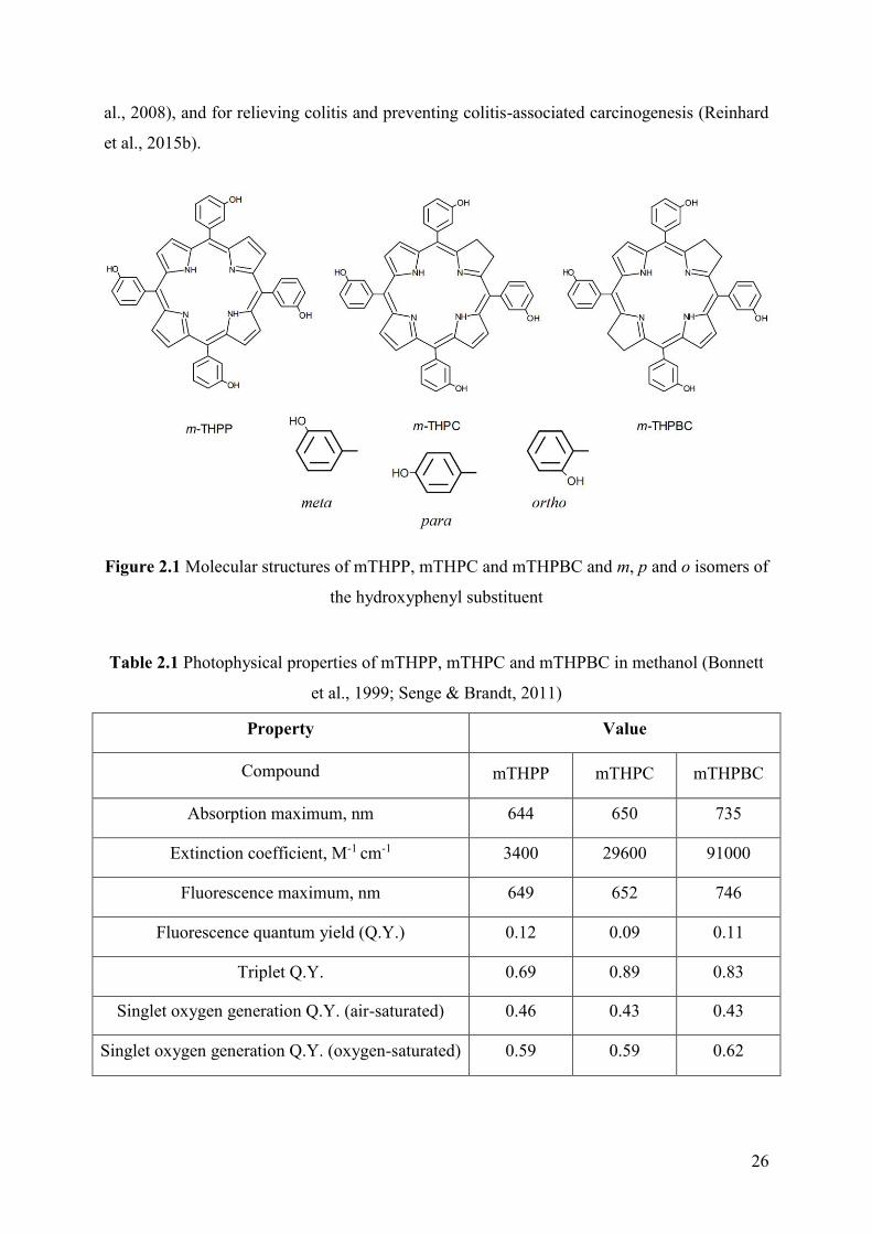
2.1. General properties and usage
5,10,15,20-Tetra(m-hydroxyphenyl)chlorin (mTHPC, temoporfin) is one of the most
potent second generation PS. The discovery and the chemical synthesis of PSs of
tetra(hydroxyphenyl)porphyrinoid series (Fig. 2.1) was performed by the group of professor
Raymond Bonnett (Berenbaum et al., 1986; Bonnett et al., 1989). The ortho, meta and para
isomers (Fig. 2.1) of these compounds have been tested and after thorough comparison the
meta isomers of the PSs demonstrated the greatest efficiency in vitro and in vivo. They are
namely meta-tetra(hydroxyphenyl)porphyrin (mTHPP), mTHPC and meta-
tetra(hydroxyphenyl)-bacteriochlorin (mTHPBC). These PSs have attractive photophysical
characteristics compared with the first generation PSs (Table 2.1). All three compounds have
a high triplet state QY formation ranging between 0.69-0.89 and a good QY of singlet oxygen
formation (0.43-0.45) in aerated methanol (Bonnett et al., 1999). Besides, they are
characterized by a strong absorption in the far red region: 3400 M-1cm-1 at 644 nm for
mTHPP, 29600 M-1cm-1 at 650 nm for mTHPC and 91000 M-1 cm-1 at 735 nm for mTHPBC.
Although the photophysical characteristics of mTHPBC from PDT point of view look more
attractive compared to mTHPC, the rapid degradation of mTHPBC precludes a practical use
in PDT (Grahn et al., 1997; Lassalle et al., 2005).
mTHPC use in PDT is characterized by a very low drug dose (order of 0.1 mg·kg-1),
light intensity (order of 10 J·cm-2) and total PDT doses (light dose x PS dose) more than 100
times higher compared to HpD (Savary et al., 1997, 1998), which explains its popularity in
PDT. Along with Photofrin, mTHPC is the only other PS approved for use in systemic cancer
therapy. In 2001 mTHPC was approved in the EU and used as a solvent-based formulation
(Foscan®; Biolitec Research GmbH, Jena, Germany) for the palliative treatment of head and
neck cancers (Senge & Brandt, 2011). In addition, mTHPC has been successfully used for the
treatment of early squamous cell carcinoma (de Visscher et al., 2013; Jerjes et al., 2011),
basal cell carcinoma (Betz et al., 2012), biliary tract carcinoma (Wagner et al., 2015), prostate
(Swartling et al., 2010, 2016), pancreatic cancer (Huggett et al., 2014) and nonmelanoma skin
cancers (Horlings et al., 2015). Moreover, mTHPC finds application in areas other than
cancer treatment. For examples, there are studies demonstrating the prospects of mTHPC
application for antibacterial therapy (Ossmann et al., 2015), rheumatoid arthritis (Hansch et
26
al., 2008), and for relieving colitis and preventing colitis-associated carcinogenesis (Reinhard
et al., 2015b).
Figure 2.1 Molecular structures of mTHPP, mTHPC and mTHPBC and m, p and o isomers of
the hydroxyphenyl substituent
Table 2.1 Photophysical properties of mTHPP, mTHPC and mTHPBC in methanol (Bonnett
et al., 1999; Senge & Brandt, 2011)
Property Value
Compound
mTHPP
mTHPC
mTHPBC
Absorption maximum, nm 644 650 735
Extinction coefficient, M-1 cm-1 3400 29600 91000
Fluorescence maximum, nm 649 652 746
Fluorescence quantum yield (Q.Y.) 0.12 0.09 0.11
Triplet Q.Y. 0.69 0.89 0.83
Singlet oxygen generation Q.Y. (air-saturated) 0.46 0.43 0.43
Singlet oxygen generation Q.Y. (oxygen-saturated) 0.59 0.59 0.62

27
2.2. Biological behavior of mTHPC
The high PDT-efficiency of mTHPC is usually associated with the features of its
biological behavior. In many papers it is shown that temoporfin forms large aggregates in
aqueous medium (Kruijt et al., 2009; Tikhomirov et al., 2009). As a rule, mTHPC is IV-
introduced to patients and the PS’s hydrophobic nature defines its pharmacokinetics and
biodistribution behavior. Plasma protein binding pattern is an important factor governing
mTHPC biological behavior. Previous studies have revealed two main features of mTHPC
interaction with plasma proteins. First, about 95% of the sensitizer in plasma is associated
with HDL and LDL, and the binding with HDL is twice higher than that with LDL (Reshetov
et al., 2012; Triesscheijn et al., 2007). Secondly, as compared with other non-polar drugs,
mTHPC releases from lipoprotein carriers very slowly and proceeds on a timescale of hours
(Sasnouski et al., 2005, 2006). High affinity to cell membranes and plasma proteins and the
presence of PS aggregates modifies the interaction of mTHPC with biological objects, as will
be more fully described hereinafter.
Interaction with cells in vitro
Time-dependent mTHPC accumulation demonstrates a continuous increase in PS
cellular uptake within the first 20 h, reaching a plateau at 24 h (Berlanda et al., 2010).
Temoporfin is rigidly fixed in model membranes (Kachatkou et al., 2009) and is strongly
retained in cells in vitro (Ball et al., 1999; Berlanda et al., 2010). Cellular uptake of mTHPC
is affected by many factors. Temoporfin seems to be taken up by cells in its aggregated form,
followed by slow monomerization (Rezzoug et al., 1998) attested by fluorescence lifetime
imaging microscopy data (Lassalle et al., 2008). The uptake of PS into cells appears to be pH
independent in the rage 6.8-7.8; however, photosensitivity of cells increased with decreasing
pH value (Ma et al., 1999). As discussed above, due to the high affinity of mTHPC to
lipoproteins, the PS uptake may be mediated by LDL. However, HDL-mediated endocytosis
was proposed as the main mode of temoporfin transport into the cells (Sasnouski et al., 2006).
Primary research reported that mTHPC is localized generally within cellular
organelles, but not in the nucleus (Melnikova et al., 1999). It has been demonstrated that the
Golgi apparatus and endoplasmic reticulum are preferential sites of mTHPC accumulation in
MCF-7 human adenocarcinoma cells (Teiten et al., 2003a) and the latter two were the sites of
primary PDT damage (Teiten et al., 2003b). On the other side, confocal fluorescence
microscopy study showed only weak localization in lysosomes and mitochondria. A study by
Kessel (1999) showed mitochondrial damage, release of cytochrome c and activation of

28
caspase-3 resulting in an apoptotic response after mTHPC-PDT in vitro. Mitochondrial
damage and cytochrome c release has been described for various cancer cells (Marchal et al.,
2004; Yow et al., 2000). However, investigation of the relationship between mTHPC
subcellular localization and post-PDT intrinsic apoptotic pathway demonstrated that
temoporfin localization in endoplasmic reticulum improves the photoactivation of the
caspase-7 apoptotic pathway, which is poorly related to photoinduced mitochondrial damage
(Marchal et al., 2007). Thus, although mitochondria are not affected directly they will
significantly contribute to late and indirect apoptotic effects originating in Endoplasmic
reticulum and/or Golgi apparatus.
Biodistribution and pharmacokinetic properties
The pharmacokinetics of mTHPC depends on using of animal models (Senge &
Brandt, 2011). Early studies showed that mTHPC displays an unusual plasma
pharmacokinetic behavior in humans and rabbits, with a secondary peak at about 10 h and 6 h
after injection, respectively (Glanzmann et al., 1998; Ronn et al., 1996). These phenomena
were interpreted by the initial retention of the PS in the liver or PS aggregates in the
vasculature, with subsequent disaggregation, binding to lipoproteins and mTHPC release from
the depot. Depending on the study and animal models a half-life of the drug of 26–45 h was
established (Campbell et al., 2002; Glanzmann et al., 1998; Ronn et al., 1996; Triesscheijn et
al., 2007). It was shown that mean values for plasma drug concentrations were in good
agreement with the injected dose of mTHPC. Pharmacokinetic study of radiolabeled mTHPC
in tumor-bearing rats demonstrated a tri-exponential model with half-lives of 0.46, 6.91 and
82.5 h, respectively (Jones et al., 2003). However, a detailed study comparing the
pharmacokinetics in mice and humans showed the different pharmacokinetics between
rodents and human (Triesscheijn et al., 2007). The initial (5 min) plasma drug levels in
humans were on average 86% of the maximal plasma concentration, which occurred at about
5 h after injection. On the contrary plasma pharmacokinetics in mice was characterized by a
standard bi-exponential decline of the drug concentration. Besides, the drug distribution over
the lipoproteins and the metabolism of the lipoproteins had no impact on the plasma
pharmacokinetics.
Temoporfin biodistribution studies have shown that the time course of the uptake
differs from organ to organ. Highly perfused organs (liver, spleen, kidney and lung) contained
a large amount of the drug and the less perfused organs contain less of the PS (Campbell et
al., 2002; Whelpton et al., 1996). However, the maximum level of mTHPC can be different

29
for different animal models and doses of administration (Ronn et al., 1996, 1997). The
intratumoral distribution of mTHPC depends on the time of circulation and the distance to
blood vessels. Peng et al. (1995) showed that mTHPC was distributed in the vascular
interstitial and neoplastic cells of the breast cancer implanted in mice. Mitra et al. (2005) used
high-resolution confocal fluorescence imaging to simultaneously map microscopic
intratumoral mTHPC localization with respect to perfused vasculature as a function of time
after injection. Three hours after injection, maximal mTHPC fluorescence was detected in the
periluminal structures, but after 24 h, mTHPC was mainly localized in parenchyma of tumor.
All these aspects have clear clinical applications, which will be discussed below.
A study of mTHPC excretion in a murine model showed that nearly 40% of the drug
was excreted in the faeces during the first day, and less than 0.2% of the dose was recovered
from the urine (Whelpton et al., 1996). Furthermore, in vitro and in vivo experiments, using
HPLC and electrospray mass spectrometry, have confirmed that mTHPC is not metabolized
and is excreted unchanged via biliary excretion in the faeces (Cai et al., 1999a, 1999b).
Mechanism of action
Early pre-clinical studies have demonstrated that in PC6 tumor bearing mice the
depth of necrosis was 3.79 ± 0.28 mm after mTHPC administration (Bonnett et al., 1989).
Melnikova et al. (1999) showed that mTHPC mediates cell photodamage, principally through
singlet oxygen formation and its efficacy is sensitive to oxygenation conditions (Coutier et al.,
2002). Moreover, in the case of mTHPC-PDT, singlet oxygen dose to the tumor volume does
not track even qualitatively tumor response, so in this case any PDT dose metric that is
proportional to singlet oxygen creation and/or deposition would fail to predict the tumor
response (Wang et al., 2008)
mTHPC has a small initial volume of distribution with important retention in the
vasculature together with two peaks of PDT efficacy (2 and 24 h) in rats (Jones et al., 2003).
Thus, PDT at early time of irradiation resulted mainly in the destruction of the
microvasculature of the tumor while the latter one affected both the vascular walls and the
tumor cells. Features of such intratumoral distribution were used in mTHPC-PDT of mice
bearing EMT6 tumors (Garrier et al., 2010). Indeed, a fractionated double injection (3 and 24
h prior to PDT) was superior to any single dose of administration of the drug at 3 h, 6 h and
24 h of DLI. The absence of correlation between the mTHPC concentration in tumor and PDT
efficiency was observed (Garrier et al., 2010; Ris et al., 1998; Veenhuizen et al., 1997), while
accumulation of PS in leukocytes exhibited a good correlation with PDT efficacy (Maugain et

30
al., 2004). Thus, photodynamic effect will depend on the treatment protocol (i.e.
photodynamic dose and DLI). In fine, although direct cells damage is a predominated in
tumor destruction by mTHPC-PDT, other mechanisms play an important role for the
complete tumor destruction and prolonged PDT-effect.
Side effects
mTHPC is a highly active PS with a significant clinical efficacy, however the skin
photosensitivity still remains a major issue in clinical management. For example, there is the
evidence indicating a mild to moderate pain in the treated area (Allison & Sibata, 2004). The
main side effect of mTHPC is its photosensitivity. Although several studies have indicated
that mTHPC is less photosensitivity than Photofrin® (van Geel et al., 1995; Wagnieres et al.,
1998), skin photosensitivity persists for up to 6 weeks (usually 2-3 weeks) post-mTHPC
administration (Allison & Sibata, 2004). Usually, Foscan-based PDT resulted in mild to
moderate skin photosensitivity and caused neither functional nor cosmetic impairments after
treatment of early oral squamous cell carcinoma (O’Connor et al., 2009). There are also other
side effects, but they are very rare and specific (http://sideeffects.embl.de/drugs/60751/pt).
Moreover, as noted earlier, mTHPC molecule is hydrophobic, leading to its poor water
solubility and aggregation within the vasculature requiring the use of special systems suitable
for clinical applications.
2.3. Strategies for improving delivery and efficacy of photodynamic
therapy with mTHPC
The side effects of mTHPC clearly accentuate the need to improve the treatment
protocols by optimizing the DLI, increasing bioavailability and selectivity of PS accumulation
thus reducing the damage to healthy tissues. Various solutions were proposed over the past
decades, including mTHPC conjugation with different molecules and using special
formulations, such as liposomes, polymer solutions and nanoparticles (Senge, 2012; Senge &
Brandt, 2011). The first attempt to improve the pharmacokinetic properties of mTHPC is
related to the conjugation with polyethylene glycol (PEG) (Westermann et al., 1995; Morlet et
al., 1997). Although mTHPC-PEG conjugate showed increased water solubility and selective
retention of the drug in tumor tissue, PDT-effect of unconjugated form of mTHPC
demonstrated the same or superior efficiency (Kübler et al., 2001; Westermann et al., 1999).
Besides, various glucoconjugated analogues of mTHPC were investigated (Bautista-Sanchez
et al., 2005; Laville et al., 2003), along with folic acid conjugate (Gravier et al., 2008).

31
More significant advances were made through the use of nanocarriers for mTHPC
development. In order to improve its bioavailability and PDT-efficacy and to reduce side
effects of mTHPC, various nanosized vehicles such as nanoemulsion (Primo et al., 2008),
lipid vesicle systems (liposomes, invasosome, ethosomes) (Bovis et al., 2012; Chen et al.,
2011; Reshetov et al., 2013; Xie et al., 2015), nanoparticles (mainly biodegradable) (Navarro
et al., 2014; Rojnik et al., 2012; Silva et al., 2015) were proposed. A study of mTHPC
encapsulated in polyethylene glycol (PEG) poly-(D,L-lactide-co-glycolide) nanoparticles
showed significant decrease of mTHPC dark cytotoxicity, but also significantly reduced
mTHPC cellular uptake, whereas phototoxicity was similar to that of Foscan® (Rojnik et al.,
2012). Silva et al. (2015) demonstrated that using mTHPC-loaded magnetic iron oxide
microvesicles, the uptake of mTHPC by cancer cells could be kinetically modulated and
spatially controlled under magnetic field and that photoinduced cell death was enhanced by
magnetic targeting. Another study compared vesicular systems (liposomes, invasosome and
ethosomes) with non-vesicular systems on the skin penetration of mTHPC. Both vesicular and
non-vesicular formulations provided drug localization predominantly in the superficial skin
layer (Chen et al., 2011).
Two mTHPC liposomal formulations (Foslip® and Fospeg®; Biolitec Research
GmbH) are the most studied and widely used in experimental research. Foslip® is a
formulation of mTHPC embedded in conventional (non-pegylated) liposomes. Fospeg® is a
sterically stabilized form of Foslip®, containing a small amount of PEG-
phosphatidylethanolamine layer on the surface. Compared to the standard solution of mTHPC
(Foscan®), liposomal formulations Foslip® and Fospeg® provide better bioavailability of the
PS, higher tumor/tissue ratios and result in better PDT-efficacy (Bovis et al., 2012; Buchholz
et al., 2005; Svensson et al., 2007). PDT efficiency with Foslip® in EMT6-grafted mice,
studied by Lassalle et al. (2009), was maximal at the DLI 6 h, indicating the presence of both
direct and vascular PDT effects. Reshetov et al. (2013) showed that Fospeg®-PDT of HT29
human colon adenocarcinoma bearing mice was more effective than Foslip® treatment at
almost all DLIs, except 24 h. Its higher efficiency can be achieved by combination of
enhanced permeability and retention-based tumor accumulation, stability in the circulation,
and release properties of Fospeg® compared to Foslip® (Reshetov et al., 2013; Xie et al.,
2015).
These different nanocarriers of mTHPC, in particular Foslip has now closely
approached the clinical stage. Further development of PDT in general and mTHPC-PDT in
particular is associated with nanotechnology-based drug delivery systems.
32
3. Nanotechnology-based drug delivery systems
The application of many drugs is hampered by their non-optimal pharmacological
properties, such as low aqueous solubility, irritating nature, lack of stability, rapid
metabolism, and non-selective drug distribution. Nanocarriers were developed to palliate
these problems by improving drug delivery, opening the era of nanomedicine in oncology
including PDT (Calixto et al., 2016; Marchal et al., 2015). Significant efforts have been made
toward this goal by developing nanoparticle drug delivery systems (DDSs), having particle
diameters up to 200 nm (Khodabandehloo et al., 2016). Nanocarriers can deliver drugs in a
spatiotemporally controlled manner that potentially increase therapeutic efficacy of the drugs,
reduce their systemic side effects, and improve patient's adherence to regimen by reducing the
dose and administration frequency (Lee & Yeo, 2015).
3.1. A rational design and advantages of drug delivery systems
It is well known that drugs should ideally be released at the target sites in a
controlled manner to enhance their therapeutic efficiency and reduce the side effects. The
design of an efficient DDS requires the knowledge of the drug physicochemical properties,
specific intended therapeutic application of the drug and the characteristics of interaction of
the DDS with the biological structures (Khodabandehloo et al., 2016).
Smart DDSs are defined as the process when the drugs are not released before
reaching target tissues/organs (or with extremely slow rate), and are released only at the sites
of action (Liu et al., 2016). Although the drug molecules themselves sometimes can be smart
components, here we primarily discuss smart DDSs by means of nanotechnology. Stimuli-
responsive DDS can respond to endogenous and/or exogenous stimulus. The endogenous
triggers such as pH variations, hormone level, enzyme concentration, small bio-molecules,
glucose or redox gradient (Mura et al., 2013; Kelley et al., 2013) are related to the disease
pathological characteristics. While the exogenous triggers, including temperature, magnetic
field, ultrasound, light, electric pulse/ high energy radiation, can also be used to trigger or
enhance the drug release at diseased areas.
For the drug that is already in clinical application, the advantages of a particular DDS
shall be compared to a free drug. Besides, toxicity profile (not degree) of the free drug is
generally similar to its DDS-entrapped form (Allen & Cullis, 2004), thus facilitating the
estimation of the side effects. Hence, in many cases the formulation of the already-approved

33
drug into the DDS is more efficient than the development of entirely new drugs and their
DDS formulation. The mechanism of action of a drug also dictates its suitability for delivery
in a particular DDS. If release from the DDS is required, a question arises as to whether the
DDS leads to appropriate rates and levels of drug bioavailability. Bioavailability will depend
on the release rate. Thus, the methods to measure drug release rates should also be integrated
into the development of DDSs, not only the measurements of the total drug level (e.g., in
plasma).
Although final properties will depend on the particular design of the DDS, there are
several general advantages of using DDS for cancer therapy:
DDSs can carry a large payload of drug molecules, protect them from degradation
and increase drug water solubility.
Changes in the biodistribution of the DDS generally occur through passive or
active targeting. Passive targeting exploits the characteristics of tumor biology
that allow nanocarriers to accumulate in the tumor by the enhanced permeability
and retention effect (Peer et al., 2007). Chauhan et al. (2012) findings emphasize
the importance of size in nanomedicine design by demonstrating that 12 nm
particles penetrate tumors better than larger particles. Active approaches achieve
this by conjugating nanocarriers containing chemotherapeutics with molecules
that bind to overexpressed antigens or receptors on the target cells.
The alteration of pharmacokinetics and biodistribution compared to a free drug
must match the current use of medications. Surface modifications of the DDS
such as PEGylation may dramatically change the circulation time, as discussed in
the next section. Surface characteristics contribute to the DDS solubility,
aggregation tendency, ability to traverse biological barriers (such as the cell wall),
biocompatibility, and targeting ability (Mrsny et al., 2006).
Nanocarriers composed of biocompatible materials are safe alternatives to
existing vehicles such as Cremophor® that may cause severe adverse effects
(Fang & Al-Suwayeh, 2012). Biodegradability implies that the unloaded carrier is
degraded or metabolized into nontoxic components and cleared from the
circulation (Petros & DeSimone, 2010). Thus, toxicities associated with the
carrier molecules per se tend to be mild.

34
3.2. Types of nanosized antitumor drug delivery systems
To increase the effectiveness of treatment and reduce side effects, the incorporation
of nanotechnology-based DDS such as polymeric nanoparticles, solid lipid nanoparticles,
nanostructured lipid carriers, gold nanoparticles, protein-based nanoparticles, cyclodextrin
complexes and host–guest supramolecular nanosystems represents an interesting option for
drug delivery, as demonstrated in Fig. 3.1. These structures are different in the way of drug
incorporation (i.e., conjugation, non-covalent inclusion, enzyme binding), have distinct
structural features (the composition, surface labeling) and size (1-400 nm), as well as different
mechanisms of cellular and tissue targeting (active, passive). Examples of nanotherapeutics
approved for clinical use or subjected to advanced clinical trials are listed in Table 3.1.
Polymeric carriers
Important range of application of polymers is its conjugation with the drug
molecules. Several polymer-drug conjugates have entered clinical trials and some of them,
like Zinostatin stimalamer® and Oncaspar®, are approved at the market (Table 3.1). They are
especially useful to increase water solubility and to target blood vessels in tumors. Apart from
polymers, there are several ways to functionalize drugs, including conjugation to peptides,
saccharides and proteins, which leads to improved bioavailability and delivery specificity (Liu
et al., 2016). However, polymer–drug conjugates require chemical modification of the
existing drugs; as a consequence, their production could cost more, and further purification is
needed. Moreover, the drug-polymer conjugates are usually considered as new chemical
entities owing to a pharmacokinetic profile distinct from that of the parent drugs (Bamrungsap
et al., 2012).
Micelles
Micelles are spherical colloidal amphiphilic systems, having a particle diameter
within the range of 10-100 nm. They consist of self-assembled amphiphilic block copolymers
with a hydrophobic core and hydrophilic shell. Among drug carrier systems, micelles provide
considerable advantages, since they can solubilize and increase the bioavailability of poorly
soluble drugs, and offer long blood circulation (Biswas et al., 2013). Drug release from
micelles is dependent on the interactions between drugs and micellar cores (e.g., hydrogen
bonding) and, therefore, is determined by molecular weight and hydrophobicity of
amphiphilic block copolymers (Lee & Yeo, 2015). To date, several polymeric micelle
anticancer drug-targeting systems are at different stages of clinical studies.

35
Table 3.1 Nanotherapeutics approved for clinical use or being evaluated in clinical trials
(adapted from Egusquiaguirre et al, 2012; Gidwani & Vyas, 2015; Marchal et al., 2015)
Nanocarrier Name Formulation Drug Indications Status
Liposomes
Doxil® PEGylated liposome
Doxorubicin
Karposi's sarcoma Ovarian cancer Multiple myeloma Market
Myocet® conventional liposome Breast cancer (cyclophosphamide) Market
Caelyx® PEGylated liposome
Karposi's sarcoma, multiple myeloma, breast and ovarian cancer Market
ThermoDox® Heat-activated
PEGylated liposome
Hepatocellular carcinoma Phase III
DaunoXome® conventional liposome Daunorubicin Karposi's sarcoma Market
DepoCyt® conventional liposome Cytarabine Lymphomatous meningitis, leukaemia, Market
Marqibo® sulfate liposome Vincristine acute lymphoblastic leukemia FDA 2012
Polymeric and Polymer-drug
conjugate nanoparticles
Zinostatin stimalamer®
conjugate protein or copolymer
styrene-maleic acid Hepatocellular carcinoma Market
Oncaspar® PEG-drug L-Asparaginase acute lymphoblastic leukemia Market
Docetaxel-PNP
Polymeric nanoparticle Docetaxel
Various solid malignancies Phase I
BIND-014 PEG-PLGA nanoparticle Various solid malignancies Phase II
Abraxane® (ABI-007)
Albumin-bound nanoparticle Paclitaxel
Breast cancer, non-small cell lung cancer, Pancreatic cancer Market
Opaxio®
(Xyotax) Polyglutamic
acid-drug Lung cancer, ovarian cancer FDA 2012
XMT-1001 Fleximer®-drug Camptothecin Gastric cancer, lung cancer Phase I NKTR-102 PEG-drug Irinotecan Breast cancer Phase III
Host–Guest Supramolecular Nanosystems
CRLX101 CD-PEG nanoparticle Camptothecin Various malignancies Phase II
CALAA-01 CD-PEG-
transferrin-nanoparticle
Anti-RRM2 siRNA Various solid malignancies Phase I
Vfend SBE-β-CD/drug solution for IV
injection voriconazole Invasive Fungal Infection
Hematological Malignancy Phase III
Inorganic nanoparticles NanoTherm® nanoparticle Iron oxide Glioblastoma Market
Polymeric micelles
Paclical® Polymeric micelle Paclitaxel
Ovarian cancer Phase III
Genexol-PM® PEG-PLA micelle Breast cancer Market
Livatag® Polymeric micelle Doxorubicin Hepatocellular carcinoma FDA 2014
Other drug-conjugated
based nanoparticles
Rexin-G Retroviral-
vector-dnG1 plasmid DNA
G1 gene Various solid malignancies Market/ Phase III
Kadcyla® Antibody-drug Emtansine/trastuzumab Breast cancer Market
Adcetris® Antibody-drug Brentuximab vedotin CD30+ lymphomas Market

36
Figure 3.1 Different types of nanocarriers have used for drug delivery (from Bamrungsap et
al., 2012; Khodabandehloo et al., 2016; Wang et al., 2013)
Liposomes and solid lipid carriers
Constituted by an aqueous core surrounded by one or several phospholipid bilayers,
liposomes are biocompatible and biodegradable entities able to entrap hydrophilic drugs into
their cavity, while allowing water insoluble drugs to be inserted into the lipid bilayers
(Marchal et al., 2015). PEGylation of liposomes can increase circulation time and protect the
cargo during the circulation in the body. Liposomes, together with drug-polymer conjugates,
are the most widely used for DDS. Some liposome formulations have been approved for
commercial use as shown in Table 3.1. Except for liposomes on the market, a number of
lipidic nanoparticles are in the pipeline from concept to clinical application (Liu et al., 2016).
Solid lipid carriers, also referred as lipospheres or solid lipid nanospheres are solid at
human physiological temperature and had a diameter of 50-100 nm. The solid lipid carriers
could be made from diverse range of lipids including mono-, di- and triacylglycerols, fatty
acids, waxes and combination of these materials. The solid lipid carriers could be formed by
replacing oil with solid lipid in oil and water emulsion. Also, the solid lipid carriers were
biodegradable and biocompatible and could be used in humans because of their reduced
cytotoxicity (Wong et al., 2007). These carriers have formed a lipophilic matrix which

37
enabled the drug to load onto it. The important factors affecting drugs loading into the solid
lipid carriers matrix were solubility of drug in lipid (drug must be lipophilic), physical and
chemical properties of lipid or mixture of lipids, crystalline properties of lipids in biological
temperature, and polymorphic form of the lipids. Solid lipid carriers have been investigated
for delivery of various anticancer drugs with promising results in mouse preclinical models
(Khodabandehloo et al., 2016).
Dendrimers
Dendrimers are natural and synthetic polymers with a central structure labeled as
“the core”, from which the branches of other groups called “dendrons” grow through various
reactions (Lee & Yeo, 2015). The unique surface characteristics of dendrimers enable them to
be simultaneously conjugated with several molecules, such as imaging contrast agents,
ligands or therapeutic drugs, and hence have created a multifunctional drug delivery system
(Khodabandehloo et al., 2016). Drug molecules may be covalently conjugated to the
peripheral groups of a dendrimer or included inside the core via hydrogen bonding,
hydrophobic or electro-static interactions. In regards of biomedical applications, the
biological properties of dendrimers are important. For example, cationic dendrimers were
usually hemolytic and cytotoxic. On the other hand, anionic dendrimers had longer circulation
time and higher bioavailability. Dendrimers clearance rate was generation-dependent, and
lower generation would have longer circulation time (Jain & Tekade, 2013). Our laboratory
together with the laboratory of photobiophysics (Berlin, German) demonstrated that
dendrimer formulations of chlorin e6 may offer effective biocompatible nanoparticles for
improved PDT in a preclinical tumor model (Bastien et al., 2015; Pfitzner et al., 2016)
Host–guest supramolecular nanosystems
The typical supramolecular hosts (i.e., cyclodextrins, calixarenes, and cucurbiturils)
and their nanosystems have a promising application in cancer diagnostics and therapeutics
(Gidwani & Vyas, 2015; Wang et al., 2013). A few examples have been reported where the
supramolecular nanosystems could serve as cancer imaging probes and/or drug delivery
vehicles in clinical trials (Davis, 2009; Davis et al., 2010). The use of DDSs, based on
cyclodextrins or its colloidal silica nanoparticles, represents a promising and innovative
strategy that enables effective therapy with minimum side effects. The focus of the following
sections will be on cyclodextrins as one of the most promising delivery systems.

38
4. Cyclodextrins for drug delivery
The inclusion complexes of cyclodextrins (CDs) with wide variety of organic and
non-organic compounds refer to a class of the simplest supramolecular systems extensively
studied for the last decades. The inclusion complexes based on CDs are of interest from both
a theoretical and a practical point of view. On the one hand, their study facilitates
understanding of the mechanisms of interaction of molecules and molecular recognition, on
the other hand the unique properties of CDs enables using them as additives to improve the
properties of various substances (Challa et al., 2005). As a result CDs have been explored as
additives in the food, cosmetic and pharmaceutical industries to improve product stability and
solubility (Loftsson & Brewster, 2010). Applications of CDs in the pharmaceutical industry
are constantly expanding. To date there are above 40 products or formulations containing
various CDs, especially β-CD and its derivative (Gidwani & Vyas, 2015). Moreover, CDs are
promising delivery systems for PDT purpose (Kryjewski et al., 2015; Mazzaglia, 2012).
4.1. Cyclodextrins: classification, structure and basic properties
The cyclodextrins (CDs) of biomedical and pharmaceutical interest are a family of
cyclic oligosaccharides with a hydrophobic internal cavity and a hydrophilic outer surface
consisting of six α-cyclodextrin (α-CD), seven β-cyclodextrin (β-CD), eight γ-cyclodextrin (γ-
CD) D-glucopyranose units linked by α -(1,4) bonds. The general properties of the native CDs
are summarized in the Table 4.1. These so-called “parent CDs” have been used in food (e.g.,
β-CD approved as a food additive E459) and pharmaceutical products (including anticancer
drugs) for many years (Gidwani & Vyas, 2015). CDs are also known by other names
(cycloamyloses, cellulosine, cyclomaltoses, etc.) (Loftsson & Duchêne, 2007). Usually CDs
are formed as a result of intermolecular transglycosylation reaction (Szejtli, 1998). Due to the
steric hindrances CDs with fewer than 6 units cannot be formed. On the other hand, the higher
than eight glucopyranose units do exist but are of limited interest as drug solubilizers and
stabilizers because they are rapidly reduced to smaller products (Loftsson & Brewster, 1996).
Due to the chair structure of the glucopyranose molecules, CDs have the shape of a
truncated cone (Table 4.1). According to X-ray study, the secondary hydroxyl groups (C2 and
C3) are located on the wider rim and the primary hydroxyl groups (C6) on the narrow side
(Kusmin et al., 2008). It should be noted that the primary hydroxy groups can rotate, while the
secondary ones are relatively fixed. The apolar C3 and C5 hydrogens are located in the cavity

39
of CDs (Del Valle 2004). As a result CD molecules have a hydrophilic outside (LgKoctanol/water
between (−13) and (−17)), and an apolar cavity, which provides a hydrophobic matrix
comparable to about a 60–70% aqueous ethanolic solution (Connors, 1997; Loftsson &
Brewster, 2013). The presence of a hydrophobic interior cavity allows forming inclusion host-
guest complexes with a large variety of organic and non-organic compounds. While the height
of the CDs torus remains constant, the diameter of the cavity increases considerably with
increase in the number glucopyranose units (Table 4.1). It is worth noting that despite the fact
that β-CD has been more widely used in the pharmaceutical applications than other native
CDs (Rajewski &Stella, 1996), for each specific drug, it is necessary to select the type of CD,
depending on the size of the guest molecules or its side substituents. Thus, molecules with
aliphatic chains fit better into the small α-CD cavity, whereas molecules containing phenyl
groups fit better into the larger cavity of either β- or γ-CD (Duchêne, 2011; Gabelica et al.,
2002)
An important characteristic in the pharmaceutical industry is CDs solubility in water.
In the case of native CDs solubility changes in the range 18-250 mg/ml (Table 4.1) that is
much lower than that of similar acyclic saccharides (Duchêne, 2011). Relatively low water
solubility of CDs, especially of β-CD, significantly reduces their applicability. Chemically
modified CD derivatives can overcome these problems and extend the physicochemical
properties and inclusion capacity of parent CDs. These derivatives usually are produced by
aminations, esterifications or etherifications of primary and secondary hydroxyl groups of the
CDs (Szente & Szejtli, 1999). Almost all derivatives have a changed hydrophobic cavity
volume and can improve solubility, physical and microbiological stability, reduce parenteral
toxicity and help to control the chemical activity of guest molecules (Challa at al., 2005;
Davis & Brewster, 2004).
Structural and physicochemical properties of some commercially available CD
derivatives of pharmaceutical interest are summarized in the Table 4.2. The physicochemical
properties of CDs, including their complexation ability, depend on the structure of the
attached substituents but also on their number and location within the CD molecule. The
molar degree of substitution (MS) is defined as the average number of substituents per one
glucopyranose repeat unit (Table 4.2). However, the “degree of substitution” does not
uniquely characterize a CD derivative due to the variation of possible locations of substituted
groups (e.g., hydroxypropyl) at different positions on the parent CD molecule (Blanchard &
Proniuk, 1999).

40
Table 4.1 Structure and physicochemical properties of native CDs (adapted from Loftsson & Brewster 2013; Del Valle 2004)
Property α-Cyclodextrin β-Cyclodextrin γ-Cyclodextrin
Number of glucopyranose units: 6 7 8
Molecular weight of anhydrous compound (Da): 973 1135 1297
Melting temperature range (°C): 255–260 255–265 240–245
Crystal water content (% w/w): 10.2 13.0–15.0 8–18
Height of torus (nm): 0.78 0.78 0.78
Inner diameter (nm): 0.50 0.62 0.80
Outer diameter (nm): 1.46 1.54 1.75
Cavity volume (nm3) 0.174 0.262 0.427
Solubility in water at 25 °C (mg/ml): 129.5 ± 0.7 18.4 ± 0.2 249.2 ± 0.2
Calculated LgK(octanol/water) at 25 °C: −13 −14 −17

41
Table 4.2 Structural and physiochemical properties of some CDs of pharmaceutical interest
(adapted from Loftsson & Brewster, 2013; Vyas et al., 2008)
Cyclodextrin n R MSa MWb (Da)
Solubilityc (mg/ml)
LgKo/wd
2-Hydroxypropyl-α-cyclodextrin (HP-α-CD)
6 -CH2CHOHCH3
or -H 0.65 1199 >500 < -10
2-Hydroxypropyl-β- cyclodextrin (HP-β-CD)
7 -H2CHOHCH3
or -H 0.65 1400 >600 -11
Sulfobutylether-β- cyclodextrin sodium salt
(SBE-β-CD) 7
-(CH2)4SO2–Na+
or -H 0.9 2163 >500 < -10
Randomly methylated-β-cyclodextrin (M-β-CD)
7 -CH3 or -H 1.8 1312 >600 -6
Maltosyl β-cyclodextrin (Mal-β-CD)
7 Maltaosyl-
or -H 0.14 1459 >500 < -10
2-Hydroxypropyl- γ -cyclodextrin(HP-γ-CD)
8 -H2CHOHCH3
or -H 0.6 1576 >600 -13
aMolar degree of Substitution : average number of substitution (R) per glucose repeat unit;
derivatives may have differing degrees of substitution on the 2, 3, and 6 positions (inserted figure); bMW, molecular weight; cSolubility in pure water at approx. 25 °C; dPartition coefficient octanol/water;
n, number of glucopyranoside units
In aqueous solutions CDs are chemically stable under neutral and basic conditions
and they are more resistant to both enzymatic and non-enzymatic hydrolysis than the linear
sugars (Frömming & Szejtli, 1994). The stability of CDs toward acid hydrolysis depends on
the temperature and acidity. Under normal experimental conditions (pH higher than 3.5 and
temperature below 60 °C), CDs are fairly stable (Hirayama et al., 1992). Besides, CDs are
quite resistant to light within the ultraviolet-visible spectrum and infrared ranges (Loftsson &
Brewster, 1996).
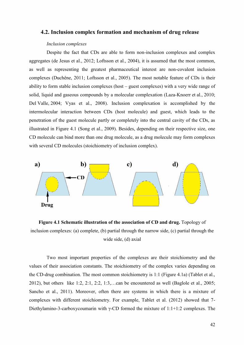
42
4.2. Inclusion complex formation and mechanism of drug release
Inclusion complexes
Despite the fact that CDs are able to form non-inclusion complexes and complex
aggregates (de Jesus et al., 2012; Loftsson et al., 2004), it is assumed that the most common,
as well as representing the greatest pharmaceutical interest are non-covalent inclusion
complexes (Duchêne, 2011; Loftsson et al., 2005). The most notable feature of CDs is their
ability to form stable inclusion complexes (host – guest complexes) with a very wide range of
solid, liquid and gaseous compounds by a molecular complexation (Laza-Knoerr et al., 2010;
Del Valle, 2004; Vyas et al., 2008). Inclusion complexation is accomplished by the
intermolecular interaction between CDs (host molecule) and guest, which leads to the
penetration of the guest molecule partly or completely into the central cavity of the CDs, as
illustrated in Figure 4.1 (Song et al., 2009). Besides, depending on their respective size, one
CD molecule can bind more than one drug molecule, as a drug molecule may form complexes
with several CD molecules (stoichiometry of inclusion complex).
Figure 4.1 Schematic illustration of the association of CD and drug. Topology of
inclusion complexes: (a) complete, (b) partial through the narrow side, (c) partial through the
wide side, (d) axial
Two most important properties of the complexes are their stoichiometry and the
values of their association constants. The stoichiometry of the complex varies depending on
the CD-drug combination. The most common stoichiometry is 1:1 (Figure 4.1a) (Tablet et al.,
2012), but others like 1:2, 2:1, 2:2, 1:3,…can be encountered as well (Baglole et al., 2005;
Sancho et al., 2011). Moreover, often there are systems in which there is a mixture of
complexes with different stoichiometry. For example, Tablet et al. (2012) showed that 7-
Diethylamino-3-carboxycoumarin with γ-CD formed the mixture of 1:1+1:2 complexes. The

43
efficiency of complexation is determined by the values of association constants also known as
affinity constant, stability constant or binding constant. The value of association constant
correlates with the stability of the inclusion complexation (lower probability of dissociation).
In the case of Drug/CD 1:1 complexes the values of the binding constants are usually between
10 and 103 M−1 and values > 105 M–1 are very rare (Stella & He, 2008). One of the few
examples of high value of the binding constant (about 107 M–1) is the complexes of
rocuronium with sugammadex (modified γ-CD) (Adam et al., 2002).
Inclusion complexation with CDs is a reversible process (dynamic equilibrium), i.e.
each molecule can dissociate from the complex. There are two key factors influencing the
ability of CDs to form inclusion complex with a guest molecule: steric and thermodynamic.
Complex formation is a dimensional fit between host cavity and guest molecule (Muñoz-
Botella et al., 1995). It is important that no covalent bonds are broken or formed during
formation of the inclusion complex (Schneiderman & Stalcup, 2000). The driving forces for
the complex formation include release of enthalpy-rich water molecules from the cavity,
electrostatic interactions, van der Waals interactions, hydrophobic interactions, hydrogen
bonding, release of conformational strain and charge-transfer interactions
(Loftsson et al., 2005; Liu & Guo, 2002).
It is important to note that the physicochemical properties of free drug molecules are
different from those bound to the CD molecules. Any changes in the physico-chemical
properties of the guest molecules through complexation with CDs allow determining the
stoichiometry of the complexes and the value of binding constants (Del Valle, 2004). These
modifications include changes in solubility (Higuchi & Connors, 1965), changes in chemical
reactivity (Másson at al., 1998), changes in photophysical properties (Lang et al., 2004),
nuclear magnetic resonance (NMR) chemical shifts (Pessine et al., 2012), changes in drug
retention (e.g., in liquid chromatography), changes in the physicochemical properties of
aqueous complexation media (e.g., measurements of conductivity, viscosity), etc. Although
there are many techniques, information on the structure of the complexes can be
experimentally obtained only from 2D-NMR and induced circular dichroism spectra.
Especially interesting is the last one. Circular dichroism is essentially an absorption
phenomenon occurring when an optically active molecule absorbs to different degrees the
right and left components of a circularly polarized light beam (Berova et al., 2000). Optically
active molecules of CDs themselves do not show any significant ultraviolet absorbance and
do not fluoresce. Being symmetrical molecules, CDs have no dichroic activity, but they can
modify that of guest molecules by perturbation of the microenvironment polarity resulting

44
from the inclusion (Duchêne, 2011; Ventura et al., 2006). The formation of an inclusion
complex between an optically inactive molecule and CDs may result in the appearance of an
induced circular dichroism (Desroches et al., 2001; Nablet et al., 2012).
It is worth noting that the efficiency of the formation of inclusion complexes is
determined by many factors, including temperature, solvents composition, amount of water
(the degree of complexes dilution), complexation techniques. Ionization of the guest, salt
formation, formation of metal complexes, application of supercritical fluids and addition of
organic co-solvents to the drug/CDs aqueous solution will, if the conditions are correct, lead
to enhanced complexation efficiency (e.g., enhanced solubility) (Arun et al., 2008; Loftsson
& Duchêne, 2007; Van Hees et al., 1999).
4.3. Effects of cyclodextrins on drug properties in cyclodextrin-based
formulations
Inclusion in CDs exerts a profound effect on physicochemical properties of guest
molecules as they are temporarily locked or partially caged within the host cavity giving rise
to beneficial modification of guest molecule properties. Indeed, CDs increase drug solubility,
dissolution, circulation time thus enhancing their bioavailability (Acartürk & Çelebi, 2011).
4.3.1. Effect on drug solubility, dissolution and stability
Drug solubility and dissolution
The most common application of CDs in pharmacy and other fields is related to their
ability to increase solubility of poorly water-soluble substances. The increased apparent
solubility allows solution-based dosage forms, such as parenteral (IV) and peroral
formulation. Besides, improving the apparent solubility of a drug through CDs complexation
can increase its dissolution rate and, for compounds whose oral bioavailability is limited by
solubility or dissolution rate, can increase oral bioavailability (Acartürk & Çelebi, 2011;
Ahuja et al., 2011). For example, the solubility of cortisone acetate increased from 0.039 g/L
to 7.382 g/L (almost 200 times) in the presence of HP-β-CD that is far superior to
dimethylformamide and ethanol (Kalogeropoulos et al., 2009).
The use of CDs is usually preferred over organic solvents from both a toxicological
perspective and solubility mechanism. In the case of organic solvents or co-solvent cocktails,
significant drug solubility in aqueous solution is occurred at sufficiently high content of

45
organic solvent (> 30-40%) and solubility depends in a very nonlinear fashion on water
quantity (Stella & He, 2008).
CDs may acting as tablet dissolution enhancers for drugs at high dose (with which
use of a drug/CD complex is difficult) e.g., paracetamol (Tasić et al., 1992). Out of some
commercially available CDs, methylated CDs with a relatively low molar substitution appear
to be the most powerful solubilizers (Challa et al., 2005). The water solubility enhancement
factor of melarsoprol was about 7.2·103 times greater when the drug was complexed with M-
β-CD (Gibaud et al., 2005).
Reduction of drug crystallinity upon complexation with CDs also contributes to the
CD increased apparent drug solubility and dissolution rate (Becket et al., 1999; Vyas et al.,
2008). For example, during storage carbamazepine strongly crystallizes that significantly
reduces predictable dissolution and bioavailability. Introduction of Hp-β-CD, as a carrier,
reduced crystalline nature of carbamazepine in solid dispersions and resulted in better and
predictable dissolution profiles (Londhe & Nagarsenker, 1999). Furthermore, CDs can
significantly accelerate the drug release, for example β-CD and their derivatives enhanced the
release rate of poorly soluble naproxen and ketoprofen from inert acrylic resins and
hydrophilic swellable (high-viscosity hydroxypropyl methyl cellulose) tableted matrices (Ann
et al., 1997; Bettinetti et al., 1992; Sangalli et al., 2001).
Drug stability
CDs can improve both chemical and physical stability of drugs. CDs reduce the rate
of drug hydrolysis, oxidation, steric rearrangement, racemization, isomerization and
polymerization as well as enzymatic and photo-decomposition of labile drugs thus increasing
the shelf life of drugs (Ahuja et al., 2011; Babu & Pandit, 1999; Li et al., 2002; Loftsson &
Brewester, 1996; Lutka, 2002). Ansari et al. (2009) demonstrated that dihydroartemisinin/HP-
β-CD complexes prepared using commercial (untreated) or recrystallized dihydroartemisinin
showed a 40% increase in thermal stability and a 29-fold decrease in hydrolysis rates
compared to dihydroartemisinin. It was reported that CD-induced enhancement of drug
stability may be a result of inhibition of drug interaction with vehicles and/or inhibition of
drug bioconversion at the absorption site (Matsuda & Arima, 1999). By providing a molecular
shield, CD complexation encapsulates labile drug molecules at the molecular level and thus
insulates them against various degradation processes (Arima et al., 2011). Since the
hydrolysis of drugs encapsulated in CDs is slower than that of free drugs (Brewster et al.,

46
1992), the magnitude of the complex stability constant plays a significant role in determining
the extent of protection (Kang et al., 2002; Nagase et al., 2001).
The stabilizing effect of CDs depends on the nature and effect of the included
functional group on the drug stability and the nature of the vehicle. The large charged groups
on SBE-β-CD showed greater stability enhancement of many chemically unstable drugs than
other CDs including the Hp-β-CD and M-β-CD (Ma et al., 2000; Ueda et al., 1998). From the
solubility of phenytoin and the kinetic information, the fosphenytoin shelf life was as high as
nine years at 25 °C and pH 7.4 in the presence of 60 mM of SBE7m-β-CD, although increasing
the pH to 8 could increase the shelf life fosphenytoin even more (Narisawa & Stella, 1998).
However, there are enough examples that CDs catalyze the degradation of some drug
molecules (Jarho et al., 2000; Narisawa & Stella, 1998; Tonnesen et al., 2002). The effects of
CDs on the rate of drug degradation are pH-sensitive and depend on ionization stage of the
drug (Loftsson & Brewster, 2013).
4.3.2. Effect on drug permeation through biological membranes
CDs can enhance the permeation of poorly water-soluble drugs through different
biological membranes (Másson et al., 1999). For example, permeation studies of omeprazole
indicated a 1.7-fold increase in drug permeation when complexed with M-β-CD (Figueiras et
al., 2009). Only relatively lipophilic molecules can penetrate through the biological
membranes. Through CD complexation it is possible to increase aqueous solubility of drugs
without affecting their intrinsic abilities to penetrate through lipophilic membranes. However,
the effect of CDs cannot be explained by only increasing of the drug solubility in the aqueous
solution or by assuming that CDs act as classical permeation enhancers (reducing the barrier
function of the lipophilic membrane). The CDs allow delivering the drug to the biological
membrane via the water phase. In general, the resulting unstirred water layer formed by water
molecules in the skin is relatively thin and permeable (Wertz, 2004). On the other hand,
mucosal epithelium is covered by the layer of mucus, a gel-like fluid containing mainly water
(∼95%) and mucin (Bansil & Turner, 2006). Researchers have shown that passive drug
diffusion through mucus is up to 100-times slower than through pure water (Khanvilkar et al.,
2001).
Complexation of hydrophobic drugs with CDs makes them available at the surface of
the biological barrier (e.g., eye cornea, skin). Besides, inclusion complexes permeate into the
membrane without disrupting the lipid layers of the barrier. After analyzing the existing

47
studies (Ahuja et al., 2011; Khanvilkar et al., 2001; Loftsson et al., 2007; Loftsson &
Brewster, 2013), the following patterns can be identified:
introduction of excess amount of CDs, more than is needed to solubilize the drug,
leads to decreasing drug permeation. Study by Másson et al. (1999) demonstrated
that the flux of hydrocortisone at different M-β-CD and Mal-β-CD concentrations
increased sharply with increase of CD concentration and decreased when CDs
were in excess;
only with considerable resistance of unstirred water layer (equal or greater than
resistance of the membrane), the use of CDs can improve the delivery of drugs
across biological membranes;
CDs allow better penetration through the skin surface only water-based dermal
vehicles, i.e. requires the presence of unstirred water layer at the membrane
surface. For example, CDs enhance dermal delivery of hydrocortisone from o/w
(oil in water) creams but not w/o creams (Preiss et al., 1995);
CDs themselves and their inclusion complexes do not penetrate biological
membranes. Therefore drug molecules have to be released from the complexes
before they can permeate the membranes;
permeation of labile drugs may be increased through CD-stabilization, i.e. CDs
are able to prevent enzymatic degradation of drugs at the exterior aqueous
membrane (Majumdar & Srirangam, 2009);
usually, CDs do not enhance membrane permeation of water-soluble drugs;
CDs can significantly increase the solubility and enhanced permeation of ionized
drugs. For example, the flux of the ionized ketoprofen at 10% w/v HP-β-CD
concentration was enhanced approximately 8 times compared to the intrinsic
permeability of the nonionized drug (Sridevi & Diwan, 2002).
4.3.3. Effect on drug bioavailability and safety
CDs enhance the bioavailability of wide range of drugs (especially hydrophobic) by
increasing the drug solubility, dissolution, and/or drug permeability. Literature data have
shown that CDs are used to increase oral (Corti et al., 2008), nasal (Tas et al., 2009), ocular
(Cappello et al., 2001), rectal (Değim et al., 2005) and dermal–transdermal (Kear et al., 2008),
bioavailability. The mechanisms enhancing bioavailability of the drugs complexed with CDs
are summarized as follows (Acartürk & Çelebi, 2011; Ahuja et al., 2011; Davis & Brewster,

48
2004; Miyake et al., 2000; Rajewski & Stella, 1996; Stella et al., 1999; Vyas et al., 2008;
Uekama et. al., 2006; Uekama, 2004):
increasing the solubility, dissolution rate, and wettability of hydrophobic drugs in
inclusion complexes;
CDs prevent the degradation of chemically unstable drugs, as well as during
storage;
competitive inclusion complexation with endogenous components of blood (e.g.,
cholesterol) to release the included drug;
CDs disturb the membranes fluidity lowering their barrier function, which
consequently enhances the absorption of medication through the mucosa;
CDs can also allow reducing drug-related irritation and masking bitter tastes;
CDs (e.g., DM-β-CD) can affect multidrug efflux pump, P-glycoprotein (P-gp)
and multidrug resistant-associated protein 2 enabling oral bioavailability of
hydrophobic drugs (e.g., tacrolimus and vinblastine) (Carrier et al., 2007;
Yunomae et al., 2003).
Various factors associated with CD complexation affect the bioavailability of drugs:
(1) the binding constant, (2) the type of CDs, (3) the properties of the drug, (4) the
complexation methods and (5) the composition of ingredients used in formulation (Acartürk
& Çelebi, 2011). For example, the high association constant (106 M-1) between ozonide
antimalarial and SBE7-β-CD resulted in slow dissociation of the complex after IV
administration (Charman et al., 2006). It causes significant alteration of pharmacokinetics of
the ozonide expressed as an 8.5-fold decrease in the steady-state blood volume of distribution,
a 6.6-fold decrease in the mean residence time and a greater than 200-fold increase in renal
clearance of the drug. Moreover, CDs with such a high association constant can alter the
pharmacokinetics and bioavailability of the already administered drug through an additional
injection of CDs. (Mannila et al., 2012). In addition, CDs also prevent crystallization of active
ingredients by complexing individual drug molecules so that they can no longer self-assemble
into a crystal lattice (Arima et al., 2001). Thus, CDs can enhance the oral bioavailability of
drugs in different ways (Challa et al., 2005).
The increased drug efficacy and potency (i.e., reduction of the dose required for
optimum therapeutic activity), caused by CD-increased drug solubility, may reduce drug
toxicity by making the drug effective at lower doses. For example, β-CD enhanced the
antiviral activity of ganciclovir on human cytomegalovirus clinical strains and the resultant

49
increase in the drug potency reduced the drug toxicity (Nicolazzi et al., 2002). The toxicities
associated with crystallization of poorly water-soluble drugs in parenteral formulations can
often be reduced by formation of soluble drug/CD complexes (Blanchard et al., 2000).
Finally, inclusion of drugs into CDs cavity at the molecular level prevents their direct contact
with biological membranes and thus reduces their side effects and local irritation with no
drastic loss of therapeutic benefits (Challa et al., 2005; Uekama et al., 1998).
4.4. Pharmacokinetics, biological activity and toxicology of
cyclodextrins
As shown earlier, CDs have a great potential in the pharmaceutical field due to their
ability to improve the drug properties. However, application of CDs as drug delivery systems
requires knowledge of its pharmacokinetic and pharmacodynamic properties including in
vitro cellular interactions and in vivo safety profiles. Moreover, CDs have ability to interact
with various endogenous and exogenous molecules (e.g., cholesterol), and affect cellular
processes. We shall further consider the most important properties of the cyclodextrin
behavior in biological systems.
Pharmacokinetics and biodistribution
Pharmacokinetics of CDs and their excretion from the body will be defined inter alia
by route of administration. However, there are general patterns, which will be described, for
example, for oral and parenteral routes of CDs administration. Due to the relatively high
molecular weight (from 973 to over 2000 Da) and hydrophilic nature only insignificant
amounts of intact CDs are absorbed from the gastrointestinal tract, thus, their oral
bioavailability is generally below 3% (Loftsson & Brewster, 2011). The absorbed CDs are
rapidly excreted with the urine without any significant metabolism. The native α-CD and β-
CD are practically resistant to stomach acid and pancreatic enzyme digestion, whereas γ-CD
is metabolized partly by amylases in the gastrointestinal tract. The oral bioavailability of Hp-
β-CD in humans is between 0.5 to 3.3% depending on the dosage and formulation (Gould &
Scott, 2005; Szathmary et al., 1990; Zhou et al., 1998). Besides, more than half of the initial
oral dose is excreted intact in the feces (Loftsson & Brewster, 2013). At the same time more
lipophilic methylated CDs have a strong absorption in the body. According to Mosher &
Thompson (2007) the absorption of DM-β-CD in rats is 6.3% to 9.6% and in the case of M-β-
CD can reach 15% (Antlsperger & Schmid, 1996).
50
Parenteral administration of CDs, on the other hand, can be somewhat more limited.
IV-injected CDs eliminate rapidly from systemic circulation and more than 90% of introduced
CDs are renally excreted in intact form (Stella & He, 2008). For example, IV-administrated to
humans, Hp-β-CD has a relatively small volume of distribution VD ≈ 200-250 mL/kg and
short half-life t1/2≈1.8 h, and is mainly excreted unchanged with the urine (Szathmary et al.,
1990; Zhou et al., 1998). Rapid elimination and a small volume of distribution are inherent
for the majority of CDs administered to animals or humans (see Table 4.3). The small VD
indicates that systemically absorbed CDs distribute mainly in the extracellular compartments
without involving deep compartments or storage pools (Stella & He, 2008). However, due to
the ability to interact with components of blood and cellular membranes, the methylated β-CD
has a larger volume of distribution (VD ≈ 2.5 L/kg) and longer half-life (t1/2 ≈7 h) compared
to other CDs derivatives (Thompson, 1997). For parenterally administered Hp-β-CD in
humans, a non-saturable excretion mechanism is observed even at high doses. The
pharmacokinetics of SBE-β-CD is very similar to that of Hp-β-CD. Indeed, the total plasma
clearance for HP-β-CD and SBE-β-CD in all tested species is similar to the glomerular
filtration rate (Davies & Morris, 1993; Rajewski & Stella, 1996) and almost all introduced
CDs are excreted in the urine within 12 h (Zhou et al., 1998), except methylated β-CD. For
example, M-β-CD persists for at least 6 days after a single IV administration (10 mg /kg) and
is almost at the same level in the renal cortex (50-70% of all absorbed CD according to
radioactivity study) as at 24 h after injection (Antlsperger & Schmid, 1996). In this regard, it
is necessary to use CDs with extreme caution in patients with renal failure, because CDs, in
this case, will accumulate in the body (Slain et al., 2001; von Mach et al., 2006).
The main distribution areas of CDs after their absorption include the following
tissues: kidney, bladder, adrenal glands and liver (Antlsperger & Schmid, 1996; De Bie et al.,
1998; Gerloczy et al., 1990; Kubota et al., 1996; Van Ommen et al., 2004). The highest
concentration of absorbed CDs is in the kidneys. In regards to biodistribution in tumor-
bearing rodents, the results of several studies have showed that a significant amount of
methylated CDs is distributed in the tumor tissue, e.g. primary effusion non-Hodgkin
lymphoma (Gotoh et al., 2014), and various carcinoma in xenograft tumor bearing mice
(Grosse et al., 1998; Onodera et al., 2013a). Grosse et al. (1998) have showed that M-β-CD
after 3 hours of parenteral administration in MCF7 xenografts was accumulated mostly in
tumor and kidneys and a small quantity in intestine and liver. Concentrations in lungs and
muscle were almost undetectable.

51
Table 4.3 Pharmacokinetic parameters of some CDs IV administered
(from Stella & He, 2008)
Cyclodextrin Species Dose
(mg/kg)
t1/2
(min)
VD
(mL/kg)
Plasma
clearance
(mL/min/kg)
β-CD Rat 50 21.6 201.8 7.53
HP-β-CD Rat 200 23.9 194 7.2
HP-β-CD Human 43 102 222 1.57
HP-β-CD Human 114 114 253 1.54
SBE-β-CD Rat 600 18 300 9.8
SBE-β-CD Rabbit 600 30 200 5.2
SBE-β-CD Dog 240 66 430 4.7
SBE-β-CD Human 100 84 200 1.9
M-β-CD Rabbit 200 420 2500 3.8
Toxicology of CDs
To date, there are a huge number of publications, including a detailed review of the
toxicological status of CDs and their biological activity (Antlsperger & Schmid, 1996; Arima
et al., 2011; Irie & Uekama, 1997; Gould & Scott, 2005; Loftsson & Brewster, 2011; Mosher
& Thompson, 2007; Thompson, 1997; Zidovetzki & Levitan, 2007). We shall further briefly
review the toxicology issues.
Toxicological studies of various CDs have been conducted following different
administration routes in numerous formulations. In the case of oral administration of CDs the
following abnormalities were observed: soft feces or diarrhea, cecal enlargement on oral
administration, and renal effects subsequent to systemic absorption (Mosher & Thompson,
2007; Thompson, 1997). The major findings following parenteral administration of CDs at
high doses or during a long period of application include vacuolation of hepatocytes and
urinary bladder and foamy macrophages in the liver, lung, and lymph nodes (Stella & He,
2008). With the exception of α-CD, β-CD, and methylated-β-CD, no evidence of
inflammatory responses, cell death, and cell degeneration or regeneration is observed in
studies reported to date (Mosher & Thompson, 2007). Furthermore, in the case of γ-CD, Hp-

52
β-CD, and SBE-β-CD vacuolation is reversible after discontinuation of the CDs (Frijlink et
al., 1991a; Rajewski & Stella, 1996).
Since kidneys are the main absorption site of CDs in the body, the renal toxicity
should be carefully considered. The main effect of CDs in the kidney is vacuolation of
proximal tubular epithelium. Many studies have demonstrated that CDs, especially β-CD and
its methylated derivatives cause renal toxicity (Frank et al., 1976; Irie & Uekama, 1997;
Thompson, 1997). These changes begin with an increase of apical vacuoles and the
appearance of giant lysosomes. Prominent acicular microcrystals are observed in lysosomes.
These changes are followed by disruption of mitochondria and other organelles, and
eventually, irreversible cell injury and kidney dysfunction occurs. The effective dose of M-β-
CD to induce kidney damage is even lower than that of β-CD (Antlsperger & Schmid, 1996;
Irie & Uekama, 1997). The renal toxicity may be caused by CDs according to the following
scheme: (1) IV-injected cyclodextrin binds endogenous cholesterol and some phospholipids
(also CDs may extracted its from the cells); (2) glomerular filtration of the cholesterol-
cyclodextrin complex is followed by dissociation of the complex in the ultrafiltrate, resulting
in cholesterol accumulation in the proximal tubule cells; (3) due to limited aqueous solubility
of the complexes in renal tissue, the complexes crystalize and formed needle-like crystals
cause kidney damage (Frijlink et al., 1991a). However, CDs like γ-CD, Hp-β-CD, and SBE-β-
CD may be used in high doses without causing any significant effects on the human kidney.
For example, IV-introduction of Hp-β-CD in healthy volunteers has been studied at a single
dose up to 3 g and was well tolerated (Gould & Scott, 2005). CDs such as Hp-β-CD and SBE-
β-CD are widely used in marketed parenteral drug formulations (Kurkov et al., 2012).
It should be noted that at the joint introduction of CDs with drugs, a decrease of
toxicological effects of both substances was reported (Arima et al., 2011; Mosher &
Thompson, 2007). For example, IV injection of M-β-CD (5 mg/kg) to tumor-bearing mice
significantly increased blood urea nitrogen, aspartate aminotransferase and lactate
dehydrogenase levels, compared to control, suggesting induction of systemic side effects
(Onodera et al., 2013a). On the other hand, co-administration of doxorubicin (5 mg/kg) with
M-β-CD (12 mg/kg) significantly reduces systemic side effects of both compounds (in the
case of M-β-CD compared to 5 mg/kg dosage) (Motoyama et al., 2013).
Biological activity
Numerous studies have shown that a variety of cellular functions are affected when
cells are treated by β-CD and its derivatives. For example, M-β-CD has the high affinity for

53
cholesterol and is used widely as a lipid raft–disrupting agent (Kilsdonk et al., 1995;
Mohammad et al., 2014; Zidovetzki & Levitan, 2007). Due to the highest affinity for
inclusion, cholesterol is a primary target of CDs in the cell membrane (Irie et al., 1992;
Ohtani et al., 1989; Ohvo & Slotte, 1996). Depletion of membrane cholesterol results in
increased membrane fluidity and induces membrane invagination through a loss of bending
resistance and as such causes cell lysis (Challa et al., 2005). It has been shown that under
certain conditions β-CD and its methylated derivatives induce apoptosis via the activator
caspase-8, which subsequently activates the effector caspases-3/-7 (Onodera et al., 2013b;
Schönfelder et al., 2006). Removal of cholesterol disrupts integrity of lipid rafts and
concurrently enhances the permeability of ions and small non-electrolytes (Chen et al., 1978;
Mohammad et al., 2014). On the other hand, membrane cholesterol depletion at high CD
concentration (higher than 1-5 mM) inhibits endocytotic processes (O' Neill et al., 2011;
Zuhorn et al., 2002) and increases exocytosis (Chen et al., 2010).
Decreasing the concentration of the CDs and/or using shorter incubation time result
in moderate depletion effects. On the other hand, depletion of membrane phospholipids (e.g.,
phosphatidylcholine and sphingomyelin) by CDs results in bilayer imbalance. The removal of
lipids may also promote in part the formation of stomatocytes through an inward bending of
biological membranes (Figure 4.2). A similar mechanism leads to the lysis of artificial
membranes treated by CDs (Hatzi et al., 2007). However, unlike detergents, CDs solubilize
membrane components without entering inside cells and, thus, the perturbing effects of CDs
can be mild and reversible (Challa et al., 2005; Irie & Uekama, 1997). Besides, CDs may
function as a cholesterol shuttle, enhancing cholesterol bidirectional flux without changing the
equilibrium cholesterol distribution between cells and medium (Atger et al., 1997). Leventis
& Silvius (2001) demonstrate that β-CDs and γ-CDs can dramatically accelerate the rate of
cholesterol transfer between lipid vesicles.
At the initial stage after administration, CDs interact with blood cells, thus,
erythrocytes are good and simple model for assessing the instinct toxicity of CDs. The in vitro
hemolytic activity of CDs is reported in the order β-CD >α-CD > Hp-β-CD >γ-CD >> Hp-γ-
CD ≥ Hp-α-CD in red blood cells freshly collected from human and P388 murine leukemic
cells (Leroy-Lechat et al., 1994; Rajewski & Stella, 1996). Irie & Uekama (1997) reported
that hemolytic effects of various CD derivatives decreased in the order dimethyl-β-CD >>
trimethyl-β-CD >β-CD > Hp-β-CD ≥ α-CD >>> γ-CD. The concentration of Hp-β-CD less
than 5 mg/ml did not cause hemolysis. It is clear that these differences are attributed to the
differential solubilization effects of membrane components by each CDs rather than their

54
intrinsic solubility or surface activity (Arima et al., 2011). This is supported by the fact that a
positive correlation arises between the hemolytic activity of several CDs and their capacity to
solubilize cholesterol and some phospholipids (Irie & Uekama, 1997; Thompson, 1997). The
suggested mechanisms of morphological changes in erythrocytes induced by different CDs
are represented in the Figure 4.2.
Figure 4.2 Mechanism proposed for morphological changes in red blood cells induced by
CDs (from Arima et al., 2011)

55
4.5. Mechanism of drug release from cyclodextrin complexes
Studies on drug release mechanism provide important information about the behavior
and functioning of drug/carrier complex in biological systems. Although it is possible to
predict the pharmacokinetics of the drug after administration from the value of drug/CD
association constants, physiological environment has to be considered (e.g., plasma proteins
competition with CDs for drug binding) (Loftsson et al., 2016). Different mechanisms play an
important role in drug release from the drug/CD inclusion complex. Drug/CD complexes are
dynamic system continually forming and dissociating with life-times in the range of
milliseconds or less (García-Ruiz et al., 2005; Stella et al., 1999). Therefore, it will not be the
governing factor in the study of the kinetics of drug release. Even at high affinity the drug is
quickly released from the complexes. Despite the fact that the major driving force for
dissociation of drug from the complex is related to simple dilution (Stella & He, 2008), other
mechanisms have a significant impact, such as competitive displacements of the drug from
the complex, binding to protein, uptake by tissues (Stella et al., 1999).
Dilution
Dissociation due to dilution appears to be a major release mechanism for the drug
from CD complexes. Indeed, a simple dilution of 1000 times of drug/CD complex with the
association constant 610 M-1 leads to the release of more than 95% of the drug from the
complex (Stella et al., 1999). Only for strongly bound drugs, with association constants of
104 M-1 or higher, other ways to of release may be considered (Loftsson et al., 2016; Stella &
He, 2008). Dilution is minimal when a drug/CD complex is administered by ophthalmic,
transdermal and transmucosal routes (Rajewski & Stella, 1996). Peroral administrated
complexes also dissociate rapidly upon dilution in the stomach and intestine and it is
considered that only the drug is absorbed (not the complex) (Loftsson & Masson, 2001; Tong
& Wen, 2008).
Protein binding
Drug binding to plasma proteins is an important mechanism of drug release from a
drug/CD complex. Obviously the proteins may effectively compete with CDs for drug
binding and thus facilitate the in vivo drug release. Frijlink et al. (1991b) studied the effect of
Hp-β-CD on the displacement of both naproxen and flurbiprofen from plasma proteins in
vitro. The complexes of Hp-β-CD with naproxen (K = 2207 M-1) or flurbiprofen
(K = 12515 M-1) characterized by 2207 M-1 and 12515 M-1 binding constants, respectively.
Only in the case of flurbiprofen a significant fraction of the drug associated with the CD in

56
serum was observed in vitro, which is reflected with altered biodistribution and
pharmacokinetic of flurbiprofen by Hp-β-CD immediately after IV-injection (10 min) in vivo.
In the case of complexes with moderate binding constant (less than 104 M-1) and relatively
high affinity to plasma proteins, binding of drug to plasma proteins will play a major role in
the drug release from the compexes and its further distribution (Kurkov et al., 2012).
Oppositely, for drugs showing large CD complex binding constant and low protein binding
like rocuronium in presence of sugammadex (K = 2.5·107 M−1 and 30 % protein binding)
(Adam et al., 2002), significant decrease in distribution volume and enhanced excretion of
unmetabolized drug are observed (Loftsson et al., 2016).
Competitive displacement
Another important mechanism influencing the drug release from the complexes is
competitive displacement by endogenous molecules in vivo. Frijlink et al. (1991b) study
showed that the cholesterol could displace both naproxen and flurbiprofen from CD by
competitive binding. On the other hand, the addition of a competing agent allows a controlled
release of the drug from the CD complexes (Hirayama & Uekama, 1999). Oral administration
of the complex of cinnarizine with β-CD showed decrease in the drug bioavailability due to
strong complexation (Tokomura et al., 1986). Co-administration of phenylalanine (competing
agent) improved the bioavailability of cinnarizine in comparison to the conventional tablets
formulation.
Drug uptake by tissue
In the case of highly bound drugs or when the complex is administered at a site
where dilution is minimal (e.g., ocular, nasal, rectal administration) drug uptake by tissues is a
potential contributing mechanism for drug release from CD complex. If the lipophilic drug
has an access to tissue, which in ite turn is not available to the CD or the complex, the tissue
then acts as an absorber causing dissociation of the complex based on simple mass action
principles (Stella et al., 1999). The scheme of drug absorption from the CD complex by
tissues can be demonstrated on ocular drug administration. Upon ocular administration, the
free drug is transported across the cornea competing with elimination of the free CDs and the
complex via the nasolacrimal drainage (Loftsson & Stefansson, 1997). The absorption rate
will depend on CD concentration (laws of mass action).

57
Change in ionic strength and temperature
Binding of drug to CD is an exothermic process, i.e. any increase in temperature
results in weakening of the complex, and thus increasing the release of drug (Inoue et al.,
1993). Most of the drug/CD complexes are usually prepared and stored at/or below room
temperature. Normal body tissue temperatures (37°C) may contribute to drug dissociation in
physiological environment (Stella et al., 1999). In the case of a weak electrolyte, the strength
of binding to CD is determined by the charged state of the drug and, hence, depends on the
pH of environment. For most molecules, the ionized or charged form of the molecule has
weak affinity to CDs compared to the non-ionized or neutral form of the drug especially when
bound to a neutral CD (Patel et al., 2007).
In summary, the relative contribution of all mentioned mechanisms will however
depend on the route of administration, volume of distribution of drug and CD, binding
strength and concentrations of drug and CD, as well as relative affinity and concentration of
competing agent (e.g., proteins, cholesterol).

58
5. Use of cyclodextrins in photodynamic therapy
In previous chapters it was shown that CDs effectively interact with a large number
of compounds improving their properties, hence, it is not surprising that the CDs were tested
as delivery systems in PDT. Although the first mention of the use of CDs in the field of PDT
dates back to the early 1990s (Kessel et al., 1991; Garbo, 1990), intensive research began only
in the last decade (Kryjewski et al., 2015; Lang et al., 2004; Mazzaglia, 2011). Nowadays,
over than 150 papers and conference reports have been published on CDs application in PDT.
Most of these studies are related to the tetrapyrrolic (porphyrinoid) PSs. At the same time
literature data indicate the successful use of CDs with other PSs, such as hypericin (Sattler et
al., 1997; Zhang et al., 2013), aminolevulinic acid (Aggelidou et al., 2014), fullerene (Ikeda et
al., 2013; Iohara et al., 2012; Zhao et al., 2009), squaraine dyes (Arun et al., 2011), curcumin
(Haukvik et al., 2009; Wikene et al., 2014) and phenosafranin (Kundu et al., 2016). However,
in this section we will focus exclusively on the porphyrinoid (e.g., porphyrins, chlorins and
phthalocyanines) PSs.
The following problems were tried to be solved by means of the use of CDs in PDT:
i) enhancement of PSs water solubility, ii) changes of the photophysical characteristics of
PSs, including the generation of ROS and iii) improving of PS delivery to targets cells/tissues.
The solution to these problems was carried out by various ways, such as non-covalent
inclusion complexation, covalent conjugation or formation of nanoassamblies between PSs
and CDs (Figure 5.1).
5.1 Inclusion complexes of cyclodextrins with photosensitizers
Host–guest complexes between tetrapyrrole PSs and CDs are usually formed by
incorporating a part of the sensitizer (e.g. aryl substitutes) into CD cavity due to relatively
large size. The small size of the α-CD cavity does not allow forming inclusion complexes
with porphyrinoid PS. The binding modes of aryl substituted tetrapyrroles can be classified
into three types (Lang et al., 2004):
Inclusion through the secondary face (typical for β-CDs). Inclusion complexes of
tetrakis(3,5-dibromo-2-hydroxylphenyl)porphyrin with β-CD is an example of this
inclusion mode (Zhang et al., 2005).
Inclusion through the primary face (typical for γ-CD). For example, complexes
of tetrakis(4-sulfonatophenyl)porphyrin (TTPS4) with β-CD and γ-CD show

59
Figure 5.1 The type of CDs binding with tetrapyrrole photosensitizers: host-guest (Lang et
al., 2004), conjugate (Kirejev et al. 2014), nanoassemblies (Mazzaglia et al. 2013)
different geometry: insertion of the phenyl substituent through the CD secondary
face for β-CD and through the primary face for γ-CD (Ribó et al., 1995).
Non-specific external binding. This type of binding is of interest in the formation
of CD-PS nanoassembly and will be discussed later (chapter 5.3)
Binding of PS to CDs increases water solubility of PS, usually leading to spectro-
fluorescence changes and minimally affecting the formation of singlet oxygen compared to
monomer form of PS. For example, inclusion complexation between mTHPP and Hp-β-CD
results in nearly 300 times enhancement of fluorescence intensity of PS in PBS solution (Guo
et al., 2005). It is worth noting that PS:CD 1:2 stoichiometry along with the 1:1 is the most
common. Although, computer modeling of interaction between Zn-phthalocyanines and
native CDs showed 1:4 stoichiometry in all cases (Lu et al., 2016). The inclusion of PS in the
CD does not alter the equilibrium between Type I and Type II pathways in the ROS
production (Cellamare et al., 2013; Lang et al., 2004).
The study of Mosinger et al. (2002) showed the influence of different CDs on the
photophysical properties of porphyrin PSs (TPPS4, Zn-TPPS4, Pd-TPPS4 and tetrakis(4-
carboxyphenyl)porphyrin). Almost all obtained complexes were characterized by 1:1
stoichiometry of binding. The binding constants (102–105 M−1) depend on the cavity size and
the functionalization of the primary and secondary faces of CDs. The highest binding

60
constants and relevant changes in photophysical characteristics were obtained for Hp-β-CD
(~105 M-1). While the magnitude of the QY of fluorescence and triplet state are practically not
changed, the lifetime of the triplet states of the bound porphyrins in the absence of oxygen is
considerably extended (in some cases more than an order of magnitude). This fact is due to
the exclusion of water molecules from the solvation shell of porphyrins and reduction of
collisional quenching (Lang et al., 2004). Furthermore, in the case of Hp-β-CD complexes the
QY of singlet oxygen generation increased by 15-20% compared to free PS, which is
associated with an increase in the lifetime of the triplet state. Similar results were obtained for
the other porphyrinoid/CD inclusion complexes (Cosma et al., 2006; Guo et al., 2005; Kano et
al., 2002, 2005; Wang et al., 2012; Wu et al., 2005; Yang et al., 2001). On the other hand,
cationic sensitizer tetrakis(N-methylpyridinium-4-yl)porphyrin binds with the external surface
of native CDs and complex formation do not affect the behavior of the PS triplet states
(Mosinger et al., 2009). Typically, methylated CDs have the highest affinity for aryl
substituted tetrapyrrole PS. Indeed, in the case of TTPS4 the association constants for
different CDs keep the following order: M-β-CD> Hp-β-CD> β-CD (Wu et al., 2005).
In addition, the complexes of CDs with PSs such as chlorophyll a and chlorin e6
were studied (Cellamare et al., 2013; Cosma et al., 2008; Dentuto et al., 2005, 2007; Paul et
al., 2016). Paul et al. (2016) showed that chlorin e6 effectively interacts with Hp-β-CD
(K = 5.6·103 M-1 at pH 7) with 1:1 stoichiometry. The solubility of chlorin e6 increased by
about 14-fold at pH 7. Contrary, due to low affinity of CDs to chlorophyll a (K ~ 10 M-1)
(Dentuto et al., 2005), a complexation with CDs leads to only to a partial dissolution of PS in
PBS showing the appearance of a different aggregates (Dentuto et al., 2007; Cosma et al.,
2008). Besides, the quantum yield of 1O2 generation of TM-β-CD/chlorophyll a complexes is
low too (0.076 relative to chlorophyll a in CH3-CN) (Dentuto et al., 2005).
One way to improve the delivery of a PS to target tissues is conjugation of specific
molecules (e.g., antibodies, aptamers, and peptides) to the DDS surface or to the sensitizer
molecules. Kitagishi et al. (2013, 2015) studied the interaction of octaarginine-conjugated
TPPS4 with permethylated-β-CD and TPPS4 with an octaarginine-conjugated permethylated-
β-CD aiming to improve tumor cells targeting (octaarginine chain is used as a cell-penetrating
peptide). In both cases CDs demonstrated high affinity to sensitizers and formed 1:2 inclusion
complexes preventing self-aggregation of PSs. Furthermore, in vitro studies demonstrated that
conjugation (§ 5.2) of octaarginine to CD was a more effective compared to conjugation of
octaarginine to PS.

61
5.2 Photosensitizer/cyclodextrin conjugates
One of the possible ways to improve the physicochemical properties of the PS is its
conjugation with various biomolecules and therefore a covalent conjugation of porphyrinoid
molecules to CDs has been explored (Gonzalez et al., 1984; Kano et al., 2003; Kryjewski et
al., 2015). In most cases, conjugation of tetrapyrroles with CDs enhances the water solubility
of PSs, improves their photophysical characteristics and increases the generation of singlet
oxygen (Kryjewski et al., 2015). According to absorption and fluorescence spectra, the
conjugation of protoporphyrin IX with β-CD leads to increase of aqueous solubility of PS at
biologically relevant concentrations (µM) range, while protoporphyrin IX alone was soluble
only in DMSO (Aggelidou et al., 2013). Kirejev et al. (2014) showed that CD/mTHPP
conjugate had a significantly improved solubility of CD/mTHPP in aqueous solution
compared to unconjugated mTHPP resulting in more than tenfold increase of fluorescence
intensity compared to free mTHPP in 4% DMSO-PBS solution (v/v). Conjugation of meso-
tetrakis(pentafluorophenyl)-chlorin to four β-CD leads to about 250 times enhancement of PS
fluorescence QY in PBS, as well as causes increase of the QY of singlet oxygen generation
from less than 0.05 (free PS) to 0.4 (conjugated PS) in PBS (Silva et al., 2008). On the other
hand, Lang et al. (2002) demonstrated that the attachment of β-CD to meso-
tetrakis(pentafluorophenyl)porphyrin does not affect the spectral properties including
quantum yields of the triplet states of the PS. Contrary, 5,10,15-tris(pentafluorophenyl)corrole
conjugates with one or two molecules of β-CD demonstrated a 25 % decrease in the
fluorescence QY in the case of one β-CD conjugation and 40-50 % decrease in the QY of
singlet oxygen generation for both conjugates compared to unconjugated PS in
dimethylformamide solution (Barata et al., 2015).
Increasing the amount of CDs conjugated to a porphyrinoid molecules (e.g.,
silicon(IV) phthalocyanines and tetrakis(pentafluorophenyl)porphyrin) leads to a greater
solubility of PS (Králová et al., 2006; Lau et al., 2011a). On the other hand, the water-
solubility of such systems primarily depends on the solubility of the cyclodextrin. Indeed,
Lourenço et al. (2014) showed that phthalocyanines conjugated with α-CDs and γ-CDs were
more water soluble compared to conjugates with β-CDs. In addition, the CD cavity can
include (non-covalently) another PS, anticancer or antibiotic compound making possible a
multimodal therapy against cancer or pathogens (Ermilov et al., 2011; Králová et al., 2010;
Theodossiou et al., 2015). For example, Leng et al. (2007) demonstrated that formation of a
stable 1:1 host-guest complex between a silicon(IV) phthalocyanine conjugated axially with

62
two permethylated beta-cyclodextrin units and a tetrasulfonated porphyrin exhibits a light-
harvesting property and works as an efficient photosensitizing system against HT29 human
colon adenocarcinoma cells.
5.3. Photosensitizers encapsulated in cyclodextrin nanoassemblies
Chemical modifications of native CDs by grafting substituent groups to different
positions (narrow rim, wide rim, or both rims) provides non-ionic or cationic CD derivatives
able to form supramolecular nanoassemblies. Nanoassemblies are supramolecular colloidal
systems formed by drugs and usually amphiphilic CDs (Zerkoune et al., 2014). In addition,
the grafting of small portions of polyethylene glycol at the secondary rim of CD can increase
drug bioavailability and potentially decrease the immunogenicity (Mazzaglia, 2011). The size
of obtained nanoassemblies is in a range 20 nm- 1000 nm depending on CD nature,
preparation method, included drug and so on (Ferro et al., 2009; Mazzaglia et al., 2005,
2006). For example, self-aggregation of natural β-CD leads to the formation of polydispersed
stable nanoaggregates with the size of 200–300 nm (Bonini et al., 2006). Mazzaglia et al.
(2011) demonstrated that the presence of the dansyl group gives rise to 2-o-amino oligo-
ethylene amphiphilic CD aggregates with sizes smaller (50 nm) than those of the CD
precursor alone (190 nm). The formation of nanoassemblies between PSs and CDs leads to
the enhancement of water solubility of PS, causes spectro-fluorescence changes of PS
properties, provides a high yield of singlet oxygen photogeneration and as a result high PDT-
efficiency toward tumor cells (Conte et al., 2014; Sortino et al., 2006). Inclusion of 5-[4-(1-
dodecanoylpyridinium)]-10,15,20-triphenyl-porphyrinyl chloride in CD nanoagregates at a
molar ratio 1:10 causes drastic alterations in their spectroscopic properties such as 4-times
increasing of its fluorescence QY and provides a high QY of PS triplet formation (≥ 0.9) and 1O2 generation (≈ 0.9) (Ferro et al., 2009). The physical–chemical properties of PS-CD
nanoassemblies such as size, fluorescence lifetimes and QY of singlet oxygen generation are
strongly dependent on the PS:CD molar ratio. For example, Sortino et al. (2006) demonstrated
that high loading of the nanoassemblies (i.e., 1:1, 1:2) causes aggregation of the PS with the
subsequent loss of its photodynamic activity. Increasing the amount of CDs in the
nanoassemblies (1:10-1:50) leads to PS monomerization, which is reflected in the increase of
the quantum yield of singlet oxygen generation. Moreover, cross-linking CD with polymer
molecules allows forming nanoparticles combining two-photon fluorescence imaging and
bimodal phototherapeutic activity in a single nanostructure that was achieved through the

63
appropriate choice of a PS (singlet oxygen) and a nitric oxide (NO) photodonor (Fraix et al.,
2013, 2014; Kandoth et al., 2014).
5.4. Influence of cyclodextrins on photosensitizers in biological systems
The effective photophysical characteristics and the high QY of generation of singlet
oxygen not always allow achieving high parameters of PDT effectiveness in biological
systems. In the case of complexes of CDs and PSs it is important to determine which
parameters will have the greatest impact. While the main part of the researches was focused
on the study of the photophysical characteristics of such systems and their ability to sensitize
singlet oxygen (Cellamare et al., 2013; Lu et al., 2016; Mazzaglia et al., 2006; Silva et al.,
2008), there are a number of studies which demonstrated that CDs can be successfully used as
carriers in photodynamic anti-cancer therapy (Garbo, 1990; Králová et al., 2006) and
photodynamic antimicrobial therapy (Ferro et al., 2009; Hanakova et al., 2014).
Inclusion complexation
Influence of inclusion complexation with CDs on the accumulation of arylporphyrins
and some other porphyrin-like sensitizers in cellular systems have been studied by several
research groups (Dentuto et al., 2007; Kitagishi et al., 2015; Kolárová et al., 2003, 2004).
TPPS4 and Zn-TPPS4 in the inclusion complex with Hp-β-CD represent efficient PSs with a
high phototoxicity towards G361 human melanoma cells (Kolárová et al., 2003, 2005).
Nevřelová et al. (2005) determined that the generation of ROS and H2O2 in photosensitised
cells was higher for free Zn-TPPS4 than for the sensitiser bound to Hp-β-CD. In the study of
Kolárová et al. (2004) the one hundred excess of Hp-β-CD in relation to the PSs TTPS4 and
Pd-TTPS4 leads to a 2 fold increase of TTPS4 and Pd-TTPS4 uptake in melanoma G361 cells
with a simultaneous (~1.5 times) increase in phototoxicity. At the same time, Kitagishi et al.
(2015) observed that the addition of TTPS4 complexed with 2.4 time excess of trimethyl-β-
cyclodextrin led to approximately 30% reduction of PS cellular uptake and consequently cell-
photokilling ability in HeLa cells. Similarly, Dentuto et al. (2007) showed that the interaction
of chlorophyll a with trimethyl-β-cyclodextrin at a ratio 1:10, Hp-β-CD (1:100) or Hp-γ-CD
(1:100) resulted in a 2-4 fold decrease in phototoxicity observed in Jurkat cell line. Despite
the lower cells accumulation, inclusion complexes of chlorin e6 with Hp-β-CD demonstrated
significantly higher phototreatment efficacy of human oral squamous carcinoma compared to
unbounded PS (the value IC50 decreses 3 times) (Paul et al., 2016).

64
Surprisingly, to date, there is only one publication on the influence of inclusion
complexation on PDT efficacy in vivo (Garbo, 1990) and no one study on the effect of CDs
on the PS distribution in vivo. Garbo (1990) showed that PDT of the bladder tumor
transplanted into rats with inclusion complexes of tin etiopurpurin and γ-CD was 3.5 and 5
times more efficient compared to emulsion and liposome delivery systems respectively.
Conjugates
Conjugates of PS-CD demonstrated significant PDT-efficiency in vitro and in vivo.
The results of Kirejev et al. (2014) indicated that CD-porphyrin conjugates exhibit improved
skin distribution compared to PS alone using aqueous vehicles. Lau et al. (2011a, 2011b)
investigated PDT efficacy of different phthalocyanines/CD conjugates in vitro and in vivo. In
the case of most active permethylated-β-CD bis-substituted phthalocyanines, the values of
IC50 for phototreated HT29 and HepG2 cells in vitro were 0.04 and 0.05 μM respectively. The
study of subcellular localization of this conjugate showed that PS targets lysosomes and not
mitochondria, and that the major cell death pathway was apoptosis. In vivo tests of this PS-
CD conjugate in HT29 tumor bearing mice demonstrated that the tumor size in the treated
group was less than half of that found in the control group on 15 day post PDT treatment (Lau
et al., 2011a). In another study, the phthalocyanine axial conjugated with permethylated-β-
CD from one side and galactose unit from other side demonstrated a 2-times improvement of
in vitro PDT efficacy toward the same cells line compared to describe above PS-bis-CD
conjugate, but reduction in tumor growth was similar at the same condition (Lau et al.,
2011b). It is worth noting that the amount of conjugated CD molecules has an ambiguous
effect on the PDT-efficiency in various models. Králová et al. (2006) demonstrated that
mono-CD porphyrin conjugates exhibited stronger in vitro phototoxic effect than bis-CD
conjugates. Hovewer, bis-analogue was found to be a better PS in vivo, exhibiting fast and
selective tumor uptake, high PDT-antitumor activity, and fast elimination from body.
The synergistic therapeutic efficiency of PDT with PS-CD conjugates and
chemotherapy drugs incorporated into CD cavity was an object of several works (Aggelidou
et al., 2013; Kejík et al., 2011; Králová et al., 2010; Theodossiou et al., 2015). For example,
the combination of porphyrin-bis-CD (β-CD or γ-CD) conjugates with the corresponding drug
(paclitaxel and doxorubicin, respectively) in mice bearing subcutaneously growing mammary
carcinoma 4T1 leads to increased therapeutic efficiency of both PDT (20-30 %) and
chemotherapy (40-50 %) (Králová et al., 2010). Moreover, using metalloporphyrin rends
possible forming a supramolecular coordination complexes with immunoglobulins that in the

65
case of PS-CD conjugate allows combined PDT, chemotherapy and immunotherapy (Kejík et
al., 2011).
Nanoassemblies
It was shown that encapsulation of PSs into CD nanoassemblies improves PS
delivery and PDT-efficacy in vitro (Mazzaglia, 2011). For example, Sortino et al. (2006)
demonstrated that anionic porphyrin binding to nanoassemblies leads to a decrease in singlet
oxygen production, but enhances the delivery of PS to the HEP-2 cells resulting in a higher
phototoxicity compared to free PS. Different studies suggest that the uptake of the
nanoassembly is mediated by endocytosis (Conte et al., 2014; Mazzaglia et al., 2011).
Mazzaglia et al. (2013) showed that delivery of poor-water-soluble porphyrin by
nanoassemblies based on nonionic and hydrophilic CD increases the internalization efficiency
and photocytotoxicity (apoptosis) and, at lower concentrations, changes the morphology of
A375 melanoma cells and prevents its proliferation. Besides, the monocationic meso-
substituted porphyrin complexed into supramolecular aggregates of cationic amphiphilic CDs
demonstrated high antimicrobial activity towards both gram-positive and gram-negative
bacterial pathogens (Ferro et al., 2009). Further development of the nanoassembly system
was achieved by introducing NO photodonor (Kandoth et al., 2012). Studies on HeLa cells
proved that there was a synergistic cytotoxic effect, caused by singlet oxygen and NO. In
addition, in vitro study of Conte et al. (2016) demonstrated that CD based nanoparticles may
be used for dual delivery of zinc-phthalocyanine and docetaxel combining photodynamic and
antimitotic effects toward tumor cells. However, to date, there are no studies devoted to the
PS-CD nanoassemblies in vivo.
5.5. Inclusion complexes of mTHPC with cyclodextrins
The complexes of mTHPC with native CDs, M-β-CD and Hp-β-CD were studied by
P. Prognon and A. Kasselouri group from France (Demore et al., 1999; Desroches et al.,
2001; Bautista-Sanchez et al., 2005). It was shown that α-CD does not form inclusion
complexes with mTHPC while β-CD has limited effect on the PS solubility due to relatively
low own water solubility. On the contrary, mTHPC effectively forms inclusion complexes
with derivatives of β-CD and γ-CD resulting in the destruction of mTHPC aggregated forms
with the restoration of the absorption spectrum of the monomer form (like in methanol)
(Demore et al., 1999). The formation of such inclusion complex leads to enhanced mTHPC
fluorescence intensity in water solution up to 300-times in the case of M-β-CD. Indeed, M-β-

66
CD demonstrates a higher affinity to mTHPC molecules as reflected by the high value of the
association constant: 1.2·105 M-1 for 1:1 (Bautista-Sanchez et al., 2005) and 2.8·1010 M-2 for
1:2 complexes (Desroches et al., 2001). Besides, in the case of M-β-CD and Hp-β-CD
association with mTHPC, the authors observed an increase in the QY fluorescence about 1.4
times compared to monomeric PS (Bautista-Sanchez et al., 2005). Taking into account the
molecular size and Van der Waals interactions between mTHPC and CDs, molecular
modeling demonstrates that one mTHPC molecule cannot bind more than two CDs molecule.
According to Demore et al. (1999) calculations, the most convenient model appears to be the
one-step 1:2 mTHPC:CD association. The data on circular dichroism confirm the formation
of inclusion complexes (Desroches et al., 2001). Insertion of mTHPC phenyl group into CD
cavity causes an appearance of induced circular dichroism spectrum with three main circular
dichroic signals: a positive maximum at 416 nm and two negative signals at 432 and 652 nm
that well correlate with three main mTHPC absorption bands.
The study of Bautista-Sanchez et al. (2005) showed ambiguous effect of CDs on
triplet state characteristics and QY of singlet oxygen generation of mTHPC. On the one hand,
calculated quantum yields values of triplet state and singlet oxygen generation of mTHPC
were not affected by the presence of M-β-CD, but triplet lifetimes were strongly enhanced
(about 5 times). On the other hand, the value of QY of singlet oxygen generation determined
from infra-red measurement of 1O2 luminescence was lowered in 1.5 times. The authors
suggest that such changes can be related to some protective effect of the CD ring, i.e. the
formation of the complex reduces the interaction of mTHPC with molecular quenchers,
including oxygen.
The effective formation of mTHPC/β-CD inclusion complex can be obtained in the
whole plasma (Desroches et al., 2001). This study demonstrated that competition between
lipoprotein and CDs for mTHPC binding occurs in human plasma. Besides, competing
displacement of mTHPC from CD cavity by lipid molecules was observed. The addition of
M-β-CD (0.01 M) to plasma containing mTHPC leads to an increase of PS fluorescence
intensity by a factor of 1.9. Such effect allows exploiting fluorescence measurements for
direct mTHPC pharmacokinetic studies with improvement of the sensitivity of mTHPC
detection in human plasma (Desroches et al. 2001).
Despite the wealth of data about mTHPC/CD inclusion complexes, there is no
available data on the use of CDs as a delivery system of mTHPC on in vitro and in vivo
models so far.


67
OBJECTIVES
Supramolecular systems of mTHPC with derivatives of β-CD are the general objects
of this study. Comprehensive characterization of drug/carrier (PS/CD) complex is essential to
achieve an adequate understanding of drug behavior after introduction to biological systems
and resulting therapy outcome. To this end, it is necessary to investigate the physicochemical,
photophysical and biological properties of the system and how these parameters correlated
with each other. An important parameter is the drug release from the DDS and its distribution
between components of the biological system. Investigations of the drug release from DDS in
vitro and in vivo require developing of suitable and preferably minimally invasive methods
for monitoring and control of these processes.
The objective of the work was to determine the effect of β-CD derivatives on
mTHPC properties in vitro and in vivo and estimate its release from CD vehicles. For this
purpose, the following studies were carried out:
Determination the kinetic and equilibrium distribution of mTHPC between β-
CDs and serum.
Investigation of β-CDs influence on the rate of mTHPC cellular uptake.
Estimation the effect of β-CD derivatives on mTHPC intracellular distribution
and photodynamic efficiency in vitro.
Study of mTHPC/CD complexes distribution processes in tumor bearing mice.
These tasks are addressed in the first research section (Chapter II, section 1).
Determination of the absorption and fluorescence characteristics of mTHPC in
solutions and model biological systems. Identification of differences between
the spectral-fluorescent characteristics of the sensitizer bound to CDs and other
biological structures (e.g., serum proteins, lipid membranes).
Development the method based on the variation of mTHPC Soret band to
evaluate PS redistribution from CD carriers in biological systems. Evaluation
of the possible application fields of this technique.
Comparison the spectroscopic-based methods suitable for mTHPC release
studies from β-CD carriers in biological systems.
These points are addressed in the second research section (Chapter II, section 2).

CHAPITRE II. RÉSULTATS 1. Influence des cyclodextrines sur la distribution et l’activité
photodynamique de la mTHPC en milieu biologique
La première partie des résultats est décrite dans l’article « Inclusion complexation with β-
cyclodextrin derivatives alters photodynamic activity and biodistribution of meta-
tetra(hydroxyphenyl)chlorin » et décrit l’effet des dérivés β-CD sur le comportement de la
mTHPC aux différents niveau de sa distribution in vitro et in vivo.
Les propriétés photophysiques de la mTHPC libre ou complexée aux β-CDs ont été
comparées. Il a été démontré que l’association de la mTHPC avec les β-CDs abolissait
complétement son agrégation après son introduction dans le sang. La cinétique et l’équilibre
de distribution de la mTHPC entre les β-CDs et les protéines du sérum ont été étudié par
chromatographie d’exclusion de taille et par mesure de polarisation de fluorescence. Les β-
CDs augmentent les mouvements de diffusion de la mTHPC entre les structures biologiques.
De plus, il a été démontrés que les β-CDs ont un effet concentrations dépendantes sur le
procédé de distribution dans le sérum et d’accumulation dans les cellules de la mTHPC. Les
études de photosensibilisation ont montré que les β-CDs affectent la distribution
intracellulaire de la mTHPC et améliore sa phototoxicité sur une lignée cellulaire HT29. Les
procédés de biodistribution de la mTHPC sur des souris porteuses de tumeur HT29 après
l’injection du PS seul ou accompagné des β-CDs ont été comparés. L’étude in vivo confirme
le fait que l’utilisation des β-CDs permet de modifier la biodistribution de la mTHPC sur des
animaux porteurs de tumeur, se traduisant par une diminution du taux d’accumulation du PS
dans la peau et dans le muscle accompagné d’une augmentation de son accumulation
tumorale.
Cette partie du travail a été publié dans le journal « European Journal of Pharmaceutical
Sciences » et présenté si après sous sa version publiée.

68
CHAPTER II. RESULTS
1. Influence of cyclodextrins on mTHPC distribution and
photodynamic activity in biological systems
The first part of the results is described in the article “Inclusion complexation with β-
cyclodextrin derivatives alters photodynamic activity and biodistribution of meta-
tetra(hydroxyphenyl)chlorin” on the effect of β-CD derivatives on mTHPC behavior at
various stages of its distribution in vitro and in vivo.
Comparison of mTHPC photophysical characteristics in the serum and β-CDs
solutions was carried out. It was showed that association of mTHPC with the β-CDs
completely abolishes its aggregation after introduction into blood. Kinetic and equilibrium
distribution of mTHPC between β-CDs and serum were studied by using size-exclusion
chromatography and fluorescence polarization measurements. β-CDs increase diffusion
movement of mTHPC molecules between biological structures. It was demonstrated that the
β-CDs have a concentration-dependent effect on the process of mTHPC distribution in blood
serum and accumulation in cellular culture. Photosensitization studies have shown that the β-
CDs affect intracellular distribution of mTHPC and enhance its photocytotoxicity in HT29
cultured cells. The processes of mTHPC biodistribution in HT29 tumor bearing mice after
intravenous injection of PS alone or with the β-CDs were compared. In vivo study confirms
the fact that the use of β-CDs allows modifying mTHPC distribution processes in tumor
bearing animals that is reflected in the decreased level of PS accumulation in skin and
muscles, as well as in the increased PS accumulation in tumor.
This part of the work was published in European Journal of Pharmaceutical
Sciences and is presented thereafter in its published form.

Inclusion complexation with β-cyclodextrin derivatives altersphotodynamic activity and biodistributionof meta-tetra(hydroxyphenyl)chlorin
Igor Yankovsky a,b,c, Estelle Bastien b,c, Ilya Yakavets a,b,c, Ivan Khludeyev a, Henri-Pierre Lassalle b,c,Susanna Gräfe d, Lina Bezdetnaya b,c, Vladimir Zorin a,⁎a Laboratory of Biophysics and Biotechnology, Physics Faculty, Belarusian State University, 4 Nezavisimosti Ave, 220030 Minsk, Belarusb Centre de Recherche en Automatique de Nancy (CRAN), CNRS UMR 7039 (Centre National de la Recherche Scientifique), Université de Lorraine, Campus Sciences, Vandœuvre-lès-Nancy, Francec Institut de Cancérologie de Lorraine, Research Department, Avenue de Bourgogne, 54519 Vandœuvre-lès-Nancy, Franced Biolitec Research GmbH, Otto-Schott-Str. 15, 07745 Jena, Germany
a b s t r a c ta r t i c l e i n f o
Article history:Received 8 April 2016Received in revised form 25 May 2016Accepted 15 June 2016Available online 16 June 2016
Application ofmeta-tetra(hydroxyphenyl)chorin (mTHPC) one of themost effective photosensitizer (PS) in pho-todynamic therapy of solid tumors encounters several complications resulting from its insolubility in aqueousmedium. To improve its solubility and pharmacokinetic properties, twomodified β-cyclodextrins (β-CDs)meth-yl-β-cyclodextrin (M-β-CD) and 2-hydroxypropyl-β-cyclodextrin (Hp-β-CD) were proposed. The aim of thiswork was to evaluate the effect of β-CDs on mTHPC behavior at various stages of its distribution in vitro and invivo. For this purpose, we have studied the influence of the β-CDs on mTHPC binding to the serum proteins, itsaccumulation, distribution and photodynamic efficiency in HT29 cells. In addition, the processes of mTHPCbiodistribution in HT29 tumor bearing mice after intravenous injection of PS alone or with the β-CDs were com-pared. Interaction of mTHPC with studied β-CDs leads to the formation of inclusion complexes that completelyabolishes its aggregation after introduction into serum. It was demonstrated that the β-CDs have a concentra-tion-dependent effect on the process of mTHPC distribution in blood serum. At high concentrations, β-CDs canform inclusion complexes with mTHPC in the blood that can have a significant impact on PS distribution out ofthe vascular system in solid tissues. Besides, the β-CDs increase diffusion movement of mTHPC molecules thatcan significantly accelerate the delivery of PS to the targets cells and tissues. In vivo study confirms the factthat the use of β-CDs allows to modify mTHPC distribution processes in tumor bearing animals that is reflectedin the decreased level of PS accumulation in skin and muscles, as well as in the increased PS accumulation intumor. Further studies are underway to verify the optimal protocols of mTHPC/β-CD formulation for photody-namic therapy.
© 2016 Elsevier B.V. All rights reserved.
Keywords:mTHPCβ-CyclodextrinsPhotosensitizerInclusion complexationPhotodynamic therapyBiodistribution
1. Introduction
Photodynamic therapy (PDT) is a minimally invasive photochemi-cal-based treatment with a promising clinical track record for oncologi-cal and some other diseases (Agostinis et al., 2011; François et al., 2013).PDT uses light-activated drugs (photosensitizers, PSs) to induce celldeathmediated by theproduction of singlet oxygen andoxygen radicals.These reactive oxygen species induce damage inmalignant and nonma-lignant cells alike, depending on the PS distribution (Castano et al.,2005a). Clinical application of PDT encounters several difficulties due
to the high hydrophobicity of most PSs, thus leading to poor water solu-bility and aggregationwithin the vasculature after intravenous injection(Josefsen and Boyle, 2012). In fine, a decrease in photosensitizing effi-ciency and extended skin photosensitivity caused by unfavorablebiodistribution are considered as major drawbacks of PDT.
The use of biocompatible nanoscale systems for PSs delivery demon-strated a significant improvement in their bioavailability, biodistributionand in treatment efficacy (Marchal et al., 2015). Special delivery formssuch as liposomes (Reshetov et al., 2013), polymeric nanoparticle(Ogawara et al., 2016) and dendrimers (Bastien et al., 2015)were exten-sively studied and showed important progress in drug delivery and PDT.Among the investigated drug delivery systems, cyclodextrins are prom-ising PSs carriers (Kryjewski et al., 2015; Mazzaglia, 2011). Cyclodex-trins are a family of cyclic oligosaccharides with a hydrophobic internal
European Journal of Pharmaceutical Sciences 91 (2016) 172–182
⁎ Corresponding author at: Head Laboratory of Biophysics and Biotechnology, PhysicsFaculty, Belarusian State University, Minsk 220030, Belarus.
E-mail address: [email protected] (V. Zorin).
http://dx.doi.org/10.1016/j.ejps.2016.06.0120928-0987/© 2016 Elsevier B.V. All rights reserved.
Contents lists available at ScienceDirect
European Journal of Pharmaceutical Sciences
j ourna l homepage: www.e lsev ie r .com/ locate /e jps
69

cavity and a hydrophilic outer surface consisting of six α-cyclodextrin,sevenβ-cyclodextrin (β-CD), eightγ-cyclodextrin ormore glucopyranoseunits. Due to its appropriate cavity size, β-CD derivatives are most com-monly used for inclusion complexationwith PSs, especially aryl substitut-ed porphyrins. Sensitizers binding to cyclodextrins usually increased thesolubility of highly hydrophobic drugs, their physical and chemical stabil-ity (Kryjewski et al., 2015; Lang et al., 2004). Several reports documentedthat cyclodextrins change PSs delivery into the cells and theirphotosensitizing activity (Dentuto et al., 2007; Kitagishi et al., 2015;Kolárová et al., 2003, 2004). It should be noted that currently available ex-perimental data on the influence of cyclodextrins on PSs accumulationand photosensitized processes in cells are highly contradictory.
One of the most potent clinically approved PS is meta-tetra(hydroxyphenyl)chlorin (mTHPC). The main limitation of mTHPCapplication is related to its low water solubility necessitating the use ofspecial delivery systems (Senge and Brandt, 2011). It was shown thatthis PS forms effectively inclusion complexes with derivatives of β-CD(Demore et al., 1999). Introduction of β-CDs into aqueous solutions ofmTHPC causes PS disaggregation with the restoration of the absorptionand fluorescence spectra of the monomer form (Bautista-Sanchez et al.,2005; Demore et al., 1999; Desroches et al., 2001). However, there is noavailable data on the use of cyclodextrins as a delivery system ofmTHPCon in vitro and in vivo models so far. In the present study, we analyzedthe influence of methyl-β-cyclodextrin (M-β-CD) and 2-hydroxypro-pyl-β-cyclodextrin (Hp-β-CD) at wide range concentrations on the pro-cesses of mTHPC biodistribution and its photosensitizing activity.
The aim of this work was to evaluate the effect of two β-CDs onmTHPC behavior at various stages of its distribution in vitro and invivo. For this purpose we have studied the influence of β-CDs onmTHPC binding to the serum proteins, its accumulation, distributionand photodynamic efficiency in HT29 cells. In addition, the processesofmTHPC biodistribution in HT29 tumor bearingmice after intravenousinjection of PS alone or with β-CDs were compared.
2. Material and methods
2.1. Chemical and reagent preparation
mTHPCwas kindly provided by Biolitec Research GmbH (Germany).Phosphate buffered saline (PBS), phenol-red-free RPMI-1640 medium,penicillin-streptromycin and L-glutamine were supplied by GibcoBRL(Belgium). Fetal bovine serum (FBS) was purchased from PAN™ BIO-TECH GmbH (South America). M-β-CD and Hp-β-CD were purchasedfrom AraChem (Netherlands).
mTHPC stock solution (1 mM) was prepared in absolute ethanol(99.6%) and was kept at 4 °C in the dark. The concentration of mTHPCin the solution was estimated by a spectrophotometric method usingan mTHPC molar extinction coefficient of 30,000 M−1 cm−1 at650 nm in ethanol.
β-CDs and mTHPC complexes were formed using co-precipitationmethod (Martin Del Valle, 2004). Briefly, β-CDs were dissolved in PBSat the required concentrations with the subsequent addition ofmTHPC stock solution. The final content of ethanol in the mTHPC/β-CD solutions did not exceed 0.5%. Solution was thoroughly mixed over15 min under magnetic stirring.
For in vitro cell culture experimentsmTHPC stock solutionwas dilut-ed in RPMI medium, supplemented with 2% FBS to reach unless other-wise indicated the final mTHPC concentration of 1.47 μM.
All other chemicals and solvents used in our experimentswere com-mercial products of the highest grade of purity and its manufacturerswill be specified below.
2.2. Spectroscopic measurements
The absorption spectra were measured on PerkinElmer Lambda 35UV/Vis (PerkinElmer, Waltham, USA) and Solar PV1251 (Solar, Belarus)
spectrophotometers using 1 cm optical path quartz cuvettes. Fluores-cent measurements were performed on PerkinElmer LS55B (Perkin-Elmer, USA) and Solar SFL 1211A (Solar, Belarus) fluorescence spec-trometers equipped with thermostat cuvette compartments (PTP-1Peltier temperature controller in the case of LS55B) and magnetic stir-ring. mTHPC fluorescence lifetime was measured in a pulsed fluorome-ter PRA-3000 (Photochemical Research Associates, Canada), working inphoton counting mode.
Absorption andfluorescent characteristics ofmTHPC associatedwithβ-CDs or serum proteins were carried out for samples containing50 μMM-β-CD, 500 μMHp-β-CDor 2% FBS (vol/vol) providing completePS binding with studied structures. All measurements were carried outat room temperature 22–24 °C.
2.3. Fluorescence polarization measurements
Measurements of mTHPC fluorescence polarization degree (P) wereperformed on PerkinElmer LS55B equippedwith polarizers. In this case,samples were excited at 435 nm and fluorescence was registered at652nmsimilar to (Reshetov et al., 2011b). Kinetic and equilibriumchar-acteristics of mTHPC distribution between β-CDs and serum proteinswere controlled on the base of fluorescence polarizationmeasurementsas described in (Rossi and Taylor, 2011). Quantitative characteristics ofmTHPC distribution were estimated from P values taking into accountthe difference in the fluorescence quantum yields of mTHPC in β-CDsand serum solutions.
Fluorescence polarization technique was also used to estimatethe cyclodextrin effect on the molecular movement of mTHPC inblood plasma. For this purpose, different concentrations of mTHPC(0.1–1 μM) were added to 5% FBS solution and incubated for 10 hat room temperature (22–24 °C). Binding of several mTHPC mole-cules to a molecule of low density lipoproteins (LDL) or high densitylipoproteins (HDL) was accompanied with concentration-dependentdepolarization of PS fluorescence (effect occurs at mTHPC concentra-tions above 0.5 μM). Mixing mTHPC loaded serum with an excess ofPS-free serum (ratio 1:19) was accompanied by the PS redistribu-tion, expressed as the change in P value. Final concentration of FBSwas kept as 5%.
2.4. Size-exclusion chromatography
mTHPC (1.47 μM) was incubated during 2 h with different M-β-CDconcentrations in PBS supplemented with 2% FBS at 37 °C under lightprotection.
Chromatography experiments and biochemical analysis of the pro-tein fraction were performed as described earlier (Reshetov et al.,2012) except that we used Sigma 1.5 × 35 cm column filled withSephadex G-200 gel (GE Healthcare, USA), pre-equilibrated with PBS.Concentration of mTHPC in the chromatographic fractionswas estimat-ed by fluorescence intensity measured after the addition of 0.2% neutraldetergent Triton®X-100 (Sigma–Aldrich, USA) to the samples. Intensi-ties were corrected for fraction volume.
2.5. Cell culture
HT29 human adenocarcinoma cells were obtained from the ATCC®(LGC Standards, France) and regularly controlled for mycoplasma con-tamination. HT29 cells were maintained in RPMI medium supplement-ed with 9% (vol/vol) heat-inactivated FBS, penicillin (10,000 IU),streptomycin (10,000 μg/mL), and 1% (vol/vol) 0.1 M glutamine. Cellswere kept as a monolayer culture in a humidified incubator (5% CO2)at 37 °C. Cell culture was reseeded every week to ensure exponentialgrowth.
173I. Yankovsky et al. / European Journal of Pharmaceutical Sciences 91 (2016) 172–182
70

2.6. Cellular uptake of mTHPC
HT29 cells (5 · 104 cells perml)were seeded in the 6-well plate, 48 hlater the cells were washed twice with PBS and incubated in RPMI sup-plementedwith 2% FBS, containing 1.47 μMmTHPC alone or complexedwith β-CDs at various concentrations. After incubation, the cells werethree times consecutively washed with PBS, trypsinized and centri-fuged. Cells were counted with a Thoma hemocytometer by usingtrypan blue exclusion assay in order to determine cell viability. The via-bility of the control (untreated cells)was regarded as 100%. Then the su-pernatant was discarded and the methanol was added to the pellet.Afterwards, the suspension was sonicated during 15 min and centri-fuged. The mTHPC fluorescence contained in the supernatant wasmeasured with Perkin-Elmer LS 55B spectrometer and mTHPC concen-tration was evaluated by fluorescence calibration curves constructed byplotting the peak height of PS standard solutions in methanol. TheobtainedmTHPC concentrationwas normalized to the number of viablecells.
2.7. Fluorescence microscopy
HT29 cells (1.5 · 104 cells per ml) were plated into Lab-Tek II cham-ber Slide (Roskilde, Denmark), incubated in the dark at 37 °C with1.47 μM of mTHPC alone or complexed with M-β-CD (10 μM) during1 h, then rinsed with PBS. Fluorescence of mTHPC was observed underanupright epifluorescencemicroscope (AX-70 Provis, Olympus, France)equipped with a 100 W mercury vapor lamp and a Peltier cooled CCDcamera (DP70, Olympus). The filter was set at 400–440 nm band passexcitation associated with a 570 nm dichroic mirror and a 590 nmlong pass emission filter for mTHPC fluorescence measurements. Fluo-rescence images were recorded using an oil immersion ×40 objective.
Fluorescence microscopy technique based on filipin III cholesterolassay kit (ab133116 Abcam, UK) was used to estimate of β-CD effecton plasma membrane cholesterol content. The filter was set at 330–385 nm band pass excitation associated with a 400 nm dichroic mirrorand a 420–460 nm long pass emission filter for filipin III fluorescencemeasurements. A cholesterol inhibitor, U-18666A, was used as a posi-tive control. In vitro detection and quantification of cholesterol levelsin HT29 cells by filipin III staining were performed as described earlier(Wolfe et al., 2015).
To prevent the photobleaching of dyes, the samples were analyzedimmediately after staining. Fluorescence microscopy data were ana-lyzed using ImageJ software.
2.8. Assessment of cell viability
Cell viability was assessed by the MTT [3-(4,5-dimethylthiazol-2-yl)-2,5-diphenyl-2H-tetrazolium bromide; Sigma] colorimetric assay,which measures the capacity of mitochondrial dehydrogenase in viablecells to reduceMTT to purple formazan crystals. HT29 cells (5 · 104 cellsper ml) were seeded in the 96-well plates, 48 h later the cells werewashed twice with PBS and incubated during 3 h or 24 h in RPMI sup-plemented with 2% FBS containing 1.47 μM of mTHPC only or in pres-ence of 10 μM of M-β-CD or 200 μM of Hp-β-CD. Each series ofsamples contained non-irradiated plate as the control of the dark cyto-toxicity. Irradiation was performed at room temperature with a laserdiode (Biolitec AG, Germany) at 652 ± 4 nm at different expositiontimes varying from 4 s to 500 s, power output was adjusted to 2 mW/cm2. Following irradiation, cells were washed twice with cold PBS, thefresh medium was added and cells were maintained in the humidifiedincubator. 24 h later, the medium was removed, the MTT solution wasadded to each dish and cells were kept in the humidified incubator for3 h. The formazan crystals were solubilized by adding dimethyl sulfox-ide and absorbance was measured at 540 nm with a Multiskan Ascentspectrometer (Labsystems, USA). From these values, cell viability per-centages of treated and non-irradiated control samples were calculated.
All experiments were carried out in triplicate. The efficacy of mTHPCphototoxicity was based on LD50 (Lethal Dose inducing 50% cell death).
2.9. Fluorescence analysis of in vivo distribution of mTHPC
All experiments were performed in accordance with animal careguidelines from European Union and were carried out in an establish-ment approved by the appropriate authority. The animal project regis-tered under the number 1353–2015080615338957, received afavorable opinion from the Ethics Committee and was definitely ap-proved by the French Higher Education and Research Minister.
All procedures involving animals were performed under general an-esthesia with inhaled isoflurane (Forene; Abbott Laboratories, AbbottPark, IL, USA) using a Univentor 400 anesthesia unit (Genestil,Royaucourt, France), with every effort made to minimize suffering. Fe-male NMRInu/nu mice (Janvier, St Berthevin, France) aged 9–10 weekswere used, with a mean bodyweight of 29 ± 2 g. Mice were inoculatedsubcutaneously in the right flank with 8 ∙106 exponentially growingHT29 cells. When the tumor volume reached ~100 mm3, the micewere randomized into three groups (n = 3–4 mice/group) accordingto the injected formulation: mTHPC alone, mTHPC/M-β-CD andmTHPC/Hp-β-CD. All drugs were administered intravenously by a tailvein injection at a dose of 0.5 mg/kg of mTHPC. Following injection,mice were kept in the dark and experiments were undertaken withminimal ambient light.
PDT fluorometer (JETI Technische Instrumente GmbH, Germany)was used tomeasure the in vivomTHPC fluorescence after injection. Ex-citationwavelengthwas 405 nm, emissionwavelength 600–700 nm. Ateach time interval the mTHPC fluorescence was measured in threepoints on the surface of tumor through the skin.
To compare tissues and organs distribution of mTHPC alone andcomplexed to β-CD 24 h after i.v. injection, the excised tissues includingliver, spleen, kidney, muscle, lungs, heart and skin as well as the tumorwere analyzed byfluorescence imaging. The FluorVivo™ 300 image sys-tem (Indec Systems, Santa Clara, CA, USA) equipped with a LED at 510–550 nm and a long wave emission filter (630–690 nm). Imaging datawere analyzed using ImageJ software.
2.10. Statistical analysis
Each experimentwas repeated at least three times. Data are present-ed as mean ± SD. The data were evaluated using Student's t-test. Thedifference was considered significant at p b 0.05. Data analysis was car-ried out with the STATISTICA 10 (StatSoft, Inc., USA).
3. Results
3.1. The spectral characteristics ofmTHPC in the serum and β-CDs solutions
Absorption andfluorescence characteristics ofmonomericmTHPC inorganic solutions are typical for chlorin-type compounds (absorptionspectrum with two main peaks at 418 nm and 650 nm and emissionspectrumwith a peak at 652 nm). Like other hydrophobic tetrapyrroles,mTHPC molecules aggregate in aqueous solutions resulting in signifi-cant changes of the spectral properties and almost complete disappear-ance of PS fluorescence. Introduction of the serum or β-CDs to aqueoussolution prevents mTHPC aggregation and restores its spectral proper-ties (Fig. S1–4 in Supplementary material). A comparison of mTHPCspectral features in the serum and β-CDs solution showed only slightdifferences in the absorption and fluorescence spectra (1–2 nm shiftsof the spectra maxima). Fluorescence capacity of mTHPC is increasedin 2.0 fold in M-β-CD solution and 1.7 fold in HP-β-CD solutions com-pared to PS in 2% serum solution. The fluorescence decay time variesslightly from 10.2 ± 0.1 ns in the serum to 10.8 ± 0.1 ns in β-CDs(Fig. S5 and Table S1 in Supplementarymaterial). Significant differencesin the presence of polarized fluorescence in the compared solutions are
174 I. Yankovsky et al. / European Journal of Pharmaceutical Sciences 91 (2016) 172–182
71

observed (Fig. S2 and Table S1 in Supplementarymaterial). The P valuesof mTHPC complexed with β-CDs molecules are about 4.5% while thefluorescence of PS associated with the serum proteins is characterizedby high polarization degree (P = 25%). The gap in P values at equaldecay times indicates the difference in hydrodynamic volumes ofsmall β-CDs molecules and large LDL and HDL being the main carriersfor mTHPC in plasma.
3.2. Kinetic and equilibrium distribution of mTHPC between β-CDs andserum
Complexation of mTHPC with β-CDs is a reversible process, i.e. eachmolecule can dissociate from the inclusion complex with the subse-quent binding to other molecules (e.g. plasma proteins). Therefore, theintroduction of mTHPC/β-CD complexes into the serum is accompaniedby the processes of PS redistribution to plasma proteins. We have esti-mated the rates of mTHPC transfer from complexes with β-CDs to theserum proteins and in the opposite direction from the serum to β-CDs.A convenient tool for this purpose ismeasuringmTHPCfluorescence po-larization changes (see 2.3). Fig. 1a shows typical kinetics of P changeswhen mTHPC/M-β-CD complexes are mixed with 2% serum solution.A competition between M-β-CD and the serum proteins leads to redis-tribution of mTHPC molecules from inclusion complexes to proteins,which results in the growth of fluorescence polarization degree. WhenM-β-CD concentration is low (Fig. 1a, curve 1) the P value reaches
24–25% in 5–7min of incubation indicating that almost all mTHPCmol-ecules have changed their location. In the samples containingmoderateβ-CDs concentration only limited amount of mTHPC transfer from β-CDs to the serum proteinswith the achievement of transitional P valuesat equilibrium (Fig. 1a, curve 2). Return transfer of mTHPC moleculesfrom serum proteins to β-CDs is controlled in a similar way (Fig. 1a,curves 3, 4). After adding an excess amount of M-β-CD (100 μM) tomTHPC stained 2% serum solution, containing mTHPC, the P valuedrops from 25% to 4–5% in several minutes reflecting complete PS redis-tribution to β-CDs.
Estimation of cyclodextrin effect on mTHPC movement in serummay be obtain from the polarization measurements (Fig. 1b,c). As men-tioned above, the binding of mTHPC with LDL and HDL limits PS mobil-ity as manifested in corresponding to the increase in the P value. The Pvalue does not change at small mTHPC concentrations (b0.5 μM) in 5%FBS solution (Fig. 1b). When mTHPC concentration overcomes 0.5 μM,a significant decrease of P is observed and at PS concentrations above5 μMa completely depolarizedfluorescenceofmTHPC in serum solutionis observed. The mechanism of this phenomenon is well known and isassociatedwith the concentration depolarization.When one lipoproteinmolecule bindsmore than onemolecule ofmTHPC, there ismigration ofenergy between the dye molecules. A decrease in the distance betweendye molecules increases the probability of resonance energy transfer,which leads to significant depolarization (Reshetov et al., 2011a). Onthe basis of changes in the P values it is possible to estimate the degree
Fig. 1. (a) Kinetics ofmTHPC fluorescence polarization degree P changes aftermixing of FBSwithmTHPC/M-β-CD solution (curves 1, 2) and aftermixing ofM-β-CD solutionwithmTHPC/FBS solution (curves 3, 4). Concentration of mTHPC was 0.2 μM, concentration of FBS was 2% (vol/vol). Concentrations of M-β-CD were 1 μM (curve 1), 10 μM (curves 2, 3) and 100 μM(curve 4). (b) Influence of PS concentration on theP value ofmTHPC fluorescence in 5% FBS solution. (c) Influence ofM-β-CD on the rate of mTHPC P value changes aftermixing ofmTHPCloaded serum (100 μL) with PS-free FBS solution (1.9mL) (curves 1-3). Curve 4 corresponds to the changes of mTHPC P value after introduction of 5 μMM-β-CD tomTHPC-loaded serumsolution (0.25 μMmTHPC, 5% FBS). Final concentration ofmTHPCwas0.25 μM, concentration of FBSwas 5% (vol/vol) (curves 1-3). Concentrations ofM-β-CDwere 0 (curve 1), 2 μM(curve2) and 5 μM (curve 3).
175I. Yankovsky et al. / European Journal of Pharmaceutical Sciences 91 (2016) 172–182
72

of mTHPC redistribution between the serumproteins. Mixing of mTHPCloaded serum with an excess of PS-free serum leads to a gradual slowredistribution of PS, accompanied by the restoration (increase) of Pvalue (Fig. 1c, curve 1). Introduction of a small amount of β-CDs signif-icantly accelerates the processes of mTHPC redistribution between theserum proteins (Fig. 1c, curves 2,3). It should be noted that the concen-tration of β-CDs did not affect significantly the equilibrium distributionof mTHPC (Fig. 1c, curve 4).
Fig. 2 shows the dependence ofmTHPCfluorescence polarization de-gree in FBS as the function of cyclodextrins (Fig. 2a) or FBS (Fig. 2b) con-centrations. According to the data obtained, the detectable amount ofcyclodextrin-bound mTHPC in the 2% serum appears above 2 μM M-β-CD and 50 μM HP-β-CD. The minimal P values corresponding to com-plete mTHPC transfer from the serum proteins to β-CDs are observedat 100 μM M-β-CD and 2 mM HP-β-CD. Taking into account the differ-ences in fluorescence capacity of mTHPC we calculated the relativeweights of mTHPC fractions bound to β-CDs and serum proteins for in-termediate P values (Fig. 2). This approach allows also analyzing com-plexation of mTHPC and β-CDs at different serum content. As can beseen from Fig. 2b, at a fixed concentration of β-CDs, the increase in theFBS content leads to the increase of the relative weight of mTHPC frac-tion bound to serum proteins. It should be noted that M-β-CD bindsmore molecules of PS even at considerably lower concentrations incomparison with HP-β-CD at the same condition (serum concentra-tion). Evidently, this is a consequence of the greater affinity of M-β-CDto mTHPC molecules. In the case of 100 μM M-β-CD, a greater part ofmTHPC associated with cyclodextrin fraction, even at 10% of FBS (Fig.2b).
To further characterize the influence of β-CDs on the processes ofmTHPC distribution in the serum, we used the size exclusive gel chro-matography technique. Fig. 3 shows elution profiles for 2% serum sam-ples, containing 1.47 μM mTHPC and different bulk of M-β-CD. In thecontrol samples (mTHPC in FBS) PS passed through the column withthe main transport proteins in the range of 20–50 mL (Fig. 3 curve 1).The biochemical analysis of protein eluting through the column showsthat mTHPC elution peaks correspond to LDL (elution volume 23 mL)andHDL (elution volume 31mL) fraction (data not shown). The relativeweight of mTHPC bound to the serum albumin (a band with maximumat 40 mL) does not exceed 5%. In serum samples containing higher than1 μMM-β-CD a decrease in themain elution protein-bound peaks is ob-served. Simultaneous a new protein free peak of mTHPC bound toM-β-CD appears in the range of 50–70 mL. The relative weight of this newfraction increases with increasing M-β-CD concentrations and it con-tains all bulk of mTHPC when M-β-CD concentration is above 200 μM(Fig. 3, curve 5). It should be noted that β-CDs do not affect the
distribution profile of the drug between themajor serum transport pro-teins (except free inclusion complexes mTHPC/β-CD). The data ofmTHPC distribution between the serum proteins and β-CDs obtainedby chromatography are in full agreement with those obtained by fluo-rescence polarization.
3.3. Influence of the β-CDs on the rate of mTHPC cellular uptake
HT29 cells incubated in cyclodextrin-free medium accumulatesmTHPC very slow (Fig. 4a). Estimated initial rate of PS accumulationwas 0.21 pg cell−1 h−1 and its value did not changed significantlythroughout studied duration of incubation 0-24 h. The presence of β-CDs in incubation medium alters mTHPC uptake by cells, and this effectcritically depends on M-β-CD and Hp-β-CD concentrations (Fig. 4b,c).Introduction of β-CDs at low concentrations results in considerable in-crease of mTHPC cellular uptake. A maximal level of mTHPC accumula-tion is observed at 10 μMM-β-CD and 200 μMHp-β-CD independentlyof incubation time. Further increase in β-CDs concentrations is accom-panied by a gradual decrease of mTHPC uptake, so at concentrations
Fig. 2. (a) Influence of β-CD derivatives concentration on the equilibrium distribution of mTHPC in 2% FBS solution. (b) Influence of FBS concentration on the equilibrium distribution ofmTHPC in PBS solution containing M-β-CD or Hp-β-CD. mTHPC concentration was 0.2 μM. The left ordinate is equilibrium level for mTHPC fluorescence polarization degree P in mixedsolution of β-CD derivatives and FBS. The right ordinate is mTHPC fraction bound to serum proteins calculated from polarization measurements.
Fig. 3. Elution of mTHPC from column during gel chromatography analysis of 2% FBSsolution, containing 1.47 μM of PS and different concentration ofM-β-CD. Concentrationof M-β-CD in the samples was (1) 0, (2) 5 μM, (3) 10 μM, (4) 20 μM or (5) 200 μM.mTHPC was preincubated with FBS at 37 °C during 2 h until complete binding of PS.
176 I. Yankovsky et al. / European Journal of Pharmaceutical Sciences 91 (2016) 172–182
73

above 60 μM for M-β-CD and 2 mM for Hp-β-CD cells accumulates lessquantities of mTHPC as compared to control sample.
It is interesting to estimate the influence of β-CDs at their optimalconcentrations (10 μM M-β-CD and 200 μM Hp-β-CD) on the rate ofmTHPC uptake by HT29 cells (Fig. 4a). The highest rate of mTHPC accu-mulation is observed immediately after adding β-CDs: 0.77 and1.75 pg cell−1 h−1 forM-β-CD andHp-β-CD, respectively. Further incu-bation is accompanied with gradual decrease in the rate of mTHPC up-take until this parameter reaches a value 0.16 pg cell−1 h−1 for bothβ-CDs after 10–24 h interval.
It should be noted that all determined cyclodextrin-dependentchanges in mTHPC accumulation by cells are not associated with a de-crease in cell viability, because its level was not b95% compared to acontrol sample (according to trypan blue assay). Special studies of cy-clodextrins cytotoxicity showed that used in our articles β-CDs concen-trations were far from toxic levels (Fig. S6 in Supplementary material).
3.4. Photocytotoxicity of mTHPC/β-CD inclusion complexes
To evaluate photosensitized cytotoxicity of mTHPC alone or its com-plexeswithβ-CDs at concentrations providing optimal PS accumulationwe have determined the parameter LD50 from the corresponding dose-response curves. In this experiments cells, preincubated in dark for 3 hand 24 h with mTHPC, were irradiated by a laser diode (λ = 652 nm)and in 24 h interval cell viability was determined. LD50 values for cellsincubated for 3 h and 24 h in the presence of mTHPC alone are equalto 330 mJ cm−2 and 39 mJ cm−2, respectively (Fig. 5).
Introduction of β-CDs into the samples enhances mTHPCphotosensitizing activity towards HT29 cells. For M-β-CD LD50 valuesare 70 mJ cm−2 at 3 h and 14 mJ cm−2 at 24 h and for Hp-β-CD thesevalues are 27 mJ cm−2 at 3 h and 8 mJ cm−2 at 24 h.
Fig. 4. (a) Influence of 10 μM M-β-CD (2) and 200 μM Hp-β-CD (3) on the rate of mTHPC uptake by HT29 cells. In control samples (1) HT29 cells incubated with mTHPC in β-CDs freemedium. (b,c) Effect of (b) M-β-CD and (c) Hp-β-CD on relative level of mTHPC uptake by HT29 cells in culture at 3 h and 24 h of incubation. The concentrations of CDs are shown inthe abscissa. mTHPC concentration was 1.47 μM. mTHPC cellular uptake in CDs free medium (control) taken as 100%. mTHPC uptake with CDs was normalized to control sample. Theresults of three independent experiments are expressed as the mean with the vertical bar showing S.D.
Fig. 5. Influence of 10 μMM-β-CD and 200 μMHp-β-CD on photosensitivity of HT29 cellsincubated in the presence of 1.47 μMmTHPC during 3 h and 24 h. LD50 was determined inthree independent experiments and expressed as themean with the vertical bar showingS.D.
177I. Yankovsky et al. / European Journal of Pharmaceutical Sciences 91 (2016) 172–182
74

3.5. Fluorescent microscopy studies of mTHPC uptake by HT29 cells
The intracellular distribution of mTHPC alone and mTHPC/β-CDcomplexes was assessed using epifluorescence microscopy (Fig. 6).Fluorescence images of cells treated with mTHPC alone revealed pre-dominant fluorescence in the plasma membrane with a weak diffusedistribution throughout the cytoplasm. After incubation with mTHPC/M-β-CD complexes, the overall level of mTHPC fluorescence in cells in-creased. In this case, we observed an appearance of bright spots in theperiphery of cells fluorescent image indicating about heterogeneousPS localization in the plasma membrane.
Using cholesterol-specific stain filipin III we assumed the involve-ment of cyclodextrin-mediated changes of plasma membrane choles-terol content into regulation of mTHPC binding to cells. Treatment ofcells by 10 μMofM-β-CDor 200 μMofHp-β-CD causes 15–20% decreaseof mean fluorescence intensity of fillipin III in cells indicating reductionof cholesterol content in plasma membrane (data not shown).
3.6. mTHPC biodistribution in tumor-bearing mice
In vivo kinetics of mTHPC fluorescence from the surface of graftedHT29 tumor after i.v. injection of PS alone (curve 1) or its complexes
with M-β-CD (curve 2) are presented in Fig. 7. Very low fluorescencelevel is observed during several hours when mTHPC is injected alone.Then, fluorescence intensity increases gradually for 24 h. In the case ofmTHPC/M-β-CD complexes fluorescence signal reaches maximal levelvery quickly after introduction and keeps it during 24 h.
Several organs and tissues of the injected mice were harvested forfluorescence imaging to visualize mTHPC distribution (Fig. 8) and toquantify PS accumulation in these samples (Table 1) 24h after injection.According to the data obtained, studied β-CDs strongly influencemTHPC distribution between different organs and tissues. Indeed, intro-duction ofmTHPCwithβ-CDs results in 1.5–3 times decreasing of PS ac-cumulation in skin andmuscles tissues. Another interesting observationdealswith opposite changes inmTHPC accumulation in the kidneys andliver. Introduction of mTHPC/M-β-CD based formulation reduces thelevel of mTHPC in the liver in 1.5 times and increasesmTHPC accumula-tion by kidneys in 1.8 times compared to introduction of mTHPC alone.Similar changes were also observed in the case of mTHPC/Hp-β–CDbased formulation. Changes in the level of PS accumulation associatedwith the use of both studied β-CDs were expressed weaker in lungsand spleen, and no significant changes were observed in the heart.The PS distribution between the tumor (T) and normal muscle (M) tis-sue is of a special interest. The highest mTHPC fluorescent signal in thetumor is observedwhenmTHPCwas introducedwithM-β-CD. Calculat-ed from the fluorescent data obtained T/M ratio for the formulationsmTHPC/M-β-CD, mTHPC/Hp-β-CD and mTHPC alone decreased in thefollowing order 12 N 7 N 5.
4. Discussion
Despite the high efficacy of mTHPC as a photosensitizer for PDT, itslow water solubility distorts the biodistribution processes and leads tonon-optimal pharmacokinetics and onset of a number of side effects.The use of cyclodextrins as carriers of PSs can be useful to avoid aggre-gation and, hence, provide favorable characteristics of pharmacokineticsand biodistribution of photosensitizing drugs (Kryjewski et al., 2015). Itiswell documented thatmTHPC similarly to other arylporphyrins is ableto bind 1 or 2 molecules of β-CD derivatives and the side phenyl groupspenetrate into cyclodextrin cavity (Demore et al., 1999). According toBautista-Sanchez et al. (2005) binding constants for mTHPCwith sever-alβ-CDs are very high especially in the case of Hp-β-CD (4.8 · 104M−1)and M-β-CD (1.2 · 105 M−1). So, the effective formation of mTHPC/β-CD inclusion complex can be obtained in the whole plasma. Such an ef-fect allows exploiting fluorescence measurements for direct mTHPCpharmacokinetic studies (Desroches et al., 2001).
Inclusion in cyclodextrins exerts a profound effect on physicochem-ical properties of guestmolecules as they are temporarily locked or par-tially caged within the host cavity giving rise to beneficial modification
Fig. 6. Epifluorescence images of HT29 cells incubated with 1.47 μMmTHPC alone or with 10 μMM-β-CD. Incubation time was 1 h. Incubation temperature was 37 °C. Scale bar =10 μm.
Fig. 7. Kinetics of in vivo fluorescence (λex = 405 nm, λem = 600–700 nm) from thesurface of HT29 tumor grafted to NMRInu/nu mice after injection in tale vein of 0.5 mg/kgmTHPC alone (curve 1) or with 2.5 mg/kg M-β-CD (curve 2). Results are expressed asthe mean of 3–4 measurements with the vertical bar showing S.D.
178 I. Yankovsky et al. / European Journal of Pharmaceutical Sciences 91 (2016) 172–182
75

of guest molecule properties. Indeed, in the case of hydrophobic drugs,cyclodextrins increase drug solubility, dissolution, circulation timethus enhancing their bioavailability (Loftsson and Brewster, 2013;Stella and He, 2008). Following this approach, it is reasonable to assumethat β-CDs can be used to improve the pharmacokinetic properties ofmTHPC.
It is well recognized that the distribution of PSmolecules during PDTof solid tumors may be defined by three main stages: i) PS interactionswith plasma proteins after injection and transport in bloodstream, ii) PStransfer from the bloodstream to the extravascular tissue and iii) PS in-teraction with intracellular targets within the tumor environment. Dataobtained in this work allow assessing an involvement of β-CDs inmTHPC distribution at all these stages.
Plasma protein binding pattern is an important factor governing PSsbiodistribution and photosensitizing efficiency during PDT (Castano etal., 2005b). Previous studies have revealed two main features ofmTHPC interaction with plasma proteins. First, about 95% of the sensi-tizer in plasma is associated with HDL and LDL, and the binding withHDL is twice higher than with LDL (Reshetov et al., 2012; Triesscheijnet al., 2007). Secondly, as compared with other non-polar drugs,mTHPC releases from lipoprotein carriers very slowly and proceeds ona timescale of hours (Sasnouski et al., 2005, 2006). According to ourfluorescence polarizationmeasurements, the addition ofM-β-CD causesa rapid dissociation of mTHPCmolecules from complexes with proteins,resulting in an equilibrium distribution attained in 10–15 min (Fig. 1acurve 4). Since mTHPC dissociation from the inclusion complexes andsubsequent binding to the serum proteins is achieved even faster, itcan be concluded that β-CDs cause a significant acceleration of PS diffu-sion movement in plasma. This conclusion is consistent with the evalu-ation of β-CDs effect on the rate of mTHPC redistribution betweenserumproteins following from themeasurements of fluorescence depo-larization after mixing of PS loaded serumwith an excess of the PS-freeserum (Fig. 1c). Indeed, introduction of a small concentration of β-CDs
in medium significantly reduces the time of achieving the equilibriumdistribution of mTHPC in mixture.
According to our chromatography data, the equilibrium distributionof mTHPC in serum is dependent on β-CDs concentrations. At low con-centrations of M-β-CD (high concentration of FBS) almost all mTHPCmolecules are bound to the serum proteins (Fig. 3). That conclusion isconsistent with the results of fluorescence polarization measurements(Fig. 2). At M-β-CD concentrations b5 μM and HP-β-CD b50 μMmTHPC fluorescence polarization P takes the maximum value about25% and, hence, the relative weight of mTHPC inclusion complexes inthe samples is minor. Increasing β-CDs concentration shifts equilibriumof PS distribution towards the formation of inclusion complexes. As a re-sult, when β-CDs concentration is high,most of the sensitizermoleculesare not bound to plasma proteins. It is obvious that concentration levelof cyclodextrins required to change themechanism ofmTHPC transportin the blood is determined by the constant of inclusion complex forma-tion andmay differ significantly in the case of various β-CD derivatives.
The formation of inclusion complexes in the plasma can have a sig-nificant impact onmTHPC distribution from the bloodstream and its lo-calization in extravascular tissues including tumor transformed one.Indeed, the interstitial transport of molecules and particles is character-istically described by their effective interstitial diffusion coefficient,which decreases as their size is increased (Pluen et al., 2001). The small-er mTHPC/β-CDs inclusion complexes, which size is 1.5–2.0 nm, mustpenetrate into extravascular tissues much faster as compare tomTHPC/HDL (8–12 nm) ormTHPC/LDL (20–25 nm) complexes. In addi-tion, lowered cyclodextrins concentration in solid tissue compared tothe bloodstream should stimulate dissociation of entering inclusioncomplexes with subsequent mTHPC binding by tissue structures (cells,interstitial proteins).
Influence of cyclodextrins on the accumulation of arylporphyrinsand some other porphyrin-like sensitizers in cellular systems havebeen studied by several research groups (Dentuto et al., 2007;
Fig. 8. Fluorescence imaging of different organs and tissues isolated from HT29 tumor bearing mice after 24 h of introduction of 0.5 mg/kg mTHPC alone, with 2.5 mg/kg M-β-CD or with25 mg/kg Hp-β-CD.
Table 1Fluorescence intensity of mTHPC in various organs and tissues isolated from HT29 tumor bearing mice 24 h after injection obtained from fluorescence imaging data.
Formulations of mTHPC
Fluorescence intensity (a.u.)
Tumor Muscles Skin Liver Kidney Spleen Heart Lung
Alone 196 ± 26 38.7 ± 5.6 323 ± 45 198 ± 41 135 ± 32 60 ± 6 85 ± 12 248 ± 6M-β-CD 247 ± 30* 20.6 ± 8.9* 203 ± 87* 130 ± 36* 241 ± 39** 54.3 ± 4.2 85 ± 11 150 ± 5**Hp-β-CD 143 ± 28* 20.3 ± 9.6* 119 ± 23** 157 ± 26 183 ± 72 39 ± 14* 77 ± 5 204 ± 33
The experimental conditions as indicated in the legend to Fig. 8.Results are presented as mean ± SD (n = 3–4).Significant difference compared to mTHPC alone: *р b 0.05; **р b 0.01.
179I. Yankovsky et al. / European Journal of Pharmaceutical Sciences 91 (2016) 172–182
76

Kitagishi et al., 2015; Kolárová et al., 2003, 2004). It is worth noting thatthe results on these studies are contradictory. In the study by Kolárováet al. (2004) the one hundred excess of Hp-β-CD in relation to the PSs5,10,15,20-tetrakis(4-sulfonatophenyl)porphyrin (TTPS4) and Pd-TTPS4 leads to a 2 fold increase of TTPS4 and Pd-TTPS4 uptake inmelano-ma G361 cells. At the same time, Kitagishi et al. (2015) observed thatthe addition of TTPS4 complexed with 2.4 times excess of trimethyl-β-cyclodextrin led to approximately 30% reduction of PS cellular uptakeand consequently cell-photokilling ability in HeLa cells. Similarly,Dentuto et al. (2007) showed that the interaction of chlorophyll a withtrimethyl-β-cyclodextrin at a ratio 1:10, Hp-β-CD (1:100) or Hp-γ-CD(1:100) resulted in a 2–4 fold decrease in phototoxicity observed inJurkat cell line.
Results presented in this paper confirm that mTHPC accumulationby HT29 cells strongly depends on Hp-β-CD and M-β-CD concentra-tions, the β-CDs can cause both acceleration and inhibition of mTHPCcellular uptake (Fig. 4). Most of water soluble cyclodextrins are notable to penetrate into cells. So, their effect on accumulation of hydro-phobic drugs should be attributed to an increase of the permeabilityby enhancing drug solubility or dissolution and thus making the drugavailable at the surface of plasmamembrane of cells, fromwhere it par-titions into intracellular compartments (Loftsson and Brewster, 2013).In such cases it is important to use proper cyclodextrins concentrationsince excess may decrease the drug availability. Actually, at high β-CDs concentrations, the rate of mTHPC accumulation in the cells is re-duced due to the high probability of formation of inclusion complexesin the extracellular environment.
Another mechanism affecting mTHPC absorption by cells may bemediated by cyclodextrin-dependent perturbations in cellular mem-brane. Cyclodextrins are able to bind several components of plasmamembrane of cell that causes changes in membrane composition andstructure, modifying its binding and barrier functions (López et al.,2011; Zidovetzki and Levitan, 2007). In this connection, it is interestingto consider the involvement of cellular cholesterol, the content ofwhichsignificantly affects the relative affinity of porphyrins to cellular andmodel biological membranes (Cohen and Margalit, 1985; Zorin et al.,1997). Much of evidence reported suggests that β-CD derivatives veryeffectively (up to 90%) eliminate cholesterol from plasma membraneof various cells (Zidovetzki and Levitan, 2007). According to our resultson the fluorescence of cholesterol-specific stain filipin III the treatmentof HT29 cells with 10 μMofM-β-CD or 200 μMof Hp-β-CD led to deple-tion of 15–20% cholesterol from the plasma membrane. Such a moder-ate change in the level of cholesterol apparently cannot be the mainreason for multifold acceleration of sensitizer accumulation by treatedcells.
It is essential to take into account that exposing cells to β-CDs alsoalter the relative distribution of cholesterol between different mem-brane compartments with corresponding physiological effects(Zidovetzki and Levitan, 2007). According to Decker et al. (2013)mTHPC prefers cholesterol poor membrane compartments. In this con-nection an appearance of bright spots at the fluorescent images of theHT29 cells treated with β-CDs apparently could be attributed to an in-creased level of mTHPC accumulation in cholesterol depleted areas ofplasma membrane.
Photosensitization studies have shown that the β-CDs enhancemTHPC photocytotoxicity in respects of HT29 cells in culture (Fig. 5).It is unlikely that this effect is connected with the direct influence ofβ-CDs on photodynamic activity in inclusion complexes. According toIR luminescence measurements of singlet oxygen described in(Bautista-Sanchez et al., 2005)mTHPC associationwith β-CDs is accom-panied with decreasing in the value of singlet oxygen quantum yield in1.5 times. Beside that as it was mentioned above, the penetration ofmTHPC into the cell follows processes of its dissociation from the inclu-sion complexes with β-CDs. It is well known that the photocytotoxicityof PS depends on the level of its accumulation in cells (Castano et al.,2005a). According to the data obtained in this work cyclodextrin-
depending acceleration of mTHPC uptake by HT29 cells provides in-creasing in cells sensitivity to the phototreatment. There are strong cor-relations between increasing of PS cellular uptake and decreasing ofLD50 value at different protocols of cells treatment (type of β-CD, incu-bation time).
Changes of physico-chemical properties of sensitizer via inclusioncomplexation with β-CDs are ought also to modify its biodistributionin the whole body. Our results of in vivo fluorescence kinetics (Fig. 7)and fluorescent imaging of mTHPC distribution in various tissues (Fig.8) confirm this assumption. Indeed, intravenous administration ofmTHPC alone leads to formation of large non-fluorescent aggregatesdistorting the pharmacokinetics and reducing drug bioavailability(Sasnouski et al., 2005; Triesscheijn et al., 2007). The gradual destruc-tion of these aggregates is accompanied by slow increase of monomericmTHPC concentration in bloodstreamand consequently itsfluorescenceintensity (Fig. 7, curve 1). Co-administration with β-CDs abolishesmTHPC aggregation in blood, thus the highest levels of mTHPC fluores-cence intensity are achieved in 15–30 min (Fig. 7, curve 2). Change ofmTHPC kinetic pattern should enhance of PS bioavailability immediate-ly after injection.
In our work fluorescence imaging technique was used to evaluatetheβ-CDs effect onmTHPC accumulation by different tissue and organs.It should be noted that the correlation between PS accumulation and itsfluorescent signal obtained by this non-invasive technique is a functionof tissue optical properties (scattering, reflection, endogenous chromo-spheres etc.). For this reason, our fluorescent imaging data are suitableonly for qualitative comparison of PS accumulation in different tissues.For the same tissuemeasured changes in the level of fluorescence signalshould clearly reflect variations in the accumulation of PS and fluores-cent imaging technique allows quantifying β-CDs effect on mTHPC ac-cumulation in different tissues.
Data obtained in this article suggest that co-administration ofmTHPC with M-β-CD and Hp-β-CD modifies mTHPC distribution intumor bearingmice. The tumor andmuscles uptake indicates that com-plexation of mTHPC with β-CDs especially with M-β-CD results inhigher tumor-to-muscles ratio than mTHPC alone that may induce abetter selectivity of PDT treatment (Roby et al., 2007).Moreover, formu-lation of mTHPC with M-β-CD and Hp-β-CD showed lower drug levelsin the skin compared to mTHPC alone (Fig. 8), thus indicating that theside effects with both β-CDs may prove less severe. It should be noted,that cyclodextrins are rapidly eliminated in the urine and can increaserenal clearance of lipophilic water-insoluble drugs (Stella et al., 1999).This is in accordance with a significant increase in the fluorescencelevel of mTHPC in the kidney as well as a reduction in the PS fluores-cence level in the liver when applying a formulation withM-β-CD com-pared to mTHPC alone. This indirectly confirms that even afteradministration, a certain part of mTHPC remains associated with M-β-CD.
5. Conclusion
Application of β-CD derivatives can open up new possibilities tomodify mTHPC biodistribution in the course of PDT. mTHPC formula-tions with Hp-β-CD andM-β-CD showed sufficiently different behaviorin several studied biological systems: serum solutions, cellular culturesand HT29 tumor bearing mice. Association of mTHPC with the β-CDscompletely abolishes its aggregation after introduction into blood thatshould improve its pharmacokinetics and bioavailability. It was demon-strated that the β-CDs have a concentration-dependent effect on theprocess of mTHPC distribution in blood serum. At high concentrationsthe β-CDs can form inclusion complexes with mTHPC in the bloodthat can have a significant impact on PS distribution out of the vascularsystem in solid tissues. Besides, the β-CDs increase diffusion movementof mTHPC molecules that can significantly accelerate the delivery of PSto the targets cells and tissues. In vivo study confirms the fact that theuse of β-CDs allows to modify mTHPC distribution processes in tumor
180 I. Yankovsky et al. / European Journal of Pharmaceutical Sciences 91 (2016) 172–182
77
bearing animals that is reflected in the decreased level of PS accumula-tion in skin and muscles, as well as in the increased PS accumulation intumor. Further studies are underway to verify the optimal protocols ofmTHPC/β-CD formulation for PDT.
AbbreviationsPDT Photodynamic therapyPS Photosensitizerβ-CD β-CyclodextrinM-β-CD Methyl-β-cyclodextrinHp-β-CD 2-Hydroxypropyl-β-cyclodextrinmTHPC Meta-tetra(hydroxyphenyl)chlorinPBS Phosphate buffered salineFBS Fetal bovine serumP The degree of fluorescence polarizationLD50 Lethal dose, inducing 50% cell deathLDL Low density lipoproteinsHDL High density lipoproteins
Acknowledgements
Thisworkwas supported by the Institut de Cancérologie de Lorraine,French “Ligue Nationale contre le Cancer (CCIR-GE)” and Belarusian Re-publican Foundation for Fundamental Research (grant № М16М-049).I.Y. acknowledges the fellowship of the Ministry of Foreign and Europe-an affairs of France. We thank Biolitec (Jena, Germany) for providingmTHPC. The authors thank Dr. Alexander Stasheuski of the BI StepanovInstitute of Physics (Minsk, Belarus) for help with the singlet oxygenmeasurements, Dr. Vadim Reshetov (Minsk, Belarus) for help with invivo fluorescence measurements and Dominique Marius Le Prince(Nancy, France) for help with manuscript preparation.
Appendix A. Supplementary data
Supplementary data to this article can be found online at http://dx.doi.org/10.1016/j.ejps.2016.06.012.
References
Agostinis, P., Berg, K., Cengel, K.A., Foster, T.H., Girotti, A.W., Gollnick, S.O., Hahn, S.M.,Hamblin, M.R., Juzeniene, A., Kessel, D., Korbelik, M., Moan, J., Mroz, P., Nowis, D.,Piette, J., Wilson, B.C., Golab, J., 2011. Photodynamic therapy of cancer: an update.CA Cancer J. Clin. 61, 250–281. http://dx.doi.org/10.3322/caac.20114.
Bastien, E., Schneider, R., Hackbarth, S., Dumas, D., Jasniewski, J., Röder, B., Bezdetnaya, L.,Lassalle, H.-P., 2015. PAMAM G4.5-chlorin e6 dendrimeric nanoparticles for en-hanced photodynamic effects. Photochem. Photobiol. Sci. 14, 2203–2212. http://dx.doi.org/10.1039/C5PP00274E.
Bautista-Sanchez, A., Kasselouri, A., Desroches, M.-C., Blais, J., Maillard, P., de Oliveira,D.M., Tedesco, A.C., Prognon, P., Delaire, J., 2005. Photophysical properties ofglucoconjugated chlorins and porphyrins and their associations with cyclodextrins.J. Photochem. Photobiol. B 81, 154–162. http://dx.doi.org/10.1016/j.jphotobiol.2005.05.013.
Castano, A.P., Demidova, T.N., Hamblin, M.R., 2005a. Mechanisms in photodynamic ther-apy: part two—cellular signaling, cell metabolism and modes of cell death.Photodiagnosis Photodyn. Ther. 2, 1–23. http://dx.doi.org/10.1016/S1572-1000(05)00030-X.
Castano, A.P., Demidova, T.N., Hamblin, M.R., 2005b. Mechanisms in photodynamic ther-apy: part three—photosensitizer pharmacokinetics, biodistribution, tumor localiza-tion and modes of tumor destruction. Photodiagnosis Photodyn. Ther. 2, 91–106.http://dx.doi.org/10.1016/S1572-1000(05)00060-8.
Cohen, S., Margalit, R., 1985. Binding of hematoporphyrin derivative to membranes. Ex-pression of porphyrin heterogeneity and effects of cholesterol studied in largeunilamellar liposomes. Biochim. Biophys. Acta 813, 307–312.
Decker, C., Steiniger, F., Fahr, A., 2013. Transfer of a lipophilic drug (temoporfin) betweensmall unilamellar liposomes and human plasma proteins: influence of membranecomposition on vesicle integrity and release characteristics. J. Liposome Res. 23,154–165. http://dx.doi.org/10.3109/08982104.2013.770017.
Del Valle, E.M.M., 2004. Cyclodextrins and their uses: a review. Process Biochem. 39,1033–1046. http://dx.doi.org/10.1016/S0032-9592(03)00258-9.
Demore, D., Kasselouri, A., Bourdon, O., Blais, J., Mahuzier, G., Prognon, P., 1999. Enhance-ment of 5,10,15,20-tetra(m-hydroxyphenyl)chlorin fluorescence emission by inclu-sion in natural and modified cyclodextrins. Appl. Spectrosc. 53, 523–527. http://dx.doi.org/10.1366/0003702991947063.
Dentuto, P.L., Catucci, L., Cosma, P., Fini, P., Agostiano, A., Hackbarth, S., Rancan, F., Roeder,B., 2007. Cyclodextrin/chlorophyll a complexes as supramolecular photosensitizers.Bioelectrochemistry 70, 39–43. http://dx.doi.org/10.1016/j.bioelechem.2006.03.026.
Desroches, M.-C., Kasselouri, A., Chaminade, P., Prognon, P., Bourdon, O., Blais, J., 2001. Adirect sensitized fluorimetric determination of 5,10,15,20-tetra(m-hydroxyphenyl)chlorin [m-THPC (Foscan)®] in human plasma using a cyclodextrininclusion complex. Analyst 126, 923–927. http://dx.doi.org/10.1039/b100808k.
François, A., Salvadori, A., Bressenot, A., Bezdetnaya, L., Guillemin, F., D'Hallewin, M.A.,2013. How to avoid local side effects of bladder photodynamic therapy: impact ofthe fluence rate. J. Urol. 190, 731–736. http://dx.doi.org/10.1016/j.juro.2013.01.046.
Josefsen, L.B., Boyle, R.W., 2012. Unique diagnostic and therapeutic roles of porphyrinsand phthalocyanines in photodynamic therapy, imaging and theranostics.Theranostics 2, 916–966. http://dx.doi.org/10.7150/thno.4571.
Kitagishi, H., Chai, F., Negi, S., Sugiura, Y., Kano, K., 2015. Supramolecular intracellular de-livery of an anionic porphyrin by octaarginine-conjugated per-O-methyl-β-cyclodex-trin. Chem. Commun. 51, 2421–2424. http://dx.doi.org/10.1039/C4CC09042J.
Kolárová, H., Mosinger, J., Lenobel, R., Kejlová, K., Jı́rová, D., Strnad, M., 2003. In vitro tox-icity testing of supramolecular sensitizers for photodynamic therapy. Toxicol. In Vitro17, 775–778. http://dx.doi.org/10.1016/S0887-2333(03)00094-8.
Kolárová, H., Huf, M., Macedek, J., Nevrelová, P., Tomecka, M., Bajgar, R., Mosinger, J.,Strnad, M., 2004. The cellular uptake of sensitizers bound to cyclodextrin carriers.Acta Medica (Hradec Kralove) 47, 313–315.
Kryjewski, M., Goslinski, T., Mielcarek, J., 2015. Functionality stored in the structures of cy-clodextrin–porphyrinoid systems. Coord. Chem. Rev. 300, 101–120. http://dx.doi.org/10.1016/j.ccr.2015.04.009.
Lang, K., Mosinger, J., Wagnerová, D.M., 2004. Photophysical properties of porphyrinoidsensitizers non-covalently bound to host molecules; models for photodynamic ther-apy. Coord. Chem. Rev. 248, 321–350. http://dx.doi.org/10.1016/j.ccr.2004.02.004.
Loftsson, T., Brewster, M.E., 2013. Drug solubilization and stabilization by cyclodextrindrug carriers. In: Douroumis, D., Fahr, A. (Eds.), Drug Delivery Strategies for PoorlyWater-Soluble Drugs. John Wiley & Sons Ltd., Oxford, UK, pp. 67–101.
López, C.A., de Vries, A.H., Marrink, S.J., 2011. Molecular mechanism of cyclodextrin medi-ated cholesterol extraction. PLoS Comput. Biol. 7, e1002020. http://dx.doi.org/10.1371/journal.pcbi.1002020.
Marchal, S., Hor, A.E., Millard, M., Gillon, V., Bezdetnaya, L., 2015. Anticancer drug deliv-ery: an update on clinically applied Nanotherapeutics. Drugs 75, 1601–1611. http://dx.doi.org/10.1007/s40265-015-0453-3.
Mazzaglia, A., 2011. Photodynamic tumor therapy with cyclodextrin nanoassemblies. In:Bilensoy, E. (Ed.), Cyclodextrins in Pharmaceutics, Cosmetics, and Biomedicine. JohnWiley & Sons, Inc., Hoboken, NJ, USA, pp. 343–361.
Ogawara, K., Shiraishi, T., Araki, T., Watanabe, T., Ono, T., Higaki, K., 2016. Efficient anti-tumor effect of photodynamic treatment with polymeric nanoparticles composed ofpolyethylene glycol and polylactic acid block copolymer encapsulating hydrophobicporphyrin derivative. Eur. J. Pharm. Sci. 82, 154–160. http://dx.doi.org/10.1016/j.ejps.2015.11.016.
Pluen, A., Boucher, Y., Ramanujan, S., McKee, T.D., Gohongi, T., di Tomaso, E., Brown, E.B.,Izumi, Y., Campbell, R.B., Berk, D.A., Jain, R.K., 2001. Role of tumor–host interactions ininterstitial diffusion of macromolecules: cranial vs. subcutaneous tumors. Proc. Natl.Acad. Sci. U. S. A. 98, 4628–4633. http://dx.doi.org/10.1073/pnas.081626898.
Reshetov, V., Kachatkou, D., Shmigol, T., Zorin, V., D'Hallewin, M.-A., Guillemin, F.,Bezdetnaya, L., 2011a. Redistribution of meta-tetra(hydroxyphenyl)chlorin (m-THPC) from conventional and PEGylated liposomes to biological substrates.Photochem. Photobiol. Sci. 10, 911. http://dx.doi.org/10.1039/c0pp00303d.
Reshetov, V., Zorina, T.E., D'Hallewin, M.-A., Bolotina, L.N., Zorin, V.P., 2011b. Fluorescencemethods for detecting the kinetics of photosensitizer release from nanosized carriers.J. Appl. Spectrosc. 78, 103–109. http://dx.doi.org/10.1007/s10812-011-9431-z.
Reshetov, V., Zorin, V., Siupa, A., D'Hallewin, M.-A., Guillemin, F., Bezdetnaya, L., 2012. In-teraction of liposomal formulations of meta-tetra(hydroxyphenyl)chlorin(temoporfin) with serum proteins: protein binding and liposome destruction.Photochem. Photobiol. 88, 1256–1264. http://dx.doi.org/10.1111/j.1751-1097.2012.01176.x.
Reshetov, V., Lassalle, H.-P., François, A., Dumas, D., Hupont, S., Gräfe, S., Filipe, V., Jiskoot,W., Guillemin, F., Zorin, V., Bezdetnaya, L., 2013. Photodynamic therapy with conven-tional and PEGylated liposomal formulations of mTHPC (temoporfin): comparison oftreatment efficacy and distribution characteristics in vivo. Int. J. Nanomedicine 3817http://dx.doi.org/10.2147/IJN.S51002.
Roby, A., Erdogan, S., Torchilin, V.P., 2007. Enhanced in vivo antitumor efficacy of poorlysoluble PDT agent, meso-tetraphenylporphine, in PEG-PE-based tumor-targetedimmunomicelles. Cancer Biol. Ther. 6, 1136–1142.
Rossi, A.M., Taylor, C.W., 2011. Analysis of protein-ligand interactions by fluorescence po-larization. Nat. Protoc. 6, 365–387. http://dx.doi.org/10.1038/nprot.2011.305.
Sasnouski, S., Zorin, V., Khludeyev, I., D'Hallewin, M.-A., Guillemin, F., Bezdetnaya, L., 2005.Investigation of Foscan interactions with plasma proteins. Biochim. Biophys. Acta1725, 394–402. http://dx.doi.org/10.1016/j.bbagen.2005.06.014.
Sasnouski, S., Kachatkou, D., Zorin, V., Guillemin, F., Bezdetnaya, L., 2006. Redistribution ofFoscan from plasma proteins to model membranes. Photochem. Photobiol. Sci. 5,770–777. http://dx.doi.org/10.1039/b603840a.
Senge, M.O., Brandt, J.C., 2011. Temoporfin (Foscan®, 5,10,15,20-tetra(m-hydroxyphenyl)chlorin)—a second-generation photosensitizer. Photochem.Photobiol. 87, 1240–1296. http://dx.doi.org/10.1111/j.1751-1097.2011.00986.x.
Stella, V.J., He, Q., 2008. Cyclodextrins. Toxicol. Pathol. 36, 30–42. http://dx.doi.org/10.1177/0192623307310945.
Stella, V.J., Rao, V.M., Zannou, E.A., Zia, V., 1999. Mechanisms of drug release from cyclo-dextrin complexes. Adv. Drug Deliv. Rev. 36, 3–16.
Triesscheijn, M., Ruevekamp, M., Out, R., Van Berkel, T.J.C., Schellens, J., Baas, P., Stewart,F.A., 2007. The pharmacokinetic behavior of the photosensitizer meso-tetra-
181I. Yankovsky et al. / European Journal of Pharmaceutical Sciences 91 (2016) 172–182
78

hydroxyphenyl-chlorin in mice and men. Cancer Chemother. Pharmacol. 60,113–122. http://dx.doi.org/10.1007/s00280-006-0356-9.
Wolfe, A.R., Atkinson, R.L., Reddy, J.P., Debeb, B.G., Larson, R., Li, L., Masuda, H., Brewer, T.,Atkinson, B.J., Brewster, A., Ueno, N.T., Woodward, W.A., 2015. High-density andvery-low-density lipoprotein have opposing roles in regulating tumor-initiatingcells and sensitivity to radiation in inflammatory breast cancer. Int. J. Radiat. Oncol.Biol. Phys. 91, 1072–1080. http://dx.doi.org/10.1016/j.ijrobp.2014.12.039.
Zidovetzki, R., Levitan, I., 2007. Use of cyclodextrins to manipulate plasma membranecholesterol content: evidence, misconceptions and control strategies. Biochim.Biophys. Acta 1768, 1311–1324. http://dx.doi.org/10.1016/j.bbamem.2007.03.026.
Zorin, V.P., Mikhalovsky, I.S., Zorina, T.E., 1997. Kinetics of porphyrin partitioning in cellsand membranes: investigation of cholesterol. Proc. SPIE 3191, Photochemotherapy:Photodynamic Therapy and Other Modalities III, pp. 325–332 http://dx.doi.org/10.1117/12.297823.
182 I. Yankovsky et al. / European Journal of Pharmaceutical Sciences 91 (2016) 172–182
79

2. Développement de méthodes de fluorescences pour le
monitoring du relargage de la mTHPC du complexe
d’inclusion.
La seconde partie des résultats est présentée dans l’article « Soret band shape indicates
mTHPC distribution in biological systems » accepté dans le journal « Dyes and Pigments » et
relate les méthodes de mesure de relargage et de distribution de la mTHPC.
Les caractéristiques de fluorescence et d’absorbance de la mTHPC solubilisée dans
différents solvants organiques et complexée avec différentes structures biologiques ont été
analysées. Une nouvelle technique de mesure des spectres de fluorescence pour le contrôle
non-invasif de la distribution de la mTHPC entre les nanotransporteurs β-CD et différentes
structures biologiques a été décrite. Une analyse des données obtenues permet de conclure
que les caractéristiques spectrales de la bande de Soret sont les plus sensible à la liaison de la
mTHPC aux structures biologiques. La forme de la bande de Soret du spectre d’absorbance de
la mTHPCs et la bande correspondante du spectre d’excitation de fluorescente sont fortement
dépendantes du type de structures biologiques liées aux molécule pigmentaires.
Le ratio de deux pics de bande de Soret est fortement dépendant du type de structures
biologiques liée aux molécule pigmentaires. Une comparaison des résultats numériques
obtenu à la suite de la déconvolution des spectres d’absorption et d’excitation démontre que le
ratio Bx/By varie d’une valeur inférieure à 0,7 (mTHPC conjuguée aux vésicules lipidiques et
aux protéines du sérum) à des valeurs de 1,03 – 1,42 pour le complexe mTHPC/β-CD. En
prenant en compte les mécanismes possibles d’incorporation des molécules de mTHPC dans
les cavités des β-CDs ou dans les membranes lipidiques, on peut suggérer que la variabilité de
la forme de la bande de Soret est associée aux changements de conformation des molécules de
pigment. En se basant sur les variations de formes de la bande de Soret, la technique spectrale
de monitoring en continu du relargage de la mTHPC des transporteurs β-CD dans un système
biologique (e.g., distribution dans le sérum) a été développée. Cette approche peut être aussi
utilisée dans l’étude de l’équilibre de distribution de la mTHPC dans un system complexe.
Ces découvertes sont intéressantes pour le développement des techniques spectrales de
monitoring non-invasif de la distribution de la mTHPC dans un system biologique.
Cette partie du travail a été accepté dans le journal « Dyes and Pigments » et présenté si
après sous sa version publiée.

80
2. Development of fluorescence methods suitable for monitoring of
mTHPC release from inclusion complexes
The second part of the results is presented in the article “Soret band shape indicates
mTHPC distribution between β-cyclodextrins and serum proteins” accepted to Dyes and
Pigments and is related to the methods of mTHPC release and distribution measurements.
Fluorescence and absorbance characteristics of mTHPC in several organic solutions
and in complexes with various biological structures have been analyzed. A new spectroscopic
technique for noninvasive control of mTHPC distribution between β-CD nanocarriers and
various biological structures was described. An analysis of received data allows concluding
that Soret band is the most variable spectral characteristic sensitive to the binding of mTHPC
to biological structures. The shapes of the Soret band of mTHPC absorbance spectrum and
corresponding band of fluorescence excitation spectrum are strongly depended on the kind of
biological structure bound with the pigment molecule.
The ratio of two Soret band peaks are strongly depended on the kind of biological
structure bound with the pigment molecule. A comparison of the numerical results for
mTHPC absorption and excitation spectra deconvolution shows that the ratio Bx/By varies
from less than 0.7 (mTHPC bound to lipid vesicles and serum proteins) to 1.03-1.42 for
mTHPC/β-CD complexes. Taking in account the possible mechanism of incorporation of
mTHPC molecule into the β-CD cavities or lipid membranes, one can suggest that the
variability of Soret band shape is associated with the changes of pigment molecule
conformation. On the basis of Soret band shape variations, the spectral technique for
continuous monitoring of the release of mTHPC from β-CD carriers in biological systems
(e.g., distribution in blood serum) was developed. This approach can be also applied in a
study of equilibrium distribution of mTHPC in complex system. This finding is useful for the
development of spectral technique for noninvasive continuous control of mTHPC distribution
in biological systems.
This part of the work was accepted in Dyes and Pigments and is presented thereafter
in its published form.
Soret band shape indicates mTHPC distribution between b-cyclodextrins and serum proteins
Ilya Yakavets a, b, c, 1, Igor Yankovsky a, b, c, 1, Lina Bezdetnaya b, c, Vladimir Zorin a, d, *
a Laboratory of Biophysics and Biotechnology, Physics Faculty, Belarusian State University, 4 Nezavisimosti Avenue, 220030, Minsk, Belarusb Centre de Recherche en Automatique de Nancy (CRAN), CNRS UMR 7039 (Centre National de la Recherche Scientifique), Universit�e de Lorraine, CampusSciences, Vandœuvre-l�es-Nancy, Francec Institut de Canc�erologie de Lorraine, Research Department, 6 Avenue de Bourgogne, 54519, Vandœuvre-l�es-Nancy, Franced International Sakharov Environmental Institute, Dauhabrodskaja 23, 220070, Minsk, Belarus
a r t i c l e i n f o
Article history:Received 29 September 2016Received in revised form4 November 2016Accepted 5 November 2016Available online 8 November 2016
Keywords:Soret bandmTHPCb-CyclodextrinsSerum proteinsLiposomes
a b s t r a c t
Fluorescence and absorbance characteristics of 5,10,15,20-tetra(m-hydroxyphenyl)chlorin (mTHPC) inseveral organic solutions and in complexes with various biological structures have been analyzed. Acomparison of data showed that the most variable mTHPC spectral characteristic is the Soret band shape.The ratio of two Soret band peaks are strongly dependent on the kind of biological structure bound withthe pigment molecule. It has been suggested, that the observed variations of Soret band shape areassociated with conformational changes of mTHPC molecule, caused by the forced rotation of the aryl-substitutes. On the basis of Soret band shape variations, the spectral technique for noninvasive moni-toring of mTHPC release from inclusion complex with b-cyclodextrin carriers in blood serum wasdeveloped.
© 2016 Elsevier Ltd. All rights reserved.
1. Introduction
The expanding application of tetrapyrroles in photomedicinetechniques (photoimaging, photodynamic diagnosis and therapy(PDT)) increases interest in the study of the porphyrin spectralcharacteristics in biological systems [1e4]. The spectral propertiesof porphyrins are often highly informative in analysis of theporphyrin interaction with biological structures, for instance instudy of photosensitizer (PS) distribution in cellular and tissuesystems.
5,10,15,20-Tetra(m-hydroxyphenyl)chlorin (mTHPC) (Scheme1), the potent second-generation PS, which is currently under
clinical trial for the palliative treatment of head and neck cancer,appears as a promising PS exhibiting a high therapeutic ratio [5e7].mTHPC possesses optimal photophysical characteristics for PDT [8],selectively accumulates in the tumor tissue and sensitizes itsphotodamage [7]. However, this drug is hydrophobic and is proneto aggregation in biological media, resulting in a decreasedphotodynamic efficacy, moderate selectivity and prolonged skinphotosensitivity [6,9]. To abolish these problems, special pharma-cological forms such as liposomes [10,11], polymeric nanoparticles[12] and bioconjugates [13] are proposed to use for mTHPCadministration.
b-Cyclodextrin (b-CD) derivatives can be successfully used fornonpolar porphyrin administration [14e16]. Introduction of b-CDsinto aqueous solutions of porphyrins causes PS disaggregation byforming the inclusion complexes [17]. It is also reported that por-phyrins in combination with b-CDs efficiently accumulate in tumorcells in vitro [18e20]. In our recent study we have proposed to useb-CDs as a delivery system in mTHPC-PDT [20]. It was demon-strated that the b-CDs have a concentration-dependent effect onthe process of mTHPC distribution in blood serum and significantlyalter the selectivity of mTHPC accumulation in the tumor tissue. Itwas suggested, that b-CDs concentration dependent effect stronglydepends on mTHPC distribution between inclusion complexes and
Abbreviations: PDT, photodynamic therapy; PS, photosensitizer; mTHPC,5,10,15,20-Tetra(m-hydroxyphenyl)chlorin; b-CD, b-cyclodextrin; DPPC, dipalmitoylphosphatidyl choline; DPPG, dipalmitoylphosphatidylglycerol; Me-b-CD, methyl-b-cyclodextrin; TM-b-CD, trimethyl-b-cyclodextrin; DPBS, dulbecco's phosphate-buffered saline; TGF, tetrahydrofuran; 4f, fluorescence quantum yields; P, fluor-escence polarization degree; r2, adjusted coefficient of determination; N, the frac-tion of mTHPC molecules bound to Me-b-CD.* Corresponding author. Laboratory of Biophysics and Biotechnology, Physics
Faculty, Belarusian State University, 4 Nezavisimosti Ave, 220030, Minsk, Belarus.E-mail address: [email protected] (V. Zorin).
1 These authors have contributed equally to this work.
Contents lists available at ScienceDirect
Dyes and Pigments
journal homepage: www.elsevier .com/locate/dyepig
http://dx.doi.org/10.1016/j.dyepig.2016.11.0070143-7208/© 2016 Elsevier Ltd. All rights reserved.
Dyes and Pigments 137 (2017) 299e306
81

biological structures in the blood stream or interstitial media(serum proteins and cellular membranes). From this point of view,the rate and the extent of mTHPC transfer from the inclusioncomplex is an important determinant of this PS pharmacokinetics.In the present work we describe a new fluorescent spectral tech-nique for noninvasive control of mTHPC distribution between b-CDnanocarriers and various biological structures.
2. Materials and methods
2.1. Chemicals and reagent preparation
The photosensitizer mTHPC was kindly provided by BiolitecResearch GmbH (Jena, Germany) with the purity > 99%. Neutraldetergent Triton® X-100 was purchased from Sigma Aldrich(France). Embryonal calf serum, dipalmitoylphosphatidyl choline(DPPC) and dipalmitoylphosphatidylglycerol (DPPG) were pur-chased from Sigma Chemical Co. (USA). The cyclodextrins methyl-b-cyclodextrin (Me-b-CD) and trimethyl-b-cyclodextrin (TM-b-CD)were purchased from AraChem (Tilburg, NL).
mTHPC stock solution (1 mM) was prepared in absolute ethanol(99.6%) and was kept at 4 �C in the dark. The concentration ofmTHPC in the solution was estimated by a spectrophotometricmethod using molar extinction coefficient of 30 000 M-1 cm-1 at650 nm in ethanol.
All aqueous solutions were prepared in Dulbecco's phosphate-buffered saline (DPBS) (pH 7.4) at 25 �C. To prepare mTHPC solu-tion in organic solvents, mTHPC stock solution was dissolved 100times. To prepare the solution with serum proteins, mTHPC wasincubated for 3 h with 2% serum at 37 �C under light protection.
Me-b-CD and TM-b-CD complexes with mTHPC were formedusing the co-precipitation method [21]. Briefly, b-CDs were dis-solved in DPBS at the required concentrations with subsequentaddition of the stock solution of mTHPC. The final content ofethanol in the mTHPC/b-CD solutions did not exceed 0.5%. Thesolution was thoroughly mixed for 15 min under magnetic stirring.
All other chemicals and solvents (ethanol, methanol, tetrahy-drofuran (TGF), acetone) used in our experiments were commercialproducts of the highest grade of purity and its manufacturers willbe specified below.
2.2. Preparation of liposomes
Unilamellar liposomes containing mTHPC were made by filter
extrusion technique as described in Ref. [22]. Briefly, 18 mg mL�1 ofDPPC (Avanti, USA) and 2mgmL�1 of DPPG (Avanti) were dissolvedin 1mL of 99.6% ethanol. A thin filmwas obtained by removal of thesolvent by rotary evaporation at 60 �C. The film was hydrated in1 mL of DPBS and underwent three freeze-thaw cycles. The sus-pension was extruded 21 times through 100 nm polycarbonateNuclepore® membranes using Avanti Mini-Extruder (Avanti) at50 �C. After extrusion liposomes were stored at 4 �C. mTHPC wasadded to the lipid vesicles on the stage of receiving of the lipid filmto obtain dye: lipid ratio of 1: 100.
2.3. Spectroscopic measurements
The absorption spectra were measured on Solar PV1251 (Solar,Belarus) spectrophotometer using 1 cm optical path quartz cu-vettes. Fluorescent measurements were performed on Solar CM2303 (Solar, Belarus) fluorescence spectrometer equipped withthermostat cuvette compartment and magnetic stirring. The errorin the wavelength measurements was estimated as ± 1 nm.
The concentration of mTHPC in all samples was 0.5 mM. Thespectroscopicmeasurements were carried out at room temperature22e24 �C. Optical density at all measurements did not exceed 0.15a.u. The concentrations of serum and Triton® X-100 were 5% (vol/vol) and 0.2% (vol/vol) respectively. The concentration of b-CDs was0.1 mM.
2.4. Quantum yield measurements
The fluorescence quantum yields (4f) were determined usingmTHPC in methanol (4f ¼ 0.089) [8] as the standard, and an exci-tation wavelength of 416 nm. The absolute error in quantum yieldsvalues was estimated as ± 0.01.
2.5. Fluorescence polarization measurements
Measurements of mTHPC fluorescence polarization degree (P)were performed on Solar CM 2303 equipped with polarizers. In thiscase, samples were excited at 430 nm and fluorescence was regis-tered at 652 nm as published before [20]. The polarization degreewas calculated using a mean values of fluorescence intensities withthe polarizers in parallel and perpendicular positions. The absoluteerror value for all measurements was less than 0.02.
2.6. Deconvolution of curve into component Gaussians
The experimental spectra were obtained and then weredeconvoluted using a MATLAB software (The Math Works, USA).Using this program for an experimental curve, a multiple Gaussianfit can be done. With the approximate input parameters as abovefor each of the Gaussian curves an iterative nonlinear least squarefit method was used. For optimization of the fit conditions, itera-tions ware carried out until the difference in the value of chi-squarefor successive iterations was less than 5% of the previous value. Theadjusted coefficient of determination (r2) was more than 0.995 forall calculations.
2.7. Statistical analysis
Each experiment was repeated at least three times. Data arepresented as mean ± SD. The datawere evaluated using Student's t-test. The difference was considered significant at p < 0.05. Dataanalysis was carried out with the STATISTICA 10 (StatSoft, Inc.,USA).
Scheme 1. Representation of the molecular structure of the 5,10,15,20-tetra(m-hydroxyphenyl)chlorin (mTHPC).
I. Yakavets et al. / Dyes and Pigments 137 (2017) 299e306300
82

3. Results and discussion
3.1. Absorption and fluorescence characteristics of mTHPC insolvents and solutions with different biological structures
The results obtained from spectral studies of mTHPC in neatsolvents and different biological solutions are summarized inTable 1. The spectral characteristics of mTHPC are generally definedby PS belonging to chlorin-type compounds. Absorption spectra ofmTHPC reveal most prominent peaks in the region of 420 nm (B-band or Soret band) and 651 nm (Q-band). In organic solventsmTHPC intensively fluoresces at 652 nm with a quantum yield (4f)around 0.09 [8] and the fluorescence lifetime of 10 ns [23]. Indifferent organic solvents we detected onlyminor spectral changes:shifts of 1e2 nm in the peaks and the relative fluorescence quan-tum yield varying within 10% compared to ethanol solution.
Like many hydrophobic tetrapyrroles, mTHPC molecules inaqueous solutions tend to aggregate. The formation of large J-ag-gregates leads to a significant change in the absorption and mTHPCfluorescence properties [24,25]. The aggregated species are char-acterized by the decreased extinction coefficients for all bands andexhibit bathochromic shifts of the peaks (Table 1). According to thedata obtained, the fluorescence quantum yield of mTHPC in DPBSdecreases hundreds times.
The aggregation of mTHPC in aqueous media is completelyprevented via interaction and binding with different biologicalstructures i.e. liposomes, proteins, cyclodextrins [17,26e28].Depending on the type of biological structure several features ofmTHPC spectral properties are observed (Table 1):
i) the positions of all spectral bands (except Soret band) remainpractically unchanged;
ii) an enhanced quantum yield of mTHPC fluorescence in solu-tions with b-CDs and liposomes, while in Triton® X-100 andserum solution this parameter is decreased;
iii) the presence of mTHPC polarized fluorescence (in liposomes,neutral detergent micelles and serum proteins P up to 0.30,in inclusion complexes with b-CDs P up to 0.05 only), whilein organic solutions fluorescence is totally depolarized (P lessthan 0.02). The fluorescence decay time varies slightly instudied samples [20], that allow us to use the detection ofpolarized fluorescence to confirm mTHPC binding to studiedbiological structures.
An analysis of the collected data allows the conclusion that theSoret band is the most variable characteristic, when we comparedspectra of mTHPC bound to the studied biological structures (Fig.1).This statement concerns both the position and the shape of theband.
Soret band has a semi-resolved structure with two distinctpeaks. In organic solvents the maxima of these peaks are located at410 nm and 417 nm respectively with the intensity ratio 0.85. Thebinding of mTHPC to liposomes, neutral detergent micelles andserum proteins leads to 3e4 nm bathochromic shift of Soret bandand the peaks intensity ratio decreasing by 15e20% compared toethanol solution. In b-CDs solutions similar spectral shift (2e3 nm)is accompanied by the opposite changes of its shape (intensity ofshortwave shoulder almost reaches the value of main peak).
3.2. Formalization of mTHPC Soret band
To formalize the description of mTHPC Soret band shape vari-ations in different solutions we used the deconvolution technique.According to Palummo et al. [29], the absorption spectrum of free-base porphyrins and their derivatives over the range 350e450 nmmay be approximated using five Gaussian components: Bx, By andNx, Ny, Lx (Fig. 2). The components of this system are labeled ac-cording to the directions of polarization in the (x,y) planes of amacrocyclic system. B-band also known as the Soret band is locatedwithin the range 370e450 nm and is associated with the high-
Table 1Spectral-fluorescent characteristics of mTHPC.
Surrounding Absorbance Fluorescence
lmax, nm (the Soret band) lmax, nm (Q(0,0)-band) lmax, nm 4f P, a.u
Ethanol 417 651 652 0.092 0.02 ± 0.01Methanol 416 650 652 0.089 0.01 ± 0.01Acetone 417 651 653 0.085 0.01 ± 0.01TGF 418 653 654 0.083 0.01 ± 0.01DPBS 430e438 654 654 <0.001 e
Triton X-100® 421 653 654 0.074 0.23 ± 0.02Liposomes 420 651 652 0.132 0.30 ± 0.01Serum proteins 421 653 654 0.063 0.26 ± 0.02Me-b-CD 420 650 652 0.125 0.04 ± 0.01TM-b-CD 419 650 652 0.142 0.05 ± 0.01
Fig. 1. Normalized fluorescence excitation spectra of mTHPC in various biologicalmedia. The solid black line is the mTHPC Soret band spectrum in ethanol, the dots arein liposomes, the dash dots are spectrum of mTHPC in complexes with TM-b-CD andthe dash line is Soret band spectral region of mTHPC in micelles of Trinton® X-100. Theconcentration of mTHPC was 0.5 mM, the concentrations of serum and Triton® X-100were 5% (vol/vol) and 0.2% (vol/vol) respectively. The concentration of TM-b-CD was0.1 mM.
I. Yakavets et al. / Dyes and Pigments 137 (2017) 299e306 301
83

energy p-p* transition to the 2nd degenerate excited state,ðS0/S2Þ [29e31]. The B-band degenerates for the D4h high sym-metry porphyrins and splits in the case of lower symmetry de-rivatives such as mTHPC [31,32]. In the near UV region from 360 to400 nm the higher-energy transitions known as the N-, L-bands arealso found. These bands are associated with the allowed p-p* (N-band) and n-p* (L-band) electronic transitions to the high-energystates ðS0/SnÞ [33,34].
The applied model may be used for the description and quan-titative analysis of the observed spectral variations by Gaussianfunction expansion of the electronic spectra using the wave-numbers as coordinates [35,36]. The applicability of model pro-posed was checked by adjusted coefficient of determination (r2) foreach sample.
To compare the contribution of each component to the changesin the shape of the Soret band the relative weights of sub-bandswere calculated (Table 2). A comparison of the numerical resultsfor mTHPC absorption and excitation spectra deconvolution showsthat themain factor determining observedmTHPC spectral featuresin the 360e450 nm region is a variation of ratio Bx/By. For mono-meric unbound mTHPC molecules in organic solvents, the ratio By/Bx is within the range 0.73e0.93, whereas for mTHPC/b-CD com-plexes its values are 1.03e1.42. For mTHPC bound to lipid vesiclesand serum proteins, the value of By/Bx is less than 0.7.
3.3. The analysis of Soret band shape of mTHPC in solvents andsolutions with different biological structures
It is recognized, that the solvent impacts the spectral charac-teristics of free base porphyrins and chlorins [3]. Quantum calcu-lations suggest that the main mechanism of this effect deals withthe slight change of natural porphyrin ring conformation due to theinteractions of solvent molecules with the lateral substituents.Based on mTHPC crystallographic data [37], we can suggest that insolvents the planar chlorin ring of free mTHPC molecule is sur-rounded by pairs of face-to-face positioned phenyl groups, withtwo of them related to the macrocycle meanplane dihedral angles(Fig. 3a). Organic solvents can have a minor impact on the rotationof phenyl groups of mTHPC and only slightly change the mTHPCSoret band shape. More significant changes of mTHPC conforma-tion related to the phenyl groups are supposed to occur in orga-nized biological structures as liposomes, serum proteins andinclusion complexes.
An incorporation in lipid membrane suggests strong interactionbetween mTHPC and lipid molecules [38]. Data concerning themTHPC fluorescence quenching by membrane impermeablequenchers, mTHPC molecules penetrate into the lipid bilayer [39].The inclusion of the mTHPC molecule into biological membranes inintact conformation should lead to a significant disordering of lipidbilayer and as the consequence to the modification of structuralcharacteristics and stability of lipid vesicles. In fact, mTHPC loadinginto liposomes has practically no effect on the stability of the ves-icles. Even at high dye: lipid ratio of 1: 12 the size of DPPC lipo-somes changes by only a few nanometers while the liposomalmembrane retains the ability to change phase state when heated[40].
Absorption and fluorescence wavelengths provide valuable in-formation of the microenvironment experienced by mTHPC in lipidmembranes. It is proposed that the influence of the lipid bilayer onmTHPC spectral characteristics can occur through the effect ofapolar environment or as a result of steric interactions in the bulk ofthe lipid bilayer. In accordance with our results an alteration ofsolvent polarity keeps the parameters of the Soret band unchanged(Table 1). At the same time for mTHPC molecules incorporated intolipid vesicles the membrane lateral pressure should change theorientation of two or more phenyl meso-substituents and arrangethem in parallel relatively tetrapyrrolic cycle (Fig. 3b). Thisconformation of mTHPC molecule provides a minimal effect on thelipid bilayer structure. Discussed structural modification of mTHPCmolecule in lipid membrane is associated with a significant in-crease of overlapping of the phenyl rings and chlorin core p-sys-tems, and consequently with changes in the energy states andelectronic spectra.
In accordancewith our assumption, modifications of the mTHPCmolecule conformation should depend on the rigidity of lipidbilayer. In this connection it is interesting to compare thetemperature-depended variations of the ratio Bx/By and polariza-tion degree of fluorescence of mTHPC incorporated into DPPC lipidvesicles (Fig. 4).
Highly polarized fluorescence (P ¼ 33%) of mTHPC in liposomesin a quasi-crystal state (at the temperature less than 35 �C) in-dicates a significant restriction of fluorophore rotation in the lipidmatrix [42]. The heating above 40 �C leads to the DPPC lipid bilayerphase transition, accompanied with the changing of lipid packingtype [39]. The noted structural changes result in the increasedmobility of mTHPC molecules as evidenced by the lowering of thefluorescence polarization degree by up to 24%. The decreasedpacking density of lipid molecules and lateral pressure in a bilayer[43], high conformational mobility of fatty-acid chains at temper-atures above the phase-transition temperature should provide
Fig. 2. Gaussian deconvolution for mTHPC in ethanol for the Soret band region. Theopen circles are the experimental data, the solid black lines are the theoretical curvesobtained from Gaussian deconvolution.
Table 2Results for deconvolution of the mTHPC Soret band in various solvents and solutionswith different biological structures.
Medium Bx By Nx, Ny, Lx By/Bx r2
TM-b-CD 0.31 0.44 0.25 1.42 0.995Me-b-CD 0.38 0.39 0.23 1.03 0.997Ethanol 0.44 0.33 0.23 0.74 0.999Methanol 0.45 0.33 0.22 0.73 0.998Aceton 0.42 0.36 0.22 0.86 0.998TGF 0.40 0.37 0.23 0.93 0.997Triton X-100 0.45 0.35 0.20 0.78 0.998Serum proteins 0.48 0.32 0.20 0.67 0.999Liposomes 0.49 0.31 0.20 0.63 0.998
I. Yakavets et al. / Dyes and Pigments 137 (2017) 299e306302
84

enhanced freedom of mTHPC aryl substituents rotation. As a result,for mTHPC in liposomes at high temperatures the By/Bx ratio isgrowing.
It is recognized, that serum lipoproteins are the main bindingsites for mTHPC (95% of mTHPC in serum plasma are bound to highand low density lipoproteins) [26]. This type of protein consists of acore of hydrophobic lipids surrounded by a shell of proteins andphosphatidyl glycerols and has quite similar structure as lipid mi-celles. As in the liposomes, a high value of the fluorescence polar-ization degree of mTHPC in serum indicates an extremely strongfixation of PS molecules. Thus, based on studies of mTHPC spectralcharacteristics in solutions of serum proteins, we can conclude thatthe interaction of the PS molecule with lipoproteins leads to thesame conformational modifications as were observed in a lipidbilayer.
mTHPC effectively forms inclusion complexes with b-CDs[15,17]. b-CD/mTHPC complexes are reported to have a 1: 2 stoi-chiometry [17] and are characterized by a high values of bindingconstants (1.2 105 M�1 for Me-b-CD) [15]. Molecular modeling andNMR spectroscopy of b-CD/aryl-porphyrins complexes structureindicate that two opposite porphyrin aryl-groups would be totallylocated in the hydrophobic b-CD cavity from the secondaryhydroxyl-groups side (Fig. 3c) [17,44,45]. The proximity of b-CD'sface groups has to limit the rotation of two hydroxyphenyl-groups,which are not involved into the formation of host-guest complex.On the contrary to mTHPC in lipid bilayer, mTHPC binding to b-CDinduces steric restrictions leading to the increase of the averagevalue of dihedral angles between macrocycle and phenyl
Fig. 3. Possible steric structures of mTHPC molecules: а e in organic solvent (natural conformation: phenyl groups are related to the macrocycle meanplane with dihedral angles of61� and 76�), b e in lipid bilayer or serum lipoproteins (phenyl groups (grey coloured) tends to turn parallel to the macrocycle), c e in inclusion complex with b-CDs tends to turnperpendicular to the macrocycle). The structures were modeled in UCSF Chimera [41].
Fig. 4. The dependence of polarization degree of mTHPC fluorescence (1) and I405/I418value (2) from temperature for mTHPC in lipid bilayer. mTHPC concentration was0.5 mM. The liposome ratio dye: lipid ¼ 1: 100. The results of three independent ex-periments are expressed as the mean with the vertical bar showing S.D.
I. Yakavets et al. / Dyes and Pigments 137 (2017) 299e306 303
85

substituents (Fig. 3c). A similar conclusion on conformationalchanges of aryl-substituted porphyrins in complexes with b-CD hasbeen obtained by computational studies [17,44]. The conformationchanges of mTHPC molecule result in decreasing conjugation de-gree between hydroxyphenyl-group and chlorin-ring systems.Alteration of mTHPC electronic system is manifest as the decreaseof By and Bx relative weight disbalancing, which corresponds toincreasing of mTHPC By/Bx value in the complexes with b-CD.
It is obvious, that the role of discussed conformation-linkedmechanism of mTHPC spectral alterations depends on character-istics of biological structures. Values of dihedral angles determinethe degree of orbital-overlapping p-conjugated electron systems ofaryl-groups and chlorin ring that affects the energy spectrum ofmTHPC in complexes with biological structures [44,45]. It has beenshown that increasing the degree of etherification of the hydroxylgroups of the b-CD leads to the enhancement of affinity between b-
CD and mTHPC [17]. According to the obtained data, the ether-ification of hydroxyl-groups results in increasing of binding con-stants as well as increasing of the penetration depth of mTHPC'saryl-groups in the b-CD's molecule.
3.4. Application of mTHPC spectral features for the control itsredistribution in serum
Our results show that Soret band shape is dependent on mTHPCbinding to the different biological structures (b-CDs and serumproteins/lipid membranes). We suggest that this feature allow oneto control whethermTHPCmolecules are in inclusion complexwithb-CDs or they are bound to the serum protein/membrane.
Fig. 5 shows the kinetics of Il1/Il2 value changes when mTHPC/Me-b-CD complexes are mixed to the 5% serum solution (curve 1)and whenMe-b-CD is added to the 5% serum solution preincubatedwith mTHPC (curve 2). We analyzed mTHPC excitation spectrum at652 nm using l1¼397 nm and l2¼ 422 nm excitationwavelengths,providing the greatest difference in mTHPC emission intensitiesratio. The I397/I422 value of mTHPC complexed with Me-b-CD mol-ecules is equal to 0.63, while the fluorescence spectrum of PSassociated with serum proteins is characterized by I397/I422 ¼ 0.41.A competition between Me-b-CD and the serum proteins leads toredistribution of mTHPC molecules from inclusion complexes toserum proteins, which results in the drop of I397/I422 value. Theequilibrium value corresponding to used Me-b-CD and serumconcentrations is equal to 0.55. Return transfer of mTHPC mole-cules from serum proteins to Me-b-CDs is controlled in a similarway (Fig. 5, curve 2). The application of this technique allows theestimation of the fraction of mTHPC molecules bound to serumproteins/Me-b-CD at each time point and the kinetic characteristicsof mTHPC transfer.
Fig. 6a illustrates the kinetics of mTHPC redistribution fromserum proteins to Me-b-CD depending on b-CD concentration.When Me-b-CD concentration is low (less that 20 mM) the Soretband shape changes not significantly indicating that almost allmTHPC molecules remained bound to serum proteins (Fig. 6a,curves 1, 2, 3). In the samples containingmoderate concentration ofMe-b-CD, the I397/I422 ratio value reaches 0.52 in 10 min of incu-bation meaning the transport of only limited amount of mTHPCmolecules from the serum proteins to Me-b-CD (Fig. 6a, curve 4).Finally, after adding an excess amount of Me-b-CD (200 mM) to the
Fig. 5. Kinetics of I397/I422 value changes after mixing of serum with mTHPC/Me-b-CDsolution (curve 1) and after mixing of Me-b-CD solution with mTHPC/serum solution(curve 2). The concentration of mTHPC was 0.5 mM, concentration of serum was 5%(vol/vol). The concentration of Me-b-CD was 60 mM.
Fig. 6. A e The kinetics of I397/I422 value changes after mixing of Me-b-CD solutions with mTHPC/serum solution. The concentration of Me-b-CD in the samples was (1) 5 mM, (2)10 mM, (3) 20 mM, (4) 50 mM or (5) 200 mM. B e Influence of Me-b-CD concentration on the equilibrium distribution of mTHPC in serum solution after 24 h of incubation at 37 �C. N e
the fraction of mTHPC molecules bound to Me-b-CD. The concentration of mTHPC was 0.5 mM, the concentration of serumwas 5% (vol/vol). mTHPC was preincubated with serum at37 �C during 2 h until complete binding of photosensitizer.
I. Yakavets et al. / Dyes and Pigments 137 (2017) 299e306304
8586

serum solution, containing mTHPC, the I397/I422 value grows from0.41 to 0.62 in several minutes confirming complete PS redistri-bution to Me-b-CD (Fig. 6a, curve 5).
Taking into account spectral shifts and variations of quantumyield of mTHPC fluorescence, it is possible to convert the value ofIl1/Il2 ratio to the relative weights of fractions of mTHPC moleculesbound to serum proteins or b-CD. The dependence of the fraction ofmTHPC molecules bound to Me-b-CD (N) on the concentration ofMe-b-CD is shown on Fig. 6b. The data obtained allow to estimatethe interval of Me-b-CD concentrations in which mTHPC distribu-tion pattern in serum has undergone changes.
Similar results of mTHPC distribution between b-CD and serumproteins were obtained recently by means of fluorescence polari-zation measurements [20]. In this connection it is worth noting,that the spectral technique described in this paper is less sensitiveto the influence of various physical factors, such as light scattering,fluorescence quenching. And for this reason the technique based onspectral features of the Soret band shape is more reliable for thenon-invasive studies of mTHPC distribution in blood serum.
4. Conclusion
Spectral characteristics of Soret band is considered to be unin-formative for the analysis of peculiarities of aryl-porphyrin micro-environment. In the present study it was shown that the shapes ofthe Soret band of mTHPC absorbance spectrum and correspondingband of fluorescence excitation spectrum are strongly dependenton the kind of biological structure bound with the pigment mole-cule. The well-detected difference in ratio of two Soret band peaksis observed for mTHPC molecules bound to b-CD on the one handand to serum proteins/lipid membranes on the other. Taking inaccount the possible mechanism of incorporation of mTHPCmolecule into the b-CD cavities or lipid membranes, one can sug-gest that the variability of Soret band shape is associated with thechanges of pigment molecule conformation. This finding is usefulfor the development of spectral technique for noninvasive contin-uous control of mTHPC distribution from b-CDs to biologicalstructures.
Acknowledgments
This work was supported by the Institut de Canc�erologie deLorraine, French “Ligue Nationale contre le Cancer (CCIR-GE)”,Belarusian Republican Foundation for Fundamental Research(BRFFR) (grant number М16М-049) and the Ministry of Educationof Belarus (joint grant with BRFFR number Ф16МВ-006). I.Y. ac-knowledges the fellowship of the Ministry of Foreign and Europeanaffairs of France. We thank Biolitec research GmbH (Jena, Germany)for providing mTHPC. The authors thank Dr. N. Kruk (Minsk,Belarus) for precious and precise discussion of this work andDominique Marius Le Prince (Nancy, France) for help with manu-script preparation.
References
[1] Ion R-M. Derivative UV-vis spectrophotometry for porphyrins interactions inphotodynamic therapy. Anal Lett 2010;43:1277e86. http://dx.doi.org/10.1080/00032710903518690.
[2] Fujita AKL, Rodrigues PGS, Requena MB, Escobar A, da Rocha RW, de Nardi AB,et al. Fluorescence evaluations for porphyrin formation during topical PDTusing ALA and methyl-ALA mixtures in pig skin models. PhotodiagnosisPhotodyn Ther 2016;15:236e44. http://dx.doi.org/10.1016/j.pdpdt.2016.05.008.
[3] Sampaio RN, Machado AEH, Piovesan E, Gonçalves PJ, De Paula R,Cavaleiro JAS, et al. Influence of the solvent-porphyrin interaction on theUVeVis absorption of free base imidazol cationic porphyrin. Dye Pigment2014;100:73e8. http://dx.doi.org/10.1016/j.dyepig.2013.07.023.
[4] Zorin VP, Michalovsky I, Zorina TE, Khludeyev II . Distribution of chlorin-e6
derivatives in biological systems: investigation of pH-effect. In: Ehrenberg B,Jori G, Moan J, editors. International society for optics and photonics; 1996.p. 146e55. http://dx.doi.org/10.1117/12.230981.
[5] Bonnett R, White RD, Winfield UJ, Berenbaum MC. Hydroporphyrins of themeso-tetra(hydroxyphenyl)porphyrin series as tumour photosensitizers.Biochem J 1989;261:277e80.
[6] Senge MO. mTHPC e a drug on its way from second to third generationphotosensitizer? Photodiagnosis Photodyn Ther 2012;9:170e9. http://dx.doi.org/10.1016/j.pdpdt.2011.10.001.
[7] Senge MO, Brandt JC. Temoporfin (Foscan®, 5,10,15,20-tetra(m-hydrox-yphenyl)chlorin) -a second-generation photosensitizer. Photochem Photobiol2011;87:1240e96. http://dx.doi.org/10.1111/j.1751-1097.2011.00986.x.
[8] Bonnett R, Charlesworth P, Djelal BD, Foley S, McGarvey DJ, Truscott TG.Photophysical properties of 5,10,15,20-tetrakis(m-hydroxyphenyl)porphyrin(m-THPP), 5,10,15,20-tetrakis(m-hydroxyphenyl)chlorin (m-THPC) and5,10,15,20-tetrakis(m-hydroxyphenyl)bacteriochlorin (m-THPBC): a compar-ative study. J Chem Soc Perkin Trans 1999;2:325e8. http://dx.doi.org/10.1039/A805328F.
[9] Kachatkou D, Sasnouski S, Zorin V, Zorina T, D'Hallewin M-A, Guillemin F, et al.Unusual photoinduced response of mTHPC liposomal formulation (Foslip).Photochem Photobiol 2009;85:719e24. http://dx.doi.org/10.1111/j.1751-1097.2008.00466.x.
[10] Reshetov V, Lassalle H-P, François A, Dumas D, Hupont S, Gr€afe S, et al.Photodynamic therapy with conventional and PEGylated liposomal formula-tions of mTHPC (temoporfin): comparison of treatment efficacy and distri-bution characteristics in vivo. Int J Nanomedicine 2013;8:3817e31. http://dx.doi.org/10.2147/IJN.S51002.
[11] Garrier J, Reshetov V, Gr€afe S, Guillemin F, Zorin V, Bezdetnaya L. Factorsaffecting the selectivity of nanoparticle-based photoinduced damage in freeand xenografted chorioallantoïc membrane model. J Drug Target 2013. http://dx.doi.org/10.3109/1061186X.2013.860981.
[12] Ogawara K, Shiraishi T, Araki T, Watanabe T, Ono T, Higaki K. Efficient anti-tumor effect of photodynamic treatment with polymeric nanoparticlescomposed of polyethylene glycol and polylactic acid block copolymerencapsulating hydrophobic porphyrin derivative. Eur J Pharm Sci Off J Eur FedPharm Sci 2016;82:154e60. http://dx.doi.org/10.1016/j.ejps.2015.11.016.
[13] Rogers L, Sergeeva NN, Paszko E, Vaz GMF, Senge MO. Lead structures forapplications in photodynamic therapy. 6. Temoporfin anti-inflammatoryconjugates to target the tumor microenvironment for in vitro PDT. PLoSOne 2015;10:e0125372. http://dx.doi.org/10.1371/journal.pone.0125372.
[14] Vinodh M, Alipour FH, Mohamod AA, Al-Azemi TF. Molecular assemblies ofporphyrins and macrocyclic receptors: recent developments in their synthesisand applications. Molecules 2012;17:11763e99. http://dx.doi.org/10.3390/molecules171011763.
[15] Bautista-Sanchez A, Kasselouri A, Desroches M-C, Blais J, Maillard P, deOliveira DM, et al. Photophysical properties of glucoconjugated chlorins andporphyrins and their associations with cyclodextrins. J Photochem Photobiol B2005;81:154e62. http://dx.doi.org/10.1016/j.jphotobiol.2005.05.013.
[16] Kryjewski M, Goslinski T, Mielcarek J. Functionality stored in the structures ofcyclodextrineporphyrinoid systems. Coord Chem Rev 2015;300:101e20.http://dx.doi.org/10.1016/j.ccr.2015.04.009.
[17] Demore D, Kasselouri A, Bourdon O, Blais J, Mahuzier G, Prognon P.Enhancement of 5,10,15,20-Tetra(m-Hydroxyphenyl)chlorin fluorescenceemission by inclusion in natural and modified cyclodextrins. Appl Spectrosc1999;53:523e7.
[18] Kralova J, Synytsya A, Pouckova P, Koc M, Dvorak M, Kral V. Novel porphyrinconjugates with a potent photodynamic antitumor effect: differential efficacyof mono- and bis-beta-cyclodextrin derivatives in vitro and in vivo. Photo-chem Photobiol 2006;82:432e8. http://dx.doi.org/10.1562/2005-05-06-RA-516.
[19] Mazzaglia A. Photodynamic tumor therapy with cyclodextrin nanoassemblies.In: Bilensoy E, editor. Cyclodextrins pharm. Cosmet. Biomed. John Wiley &Sons, Inc; 2011. p. 343e61.
[20] Yankovsky I, Bastien E, Yakavets I, Khludeyev I, Lassalle H-P, Gr€afe S, et al.Inclusion complexation with b-cyclodextrin derivatives alters photodynamicactivity and biodistribution of meta-tetra(hydroxyphenyl)chlorin. Eur J PharmSci 2016;91:172e82. http://dx.doi.org/10.1016/j.ejps.2016.06.012.
[21] Del Valle EMM. Cyclodextrins and their uses: a review. Process Biochem2004;39:1033e46. http://dx.doi.org/10.1016/S0032-9592(03)00258-9.
[22] Batzri S, Korn ED. Single bilayer liposomes prepared without sonication.Biochim Biophys Acta 1973;298:1015e9.
[23] Howe L, Sucheta A, Einarsd�ottir O, Zhang JZ. Time-resolved studies of theexcited-state dynamics of meso-tetra(hydroxylphenyl)chlorin in solution.Photochem Photobiol 1999;69:617e23.
[24] Desroches M-C, Kasselouri A, Bourdon O, Chaminade P, Blais J, Prognon P.A direct sensitized fluorimetric determination of5,10,15,20-tetra(m-hydrox-yphenyl)chlorin [m-THPC(Foscan)®] in human plasma using a cyclodextrininclusion complex. Analyst 2001;126:923e7. http://dx.doi.org/10.1039/B100808K.
[25] Kasc�akov�a S, Kruijt B, de Bruijn HS, van der Ploeg-van den Heuvel A,Robinson DJ, Sterenborg HJCM, et al. Ex vivo quantification of mTHPC con-centration in tissue: influence of chemical extraction on the optical properties.J Photochem Photobiol B 2008;91:99e107. http://dx.doi.org/10.1016/j.jphotobiol.2008.02.003.
[26] Reshetov V, Zorin V, Siupa A, D'Hallewin M-A, Guillemin F, Bezdetnaya L.
I. Yakavets et al. / Dyes and Pigments 137 (2017) 299e306 305
87

Interaction of liposomal formulations of meta-tetra(hydroxyphenyl)chlorin(temoporfin) with serum proteins: protein binding and liposome destruc-tion. Photochem Photobiol 2012;88:1256e64. http://dx.doi.org/10.1111/j.1751-1097.2012.01176.x.
[27] Belitchenko I, Melnikova V, Bezdetnaya L, Rezzoug H, Merlin JL, Potapenko a,et al. Characterization of photodegradation of meta-tetra(hydroxyphenyl)chlorin (mTHPC) in solution: biological consequences in human tumor cells.Photochem Photobiol 1998;67:584e90.
[28] Compagnin C, Moret F, Celotti L, Miotto G, Woodhams JH, MacRobert AJ, et al.Meta-tetra(hydroxyphenyl)chlorin-loaded liposomes sterically stabilised withpoly(ethylene glycol) of different length and density: characterisation, in vitrocellular uptake and phototoxicity. Photochem Photobiol Sci 2011;10:1751e9.http://dx.doi.org/10.1039/c1pp05163f.
[29] Palummo M, Hogan C, Sottile F, Bagal�a P, Rubio A. Ab initio electronic andoptical spectra of free-base porphyrins: the role of electronic correlation.J Chem Phys 2009;131:84102. http://dx.doi.org/10.1063/1.3204938.
[30] Serrano-Andr�es L, Merch�an M, Rubio M, Roos BO. Interpretation of the elec-tronic absorption spectrum of free base porphin by using multiconfigurationalsecond-order perturbation theory. Chem Phys Lett 1998;295:195e203. http://dx.doi.org/10.1016/S0009-2614(98)00934-8.
[31] Steiner E, Fowler PW. Mapping the global ring currents in porphyrins andchlorins. In: Grimm B, Porra RJ, Rüdiger W, Scheer H, editors. Chlorophylls andbacteriochlorophylls. Netherlands: Springer; 2006. p. 337e47.
[32] Shkirman SF, Solov’ev KN, Kachura TF, Arabei SA, Skakovskii ED. Interpreta-tion of the soret band of porphyrins based on the polarization spectrum of N-methyltetraphenylporphin fluorescence. J Appl Spectrosc 2001;66:68e75.http://dx.doi.org/10.1007/BF02679221.
[33] Tokita Y, Hasegawa J, Nakatsuji H. SAC-CI study on the excited and ionizedstates of free-base porphin: rydberg excited states and effect of polarizationand rydberg functions. J Phys Chem A 1998;102:1843e9. http://dx.doi.org/10.1021/jp9731361.
[34] Nguyen KA, Pachter R. Ground state electronic structures and spectra of zinccomplexes of porphyrin, tetraazaporphyrin, tetrabenzoporphyrin, andphthalocyanine: a density functional theory study. J Chem Phys 2001;114:10757e67. http://dx.doi.org/10.1063/1.1370064.
[35] Nemykin VN, Hadt RG, Belosludov RV, Mizuseki H, Kawazoe Y. Influence ofmolecular geometry, exchange-correlation functional, and solvent effects inthe modeling of vertical excitation energies in phthalocyanines using time-dependent density functional theory (TDDFT) and polarized continuum
model TDDFT methods: ca. J Phys Chem A 2007;111:12901e13. http://dx.doi.org/10.1021/jp0759731.
[36] Tian F, Johnson EM, Zamarripa M, Sansone S, Brancaleon L. Binding of por-phyrins to tubulin heterodimers. Biomacromolecules 2007;8:3767e78. http://dx.doi.org/10.1021/bm700687x.
[37] Krupitsky H, Stein Z, Goldberg I. Molecular structure and intermolecular or-ganization in the crystalline 1:2 complex of Zn(II)-tetra(3-hydroxyphenyl)porphyrin with dimethylsulphoxide. Z Für Krist - Cryst Mater 2010;210:665e8. http://dx.doi.org/10.1524/zkri.1995.210.9.665.
[38] Gravier J, Korchowiec B, Schneider R, Rogalska E. Interaction of amphiphilicchlorin-based photosensitizers with 1,2-dipalmitoyl-sn-glycero-3-phosphocholine monolayers. Chem Phys Lipids 2009;158:102e9. http://dx.doi.org/10.1016/j.chemphyslip.2009.01.004.
[39] Reshetov V, Kachatkou D, Shmigol T, Zorin V, D'Hallewin M-A, Guillemin F,et al. Redistribution of meta-tetra(hydroxyphenyl)chlorin (m-THPC) fromconventional and PEGylated liposomes to biological substrates. PhotochemPhotobiol Sci 2011;10:911e9. http://dx.doi.org/10.1039/C0PP00303D.
[40] Reshetov VA, Zorina TE, D'Hallewin M-A, Bolotina LN, Zorin VP. Fluorescencemethods for detecting the kinetics of photosensitizer release from nanosizedcarriers. J Appl Spectrosc 2011;78:103e9. http://dx.doi.org/10.1007/s10812-011-9431-z.
[41] Pettersen EF, Goddard TD, Huang CC, Couch GS, Greenblatt DM, Meng EC, et al.UCSF Chimera - a visualization system for exploratory research and analysis.J Comput Chem 2004;25:1605e12. http://dx.doi.org/10.1002/jcc.20084.
[42] Antonenko YN, Kotova EA, Omarova EO, Rokitskaya TI, Ol’shevskaya VA,Kalinin VN, et al. Photodynamic activity of the boronated chlorin e6 amide inartificial and cellular membranes. Biochim Biophys Acta - Biomembr2014;1838:793e801. http://dx.doi.org/10.1016/j.bbamem.2013.11.012.
[43] Konttila R, Salonen I, Virtanen JA, Kinnunen PKJ. Estimation of the equilibriumlateral pressure in liposomes of 1-palmitoyl-2-[10-(pyren-1-yl)-10-ketodecanoyl]-sn-glycero-3-phosphocholine and the effect of phospholipidphase transition. Biochemistry 1988;27:7443e6. http://dx.doi.org/10.1021/bi00419a040.
[44] Noss L, Liddell PA, Moore AL, Moore TA, Gust D. Aryl ring rotation in por-phyrins. A carbon -13 NMR spin � lattice relaxation time study. J Phys Chem B1997;101:458e65. http://dx.doi.org/10.1021/jp962209y.
[45] Rosa A, Ricciardi G, Baerends EJ. Synergism of porphyrin-core saddling andtwisting of meso-aryl substituents. J Phys Chem A 2006;110:5180e90. http://dx.doi.org/10.1021/jp060931i.
I. Yakavets et al. / Dyes and Pigments 137 (2017) 299e306306
88

89
GENERAL DISCUSSION
The most effective clinically approved PSs have poor solubility in water resulting in
unfavorable physico-chemical and biopharmaceutical properties. An important approach in
this regard is the use of appropriate DDS. Since both CDs and tetrapyrrole PSs have been
widely examined for applications in supramolecular chemistry (Kryjewski et al., 2015), it is
not surprising that the CDs have been proposed as a DDS in PDT (Mazzaglia, 2011).
Numerous studies have shown that CDs have a high affinity to porphyrinoid PSs especially
aryl substituted (Lang et al., 2004; Wu et al., 2005). Inclusion in CDs exerts a profound effect
on physicochemical properties of guest molecules as they are temporarily locked or partially
caged within the host cavity giving rise to beneficial modifications of guest molecule
properties. These modifications may enhance the therapeutic potential of PSs by increasing
their aqueous solubility and dissolution rate, diminishing their decomposition before drugs
enter tissues and by altering how they enter tissues (Gidwani & Vyas, 2015). In this
perspective, it is reasonable to assume that β-CDs can be used to improve the
pharmacokinetic and biodistribution properties of mTHPC affecting its PDT efficiency.
It is known that in water solutions mTHPC forms large J-aggregates leading to a
significant change in the absorption and fluorescence properties (Ball et al., 1998; Sasnouski
et al., 2005). Introduction of β-CD derivatives to the aqueous mTHPC solution results in
monomerisation of mTHPC as shown by its spectral and fluorescence parameters: the value
of relative quantum yield of mTHPC fluorescence in PBS shows more than 100 times
increase in the presence of M-β-CD. Similar effects are observed when mTHPC aggregates
interact with different structures, such as serum, lipid vesicles or detergent molecules, and the
mechanism of this dissociation have been studied in details experimentally (Ball et al., 1998;
Kachatkou et al., 2009; Rezzoug et al., 1998; Sasnouski et al., 2005). On the other hand,
simultaneous administration of mTHPC with β-CD derivatives prevents its aggregation.
Demore et al. (1999) showed that in water solution mTHPC forms readily inclusion
complexes with β-CD derivatives confirmed by the presence of strong induced circular
dichroism at the main band of mTHPC absorption spectrum (Desroches et al., 2001).
mTHPC similarly to other aryl-porphyrins is able to bind 1 or 2 molecules of β-CD
derivatives and the side phenyl groups penetrate into CD cavity (Demore et al., 1999).
An interesting peculiarity of mTHPC interaction with the microenvironment is a
strong dependence of several photophysical characteristics on the type of biological

90
compound bound to mTHPC molecule. An analysis of received data allows concluding that
both the degree of fluorescence polarization and the Soret band (the position and the shape)
are the most informative and variable spectroscopic characteristics in respect to mTHPC
microenvironment. It allows us to use the registration of these characteristics to confirm
mTHPC binding to studied biological compounds. In case of mTHPC/CD inclusion
complexes the value of fluorescence polarization degree is relatively low P=0.05 compared
the PS bound to liposomes and serum proteins (P ≤ 0.30). Fluorescence of PS bound to
proteins, liposomes, micelles and CDs is polarized due to mobility restrictions. Since the
fluorescence decay time varies slightly in compared samples, the difference in the values of
fluorescence polarization degree is determined by hydrodynamic volumes of each structure
(proteins, liposomes, micelles, CDs) bound to mTHPC. It is worth noting that a measurement
of fluorescence polarization degree is sensitive to the various factors, such as sample turbidity
and PS concentration. Indeed, fluorescence polarization of mTHPC in lipid vesicles and
serum proteins is strongly dependent on the local concentration of PS and increase of PS
concentration leading to progressive depolarization of fluorescence (Kachatkou et al., 2009;
Reshetov et al., 2011). On the other hand, excitation fluorescence-based measurements of
mTHPC Soret band are more informative and less sensitive to the above mentioned factors.
Unlike porphyrins with their symmetrical structure, the chlorin-type molecules are
tetrapyrroles with the double bond in one pyrrole ring reduced, and, as a result, Soret band has
a semi-resolved structure with two distinct peaks that can be represented by two Gaussian
components (Bx, By) (Shkirman et al., 1999; Steiner & Fowler, 2006). Although in the near
UV region from 360 to 400 nm the higher-energy transitions known as the N-, L-bands are
also found (Palummo et al., 2009), according to our analysis they are not practically affected
by different mTHPC surroundings and their contribution to the Soret band can be fixed. A
comparison of the numerical results for mTHPC absorption and excitation spectra
deconvolution shows that the ratio Bx/By varies from less than 0.7 (mTHPC bound to lipid
vesicles and serum proteins) to 1.03-1.42 for mTHPC/β-CD complexes. Most probable that
the main mechanism of this effect deals with the slightly change of tetrapyrrole ring
conformation due to the interactions of the lateral substitutes with surrounding
microenvironment (Noss et al., 1997; Rosa et al., 2006). So, an incorporation of mTHPC in
lipid membrane or CD cavity should cause the rotation of phenyl groups around PS
macrocycle; however, due to the different insertion mechanisms, rotation of phenyl
substituents has a different direction in these cases.

91
After injection into the bloodstream the PS interacts primarily with plasma proteins.
Drug release from vehicles and subsequent pattern of plasma protein binding are important
factors governing the in vivo pharmacokinetics and biodistribution of PSs during PDT
(Castano et al., 2005b). In the case of mTHPC about 95% of PS in plasma is associated with
HDL and LDL (Reshetov et al. 2012, Triesscheijn et al. 2007) and mTHPC releases from
lipoprotein carriers very slowly proceeding on a timescale of hours (Sasnouski et al. 2006,
2005). According to Bautista-Sanchez et al. (2005) association constants for mTHPC with
several β-CDs are very high (~105 M-1) and evidently the effective formation of mTHPC/β-
CD inclusion complex can be obtained in the whole plasma. Desroches et al. (2001) suggested
using β-CDs for pharmacokinetic studies in order to improve the sensitivity of the detection of
mTHPC in human plasma.
We have proposed the use of techniques described above (fluorescence polarization,
Soret band) for controlling the distribution of mTHPC between inclusion complexes with β-
CDs and serum proteins. Analysis of the data showed that these methods can be used for both
kinetic and equilibrium distribution of mTHPC between β-CDs and serum. The data of
mTHPC distribution between the serum proteins and β-CDs obtained by Soret band correlate
well with those received by fluorescence polarization that is supported by the gel
chromatography data. Complexation of mTHPC with β-CDs is a reversible process, i.e. each
molecule can dissociate from the inclusion complex with the subsequent binding to other
molecules like plasma proteins. Indeed, the introduction of mTHPC/β-CD complexes into the
serum is followed by a competition between CD and the serum proteins leading to
redistribution of mTHPC molecules from inclusion complexes to serum proteins. It is
obviously that return transfer of mTHPC molecules from serum proteins to CDs is controlled
in a similar way.
The rate and the extent of mTHPC transfer from inclusion complex is an important
determinant of PS pharmacokinetics. According to our studies, β-CDs cause a significant
acceleration of mTHPC diffusion movement in plasma. The addition of M-β-CD causes a
rapid dissociation of mTHPC molecules from complexes with proteins, resulting in an
equilibrium distribution attained in 10-15 min. Further, introduction of a small content of β-
CDs in serum significantly accelerates the rate of mTHPC redistribution between serum
proteins. Besides studying the release rate of mTHPC from CD carriers to serum proteins, it is
equally important to determine the exact protein fractions that bind mTHPC, since this has a
significant effect on the drug tumor binding. According to gel chromatography data, β-CDs
do not affect the distribution profile of the drug between the major serum transport proteins

92
except an appearance of free inclusion complexes of mTHPC/β-CD. It should be noted that
the β-CDs effect on the process of mTHPC distribution in blood serum is a concentration-
dependent. At low concentrations of CD (high concentration of serum) almost all mTHPC
molecules are bound to the serum proteins. Increasing β-CDs concentration shifts equilibrium
of PS distribution towards the formation of inclusion complexes. The final pattern of mTHPC
equilibrium distribution in the blood serum is determined by relative content of CDs and
affinity of CDs to the PS. From this we can conclude that mTHPC transport in the blood is
defined by the constant of inclusion complex formation and may differ significantly in the
case of various β-CD derivatives.
Despite a considerable amount of papers relate to study the influence of CDs on PS
interaction with cells in vitro, there are no clear criteria for determining obtained effect of
CDs (an increase or inhibition of PS accumulation). Moreover, an increase in the
accumulation due to the increase of water solubility of the PS is not always the case (Dentuto
et al., 2007; Kitagishi et al., 2015). The obtained results showed that mTHPC accumulation
by cells strongly depends on type of CDs and its concentration: the β-CDs can cause both
acceleration and inhibition of mTHPC cellular uptake. The effect of CDs on accumulation of
hydrophobic drugs should be attributed to an increase of the permeability by enhancing drug
solubility and thus making the drug available at the surface of plasma membrane of cells,
from where it partitions into intracellular compartments (Loftsson & Brewster, 2013). Since
most of water soluble CDs are not able to penetrate into cells, it is important to use
appropriate CD concentration because excess may decrease the drug availability (Másson et
al., 1999). Actually, at high β-CDs concentrations, the rate of mTHPC accumulation in the
cells is reduced due to the high probability of formation of inclusion complexes in the
extracellular environment. Furthermore, accumulation of mTHPC/CD inclusion complexes
may be determined by specific mechanisms of CD interaction with cells. CDs are able to bind
several components of cellular plasma membrane especially cholesterol that causes changes in
membrane composition and structure, modifying its binding and barrier functions (López et
al., 2011; Zidovetzki & Levitan, 2007). On the other hand, the level of membrane cholesterol
significantly affects the relative affinity of porphyrins to cellular and model biological
membranes (Cohen & Margalit, 1985; Zorin et al., 1997). According to our results on the
fluorescence of cholesterol-specific stain filipin III the treatment of HT29 cells with 10 μM of
M-β-CD or 200 μM of Hp-β-CD led to depletion of 15-20% cholesterol from the plasma
membrane. Such a moderate change in the level of cholesterol apparently can not be the main

93
reason for multifold acceleration of mTHPC accumulation by treated cells, but it may be the
cause of bright spots in the periphery of cells loaded with mTHPC.
It is well known that the photocytotoxicity of PS depends on the level of its
accumulation in cells (Castano et al., 2005a). According to the data obtained CD-depending
acceleration of mTHPC uptake by cells provides better cells sensitivity to phototreatment. It
is unlikely that this effect is connected with the improved photophysical properties of mTHPC
in the presence of β-CDs on mTHPC. According to Bautista-Sanchez et al. (2005), depending
on used techniques of singlet oxygen measurements, the values ΦΔ(1O2) were either not
affected or were even 1.5 lower by the mTHPC/CDs associations. Besides, the penetration of
mTHPC into the cell follows the processes of its dissociation from the inclusion complexes
with β-CDs.
Finally, the processes of mTHPC biodistribution in HT29 tumor bearing mice after
intravenous injection of PS alone or with the β-CDs were compared. Co-administration with
β-CDs abolishes mTHPC aggregation in blood which was observed in the case of standard
formulation, thus probably enhancing PS bioavailability immediately after injection. The
formation of inclusion complexes in the plasma can have a significant impact on mTHPC
distribution from the bloodstream and its localization in extravascular tissues including
tumors. Indeed, the interstitial transport of molecules and particles is characteristically
described by their effective interstitial diffusion coefficient, which decreases as their size is
increased (Pluen et al. 2001). In addition, Chauhan et al. (2012) showed that smaller particles
penetrate tumors better than larger particles. Thus, the smaller mTHPC/β-CDs inclusion
complexes, with the size about 1.5-2.0 nm, must penetrate into extravascular tissues much
faster as compare to mTHPC/HDL (8-12 nm) or mTHPC/LDL (20-25 nm) complexes.
In vivo study confirms the fact that the use of β-CDs allows modifying mTHPC
distribution processes in tumor bearing animals that is reflected in the decreased level of PS
accumulation in skin and muscles, as well as in the increased PS accumulation in tumor.
Enhancement tumor-to-muscles ratio may induce a better selectivity of PDT treatment (Roby
et al., 2007). Moreover, formulation of mTHPC with M-β-CD and Hp-β-CD showed lower
drug levels in skin compared to mTHPC alone, thus indicating that the side effects with both
β-CDs may prove less severe. We can assume that even after administration, a certain part of
mTHPC remains associated with M-β-CD. According to the literature the highest level of
mTHPC was found in the liver (Senge & Brandt, 2011) and the PS is excreted unchanged via
biliary excretion in the faeces (Cai et al., 1999a, 1999b). On the other hand, CDs are rapidly
eliminated in the urine and can increase renal clearance of lipophilic water-insoluble drugs

94
(Stella et al., 1999). This is in accordance with a significant increase in the fluorescence level
of mTHPC in the kidney as well as a reduction in the PS fluorescence level in the liver when
applying a formulation with M-β-CD compared to mTHPC alone.
Taken as a whole, complexation of mTHPC with β-CDs leads to increased water
solubility, accelerated delivery of PS to the targets cells and tissues, improved the tumor-to-
muscles ratio and expected low skin photosensitivity. Thus, β-CDs are very attractive delivery
systems of aryl-substituted PSs and these findings might have potential relevance in
improvement of PDT treatment with mTHPC.

95
CONCLUSIONS AND OUTLOOK
The present thesis covers the study of inclusion complexes of CDs and second
generation photosensitizer mTHPC. The main research results are summarized as follows:
1. According to the spectral data, mTHPC efficiently forms the inclusion complexes
with β-CD molecules in solutions and in biological systems. The shape of mTHPC Soret band
and fluorescence polarization degree are the most informative spectral features for the
purposes of controlling mTHPC distribution between inclusion complex and biological
compounds (e.g., serum proteins and biological membranes).
2. β-CDs have a concentration-dependent effect on the process of mTHPC
distribution in blood serum. At low concentrations (10-6-10-5 M) the CDs do not affect the
equilibrium distribution of mTHPC in blood serum, but significantly increase its diffusion
mobility accelerating sensitizer binding to serum proteins. At high concentrations (>10-4 M)
β-CDs are strongly bound to mTHPC molecules.
3. mTHPC accumulation by HT29 cells is strongly depended on β-CD
concentrations. The β-CDs can cause both acceleration and inhibition of mTHPC cellular
uptake. The main mechanism of mTHPC cellular uptake increase is the acceleration of
mTHPC transfer from serum proteins to cellular membranes. On the other hand, most of
water soluble CDs are not able to penetrate into cells. Thus, at high β-CDs concentrations, the
rate of mTHPC accumulation in the cells is reduced due to the high probability of inclusion
complexes formation in the extracellular environment.
4. Variation of β-CD type leads to the shifts of optimal concentrations providing
maximal increase of the PS accumulation due to the changes in binding constants with
mTHPC. β-CDs increase mTHPC photocytotoxicity towards HT29 cells due to the increase of
mTHPC accumulation level.
5. Co-administration with β-CDs completely abolishes mTHPC aggregation in blood
and enhances mTHPC bioavailability immediately after injection. Besides, use of β-CDs
modifies mTHPC biodistribution pattern in tumor bearing mice in vivo.
6. The tumor and muscles uptake indicates that complexation of mTHPC with β-CD,
especially with M-β-CD, results in higher tumor-to-muscles ratio than mTHPC alone that may
probably induce a better selectivity of PDT treatment and overcome some side effects.
On the basis of data obtained, new pharmacological forms of the sensitizer based on
CDs in the PDT may be designed.

96
Outlook
Further continuation of research is related to the definition of optimal parameters of
PDT treatment with mTHPC/CD inclusion complexes. Optimization of PDT regimens
necessarily requires knowledge of mTHPC/CD complexes pharmacokinetics and
biodistribution, including the intratumoral spatial distribution of the PS.
Firstly, the in vivo study shall be conducted, comparing the pharmacokinetics
(including drug release) and biodistribution of mTHPC inclusion complexes with different
CDs. Based on these findings the type of CD providing the most favorable biodistribution of
mTHPC in tumor-bearing animals (i.e., higher tumor/muscles ration and low skin
photosensitivity) will be defined. Further, the optimal concentrations of the CDs and PSs will
be determined.
Secondly, the localization of the mTHPC/CD complexes will be studied in the
parenchyma and the vascular system of the tumor. The results obtained above suggest that
CDs allow accelerating the rate of mTHPC uptake in target tissues, thus optimizing DLI of
PDT. Based on the intratumoral mTHPC localization the optimal DLI for PDT treatment will
be selected.
Thirdly, another direction of future research would be development of spectral
technique based on Sored band variation for continuous control of mTHPC distribution
in vitro and in vivo.
Fourthly, CDs can be used for controlled acceleration of mTHPC release from the
various types of nanocarriers (liposomes, micelles, etc.). Indeed, CDs have high affinity to
mTHPC molecules and, therefore, additional introduction of CDs should cause mTHPC
release from nanocarriers in biological systems (in vitro and in vivo).
Finally, the results of this work can be useful for the development of CD-based
nanocarriers for PDT, including CD nanoassemblies, CD-nanoconjugates with different
targeting molecule and different nanoparticles functionalized with CD molecules.
The overall results of such work would be beneficial for understanding the behavior
of CD-PDT drug.

97
REFERENCES
Abrahamse, H., Hamblin, M.R., 2016. New photosensitizers for photodynamic therapy. Biochemical Journal 473, 347–364. doi:10.1042/BJ20150942
Acartürk, F., Çelebi, N., 2011. Cyclodextrins as Bioavailability Enhancers, in: Bilensoy, E. (Ed.), Cyclodextrins in Pharmaceutics, Cosmetics, and Biomedicine. John Wiley & Sons, Inc., Hoboken, NJ, USA, pp. 45–64.
Adam, J.M., Bennett, D.J., Bom, A., Clark, J.K., Feilden, H., Hutchinson, E.J., Palin, R., Prosser, A., Rees, D.C., Rosair, G.M., Stevenson, D., Tarver, G.J., Zhang, M.-Q., 2002. Cyclodextrin-derived host molecules as reversal agents for the neuromuscular blocker rocuronium bromide: synthesis and structure-activity relationships. J. Med. Chem. 45, 1806–1816.
Aggelidou, C., Theodossiou, T.A., Gonçalves, A.R., Lampropoulou, M., Yannakopoulou, K., 2014. A versatile δ-aminolevulinic acid (ΑLA)-cyclodextrin bimodal conjugate-prodrug for PDT applications with the help of intracellular chemistry. Beilstein Journal of Organic Chemistry 10, 2414–2420. doi:10.3762/bjoc.10.251
Aggelidou, C., Theodossiou, T.A., Yannakopoulou, K., 2013. Protoporphyrin IX-β-Cyclodextrin Bimodal Conjugate: Nanosized Drug Transporter and Potent Phototoxin. Photochemistry and Photobiology 89, 1011–1019. doi:10.1111/php.12127
Agostinis, P., Berg, K., Cengel, K.A., Foster, T.H., Girotti, A.W., Gollnick, S.O., Hahn, S.M., Hamblin, M.R., Juzeniene, A., Kessel, D., Korbelik, M., Moan, J., Mroz, P., Nowis, D., Piette, J., Wilson, B.C., Golab, J., 2011. Photodynamic therapy of cancer: An update. CA: A Cancer Journal for Clinicians 61, 250–281. doi:10.3322/caac.20114
Agostinis, P., Buytaert, E., Breyssens, H., Hendrickx, N., 2004. Regulatory pathways in photodynamic therapy induced apoptosis. Photochem. Photobiol. Sci. 3, 721–729. doi:10.1039/b315237e
Ahuja, A., Baboota, S., Ali, J., Mustafa, G., 2011. Cyclodextrins as Potential Excipients in Pharmaceutical Formulations: Solubilizing and Stabilizing Effects, in: Bilensoy, E. (Ed.), Cyclodextrins in Pharmaceutics, Cosmetics, and Biomedicine. John Wiley & Sons, Inc., Hoboken, NJ, USA, pp. 19–43.
Allen, T.M., Cullis, P.R., 2004. Drug delivery systems: entering the mainstream. Science 303, 1818–1822. doi:10.1126/science.1095833
Allison, R.R., Sibata, C.H., 2010. Oncologic photodynamic therapy photosensitizers: a clinical review. Photodiagnosis Photodyn Ther 7, 61–75. doi:10.1016/j.pdpdt.2010.02.001
Almeida, R.D., Manadas, B.J., Carvalho, A.P., Duarte, C.B., 2004. Intracellular signaling mechanisms in photodynamic therapy. Biochim. Biophys. Acta 1704, 59–86. doi:10.1016/j.bbcan.2004.05.003
Alwaeli, H.A., Al-Khateeb, S.N., Al-Sadi, A., 2015. Long-term clinical effect of adjunctive

98
antimicrobial photodynamic therapy in periodontal treatment: a randomized clinical trial. Lasers Med Sci 30, 801–807. doi:10.1007/s10103-013-1426-y
Ann, H.J., Kim, K.M., Choi, J.-S., Kim, C.-K., 1997. Effects of Cyclodextrin Derivatives on Bioavailability of Ketoprofen. Drug Development and Industrial Pharmacy 23, 397–401. doi:10.3109/03639049709146143
Ansari, M.T., Iqbal, I., Sunderland, V.B., 2009. Dihydroartemisinin-cyclodextrin complexation: solubility and stability. Arch. Pharm. Res. 32, 155–165. doi:10.1007/s12272-009-1130-4
Antlsperger, G., Schmid, G., 1996. Toxicological Comparison of Cyclodextrins, in: Szejtli, J., Szente, L. (Eds.), Proceedings of the Eighth International Symposium on Cyclodextrins. Springer Netherlands, pp. 149–155.
Arima, H., Motoyama, K., Irie, T., 2011. Recent Findings on Safety Profiles of Cyclodextrins, Cyclodextrin Conjugates, and Polypseudorotaxanes, in: Bilensoy, E. (Ed.), Cyclodextrins in Pharmaceutics, Cosmetics, and Biomedicine. John Wiley & Sons, Inc., Hoboken, NJ, USA, pp. 91–122.
Arima, H., Yunomae, K., Miyake, K., Irie, T., Hirayama, F., Uekama, K., 2001. Comparative studies of the enhancing effects of cyclodextrins on the solubility and oral bioavailability of tacrolimus in rats. J Pharm Sci 90, 690–701.
Arun, K.T., Jayaram, D.T., Avirah, R.R., Ramaiah, D., 2011. β-Cyclodextrin as a Photosensitizer Carrier: Effect on Photophysical Properties and Chemical Reactivity of Squaraine Dyes. The Journal of Physical Chemistry B 115, 7122–7128. doi:10.1021/jp201784b
Arun R., ASHOK K. C. K., SRAVANTHI V. V. N. S. S., 2008. Cyclodextrins as Drug Carrier Molecule: A Review. Scientia Pharmaceutica 76, 567–598. doi:10.3797/scipharm.0808-05
Atger, V.M., de la Llera Moya, M., Stoudt, G.W., Rodrigueza, W.V., Phillips, M.C., Rothblat, G.H., 1997. Cyclodextrins as catalysts for the removal of cholesterol from macrophage foam cells. J. Clin. Invest. 99, 773–780. doi:10.1172/JCI119223
Ausprunk, D.H., Folkman, J., 1977. Migration and proliferation of endothelial cells in preformed and newly formed blood vessels during tumor angiogenesis. Microvasc. Res. 14, 53–65.
Aveline, B.M., Sattler, R.M., Redmond, R.W., 1998. Environmental effects on cellular photosensitization: correlation of phototoxicity mechanism with transient absorption spectroscopy measurements. Photochem. Photobiol. 68, 51–62.
Babu, R.J., Pandit, J.K., 1999. Effect of aging on the dissolution stability of glibenclamide/beta-cyclodextrin complex. Drug Dev Ind Pharm 25, 1215–1219. doi:10.1081/DDC-100102291
Baglole, K.N., Boland, P.G., Wagner, B.D., 2005. Fluorescence enhancement of curcumin upon inclusion into parent and modified cyclodextrins. Journal of Photochemistry and Photobiology A: Chemistry 173, 230–237. doi:10.1016/j.jphotochem.2005.04.002

99
Baker, A., Kanofsky, J.R., 1992. Quenching of singlet oxygen by biomolecules from L1210 leukemia cells. Photochem. Photobiol. 55, 523–528.
Ball, D.J., Vernon, D.I., Brown, S.B., 1999. The high photoactivity of m-THPC in photodynamic therapy. Unusually strong retention of m-THPC by RIF-1 cells in culture. Photochem. Photobiol. 69, 360–363.
Ball, D.J., Wood, S.R., Vernon, D.I., Griffiths, J., Dubbelman, T.M., Brown, S.B., 1998. The characterisation of three substituted zinc phthalocyanines of differing charge for use in photodynamic therapy. A comparative study of their aggregation and photosensitising ability in relation to mTHPC and polyhaematoporphyrin. J. Photochem. Photobiol. B, Biol. 45, 28–35. doi:10.1016/S1011-1344(98)00156-0
Bamrungsap, S., Zhao, Z., Chen, T., Wang, L., Li, C., Fu, T., Tan, W., 2012. Nanotechnology in therapeutics: a focus on nanoparticles as a drug delivery system. Nanomedicine 7, 1253–1271. doi:10.2217/nnm.12.87
Barata, J.F.B., Zamarrón, A., Neves, M.G.P.M.S., Faustino, M.A.F., Tomé, A.C., Cavaleiro, J.A.S., Röder, B., Juarranz, Á., Sanz-Rodríguez, F., 2015. Photodynamic effects induced by meso-tris(pentafluorophenyl)corrole and its cyclodextrin conjugates on cytoskeletal components of HeLa cells. Eur J Med Chem 92, 135–144. doi:10.1016/j.ejmech.2014.12.025
Bastien, E., Schneider, R., Hackbarth, S., Dumas, D., Jasniewski, J., Röder, B., Bezdetnaya, L., Lassalle, H.-P., 2015. PAMAM G4.5-chlorin e6 dendrimeric nanoparticles for enhanced photodynamic effects. Photochem. Photobiol. Sci. 14, 2203–2212. doi:10.1039/C5PP00274E
Bautista-Sanchez, A., Kasselouri, A., Desroches, M.-C., Blais, J., Maillard, P., de Oliveira, D.M., Tedesco, A.C., Prognon, P., Delaire, J., 2005. Photophysical properties of glucoconjugated chlorins and porphyrins and their associations with cyclodextrins. J. Photochem. Photobiol. B. 81, 154–162. doi:10.1016/j.jphotobiol.2005.05.013
Becket, G., Schep, L.J., Tan, M.Y., 1999. Improvement of the in vitro dissolution of praziquantel by complexation with alpha-, beta- and gamma-cyclodextrins. Int J Pharm 179, 65–71.
Berenbaum, M.C., Akande, S.L., Bonnett, R., Kaur, H., Ioannou, S., White, R.D., Winfield, U.J., 1986. meso-Tetra(hydroxyphenyl)porphyrins, a new class of potent tumour photosensitisers with favourable selectivity. Br. J. Cancer 54, 717–725.
Berlanda, J., Kiesslich, T., Engelhardt, V., Krammer, B., Plaetzer, K., 2010. Comparative in vitro study on the characteristics of different photosensitizers employed in PDT. J. Photochem. Photobiol. B, Biol. 100, 173–180. doi:10.1016/j.jphotobiol.2010.06.004
Berova, N., Nakanishi, K., Woody, R. (Eds.), 2000. Circular dichroism: principles and applications, 2nd ed. ed. Wiley-VCH, New York.
Bettinetti, G., Gazzaniga, A., Mura, P., Giordano, F., Setti, M., 1992. Thermal behaviour and dissolution properties of naproxen in combinations with chemically modified ß-Cyclodextrins. Drug Development and Industrial Pharmacy 18, 39–53.

100
doi:10.3109/03639049209043682 Betz, C.S., Rauschning, W., Stranadko, E.P., Riabov, M.V., Volgin, V.N., Albrecht, V.,
Nifantiev, N.E., Hopper, C., 2012. Long-term outcomes following Foscan®-PDT of basal cell carcinomas. Lasers Surg Med 44, 533–540. doi:10.1002/lsm.22056
Biel, M., 2006. Advances in photodynamic therapy for the treatment of head and neck cancers. Lasers Surg Med 38, 349–355. doi:10.1002/lsm.20368
Biswas, S., Vaze, O.S., Movassaghian, S., Torchilin, V.P., 2013. Polymeric Micelles for the Delivery of Poorly Soluble Drugs, in: Douroumis, D., Fahr, A. (Eds.), Drug Delivery Strategies for Poorly Water-Soluble Drugs. John Wiley & Sons Ltd, pp. 411–476.
Bizik, J., Kankuri, E., Ristimäki, A., Taïeb, A., Vapaatalo, H., Lubitz, W., Vaheri, A., 2004. Cell-cell contacts trigger programmed necrosis and induce cyclooxygenase-2 expression. Cell Death Differ. 11, 183–195. doi:10.1038/sj.cdd.4401317
Blanchard, J., Proniuk, S., 1999. Some important considerations in the use of cyclodextrins. Pharm. Res. 16, 1796–1798.
Blanchard, J., Ugwu, S.O., Bhardwaj, R., Dorr, R.T., 2000. Development and testing of an improved parenteral formulation of phenytoin using 2-hydroxypropyl-beta-cyclodextrin. Pharm Dev Technol 5, 333–338. doi:10.1081/PDT-100100548
Bonini, M., Rossi, S., Karlsson, G., Almgren, M., Lo Nostro, P., Baglioni, P., 2006. Self-assembly of beta-cyclodextrin in water. Part 1: Cryo-TEM and dynamic and static light scattering. Langmuir 22, 1478–1484. doi:10.1021/la052878f
Bonneau, S., Morlière, P., Brault, D., 2004. Dynamics of interactions of photosensitizers with lipoproteins and membrane-models: correlation with cellular incorporation and subcellular distribution. Biochem. Pharmacol. 68, 1443–1452. doi:10.1016/j.bcp.2004.06.014
Bonneau, S., Vever-Bizet, C., 2008. Tetrapyrrole photosensitisers, determinants of subcellular localisation and mechanisms of photodynamic processes in therapeutic approaches. Expert Opinion on Therapeutic Patents 18, 1011–1025. doi:10.1517/13543776.18.9.1011
Bonnett, R., Charlesworth, P., Djelal, B.D., Foley, S., McGarvey, D.J., Truscott, T.G., 1999. Photophysical properties of 5,10,15,20-tetrakis(m-hydroxyphenyl)porphyrin (m-THPP), 5,10,15,20-tetrakis(m-hydroxyphenyl)chlorin (m-THPC) and 5,10,15,20-tetrakis(m-hydroxyphenyl)bacteriochlorin (m-THPBC): a comparative study. Journal of the Chemical Society, Perkin Transactions 2 325–328. doi:10.1039/a805328f
Bonnett, R., White, R.D., Winfield, U.J., Berenbaum, M.C., 1989. Hydroporphyrins of the meso-tetra(hydroxyphenyl)porphyrin series as tumour photosensitizers. Biochem. J. 261, 277–280.
Bovis, M.J., Woodhams, J.H., Loizidou, M., Scheglmann, D., Bown, S.G., Macrobert, A.J., 2012. Improved in vivo delivery of m-THPC via pegylated liposomes for use in photodynamic therapy. J Control Release 157, 196–205. doi:10.1016/j.jconrel.2011.09.085
101
Boyle, R.W., Dolphin, D., 1996. Structure and biodistribution relationships of photodynamic sensitizers. Photochem. Photobiol. 64, 469–485.
Brandis, A., Mazor, O., Neumark, E., Rosenbach-Belkin, V., Salomon, Y., Scherz, A., 2005. Novel water-soluble bacteriochlorophyll derivatives for vascular-targeted photodynamic therapy: synthesis, solubility, phototoxicity and the effect of serum proteins. Photochem. Photobiol. 81, 983–993. doi:10.1562/2004-12-01-RA-389
Brewster, M.E., Loftsson, T., Estes, K.S., Lin, J.-L., Fridriksdóttir, H., Bodor, N., 1992. Effect of various cyclodextrins on solution stability and dissolution rate of doxorubicin hydrochloride. International Journal of Pharmaceutics 79, 289–299. doi:10.1016/0378-5173(92)90121-H
Buchholz, J., Kaser-Hotz, B., Khan, T., Rohrer Bley, C., Melzer, K., Schwendener, R.A., Roos, M., Walt, H., 2005. Optimizing photodynamic therapy: in vivo pharmacokinetics of liposomal meta-(tetrahydroxyphenyl)chlorin in feline squamous cell carcinoma. Clin. Cancer Res. 11, 7538–7544. doi:10.1158/1078-0432.CCR-05-0490
Cai, H., Wang, Q., Luo, J., Lim, C.K., 1999b. Study of temoporfin metabolism by HPLC and electrospray mass spectrometry. Biomed. Chromatogr. 13, 354–359. doi:10.1002/(SICI)1099-0801(199908)13:5<354::AID-BMC890>3.0.CO;2-Q
Cai, H., Wang, Q., Luo, J., Lim, C.K., 1999a. In vitro and in vivo metabolism of 5,10,15,20-tetra (m-hydroxyphenyl)chlorin in rats and humans. Biomed. Chromatogr. 13, 184–186. doi:10.1002/(SICI)1099-0801(199904)13:2<184::AID-BMC875>3.0.CO;2-T
Calixto, G., Bernegossi, J., de Freitas, L., Fontana, C., Chorilli, M., 2016. Nanotechnology-Based Drug Delivery Systems for Photodynamic Therapy of Cancer: A Review. Molecules 21, 342. doi:10.3390/molecules21030342
Campbell, G.A., Bartels, K.E., Arnold, C., Healey, T., Cowell, R.L., Lucroy, M.D., Ronn, A.M., 2002. Tissue levels, histologic changes and plasma pharmacokinetics of meta-Tetra (hydroxyphenyl) chlorin (mTHPC) in the cat. Lasers Med Sci 17, 79–85. doi:10.1007/s101030200014
Canti, G., Lattuada, D., Nicolin, A., Taroni, P., Valentini, G., Cubeddu, R., 1994. Antitumor immunity induced by photodynamic therapy with aluminum disulfonated phthalocyanines and laser light. Anticancer Drugs 5, 443–447.
Cappello, B., Carmignani, C., Iervolino, M., Immacolata La Rotonda, M., Fabrizio Saettone, M., 2001. Solubilization of tropicamide by hydroxypropyl-beta-cyclodextrin and water-soluble polymers: in vitro/in vivo studies. Int J Pharm 213, 75–81.
Carrier, R.L., Miller, L.A., Ahmed, I., 2007. The utility of cyclodextrins for enhancing oral bioavailability. J Control Release 123, 78–99. doi:10.1016/j.jconrel.2007.07.018
Castano, A.P., Demidova, T.N., Hamblin, M.R., 2004. Mechanisms in photodynamic therapy: part one—photosensitizers, photochemistry and cellular localization. Photodiagnosis and Photodynamic Therapy 1, 279–293. doi:10.1016/S1572-1000(05)00007-4
Castano, A.P., Demidova, T.N., Hamblin, M.R., 2005b. Mechanisms in photodynamic

102
therapy: Part three—Photosensitizer pharmacokinetics, biodistribution, tumor localization and modes of tumor destruction. Photodiagnosis and Photodynamic Therapy 2, 91–106. doi:10.1016/S1572-1000(05)00060-8
Castano, A.P., Demidova, T.N., Hamblin, M.R., 2005a. Mechanisms in photodynamic therapy: part two—cellular signaling, cell metabolism and modes of cell death. Photodiagnosis and Photodynamic Therapy 2, 1–23. doi:10.1016/S1572-1000(05)00030-X
Castano, A.P., Mroz, P., Hamblin, M.R., 2006. Photodynamic therapy and anti-tumour immunity. Nat. Rev. Cancer 6, 535–545. doi:10.1038/nrc1894
Castedo, M., Perfettini, J.-L., Roumier, T., Andreau, K., Medema, R., Kroemer, G., 2004. Cell death by mitotic catastrophe: a molecular definition. Oncogene 23, 2825–2837. doi:10.1038/sj.onc.1207528
Cellamare, B.M., Fini, P., Agostiano, A., Sortino, S., Cosma, P., 2013. Identification of Ros Produced by Photodynamic Activity of Chlorophyll/Cyclodextrin Inclusion Complexes. Photochemistry and Photobiology 89, 432–441. doi:10.1111/j.1751-1097.2012.01238.x
Challa, R., Ahuja, A., Ali, J., Khar, R.K., 2005. Cyclodextrins in drug delivery: An updated review. AAPS PharmSciTech 6, E329–E357. doi:10.1208/pt060243
Chan, W.S., Marshall, J.F., Svensen, R., Bedwell, J., Hart, I.R., 1990. Effect of sulfonation on the cell and tissue distribution of the photosensitizer aluminum phthalocyanine. Cancer Res. 50, 4533–4538.
Charman, S.A., Perry, C.S., Chiu, F.C.K., McIntosh, K.A., Prankerd, R.J., Charman, W.N., 2006. Alteration of the intravenous pharmacokinetics of a synthetic ozonide antimalarial in the presence of a modified cyclodextrin. Journal of Pharmaceutical Sciences 95, 256–267. doi:10.1002/jps.20534
Chauhan, V.P., Stylianopoulos, T., Martin, J.D., Popović, Z., Chen, O., Kamoun, W.S., Bawendi, M.G., Fukumura, D., Jain, R.K., 2012. Normalization of tumour blood vessels improves the delivery of nanomedicines in a size-dependent manner. Nature Nanotechnology 7, 383–388. doi:10.1038/nnano.2012.45
Chen, B., Pogue, B.W., Hoopes, P.J., Hasan, T., 2005. Combining vascular and cellular targeting regimens enhances the efficacy of photodynamic therapy. Int. J. Radiat. Oncol. Biol. Phys. 61, 1216–1226. doi:10.1016/j.ijrobp.2004.08.006
Chen, F.W., Li, C., Ioannou, Y.A., 2010. Cyclodextrin induces calcium-dependent lysosomal exocytosis. PLoS ONE 5, e15054. doi:10.1371/journal.pone.0015054
Chen, H.W., Heiniger, H.J., Kandutsch, A.A., 1978. Alteration of 86Rb+ influx and efflux following depletion of membrane sterol in L-cells. J. Biol. Chem. 253, 3180–3185.
Chen, M., Liu, X., Fahr, A., 2011. Skin penetration and deposition of carboxyfluorescein and temoporfin from different lipid vesicular systems: In vitro study with finite and infinite dosage application. Int J Pharm 408, 223–234. doi:10.1016/j.ijpharm.2011.02.006

103
Cohen, S., Margalit, R., 1985. Binding of hematoporphyrin derivative to membranes. Expression of porphyrin heterogeneity and effects of cholesterol studied in large unilamellar liposomes. Biochim. Biophys. Acta 813, 307–312.
Connors, K.A., 1997. The Stability of Cyclodextrin Complexes in Solution. Chem. Rev. 97, 1325–1358.
Conte, C., Scala, A., Siracusano, G., Leone, N., Patanè, S., Ungaro, F., Miro, A., Sciortino, M.T., Quaglia, F., Mazzaglia, A., 2014. Nanoassembly of an amphiphilic cyclodextrin and Zn-phthalocyanine with the potential for photodynamic therapy of cancer. RSC Adv. 4, 43903–43911. doi:10.1039/C4RA07847K
Conte, C., Scala, A., Siracusano, G., Sortino, G., Pennisi, R., Piperno, A., Miro, A., Ungaro, F., Sciortino, M.T., Quaglia, F., Mazzaglia, A., 2016. Nanoassemblies based on non-ionic amphiphilic cyclodextrin hosting Zn(II)-phthalocyanine and docetaxel: Design, physicochemical properties and intracellular effects. Colloids Surf B Biointerfaces 146, 590–597. doi:10.1016/j.colsurfb.2016.06.047
Corti, G., Cirri, M., Maestrelli, F., Mennini, N., Mura, P., 2008. Sustained-release matrix tablets of metformin hydrochloride in combination with triacetyl-beta-cyclodextrin. Eur J Pharm Biopharm 68, 303–309. doi:10.1016/j.ejpb.2007.06.004
Cosma, P., Catucci, L., Fini, P., Dentuto, P.L., Agostiano, A., Angelini, N., Scolaro, L.M., 2006. Tetrakis(4-pyridyl)porphyrin Supramolecular Complexes with Cyclodextrins in Aqueous Solution. Photochemistry and Photobiology 82, 563. doi:10.1562/2005-09-26-RA-694
Cosma, P., Fini, P., Rochira, S., Catucci, L., Castagnolo, M., Agostiano, A., Gristina, R., Nardulli, M., 2008. Phototoxicity and cytotoxicity of chlorophyll a/cyclodextrins complexes on Jurkat cells. Bioelectrochemistry 74, 58–61. doi:10.1016/j.bioelechem.2008.04.020
Coutier, S., Bezdetnaya, L.N., Foster, T.H., Parache, R.-M., Guillemin, F., 2002. Effect of irradiation fluence rate on the efficacy of photodynamic therapy and tumor oxygenation in meta-tetra (hydroxyphenyl) chlorin (mTHPC)-sensitized HT29 xenografts in nude mice. Radiat. Res. 158, 339–345.
Dąbrowski, J.M., Arnaut, L.G., 2015. Photodynamic therapy (PDT) of cancer: from local to systemic treatment. Photochem. Photobiol. Sci. 14, 1765–1780. doi:10.1039/c5pp00132c
Dai, T., Huang, Y.-Y., Hamblin, M.R., 2009. Photodynamic therapy for localized infections—State of the art. Photodiagnosis and Photodynamic Therapy 6, 170–188. doi:10.1016/j.pdpdt.2009.10.008
Darlenski, R., Fluhr, J.W., 2012. Photodynamic therapy in dermatology: past, present, and future. Journal of Biomedical Optics 18, 61208. doi:10.1117/1.JBO.18.6.061208
Davies, B., Morris, T., 1993. Physiological parameters in laboratory animals and humans. Pharm. Res. 10, 1093–1095.
Davis, M.E., 2009. The first targeted delivery of siRNA in humans via a self-assembling,

104
cyclodextrin polymer-based nanoparticle: from concept to clinic. Mol. Pharm. 6, 659–668. doi:10.1021/mp900015y
Davis, M.E., Brewster, M.E., 2004. Cyclodextrin-based pharmaceutics: past, present and future. Nature Reviews Drug Discovery 3, 1023–1035. doi:10.1038/nrd1576
Davis, M.E., Zuckerman, J.E., Choi, C.H.J., Seligson, D., Tolcher, A., Alabi, C.A., Yen, Y., Heidel, J.D., Ribas, A., 2010. Evidence of RNAi in humans from systemically administered siRNA via targeted nanoparticles. Nature 464, 1067–1070. doi:10.1038/nature08956
De Bie, A.T., Van Ommen, B., Bär, A., 1998. Disposition of [14C]gamma-cyclodextrin in germ-free and conventional rats. Regul. Toxicol. Pharmacol. 27, 150–158. doi:10.1006/rtph.1998.1219
De Jesus, M.B., Fraceto, L.F., Martini, M.F., Pickholz, M., Ferreira, C.V., de Paula, E., 2012. Non-inclusion complexes between riboflavin and cyclodextrins. J. Pharm. Pharmacol. 64, 832–842. doi:10.1111/j.2042-7158.2012.01492.x
De Visscher, S.A.H.J., Melchers, L.J., Dijkstra, P.U., Karakullukcu, B., Tan, I.B., Hopper, C., Roodenburg, J.L.N., Witjes, M.J.H., 2013. mTHPC-mediated photodynamic therapy of early stage oral squamous cell carcinoma: a comparison to surgical treatment. Ann. Surg. Oncol. 20, 3076–3082. doi:10.1245/s10434-013-3006-6
Değim, Z., Değim, T., Acartürk, F., Erdoğan, D., Ozoğul, C., Köksal, M., 2005. Rectal and vaginal administration of insulin-chitosan formulations: an experimental study in rabbits. J Drug Target 13, 563–572. doi:10.1080/10611860500441933
Del Valle, E.M.M., 2004. Cyclodextrins and their uses: a review. Process Biochem. 39, 1033–1046. doi:10.1016/S0032-9592(03)00258-9
Demore, D., Kasselouri, A., Bourdon, O., Blais, J., Mahuzier, G., Prognon, P., 1999. Enhancement of 5,10,15,20-Tetra(m-Hydroxyphenyl)chlorin Fluorescence Emission by Inclusion in Natural and Modified Cyclodextrins. Appl. Spectrosc. 53, 523–527. doi:10.1366/0003702991947063
Dentuto, P.L., Catucci, L., Cosma, P., Fini, P., Agostiano, A., D’Accolti, L., Trevithick-Sutton, C.C., Foote, C.S., 2005. Effect of Cyclodextrins on the Physicochemical Properties of Chlorophyll a in Aqueous Solution. The Journal of Physical Chemistry B 109, 1313–1317. doi:10.1021/jp047132p
Dentuto, P.L., Catucci, L., Cosma, P., Fini, P., Agostiano, A., Hackbarth, S., Rancan, F., Roeder, B., 2007. Cyclodextrin/chlorophyll a complexes as supramolecular photosensitizers. Bioelectrochemistry 70, 39–43. doi:10.1016/j.bioelechem.2006.03.026
Desroches, M.-C., Kasselouri, A., Chaminade, P., Prognon, P., Bourdon, O., Blais, J., 2001. A direct sensitized fluorimetric determination of 5,10,15,20-tetra(m-hydroxyphenyl)chlorin [m-THPC (Foscan)®] in human plasma using a cyclodextrin inclusion complex. The Analyst 126, 923–927. doi:10.1039/b100808k
Dewaele, M., Maes, H., Agostinis, P., 2010. ROS-mediated mechanisms of autophagy

105
stimulation and their relevance in cancer therapy. Autophagy 6, 838–854. Dougherty, T.J., 2002. An update on photodynamic therapy applications. J Clin Laser Med
Surg 20, 3–7. doi:10.1089/104454702753474931 Dougherty, T.J., 1974. Activated dyes as antitumor agents. J. Natl. Cancer Inst. 52, 1333–
1336. Dougherty, T.J., Grindey, G.B., Fiel, R., Weishaupt, K.R., Boyle, D.G., 1975. Photoradiation
therapy. II. Cure of animal tumors with hematoporphyrin and light. J. Natl. Cancer Inst. 55, 115–121.
Dougherty, T.J., Kaufman, J.E., Goldfarb, A., Weishaupt, K.R., Boyle, D., Mittleman, A., 1978. Photoradiation therapy for the treatment of malignant tumors. Cancer Res. 38, 2628–2635.
Duchêne, D., 2011. Cyclodextrins and Their Inclusion Complexes, in: Bilensoy, E. (Ed.), Cyclodextrins in Pharmaceutics, Cosmetics, and Biomedicine. John Wiley & Sons, Inc., Hoboken, NJ, USA, pp. 1–18.
Dysart, J.S., Patterson, M.S., 2005. Characterization of Photofrin photobleaching for singlet oxygen dose estimation during photodynamic therapy of MLL cells in vitro. Phys Med Biol 50, 2597–2616. doi:10.1088/0031-9155/50/11/011
Eberhard, A., Kahlert, S., Goede, V., Hemmerlein, B., Plate, K.H., Augustin, H.G., 2000. Heterogeneity of angiogenesis and blood vessel maturation in human tumors: implications for antiangiogenic tumor therapies. Cancer Res. 60, 1388–1393.
Eggener, S.E., Scardino, P.T., Carroll, P.R., Zelefsky, M.J., Sartor, O., Hricak, H., Wheeler, T.M., Fine, S.W., Trachtenberg, J., Rubin, M.A., Ohori, M., Kuroiwa, K., Rossignol, M., Abenhaim, L., International Task Force on Prostate Cancer and the Focal Lesion Paradigm, 2007. Focal therapy for localized prostate cancer: a critical appraisal of rationale and modalities. J. Urol. 178, 2260–2267. doi:10.1016/j.juro.2007.08.072
Egorin, M.J., Zuhowski, E.G., Sentz, D.L., Dobson, J.M., Callery, P.S., Eiseman, J.L., 1999. Plasma pharmacokinetics and tissue distribution in CD2F1 mice of Pc4 (NSC 676418), a silicone phthalocyanine photodynamic sensitizing agent. Cancer Chemother. Pharmacol. 44, 283–294. doi:10.1007/s002800050979
Egusquiaguirre, S.P., Igartua, M., Hernández, R.M., Pedraz, J.L., 2012. Nanoparticle delivery systems for cancer therapy: advances in clinical and preclinical research. Clinical and Translational Oncology 14, 83–93. doi:10.1007/s12094-012-0766-6
Eming, S.A., Krieg, T., Davidson, J.M., 2007. Inflammation in wound repair: molecular and cellular mechanisms. J. Invest. Dermatol. 127, 514–525. doi:10.1038/sj.jid.5700701
Ermilov, E.A., Menting, R., Lau, J.T.F., Leng, X., Röder, B., Ng, D.K.P., 2011. Switching the photoinduced processes in host–guest complexes of β-cyclodextrin-substituted silicon(iv) phthalocyanines and a tetrasulfonated porphyrin. Physical Chemistry Chemical Physics 13, 17633. doi:10.1039/c1cp21930h
Fang, J.-Y., Al-Suwayeh, S.A., 2012. Nanoparticles as delivery carriers for anticancer prodrugs. Expert Opin Drug Deliv 9, 657–669. doi:10.1517/17425247.2012.679927

106
Fayter, D., Corbett, M., Heirs, M., Fox, D., Eastwood, A., 2010. A systematic review of photodynamic therapy in the treatment of pre-cancerous skin conditions, Barrett’s oesophagus and cancers of the biliary tract, brain, head and neck, lung, oesophagus and skin. Health Technol Assess 14, 1–288. doi:10.3310/hta14370
Ferro, S., Jori, G., Sortino, S., Stancanelli, R., Nikolov, P., Tognon, G., Ricchelli, F., Mazzaglia, A., 2009. Inclusion of 5-[4-(1-Dodecanoylpyridinium)]-10,15,20-triphenylporphine in Supramolecular Aggregates of Cationic Amphiphilic Cyclodextrins: Physicochemical Characterization of the Complexes and Strengthening of the Antimicrobial Photosensitizing Activity. Biomacromolecules 10, 2592–2600. doi:10.1021/bm900533r
Figueiras, A., Hombach, J., Veiga, F., Bernkop-Schnürch, A., 2009. In vitro evaluation of natural and methylated cyclodextrins as buccal permeation enhancing system for omeprazole delivery. Eur J Pharm Biopharm 71, 339–345. doi:10.1016/j.ejpb.2008.08.016
Fingar, V.H., Siegel, K.A., Wieman, T.J., Doak, K.W., 1993a. The effects of thromboxane inhibitors on the microvascular and tumor response to photodynamic therapy. Photochem. Photobiol. 58, 393–399.
Fingar, V.H., Taber, S.W., Haydon, P.S., Harrison, L.T., Kempf, S.J., Wieman, T.J., 2000. Vascular damage after photodynamic therapy of solid tumors: a view and comparison of effect in pre-clinical and clinical models at the University of Louisville. In Vivo 14, 93–100.
Fingar, V.H., Wieman, T.J., Karavolos, P.S., Doak, K.W., Ouellet, R., van Lier, J.E., 1993b. The effects of photodynamic therapy using differently substituted zinc phthalocyanines on vessel constriction, vessel leakage and tumor response. Photochem. Photobiol. 58, 251–258.
Foote, C.S., 1991. Definition of type I and type II photosensitized oxidation. Photochem. Photobiol. 54, 659.
Fraix, A., Gref, R., Sortino, S., 2014. A multi-photoresponsive supramolecular hydrogel with dual-color fluorescence and dual-modal photodynamic action. Journal of Materials Chemistry B 2, 3443. doi:10.1039/c4tb00257a
Fraix, A., Kandoth, N., Manet, I., Cardile, V., Graziano, A.C.E., Gref, R., Sortino, S., 2013. An engineered nanoplatform for bimodal anticancer phototherapy with dual-color fluorescence detection of sensitizers. Chemical Communications 49, 4459. doi:10.1039/c3cc40714d
Frank, D.W., Gray, J.E., Weaver, R.N., 1976. Cyclodextrin nephrosis in the rat. Am J Pathol 83, 367–382.
Frijlink, H.W., Eissens, A.C., Hefting, N.R., Poelstra, K., Lerk, C.F., Meijer, D.K., 1991a. The effect of parenterally administered cyclodextrins on cholesterol levels in the rat. Pharm. Res. 8, 9–16.
Frijlink, H.W., Franssen, E.J., Eissens, A.C., Oosting, R., Lerk, C.F., Meijer, D.K., 1991b.

107
The effects of cyclodextrins on the disposition of intravenously injected drugs in the rat. Pharm. Res. 8, 380–384.
Frömming, K.-H., Szejtli, J., 1994. Cyclodextrins in Pharmacy, Topics in Inclusion Science. Springer Netherlands, Dordrecht.
Gabelica, V., Galic, N., De Pauw, E., 2002. On the specificity of cyclodextrin complexes detected by electrospray mass spectrometry. J. Am. Soc. Mass Spectrom. 13, 946–953. doi:10.1016/S1044-0305(02)00416-6
Gao, F., Bai, Y., Ma, S.-R., Liu, F., Li, Z.-S., 2010. Systematic review: photodynamic therapy for unresectable cholangiocarcinoma. J Hepatobiliary Pancreat Sci 17, 125–131. doi:10.1007/s00534-009-0109-3
Garbo, G.M., 1990. Use of liposomes, emulsions, or inclusion complexes may potentiate in-vivo effects of SnET2. Proc. SPIE 1203, Photodynamic Therapy: Mechanisms II 118–125. doi:10.1117/12.17655
Garcia, V.G., de Lima, M.A., Okamoto, T., Milanezi, L.A., Júnior, E.C.G., Fernandes, L.A., de Almeida, J.M., Theodoro, L.H., 2010. Effect of photodynamic therapy on the healing of cutaneous third-degree-burn: histological study in rats. Lasers Med Sci 25, 221–228. doi:10.1007/s10103-009-0694-z
Garciaruiz, C., Scholtes, M., Ariese, F., Gooijer, C., 2005. Enantioselective detection of chiral phosphorescent analytes in cyclodextrin complexes. Talanta 66, 641–645. doi:10.1016/j.talanta.2004.12.010
Garrier, J., Bezdetnaya, L., Barlier, C., Gräfe, S., Guillemin, F., D’Hallewin, M.-A., 2011. Foslip®-based photodynamic therapy as a means to improve wound healing. Photodiagnosis Photodyn Ther 8, 321–327. doi:10.1016/j.pdpdt.2011.06.003
Garrier, J., Bressenot, A., Gräfe, S., Marchal, S., Mitra, S., Foster, T.H., Guillemin, F., Bezdetnaya, L., 2010. Compartmental targeting for mTHPC-based photodynamic treatment in vivo: Correlation of efficiency, pharmacokinetics, and regional distribution of apoptosis. Int. J. Radiat. Oncol. Biol. Phys. 78, 563–571. doi:10.1016/j.ijrobp.2010.04.009
Gerloczy, A., Antal, S., Szathmari, I., Muller-Horvath, R., Szejtli, J., 1990. Absorption, distribution and excretion of 14C-labelled hydroxypropyl βcyclodextrin in rats following oral administration. In Minutes, 5th International Symposium on Cyclodextrins, Paris,March 1990 (D. Duchéne, ed., pp. 507-13). de Sante, Paris.
Gibaud, S., Zirar, S.B., Mutzenhardt, P., Fries, I., Astier, A., 2005. Melarsoprol-cyclodextrins inclusion complexes. Int J Pharm 306, 107–121. doi:10.1016/j.ijpharm.2005.09.003
Gidwani, B., Vyas, A., 2015. A Comprehensive Review on Cyclodextrin-Based Carriers for Delivery of Chemotherapeutic Cytotoxic Anticancer Drugs. BioMed Research International 2015, 1–15. doi:10.1155/2015/198268
Glanzmann, T., Hadjur, C., Zellweger, M., Grosiean, P., Forrer, M., Ballini, J.P., Monnier, P., van den Bergh, H., Lim, C.K., Wagnières, G., 1998. Pharmacokinetics of tetra(m-hydroxyphenyl)chlorin in human plasma and individualized light dosimetry in

108
photodynamic therapy. Photochem. Photobiol. 67, 596–602. Gollnick, S.O., Liu, X., Owczarczak, B., Musser, D.A., Henderson, B.W., 1997. Altered
expression of interleukin 6 and interleukin 10 as a result of photodynamic therapy in vivo. Cancer Res. 57, 3904–3909.
Gollnick, S.O., Vaughan, L., Henderson, B.W., 2002. Generation of effective antitumor vaccines using photodynamic therapy. Cancer Res. 62, 1604–1608.
Gonzalez, M.C., Mclntosh, A.R., Bolton, J.R., Weedon, A.C., 1984. Intramolecular photochemical electron transfer to acceptors in a β-cyclodextrin linked to a porphyrin. J. Chem. Soc., Chem. Commun. 1138–1140. doi:10.1039/C39840001138
Gotoh, K., Kariya, R., Alam, M.M., Matsuda, K., Hattori, S., Maeda, Y., Motoyama, K., Kojima, A., Arima, H., Okada, S., 2014. The antitumor effects of methyl-β-cyclodextrin against primary effusion lymphoma via the depletion of cholesterol from lipid rafts. Biochem. Biophys. Res. Commun. 455, 285–289. doi:10.1016/j.bbrc.2014.11.006
Gould, S., Scott, R.C., 2005. 2-Hydroxypropyl-β-cyclodextrin (HP-β-CD): A toxicology review. Food and Chemical Toxicology 43, 1451–1459. doi:10.1016/j.fct.2005.03.007
Grahn, M.F., McGuinness, A., Benzie, R., Boyle, R., de Jode, M.L., Dilkes, M.G., Abbas, B., Williams, N.S., 1997. Intracellular uptake, absorption spectrum and stability of the bacteriochlorin photosensitizer 5,10,15, 20-tetrakis (m-hydroxyphenyl) bacteriochlorin (mTHPBC). Comparison with 5,10,15,20-tetrakis (m-hydroxyphenyl) chlorin (mTHPC). Journal of Photochemistry and Photobiology B: Biology 37, 261–266. doi:10.1016/S1011-1344(96)07421-0
Gravier, J., Schneider, R., Frochot, C., Bastogne, T., Schmitt, F., Didelon, J., Guillemin, F., Barberi-Heyob, M., 2008. Improvement of meta-tetra(hydroxyphenyl)chlorin-like photosensitizer selectivity with folate-based targeted delivery. synthesis and in vivo delivery studies. J. Med. Chem. 51, 3867–3877. doi:10.1021/jm800125a
Grosse, P.Y., Bressolle, F., Pinguet, F., 1998. Antiproliferative effect of methyl-beta-cyclodextrin in vitro and in human tumour xenografted athymic nude mice. Br. J. Cancer 78, 1165–1169.
Guo, X., An, W., Shuang, S., Cheng, F., Dong, C., 2005. Study on spectroscopic characterization of meso-tetrakis (4-hydroxyphenyl) porphyrin (THPP) in β-cyclodextrin and its derivatives. Journal of Photochemistry and Photobiology A: Chemistry 173, 258–263. doi:10.1016/j.jphotochem.2005.04.004
Hanakova, A., Bogdanova, K., Tomankova, K., Pizova, K., Malohlava, J., Binder, S., Bajgar, R., Langova, K., Kolar, M., Mosinger, J., Kolarova, H., 2014. The application of antimicrobial photodynamic therapy on S. aureus and E. coli using porphyrin photosensitizers bound to cyclodextrin. Microbiological Research 169, 163–170. doi:10.1016/j.micres.2013.07.005
Hansch, A., Frey, O., Gajda, M., Susanna, G., Boettcher, J., Bräuer, R., Kaiser, W.A., 2008. Photodynamic treatment as a novel approach in the therapy of arthritic joints. Lasers

109
Surg Med 40, 265–272. doi:10.1002/lsm.20620 Hatzi, P., Mourtas, S., Klepetsanis, P.G., Antimisiaris, S.G., 2007. Integrity of liposomes in
presence of cyclodextrins: effect of liposome type and lipid composition. Int J Pharm 333, 167–176. doi:10.1016/j.ijpharm.2006.09.059
Haukvik, T., Tonnesen, H.H., Bruzell, E., 2009. Photokilling of bacteria by curcumin in different aqueous preparations. Studies on curcumin and curcuminoids XXXVll 666–673. doi:10.1691/ph.2009.9130
Henderson, B.W., Dougherty, T.J., 1992. How does photodynamic therapy work? Photochem. Photobiol. 55, 145–157.
Henderson, B.W., Waldow, S.M., Mang, T.S., Potter, W.R., Malone, P.B., Dougherty, T.J., 1985. Tumor destruction and kinetics of tumor cell death in two experimental mouse tumors following photodynamic therapy. Cancer Res. 45, 572–576.
Higuchi, T., Connors, K.A., 1965. Phase solubility techniques. Adv. Anal. Chem. Instrum 4, 117–212.
Hirayama, null, Uekama, null, 1999. Cyclodextrin-based controlled drug release system. Adv. Drug Deliv. Rev. 36, 125–141.
Hirayama, F., Yamamoto, M., Uekama, K., 1992. Acid-catalyzed hydrolysis of maltosyl-beta-cyclodextrin. J Pharm Sci 81, 913–916.
Hopper, C., Kübler, A., Lewis, H., Tan, I.B., Putnam, G., 2004. mTHPC-mediated photodynamic therapy for early oral squamous cell carcinoma. Int. J. Cancer 111, 138–146. doi:10.1002/ijc.20209
Horlings, R.K., Terra, J.B., Witjes, M.J.H., 2015. mTHPC mediated, systemic photodynamic therapy (PDT) for nonmelanoma skin cancers: Case and literature review. Lasers Surg Med 47, 779–787. doi:10.1002/lsm.22429
Huggett, M.T., Jermyn, M., Gillams, A., Illing, R., Mosse, S., Novelli, M., Kent, E., Bown, S.G., Hasan, T., Pogue, B.W., Pereira, S.P., 2014. Phase I/II study of verteporfin photodynamic therapy in locally advanced pancreatic cancer. Br. J. Cancer 110, 1698–1704. doi:10.1038/bjc.2014.95
Hunt, D.W., Levy, J.G., 1998. Immunomodulatory aspects of photodynamic therapy. Expert Opin Investig Drugs 7, 57–64. doi:10.1517/13543784.7.1.57
Ikeda, A., Iizuka, T., Maekubo, N., Aono, R., Kikuchi, J., Akiyama, M., Konishi, T., Ogawa, T., Ishida-Kitagawa, N., Tatebe, H., Shiozaki, K., 2013. Cyclodextrin Complexed [60]Fullerene Derivatives with High Levels of Photodynamic Activity by Long Wavelength Excitation. ACS Medicinal Chemistry Letters 4, 752–756. doi:10.1021/ml4001535
Inoue, Y., Hakushi, T., Liu, Y., Tong, L., Shen, B., Jin, D., 1993. Thermodynamics of molecular recognition by cyclodextrins. 1. Calorimetric titration of inclusion complexation of naphthalenesulfonates with .alpha.-, .beta.-, and .gamma.-cyclodextrins: enthalpy-entropy compensation. Journal of the American Chemical Society 115, 475–481. doi:10.1021/ja00055a017

110
Iohara, D., Hiratsuka, M., Hirayama, F., Takeshita, K., Motoyama, K., Arima, H., Uekama, K., 2012. Evaluation of Photodynamic Activity of C60/2-Hydroxypropyl-β-Cyclodextrin Nanoparticles. Journal of Pharmaceutical Sciences 101, 3390–3397. doi:10.1002/jps.23045
Irie, T., Fukunaga, K., Pitha, J., 1992. Hydroxypropylcyclodextrins in parenteral use. I: Lipid dissolution and effects on lipid transfers in vitro. J Pharm Sci 81, 521–523.
Irie, T., Uekama, K., 1997. Pharmaceutical applications of cyclodextrins. III. Toxicological issues and safety evaluation. J Pharm Sci 86, 147–162. doi:10.1021/js960213f
Jain, N.K., Tekade, R.K., 2013. Dendrimers for Enhanced Drug Solubilization, in: Douroumis, D., Fahr, A. (Eds.), Drug Delivery Strategies for Poorly Water-Soluble Drugs. John Wiley & Sons Ltd, pp. 373–409.
Jarho, P., Vander Velde, D., Stella, V.J., 2000. Cyclodextrin-catalyzed deacetylation of spironolactone is pH and cyclodextrin dependent. J Pharm Sci 89, 241–249. doi:10.1002/(SICI)1520-6017(200002)89:2<241::AID-JPS11>3.0.CO;2-0
Jayasree, R.S., Gupta, A.K., Rathinam, K., Mohanan, P.V., Mohanty, M., 2001. The influence of photodynamic therapy on the wound healing process in rats. J Biomater Appl 15, 176–186.
Jerjes, W., Upile, T., Hamdoon, Z., Alexander Mosse, C., Morcos, M., Hopper, C., 2011. Photodynamic therapy outcome for T1/T2 N0 oral squamous cell carcinoma. Lasers Surg Med 43, 463–469. doi:10.1002/lsm.21071
Jones, H.J., Vernon, D.I., Brown, S.B., 2003. Photodynamic therapy effect of m-THPC (Foscan) in vivo: correlation with pharmacokinetics. Br. J. Cancer 89, 398–404. doi:10.1038/sj.bjc.6601101
Jori, G., 1996. Tumour photosensitizers: approaches to enhance the selectivity and efficiency of photodynamic therapy. J. Photochem. Photobiol. B, Biol. 36, 87–93.
Jori, G., 1989. In vivo transport and pharmacokinetic behavior of tumour photosensitizers. Ciba Found. Symp. 146, 78-86-94.
Jori, G., Reddi, E., 1993. The role of lipoproteins in the delivery of tumour-targeting photosensitizers. Int. J. Biochem. 25, 1369–1375.
Josefsen, L.B., Boyle, R.W., 2012. Unique Diagnostic and Therapeutic Roles of Porphyrins and Phthalocyanines in Photodynamic Therapy, Imaging and Theranostics. Theranostics 2, 916–966. doi:10.7150/thno.4571
Juzeniene, A., Nielsen, K.P., Moan, J., 2006. Biophysical aspects of photodynamic therapy. J. Environ. Pathol. Toxicol. Oncol. 25, 7–28.
Kachatkou, D., Sasnouski, S., Zorin, V., Zorina, T., D’Hallewin, M.-A., Guillemin, F., Bezdetnaya, L., 2009. Unusual photoinduced response of mTHPC liposomal formulation (Foslip). Photochem. Photobiol. 85, 719–724. doi:10.1111/j.1751-1097.2008.00466.x
Kalogeropoulos, N., Konteles, S., Mourtzinos, I., Troullidou, E., Chiou, A., Karathanos, V.T., 2009. Encapsulation of complex extracts in beta-cyclodextrin: an application to

111
propolis ethanolic extract. J Microencapsul 26, 603–613. doi:10.3109/02652040802586373
Kandoth, N., Kirejev, V., Monti, S., Gref, R., Ericson, M.B., Sortino, S., 2014. Two-Photon Fluorescence Imaging and Bimodal Phototherapy of Epidermal Cancer Cells with Biocompatible Self-Assembled Polymer Nanoparticles. Biomacromolecules 15, 1768–1776. doi:10.1021/bm500156z
Kang, J., Kumar, V., Yang, D., Chowdhury, P.R., Hohl, R.J., 2002. Cyclodextrin complexation: influence on the solubility, stability, and cytotoxicity of camptothecin, an antineoplastic agent. Eur J Pharm Sci 15, 163–170.
Kano, K., Nishiyabu, R., Asada, T., Kuroda, Y., 2002. Static and Dynamic Behavior of 2:1 Inclusion Complexes of Cyclodextrins and Charged Porphyrins in Aqueous Organic Media. Journal of the American Chemical Society 124, 9937–9944. doi:10.1021/ja020253n
Kano, K., Nishiyabu, R., Doi, R., 2005. Novel Behavior of O -Methylated β-Cyclodextrins in Inclusion of m eso -Tetraarylporphyrins. The Journal of Organic Chemistry 70, 3667–3673. doi:10.1021/jo0500535
Kano, K., Nishiyabu, R., Yamazaki, T., Yamazaki, I., 2003. Convenient Scaffold for Forming Heteroporphyrin Arrays in Aqueous Media. Journal of the American Chemical Society 125, 10625–10634. doi:10.1021/ja035055q
Kear, C.L., Yang, J., Godwin, D.A., Felton, L.A., 2008. Investigation into the mechanism by which cyclodextrins influence transdermal drug delivery. Drug Dev Ind Pharm 34, 692–697. doi:10.1080/03639040701842428
Kejík, Z., Bříza, T., Králová, J., Poučková, P., Král, A., Martásek, P., Král, V., 2011. Coordination conjugates of therapeutic proteins with drug carriers: a new approach for versatile advanced drug delivery. Bioorg. Med. Chem. Lett. 21, 5514–5520. doi:10.1016/j.bmcl.2011.06.101
Kelley, E.G., Albert, J.N.L., Sullivan, M.O., Epps, T.H., 2013. Stimuli-responsive copolymer solution and surface assemblies for biomedical applications. Chem Soc Rev 42, 7057–7071. doi:10.1039/c3cs35512h
Kepczynski, M., Dzieciuch, M., Nowakowska, M., 2012. Nanostructural hybrid sensitizers for photodynamic therapy. Curr. Pharm. Des. 18, 2607–2621.
Kessel, D., 2002. Relocalization of cationic porphyrins during photodynamic therapy. Photochem. Photobiol. Sci. 1, 837–840.
Kessel, D., 1999. Transport and localisation of m-THPC in vitro. Int. J. Clin. Pract. 53, 263–267.
Kessel, D., 1992. The role of low-density lipoprotein in the biodistribution of photosensitizing agents. J. Photochem. Photobiol. B, Biol. 14, 261–262.
Kessel, D., Luo, Y., 1999. Photodynamic therapy: a mitochondrial inducer of apoptosis. Cell Death Differ. 6, 28–35. doi:10.1038/sj.cdd.4400446
Kessel, D., Morgan, A., Garbo, G.M., 1991. Sites and efficacy of photodamage by tin

112
etiopurpurin in vitro using different delivery systems. Photochem. Photobiol. 54, 193–196.
Khanvilkar, K., Donovan, M.D., Flanagan, D.R., 2001. Drug transfer through mucus. Adv. Drug Deliv. Rev. 48, 173–193.
Khodabandehloo, H., Zahednasab, H., Ashrafi Hafez, A., 2016. Nanocarriers Usage for Drug Delivery in Cancer Therapy. Iranian Journal of Cancer Prevention In Press. doi:10.17795/ijcp-3966
Kilsdonk, E.P., Yancey, P.G., Stoudt, G.W., Bangerter, F.W., Johnson, W.J., Phillips, M.C., Rothblat, G.H., 1995. Cellular cholesterol efflux mediated by cyclodextrins. J. Biol. Chem. 270, 17250–17256.
Kirejev, V., Gonçalves, A.R., Aggelidou, C., Manet, I., Mårtensson, J., Yannakopoulou, K., Ericson, M.B., 2014. Photophysics and ex vivo biodistribution of β-cyclodextrin-meso-tetra(m-hydroxyphenyl)porphyrin conjugate for biomedical applications. Photochemical & Photobiological Sciences 13, 1185. doi:10.1039/C4PP00088A
Kitagishi, H., Chai, F., Negi, S., Sugiura, Y., Kano, K., 2015. Supramolecular intracellular delivery of an anionic porphyrin by octaarginine-conjugated per-O-methyl-β-cyclodextrin. Chem. Commun. 51, 2421–2424. doi:10.1039/C4CC09042J
Kitagishi, H., Hatada, S., Itakura, T., Maki, Y., Maeda, Y., Kano, K., 2013. Cellular uptake of octaarginine-conjugated tetraarylporphyrin included by per-O-methylated β-cyclodextrin. Org. Biomol. Chem. 11, 3203–3211. doi:10.1039/c3ob27248f
Kolárová, H., Huf, M., Macedek, J., Nevrelová, P., Tomecka, M., Bajgar, R., Mosinger, J., Strnad, M., 2004. The cellular uptake of sensitizers bound to cyclodextrin carriers. Acta Medica (Hradec Kralove) 47, 313–315.
Kolárová, H., Macecek, J., Nevrelova, P., Huf, M., Tomecka, M., Bajgar, R., Mosinger, J., Strnad, M., 2005. Photodynamic therapy with zinc-tetra(p-sulfophenyl)porphyrin bound to cyclodextrin induces single strand breaks of cellular DNA in G361 melanoma cells. Toxicology in Vitro 19, 971–974. doi:10.1016/j.tiv.2005.06.015
Kolárová, H., Mosinger, J., Lenobel, R., Kejlová, K., Jı́rová, D., Strnad, M., 2003. In vitro toxicity testing of supramolecular sensitizers for photodynamic therapy. Toxicol. In Vitro. 17, 775–778. doi:10.1016/S0887-2333(03)00094-8
Konan, Y.N., Gurny, R., Allémann, E., 2002. State of the art in the delivery of photosensitizers for photodynamic therapy. Journal of Photochemistry and Photobiology B: Biology 66, 89–106. doi:10.1016/S1011-1344(01)00267-6
Korbelik, M., 1996. Induction of tumor immunity by photodynamic therapy. J Clin Laser Med Surg 14, 329–334. doi:10.1089/clm.1996.14.329
Korbelik, M., 1992. Low density lipoprotein receptor pathway in the delivery of Photofrin: how much is it relevant for selective accumulation of the photosensitizer in tumors? J. Photochem. Photobiol. B, Biol. 12, 107–109.
Korbelik, M., Krosl, G., 1994. Cellular levels of photosensitisers in tumours: the role of proximity to the blood supply. Br. J. Cancer 70, 604–610.

113
Korbelik, M., Krosl, G., Krosl, J., Dougherty, G.J., 1996. The role of host lymphoid populations in the response of mouse EMT6 tumor to photodynamic therapy. Cancer Res. 56, 5647–5652.
Koshi, E., Mohan, A., Rajesh, S., Philip, K., 2011. Antimicrobial photodynamic therapy: An overview. Journal of Indian Society of Periodontology 15, 323. doi:10.4103/0972-124X.92563
Kousis, P.C., Henderson, B.W., Maier, P.G., Gollnick, S.O., 2007. Photodynamic therapy enhancement of antitumor immunity is regulated by neutrophils. Cancer Res. 67, 10501–10510. doi:10.1158/0008-5472.CAN-07-1778
Králová, J., Kejík, Z., Bříza, T., Poučková, P., Král, A., Martásek, P., Král, V., 2010. Porphyrin−Cyclodextrin Conjugates as a Nanosystem for Versatile Drug Delivery and Multimodal Cancer Therapy. Journal of Medicinal Chemistry 53, 128–138. doi:10.1021/jm9007278
Králová, J., Synytsya, A., Pouckova, P., Koc, M., Dvorak, M., Kral, V., 2006. Novel Porphyrin Conjugates with a Potent Photodynamic Antitumor Effect: Differential Efficacy of Mono- and Bis-β-cyclodextrin Derivatives In Vitro and In Vivo. Photochemistry and Photobiology 82, 432. doi:10.1562/2005-05-06-RA-516
Krammer, B., 2001. Vascular effects of photodynamic therapy. Anticancer Res. 21, 4271–4277.
Kruijt, B., van der Ploeg-van den Heuvel, A., de Bruijn, H.S., Sterenborg, H.J.C.M., Amelink, A., Robinson, D.J., 2009. Monitoring interstitial m-THPC-PDT in vivo using fluorescence and reflectance spectroscopy. Lasers Surg Med 41, 653–664. doi:10.1002/lsm.20845
Kryjewski, M., Goslinski, T., Mielcarek, J., 2015. Functionality stored in the structures of cyclodextrin–porphyrinoid systems. Coordination Chemistry Reviews 300, 101–120. doi:10.1016/j.ccr.2015.04.009
Kübler, A.C., Reuther, T., Staff, C., Haase, T., Flechtenmacher, C., Benner, A., Scheer, M., Zillmann, U., 2001. [Clinical effectiveness of m-THPC-PEG in a new xenogenic animal tumor model for human squamous epithelial carcinomas]. Mund Kiefer Gesichtschir 5, 105–113.
Kubota, Y., Fukuda, M., Muroguchi, M., Koizumi, K., 1996. Absorption, distribution and excretion of beta-cyclodextrin and glucosyl-beta-cyclodextrin in rats. Biol. Pharm. Bull. 19, 1068–1072.
Kundu, P., Ghosh, S., Das, S., Chattopadhyay, N., 2016. Cyclodextrin induced controlled delivery of a biological photosensitizer from a nanocarrier to DNA. Phys. Chem. Chem. Phys. 18, 3685–3693. doi:10.1039/C5CP06174A
Kurkov, S.V., Madden, D.E., Carr, D., Loftsson, T., 2012. The Effect of Parenterally Administered Cyclodextrins on the Pharmacokinetics of Coadministered Drugs. Journal of Pharmaceutical Sciences 101, 4402–4408. doi:10.1002/jps.23329
Kusmin, A., Lechner, R.E., Kammel, M., Saenger, W., 2008. Native and methylated

114
cyclodextrins with positive and negative solubility coefficients in water studied by SAXS and SANS. J Phys Chem B 112, 12888–12898. doi:10.1021/jp802031w
Lakowicz, J.R. (Ed.), 2006. Principles of Fluorescence Spectroscopy. Springer US, Boston, MA.
Lang, K., Král, V., Kapusta, P., Kubát, P., Vašek, P., 2002. Photoinduced electron transfer within porphyrin–cyclodextrin conjugates. Tetrahedron Letters 43, 4919–4922. doi:10.1016/S0040-4039(02)00954-1
Lang, K., Mosinger, J., Wagnerová, D.M., 2004. Photophysical properties of porphyrinoid sensitizers non-covalently bound to host molecules; models for photodynamic therapy. Coordination Chemistry Reviews 248, 321–350. doi:10.1016/j.ccr.2004.02.004
Larisch, P., Verwanger, T., Linecker, M., Krammer, B., 2014. The interrelation between a pro-inflammatory milieu and fluorescence diagnosis or photodynamic therapy of human skin cell lines. Photodiagnosis and Photodynamic Therapy 11, 91–103. doi:10.1016/j.pdpdt.2014.01.002
Lassalle, H.-P., Dumas, D., Gräfe, S., D’Hallewin, M.-A., Guillemin, F., Bezdetnaya, L., 2009. Correlation between in vivo pharmacokinetics, intratumoral distribution and photodynamic efficiency of liposomal mTHPC. J Control Release 134, 118–124. doi:10.1016/j.jconrel.2008.11.016
Lassalle, H.-P., Lourette, N., Maunit, B., Muller, J.-F., Guillemin, F., Bezdetnaya-Bolotine, L., 2005. MALDI-TOF mass spectrometric analysis for the characterization of the 5,10,15,20-tetrakis- (m-hydroxyphenyl)bacteriochlorin (m-THPBC) photoproducts in biological environment. J Mass Spectrom 40, 1149–1156. doi:10.1002/jms.886
Lassalle, H.-P., Wagner, M., Bezdetnaya, L., Guillemin, F., Schneckenburger, H., 2008. Fluorescence imaging of Foscan and Foslip in the plasma membrane and in whole cells. J. Photochem. Photobiol. B, Biol. 92, 47–53. doi:10.1016/j.jphotobiol.2008.04.007
Lau, J.T.F., Lo, P.-C., Fong, W.-P., Ng, D.K.P., 2011a. Preparation and Photodynamic Activities of Silicon(IV) Phthalocyanines Substituted with Permethylated β-Cyclodextrins. Chemistry - A European Journal 17, 7569–7577. doi:10.1002/chem.201100621
Lau, J.T.F., Lo, P.-C., Tsang, Y.-M., Fong, W.-P., Ng, D.K.P., 2011b. Unsymmetrical β-cyclodextrin-conjugated silicon(iv) phthalocyanines as highly potent photosensitisers for photodynamic therapy. Chemical Communications 47, 9657–9659. doi:10.1039/c1cc13783b
Laville, I., Figueiredo, T., Loock, B., Pigaglio, S., Maillard, P., Grierson, D.S., Carrez, D., Croisy, A., Blais, J., 2003. Synthesis, cellular internalization and photodynamic activity of glucoconjugated derivatives of tri and tetra(meta-hydroxyphenyl)chlorins. Bioorg. Med. Chem. 11, 1643–1652.
Laza-Knoerr, A.L., Gref, R., Couvreur, P., 2010. Cyclodextrins for drug delivery. Journal of Drug Targeting 18, 645–656. doi:10.3109/10611861003622552

115
Ledoux-Lebards, C., 1902. . Annales de l’institut Pasteur 16, 593. Lee, J.H., Yeo, Y., 2015. Controlled drug release from pharmaceutical nanocarriers. Chemical
Engineering Science 125, 75–84. doi:10.1016/j.ces.2014.08.046 Leist, M., Jäättelä, M., 2001. Triggering of apoptosis by cathepsins. Cell Death Differ. 8,
324–326. doi:10.1038/sj.cdd.4400859 Leng, X., Choi, C.-F., Lo, P.-C., Ng, D.K.P., 2007. Assembling a mixed phthalocyanine-
porphyrin array in aqueous media through host-guest interactions. Org. Lett. 9, 231–234. doi:10.1021/ol0626645
Leventis, R., Silvius, J.R., 2001. Use of cyclodextrins to monitor transbilayer movement and differential lipid affinities of cholesterol. Biophys J 81, 2257–2267.
Li, J., Guo, Y., Zografi, G., 2002. The solid-state stability of amorphous quinapril in the presence of beta-cyclodextrins. J Pharm Sci 91, 229–243.
Lipson, R.L., Baldes, E.J., 1960. The photodynamic properties of a particular hematoporphyrin derivative. Arch Dermatol 82, 508–516.
Lipson, R.L., Baldes, E.J., Olsen, A.M., 1961. The use of a derivative of hematoporhyrin in tumor detection. J. Natl. Cancer Inst. 26, 1–11.
Liu, D., Yang, F., Xiong, F., Gu, N., 2016. The Smart Drug Delivery System and Its Clinical Potential. Theranostics 6, 1306–1323. doi:10.7150/thno.14858
Liu, L., Guo, Q.-X., 2002. The Driving Forces in the Inclusion Complexation of Cyclodextrins. Journal of Inclusion Phenomena 42, 1–14. doi:10.1023/A:1014520830813
Loftsson, T., Brewster, M.E., 2013. Drug Solubilization and Stabilization by Cyclodextrin Drug Carriers, in: Douroumis, D., Fahr, A. (Eds.), Drug Delivery Strategies for Poorly Water-Soluble Drugs. John Wiley & Sons Ltd, Oxford, UK, pp. 67–101.
Loftsson, T., Brewster, M.E., 2010. Pharmaceutical applications of cyclodextrins: basic science and product development. J. Pharm. Pharmacol. 62, 1607–1621. doi:10.1111/j.2042-7158.2010.01030.x
Loftsson, T., Brewster, M.E., 1996. Pharmaceutical applications of cyclodextrins. 1. Drug solubilization and stabilization. J Pharm Sci 85, 1017–1025. doi:10.1021/js950534b
Loftsson, T., Duchene, D., 2007. Cyclodextrins and their pharmaceutical applications. International Journal of Pharmaceutics 329, 1–11. doi:10.1016/j.ijpharm.2006.10.044
Loftsson, T., Jarho, P., Másson, M., Järvinen, T., 2005. Cyclodextrins in drug delivery. Expert Opin Drug Deliv 2, 335–351. doi:10.1517/17425247.2.1.335
Loftsson, T., Masson, M., 2001. Cyclodextrins in topical drug formulations: theory and practice. Int J Pharm 225, 15–30.
Loftsson, T., Másson, M., Brewster, M.E., 2004. Self-association of cyclodextrins and cyclodextrin complexes. J Pharm Sci 93, 1091–1099. doi:10.1002/jps.20047
Loftsson, T., Moya-Ortega, M.D., Alvarez-Lorenzo, C., Concheiro, A., 2016. Pharmacokinetics of cyclodextrins and drugs after oral and parenteral administration of drug/cyclodextrin complexes: Effects of cyclodextrins on drug pharmacokinetics.

116
Journal of Pharmacy and Pharmacology 68, 544–555. doi:10.1111/jphp.12427 Loftsson, T., Stefánsson, E., 1997. Effect of Cyclodextrins on Topical Drug Delivery to the
Eye. Drug Development and Industrial Pharmacy 23, 473–481. doi:10.3109/03639049709148496
Londhe, V., Nagarsenker, M., 1999. Comparison between Hydroxypropyl ß-Cyclodextrin and Polyvinylpyrrolidone as Carriers for Carbamazepine Solid Despersions. Indian Journal of Pharmaceutical Sciences 61, 237–240.
Lourenço, L.M.O., Pereira, P.M.R., Maciel, E., Válega, M., Domingues, F.M.J., Domingues, M.R.M., Neves, M.G.P.M.S., Cavaleiro, J.A.S., Fernandes, R., Tomé, J.P.C., 2014. Amphiphilic phthalocyanine–cyclodextrin conjugates for cancer photodynamic therapy. Chemical Communications 50, 8363. doi:10.1039/c4cc02226b
Lu, S., Wang, A., Ma, Y.J., Xuan, H.Y., Zhao, B., Li, X.D., Zhou, J.H., Zhou, L., Wei, S.H., 2016. Cyclodextrin type dependent host-guest interaction mode with phthalocyanine and their influence on photodynamic activity to cancer. Carbohydrate Polymers 148, 236–242. doi:10.1016/j.carbpol.2016.04.062
Lu, Z., Dai, T., Huang, L., Kurup, D.B., Tegos, G.P., Jahnke, A., Wharton, T., Hamblin, M.R., 2010. Photodynamic therapy with a cationic functionalized fullerene rescues mice from fatal wound infections. Nanomedicine 5, 1525–1533. doi:10.2217/nnm.10.98
Lucky, S.S., Soo, K.C., Zhang, Y., 2015. Nanoparticles in Photodynamic Therapy. Chemical Reviews 115, 1990–2042. doi:10.1021/cr5004198
Lutka, A., 2002. Investigation of interaction of promethazine with cyclodextrins. Acta Pol Pharm 59, 45–51.
Lynch, D.H., Haddad, S., King, V.J., Ott, M.J., Straight, R.C., Jolles, C.J., 1989. Systemic immunosuppression induced by photodynamic therapy (PDT) is adoptively transferred by macrophages. Photochem. Photobiol. 49, 453–458.
Ma, D.Q., Rajewski, R.A., Vander Velde, D., Stella, V.J., 2000. Comparative effects of (SBE)7m-beta-CD and HP-beta-CD on the stability of two anti-neoplastic agents, melphalan and carmustine. J Pharm Sci 89, 275–287. doi:10.1002/(SICI)1520-6017(200002)89:2<275::AID-JPS15>3.0.CO;2-C
Ma, L.W., Bjørklund, E., Moan, J., 1999. Photochemotherapy of tumours with mesotetrahydroxyphenyl chlorin is pH dependent. Cancer Letters 138, 197–201. doi:10.1016/S0304-3835(99)00014-2
Majumdar, S., Srirangam, R., 2009. Solubility, stability, physicochemical characteristics and in vitro ocular tissue permeability of hesperidin: a natural bioflavonoid. Pharm. Res. 26, 1217–1225. doi:10.1007/s11095-008-9729-6
Mallidi, S., Spring, B.Q., Chang, S., Vakoc, B., Hasan, T., 2015. Optical Imaging, Photodynamic Therapy and Optically Triggered Combination Treatments: The Cancer Journal 21, 194–205. doi:10.1097/PPO.0000000000000117
Mannila, A., Morizzi, J., Nguyen, T.T., Charman, S.A., Mcintosh, M.P., Shackleford, D.M.,

117
2012. Probing a Potential In Vivo Drug–Excipient Interaction: Temporal Effects of a Modified β-Cyclodextrin on the Intravenous Pharmacokinetics of a Model High-Affinity Drug Ligand. Journal of Pharmaceutical Sciences 101, 3381–3389. doi:10.1002/jps.23177
Marchal, S., Bezdetnaya, L., Guillemin, F., 2004. Modality of cell death induced by Foscan-based photodynamic treatment in human colon adenocarcinoma cell line HT29. Biochemistry Mosc. 69, 45–49.
Marchal, S., François, A., Dumas, D., Guillemin, F., Bezdetnaya, L., 2007. Relationship between subcellular localisation of Foscan and caspase activation in photosensitised MCF-7 cells. Br. J. Cancer 96, 944–951. doi:10.1038/sj.bjc.6603631
Marchal, S., Hor, A.E., Millard, M., Gillon, V., Bezdetnaya, L., 2015. Anticancer Drug Delivery: An Update on Clinically Applied Nanotherapeutics. Drugs 75, 1601–1611. doi:10.1007/s40265-015-0453-3
Másson, M., Loftsson, T., Jónsdóttir, S., Fridriksdóttir, H., Petersen, D.S., 1998. Stabilisation of ionic drugs through complexation with non-ionic and ionic cyclodextrins. International Journal of Pharmaceutics 164, 45–55. doi:10.1016/S0378-5173(97)00387-6
Másson, M., Loftsson, T., Másson, G., Stefánsson, E., 1999. Cyclodextrins as permeation enhancers: some theoretical evaluations and in vitro testing. J Control Release 59, 107–118.
Matsuda, null, Arima, null, 1999. Cyclodextrins in transdermal and rectal delivery. Adv. Drug Deliv. Rev. 36, 81–99.
Maugain, E., Sasnouski, S., Zorin, V., Merlin, J.-L., Guillemin, F., Bezdetnaya, L., 2004a. Foscan-based photodynamic treatment in vivo: correlation between efficacy and Foscan accumulation in tumor, plasma and leukocytes. Oncol. Rep. 12, 639–645.
Maugain, E., Sasnouski, S., Zorin, V., Merlin, J.-L., Guillemin, F., Bezdetnaya, L., 2004b. Foscan-based photodynamic treatment in vivo: correlation between efficacy and Foscan accumulation in tumor, plasma and leukocytes. Oncol. Rep. 12, 639–645.
Mazzaglia, A., 2011. Photodynamic Tumor Therapy with Cyclodextrin Nanoassemblies, in: Bilensoy, E. (Ed.), Cyclodextrins in Pharmaceutics, Cosmetics, and Biomedicine. John Wiley & Sons, Inc., Hoboken, NJ, USA, pp. 343–361.
Mazzaglia, A., Angelini, N., Lombardo, D., Micali, N., Patané, S., Villari, V., Scolaro, L.M., 2005. Amphiphilic cyclodextrin carriers embedding porphyrins: charge and size modulation of colloidal stability in heterotopic aggregates. J Phys Chem B 109, 7258–7265. doi:10.1021/jp0501998
Mazzaglia, A., Bondì, M.L., Scala, A., Zito, F., Barbieri, G., Crea, F., Vianelli, G., Mineo, P., Fiore, T., Pellerito, C., Pellerito, L., Costa, M.A., 2013. Supramolecular Assemblies Based on Complexes of Nonionic Amphiphilic Cyclodextrins and a meso -Tetra(4-sulfonatophenyl)porphine Tributyltin(IV) Derivative: Potential Nanotherapeutics against Melanoma. Biomacromolecules 14, 3820–3829. doi:10.1021/bm400849n

118
Mazzaglia, A., Micali, N., Monsù Scolaro, L., 2006. Structural Characterization of Colloidal Cyclodetrins, in: Cyclodextrin Materials Photochemistry, Photophysics and Photobiology. Elsevier, pp. 203–222.
Mazzaglia, A., Valerio, A., Micali, N., Villari, V., Quaglia, F., Castriciano, M.A., Scolaro, L.M., Giuffrè, M., Siracusano, G., Sciortino, M.T., 2011. Effective cell uptake of nanoassemblies of a fluorescent amphiphilic cyclodextrin and an anionic porphyrin. Chemical Communications 47, 9140. doi:10.1039/c1cc12821c
McMahon, K.S., Wieman, T.J., Moore, P.H., Fingar, V.H., 1994. Effects of photodynamic therapy using mono-L-aspartyl chlorin e6 on vessel constriction, vessel leakage, and tumor response. Cancer Res. 54, 5374–5379.
Melnikova, V.O., Bezdetnaya, L.N., Bour, C., Festor, E., Gramain, M.P., Merlin, J.L., Potapenko AYa, null, Guillemin, F., 1999. Subcellular localization of meta-tetra (hydroxyphenyl) chlorin in human tumor cells subjected to photodynamic treatment. J. Photochem. Photobiol. B, Biol. 49, 96–103.
Mitra, S., Foster, T.H., 2005. Photophysical parameters, photosensitizer retention and tissue optical properties completely account for the higher photodynamic efficacy of meso-tetra-hydroxyphenyl-chlorin vs Photofrin. Photochem. Photobiol. 81, 849–859. doi:10.1562/2005-02-22-RA-447
Mitra, S., Maugain, E., Bolotine, L., Guillemin, F., Foster, T.H., 2005. Temporally and spatially heterogeneous distribution of mTHPC in a murine tumor observed by two-color confocal fluorescence imaging and spectroscopy in a whole-mount model. Photochem. Photobiol. 81, 1123–1130. doi:10.1562/2005-03-24-RA-471
Miyake, K., Arima, H., Hirayama, F., Yamamoto, M., Horikawa, T., Sumiyoshi, H., Noda, S., Uekama, K., 2000. Improvement of solubility and oral bioavailability of rutin by complexation with 2-hydroxypropyl-beta-cyclodextrin. Pharm Dev Technol 5, 399–407. doi:10.1081/PDT-100100556
Moan, J., Berg, K., 1991. The photodegradation of porphyrins in cells can be used to estimate the lifetime of singlet oxygen. Photochem. Photobiol. 53, 549–553.
Moan, J., Berg, K., Kvam, E., Western, A., Malik, Z., Rück, A., Schneckenburger, H., 1989. Intracellular localization of photosensitizers. Ciba Found. Symp. 146, 95-107-111.
Mohammad, N., Malvi, P., Meena, A.S., Singh, S.V., Chaube, B., Vannuruswamy, G., Kulkarni, M.J., Bhat, M.K., 2014. Cholesterol depletion by methyl-β-cyclodextrin augments tamoxifen induced cell death by enhancing its uptake in melanoma. Mol. Cancer 13, 204. doi:10.1186/1476-4598-13-204
Moriwaki, S.I., Misawa, J., Yoshinari, Y., Yamada, I., Takigawa, M., Tokura, Y., 2001. Analysis of photosensitivity in Japanese cancer-bearing patients receiving photodynamic therapy with porfimer sodium (Photofrin). Photodermatol Photoimmunol Photomed 17, 241–243.
Morlet, L., Vonarx, V., Foultier, M.T., Gouyette, A., Stewart, C., Lenz, P., Patrice, T., 1997. In vitro and in vivo spectrofluorometry of a water-soluble meta-
119
(tetrahydroxyphenyl)chlorin (m-THPC) derivative. J. Photochem. Photobiol. B, Biol. 39, 249–257.
Morley, S., Griffiths, J., Philips, G., Moseley, H., O’Grady, C., Mellish, K., Lankester, C.L., Faris, B., Young, R.J., Brown, S.B., Rhodes, L.E., 2013. Phase IIa randomized, placebo-controlled study of antimicrobial photodynamic therapy in bacterially colonized, chronic leg ulcers and diabetic foot ulcers: a new approach to antimicrobial therapy. Br. J. Dermatol. 168, 617–624. doi:10.1111/bjd.12098
Mosher, G., Thompson, D.O., 2007. Complexation: Cyclodextrins, in: Encyclopedia of Pharmaceutical Technology, Third Edition. Taylor & Francis, pp. 671–696.
Mosinger, J., Kliment, V., Sejbal, J., Kubát, P., Lang, K., 2002. Host-guest complexes of anionic porphyrin sensitizers with cyclodextrins. J. Porphyrins Phthalocyanines 6, 514–526. doi:10.1142/S1088424602000646
Mosinger, J., Slavětínská, L., Lang, K., Coufal, P., Kubát, P., 2009. Cyclodextrin carriers of positively charged porphyrin sensitizers. Organic & Biomolecular Chemistry 7, 3797. doi:10.1039/b908772a
Motoyama, K., Onodera, R., Okamatsu, A., Higashi, T., Kariya, R., Okada, S., Arima, H., 2014. Potential use of the complex of doxorubicin with folate-conjugated methyl-beta-cyclodextrin for tumor-selective cancer chemotherapy. Journal of Drug Targeting 22, 211–219. doi:10.3109/1061186X.2013.856012
Munoz-Botella, S., Castillo, B.D., Martin, M.A., 1995. Cyclodextrins: Properties and applications of inclusion complex formation. Ars Pharmaceutica 36, 187–198.
Mura, S., Nicolas, J., Couvreur, P., 2013. Stimuli-responsive nanocarriers for drug delivery. Nat Mater 12, 991–1003. doi:10.1038/nmat3776
Nagase, Y., Hirata, M., Wada, K., Arima, H., Hirayama, F., Irie, T., Kikuchi, M., Uekama, K., 2001. Improvement of some pharmaceutical properties of DY-9760e by sulfobutyl ether beta-cyclodextrin. Int J Pharm 229, 163–172.
Narisawa, S., Stella, V.J., 1998. Increased shelf-life of fosphenytoin: solubilization of a degradant, phenytoin, through complexation with (SBE)7m-beta-CD. J Pharm Sci 87, 926–930. doi:10.1021/js980041h
Navarro, F.P., Creusat, G., Frochot, C., Moussaron, A., Verhille, M., Vanderesse, R., Thomann, J.-S., Boisseau, P., Texier, I., Couffin, A.-C., Barberi-Heyob, M., 2014. Preparation and characterization of mTHPC-loaded solid lipid nanoparticles for photodynamic therapy. J. Photochem. Photobiol. B, Biol. 130, 161–169. doi:10.1016/j.jphotobiol.2013.11.007
Nevřelová P., Kolářová H., Bajgar R., Maceček J., Tomečka M., Tománková K., Strnad M., 2005. Measurement of reactive oxygen species after photodynamic therapy in vitro. Scripta Medica (BRNO) 281–290.
Nicolazzi, C., Venard, V., Le Faou, A., Finance, C., 2002. In vitro antiviral efficacy of the ganciclovir complexed with beta-cyclodextrin on human cytomegalovirus clinical strains. Antiviral Res. 54, 121–127.

120
Noss, L., Liddell, P.A., Moore, A.L., Moore, T.A., Gust, D., 1997. Aryl Ring Rotation in Porphyrins. A Carbon-13 NMR Spin−Lattice Relaxation Time Study. J. Phys. Chem. B 101, 458–465. doi:10.1021/jp962209y
Nseyo, U.O., DeHaven, J., Dougherty, T.J., Potter, W.R., Merrill, D.L., Lundahl, S.L., Lamm, D.L., 1998. Photodynamic therapy (PDT) in the treatment of patients with resistant superficial bladder cancer: a long-term experience. J Clin Laser Med Surg 16, 61–68. doi:10.1089/clm.1998.16.61
Nyst, H.J., van Veen, R.L.P., Tan, I.B., Peters, R., Spaniol, S., Robinson, D.J., Stewart, F.A., Levendag, P.C., Sterenborg, H.J.C.M., 2007. Performance of a dedicated light delivery and dosimetry device for photodynamic therapy of nasopharyngeal carcinoma: phantom and volunteer experiments. Lasers Surg Med 39, 647–653. doi:10.1002/lsm.20536
O’ Neill, M.J., Guo, J., Byrne, C., Darcy, R., O’ Driscoll, C.M., 2011. Mechanistic studies on the uptake and intracellular trafficking of novel cyclodextrin transfection complexes by intestinal epithelial cells. Int J Pharm 413, 174–183. doi:10.1016/j.ijpharm.2011.04.021
Ocakoglu, K., Er, O., Kiyak, G., Lambrecht, F.Y., Gunduz, C., Kayabasi, C., 2015. 131I–Zn–Chlorophyll derivative photosensitizer for tumor imaging and photodynamic therapy. International Journal of Pharmaceutics 493, 96–101. doi:10.1016/j.ijpharm.2015.07.047
O’Connor, A.E., Gallagher, W.M., Byrne, A.T., 2009. Porphyrin and Nonporphyrin Photosensitizers in Oncology: Preclinical and Clinical Advances in Photodynamic Therapy. Photochemistry and Photobiology 85, 1053–1074. doi:10.1111/j.1751-1097.2009.00585.x
Ohtani, Y., Irie, T., Uekama, K., Fukunaga, K., Pitha, J., 1989. Differential effects of alpha-, beta- and gamma-cyclodextrins on human erythrocytes. Eur. J. Biochem. 186, 17–22.
Ohvo, H., Slotte, J.P., 1996. Cyclodextrin-mediated removal of sterols from monolayers: effects of sterol structure and phospholipids on desorption rate. Biochemistry 35, 8018–8024. doi:10.1021/bi9528816
Oleinick, N.L., Morris, R.L., Belichenko, I., 2002. The role of apoptosis in response to photodynamic therapy: what, where, why, and how. Photochem. Photobiol. Sci. 1, 1–21. doi:10.1039/B108586G
Onodera, R., Motoyama, K., Okamatsu, A., Higashi, T., Arima, H., 2013a. Potential use of Folate-appended Methyl-β-Cyclodextrin as an Anticancer Agent. Scientific Reports 3. doi:10.1038/srep01104
Onodera, R., Motoyama, K., Okamatsu, A., Higashi, T., Kariya, R., Okada, S., Arima, H., 2013b. Involvement of cholesterol depletion from lipid rafts in apoptosis induced by methyl-β-cyclodextrin. Int J Pharm 452, 116–123. doi:10.1016/j.ijpharm.2013.04.071
Ossmann, A., Kranz, S., Andre, G., Völpel, A., Albrecht, V., Fahr, A., Sigusch, B.W., 2015. Photodynamic killing of Enterococcus faecalis in dentinal tubules using mTHPC

121
incorporated in liposomes and invasomes. Clin Oral Investig 19, 373–384. doi:10.1007/s00784-014-1271-9
Palummo, M., Hogan, C., Sottile, F., Bagalá, P., Rubio, A., 2009. Ab initio electronic and optical spectra of free-base porphyrins: The role of electronic correlation. The Journal of Chemical Physics 131, 84102. doi:10.1063/1.3204938
Patel, H.M., Suhagia, B.N., Shah, S.A., Rathod, I.S., Parmar, V.K., 2007. Preparation and characterization of etoricoxib-beta-cyclodextrin complexes prepared by the kneading method. Acta Pharm 57, 351–359. doi:10.2478/v10007-007-0028-2
Paul, S., Heng, P.W.S., Chan, L.W., 2016. pH-dependent complexation of hydroxypropyl-beta-cyclodextrin with chlorin e6: effect on solubility and aggregation in relation to photodynamic efficacy. J. Pharm. Pharmacol. 68, 439–449. doi:10.1111/jphp.12535
Peer, D., Karp, J.M., Hong, S., Farokhzad, O.C., Margalit, R., Langer, R., 2007. Nanocarriers as an emerging platform for cancer therapy. Nat Nano 2, 751–760. doi:10.1038/nnano.2007.387
Peng, Q., Moan, J., Ma, L.W., Nesland, J.M., 1995. Uptake, localization, and photodynamic effect of meso-tetra(hydroxyphenyl)porphine and its corresponding chlorin in normal and tumor tissues of mice bearing mammary carcinoma. Cancer Res. 55, 2620–2626.
Pessine T., F.B., Calderini, A., L., G., 2012. Review: Cyclodextrin Inclusion Complexes Probed by NMR Techniques, in: Kim, D.-H. (Ed.), Magnetic Resonance Spectroscopy. InTech.
Peters, T., 1996. All about albumin: biochemistry, genetics, and medical applications. Academic Press, San Diego.
Petros, R.A., DeSimone, J.M., 2010. Strategies in the design of nanoparticles for therapeutic applications. Nat Rev Drug Discov 9, 615–627. doi:10.1038/nrd2591
Pfitzner, M., Schlothauer, J.C., Bastien, E., Hackbarth, S., Bezdetnaya, L., Lassalle, H.-P., Röder, B., 2016. Prospects of in vivo singlet oxygen luminescence monitoring: Kinetics at different locations on living mice. Photodiagnosis Photodyn Ther 14, 204–210. doi:10.1016/j.pdpdt.2016.03.002
Plaetzer, K., Krammer, B., Berlanda, J., Berr, F., Kiesslich, T., 2009. Photophysics and photochemistry of photodynamic therapy: fundamental aspects. Lasers Med Sci 24, 259–268. doi:10.1007/s10103-008-0539-1
Pluen, A., Boucher, Y., Ramanujan, S., McKee, T.D., Gohongi, T., di Tomaso, E., Brown, E.B., Izumi, Y., Campbell, R.B., Berk, D.A., Jain, R.K., 2001. Role of tumor-host interactions in interstitial diffusion of macromolecules: cranial vs. subcutaneous tumors. Proc Natl Acad Sci U S A 98, 4628–4633. doi:10.1073/pnas.081626898
Pollock, B., Turner, D., Stringer, M.R., Bojar, R.A., Goulden, V., Stables, G.I., Cunliffe, W.J., 2004. Topical aminolaevulinic acid-photodynamic therapy for the treatment of acne vulgaris: a study of clinical efficacy and mechanism of action. Br. J. Dermatol. 151, 616–622. doi:10.1111/j.1365-2133.2004.06110.x
Pottier, R., Kennedy, J.C., 1990. The possible role of ionic species in selective biodistribution

122
of photochemotherapeutic agents toward neoplastic tissue. J. Photochem. Photobiol. B, Biol. 8, 1–16.
Preiss, A., Mehnert, W., Frömming, K.H., 1995. Penetration of hydrocortisone into excised human skin under the influence of cyclodextrins. Pharmazie 50, 121–126.
Primo, F.L., Bentley, M.V.L.B., Tedesco, A.C., 2008. Photophysical studies and in vitro skin permeation/retention of Foscan/nanoemulsion (NE) applicable to photodynamic therapy skin cancer treatment. J Nanosci Nanotechnol 8, 340–347.
Qian, P., Evensen, J.F., Rimington, C., Moan, J., 1987. A comparison of different photosensitizing dyes with respect to uptake C3H-tumors and tissues of mice. Cancer Lett. 36, 1–10.
Raab, O., 1900. Über die wirkung fluoreszierender stoffe auf infusoren. Z. Biol (Munich) 39, 524–546.
Rajewski, R.A., Stella, V.J., 1996. Pharmaceutical applications of cyclodextrins. 2. In vivo drug delivery. J Pharm Sci 85, 1142–1169. doi:10.1021/js960075u
RandallJ Mrsny, 2006. Active Targeting Strategies in Cancer with a Focus on Potential Nanotechnology Applications, in: Nanotechnology for Cancer Therapy. CRC Press, pp. 19–42.
Rapozzi, V., Jori, G., 2015. Basic and Clinical Aspects of Photodynamic Therapy, in: Rapozzi, V., Jori, G. (Eds.), Resistance to Photodynamic Therapy in Cancer. Springer International Publishing, Cham, pp. 3–26.
Reiners, J.J., Agostinis, P., Berg, K., Oleinick, N.L., Kessel, D., 2010. Assessing autophagy in the context of photodynamic therapy. Autophagy 6, 7–18.
Reinhard, A., Bressenot, A., Dassonneville, R., Loywick, A., Hot, D., Audebert, C., Marchal, S., Guillemin, F., Chamaillard, M., Peyrin-Biroulet, L., Bezdetnaya, L., 2015b. Photodynamic therapy relieves colitis and prevents colitis-associated carcinogenesis in mice. Inflamm. Bowel Dis. 21, 985–995. doi:10.1097/MIB.0000000000000354
Reinhard, A., Sandborn, W.J., Melhem, H., Bolotine, L., Chamaillard, M., Peyrin-Biroulet, L., 2015a. Photodynamic therapy as a new treatment modality for inflammatory and infectious conditions. Expert Review of Clinical Immunology 11, 637–657. doi:10.1586/1744666X.2015.1032256
Reshetov, V., Lassalle, H.-P., François, A., Dumas, D., Hupont, S., Gräfe, S., Filipe, V., Jiskoot, W., Guillemin, F., Zorin, V., Bezdetnaya, L., 2013. Photodynamic therapy with conventional and PEGylated liposomal formulations of mTHPC (temoporfin): comparison of treatment efficacy and distribution characteristics in vivo. Int J Nanomedicine 8, 3817–3831. doi:10.2147/IJN.S51002
Reshetov, V., Zorin, V., Siupa, A., D’Hallewin, M.-A., Guillemin, F., Bezdetnaya, L., 2012. Interaction of Liposomal Formulations of Meta-tetra(hydroxyphenyl)chlorin (Temoporfin) with Serum Proteins: Protein Binding and Liposome Destruction. Photochem. Photobiol. 88, 1256–1264. doi:10.1111/j.1751-1097.2012.01176.x
Reshetov, V., Zorina, T.E., D’Hallewin, M.-A., Bolotina, L.N., Zorin, V.P., 2011.

123
Fluorescence methods for detecting the kinetics of photosensitizer release from nanosized carriers. J. Appl. Spectrosc. 78, 103–109. doi:10.1007/s10812-011-9431-z
Rezzoug, H., Bezdetnaya, L., A’amar, O., Merlin, J.L., Guillemin, F., 1998. Parameters Affecting Photodynamic Activity of Foscan® or Meta-tetra(hydroxyphenyl)chlorin (mTHPC) In Vitro and In Vivo. Lasers Med Sci 13, 119–125. doi:10.1007/s101030050064
Richter, A.M., Cerruti-Sola, S., Sternberg, E.D., Dolphin, D., Levy, J.G., 1990. Biodistribution of tritiated benzoporphyrin derivative (3H-BPD-MA), a new potent photosensitizer, in normal and tumor-bearing mice. Journal of Photochemistry and Photobiology B: Biology 5, 231–244. doi:10.1016/1011-1344(90)80008-L
Richter, A.M., Waterfield, E., Jain, A.K., Canaan, A.J., Allison, B.A., Levy, J.G., 1993. Liposomal delivery of a photosensitizer, benzoporphyrin derivative monoacid ring A (BPD), to tumor tissue in a mouse tumor model. Photochem. Photobiol. 57, 1000–1006.
Ris, H.B., Li, Q., Krueger, T., Lim, C.K., Reynolds, B., Althaus, U., Altermatt, H.J., 1998. Photosensitizing effects of m-tetrahydroxyphenylchlorin on human tumor xenografts: correlation with sensitizer uptake, tumor doubling time and tumor histology. Int. J. Cancer 76, 872–874.
Rkein, A.M., Ozog, D.M., 2014. Photodynamic Therapy. Dermatologic Clinics 32, 415–425. doi:10.1016/j.det.2014.03.009
Roby, A., Erdogan, S., Torchilin, V.P., 2007. Enhanced in vivo antitumor efficacy of poorly soluble PDT agent, meso-tetraphenylporphine, in PEG-PE-based tumor-targeted immunomicelles. Cancer Biol. Ther. 6, 1136–1142.
Rojnik, M., Kocbek, P., Moret, F., Compagnin, C., Celotti, L., Bovis, M.J., Woodhams, J.H., Macrobert, A.J., Scheglmann, D., Helfrich, W., Verkaik, M.J., Papini, E., Reddi, E., Kos, J., 2012. In vitro and in vivo characterization of temoporfin-loaded PEGylated PLGA nanoparticles for use in photodynamic therapy. Nanomedicine (Lond) 7, 663–677. doi:10.2217/nnm.11.130
Ronn, A.M., Batti, J., Lee, C.J., Yoo, D., Siegel, M.E., Nouri, M., Lofgren, L.A., Steinberg, B.M., 1997. Comparative biodistribution of meta-Tetra(Hydroxyphenyl) chlorin in multiple species: clinical implications for photodynamic therapy. Lasers Surg Med 20, 437–442.
Ronn, A.M., Nouri, M., Lofgren, L.A., Steinberg, B.M., Westerborn, A., Windahl, T., Shikowitz, M.J., Abramson, A.L., 1996. Human tissue levels and plasma pharmacokinetics of temoporfin (Foscan®, mTHPC). Laser Med Sci 11, 267–272. doi:10.1007/BF02134918
Rosa, A., Ricciardi, G., Baerends, E.J., 2006. Synergism of Porphyrin-Core Saddling and Twisting of meso-Aryl Substituents. J. Phys. Chem. A 110, 5180–5190. doi:10.1021/jp060931i
Sancho, M.I., Gasull, E., Blanco, S.E., Castro, E.A., 2011. Inclusion complex of 2-
124
chlorobenzophenone with cyclomaltoheptaose (β-cyclodextrin): temperature, solvent effects and molecular modeling. Carbohydrate Research 346, 1978–1984. doi:10.1016/j.carres.2011.05.002
Sangalli, M.E., Zema, L., Maroni, A., Foppoli, A., Giordano, F., Gazzaniga, A., 2001. Influence of betacyclodextrin on the release of poorly soluble drugs from inert and hydrophilic heterogeneous polymeric matrices. Biomaterials 22, 2647–2651.
Sasnouski, S., Kachatkou, D., Zorin, V., Guillemin, F., Bezdetnaya, L., 2006. Redistribution of Foscan from plasma proteins to model membranes. Photochem. Photobiol. Sci. 5, 770–777. doi:10.1039/b603840a
Sasnouski, S., Zorin, V., Khludeyev, I., D’Hallewin, M.-A., Guillemin, F., Bezdetnaya, L., 2005. Investigation of Foscan interactions with plasma proteins. Biochim. Biophys. Acta 1725, 394–402. doi:10.1016/j.bbagen.2005.06.014
Sattler, S., Schaefer, U., Schneider, W., Hoelzl, J., Lehr, C.M., 1997. Binding, uptake, and transport of hypericin by Caco-2 cell monolayers. J Pharm Sci 86, 1120–1126. doi:10.1021/js970004a
Savary, J.F., Grosjean, P., Monnier, P., Fontolliet, C., Wagnieres, G., Braichotte, D., van den Bergh, H., 1998. Photodynamic therapy of early squamous cell carcinomas of the esophagus: a review of 31 cases. Endoscopy 30, 258–265. doi:10.1055/s-2007-1001252
Savary, J.F., Monnier, P., Fontolliet, C., Mizeret, J., Wagnières, G., Braichotte, D., van den Bergh, H., 1997. Photodynamic therapy for early squamous cell carcinomas of the esophagus, bronchi, and mouth with m-tetra (hydroxyphenyl) chlorin. Arch. Otolaryngol. Head Neck Surg. 123, 162–168.
Schneiderman, E., Stalcup, A.M., 2000. Cyclodextrins: a versatile tool in separation science. J. Chromatogr. B Biomed. Sci. Appl. 745, 83–102.
Schönfelder, U., Radestock, A., Elsner, P., Hipler, U.-C., 2006. Cyclodextrin-induced apoptosis in human keratinocytes is caspase-8 dependent and accompanied by mitochondrial cytochrome c release. Exp. Dermatol. 15, 883–890. doi:10.1111/j.1600-0625.2006.00481.x
Schwartz, S., Absolon, K., Vermund, H., 1955. Some relationships of porphyrins, x-rays and tumors. Univ. Minnesota Med. Bull. 27, 7–13.
Senge, M.O., 2012. mTHPC – A drug on its way from second to third generation photosensitizer? Photodiagnosis and Photodynamic Therapy 9, 170–179. doi:10.1016/j.pdpdt.2011.10.001
Senge, M.O., Brandt, J.C., 2011. Temoporfin (Foscan®, 5,10,15,20-tetra(m-hydroxyphenyl)chlorin)--a second-generation photosensitizer. Photochem. Photobiol. 87, 1240–1296. doi:10.1111/j.1751-1097.2011.00986.x
Senge, M.O., Radomski, M.W., 2013. Platelets, photosensitizers, and PDT. Photodiagnosis and Photodynamic Therapy 10, 1–16. doi:10.1016/j.pdpdt.2012.08.004
Sharman, W.M., Allen, C.M., van Lier, J.E., 2000. Role of activated oxygen species in

125
photodynamic therapy. Meth. Enzymol. 319, 376–400. Shea, C.R., Sherwood, M.E., Flotte, T.J., Chen, N., Scholz, M., Hasan, T., 1990. Rhodamine
123 phototoxicity in laser-irradiated MGH-U1 human carcinoma cells studied in vitro by electron microscopy and confocal laser scanning microscopy. Cancer Res. 50, 4167–4172.
Shkirman, S.F., Solov’ev, K.N., Kachura, T.F., Arabei, S.A., Skakovskii, E.D., 1999. Interpretation of the soret band of porphyrins based on the polarization spectrum of N-methyltetraphenylporphin fluorescence. J Appl Spectrosc 66, 68–75. doi:10.1007/BF02679221
Siemann, D.W., 2011. The Unique Characteristics of Tumor Vasculature and Preclinical Evidence for its Selective Disruption by Tumor-Vascular Disrupting Agents. Cancer Treat Rev 37, 63–74. doi:10.1016/j.ctrv.2010.05.001
Silva, A.K.A., Luciani, N., Gazeau, F., Aubertin, K., Bonneau, S., Chauvierre, C., Letourneur, D., Wilhelm, C., 2015. Combining magnetic nanoparticles with cell derived microvesicles for drug loading and targeting. Nanomedicine 11, 645–655. doi:10.1016/j.nano.2014.11.009
Silva, J.N., Silva, A.M.G., Tomé, J.P., Ribeiro, A.O., Domingues, M.R.M., Cavaleiro, J.A.S., Silva, A.M.S., Neves, M.G.P.M.S., Tomé, A.C., Serra, O.A., Bosca, F., Filipe, P., Santus, R., Morlière, P., 2008. Photophysical properties of a photocytotoxic fluorinated chlorin conjugated to four β-cyclodextrins. Photochemical & Photobiological Sciences 7, 834. doi:10.1039/b800348c
Slain, D., Rogers, P.D., Cleary, J.D., Chapman, S.W., 2001. Intravenous itraconazole. Ann Pharmacother 35, 720–729.
Smith, G., McGimpsey, W.G., Lynch, M.C., Kochevar, I.E., Redmond, R.W., 1994. An efficient oxygen independent two-photon photosensitization mechanism. Photochem. Photobiol. 59, 135–139.
Smits, T., Kleinpenning, M.M., van Erp, P.E.J., van de Kerkhof, P.C.M., Gerritsen, M.-J.P., 2006. A placebo-controlled randomized study on the clinical effectiveness, immunohistochemical changes and protoporphyrin IX accumulation in fractionated 5-aminolaevulinic acid-photodynamic therapy in patients with psoriasis. Br. J. Dermatol. 155, 429–436. doi:10.1111/j.1365-2133.2006.07290.x
Song, L.X., Bai, L., Xu, X.M., He, J., Pan, S.Z., 2009. Inclusion complexation, encapsulation interaction and inclusion number in cyclodextrin chemistry. Coordination Chemistry Reviews 253, 1276–1284. doi:10.1016/j.ccr.2008.08.011
Sortino, S., Mazzaglia, A., Monsù Scolaro, L., Marino Merlo, F., Valveri, V., Sciortino, M.T., 2006. Nanoparticles of cationic amphiphilic cyclodextrins entangling anionic porphyrins as carrier-sensitizer system in photodynamic cancer therapy. Biomaterials 27, 4256–4265. doi:10.1016/j.biomaterials.2006.03.035
Spikes, J.D., 1985. The Historical Development of Ideas on Applications of Photosensitized Reactions in the Health Sciences, in: Bensasson, R.V., Jori, G., Land, E.J., Truscott,

126
T.G. (Eds.), Primary Photo-Processes in Biology and Medicine. Springer US, Boston, MA, pp. 209–227.
Sridevi, S., Diwan, P.V.R., 2002. Optimized transdermal delivery of ketoprofen using pH and hydroxypropyl-beta-cyclodextrin as co-enhancers. Eur J Pharm Biopharm 54, 151–154.
Steiner, E., Fowler, P.W., 2006. Mapping the Global Ring Currents in Porphyrins and Chlorins, in: Grimm, B., Porra, R.J., Rüdiger, W., Scheer, H. (Eds.), Chlorophylls and Bacteriochlorophylls, Advances in Photosynthesis and Respiration. Springer Netherlands, pp. 337–347.
Stella, null, Rao, null, Zannou, null, Zia, null, 1999. Mechanisms of drug release from cyclodextrin complexes. Adv. Drug Deliv. Rev. 36, 3–16.
Stella, V.J., He, Q., 2008. Cyclodextrins. Toxicologic Pathology 36, 30–42. doi:10.1177/0192623307310945
Svensson, J., Johansson, A., Gräfe, S., Gitter, B., Trebst, T., Bendsoe, N., Andersson-Engels, S., Svanberg, K., 2007. Tumor selectivity at short times following systemic administration of a liposomal temoporfin formulation in a murine tumor model. Photochem. Photobiol. 83, 1211–1219. doi:10.1111/j.1751-1097.2007.00146.x
Swartling, J., Axelsson, J., Ahlgren, G., Kälkner, K.M., Nilsson, S., Svanberg, S., Svanberg, K., Andersson-Engels, S., 2010. System for interstitial photodynamic therapy with online dosimetry: first clinical experiences of prostate cancer. J Biomed Opt 15, 58003. doi:10.1117/1.3495720
Swartling, J., Höglund, O.V., Hansson, K., Södersten, F., Axelsson, J., Lagerstedt, A.-S., 2016. Online dosimetry for temoporfin-mediated interstitial photodynamic therapy using the canine prostate as model. J Biomed Opt 21, 28002. doi:10.1117/1.JBO.21.2.028002
Szathmary, S. C., Seiler, K.-U., Luhmann, I., Huss, H.-J., 1990. Pharmacokinetic behavior and absolute bioavailability of hydroxypropyl β-cyclodextrin after increasing dosing in volunteers. In Minutes, 5th International Symposium on Cyclodextrins (March 1990, D. Duchéne, ed., pp. 535-40). de Sante, Paris.
Szejtli, J., 1998. Introduction and General Overview of Cyclodextrin Chemistry. Chem. Rev. 98, 1743–1754.
Szente, L., 1999. Highly soluble cyclodextrin derivatives: chemistry, properties, and trends in development. Advanced Drug Delivery Reviews 36, 17–28. doi:10.1016/S0169-409X(98)00092-1
Tablet, C., Matei, I., Hillebr, M., 2012. The Determination of the Stoichiometry of Cyclodextrin Inclusion Complexes by Spectral Methods: Possibilities and Limitations, in: Innocenti, A. (Ed.), Stoichiometry and Research - The Importance of Quantity in Biomedicine. InTech.
Tannock, I.F., Rotin, D., 1989. Acid pH in tumors and its potential for therapeutic exploitation. Cancer Res. 49, 4373–4384.

127
Tas, C., Ozkan, C.K., Savaser, A., Ozkan, Y., Tasdemir, U., Altunay, H., 2009. Nasal administration of metoclopramide from different dosage forms: in vitro, ex vivo, and in vivo evaluation. Drug Deliv 16, 167–175. doi:10.1080/10717540902764172
Tasić, L.M., Jovanović, M.D., Djurić, Z.R., 1992. The influence of beta-cyclodextrin on the solubility and dissolution rate of paracetamol solid dispersions. J. Pharm. Pharmacol. 44, 52–55.
Teiten, M.-H., Bezdetnaya, L., Morlière, P., Santus, R., Guillemin, F., 2003a. Endoplasmic reticulum and Golgi apparatus are the preferential sites of Foscan localisation in cultured tumour cells. Br. J. Cancer 88, 146–152. doi:10.1038/sj.bjc.6600664
Teiten, M.-H., Marchal, S., D’Hallewin, M.A., Guillemin, F., Bezdetnaya, L., 2003b. Primary photodamage sites and mitochondrial events after Foscan photosensitization of MCF-7 human breast cancer cells. Photochem. Photobiol. 78, 9–14.
Theodossiou, T.A., Gonçalves, A.R., Yannakopoulou, K., Skarpen, E., Berg, K., 2015. Photochemical Internalization of Tamoxifens Transported by a “Trojan-Horse” Nanoconjugate into Breast-Cancer Cell Lines. Angewandte Chemie International Edition 54, 4885–4889. doi:10.1002/anie.201500183
Thompson, D.O., 1997. Cyclodextrins--enabling excipients: their present and future use in pharmaceuticals. Crit Rev Ther Drug Carrier Syst 14, 1–104.
Tikhomirov, A.M., Shmigol’, T.A., Kozhinova, E.A., Kiagova, A.A., Bezdetnaia, L.N., Potapenko, A.I., 2009. [Investigation of aggregates of dyes by the method of resonance light scattering: correction of spectra]. Biofizika 54, 824–830.
Tokumura, T., Nanba, M., Tsushima, Y., Tatsuishi, K., Kayano, M., Machida, Y., Nagai, T., 1986. Enhancement of bioavailability of cinnarizine from its beta-cyclodextrin complex on oral administration with DL-phenylalanine as a competing agent. J Pharm Sci 75, 391–394.
Tong, W.-Q., Wen, H., 2008. Applications of Complexation in the Formulation of Insoluble Compounds, in: Liu, R. (Ed.), Water-Insoluble Drug Formulation, Second Edition. CRC Press, pp. 133–159.
Tønnesen, H.H., Másson, M., Loftsson, T., 2002. Studies of curcumin and curcuminoids. XXVII. Cyclodextrin complexation: solubility, chemical and photochemical stability.Int J Pharm 244, 127–135.
Trachtenberg, J., Bogaards, A., Weersink, R.A., Haider, M.A., Evans, A., McCluskey, S.A., Scherz, A., Gertner, M.R., Yue, C., Appu, S., Aprikian, A., Savard, J., Wilson, B.C., Elhilali, M., 2007. Vascular targeted photodynamic therapy with palladium-bacteriopheophorbide photosensitizer for recurrent prostate cancer following definitive radiation therapy: assessment of safety and treatment response. J. Urol. 178, 1974–1979; discussion 1979. doi:10.1016/j.juro.2007.07.036
Triesscheijn, M., Ruevekamp, M., Out, R., Van Berkel, T.J.C., Schellens, J., Baas, P., Stewart, F.A., 2007. The pharmacokinetic behavior of the photosensitizer meso-tetra-hydroxyphenyl-chlorin in mice and men. Cancer Chemother. Pharmacol. 60, 113–122.

128
doi:10.1007/s00280-006-0356-9 Tromberg, B.J., Orenstein, A., Kimel, S., Barker, S.J., Hyatt, J., Nelson, J.S., Berns, M.W.,
1990. In vivo tumor oxygen tension measurements for the evaluation of the efficiency of photodynamic therapy. Photochem. Photobiol. 52, 375–385.
Ueda, H., Ou, D., Endo, T., Nagase, H., Tomono, K., Nagai, T., 1998. Evaluation of a sulfobutyl ether beta-cyclodextrin as a solubilizing/stabilizing agent for several drugs. Drug Dev Ind Pharm 24, 863–867. doi:10.3109/03639049809088532
Uekama, K., 2004. Design and evaluation of cyclodextrin-based drug formulation. Chem. Pharm. Bull. 52, 900–915.
Uekama, K., Hirayama, F., Arima, H., 2006. Recent Aspect of Cyclodextrin-Based Drug Delivery System. Journal of Inclusion Phenomena and Macrocyclic Chemistry 56, 3–8. doi:10.1007/s10847-006-9052-y
Uekama, K., Hirayama, F., Irie, T., 1998. Cyclodextrin Drug Carrier Systems. Chem. Rev. 98, 2045–2076.
Usuda, J., Kato, H., Okunaka, T., Furukawa, K., Tsutsui, H., Yamada, K., Suga, Y., Honda, H., Nagatsuka, Y., Ohira, T., Tsuboi, M., Hirano, T., 2006. Photodynamic therapy (PDT) for lung cancers. J Thorac Oncol 1, 489–493.
Van Geel, I.P., Oppelaar, H., Oussoren, Y.G., van der Valk, M.A., Stewart, F.A., 1995. Photosensitizing efficacy of MTHPC-PDT compared to photofrin-PDT in the RIF1 mouse tumour and normal skin. Int. J. Cancer 60, 388–394.
Van Hees, T., Piel, G., Evrard, B., Otte, X., Thunus, L., Delattre, L., 1999. Application of supercritical carbon dioxide for the preparation of a piroxicam-beta-cyclodextrin inclusion compound. Pharm. Res. 16, 1864–1870.
Van Lier, J.E., Spikes, J.D., 1989. The chemistry, photophysics and photosensitizing properties of phthalocyanines. Ciba Found. Symp. 146, 17-26-32.
Van Ommen, B., De Bie, A.T.H.J., Bär, A., 2004. Disposition of 14C-alpha-cyclodextrin in germ-free and conventional rats. Regul. Toxicol. Pharmacol. 39 Suppl 1, 57–66. doi:10.1016/j.yrtph.2004.05.011
Vanlangenakker, N., Vanden Berghe, T., Krysko, D.V., Festjens, N., Vandenabeele, P., 2008. Molecular mechanisms and pathophysiology of necrotic cell death. Curr. Mol. Med. 8, 207–220.
Veenhuizen, R., Oppelaar, H., Ruevekamp, M., Schellens, J., Dalesio, O., Stewart, F., 1997. Does tumour uptake of Foscan determine PDT efficacy? Int. J. Cancer 73, 236–239.
Ventura, C.A., Giannone, I., Musumeci, T., Pignatello, R., Ragni, L., Landolfi, C., Milanese, C., Paolino, D., Puglisi, G., 2006. Physico-chemical characterization of disoxaril-dimethyl-beta-cyclodextrin inclusion complex and in vitro permeation studies. Eur J Med Chem 41, 233–240. doi:10.1016/j.ejmech.2005.11.002
Von Mach, M.A., Burhenne, J., Weilemann, L.S., 2006. Accumulation of the solvent vehicle sulphobutylether beta cyclodextrin sodium in critically ill patients treated with intravenous voriconazole under renal replacement therapy. BMC Clin Pharmacol 6, 6.

129
doi:10.1186/1472-6904-6-6 Von Tappeiner, H., Jesionek, A., 1903. Therapeutische versuche mit fluoreszierenden stoffen.
Münch. Med. Wochenschr. 47, 2042–2044. Vyas, A., Saraf, S., Saraf, S., 2008. Cyclodextrin based novel drug delivery systems. Journal
of Inclusion Phenomena and Macrocyclic Chemistry 62, 23–42. doi:10.1007/s10847-008-9456-y
Wagner, A., Denzer, U.W., Neureiter, D., Kiesslich, T., Puespoeck, A., Rauws, E.A.J., Emmanuel, K., Degenhardt, N., Frick, U., Beuers, U., Lohse, A.W., Berr, F., Wolkersdörfer, G.W., 2015. Temoporfin improves efficacy of photodynamic therapy in advanced biliary tract carcinoma: A multicenter prospective phase II study. Hepatology 62, 1456–1465. doi:10.1002/hep.27905
Wagnieres, G., Hadjur, C., Grosjean, P., Braichotte, D., Savary, J.F., Monnier, P., van den Bergh, H., 1998. Clinical evaluation of the cutaneous phototoxicity of 5,10,15,20-tetra(m-hydroxyphenyl)chlorin. Photochem. Photobiol. 68, 382–387.
Wang, K.K.-H., Mitra, S., Foster, T.H., 2008. Photodynamic dose does not correlate with long-term tumor response to mTHPC-PDT performed at several drug-light intervals. Med Phys 35, 3518–3526. doi:10.1118/1.2952360
Wang, L., Li, L., Fan, Y., Wang, H., 2013. Host-Guest Supramolecular Nanosystems for Cancer Diagnostics and Therapeutics. Advanced Materials 25, 3888–3898. doi:10.1002/adma.201301202
Wang, Y., Cohen, B., Jicsinszky, L., Douhal, A., 2012. Femtosecond to Second Studies of a Water-Soluble Porphyrin Derivative in Chemical and Biological Nanocavities. Langmuir 28, 4363–4372. doi:10.1021/la204949e
Wertz, P.W., 2005. Stratum corneum Lipids and Water. Exogenous Dermatology 3, 53–56. doi:10.1159/000086155
West, C.M.L., West, D.C., Kumar, S., Moore, J.V., 1990. A Comparison of the Sensitivity to Photodynamic Treatment of Endothelial and Tumour Cells in Different Proliferative States. International Journal of Radiation Biology 58, 145–156. doi:10.1080/09553009014551501
Westermann, P., Glanzmann, T.M., Folli, S., Braichotte, D., Forrer, M., Andrejevic-Blant, S., Mach, J.-P., Monnier, P., van den Bergh, H., 1995. <title>Comparison of the influence of a water-soluble polymer carrier on the tumor localization and biodistribution of mesotetrametahydroxyphenylchlorin (mTHPC) in two animal models</title>, in: Cortese, D.A. (Ed.), . pp. 45–50. doi:10.1117/12.203361
Whelpton, R., Michael-Titus, A.T., Basra, S.S., Grahn, M., 1996. Distribution of temoporfin, a new photosensitizer for the photodynamic therapy of cancer, in a murine tumor model. Photochem. Photobiol. 61, 397–401.
Wikene, K.O., Hegge, A.B., Bruzell, E., Tønnesen, H.H., 2015. Formulation and characterization of lyophilized curcumin solid dispersions for antimicrobial photodynamic therapy (aPDT): studies on curcumin and curcuminoids LII. Drug

130
Development and Industrial Pharmacy 41, 969–977. doi:10.3109/03639045.2014.919315
Winkelman, J.W., Collins, G.H., 1987. Neurotoxicity of tetraphenylporphinesulfonate TPPS4 and its relation to photodynamic therapy. Photochem. Photobiol. 46, 801–807.
Woodburn, K.W., Fan, Q., Miles, D.R., Kessel, D., Luo, Y., Young, S.W., 1997. Localization and efficacy analysis of the phototherapeutic lutetium texaphyrin (PCI-0123) in the murine EMT6 sarcoma model. Photochem. Photobiol. 65, 410–415.
Woodburn, K.W., Stylli, S., Hill, J.S., Kaye, A.H., Reiss, J.A., Phillips, D.R., 1992. Evaluation of tumour and tissue distribution of porphyrins for use in photodynamic therapy. Br. J. Cancer 65, 321–328.
Wong, H.L., Bendayan, R., Rauth, A.M., Li, Y., Wu, X.Y. Chemotherapy with anticancer drugs encapsulated in solid lipid nanoparticles. Advanced Drug Delivery Reviews, Lipid Nanoparticles: Recent Advances. 59: 491–504, 2007.
Wu, J.-J., Ma, H.-L., Mao, H.-S., Wang, Y., Jin, W.-J., 2005. Investigation on disassociation of porphyrin J-aggregates induced by β-cyclodextrins using absorption and fluorescence spectroscopy. Journal of Photochemistry and Photobiology A: Chemistry 173, 296–300. doi:10.1016/j.jphotochem.2005.04.008
Xie, H., Svenmarker, P., Axelsson, J., Gräfe, S., Kyriazi, M., Bendsoe, N., Andersson-Engels, S., Svanberg, K., 2015. Pharmacokinetic and biodistribution study following systemic administration of Fospeg®--a Pegylated liposomal mTHPC formulation in a murine model. J Biophotonics 8, 142–152. doi:10.1002/jbio.201300133
Yang, R., Wang, K., Xiao, D., Yang, X., 2001. Fluorometric study of the inclusion interaction of beta-cyclodextrin derivatives with tetraphenylporphyrin and its analytical application. Spectrochim Acta A Mol Biomol Spectrosc 57, 1595–1602.
Yin, R., Hamblin, M.R., 2015. Antimicrobial Photosensitizers: Drug Discovery Under the Spotlight. Curr. Med. Chem. 22, 2159–2185.
Yow, C.M., Chen, J.Y., Mak, N.K., Cheung, N.H., Leung, A.W., 2000. Cellular uptake, subcellular localization and photodamaging effect of temoporfin (mTHPC) in nasopharyngeal carcinoma cells: comparison with hematoporphyrin derivative. Cancer Lett. 157, 123–131.
Yu, L., Lenardo, M.J., Baehrecke, E.H., 2004. Autophagy and caspases: a new cell death program. Cell Cycle 3, 1124–1126.
Yunomae, K., Arima, H., Hirayama, F., Uekama, K., 2003. Involvement of cholesterol in the inhibitory effect of dimethyl-beta-cyclodextrin on P-glycoprotein and MRP2 function in Caco-2 cells. FEBS Lett. 536, 225–231.
Zerkoune, L., Angelova, A., Lesieur, S., 2014. Nano-Assemblies of Modified Cyclodextrins and Their Complexes with Guest Molecules: Incorporation in Nanostructured Membranes and Amphiphile Nanoarchitectonics Design. Nanomaterials 4, 741–765. doi:10.3390/nano4030741
Zhang, W., Gong, X., Cai, Y., Zhang, C., Yu, X., Fan, J., Diao, G., 2013. Investigation of

131
water-soluble inclusion complex of hypericin with β-cyclodextrin polymer. Carbohydr Polym 95, 366–370. doi:10.1016/j.carbpol.2013.03.020
Zhang, Y., Xiang, W., Yang, R., Liu, F., Li, K., 2005. Highly selective sensing of lead ion based on α-, β-, γ-, and δ-tetrakis(3,5-dibromo-2-hydroxylphenyl)porphyrin/β-CD inclusion complex. Journal of Photochemistry and Photobiology A: Chemistry 173, 264–270. doi:10.1016/j.jphotochem.2005.04.005
Zhao, B., He, Y.-Y., Chignell, C.F., Yin, J.-J., Andley, U., Roberts, J.E., 2009. Difference in Phototoxicity of Cyclodextrin Complexed Fullerene [(γ-CyD) 2 /C 60 ] and Its Aggregated Derivatives toward Human Lens Epithelial Cells. Chemical Research in Toxicology 22, 660–667. doi:10.1021/tx800478u
Zhou, H., Goldman, M., Wu, J., Woestenborghs, R., Hassell, A.E., Lee, P., Baruch, A., Pesco-Koplowitz, L., Borum, J., Wheat, L.J., 1998. A pharmacokinetic study of intravenous itraconazole followed by oral administration of itraconazole capsules in patients with advanced human immunodeficiency virus infection. J Clin Pharmacol 38, 593–602.
Zidovetzki, R., Levitan, I., 2007. Use of cyclodextrins to manipulate plasma membrane cholesterol content: evidence, misconceptions and control strategies. Biochim. Biophys. Acta 1768, 1311–1324. doi:10.1016/j.bbamem.2007.03.026
Ziemssen, F., Heimann, H., 2012. Evaluation of verteporfin pharmakokinetics--redefining the need of photosensitizers in ophthalmology. Expert Opin Drug Metab Toxicol 8, 1023–1041. doi:10.1517/17425255.2012.701617
Zorin, V.P., Mikhalovsky, I.S., Zorina, T.E., 1997. Kinetics of porphyrin partitioning in cells and membranes: investigation of cholesterol. Proc. SPIE 3191, Photochemotherapy: Photodynamic Therapy and Other Modalities III 325–332. doi:10.1117/12.297823
Zuhorn, I.S., Kalicharan, R., Hoekstra, D., 2002. Lipoplex-mediated transfection of mammalian cells occurs through the cholesterol-dependent clathrin-mediated pathway of endocytosis. J. Biol. Chem. 277, 18021–18028. doi:10.1074/jbc.M111257200

132
SYNTHESE DES TRAVAUX
DE THESE

133
INTRODUCTION
Généralités
La thérapie photodynamique (PDT) est un traitement peu invasif, alternatif à la
chirurgie, employé en oncologie et dans diverses pathologies. Cette technique est basée sur
l’activation par la lumière d’une molécule, appelée photosensibilisateur (PS) qui génère sous
l’effet d’une irradiation lumineuse et en présence d’oxygène des espèces réactives de
l’oxygène (ROS) cytotoxiques responsables de dommages irréversibles dans le tissu cible.
Les composés tetrapyrroliques tels que la méta-tétra(hydroxyphenyl)-chlorine (mTHPC)
constituent la classe majeure des PS utilisée en PDT. Cependant, l’hydrophobicité élevée de
ces composés, qui entraine leur faible solubilité en milieu biologique et leur agrégation dans
le sang après injection intraveineuse, réduit leur application clinique. En effet, l’agrégation
des PS génère une biodistribution et une sélectivité vis-à-vis du tissu cible médiocre se
traduisant par une diminution de l’efficacité photodynamique combinée à une photosensibilité
cutanée prolongée.
Pour pallier ces problèmes, des formulations pharmaceutiques telles que liposomes,
nanoparticules polymériques et bioconjugués ont été proposées pour une meilleure
administration du PS. Parmi les composés biodégradables et non toxiques qui peuvent être
utilisés pour l’administration de substances actives, les cyclodextrines (CD) sont attractives.
Les CD constituent une famille d’oligosaccharides cycliques possédant une cavité interne
hydrophobe et une surface externe hydrophile. Elles sont solubles en milieu aqueux,
biocompatibles et peuvent former des complexes d’inclusion stables avec un large éventail de
composés qu’ils soient liquides ou gazeux. L’inclusion dans la cavité interne de la CD d’une
substance hydrophobe permet d’en améliorer la solubilité et d’en facilité la dissolution. Sa
stabilité chimique et physique ainsi que sa durée de circulation dans le sang est également
augmentée facilitant ainsi sa biodisponibilité. En outre, les CD sont utilisés dans l’élaboration
de complexes dirigés vers le tissu cible, y compris de façon active (incorporation d’un ligand
spécifique de certains récepteurs cellulaires), permettant la libération immédiate ou retardée
des substances médicamenteuses.
La mTHPC, actuellement évaluée en essai clinique pour le traitement palliatif des
cancers de la tête et du cou est un PS de seconde génération très puissant et attractif en raison

134
de son ratio thérapeutique élevé. Des études précédentes ont montré que la mTHPC forme des
complexes d’inclusion avec les dérivés de la ß-CD ce qui a pour conséquence d’améliorer sa
solubilité et ses propriétés photophysiques en solution aqueuse. A ce jour, l’application de
complexes mTHPC-CDs dans des modèles précliniques in vitro et/ou in vivo n’a pas été
reportée.
Nanovectorisation de substances actives
Nombreuses sont les molécules médicamenteuses possédant des propriétés
pharmacologiques non-optimales : faible solubilité, nature irritante, absence de stabilité,
métabolisme rapide et distribution non sélective. La nanovectorisation, développée pour
pallier ces inconvénients et améliorer l’administration médicamenteuse, est un domaine en
plein développement de la nanomédecine en particulier pour la thérapie oncologique y
compris la PDT (Calixto et al., 2016 ; Marchal et al., 2015). Ainsi, des nanoparticules (NP)
jusqu’à 200 nm ont été développées (Khodabandehloo et al., 2016). La libération
spatiotemporelle de la substance active à partir du nanovecteur pouvant être contrôlée, une
augmentation de l’efficacité thérapeutique tout en réduisant les effets secondaires systémiques
est attendue. L’observance thérapeutique par le patient est également améliorée car la dose et
la fréquence d’administration sont diminuées (Lee & Yeo, 2015).
Cyclodextrines
Oligosaccharides cycliques, les CD possèdent une cavité interne hydrophobe et une
surface externe hydrophile constituée par des unités D-glucopyranose à six -CD, sept ß-CD
ou huit γ-CD. Les complexes issus de CD incluant une large variété de composés organiques
ou non constituent la classe la plus simple des systèmes supramoléculaires étudiés dans les
dernières années d’un point de vue théorique et pratique. C’est pourquoi, les CD ont été
utilisés dans l’alimentation, les cosmétiques et l’industrie pharmaceutique où leur application
est en constante évolution. Actuellement, il y a 40 formulations contenant des CD, et plus
particulièrement des ß-CD et dérivés (Gidwani & Vyas, 2015).

135
Utilisation des cyclodextrines en thérapie photodynamique
A ce jour, plus de 150 communications (articles et conférences) ont été publiées sur
l’application des CD en PDT. La plupart de ces études concernent les PS tetrapyrroliques
mais d’autres PS tels que l’hypericine (Sattler et al., 1997 ; Zhang et al., 2013), l’acide
aminolévulinique (Aggelidiou et al., 2014), le fullerène (Ikeda et al., 2013 ; Iohara et al.,
2012 ; Zhao et al., 2009), le polyméthine squaraine (Arun et al., 2011), la curcumine (Hauvik
et al., 2009 ; Wikene et al., 2014) et la phéno-safranine (Kundu et al., 2016). Par l’utilisation
de CD, ces études ont abordé (i) l’augmentation de la solubilité du PS en milieu aqueux ; (ii)
les changements des caractéristiques photophysiques ; (iii) l’amélioration de la distribution du
PS dans les cellules ou tissus cibles. Les solutions à ces problématiques ont été apportées par
différentes approches telles que la complexation par inclusion non-convalente, la conjugaison
covalente ou la formation d’assemblage nanométriques entre le PS et les CD.
Complexe d’inclusion avec une molécule tétrapyrrolique

136
OBJECTIF
L’objectif de ce travail a été de déterminer l’effet de certains dérivés des ß-CD sur
les propriétés in vitro et in vivo de la mTHPC et sa libération à partir de son vecteur CD. Dans
ce but, les études suivantes ont été menées :
Déterminer la cinétique de distribution et l’état d’équilibre de la mTHPC entre les
ß-CD et les protéines sériques ;
Evaluer l’influence des dérivés de ß-CD sur le taux de mTHPC incorporée dans
les cellules ;
Estimer l’influence des dérivés de ß-CD sur la l’accumulation intracellulaire de la
mTHPC et l’efficacité photodynamique in vitro ;
Etudier la biodistribution des complexes mTHPC/CD chez la souris porteuse de
tumeur ;
Déterminer les caractéristiques d’absorption et de fluorescence de la mTHPC en
solutions et en milieux biologiques. Les différences entre les caractéristiques
spectrales de fluorescence du PS lié aux CD et à d’autres structures biologiques
(protéines du sérum, membranes lipidiques par exemple) ont été identifiées ;
Développer la méthode basée sur la variation de la bande de Soret de la mTHPC
pour estimer la redistribution du PS à partir des CD dans les milieux biologiques.
Les champs d’application potentielle de cette technique ont été examinés ;
Comparer les méthodes basées sur les caractéristiques spectrales et de
fluorescence de la mTHPC pour estimer la relargage de la mTHPC des ß-CD en
milieu biologique.
RESULTATS ET DISCUSSION
Il est connu que la mTHPC en solution aqueuse forme de larges agrégats de type J
conduisant à un changement significatif des propriétés d’absorption et de fluorescence (Ball et
al., 1998 ; Sasnouski et al., 2005). L’introduction de dérivés ß-CD à la solution aqueuse de
mTHPC provoque une monomérisation de la mTHPC se traduisant par une modification des
paramètres spectraux et la fluorescence. Des effets similaires sont observés quand les agrégats
de mTHPC interagissent avec différentes structures telles que le sérum, les vésicules
lipidiques ou les molécules de détergent et le mécanisme de cette dissociation a été étudié

137
expérimentalement en détail (Ball et al., 1998 ; Rezzoug et al., 1998 ; Sasnouski et al., 2005 ;
Kachatkou et al., 2009). L’administration simultanée des dérivés ß-CD avec la mTHPC
empêche l’agrégation de celle-ci. Demore et al. ont montré qu’en solution aqueuse la mTHPC
forme rapidement des complexes d’inclusion avec les dérivés ß-CD (Demore et al., 1999).
Les caractéristiques photophysiques de la mTHPC sont fortement dépendantes du
composé biologique à laquelle elle est liée. L’analyse des données permet de conclure qu’à la
fois le degré de polarisation de fluorescence et la bande de Soret (position et forme) sont les
plus informatives sur le microenvironnement de la mTHPC. La modification de ces
caractéristiques photochimiques permet de confirmer la liaison de la mTHPC au composé
biologique étudié.
Dans le cas des complexes d’inclusion mTHPC/CD la valeur du degré de
polarisation de fluorescence est relativement bas (P = 0,05) comparé à celle du PS lié aux
liposomes ou aux protéines du sérum (P = 0,3). La fluorescence du PS lié aux protéines, aux
liposomes ou aux CD est polarisée du fait des restrictions de mobilité. Puisque la durée de la
décroissance de la fluorescence ne varie que légèrement dans ces situations, la différence de la
valeur du degré de polarisation est due au volume hydrodynamique de chaque structure
(protéines, liposomes, CD…) liée à la mTHPC.
Les molécules de type chlorine sont tetrapyrroliques avec une double liaison dans un
des cycles pyrroles. Par conséquent, la bande de Soret est un spectre non résolu comportant
deux pics représentés par deux paramètres gaussiens (Bx, By) (Shkirman et al., 1999 ; Steiner
& Fowler, 2006). Une comparaison des résultats numériques issus de la déconvolution des
spectres d’absorption et d’excitation montre que le ratio Bx/By varie d’environ 0,7 (mTHPC
liée aux vésicules lipidiques ou aux protéines sériques) à 1,03-1,42 pour les complexes
mTHPC/CD. L’explication la plus probable est qu’une légère modification de conformation
des cycles tetrapyrroliques se produit lors des interactions des substituts latéraux avec le
microenvironnement (Noss et al., 1997 ; Rosa et al., 2006). En se basant sur les variations de
formes de la bande de Soret, la technique spectrale de monitoring en continu du relargage de
la mTHPC des transporteurs β-CD dans un système biologique (e.g., distribution dans le
sérum) a été développée. Cette approche peut être aussi utilisée dans l’étude de l’équilibre de
distribution de la mTHPC dans un system complexe. Ces découvertes sont intéressantes pour
le développement des techniques spectrales de monitoring non-invasif de la distribution de la
mTHPC dans un system biologique.

138
Après injection dans la circulation sanguine, le PS interagit immédiatement avec les
protéines du plasma. Nous avons appliqué les techniques décrites ci-dessus (polarisation de
fluorescence, bande de Soret) pour contrôler la distribution de la mTHPC entre les ß-CD et les
protéines sériques. L’analyse des data montre que ces méthodes donnent de bons résultats
pour déterminer la cinétique de distribution et l’état d’équilibre de la mTHPC entre les ß-CD
et les protéines du sérum. L’introduction des complexes mTHPC/ß-CD dans le sérum est
suivie par une compétition entre les CD et les protéines sériques se traduisant par une
redistribution des molécules de mTHPC des complexes d’inclusion vers les protéines du
sérum. Le taux et la cinétique du transfert vers ces protéines sont déterminants pour la
pharmacocinétique du PS. Selon nos études, les ß-CD causent une accélération de la diffusion
de la mTHPC dans le plasma. L’ajout de M-ß-CD produit une rapide dissociation des
molécules de mTHPC des complexes vers les protéines. De plus, l’effet des ß-CD sur le
processus de redistribution de la mTHPC dans le sérum est concentration dépendant. A faible
concentration de ß-CD presque toutes les molécules de mTHPC sont liées aux protéines
sériques. L’augmentation de la concentration en ß-CD produit un shift de l’équilibre de la
distribution vers la formation des complexes d’inclusion.
Les résultats que nous avons obtenus sur l’accumulation de la mTHPC complexée
aux CD montrent qu’elle dépend du type de CD et de leur concentration. Les ß-CD produisent
à la fois une accélération et une inhibition de l’accumulation intracellulaire. L’effet des CD
sur l’accumulation intracellulaire des molécules hydrophobes pourrait être attribué à une
augmentation de la perméabilité membranaire due à la meilleure solubilisation et dissolution
de la molécule en présence de CD (Loftsson & Brewster, 2013). Il semble cependant que le
mécanisme principal soit une accélération du transfert de la mTHPC des protéines du serum
aux membranes cellulaires. La plupart des ß-CD solubles dans l’eau n’étant pas capables de
pénétrer dans les cellules, le taux d’accumulation de la mTHPC est réduit si les ß-CD sont
hautement concentrées, ceci étant dû à la probable formation de complexes d’inclusion de
mTHPC dans l’environnement extracellulaire.
En ce qui concerne la photocytotoxicité des PS, il est évident qu’elle dépend
essentiellement du niveau d’accumulation intracellulaire (Castano et al., 2005a). D’après nos
résultats, l’accélération de l’accumulation intracellulaire en présence de ß-CD résulte en une
phtocycotoxicité accrue. Il est peu probable que cet effet soit dû aux propriétés
photophysiques améliorées de la mTHPC en présence de ß-CD. En effet, la pénétration de la
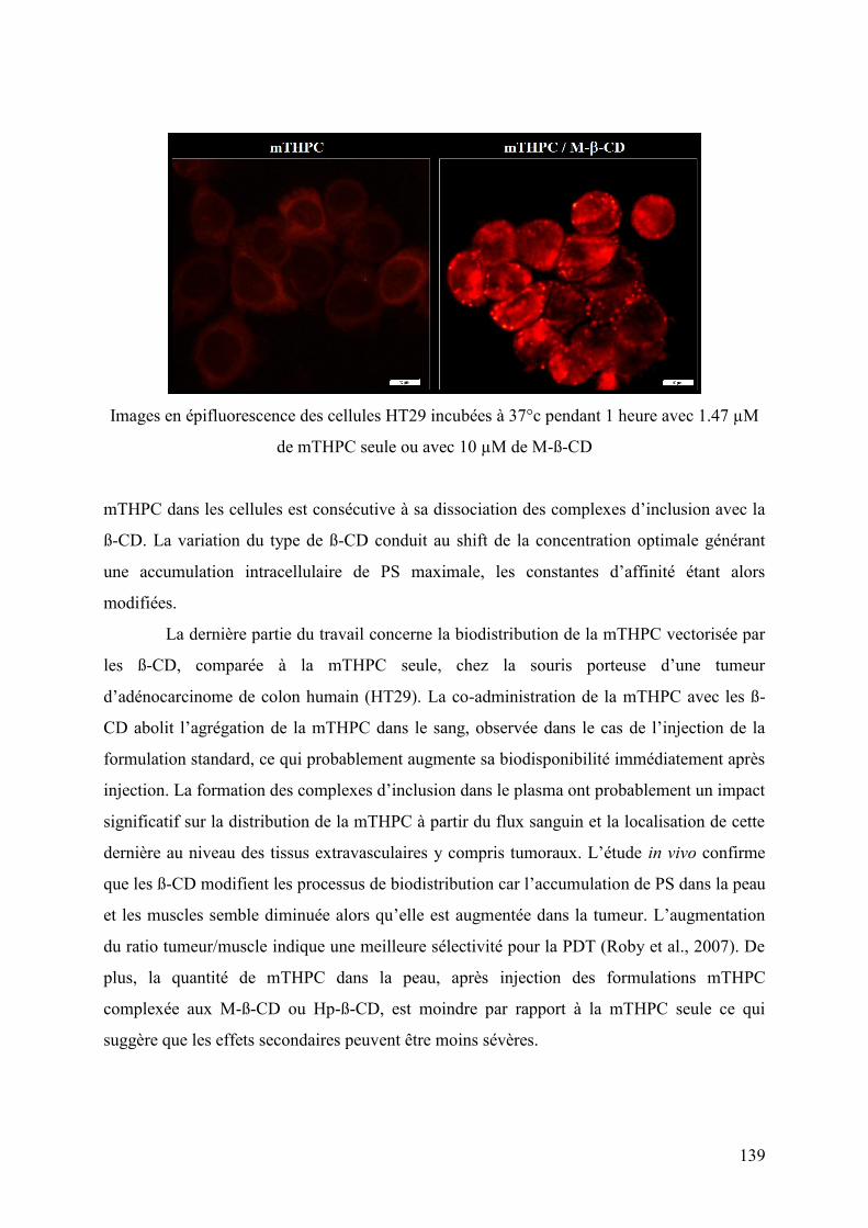
139
Images en épifluorescence des cellules HT29 incubées à 37°c pendant 1 heure avec 1.47 µM
de mTHPC seule ou avec 10 µM de M-ß-CD
mTHPC dans les cellules est consécutive à sa dissociation des complexes d’inclusion avec la
ß-CD. La variation du type de ß-CD conduit au shift de la concentration optimale générant
une accumulation intracellulaire de PS maximale, les constantes d’affinité étant alors
modifiées.
La dernière partie du travail concerne la biodistribution de la mTHPC vectorisée par
les ß-CD, comparée à la mTHPC seule, chez la souris porteuse d’une tumeur
d’adénocarcinome de colon humain (HT29). La co-administration de la mTHPC avec les ß-
CD abolit l’agrégation de la mTHPC dans le sang, observée dans le cas de l’injection de la
formulation standard, ce qui probablement augmente sa biodisponibilité immédiatement après
injection. La formation des complexes d’inclusion dans le plasma ont probablement un impact
significatif sur la distribution de la mTHPC à partir du flux sanguin et la localisation de cette
dernière au niveau des tissus extravasculaires y compris tumoraux. L’étude in vivo confirme
que les ß-CD modifient les processus de biodistribution car l’accumulation de PS dans la peau
et les muscles semble diminuée alors qu’elle est augmentée dans la tumeur. L’augmentation
du ratio tumeur/muscle indique une meilleure sélectivité pour la PDT (Roby et al., 2007). De
plus, la quantité de mTHPC dans la peau, après injection des formulations mTHPC
complexée aux M-ß-CD ou Hp-ß-CD, est moindre par rapport à la mTHPC seule ce qui
suggère que les effets secondaires peuvent être moins sévères.

140
Imagerie de fluorescence de differents organes et tissus prélevés d’une souris porteuse d’une
tumeur HT29 24 h après l’injection de 0, 5 mg/kg de mTHPC seule ou avec 2,5 mg par kg de
M-ß-CD ou 25 mg/kg de Hp-ß-CD
CONCLUSIONS ET PERSPECTIVES
Au cours de cette thèse, nous nous sommes intéressés aux complexes d’inclusion des
CD avec le PS de deuxième génération mTHPC. Les principaux résultats peuvent être
résumés ainsi :
1. Selon les données spectrales, la mTHPC forme effectivement des complexes
d’inclusion avec les molécules de ß-CD que ce soit en solution ou en milieu biologique. La
forme de la bande de Soret de la mTHPC ainsi que le degré de polarisation de fluorescence
sont les deux paramètres les plus informatifs permettant de contrôler la distribution de la
mTHPC entre le complexe d’inclusion et les composants biologiques tels que les protéines
sériques et les membranes biologiques.
2. Les ß-CD ont un effet dépendant de leur concentration sur processus de
distribution de la mTHPC dans le sérum. A faible concentrations (10-6-10-5M) les CD
n’affectent pas l’état d’équilibre de la mTHPC mais augmente significativement sa diffusion
en accélérant la liaison aux protéines. A concentrations élevées (>10-4 M) ß-CD et mTHPC
sont solidement complexés.
3. L’accumulation de la mTHPC dans les cellules HT29 est fortement dépendante
des concentrations de ß-CD. Celles-ci causent à la fois l’accélération et l’inhibition de l’entrée
de la mTHPC dans les cellules. L’accélération du transfert de la mTHPC des protéines du

141
serum aux membranes cellulaires en est le principal mécanisme. A forte concentration de ß-
CD, celles-ci solubles dans l’eau n’étant pas capables de pénétrer dans les cellules, le taux
d’accumulation de la mTHPC est réduit de fait de la formation probable de complexes
d’inclusion de mTHPC dans l’environnement extracellulaire.
4. La variation du type de ß-CD modifiant les constantes d’affinité avec la
mTHPC, il en résulte un shift de la concentration optimale générant une accumulation
intracellulaire de PS maximale. Les ß-CD augmentent la photocytotoxicité par l’augmentation
de l’accumulation intracellulaire de la mTHPC.
5. La co-administration de la mTHPC avec les ß-CD abolit l’agrégation du PS
dans le sang et augmente sa biodisponibilité. Des modifications de biodistribution in vivo de
la mTHPC issue des complexes d’inclusion en résultent.
6. Particulièrement avec la M-ß-CD, l’accumulation de la mTHPC dans la tumeur
et dans le muscle produit un ratio de mTHPC tumeur/muscle augmenté par rapport à celui de
la mTHPC seule. Ceci indique une meilleure sélectivité tumorale avec des effets secondaires
réduits.
Sur la base des résultats obtenus, des formulations de mTHPC ou d’autres PS
impliquant des complexes d’inclusion ß-CD peuvent être développées pour la PDT.
Perspectives
La continuation de ces travaux de recherche est liée à l’optimisation des paramètres
de la PDT utilisant les complexes d’inclusion mTHPC/CD. Cela demande nécessairement une
meilleure connaissance de la pharmacocinétique et de la biodistribution en particulier par une
mise en évidence de la distribution spatiotemporelle du PS issu de ces complexes dans la
tumeur.
En premier lieu une étude in vivo comparant la pharmacocinétique et la
biodistribution de la mTHPC complexée à différentes CD peut être envisagée. Ainsi, le type
de CD donnant la biodistribution la plus favorable chez la souris porteuse de tumeur pourra
être connue. La concentration optimale de CD et de PS devra être déterminée.
Ensuite, la localisation des complexes mTHPC/CD pourrait être étudiée dans le
parenchyme et le compartiment vasculaire de la tumeur. En fonction de la localisation
intratumorale de la mTHPC, l’intervalle de temps optimal entre l’injection du PS et
l’irradiation (IDL ; intervalle drogue-lumière) pourra être sélectionné.
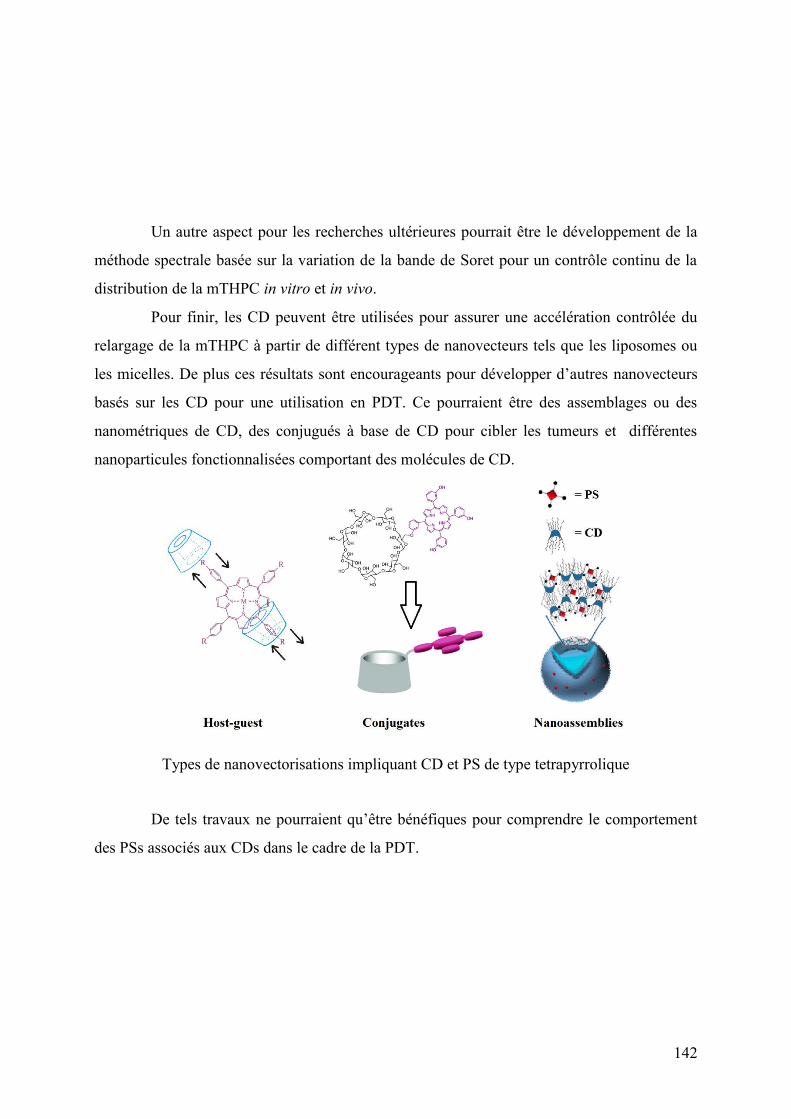
142
Un autre aspect pour les recherches ultérieures pourrait être le développement de la
méthode spectrale basée sur la variation de la bande de Soret pour un contrôle continu de la
distribution de la mTHPC in vitro et in vivo.
Pour finir, les CD peuvent être utilisées pour assurer une accélération contrôlée du
relargage de la mTHPC à partir de différent types de nanovecteurs tels que les liposomes ou
les micelles. De plus ces résultats sont encourageants pour développer d’autres nanovecteurs
basés sur les CD pour une utilisation en PDT. Ce pourraient être des assemblages ou des
nanométriques de CD, des conjugués à base de CD pour cibler les tumeurs et différentes
nanoparticules fonctionnalisées comportant des molécules de CD.
Types de nanovectorisations impliquant CD et PS de type tetrapyrrolique
De tels travaux ne pourraient qu’être bénéfiques pour comprendre le comportement
des PSs associés aux CDs dans le cadre de la PDT.


143
SCIENTIFIC OUTPUT
1. ARTICLES IN PEER-REVIEWED JOURNALS
1) Yankovsky I., Bastien E., Yakavets I., Khludeyev I., Lassalle H-P., Gräfe S.,
Bezdetnaya L., Zorin V. Inclusion complexation with β-cyclodextrin derivatives alters
photodynamic activity and biodistribution of meta-tetra(hydroxyphenyl)chlorin / Eur J
Pharm Sci. 2016 Aug 25;91 P.:172-82.
2) Ilya Yakavets1, Igor Yankovsky1, Bezdetnaya L., Zorin V. Soret band shape indicates
mTHPC distribution between β-cyclodextrins and serum proteins // Dyes and
Pigments 137 (2017) P.: 299-306. 1These authors have contributed equally to this work.
3) Zorina T.E., Yankovsky I.V., Kravchenko I.E., Shman T.V., Zorin V.P. The
Liposomal formulations of chlorin e6 esters and the features of their accumulation by
cells // J. BSU Herald, Series 2 Biology, 2013. № 3, - P. 30-35.
4) Ilya Yakavets, Igor Yankovsky, Lina Bezdetnaya, Vladimir Zorin. “Spectral
characteristics of meta-tetra(hydroxyl-phenyl)chlorin in biological systems”. J. BSU
Herald, Series 1 Physics,2015, Vol. 2, P. 39-45.
5) T.E. Zorina , I.V. Yankovsky, I.E. Kravchenko, T.V. Shman, M.V. Belevtsev, V.P.
Zorin. "Evaluation of Phototoxicity and cytotoxicity for chlorin e6 ester derivatives
and their liposomal forms" Biofizika September 2015, Volume 60, Issue 5, P.:922-930.
2. MAJOR ORAL COMMUNUCATIONS
1) Yankovsky I., V. Zorin, L. Bezdetnaya Use of cyclodextrins to improve antitumor
activity of mTHPC in human colon carcinoma HT-29 cells // 16th European Society
for Photobiology Congress – Aveiro (Portugal), 31 August - 4 September 2015: P.59.
2) Yankovsky I., Yakovets I., Khludeyev I., Bolotine L., Zorin V. Influence of methyl-β-
cyclodextrin on mTHPC interaction with blood components // «Open Readings 2015»:
58-th Scientific Conference for Students of Physics and Natural Sciences, 24-27
March, 2015 , Vilnius, Lithuania. P. 80.
3) Yankovsky I.V., Yakavets I.V., Zorin V.P., Bolotine L.N. Development of model for
an intratumoral distribution of mTHPC/cyclodextrin complexes / XII International
conference “Molecular, membrane and cell fundamentals of biosystems’ functioning”,
June 28-30, 2016: Collected articles. – Belarus. Minsk. – 2014. – P. 225-228.

144
4) Yankovsky I.V., Yakovets I.V., Bolotine L.N., Zorin V.P. Investigation of
supramolecular complexes between mthpc and methyl-β-cyclodextrin // "Low
Temperature Physics" (ICYS-LTP-2014): V Int. Conf. Young Scient., 2-6 June, 2014 ,
Kharkiv, Ukraine. P. 117.
5) Yankovsky I.V., Yakovets I.V., Bolotine L.N., Zorin V.P. meta-
tetra(hydroxyphenyl)chlorin accumulation in blood components: influence of methyl-
β-cyclodextrin // XI International conference “Molecular, membrane and cell
fundamentals of biosystems’ functioning”, June 17-20, 2014: Collected articles. –
Belarus. Minsk. – 2014. – P. 180-183.
6) Yankovsky I., Yakovets I., Bolotine L., Zorin V.P. Alteration of meta-
tetra(hydroxyphenyl)chlorin biodistribution by β-cyclodextrin derivatives // IV
International conference " Topical issues of photodynamic therapy and
photodiagnostics", Saint Petersburg, Russia, September 25-26, 2015: P. 14.
3. POSTERS
1) Yankovsky I.V., Yakavets I.V., Tihonov K.D., Bolotine L.N., Zorin V.P.
Determination of aryl-porphyrins binding constants to biological structures by indirect
spectral approach / XII International conference “Molecular, membrane and cell
fundamentals of biosystems’ functioning”, June 28-30, 2016: Collected articles. –
Belarus. Minsk. – 2014. – P. 37-39.
2) Yankovsky I., I. Yakovets, L. Bezdetnaya, V. Zorin New spectral approach for
monitoring of mTHPC redistribution in biological systems // 16th European Society for
Photobiology Congress – Aveiro (Portugal), 31 August - 4 September 2015: P.45.
4. COMMUNICATIONS PRESENTED BY COLLABORATORS
1) Yakovets I., I. Yankovsky, V. Zorin, L. Bezdetnaya The alteration of mthpc
intratumoral distribution in ht-29 spheroids by cyclodextrins // 59th Scientific
Conference for Students of Physics and Natural Sciences “Open Readings 2016”,
Vilnius, Lithuania 15-18th of March. – 2016. - P. 18.
2) Yakovets I., I. Yankovsky, L. Bezdetnaya, V. Zorin The fluorescent spectral
technique for determination the affinity of aryl-porphyrins to biological structures //
59th Scientific Conference for Students of Physics and Natural Sciences “Open
Readings 2016”, Vilnius, Lithuania 15-18th of March. – 2016. - P. 262.
3) Yakovets I., I. Yankovsky, V. Zorin, L. Bezdetnaya Spectral method for controlling
of mTHPC redistribution between cyclodextrins and biological structures // 58th

145
Scien. Conf. for Students of Physics and Natural Sciences “Open Readings 2015”,
Vilnius, Lithuania. – 2015. - P. 36.
4) Zorina T.E., Yankovsky I.V., Kravchenko I.E., Ermilova T.I., Shman T.V., Zorin
V.P. Nanosized photosensitizers accumulation and elimination from cells with
different structural and morphological properties // XII International conference
“Medico-social ecology of the person: state and perspectives”, April 11-12 2014., –
Collected articles. – Belarus. Minsk. – 2014. – P. 149-151.
5) Zorin V.P., Yankovsky I., Yakovets I., Bolotine L. Influence of β-cyclodextrin
derivatives on photosensitizer meta-tetra(hydroxyphenyl)chlorin distribution in
biologica systems // V congress of Russian biophysicists, 4-10 October 2015, Rostov-
on-Don, Russia. T. 1. – P. 340.


ABSTRACT
Photodynamic therapy (PDT) is a minimally invasive photochemical treatment with a promising clinical track record for oncological and some other diseases. Most PDT-drugs (photosensitizers) including a second-generation PS meta-tetra(hydroxyphenyl)chorin (mTHPC) are highly hydrophobic and require delivery systems. To improve mTHPC solubility and pharmacokinetic properties, β-cyclodextrins (β-CDs) derivatives were proposed.
The present study investigates the effect of β-CDs on mTHPC behavior at various stages of its distribution in vitro and in vivo. Interaction of mTHPC with β-CDs leads to the formation of inclusion complexes that completely abolishes its aggregation after introduction into serum. It was demonstrated that the β-CDs have a concentration-dependent effect on the process of mTHPC distribution in blood serum and cellular cultures in vitro. In vivo study confirms the fact that the use of β-CDs allows modifying mTHPC distribution processes in tumor bearing animals that is reflected in the decreased level of PS accumulation in skin and muscles, as well as in the increased PS accumulation in tumor. In conclusion, application of β-CD derivatives can open up new possibilities to modify and control biodistribution and pharmacokinetics of mTHPC in the course of PDT.
Key-Words : Photodynamic therapy, mTHPC, cyclodextrins, biodistribution, protein binding.
RESUME
La thérapie photodynamique (PDT) est un traitement minimalement invasif basé sur une approche photochimique. Ce traitement est prometteur en oncologie et pour d’autres pathologies. La plupart des drogues utilisées en PDT (photosensibilisateurs) y compris le PS de seconde génération, la méta-tétra (hydroxyphényl)chlorine (mTHPC), sont hautement hydrophobiques et nécessitent un système de transport. Pour améliorer la solubilité de la mTHPC et ses propriétés pharmacocinétiques, les dérivés des β-cyclodextrines (β-CDs) ont été proposés.
L’étude présente a analysé l’effet des β-CDs sur le comportement de la mTHPC à différentes étapes de sa distribution in vitro et in vivo. L’interaction de la mTHPC avec les β-CDs conduit à la formation de complexes d’inclusion qui abolissent complètement son agrégation après introduction dans le sérum. Il a été démontré que les β-CDs ont un effet lié à la concentration sur le processus de distribution de la mTHPC dans le sérum sanguin et les cultures cellulaires in vitro. L’étude in vivo confirme le fait que l’utilisation de β-CDs permet de modifier les processus de distribution de la mTHPC chez les animaux porteurs de tumeurs, ce qui se traduit par un taux moins élevé d’accumulation du PS dans la peau et les muscles, ainsi qu’une accumulation plus élevée du PS dans la tumeur. En conclusion, l’application des dérivés β-CDs peut ouvrir de nouvelles possibilités pour modifier et contrôler la biodistribution et les pharmacocinétiques de la mTHPC au cours de la PDT.
Mots-clés : Thérapie photodynamique, mTHPC, cyclodextrines, biodistribution, liaison aux protéines.
Recommended


