
RESEARCH Open Access
Cytosolic phospholipase A2 alpha amplifies earlycyclooxygenase-2 expression, oxidative stress andMAP kinase phosphorylation after cerebralischemia in miceKoji Kishimoto†, Rung-Chi Li†, Jian Zhang, Judith A Klaus, Kathleen K Kibler, Sylvain Doré, Raymond C Koehler,Adam Sapirstein*
Abstract
Background: The enzyme cytosolic phospholipase A2 alpha (cPLA2a) has been implicated in the progression ofcerebral injury following ischemia and reperfusion. Previous studies in rodents suggest that cPLA2a enhancesdelayed injury extension and disruption of the blood brain barrier many hours after reperfusion. In this study weinvestigated the role of cPLA2a in early ischemic cerebral injury.
Methods: Middle cerebral artery occlusion (MCAO) was performed on cPLA2a+/+ and cPLA2a
-/- mice for 2 hoursfollowed by 0, 2, or 6 hours of reperfusion. The levels of cPLA2a, cyclooxygenase-2, neuronal morphology andreactive oxygen species in the ischemic and contralateral hemispheres were evaluated by light and fluorescentmicroscopy. PGE2 content was compared between genotypes and hemispheres after MCAO and MCAO and6 hours reperfusion. Regional cerebral blood flow was measured during MCAO and phosphorylation of relevantMAPKs in brain protein homogenates was measured by Western analysis after 6 hours of reperfusion.
Results: Neuronal cPLA2a protein increased by 2-fold immediately after MCAO and returned to pre-MCAO levelsafter 2 hours reperfusion. Neuronal cyclooxygenase-2 induction and PGE2 concentration were greater in cPLA2a+/+
compared to cPLA2a-/- ischemic cortex. Neuronal swelling in ischemic regions was significantly greater in thecPLA2a+/+ than in cPLA2a-/- brains (+/+: 2.2 ± 0.3 fold vs. -/-: 1.7 ± 0.4 fold increase; P < 0.01). The increase inreactive oxygen species following 2 hours of ischemia was also significantly greater in the cPLA2a+/+ ischemic corethan in cPLA2a-/- (+/+: 7.12 ± 1.2 fold vs. -/-: 3.1 ± 1.4 fold; P < 0.01). After 6 hours of reperfusion ischemic cortexof cPLA2a
+/+, but not cPLA2a-/-, had disruption of neuron morphology and decreased PGE2 content.
Phosphorylation of the MAPKs-p38, ERK 1/2, and MEK 1/2-was significantly greater in cPLA2a+/+ than in cPLA2a
-/-
ischemic cortex 6 hours after reperfusion.
Conclusions: These results indicate that cPLA2a modulates the earliest molecular and injury responses aftercerebral ischemia and have implications for the potential clinical use of cPLA2a inhibitors.
BackgroundPhospholipase A2 (PLA2) enzymes hydrolyze free fattyacids from the second position (sn-2) of membraneglycerophospholipids and augment neurologic injuries ofoxidative stress (reviewed by Muralikrishna [1]). The
cytosolic phospholipase A2a (cPLA2a, also known asPLA2 group IVA) is a member of the larger PLA2 super-family and has unique properties that suggest it mayregulate formation of eicosanoids in cell-signalling path-ways. cPLA2a resides in the cytosol but translocates tointracellular membranes in response to physiologic Ca2+
changes [2]. cPLA2a has a strong preference for hydro-lysis of arachidonic acid (AA); is a major source of regu-lated, intracellular AA [3]; and is regulated by the
* Correspondence: [email protected]† Contributed equallyThe Department of Anesthesiology and Critical Care Medicine, JohnsHopkins University School of Medicine, Baltimore, MD, USA
Kishimoto et al. Journal of Neuroinflammation 2010, 7:42http://www.jneuroinflammation.com/content/7/1/42
JOURNAL OF NEUROINFLAMMATION
© 2010 Kishimoto et al; licensee BioMed Central Ltd. This is an Open Access article distributed under the terms of the CreativeCommons Attribution License (http://creativecommons.org/licenses/by/2.0), which permits unrestricted use, distribution, andreproduction in any medium, provided the original work is properly cited.

protein kinase-dependent phosphorylation of severalamino acids [4]. We previously demonstrated thatcPLA2a is a key effector of neurologic injury followingcerebral ischemia and reperfusion (I/R) by showing thatcPLA2a
-/- mice have significantly less stroke injury thando wild-type littermate (+/+) mice after transient regio-nal cerebral ischemia [5]. The presence of cPLA2a inneurons [6] and its biochemical properties suggest thatit could play a major regulatory role in neurologic sig-nalling in ischemia and other neurologic diseases [7,8].cPLA2a also has a role in the regulation of the down-
stream enzymes that metabolize AA to the eicosanoids[9,10], which are important mediators of acute andchronic neurologic injury in stroke [11]. The role ofCOX-2 is particularly well-explored in cerebral I/R andis tightly correlated with cPLA2a. Inhibition or genedeletion of COX-2 decreases while COX-2 overexpres-sion enhances neuronal injury following MCAO [12-14].In mice cPLA2a expression appears to be necessary tomaintain normal basal and induced expression of COX-2 in the brain [10,15]. cPLA2a-derived arachidonic acidis also tightly coupled to the 5-lipoxygenase enzyme [16]and in the gerbil model of global cerebral ischemia 15minutes of reperfusion caused translocation of 5-LO tothe neuron membranes and resulted in increased levelsof leukotriene C4 [17]. cPLA2a amplifies the increase inpermeability of the blood-brain barrier after transientischemia [7], and eicosanoids contribute to the subse-quent inflammatory responses [18]. The eicosanoids,particularly prostaglandins (PGs), and AA itself may alsocontribute directly to the early excitotoxicity that pre-cedes neuroinflammation [19-23]. Our lab and othersfound that cPLA2a can have a direct and early effect onexcitotoxicity in vitro [19,24,25].Here, we examined the effect of transient regional cer-
ebral I/R on cPLA2a expression and, in turn, the effectof cPLA2a on cyclooxygenase (COX)-2 expression,PGE2 levels and reactive oxygen species (ROS) early inthe cell-death cascade. We applied transient middle cer-ebral artery (MCA) occlusion (MCAO) to cPLA2a
+/+
and cPLA2a-/- mice and investigated the effect of
cPLA2a on early pathways of neurologic injury at 0, 2,and 6 hours of reperfusion. We then correlated cPLA2aexpression with ROS generation and the phosphoryla-tion of relevant MAPKs. Our results indicate thatcPLA2a contributes to I/R injury immediately afterischemia.
MethodsMaterialsUnless otherwise stated, all compounds were purchasedfrom Sigma-Aldrich Company (St. Louis, MO). Forimmunomicroscopy anti-cPLA2a (P505) was purchasedfrom Abcam Inc. (Cambridge, MA). Rabbit anti-cPLA2a
(N-216) and anti-b-actin antibodies were from SantaCruz Biotechnology (Santa Cruz, CA). Alexa Fluor 488and 568 donkey anti-rabbit IgG and NeuroTrace 435/455 Nissl Stain (NT) were purchased from InvitrogenCorporation (Carlsbad, CA).
Animal CareAll experiments were conducted in accordance with theguidelines of the National Institutes of Health andapproved by the Johns Hopkins University InstitutionalAnimal Care and Use Committee. cPLA2a
+/- mice werea gift from Takao Shimizu (Tokyo University) and weresupplied by Jim Clark (Wyeth Pharmaceutical, Cam-bridge, MA). Mice were housed in a facility with 12-hour diurnal light cycle with free access to food andwater. All experimental mice were produced by matingmale and female cPLA2a
+/- mice that were producedand maintained in the C57BL/6J strain.
Focal Cerebral IschemiaTransient focal ischemia was induced by MCAO in 10-14-week-old age-matched cPLA2a
-/- and cPLA2a+/+ lit-
termates between 20-28 g. Anesthesia was by sponta-neous ventilation of isoflurane in 30% O2. Athermostatically controlled warming pad and infraredlight were used to maintain the rectal temperature at37.5 ± 0.5°C during all phases of the surgery. Left-sidedMCAO and sham surgery were performed as previouslydescribed [5]. After 2 hours of MCAO, the mice werere-anesthetized, the occlusive suture was removed, andthe mice were placed in a temperature-controlledenvironment.In experiments to measure oxidative stress, 10 mg/kg
dihydroethidium (HE) was injected into the jugular veinat the beginning of MCAO. The mice underwent 2-hourMCAO with continuous monitoring of cerebral bloodflow (CBF) by laser-Doppler flowmetry, and at 0 or 2hours of reperfusion, the mice were sacrificed, perfusionfixed, and the brains harvested.
Regional CBF AssessmentRegional CBF (rCBF) was measured at 60 minutes ofischemia in mice of each genotype and strain, by using[14C]-iodoantipyrine ([14C]-IAP) autoradiography, aspreviously described [26]. MCAO was carried out asdescribed above, with additional placement of femoralarterial and venous catheters. At 60 minutes of MCAO,arterial blood pressure, pH, PaCO2, and PaO2 were mea-sured, and 4 μCi of [14C]-IAP was infused intravenously.Coronal brain sections (20 μm) cut on a cryostat wereexposed to BioMax film (Kodak, Rochester, NY) for10 days with [14C] standards. From each mouse, we digi-tized three autoradiographic images from five positionscorresponding to coronal sections at +2, +1, 0, -1, and
Kishimoto et al. Journal of Neuroinflammation 2010, 7:42http://www.jneuroinflammation.com/content/7/1/42
Page 2 of 13

-2 mm from bregma. Regions corresponding to the coreanterior cerebral artery (ACA) and MCA territorieswere outlined in the ipsilateral and contralateral cortex,and signal intensity was determined (ImageJ version1.36, NIH, Bethesda, MD). rCBF was calculated as pre-viously described [26], and measurements in the threeconsecutive coronal slices were averaged at each posi-tion to yield values of absolute rCBF in each region.
Fluorescence Microscopy and Quantitative DigitalImage AnalysisFollowing terminal anesthesia, mice were perfused with3 × weight/volume of normal saline, followed by 4%paraformaldehyde in PBS, and post-fixed in 4% parafor-maldehyde and 15% sucrose. For immunofluorescence,30 μm coronal sections were blocked and quenchedwith 0.5% H2O2 in 0.3% normal donkey serum in PBSand incubated with primary antibody overnight at 4°C.The samples were incubated with secondary antibodyfollowed by DAB treatment. Slides were counter-stainedwith fluorescent Nissl reagent to enable identification ofintact neurons by presence of the Nissl substance [27].Coronal brain sections were examined by confocal
microscope LSM510 META (Zeiss, Thornwood, NY). NT,Alexa Fluor 488, and Alexa Fluor 568 were excited with a405 nm diode laser, a 488 nm Argon laser, and a 561 nmhelium-neon laser, respectively. Emission was detectedthrough 420-480-nm, 505-530-nm, and 565-595-nmband-pass filters, respectively. HE was visualized by excita-tion at 561 nm and emission at 610 nm. An investigatorblinded to genotype and hemisphere used Image J soft-ware to measure total cPLA2a fluorescence in low magni-fication (10×) images obtained from representative brainsections of cPLA2a
+/+ and cPLA2a-/- mice.
For high resolution analysis, two representative imagesin the cortical subfield of interest were acquired fromeach of three brain sections per mouse, and two z-planes of ~2 μm optical thickness separated by 8 μmwere sampled. Fluorescence threshold levels were set toallow for recognition of individual neurons in sliceswithout signal saturation and were constant for analysisof all slices. The anatomical regions corresponding tothe ischemic core and penumbra were identified influorescent Nissl-stained sections. Fluorescence abovethe threshold was measured in 120-130 neurons foreach mouse in non-overlapping, randomly chosenregions in photomicrographs obtained using 100× mag-nification. Total pixel area was normalized to the totalarea analyzed and number of neurons and expressed inarbitrary units.
ImmunoblottingFor Western analysis, primary antibodies included COX-2 (1:1000, Cayman Chemical Co. Ann Arbor, MI),
cPLA2a (1:500), phospho-cPLA2a (1:500), ERK1/2 andphospho-ERK1/2 (1:1000), MEK1/2 and phospho-MEK1/2 (1:1000), p38 MAPK and phospho-p38 MAPK(1:1000) (all from Cell Signalling Technology, Inc. Dan-vers, MA). Protein samples were separated by electro-phoresis and transferred to PVDF membranes.Immunocomplexes were visualized by enhanced chemi-luminescence detection (Amersham Life Science).Subcellular fractions were prepared from brain tissue
homogenized by Dounce (20 strokes) in 10× v/w of ice-cold lysis buffer (2 mM EGTA in PBS with proteaseinhibitor), and 1/10 volume of benzonase solution (1:50dilution). The samples were gently shaken on ice for 20minutes and centrifuged at 800 × g for 10 minutes at4°C. Supernatant volumes of 100 μl were centrifuged at100,000 × g for 45 min at 4°C. The supernatants con-tained the cytosolic fraction. The pelleted nuclear frac-tion was resuspended in 0.7 w/v CHAPS lysis buffer,sonicated for 10 seconds and incubated on ice for30 minutes. Protein concentrations were measured bythe modified Bradford assay. Cell lysate proteins (25 μgper sample) were electrophoretically resolved on 4-15%polyacrylamide Tris-HCl gradient gels (BioRad, Her-cules, CA) and transferred to PVDF membranes. Eachmembrane was probed and stripped sequentially forphospho-cPLA2a, cPLA2a, and b-actin. For routineimmunodetection of proteins cortical hemispheres werehomogenized in 5 × v/w buffer, and 10 μg of crudehomogenate was used for SDS-PAGE.
Prostaglandin E2 (PGE2) Enzyme ImmunoassayCortical tissue was weighed and homogenized by polytronin 10 μl/mg wet tissue of ice-cold PBS with 10 μg/ml indo-methacin and incubated on ice for 10 min. The homoge-nate solution was brought to 40% volume aqueous ethanoland acidified with glacial acetic acid to pH 3.0, incubatedfor 5 min at room temperature, and centrifuged at 2,500 ×g for 10 min. The supernatant was applied to a condi-tioned Oasis HLB column (Waters Corp., Milford, MA),washed with 0.03% formic acid, followed by 15% aqueousethanol/0.03% formic acid followed by petroleum ether.PGs were eluted with ethyl acetate and evaporated to dry-ness under nitrogen. The eluant was dissolved in 300 μLassay buffer, and PGE2 concentration was determined byELISA according to the manufacturer’s instructions (AssayDesigns, Ann Arbor, MI.). For each extraction and ELISAthe results were normalized within the group to accountfor variation in the efficiency of lipid extraction.
Statistical AnalysisAssays that required multiple samples from a singlemouse were analyzed by averaging the intra-mouse sam-ples and then performing statistical annalysis betweenindividuals. For studies in which multiple time points
Kishimoto et al. Journal of Neuroinflammation 2010, 7:42http://www.jneuroinflammation.com/content/7/1/42
Page 3 of 13

were compared across genotypes and hemispheres ana-lysis was performed by repeated measures ANOVA andpost-hoc comparison between genotypes was made withthe Newman-Keuls test. Comparison of relative PGE2concentrations following MCAO between genotypes andhemispheres was conducted with 2-way ANOVA fol-lowed by Bonferroni testing between the genotypesusing GraphPad Prism version 5.03 (GraphPad Software,San Diego California). Densitometry analysis wasby paired t-tests. For all procedures; P < 0.05 was con-sidered statistically significant. Data are expressed asmean ± s.d.
ResultsTo examine the effect of cPLA2a expression on the cas-cade of molecular and cellular events in vivo followingcerebral I/R, we subjected cPLA2a
+/+ and cPLA2a-/-
mice to 2 hours of MCAO followed by no (0), 2, or 6hours of reperfusion and examined the expression ofcPLA2a using immunofluorescence coupled with Nisslstaining. We observed a substantial increase in the levelof cPLA2a staining in the cPLA2a
+/+ mice after 2 hoursof MCAO and no reperfusion. The averaged cPLA2afluorescence intensity in cPLA2a
+/+ ischemic hemi-spheres was 1.9 fold greater than that in contralateralhemispheres (P < 0.01). As expected, the nonspecificstaining in cPLA2a
-/- hemispheres was barely detectableand was not altered by ischemia. We then used highresolution imaging to characterize the cellular expres-sion patterns of cPLA2a that follow MCAO in theischemic core and penumbra regions. We observed avery low level of cPLA2a immunofluorescence incPLA2a
+/+ mice after sham surgery (Figure 1A a-d).After 2 hours of ischemia, the immunofluorescence wasmarkedly increased in the neurons and non-neuronalcells of the ischemic hemisphere (Figure 1A e-f) but wasunchanged in the contralateral hemisphere (Figure 1Ag-h). However, after 2 hours of reperfusion, cPLA2a wassubstantially lower in the neurons of the penumbra (Fig-ure 1A i) and almost absent in the neurons of theischemic zone (Figure 1A j). Nissl staining suggests lossof neurons in the ischemic core after 2 hours of reperfu-sion (Figure 1A j). Six hours after reperfusion, cPLA2aimmunofluorescence could not be distinguished fromthat of sham-operated mice (data not shown). ThecPLA2a
-/- mice had minimal, nonspecific backgroundstaining (Figure 1A m-x). Phosphorylated cPLA2a alsoshowed a marked increase in cPLA2a+/+ brain after2 hours of ischemia and then decreased along a timecourse similar to that of unphosphorylated cPLA2a(Figure 1B).To validate the results of the immunofluorescence
experiments, cPLA2a+/+ mice were subjected to 2-hour
MCAO and no reperfusion, or sham operation.
Following euthanasia the ipsilateral and contralateralcortices were harvested for protein extraction. We per-formed a subcellular fractionation on the cortical pro-teins and subjected these to Western blot analysis usinganti-cPLA2a and anti-phospho-cPLA2a antibodies. Theanti-cPLA2a antibody recognizes both the phosphory-lated and unphosphorylated forms of cPLA2a and thisleads to the formation of a doublet on immunoblot. Theupper band of this doublet is the phospho-cPLA2a formand this is confirmed with the anti-phospho-cPLA2aantibody. Consistent with the immunofluorescence find-ings, 2 hours of ischemia increased total and phospho-cPLA2a in the ipsilateral cytosolic fraction as comparedto the contralateral (non-ischemic) cytosolic fraction(Figure 2). Expression levels of total and phospho-cPLA2a in the membrane fraction did not differbetween the ipsilateral and contralateral hemispheres.This indicates that cPLA2a is not associated with cellu-lar membranes following 2 hours of MCAO.Nissl staining illustrated that I/R caused much greater
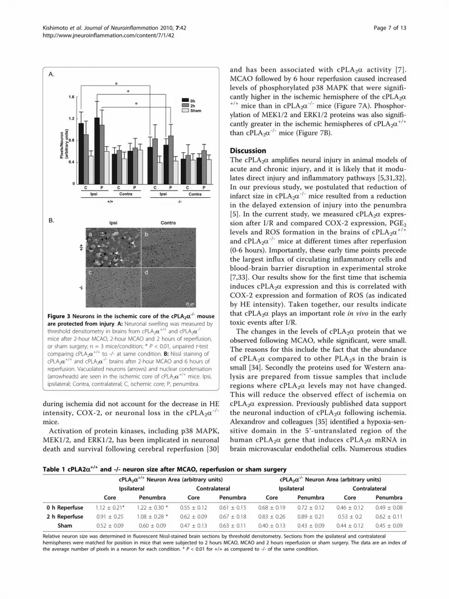
disruption of cortical pyramidal neuron morphology incPLA2a
+/+ mice than in cPLA2a-/- mice. Neurons in the
core and penumbra regions were enlarged immediatelyafter 2-hour ischemia (0 hours of reperfusion) and after2 hours of reperfusion (Figure 3A and Table 1). Theexpression of cPLA2a was associated with greater neu-ronal swelling at both time points. After 6 hours ofreperfusion, neuronal structure in the cPLA2a
+/+ ipsilat-eral hemisphere was almost completely disrupted with adramatic reduction in the number of neurons(Figure 3B, a). The structure and number of neurons incPLA2a
-/- mouse brains, however, remained intact(Figure 3B, c).cPLA2a regulates COX-2 expression in the brain
[10,15] and nonspecific PLA2 blockade prevents COX-2induction after transient focal ischemia [28]. We exam-ined the effect of cPLA2a deletion on COX-2 expressionafter I/R. In the ipsilateral cortices of cPLA2a
+/+ mice,COX-2 immunofluorescence was substantially greaterthan that in sham-operated controls immediately afterischemia (Figure 4A a-b compared to i-j) and increasedfurther 2 hours after reperfusion (Figure 4A e-f). In con-trast, COX-2 was not elevated in the ipsilateral cortex ofcPLA2a
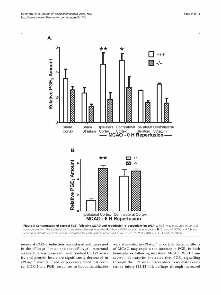
-/- mice (Figure 4A m-n) and was only slightlyincreased after 2 hours of reperfusion (Figure 4A q-r).PGE2 is produced by the coordinated enzymatic activ-
ities of COX and the PGE synthases upon AA. Previousstudies have demonstrated that PGE2 levels are elevatedfollowing MCAO in the rat hippocampus [29]. We com-pared the levels of PGE2 in the cortex of cPLA2a
+/+ and-/- mice immediately following 2 hours of ischemia andno reperfusion (Figure 5A) or after 6 hours of reperfu-sion (Figure 5B). In agreement with previous resultsthere was no significant difference between basal PGE2
Kishimoto et al. Journal of Neuroinflammation 2010, 7:42http://www.jneuroinflammation.com/content/7/1/42
Page 4 of 13

Figure 1 cPLA2a is increased by ischemia in cPLA2a+/+ neurons. A: Immunofluorescence of cPLA2a (green) and fluorescent Nissl dye (blue)in brain slices from cPLA2a
+/+ and cPLA2a-/- mice after sham surgery, 2-hour MCAO, or 2-hour MCAO and 2-hour reperfusion. Images from the
ischemic core and penumbra regions from the ipsilateral and contralateral hemispheres of each genotype are shown. B: Immunofluorescence ofSer505-phosphorylated cPLA2a (green) in cPLA2a
+/+ mice subjected to sham surgery (a-d), 2 hours of MCAO (e-h) or 2 hours of MCAO and 2hours of reperfusion (i-l). Fluorescent Nissl staining of neurons is in blue. Representative images, n = 3 mice of each genotype. Ipsi, ipsilateral;Contra, contralateral.
Kishimoto et al. Journal of Neuroinflammation 2010, 7:42http://www.jneuroinflammation.com/content/7/1/42
Page 5 of 13

levels in the cPLA2a+/+ and -/- cortex [10]. However 2
hours of MCAO caused a significant increase in thePGE2 concentration of both the contralateral and ipsilat-eral cPLA2a
+/+ cortices. In contrast the levels of PGE2were not changed by ischemia in the cPLA2a
-/- cortex.After 6 hours of reperfusion the concentration of PGE2in ischemic cPLA2a
+/+ cortex was significantly lowerthan in cPLA2a
-/- cortex or in the contralateral cortexof either genotype (Figure 5B).We also evaluated the role of cPLA2a expression in
the generation of ROS using the fluorescent probe HE.The increase in ROS in the ischemic hemisphere ofcPLA2a
+/+ mice was significantly greater than in thecPLA2a
-/- mice following ischemia without reperfusion(Figure 6A, 0 h) (+/+: 7.12 ± 1.2 fold increase vs. -/-:3.10 ± 1.4 fold increase, P < 0.01) and also 2 hours afterischemia (Figure 6A and Table 2). Levels of ROS in the
contralateral hemispheres were not different from levelsin sham-operated mice.To determine if differences in ROS levels between
cPLA2a+/+ and cPLA2a
-/- mice resulted from differencesin the vascular responses during ischemia, rCBF wasmeasured by the technique of [14C]-IAP injection. Thecortical regions corresponding to the ACA and MCAwere demarcated in coronal brain sections. MCAOcaused a significant reduction of blood flow in both theACA and MCA territories, relative to the contralateralsides in each genotype (Figure 6B). CBF was slightlylower in the ipsilateral ACA territory in the anteriorregion of the cPLA2a
-/- brain than in the correspondingregion of the cPLA2a
+/+ brain. A similar level of ACAblood flow reduction was measured in the anteriorregions of the contralateral cortex of cPLA2a
-/- mice.Therefore, differences in rCBF between the genotypes
Figure 2 cPLA2a is increased in the ischemic hemisphere following focal ischemia. A: A representative Western blot of cPLA2a+/+ brain
protein fractions (25 μg each lane) from the non-ischemic (contralateral) and ischemic (ipsilateral) hemispheres. The same membrane wasexposed to antibody and developed sequentially for phospho-cPLA2a, cPLA2a, and actin. The panel on the left represents positive controls-recombinant human cPLA2a (rh cPLA2a) and mouse spleen protein-and a negative control, cPLA2a
-/- brain protein. The antibody directedagainst cPLA2a recognizes both the phosphorylated and nonphosphorylated cPLA2a. B, C: Densitometry analysis of Western blots for (B) totalcPLA2a (n = 5 experiments) and (C) phospho-cPLA2a (n = 5 experiments). * P < 0.05 ipsilateral compared to contralateral.
Kishimoto et al. Journal of Neuroinflammation 2010, 7:42http://www.jneuroinflammation.com/content/7/1/42
Page 6 of 13
during ischemia did not account for the decrease in HEintensity, COX-2, or neuronal loss in the cPLA2a
-/-
mice.Activation of protein kinases, including p38 MAPK,
MEK1/2, and ERK1/2, has been implicated in neuronaldeath and survival following cerebral reperfusion [30]
and has been associated with cPLA2a activity [7].MCAO followed by 6 hour reperfusion caused increasedlevels of phosphorylated p38 MAPK that were signifi-cantly higher in the ischemic hemisphere of the cPLA2a+/+ mice than in cPLA2a
-/- mice (Figure 7A). Phosphor-ylation of MEK1/2 and ERK1/2 proteins was also signifi-cantly greater in the ischemic hemispheres of cPLA2a
+/+
than cPLA2a-/- mice (Figure 7B).
DiscussionThe cPLA2a amplifies neural injury in animal models ofacute and chronic injury, and it is likely that it modu-lates direct injury and inflammatory pathways [5,31,32].In our previous study, we postulated that reduction ofinfarct size in cPLA2a
-/- mice resulted from a reductionin the delayed extension of injury into the penumbra[5]. In the current study, we measured cPLA2a expres-sion after I/R and compared COX-2 expression, PGE2levels and ROS formation in the brains of cPLA2a
+/+
and cPLA2a-/- mice at different times after reperfusion
(0-6 hours). Importantly, these early time points precedethe largest influx of circulating inflammatory cells andblood-brain barrier disruption in experimental stroke[7,33]. Our results show for the first time that ischemiainduces cPLA2a expression and this is correlated withCOX-2 expression and formation of ROS (as indicatedby HE intensity). Taken together, our results indicatethat cPLA2a plays an important role in vivo in the earlytoxic events after I/R.The changes in the levels of cPLA2a protein that we
observed following MCAO, while significant, were small.The reasons for this include the fact that the abundanceof cPLA2a compared to other PLA2s in the brain issmall [34]. Secondly the proteins used for Western ana-lysis are prepared from tissue samples that includeregions where cPLA2a levels may not have changed.This will reduce the observed effect of ischemia oncPLA2a expression. Previously published data supportthe neuronal induction of cPLA2a following ischemia.Alexandrov and colleagues [35] identified a hypoxia-sen-sitive domain in the 5′-untranslated region of thehuman cPLA2a gene that induces cPLA2a mRNA inbrain microvascular endothelial cells. Numerous studies
Figure 3 Neurons in the ischemic core of the cPLA2a-/- mouseare protected from injury. A: Neuronal swelling was measured bythreshold densitometry in brains from cPLA2a+/+ and cPLA2a-/-
mice after 2-hour MCAO, 2-hour MCAO and 2 hours of reperfusion,or sham surgery; n = 3 mice/condition; * P < 0.01, unpaired t-testcomparing cPLA2a
+/+ to -/- at same condition. B: Nissl staining ofcPLA2a
+/+ and cPLA2a-/- brains after 2-hour MCAO and 6 hours of
reperfusion. Vacuolated neurons (arrows) and nuclear condensation(arrowheads) are seen in the ischemic core of cPLA2a
+/+ mice. Ipsi,ipsilateral; Contra, contralateral; C, ischemic core; P, penumbra.
Table 1 cPLA2a+/+ and -/- neuron size after MCAO, reperfusion or sham surgery
cPLA2a+/+ Neuron Area (arbitrary units) cPLA2a-/- Neuron Area (arbitrary units)
Ipsilateral Contralateral Ipsilateral Contralateral
Core Penumbra Core Penumbra Core Penumbra Core Penumbra
0 h Reperfuse 1.12 ± 0.21* 1.22 ± 0.30 * 0.55 ± 0.12 0.61 ± 0.15 0.68 ± 0.19 0.72 ± 0.12 0.46 ± 0.12 0.49 ± 0.08
2 h Reperfuse 0.91 ± 0.25 1.08 ± 0.28 * 0.62 ± 0.09 0.67 ± 0.18 0.83 ± 0.26 0.89 ± 0.21 0.53 ± 0.2 0.62 ± 0.11
Sham 0.52 ± 0.09 0.60 ± 0.09 0.47 ± 0.13 0.63 ± 0.11 0.40 ± 0.13 0.43 ± 0.09 0.44 ± 0.12 0.45 ± 0.09
Relative neuron size was determined in fluorescent Nissl-stained brain sections by threshold densitometry. Sections from the ipsilateral and contralateralhemispheres were matched for position in mice that were subjected to 2 hours MCAO, MCAO and 2 hours reperfusion or sham surgery. The data are an index ofthe average number of pixels in a neuron for each condition. * P < 0.01 for +/+ as compared to -/- of the same condition.
Kishimoto et al. Journal of Neuroinflammation 2010, 7:42http://www.jneuroinflammation.com/content/7/1/42
Page 7 of 13

have reported cPLA2a expression in glial cells [36] andmRNA expression in neurons [6], and a recent studyshowed that cPLA2a is expressed in neurons in a mousemodel of Alzheimer’s disease [8]. After transient globalischemia, late induction of cPLA2a was found only inglial cells [37]. Other investigators have noted an earlyincrease in PLA2 activity minutes after global cerebral I/R [38]. A rat model of transient cerebral ischemiashowed that cPLA2a activity increased 1 day after reper-fusion but that the levels of protein and phospho-cPLA2a did not increase until 3 days after reperfusion[7]. Changes in cPLA2a that occur hours to days follow-ing ischemia may be related to secondary injury andinflammation.In cell culture models, chemical anoxia [39] and
increased intracellular calcium [40] cause cPLA2a totranslocate to nuclear and other membranes. In ourimmunofluorescence and subcellular fractionationexperiments ischemia did not cause translocation ofcPLA2a to membranes. There are several potential
explanations for the lack of cPLA2a membrane associa-tion. In the gerbil global ischemia model, 5-LO did nottranslocate to the nucleus until minutes after reperfu-sion [17]. Similarly, reoxygenation following ischemiaappears to be a major determinant of intracellular Ca2+
flux (reviewed by Szydlowska and Tymianski [41]).Thus, it is possible that cPLA2a translocates to cellularmembranes minutes after reperfusion. Further experi-ments examining the immediate reperfusion period willbe necessary to delineate the intracellular signallingevents of cPLA2a activation and translocation inneurons.How could cPLA2a impact neuronal injury at times
that precede classical neuroinflammation? Mechanismsincluding increased PG synthesis and action, modulationof excitotoxic responses and increased ROS stress havebeen postulated.The cPLA2a-associated increase in PGE2 levels in
cPLA2a+/+ cortex following MCAO are consistent with
these postulates. In the ischemic core, we found that
Figure 4 cPLA2a regulates post-ischemic COX-2 levels. Immunofluorescence of COX-2 (green) and fluorescent Nissl dye (blue) in brain slicesfrom cPLA2a
+/+ and cPLA2a-/- mice after 2-hour MCAO, 2-hour MCAO and 2-hour reperfusion, or sham surgery. Images of the ischemic core
and penumbra regions from the ipsilateral and contralateral hemispheres of each genotype are shown. Representative images, n = 3 mice ofeach genotype. Ipsi, ipsilateral; Contra, contralateral.
Kishimoto et al. Journal of Neuroinflammation 2010, 7:42http://www.jneuroinflammation.com/content/7/1/42
Page 8 of 13
neuronal COX-2 induction was delayed and decreasedin the cPLA2a
-/- mice and that cPLA2a-/- neuronal
architecture was preserved. Basal cerebral COX-2 activ-ity and protein levels are significantly decreased incPLA2a
-/- mice [15], and we previously found that corti-cal COX-2 and PGE2 responses to lipopolysaccharide
were attenuated in cPLA2a-/- mice [10]. Systemic effects
of MCAO may explain the increase in PGE2 in bothhemispheres following unilateral MCAO. Work fromseveral laboratories indicates that PGE2 signallingthrough the EP1 or EP3 receptors exacerbates earlystroke injury [22,42-44], perhaps through increased
Sham C
ortex
Sham Stri
atum
Ipsi Corte
x
Contra C
ortex
Ipsi Stri
atum
Contra Stri
atum
0
2
4
6
+/+-/-
MCAO - 0 H ReperfusionIpsilateralCortex
ContalateralCortex
IpsilateralStriatum
ContralateralStriatum
ShamStriatum
ShamCortex
** *A.
Rel
ativ
e PG
E 2Am
ount
Ipsilateral Cortex Contralateral Cortex0
2
4
6+/+-/-
B.
MCAO - 6 H Reperfusion
**
Rel
ativ
e PG
E 2Am
ount
Figure 5 Concentration of cortical PGE2 following MCAO and reperfusion is dependent on cPLA2a. PGE2 was measured in corticalhomogenate from the ipsilateral and contralateral hemispheres after A: 2 hours MCAO or sham operation and B: 2 hours of MCAO and 6 hoursreperfusion. Results are expressed as normalized for each lipid extraction and assay. * P < 0.05; ** P < 0.01 n = 3 - 4 each condition.
Kishimoto et al. Journal of Neuroinflammation 2010, 7:42http://www.jneuroinflammation.com/content/7/1/42
Page 9 of 13

Figure 6 Oxidative stress is greater in cPLA2a+/+ than in cPLA2a-/- mice after MCAO. A: ROS were measured in cPLA2a+/+ and cPLA2a
-/-
mice that were injected with dihydroethidium (HE) immediately before MCAO or after sham surgery. The intensity of HE staining in brainsections from the indicated time points was measured by densitometry. Ipsi, ipsilateral; Contra, contralateral; n = 3 mice/condition with 120-130neurons counted/mouse; † P < 0.01. B: CBF was measured in mice by [14C]-IAP autoradiography in the ACA and MCA territories of each cortexat 60 minutes of MCAO. The left axis shows the position (in mm) of each region relative to bregma, and the right axis shows the absolute rCBF(ml/100 g/min); 24 slices from n = 4 mice/group. * P < 0.01 and † P < 0.05 as compared to the same region in cPLA2a
+/+ mice.
Kishimoto et al. Journal of Neuroinflammation 2010, 7:42http://www.jneuroinflammation.com/content/7/1/42
Page 10 of 13
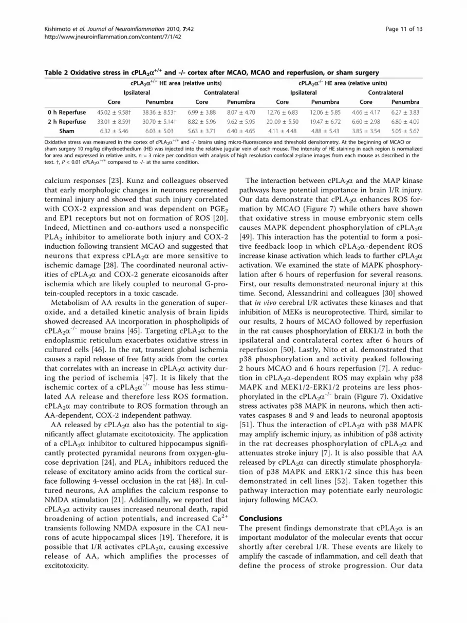
calcium responses [23]. Kunz and colleagues observedthat early morphologic changes in neurons representedterminal injury and showed that such injury correlatedwith COX-2 expression and was dependent on PGE2
and EP1 receptors but not on formation of ROS [20].Indeed, Miettinen and co-authors used a nonspecificPLA2 inhibitor to ameliorate both injury and COX-2induction following transient MCAO and suggested thatneurons that express cPLA2a are more sensitive toischemic damage [28]. The coordinated neuronal activ-ities of cPLA2a and COX-2 generate eicosanoids afterischemia which are likely coupled to neuronal G-pro-tein-coupled receptors in a toxic cascade.Metabolism of AA results in the generation of super-
oxide, and a detailed kinetic analysis of brain lipidsshowed decreased AA incorporation in phospholipids ofcPLA2a
-/- mouse brains [45]. Targeting cPLA2a to theendoplasmic reticulum exacerbates oxidative stress incultured cells [46]. In the rat, transient global ischemiacauses a rapid release of free fatty acids from the cortexthat correlates with an increase in cPLA2a activity dur-ing the period of ischemia [47]. It is likely that theischemic cortex of a cPLA2a
-/- mouse has less stimu-lated AA release and therefore less ROS formation.cPLA2a may contribute to ROS formation through anAA-dependent, COX-2 independent pathway.AA released by cPLA2a also has the potential to sig-
nificantly affect glutamate excitotoxicity. The applicationof a cPLA2a inhibitor to cultured hippocampus signifi-cantly protected pyramidal neurons from oxygen-glu-cose deprivation [24], and PLA2 inhibitors reduced therelease of excitatory amino acids from the cortical sur-face following 4-vessel occlusion in the rat [48]. In cul-tured neurons, AA amplifies the calcium response toNMDA stimulation [21]. Additionally, we reported thatcPLA2a activity causes increased neuronal death, rapidbroadening of action potentials, and increased Ca2+
transients following NMDA exposure in the CA1 neu-rons of acute hippocampal slices [19]. Therefore, it ispossible that I/R activates cPLA2a, causing excessiverelease of AA, which amplifies the processes ofexcitotoxicity.
The interaction between cPLA2a and the MAP kinasepathways have potential importance in brain I/R injury.Our data demonstrate that cPLA2a enhances ROS for-mation by MCAO (Figure 7) while others have shownthat oxidative stress in mouse embryonic stem cellscauses MAPK dependent phosphorylation of cPLA2a[49]. This interaction has the potential to form a posi-tive feedback loop in which cPLA2a-dependent ROSincrease kinase activation which leads to further cPLA2aactivation. We examined the state of MAPK phosphory-lation after 6 hours of reperfusion for several reasons.First, our results demonstrated neuronal injury at thistime. Second, Alessandrini and colleagues [30] showedthat in vivo cerebral I/R activates these kinases and thatinhibition of MEKs is neuroprotective. Third, similar toour results, 2 hours of MCAO followed by reperfusionin the rat causes phosphorylation of ERK1/2 in both theipsilateral and contralateral cortex after 6 hours ofreperfusion [50]. Lastly, Nito et al. demonstrated thatp38 phosphorylation and activity peaked following2 hours MCAO and 6 hours reperfusion [7]. A reduc-tion in cPLA2a-dependent ROS may explain why p38MAPK and MEK1/2-ERK1/2 proteins are less phos-phorylated in the cPLA2a
-/- brain (Figure 7). Oxidativestress activates p38 MAPK in neurons, which then acti-vates caspases 8 and 9 and leads to neuronal apoptosis[51]. Thus the interaction of cPLA2a with p38 MAPKmay amplify ischemic injury, as inhibition of p38 activityin the rat decreases phosphorylation of cPLA2a andattenuates stroke injury [7]. It is also possible that AAreleased by cPLA2a can directly stimulate phosphoryla-tion of p38 MAPK and ERK1/2 since this has beendemonstrated in cell lines [52]. Taken together thispathway interaction may potentiate early neurologicinjury following MCAO.
ConclusionsThe present findings demonstrate that cPLA2a is animportant modulator of the molecular events that occurshortly after cerebral I/R. These events are likely toamplify the cascade of inflammation, and cell death thatdefine the process of stroke progression. Our data
Table 2 Oxidative stress in cPLA2a+/+ and -/- cortex after MCAO, MCAO and reperfusion, or sham surgery
cPLA2a+/+ HE area (relative units) cPLA2a-/- HE area (relative units)
Ipsilateral Contralateral Ipsilateral Contralateral
Core Penumbra Core Penumbra Core Penumbra Core Penumbra
0 h Reperfuse 45.02 ± 9.58† 38.36 ± 8.53† 6.99 ± 3.88 8.07 ± 4.70 12.76 ± 6.83 12.06 ± 5.85 4.66 ± 4.17 6.27 ± 3.83
2 h Reperfuse 33.01 ± 8.59† 30.70 ± 5.14† 8.82 ± 5.96 9.62 ± 5.95 20..09 ± 5.50 19.47 ± 6.72 6.60 ± 2.98 6.80 ± 4.09
Sham 6.32 ± 5.46 6.03 ± 5.03 5.63 ± 3.71 6.40 ± 4.65 4.11 ± 4.48 4.88 ± 5.43 3.85 ± 3.54 5.05 ± 5.67
Oxidative stress was measured in the cortex of cPLA2a+/+ and -/- brains using micro-fluorescence and threshold densitometry. At the beginning of MCAO or
sham surgery 10 mg/kg dihydroethedium (HE) was injected into the relative jugular vein of each mouse. The intensity of HE staining in each region is normalizedfor area and expressed in relative units. n = 3 mice per condition with analysis of high resolution confocal z-plane images from each mouse as described in thetext. †, P < 0.01 cPLA2a
+/+ compared to -/- at the same condition.
Kishimoto et al. Journal of Neuroinflammation 2010, 7:42http://www.jneuroinflammation.com/content/7/1/42
Page 11 of 13

suggest that the late administration of a cPLA2a inhibi-tor may have limited efficacy in preventing neurologicinjury produced by I/R.
AcknowledgementsThis work was supported by an Anesthesia Departmental Grant, an AmericanHeart Association Grant-in-Aid (AS), and grants from the National Institutesof Health: NS048978 (AS), NS067525 and NS39148 (RCK), AG022971 andNS046400 (SD). The authors thank Takao Shimizu for the gift of the cPLA2a
-/-
mice. We also thank Claire Levine MS, ELS and Tzipora Sofare, MA, for theircareful reading of the manuscript and editorial assistance.
Authors’ contributionsKK carried out all the immunomicroscopy, Western blotting (kinases), andELISA and analysis of these and rCBF data and helped draft the manuscript.RCL carried out Western blotting (cPLA2a) and analysis and helped draft themanuscript. JZ carried out MCAO and drug treatments. JAK and KKKperformed MCAO and measurement of rCBF. SD and RJK participated in thedesign of the study and helped draft the manuscript. AS conceived of thestudy, and participated in its design and conduct and helped draft themanuscript. All authors read and approved the final manuscript.
Competing interestsThe authors declare that they have no competing interests.
Received: 30 November 2009 Accepted: 30 July 2010Published: 30 July 2010
References1. Muralikrishna Adibhatla R, Hatcher JF: Phospholipase A2, reactive oxygen
species, and lipid peroxidation in cerebral ischemia. Free Radic Biol Med2006, 40:376-387.
2. Glover S, de Carvalho MS, Bayburt T, Jonas M, Chi E, Leslie CC, Gelb MH:Translocation of the 85-kDa phospholipase A2 from cytosol to thenuclear envelope in rat basophilic leukemia cells stimulated withcalcium ionophore or IgE/antigen. J Biol Chem 1995, 270:15359-15367.
3. Clark JD, Milona N, Knopf JL: Purification of a 110-kilodalton cytosolicphospholipase A2 from the human monocytic cell line U937. Proc NatlAcad Sci USA 1990, 87:7708-7712.
4. Kramer RM, Roberts EF, Manetta JV, Hyslop PA, Jakubowski JA: Thrombin-induced phosphorylation and activation of Ca(2+)-sensitive cytosolicphospholipase A2 in human platelets. J Biol Chem 1993, 268:26796-26804.
5. Bonventre JV, Huang Z, Taheri MR, O’Leary E, Li E, Moskowitz MA,Sapirstein A: Reduced fertility and postischaemic brain injury in micedeficient in cytosolic phospholipase A2. Nature 1997, 390:622-625.
6. Kishimoto K, Matsumura K, Kataoka Y, Morii H, Watanabe Y: Localization ofcytosolic phospholipase A2 messenger RNA mainly in neurons in the ratbrain. Neuroscience 1999, 92:1061-1077.
7. Nito C, Kamada H, Endo H, Niizuma K, Myer DJ, Chan PH: Role of the p38mitogen-activated protein kinase/cytosolic phospholipase A2 signalingpathway in blood-brain barrier disruption after focal cerebral ischemiaand reperfusion. J Cereb Blood Flow Metab 2008, 28:1686-1696.
8. Sanchez-Mejia RO, Newman JW, Toh S, Yu GQ, Zhou Y, Halabisky B, Cisse M,Scearce-Levie K, Cheng IH, Gan L, et al: Phospholipase A2 reductionameliorates cognitive deficits in a mouse model of Alzheimer’s disease.Nat Neurosci 2008, 11:1311-1318.
9. Fujishima H, Sanchez Mejia RO, Bingham CO, Lam BK, Sapirstein A,Bonventre JV, Austen KF, Arm JP: Cytosolic phospholipase A2 is essentialfor both the immediate and the delayed phases of eicosanoidgeneration in mouse bone marrow-derived mast cells. Proc Natl Acad SciUSA 1999, 96:4803-4807.
10. Sapirstein A, Saito H, Texel SJ, Samad TA, O’Leary E, Bonventre JV: Cytosolicphospholipase A2alpha regulates induction of brain cyclooxygenase-2 ina mouse model of inflammation. Am J Physiol Regul Integr Comp Physiol2005, 288:R1774-1782.
11. Bazan NG: Lipid signaling in neural plasticity, brain repair, andneuroprotection. Mol Neurobiol 2005, 32:89-103.
Figure 7 Activation of MAPK following reperfusion is cPLA2a-dependent. A: The relative expression of phosphorylated p38MAPK (P-p38) was measured in cPLA2a
+/+ and cPLA2a-/- after
MCAO and 6 hours of reperfusion. Representative Western blots of asingle membrane are shown above the quantification from threeexperiments. P-p38 levels were quantified by densitometry and areexpressed as the ratio of P-p38 to total p38 relative to the ratio ofP-p38 to total p38 in sham-operated cPLA2a
+/+ mice. B:Representative Western blots of MEK1/2 and ERK1/2 are shown.Relative levels of phosphorylated MEK1/2 (P-MEK1/2) and ERK1/2 (P-ERK1/2) were calculated as described in A. In all cases, actin wasused as a control for loading. I, ipsilateral; C, contralateral; s/R and s/L, right and left side cortices of sham-operated mice (n = 3 from 3mice). # P < 0.01; † P < 0.05.
Kishimoto et al. Journal of Neuroinflammation 2010, 7:42http://www.jneuroinflammation.com/content/7/1/42
Page 12 of 13

12. Doré S, Otsuka T, Mito T, Sugo N, Hand T, Wu L, Hurn PD, Traystman RJ,Andreasson K: Neuronal overexpression of cyclooxygenase-2 increasescerebral infarction. Ann Neurol 2003, 54:155-162.
13. Iadecola C, Niwa K, Nogawa S, Zhao X, Nagayama M, Araki E, Morham S,Ross ME: Reduced susceptibility to ischemic brain injury and N-methyl-D-aspartate-mediated neurotoxicity in cyclooxygenase-2-deficient mice.Proc Natl Acad Sci USA 2001, 98:1294-1299.
14. Nogawa S, Zhang F, Ross ME, Iadecola C: Cyclo-oxygenase-2 geneexpression in neurons contributes to ischemic brain damage. J Neurosci1997, 17:2746-2755.
15. Bosetti F, Weerasinghe GR: The expression of brain cyclooxygenase-2 isdown-regulated in the cytosolic phospholipase A2 knockout mouse. JNeurochem 2003, 87:1471-1477.
16. Peters-Golden M: Cell biology of the 5-lipoxygenase pathway. Am J RespirCrit Care Med 1998, 157:S227-232.
17. Ohtsuki T, Matsumoto M, Hayashi Y, Yamamoto K, Kitagawa K, Ogawa S,Yamamoto S, Kamada T: Reperfusion induces 5-lipoxygenasetranslocation and leukotriene C4 production in ischemic brain. Am JPhysiol 1995, 268:H1249-1257.
18. Iadecola C, Gorelick PB: The Janus face of cyclooxygenase-2 in ischemicstroke: shifting toward downstream targets. Stroke 2005, 36:182-185.
19. Shen Y, Kishimoto K, Linden DJ, Sapirstein A: Cytosolic phospholipase A2
alpha mediates electrophysiologic responses of hippocampal pyramidalneurons to neurotoxic NMDA treatment. Proc Natl Acad Sci USA 2007,104:6078-6083.
20. Kunz A, Anrather J, Zhou P, Orio M, Iadecola C: Cyclooxygenase-2 doesnot contribute to postischemic production of reactive oxygen species. JCereb Blood Flow Metab 2007, 27:545-551.
21. Richards DA, Bliss TV, Richards CD: Differential modulation of NMDA-induced calcium transients by arachidonic acid and nitric oxide incultured hippocampal neurons. Eur J Neurosci 2003, 17:2323-2328.
22. Ahmad AS, Saleem S, Ahmad M, Doré S: Prostaglandin EP1 receptorcontributes to excitotoxicity and focal ischemic brain damage. Toxicol Sci2006, 89:265-270.
23. Kawano T, Anrather J, Zhou P, Park L, Wang G, Frys KA, Kunz A, Cho S,Orio M, Iadecola C: Prostaglandin E2 EP1 receptors: downstream effectorsof COX-2 neurotoxicity. Nat Med 2006, 12:225-229.
24. Arai K, Ikegaya Y, Nakatani Y, Kudo I, Nishiyama N, Matsuki N:Phospholipase A2 mediates ischemic injury in the hippocampus: aregional difference of neuronal vulnerability. Eur J Neurosci 2001,13:2319-2323.
25. Brady KM, Texel SJ, Kishimoto K, Koehler RC, Sapirstein A: Cytosolicphospholipase A2 alpha modulates NMDA neurotoxicity in mousehippocampal cultures. Eur J Neurosci 2006, 24:3381-3386.
26. Jay TM, Lucignani G, Crane AM, Jehle J, Sokoloff L: Measurement of localcerebral blood flow with [14C]iodoantipyrine in the mouse. J Cereb BloodFlow Metab 1988, 8:121-129.
27. Quinn B, Toga AW, Motamed S, Merlic CA: Fluoro nissl green: a novelfluorescent counterstain for neuroanatomy. Neurosci Lett 1995,184:169-172.
28. Miettinen S, Fusco FR, Yrjanheikki J, Keinanen R, Hirvonen T, Roivainen R,Narhi M, Hokfelt T, Koistinaho J: Spreading depression and focal brainischemia induce cyclooxygenase-2 in cortical neurons through N-methyl-D-aspartic acid-receptors and phospholipase A2. Proc Natl AcadSci USA 1997, 94:6500-6505.
29. Marcheselli VL, Hong S, Lukiw WJ, Tian XH, Gronert K, Musto A, Hardy M,Gimenez JM, Chiang N, Serhan CN, Bazan NG: Novel docosanoids inhibitbrain ischemia-reperfusion-mediated leukocyte infiltration and pro-inflammatory gene expression. J Biol Chem 2003, 278:43807-43817.
30. Alessandrini A, Namura S, Moskowitz MA, Bonventre JV: MEK1 proteinkinase inhibition protects against damage resulting from focal cerebralischemia. Proc Natl Acad Sci USA 1999, 96:12866-12869.
31. Klivenyi P, Beal MF, Ferrante RJ, Andreassen OA, Wermer M, Chin MR,Bonventre JV: Mice deficient in group IV cytosolic phospholipase A2 areresistant to MPTP neurotoxicity. J Neurochem 1998, 71:2634-2637.
32. Kalyvas A, David S: Cytosolic phospholipase A2 plays a key role in thepathogenesis of multiple sclerosis-like disease. Neuron 2004, 41:323-335.
33. Candelario-Jalil E, Gonzalez-Falcon A, Garcia-Cabrera M, Leon OS, Fiebich BL:Post-ischaemic treatment with the cyclooxygenase-2 inhibitornimesulide reduces blood-brain barrier disruption and leukocyte
infiltration following transient focal cerebral ischaemia in rats. JNeurochem 2007, 100:1108-1120.
34. Yang HC, Mosior M, Johnson CA, Chen Y, Dennis EA: Group-specific assaysthat distinguish between the four major types of mammalianphospholipase A2. Anal Biochem 1999, 269:278-288.
35. Alexandrov PN, Cui JG, Lukiw WJ: Hypoxia-sensitive domain in the humancytosolic phospholipase A2 promoter. Neuroreport 2006, 17:303-307.
36. Sun GY, Xu J, Jensen MD, Yu S, Wood WG, Gonzalez FA, Simonyi A, Sun AY,Weisman GA: Phospholipase A2 in astrocytes: responses to oxidativestress, inflammation, and G protein-coupled receptor agonists. MolNeurobiol 2005, 31:27-41.
37. Clemens JA, Stephenson DT, Smalstig EB, Roberts EF, Johnstone EM,Sharp JD, Little SP, Kramer RM: Reactive glia express cytosolicphospholipase A2 after transient global forebrain ischemia in the rat.Stroke 1996, 27:527-535.
38. Rordorf G, Uemura Y, Bonventre JV: Characterization of phospholipase A2
(PLA2) activity in gerbil brain: enhanced activities of cytosolic,mitochondrial, and microsomal forms after ischemia and reperfusion. JNeurosci 1991, 11:1829-1836.
39. Sheridan AM, Sapirstein A, Lemieux N, Martin BD, Kim DK, Bonventre JV:Nuclear translocation of cytosolic phospholipase A2 is induced by ATPdepletion. J Biol Chem 2001, 276:29899-29905.
40. Evans JH, Spencer DM, Zweifach A, Leslie CC: Intracellular calcium signalsregulating cytosolic phospholipase A2 translocation to internalmembranes. J Biol Chem 2001, 276:30150-30160.
41. Szydlowska K, Tymianski M: Calcium, ischemia and excitotoxicity. CellCalcium 47:122-129.
42. Ahmad M, Saleem S, Zhuang H, Ahmad AS, Echeverria V, Sapirstein A,Doré S: 1-hydroxyPGE reduces infarction volume in mouse transientcerebral ischemia. Eur J Neurosci 2006, 23:35-42.
43. Manabe Y, Anrather J, Kawano T, Niwa K, Zhou P, Ross ME, Iadecola C:Prostanoids, not reactive oxygen species, mediate COX-2-dependentneurotoxicity. Ann Neurol 2004, 55:668-675.
44. McCullough L, Wu L, Haughey N, Liang X, Hand T, Wang Q, Breyer RM,Andreasson K: Neuroprotective function of the PGE2 EP2 receptor incerebral ischemia. J Neurosci 2004, 24:257-268.
45. Rosenberger TA, Villacreses NE, Contreras MA, Bonventre JV, Rapoport SI:Brain lipid metabolism in the cPLA2 knockout mouse. J Lipid Res 2003,44:109-117.
46. Ren G, Takano T, Papillon J, Cybulsky AV: Cytosolic phospholipase A2-alphaenhances induction of endoplasmic reticulum stress. Biochim BiophysActa 1803:468-481.
47. Saluja I, Song D, O’Regan MH, Phillis JW: Role of phospholipase A2 in therelease of free fatty acids during ischemia-reperfusion in the rat cerebralcortex. Neurosci Lett 1997, 233:97-100.
48. Phillis JW, O’Regan MH: Mechanisms of glutamate and aspartate releasein the ischemic rat cerebral cortex. Brain Res 1996, 730:150-164.
49. Lee SH, Na SI, Heo JS, Kim MH, Kim YH, Lee MY, Kim SH, Lee YJ, Han HJ:Arachidonic acid release by H2O2 mediated proliferation of mouseembryonic stem cells: involvement of Ca2+/PKC and MAPKs-inducedEGFR transactivation. J Cell Biochem 2009, 106:787-797.
50. Lennmyr F, Karlsson S, Gerwins P, Ata KA, Terent A: Activation of mitogen-activated protein kinases in experimental cerebral ischemia. Acta NeurolScand 2002, 106:333-340.
51. Choi WS, Eom DS, Han BS, Kim WK, Han BH, Choi EJ, Oh TH, Markelonis GJ,Cho JW, Oh YJ: Phosphorylation of p38 MAPK induced by oxidativestress is linked to activation of both caspase-8- and -9-mediatedapoptotic pathways in dopaminergic neurons. J Biol Chem 2004,279:20451-20460.
52. Paine E, Palmantier R, Akiyama SK, Olden K, Roberts JD: Arachidonic acidactivates mitogen-activated protein (MAP) kinase-activated proteinkinase 2 and mediates adhesion of a human breast carcinoma cell lineto collagen type IV through a p38 MAP kinase-dependent pathway. JBiol Chem 2000, 275:11284-11290.
doi:10.1186/1742-2094-7-42Cite this article as: Kishimoto et al.: Cytosolic phospholipase A2 alphaamplifies early cyclooxygenase-2 expression, oxidative stress and MAPkinase phosphorylation after cerebral ischemia in mice. Journal ofNeuroinflammation 2010 7:42.
Kishimoto et al. Journal of Neuroinflammation 2010, 7:42http://www.jneuroinflammation.com/content/7/1/42
Page 13 of 13
Recommended


