
1
Deficiency of Adipocyte Fatty Acid Binding Protein Alleviates Myocardial Ischemic Reperfusion 1 Injury and Diabetes-induced Cardiac Dysfunction 2 3 Mi Zhou1, Yuqian Bao1, Haobo Li2, Yong Pan3,4, Lingling Shu3,4, Zhengyuan Xia2,3,6, Donghai Wu5, 4 Karen S.L. Lam3,4,6, Paul M. Vanhoutte3,6,7, Aimin Xu3,4,6,7 , Weiping Jia1, Ruby L.C. Hoo3,4,6 5 6
1 Department of Endocrinology and Metabolism, Shanghai Jiao Tong University Affiliated Sixth 7 People’s Hospital; Shanghai Diabetes Institute; Shanghai Key Laboratory of Diabetes Mellitus; 8
Shanghai Clinical Center for Diabetes, Shanghai, China, 2 Department of Anesthesiology, 3 State Key 9 Laboratory of Pharmaceutical Biotechnology, 4 Department of Medicine, LKS faculty of Medicine, 10 The University of Hong Kong, Hong Kong. 5 Key Laboratory of Regenerative Biology, Guangzhou 11 Institute of Biomedicine and Health, Chinese Academy of Sciences, Guangzhou, China. 6 Research 12
Center of Heart, Brain, Hormone, and Healthy Aging, 7 Department of Pharmacology and Pharmacy, 13 LKS Faculty of Medicine, The University of Hong Kong, Hong Kong. 14
15 Keywords: Diabetes, ischemic heart disease, myocardial ischemia/ reperfusion, adipocyte fatty acid 16 binding protein, cardiac dysfunction, endothelial nitric oxide synthase-nitric oxide (eNOS-NO) 17 pathway 18 Running title: A-FABP and cardiac dysfunction 19 20 Total number of figures and tables: 6 original figures, 6 supplementary figures and 2 21 supplementary tables 22 23 Corresponding authors 24
Weiping Jia 25 Department of Endocrinology and Metabolism, Shanghai Jiao Tong University Affiliated Sixth 26 People’s Hospital, 600 Yishan Road, Shanghai 200233, China 27 Tel: 86-21-64369181-8922 28 Fax: 86-21-64368031 29 E-mail: [email protected] 30 31 Ruby Lai-chong Hoo 32 State Key Laboratory of Pharmaceutical Biotechnology, Department of Medicine, LKS Faculty of 33 Medicine, The University of Hong Kong. L843, Laboratory block, 21 Sassoon Road, Pokfulam, 34 Hong Kong. 35 Tel: 852-3917-9751 36 Fax: 852-2816-2095 37 E-mail: [email protected] 38
39 Acce
pted
Man
uscr
ipt
© 2015 The Author(s) Archiving permitted only in line with the archiving policy of Portland Press Limited. All other rights reserved.

2
Abstract 1 Clinical evidence shows that circulating levels of adipocyte fatty acid binding protein (A-FABP) are 2 elevated in diabetic patients and are closely associated with ischemic heart disease. Diabetic patients 3 are more susceptible to myocardial ischemia/reperfusion (MI/R) injury. The present experiments 4 investigated the role of A-FABP in MI/R injury with or without diabetes. Non-diabetic and diabetic 5 (streptozotocin-induced) A-FABP knockout and wild-type mice were subjected to MI/R or sham 6 intervention. After MI/R, A-FABP knockout mice exhibited reductions in myocardial infarct size, 7 apoptotic index, oxidative and nitrative stress and inflammation. These reductions were accompanied 8 by an improved left ventricular function compared to the relative controls under non-diabetic or 9 diabetic conditions. After diabetes induction, A-FABP knockout mice exhibited a preserved cardiac 10 function compared to that in wild-type mice. Endothelial cells, but not cardiomyocytes, were 11 identified as the most likely source of cardiac A-FABP. Cardiac and circulating A-FABP levels were 12 significantly increased in mice with diabetes or MI/R. Diabetes-induced superoxide anion production 13 was significantly elevated in wild-type mice, but diminished in A-FABP knockout mice, and this 14 elevation contributed to the exaggeration of MI/R-induced cardiac injury. Phosphorylation of 15 endothelial nitric oxide synthase (eNOS) and production of nitric oxide (NO) were enhanced in both 16 diabetic and non-diabetic A-FABP knockout mice after MI/R injury but diminished in wild-type mice. 17 The beneficial effects of A-FABP deficiency on MI/R injury were abolished by the NOS inhibitor, 18 NG-nitro-L-arginine methyl ester. Thus, A-FABP deficiency protects mice against MI/R-induced 19 and/or diabetes-induced cardiac injury at least partially through activation of the eNOS-NO pathway 20 and reduction of superoxide anion production. 21 22 23 24 25
26
Acce
pted
Man
uscr
ipt
© 2015 The Author(s) Archiving permitted only in line with the archiving policy of Portland Press Limited. All other rights reserved.

3
Abbreviations: 1 Adipocyte fatty acid binding protein, A-FABP; myocardial ischemia-reperfusion, MI/R; knockout, 2 KO; wild-type, WT; endothelial nitric oxide synthase, eNOS; nitric oxide, NO; NG-nitro-L-arginine 3 methyl ester, L-NAME; non-diabetic, non-DM; diabetic, DM; streptozotocin, STZ; left ventricle, LV; 4 left anterior descending artery, LAD; area at risk, AAR; terminal deoxynucleotidyl 5 transferase-mediated deoxyuridine triphosphate nick-end labeling, TUNEL; dihydroethidium, DHE; 6 Hematoxylin and Eosin, H&E; quantitative real time polymerase chain reaction, Q-PCR; inducible 7 nitric oxide synthase, iNOS; reactive oxygen species, ROS; c-Jun N-terminal kinases, JNK; nuclear 8 factor kappa-light-chain-enhancer of activated B cells, NF- B; tumor necrosis factor , TNF- ; 9 monocyte chemotactic protein-1, MCP-1; interleukin 1 , IL-1 ; interleukin 6, IL-6; glyceraldehyde 10 3-phosphate dehydrogenase, GAPDH.11
Acce
pted
Man
uscr
ipt
© 2015 The Author(s) Archiving permitted only in line with the archiving policy of Portland Press Limited. All other rights reserved.

4
INTRODUCTION 1 Ischemic heart disease is one of the leading causes of death worldwide [1]. The introduction of 2
reperfusion therapy after acute ischemia limits infarct size, but may paradoxically cause additional 3 cardiac damage and complications, known as myocardial ischemia/reperfusion (MI/R) injury [2]. 4 Diabetic patients are at high risk of ischemic heart disease [3], and are more susceptible to MI/R 5 injury, as evidenced by aggravated oxidative stress, cardiomyocyte apoptosis and myocardial 6 dysfunction [4]. However, the pathological link between diabetes and MI/R induced cardiac injury is 7 unclear. 8
Adipocyte fatty acid binding protein (A-FABP), an adipokine expressed in adipose tissues, 9 activated macrophages as well as endothelial cells, is a key mediator of obesity-related metabolic 10 deterioration and inflammation [5]. A-FABP can be released into the circulation [6] and its serum 11 levels are elevated in obese patients with either type 1- or type 2- diabetes [7, 8] and are positively 12 associated with key components of the metabolic syndrome including hyperlipidemia, 13 hyperglycemia, hypertension [6] and insulin resistance [7]. Both clinical and animal studies indicate 14 that A-FABP is a key player in the pathogenesis of endothelial dysfunction and vascular diseases 15 such as atherosclerosis [9]. Raised serum A-FABP levels are also closely associated with ischemic 16 heart disease [10, 11]. 17
Clinical findings suggest that A-FABP is implicated in cardiac dysfunction since raised serum 18 A-FABP levels are associated with left ventricular diastolic dysfunction [12] and cardiac contractile 19 dysfunction [13] in obese patients, or elevated left ventricular mass and reduced myocardial 20 performance index in patients with obstructive sleep apnea [14]. The circulating level of A-FABP 21 independently correlates with the severity of heart failure in Chinese [15] and is associated with 22 increased risk of heart failure in older subjects in the USA [16]. However, whether or not A-FABP is 23 involved in the development of MI/R induced cardiac dysfunction associated with diabetes remains 24 elusive. 25
Endothelial cell-cardiomyocyte interactions play a critical role in the maintenance of cardiac 26 function [17]. Endothelial cells express and release autocrine and paracrine factors such as nitric 27 oxide (NO), endothelin-1, prostacyclin and angiotensin II, and provide a constant oxygen and 28 nutrient supply for the cardiomyocytes [17]. Endothelial dysfunction in heart failure patients is 29 associated with an elevated mortality [18]. In vitro studies suggest that A-FABP is implicated in 30 endothelial dysfunction. Indeed, exogenous A-FABP suppresses endothelial nitric oxide synthase 31 (eNOS) phosphorylation by inhibiting insulin-induced IRS-1 and Akt activity which in turn reduces 32 NO production in endothelial cells [19, 20]. By contrast, pharmacological inhibition of A-FABP 33 confers protection against endothelial dysfunction in apolipoprotein (Apo) E knockout (KO) mice 34 [19] and in the pig [21]. 35
The present study aimed to investigate, whether or not A-FABP plays a pathophysiological role in 36 MI/R-induced cardiac injury under both non-diabetic and diabetic conditions. 37
38
Acce
pted
Man
uscr
ipt
© 2015 The Author(s) Archiving permitted only in line with the archiving policy of Portland Press Limited. All other rights reserved.

5
MATERIALS AND METHODS 1 Generation of A-FABP deficient mice 2 The A-FABP KO targeting vector was constructed from genomic DNA fragments derived from a 3 C57BL/6 genomic bacterial artificial chromosome clone. The targeting vector with a 3.5kb 5’ 4 upstream fragment from the ATG codon and a 5 kb 3’ downstream from the 4th exon of the A-FABP 5 gene encompassing luciferase and neomycin genes were inserted into a pl253 vector. The linearized 6 A-FABP KO targeting vector was electroporated into the embryonic stem cells of 129SV/J mice. The 7 targeted clones were identified by polymerase chain reaction (PCR) analysis and the A-FABP 8 targeted embryonic stem cells were microinjected into blastocysts and implanted into the uterus of 9 C57BL/6N mice to generate chimerae which were backcrossed with C57BL/6N mice to generate 10 offsprings. The genotypes of the pups were confirmed by PCR analysis using specific primers for 11 WT A-FABP or A-FABP KO alleles (Supplementary Table S1). A-FABP KO mice were crossed into 12 C57BL/6N background for at least six generations before the experiments. All mice were housed in a 13 temperature-controlled facility (23°C) with a 12-hour light/dark cycle, and permitted free access to 14 standard mouse chow (Purina, Framingham, MA, USA) and water. All experimental protocols were 15 approved by the Committee on the Use of Live Animals in Teaching and Research at the University 16 of Hong Kong. 17 18 Diabetes induction 19 Eight weeks old mice were injected intra-peritoneally with a low dose (55 mg/kg) of streptozotocin 20 (STZ, Alexa Biochemicals, Enzo, Basel, Switzerland) dissolved in sterile 0.1 M citrate buffer for five 21 consecutive days to induce diabetes. Control mice were injected with citrate buffer alone (pH 4.5). 22 Mice with a blood glucose level higher than 300 mg/dl were considered to be diabetic. Blood glucose 23 and body weight were monitored once a week. 24 25 MI/R protocol 26 As previous studies demonstrated that there are gender differences in the cardioprotection against 27 global or myocardial ischemia/ reperfusion and showed that female rats or mice are less susceptible 28 than males to ischemia /reperfusion injury [22], the MI/R experiments were performed using male 29 mice. Both A-FABP KO mice and wild-type (WT) littermates were subjected to MI/R or sham 30 intervention after eight weeks of diabetes induction. MI/R was performed as described [23]. In brief, 31 age-matched WT and A-FABP KO mice with similar body weight and glucose levels were 32 anesthetized. Intratracheal intubation was performed to provide artificial ventilation by connecting 33 the tubing to a TOPO Small Animal Ventilator (Kent Scientific Corporation, MA, USA) with a 0.3 34 ml tidal volume, 120 breaths/min, and a 10-12 cm H2O peak inspiratory pressure. A left thoracotomy 35 was performed, and the left anterior descending artery (LAD) was ligated with 8–0 silk suture. A one 36 centimeter long piece of polyethylene-10 tubing was placed on the heart between the LAD and the 37 suture to facilitate reperfusion. The LAD was occluded completely for 30 minutes, and reperfused 38 for two or 24 hours (for cardiac function determination). Sham-operated control mice underwent the 39 same surgical procedures except that the suture placed under the left coronary artery was not tied. To 40 study the effect of endothelial nitric oxide synthase (eNOS) inhibition on MI/R- or diabetes- induced 41 cardiac dysfunction in , subgroups of WT and A-FABP KO mice were treated with the nitric oxide 42 synthase inhibitor, NG-nitro-L-arginine methyl ester (L-NAME, Sigma-Aldrich, St. Louis, Missouri, 43 USA) (1.5 mg/ml), added to the drinking water for four weeks. 44
Acce
pted
Man
uscr
ipt
© 2015 The Author(s) Archiving permitted only in line with the archiving policy of Portland Press Limited. All other rights reserved.

6
1 Measurement of blood pressure 2 Systolic arterial blood pressure was determined non-invasively by the tail-cuff method using 3 BP-2000 Blood Pressure Analysis System (Visitech Systems Inc., Raleigh, NC, USA) in conscious 4 mice before they were subjected to MI/R or determination of cardiac function, following a training 5 period of five days. 6 7 Determination of myocardial infarct size 8 At the end of reperfusion, the LAD was religated and 5% Evan’s blue dye was injected directly into 9 the left ventricle (LV) chamber to delineate the ischemic from the non-ischemic area. The heart was 10 then rapidly excised and cross-sectioned into 1-mm-thick sections, which were incubated with 1% 11 2,3,5-triphenyltetrazolium chloride (TTC; Sigma-Aldrich) in 0.2 mol/l phosphate solution for 20 12 minutes at 37°C to identify viable tissue. The infarcted area (unstained by TTC) and the area at risk 13 (AAR, unstained by Evans blue dye) were assessed using NIH Image J software. The infarct area 14 was expressed as a percentage of the AAR. 15 16 Determination of cardiac function 17 Cardiac function was determined by invasive ventricular catheterization 24 hours after reperfusion. A 18 1.4-Fr high-fidelity microtip catheter connected to a pressure transducer (Millar Instruments, 19 Houston, TX, USA) was positioned into the left ventricle (LV) through the right carotid artery to 20 record LV pressure changes. The left ventricular-end systolic pressure (LVESP), left ventricular-end 21 diastolic pressure (LVEDP), maximal rate of pressure development (dP/dtmax) and maximal rate of 22 decay of pressure (-dP/dtmin) representative of cardiac systolic and diastolic performance were 23 obtained using a PowerLab system (AD Instruments, Inc., Colorado Springs, CO, USA). 24 25 Determination of myocardial apoptosis 26 Myocardial apoptosis was detected by the terminal deoxynucleotidyl transferase-mediated 27 deoxyuridine triphosphate nick-end labeling (TUNEL) assay using an In Situ Cell Death Detection 28 Kit (Roche, Indianapolis, IN, USA) according to the manufacturer’s instructions. The cardiac 29 sections including the entire MI/R areas were viewed at 488-nm excitation/512-nm emission. 30 TUNEL-positive nuclei were counted in 20 random fields (×40) per animal and expressed as the 31 percentage of total nuclei. 32 33 Measurement of superoxide anion production 34 In situ levels of myocardial superoxide anions were determined by dihydroethidium (DHE, 35 Sigma–Aldrich) staining. DHE intercalates into DNA upon oxidization by intracellular superoxide 36 anions to produce fluorescent ethidium bromide. Frozen sections of LV tissue were incubated with 37 7.5 μM DHE at 37°C for 30 minutes. Fluorescence images of ethidium bromide were obtained using 38 a fluorescence microscope (BX41 System microscope; Olympus, Center Valley, PA, USA). The 39 mean fluorescence intensities of DHE-labeled positive myocyte nuclei were quantified in each of 40 five randomly selected fields and are shown as mean pixel intensity. 41 42 Determination of myocardial content of nitrotyrosine and nitric oxide products 43 The nitrotyrosine content, an index of peroxynitrite formation from O2
- and NO, was determined in 44
Acce
pted
Man
uscr
ipt
© 2015 The Author(s) Archiving permitted only in line with the archiving policy of Portland Press Limited. All other rights reserved.

7
MI/R tissue by chemiluminescent immunoassay using the competitive ELISA nitrotyrosine assay kit 1 (Millipore, Billerica, MA, USA). The total concentration of NO products was determined by 2 quantification of two stable oxidized products of NO, nitrites (NO2
−) and nitrates (NO3−), using a 3
nitrate/nitrite colorimetric assay kit (Cayman Chemical, Ann Arbor, MI, USA) according to the 4 manufacturer’s instructions. 5 6 Immunohistochemistry and immunofluorescence staining 7 Immunohistochemistry was performed on formalin-fixed, paraffin-embedded (for A-FABP, troponin 8 T, and nitrotyrosine detection) or frozen (for CD31 detection) LV tissue sections. After 9 deparafinization and rehydration, the sections were protected from endogenous peroxidases using 3% 10 H2O2 for 30 minutes. Antigen retrieval was performed by incubating the slides in sodium citrate 11 buffer (pH 6.0) at 95°C for ten minutes. The slides were incubated with primary antibody overnight 12 at 4°C in a humidity chamber, and the following primary antibodies were used: mouse 13 anti-nitrotyrosine (Rockland, Gilbertsville, PA, USA), goat anti-A-FABP (R&D Systems, 14 Minneapolis, MN, USA), and mouse anti-troponin T (Thermo Scientific, Rockford, IL, USA) at a 15 dilution of 1: 100. On the following day, the slides were incubated with anti-goat or anti-mouse 16 horseradish peroxidase-conjugated secondary antibodies (1:500, Sigma-Aldrich) at room temperature 17 for one hour, then developed with 3, 3’ diaminobenzidine (DAB) solution (Sigma-Aldrich) and 18 counter-stained for nuclei with hematoxylin solution. For immunofluorescence staining, monoclonal 19 rat anti-CD31 (BD Bioscience, Bedford, MA, USA) at a dilution of 1:100 was also used (for frozen 20 sections). Secondary antibodies were Alexa Fluor 594 chicken anti-goat IgG, Alexa Fluor 488 21 chicken anti-goat IgG, Alexa Fluor 594 rabbit anti-mouse IgG and Alexa Fluor 488 chicken anti-rat 22 IgG (Molecular Probes, Eugene, OR, USA). Primary antibody isotypes were used as negative 23 controls. Nuclei were stained with 4’, 6-diamidino-2-phenylindole (DAPI, Invitrogen, Carlsbad, CA, 24 USA). The mean fluorescence intensities of positively stained cells were quantified in each of five 25 randomly selected fields. 26 27 Hematoxylin and eosin (H&E) staining 28 H&E staining was performed as described [24]. Heart tissues were fixed overnight in buffered 29 formaldehyde (10 %) and embedded in paraffin. Five micrometer thick sections were prepared. The 30 air-dried sections were stained with hematoxylin and eosin (Sigma-Aldrich) for histological analysis. 31 The images were obtained at 400x magnification using a microscope (BX41 System; Olympus, 32 Hamburg, Germany) with a color digital camera (Olympus Model DP72). 33 34 Western blotting 35 Protein samples from MI/R tissue homogenates were separated by SDS-polyacrylamide gel, 36 transferred to polyvinylidene fluoride membranes, and probed with primary antibodies against 37 A-FABP (R&D Systems), phospho-eNOS (Ser1177, BD Biosciences), eNOS (BD Biosciences), 38 inducible nitric oxide synthase (iNOS, Abcam) and β-actin (Cell Signaling, Beverly, MA, USA). The 39 membranes were then incubated with horseradish peroxidase-conjugated secondary antibodies and 40 developed with enhanced chemiluminescence reagents (GE healthcare, Little Chalfont, 41 Buckinghamshire, UK). The blot densities were quantified using the NIH ImageJ software. 42 43 Quantitative real time PCR (Q-PCR) 44
Acce
pted
Man
uscr
ipt
© 2015 The Author(s) Archiving permitted only in line with the archiving policy of Portland Press Limited. All other rights reserved.

8
Total RNA was extracted from MI/R tissue using TRIzol reagent (Invitrogen). One microgram of 1 total RNA was reverse-transcribed into cDNA using Improm-II reverse transcription kit (Promega, 2 Madison, WI, USA). Q-PCR was performed using SYBR Green reagent (Qiagen, Venlo, the 3 Netherlands) on an ABI Prism 7000 sequence detection system (Applied Biosystems, Foster, CA, 4 USA). The primer sequences of inflammation-related genes used for real time PCR including tumor 5 necrosis factor α (TNF-α), monocyte chemoattractant protein-1 (MCP-1), interleukin-6 (IL-6), and 6 F4/80 are listed in supplementary Table S2. The level of target gene expression was analyzed using 7 the 2(- CT) method and normalized against the glyceraldehyde 3-phosphate dehydrogenase (GAPDH) 8 gene. 9 10 Biochemical analysis 11 Blood glucose was measured using an ACCU-Chek glucose meter (Roche, USA). Serum A-FABP 12 levels were measured using a mouse A-FABP ELISA kit (BioVendor Laboratory Medicine, Modrice, 13 Czech Republic). 14 15 Statistical analysis 16 All data in the present study are expressed as means ± SEM. Data that were not normally distributed, as 17 determined by the Kolmogorov-Smirnov test, were transformed logarithmically to produce normally 18 distributed data before analysis. Statistically significant differences between groups were determined 19 using One-way ANOVA analysis followed by a Bonferroni post hoc test with SPSS version 16.0 20 (SPSS, Chicago, IL, USA). Two-sided values of P less than 0.05 were considered to indicate 21 statistically significant differences. 22
23
Acce
pted
Man
uscr
ipt
© 2015 The Author(s) Archiving permitted only in line with the archiving policy of Portland Press Limited. All other rights reserved.

9
RESULTS 1 Generation of systemic A-FABP KO mice 2
A-FABP KO mice were generated using a gene targeting approach with a vector replacing exon 3 1 to 4 of the A-FABP gene with luciferase and neomycin genes (Supplementary Figure S1a). 4 Genotyping analysis using gene specific primers was performed to identify the WT, A-FABP KO and 5 heterozygous mice (Supplementary Figure S1b). The absence of A-FABP protein expression in the 6 heart of A-FABP KO mice was confirmed by immunoblotting and immunohistochemistry staining 7 using goat anti A-FABP antibodies (Supplementary Figure S1c and d). 8 9 A-FABP deficiency protects against MI/R induced cardiac injury in both non-diabetic and 10 diabetic mice 11
Wild type (WT) and A-FABP knockout (KO) mice with STZ-induced diabetes (DM) developed 12 rapid and persistent hyperglycemia throughout the experimental period which was accompanied by a 13 markedly reduced body weight (Supplementary Figure S2). No significant differences in body 14 weight and blood glucose were found between WT and A-FABP KO mice in either the diabetic or 15 control groups. 16
MI/R resulted in significant myocardial infarction in both WT and A-FABP KO mice, which was 17 exacerbated by diabetes. In both normal and diabetic mice, the percentage of infarct area to AAR was 18 significantly reduced in A-FABP KO mice compared with their respective WT controls (Non-DM 19 WT: 47.64 ± 2.48 %; Non-DM KO: 29.20 ±1.86%; DM-WT: 56.67 ± 2.62%; DM-KO: 43.75 ± 20 2.15%; P<0.01) (Figure 1a). The extent of apoptosis in the AAR regions was determined by TUNEL 21 staining. Consistent with the results of infarct size measurement, MI/R induced significant cardiac 22 cell apoptosis as indicated by the increased TUNEL positive-stained nuclei in WT and A-FABP KO 23 mice compared to sham controls. MI/R-induced apoptosis was more severe in both types of mice 24 under diabetic conditions (In WT mice, Non-DM: 54.4% vs DM: 72.6%, P=0.001; In A-FABP KO 25 mice: Non-DM: 35.1% vs DM: 45.2%, P=0.013). A-FABP KO mice exhibited a significantly lower 26 proportion of TUNEL-positive cells in their myocardium compared with their WT controls in both 27 the non-diabetic and diabetic groups (both P<0.01) (Figure 1b) suggesting that A-FABP deficiency 28 protects mice against MI/R-induced cardiac damage irrespective of the presence of diabetes. 29 30 A-FABP deficiency protects against diabetes-induced or MI/R induced cardiac dysfunction 31
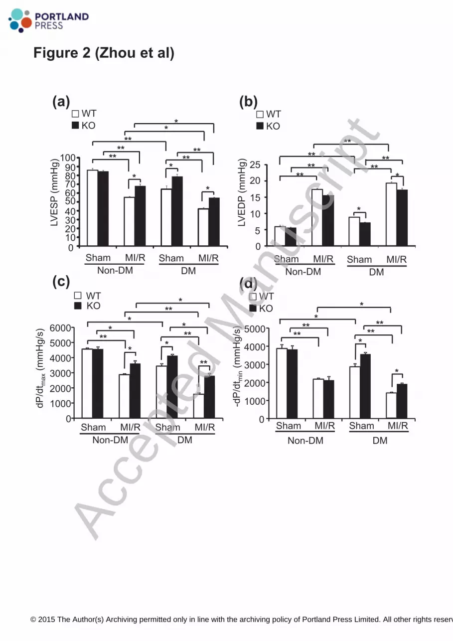
Severe hyperglycemia was paralleled by impairment of both cardiac contractile and diastolic 32 functions in WT mice while diabetic A-FABP KO mice had a preserved cardiac function (Figure 2 33 a-d). MI/R induced significant cardiac systolic dysfunction as evidenced by reductions in LVESP 34 (Figure 2a, In non–DM WT mice: Sham: 85.93±2.06 mmHg vs MI/R: 55.18±1.14 mmHg, P<0.001; 35 In non-DM KO mice: Sham: 84.51±1.22 mmHg vs MI/R: 67.93±2.41 mmHg, P=0.002) and dP/dtmax 36 (Figure 2c, In Non-DM WT mice: sham: 4561.60±62.29 mmHg/s vs MI/R: 2877.65±46.77 mmHg/s, 37 P<0.001; In Non-DM KO mice: sham: 4562.67±195.81 mmHg/s vs MI/R: 3616.30±87.88 mmHg/s, 38 P=0.023) in both WT and A-FABP KO mice while the reductions were consistently less in A-FABP 39 KO mice. Diabetes further exacerbated the MI/R-induced cardiac systolic dysfunction in both types 40 of mice (In WT mice, LVESP reduced from 55.18±1.14 mmHg to 42.09±1.52 mmHg; In A-FABP 41 KO mice, from 67.93±2.41 mmHg to 54.67±0.69 mmHg; dP/dtmax reduced from 2877.65±46.77 42 mmHg/s to 1578.54±88.33 mmHg/s in WT mice, and from 3616.30±162.74 mmHg/s to 43 2945.27±99.60 mmHg/s in A-FABP KO mice, all P<0.05) (Figure 2a and c). MI/R induced a similar 44
Acce
pted
Man
uscr
ipt
© 2015 The Author(s) Archiving permitted only in line with the archiving policy of Portland Press Limited. All other rights reserved.
10
increment of LVEDP and reduction in –dP/dtmin in both WT and KO mice while diabetes further 1 exaggerated the MI/R-induced impairment of cardiac diastolic function only in WT (all P<0.05) but 2 not in A-FABP KO mice (Figure 2 b and d). 3 4 A-FABP deficiency attenuates MI/R and diabetes-induced myocardial oxidative and nitrative 5 stresses and inflammation 6 MI/R injury alone caused a severe disruption in the geometric alignment of cardiomyocytes in WT 7
mice while the effect on A-FABP KO mice was mild. Diabetes alone also slightly altered the 8 alignment of cardiomyocytes in WT mice but not in A-FABP KO mice. Under diabetic conditions, 9 MI/R injury caused a marked reduction in the number of cardiomyocytes in WT mice as indicated by 10 the reduced number of stained nuclei while MI/R injury and diabetes induction had no synergistic 11 effect on cardiac structural changes in A-FABP KO mice (Figure 3a). 12 The superoxide anion production and peroxynitrite formation were next accessed in cardiac tissue. 13
DHE staining indicated that MI/R- or diabetes- induced superoxide anion production was 14 significantly reduced in A-FABP KO mice compared with WT mice (Figure 3b). MI/R and diabetes 15 synergistically increased superoxide anion production in both types of mice but A-FABP KO mice 16 consistently showed a significant lower level when compared to WT mice (39%-49% reduction in 17 ethidium fluorescence intensity in KO mice versus WT mice under MI/R, DM, or DM-MI/R 18 conditions). In addition, MI/R injury significantly increased nitrotyrosine formation in WT but not in 19 A-FABP KO mice. Although diabetes alone did not increase nitrotyrosine formation in both types of 20 mice, diabetes significantly exaggerated MI/R induced nitrotyrosine formation in WT mice while 21 that in A-FABP deficient hearts was significantly attenuated as indicated by immunohistological 22 staining (Figure 3c) and chemiluminescent protein quantification (Figure 3d). 23 Furthermore, MI/R-induced a significant increase in cardiac expression of inflammatory cytokines, 24
including TNF-α, MCP-1, and IL-6 in WT mice while only the expression of MCP-1 was 25 significantly enhanced in A-FABP KO mice when compared to their respective sham control. 26 Moreover, the MI/R induced increase in inflammatory cytokines was significantly attenuated in the 27 KO mice when compared to the respective WT mice (In WT mice, 3.37±0.24 fold, 10.83±1.30 fold, 28 and 8.17±0.37 fold increased, respectively, vs In A-FABP KO mice, 2.08±0.12 fold, 4.99±0.41 fold, 29 and 1.25±0.13 fold increased, respectively)(Figure 3e). Diabetes alone did not induce the cardiac 30 expression of any inflammatory cytokine in both WT and A-FABP KO mice. However, diabetes 31 significantly enhanced the MI/R-induced expression of TNF- and IL-6 in WT mice while the effect 32 on that in A-FABP-KO mice was markedly diminished (Figure 3e). There was no significant increase 33 in cardiac mRNA expression level of F4/80 in the heart of both WT and A-FABP KO mice after 34 MI/R and/or diabetes induction and the levels of F4/80 were similar between the two types of mice 35 (Supplementary Figure S3) suggesting that no significant macrophage infiltration had occurred. 36 These data are consistent with previous findings that infiltration of macrophages begins at day 3 in 37 the MI/R model [25]. Taken in conjunction, these results suggest that A-FABP deficiency attenuates 38 MI/R-induced and/or diabetes-induced cardiac injury at least partially by reducing myocardial 39 oxidative and nitrative stresses and inflammation. 40 41 The endothelium is the major cell source of cardiac A-FABP and its expression is induced 42 further by MI/R and diabetes 43
Immunofluorescence staining demonstrated that A-FABP is expressed abundantly in the heart of 44
Acce
pted
Man
uscr
ipt
© 2015 The Author(s) Archiving permitted only in line with the archiving policy of Portland Press Limited. All other rights reserved.

11
WT mice (Supplementary Figure S4a). Co-immunofluorescence staining showed that A-FABP only 1 co-localized with the endothelial marker CD31 but not with the cardiomyocyte marker troponin T 2 (Figure 4a), suggesting that the major cell source of cardiac A-FABP is the endothelium. Western 3 blot analysis also demonstrated that A-FABP was detected in adipose tissue (AT) and total heart 4 lysate (THL) but not in primary cardiomyocytes (PC) (Figure 4b). The mRNA expression of A-FABP 5 in primary cardiomyocytes was also undetectable by Q-PCR (Figure 4c). 6
MI/R or diabetes caused a significant up-regulation of mRNA expression (Figure 4d) and protein 7 presence (Figure 4e) of A-FABP in the heart tissue of WT mice. In addition, diabetes induced a 8 higher expression of A-FABP than that of MI/R injury. Under diabetic conditions, MI/R further 9 induced a mild but not significant increase of mRNA and protein expression of cardiac A-FABP and 10 of its circulating level (Supplementary Figure S4b). These data suggest that the elevated expression 11 of A-FABP in diabetic mice subjected to MI/R is contributed mainly by diabetes-induction but not by 12 MI/R injury. The cardiac expression of A-FABP was significantly higher in diabetic mice with MI/R 13 compared to non-diabetic mice with MI/R (Figure 4d and 4e). A similar trend was observed for the 14 serum A-FABP levels (Supplementary Figure S4b), in line with previous clinical findings 15 demonstrating raised circulating A-FABP levels in patients with diabetes [7, 8] and/or with ischemic 16 heart disease [10, 11]. 17 18 A-FABP deficiency enhances eNOS phosphorylation and NO production in the MI/R Heart 19
The eNOS-NO pathway exerts cardioprotective effects against apoptosis and oxidative stress [26]. 20 Exogenous A-FABP inhibits eNOS activation and NO production in cultured endothelial cells [20]. 21 Since A-FABP is expressed in cardiac endothelial cells and its expression is up-regulated by MI/R 22 and diabetes, the next experiments examined whether or not A-FABP deficiency exerts its 23 cardioprotective effect by modulating the eNOS-NO pathway. In WT mice, MI/R caused a 24 significant down-regulation of eNOS phosphorylation, and this detrimental effect was further 25 exacerbated in diabetic mice while diabetes alone did not alter eNOS phosphorylation (Figure 5a). 26 By contrast, diabetes alone or MI/R injury induced a significant, similar increase of eNOS 27 phosphorylation in A-FABP deficient hearts but MI/R injury did not further enhance eNOS 28 phosphorylation in heart tissue of diabetic A-FABP KO mice (Figure 5b). Consistent with the 29 increased eNOS phosphorylation, MI/R-subjected A-FABP deficient hearts showed a significantly 30 enhanced total NO production compared with those of WT controls under either control or diabetic 31 conditions (Figure 5c, 1.60±0.14 nmol/μg versus 0.92±0.16 nmol/μg in non-DM condition, P=0.015; 32 1.90±0.13 nmol/μg versus 0.91±0.07 nmol/μg in DM condition, P=0.005). However, diabetic 33 A-FABP KO mice only exhibited a slightly higher NO production when compared to respective WT 34 mice (1.20±0.08 nmol/μg versus 0.78±0.07 nmol/μg, P=0.05) (Figure 5c). These data suggest that 35 activation of the eNOS-NO pathway upon MI/R or diabetes induction was suppressed in the presence 36 of A-FABP. To determine whether or not the beneficial effect of A-FABP deficiency on MI/R injury 37 is attributable to the increased NO production, WT and A-FABP KO mice were pretreated with either 38 vehicle or the NOS inhibitor, NG-nitro-L-arginine methyl ester (L-NAME; 1.5mg/ml) followed by 39 either diabetes induction or MI/R injury. The systolic arterial blood pressure of both WT and 40 A-FABP KO mice was significantly increased to a similar extent after treatment with L-NAME while 41 diabetes-induction with STZ did not affect arterial blood pressure in either type of mice. Moreover, 42 STZ did not further increase the systolic blood pressure of L-NAME treated mice (Supplementary 43 Fig. S5). Furthermore, the present data also show that treatment with L-NAME did not alter the 44
Acce
pted
Man
uscr
ipt
© 2015 The Author(s) Archiving permitted only in line with the archiving policy of Portland Press Limited. All other rights reserved.

12
MI/R- or diabetes- induced cardiac injury and function in WT mice (Figure 6a-c). On the contrary, 1 the beneficial effects of A-FABP deficiency on the alleviation of MI/R-induced myocardial infarction 2 and deterioration of cardiac function was attenuated significantly when the A-FABP KO mice were 3 treated with L-NAME (Figure 6a and b). However, NOS inhibition did not exaggerate 4 diabetes-induced cardiac systolic dysfunction in A-FABP KO mice (Figure 6c). These data suggest 5 that A-FABP deficiency protects against MI/R induced cardiac injury mainly because of an increased 6 NO production. However, increased of NO production in the A-FABP deficient heart did not 7 contribute to the alleviation of diabetes-induced cardiac dysfunction. 8 To exclude other molecular sources that could be responsible for MI/R-induced increased NO 9 production in A-FABP KO mice, the protein expression of inducible NO synthase (iNOS) was also 10 determined. There was no significant change in iNOS expression after MI/R in either WT or A-FABP 11 KO animals (Supplementary Figure S6) which suggests the elevated NO production in A-FABP KO 12 mice mainly depends on eNOS activity. 13
14
Acce
pted
Man
uscr
ipt
© 2015 The Author(s) Archiving permitted only in line with the archiving policy of Portland Press Limited. All other rights reserved.

13
DISCUSSION 1 A-FABP is recognized as a key mediator of various obesity-related cardiometabolic disorders [5]. 2
Clinical and in vitro studies suggest that A-FABP is also implicated in cardiac dysfunction [12, 13, 3 15, 27]. A-FABP deficient mice are protected against diet-induced metabolic disorders such as 4 insulin resistance, hyperlipidemia and fatty liver despite of their increased susceptibility to 5 diet-induced obesity as compared to WT mice [28]. Therefore, in the present study a STZ-induced 6 type 1-, instead of a diet-induced type 2-, diabetes model was established to investigate the role of 7 A-FABP in MI/R induced cardiac injury associated with the disease to minimize the metabolic 8 discrepancy between WT and A-FABP KO mice upon diabetes induction, which may affect MI/R 9 injury. 10
The current data show that endothelial cells were identified as the major cellular source of cardiac 11 A-FABP which is supported by our previous findings [19]. Both mRNA and protein expressions of 12 A-FABP were markedly increased in the cardiac tissues of STZ-exposed and/ or MI/R-subjected 13 mice and STZ-exposed mice exhibited a higher A-FABP expression than that in MI/R-subjected 14 mice. This induction of cardiac A-FABP was accompanied by an elevated circulating level of the 15 adipokine. Under diabetic condition, MI/R can only slightly but not significantly enhance the 16 expression of A-FABP in mice. This may be due to the severe induction of A-FABP expression by 17 STZ-induced diabetes that masked the additive effect of MI/R. 18
A-FABP deficiency conferred cardio-protection with diminished infarct size, reduced cardiac 19 apoptosis and maintained better cardiac systolic and diastolic functions upon MI/R-induced cardiac 20 injury under both non-diabetic and diabetic conditions. The induction of NO production in A-FABP 21 deficient heart is specific for MI/R injury but not for diabetes induction. Moreover, treatment with 22 the NOS inhibitor L-NAME attenuates the beneficial effect of A-FABP deficiency in alleviating 23 MI/R-induced myocardial infarction and cardiac dysfunction, but not those induced by diabetes, 24 while it exerts no effect on WT mice. These data suggest that the cardio-protective effect of A-FABP 25 deficiency on MI/R injury is at least in part due to activation of the eNOS-NO pathway in endothelial 26 cells which subsequently alleviates cardiac oxidative stress and inflammation. Moreover, A-FABP 27 deficiency protects against diabetes-induced cardiac dysfunction by attenuating superoxide anion 28 production. Under diabetic condition, the presence of elevated superoxide anion further exaggerates 29 MI/R-induced oxidative and nitrative stress and inflammation leading to a more severe cardiac 30 damage and impairment of cardiac function. Thus, the present findings provide in vivo evidence that 31 A-FABP is involved in the development of MI/R- or diabetes-induced cardiac injury and dysfunction 32 as well as the exaggeration of MI/R-induced cardiac injury under diabetic condition. Consistent with 33 previous findings [3, 4], the present data show that diabetes increases the susceptibility to 34 MI/R-induced cardiac injury and that this can probably be attributed to the markedly enhanced 35 expression of A-FABP. 36
Cardiomyocyte survival depends on capillary endothelial cells for a proper supply of oxygenated 37 blood and the expression and release of protective signals and a variety of autocrine and paracrine 38 factors to maintain cardiac metabolism, growth, and an appropriate coronary vascular diameter [17]. 39 Endothelial dysfunction precedes most cardiovascular diseases and is implicated in cardiac 40 dysfunction [18]. The bioavailability of NO synthesized from L-arginine by eNOS plays an 41 obligatory role in the maintenance of a proper endothelial function [29]. Diabetic patients exhibit a 42 reduced ability to generate NO from L-arginine [30] and are more susceptible to ischemia 43 reperfusion injury as compared to healthy controls [4]. Mice deficient in eNOS also show increased 44
Acce
pted
Man
uscr
ipt
© 2015 The Author(s) Archiving permitted only in line with the archiving policy of Portland Press Limited. All other rights reserved.

14
MI/R injury [31]. Conversely, genetic overexpression of eNOS or NO donors attenuate MI/R injury 1 in mice [32, 33]. The eNOS-NO pathway exerts various cardiovascular protective effects through the 2 suppression of oxidative stress, inflammation, and apoptosis [26]. Consistent with these findings, the 3 present observation of an elevated eNOS phosphorylation and an increased NO production in the 4 heart of A-FABP KO mice explains the protective effects of the deletion against MI/R injury. 5
Exogenous A-FABP and lipid-induced A-FABP expression decrease eNOS phosphorylation and 6 NO production in cultured endothelial cells while pharmacological inhibition of A-FABP reverses 7 the condition and improves endothelial function in ApoE KO mice [19, 20] and prevents coronary 8 endothelial dysfunction in the pig [21]. The present findings demonstrate that A-FABP is expressed 9 in cardiac endothelial cells but not in cardiomyocytes and that this expression increases in response 10 to acute MI/R injury and diabetes suggesting that its adverse effect on endothelial cells contributes to 11 cardiac dysfunction. Cardiac eNOS phosphorylation and NO production following MI/R were 12 significantly reduced in WT mice but both were augmented in A-FABP KO mice, an increase 13 associated with reduced cardiac apoptosis and improved function. These data imply that a protective 14 mechanism associated with the activation of eNOS in response to MI/R is suppressed in the presence 15 of A-FABP with a resulting decreased NO bioavailability, illustrating the development of endothelial 16 dysfunction. This interpretation is comforted by the observation that the beneficial effects of 17 A-FABP deficiency were abolished by the administration of the eNOS inhibitor L-NAME. 18 Although eNOS can be also expressed in cardiomyocytes, the endothelium is the major contributor 19 of eNOS-derived NO affecting myocardial contractility [34]. In the isolated rabbit heart, the increase 20 in myocardial NO production in response to mechanical stimulation is abrogated after removing 21 cardiac endothelial and endocardial cells [35]. Furthermore, the inotropic response of cardiac 22 ventricular myocytes to β-adrenergic stimulation necessitates the production of NO from the 23 coronary microvascular endothelium [34, 36]. These findings also support the interpretation that 24 MI/R-induced cardiac dysfunction can be primarily attributed to the endothelial dysfunction caused 25 by A-FABP-mediated alteration in the eNOS-NO pathway. 26
Endothelial dysfunction resulting from impaired eNOS-NO pathway increases oxidative stress and 27 inflammation which further enhances the severity of the endothelial dysfunction. This vicious cycle 28 and additive effects eventually contribute to heart failure [29]. Elevated production of reactive 29 oxygen species (ROS) upon MI/R injury directly damage lipids, proteins and DNA, resulting in 30 irreversible oxidative modifications [37] associated with increased apoptosis, cardiac hypertrophy 31 and contractile dysfunction [38]. Elevated ROS also causes mitochondrial uncoupling leading to 32 cellular energy depletion in the diabetic heart [39]. ROS can interact with NO to generate 33 nitrotyrosine, leading to severe nitrative stress and additional cardiac damage [40]. Superoxide 34 anions scavenge NO and thus reduce NO bioavailability contributing to endothelial dysfunction and 35 apoptosis [41]. Hyperglycemia-induced oxidative stress also enhances cardiac inflammation by 36 activating the pro-inflammatory c-Jun N-terminal kinases (JNK) signaling pathway [42]. The present 37 results show that A-FABP KO mice exhibited an attenuated MI/R- and/or diabetes-induced cardiac 38 ROS production. Reduced oxidative stress diminished nitrative stress both in the absence or presence 39 of diabetes, which may contribute to the subsequent alleviation of cardiac cell apoptosis and 40 inflammation. 41
Pro-inflammatory cytokines also critically contribute to the pathogenesis of MI/R induced injury. 42 Elevated TNF- and IL6 promote cardiac hypertrophy, fibrosis and left ventricular dysfunction [43, 43 44]. TNF- and interleukin 1 (IL-1 ) increase epicardial thickness and induce cardiac contractile 44
Acce
pted
Man
uscr
ipt
© 2015 The Author(s) Archiving permitted only in line with the archiving policy of Portland Press Limited. All other rights reserved.

15
dysfunction [45]. A-FABP is a key mediator of inflammatory responses in macrophages [46, 47] 1 while both genetic disruption and pharmacological inhibition of A-FABP result in decreased 2 production of pro-inflammatory cytokines [5]. However, in the present study, no macrophage 3 infiltration was evidenced in the myocardium after acute MI/R injury and/or diabetes induction in 4 either WT or A-FABP KO mice and the cardiac expression of macrophage marker F4/80 was also 5 similar suggesting that the attenuated cardiac inflammation in A-FABP KO mice after MI/R can be 6 attributed mainly to the anti-inflammatory role of NO, the diminished ROS production in the 7 myocardium upon MI/R and diabetes induction or the low systemic inflammation state in A-FABP 8 KO mice. The elevated expression of inflammatory cytokines in response to myocardial infarction 9 may be contributed by cardiac fibroblasts and cardiomyocytes [48, 49]. Exposure to cytokines is 10 associated with induction of iNOS expression and activity mainly in infiltrated inflammatory cells 11 [50]. However, there was no change in iNOS expression among groups in the present study which 12 may be due a non-significant macrophage infiltration in the myocardium upon short term MI/R 13 injury. 14
In conclusion, the present study provides the novel insight that A-FABP plays a critical role in the 15 development of acute MI/R-induced cardiac dysfunction by inducing oxidative stress and 16 inflammation primarily via the modulation of the eNOS-NO pathway in endothelial cells. 17 Diabetes-induced elevation of A-FABP associated with raised superoxide anion production may 18 account for its adverse effect on the exaggeration of MI/R induced injury. These findings highlight 19 the importance of A-FABP in the pathogenesis of cardiac disorders. In particular, they suggest that 20 A-FABP may be a potential therapeutic target for the treatment of diabetes-associated cardiac 21 complications. 22 23 CLINICAL PERSPECTIVES 24
Circulating levels of A-FABP are elevated in diabetic patients and closely associated with 25 ischemic heart disease but the pathological link between diabetes and MI/R-induced cardiac 26 injury remains unclear. This study was designed to investigate the role of A-FABP in MI/R 27 injury under non-diabetic and diabetic condition. 28
Endothelial expression of A-FABP is increased upon diabetes-induction and acute MI/R. 29 Elevated A-FABP expression plays a critical role in the development of acute MI/R injury by 30 suppressing eNOS activation and NO production. Diabetes-induced elevation of A-FABP 31 expression associated with increased superoxide anion production may contribute to the 32 diabetes-induced cardiac dysfunction and the further exaggeration it causes of MI/R induced 33 cardiac injury. 34
A-FABP is a negative regulator of the eNOS-NO pathway causing endothelial dysfunction 35 which subsequently contributes to cardiac dysfunction by inducing oxidative stress and 36 inflammation. A-FABP may be a potential therapeutic target for the treatment of 37 diabetes-associated cardiac diseases. 38
39 AUTHOR CONTRIBUTION 40 Mi Zhou, Ruby LC Hoo and Aimin Xu completed conception and design of the research. Mi Zhou 41 and Ruby LC Hoo drafted the manuscript; Mi Zhou, Haobo Li and Yong Pan prepared the figures; 42 Yuqian Bao, Zhengyuan Xia, Karen SL Lam, Paul M. Vanhoutte, Aimin Xu, Weiping Jia edited the 43 manuscript. Donghai Wu generated A-FABP KO mice. Lingling Shu acted as technical support for 44
Acce
pted
Man
uscr
ipt
© 2015 The Author(s) Archiving permitted only in line with the archiving policy of Portland Press Limited. All other rights reserved.

16
in vivo study. Ruby LC Hoo, Paul M. Vanhoutte, Aimin Xu and Weiping Jia approved final version 1 of manuscript. 2 3 ACKNOWLEDGEMENTS 4 We would like to thank Dr. Songyan Liao, Mr. Yuelin Zhang and Dr. Kelvin Wing-hon Lai for their 5 technical support. 6 7 FUNDING 8 This study was supported by general research fund (GRF HKU767913M) and collaborative research 9 fund (HKU4/CRF/10R) from the Hong Kong Research Grant Council, (2011CB504001) from 973 10 Program, (11JC1409600) from Program of Shanghai Municipality for Basic Research and matching 11 fund for State Key Laboratory of Pharmaceutical Biotechnology from University of Hong Kong. 12 13 DISCLOSURES 14 No conflicts of interest, financial or otherwise, are declared by the authors. 15 16
17
Acce
pted
Man
uscr
ipt
© 2015 The Author(s) Archiving permitted only in line with the archiving policy of Portland Press Limited. All other rights reserved.

17
FIGURE LEGEND 1 Figure 1. A-FABP deficiency attenuates myocardial infarction and apoptosis in MI/R-induced mice 2 with (DM) or without (non-DM) diabetes. A-FABP-KO mice and their WT littermates were 3 subjected to either vehicle (citrate buffer) or STZ-injection to induce type 1 diabetes followed by 4 MI/R injury. (a) Myocardial infarct size was determined by Evans blue/TTC double staining. Upper 5 panel: Representative photomicrographs of heart sections obtained from MI/R-induced WT and 6 A-FABP KO mice with or without DM. The blue-staining areas indicate normal myocardium; 7 red-staining areas represent ischemic but non-infarcted tissue, whereas the unstained white areas 8 indicate infarcted tissue. Lower panel: Quantification of myocardial infarct size expressed as 9 percentage of area at risk (AAR), calculated as ischemic area plus infarcted area divided by total area 10 of the heart. (b) Diabetes and MI/R-induced cardiac cell apoptosis was determined by TUNEL 11 staining of the heart sections. TUNEL stained apoptotic nuclei in green and total nuclei 12 counterstained with DAPI in blue. 40x magnification. Values are expressed as means±SEM. *P < 13 0.05, **P < 0.01; n=5-6. 14 15 Figure 2. A-FABP deficiency attenuates cardiac dysfunction caused by MI/R mice with (DM) or 16 without (non-DM) diabetes or diabetes-induction. Cardiac systolic and diastolic functions were 17 determined by left ventricular catheterization as indicated by (a) left ventricular-end systolic pressure 18 (LVESP), (b) left ventricular-end diastolic pressure (LVEDP), (c) maximal rate of pressure 19 development (dP/dtmax) and (d) maximal rate of decay of pressure (-dP/dtmin). Values are expressed as 20 means±SEM. *P < 0.05, **P < 0.01; n=5-6. 21 22 Figure 3. A-FABP deficiency alleviates MI/R- and diabetes-induced cardiac histological change, 23 oxidative and nitrative stress and inflammation. (a) Histological analysis of heart sections by H&E 24 staining. (b) Cardiac superoxide anion production was determined by in situ DHE staining. The 25 fluorescence of DHE-labelled positive nuclei (red) was calculated in each of five randomly selected 26 fields and was expressed as mean pixel intensity. (c) Immunohistochemistry staining of cardiac 27 nitrotyrosine in each of five randomly selected fields. 40x magnification. (d) Myocardial 28 nitrotyrosine content was determined by chemiluminescence. (e) The mRNA abundance of 29 pro-inflammatory cytokines TNF- , MCP-1 and IL-6 in heart tissues was determined by Q-PCR and 30 normalized to the GAPDH gene. Values are expressed as means±SEM. *P < 0.05, **P < 0.01; n=5-6. 31 32 Figure 4. Endothelial cells are the major cell source of cardiac A-FABP and its expression is further 33 induced by MI/R and diabetes. (a) Co-immunofluorescence staining of cardiac A-FABP with the 34 endothelial cell marker CD31 and the cardiomyocyte marker troponin-T in the heart sections of WT 35 mice. (b) Western blot analysis of A-FABP expression and (c) the mRNA abundance of A-FABP in 36 adipose tissue (AT), total heart lysate (THL), and primary cardiomyocytes (PC) of WT mice. (d) The 37 mRNA abundance of cardiac A-FABP in MI/R- and/or diabetes-induced WT mice was determined by 38 Q-PCR. (e) Immunofluorescence staining of cardiac A-FABP of MI/R- and/or diabetes-induced WT 39 mice. Right panel: Quantification of the fluorescence intensity. Values are expressed as means±SEM. 40 *P < 0.05, **P < 0.01; n=6. 41 42 Figure 5. A-FABP deficiency protects against MI/R induced cardiac injury through activation of the 43 eNOS-NO pathway. Western blots measuring eNOS phosphorylation (Ser1177) in heart tissues of (a) 44
Acce
pted
Man
uscr
ipt
© 2015 The Author(s) Archiving permitted only in line with the archiving policy of Portland Press Limited. All other rights reserved.

18
WT and (b) A-FABP KO mice. (c) Cardiac NO production of A-FABP KO mice and WT mice. 1 Values were expressed as means±SEM. *P < 0.05, **P < 0.01; n=6. 2
3 Figure 6. Treatment of NOS inhibitor abolishes the cardioprotective effects of A-FABP KO mice 4 upon MI/R injury. A-FABP KO mice and their WT littermates were treated with either vehicle or 5 L-NAME for four weeks followed by MI/R injury or after diabetes induction. Effects of eNOS 6 inhibition on MI/R-induced (a) myocardial infarction and (b) cardiac dysfunction. (c) Effect of eNOS 7 inhibition on diabetes-induced cardiac dysfunction in A-FABP KO and WT mice. Values are 8 expressed as means±SEM. *P < 0.05, **P < 0.01; n=6. 9 10 SUPPLEMENTARY FIGURE LEGEND 11 Supplementary Figure S1. Generation of A-FABP KO mice. (a) Diagram of the A-FABP gene 12 targeting construct. An A-FABP gene targeting vector was generated with the exons (Ex) 1 to 4 of 13 A-FABP genes replaced with luciferase and neomycin. (b) Typical PCR result of genotyping analysis 14 using specific primers to identify heterozygous (HE), WT and A-FABP KO mice. (c) Western blot 15 analysis of A-FABP expression in the heart of WT and A-FABP KO mice. (d) Representative 16 immunohistochemistry staining of A-FABP in heart sections of WT and A-FABP KO mice. 40x 17 magnification; n=5-6. 18
19 Supplementary Figure S2. Streptozotocin (STZ)-induced diabetes in A-FABP KO mice and WT 20 littermates. A-FABP KO and their WT littermates were injected intra-peritoneally with either vehicle 21 (veh, citrate buffer) or STZ for five consecutive days to induce diabetes. (a) Blood glucose levels and 22 (b) body weight following STZ induction. Values are expressed as means±SEM. *P < 0.05, **P < 23 0.01; n=10. 24
25 Supplementary Figure S3. No significant change in the expression of macrophage marker F4/80 in 26 the heart of WT and A-FABP KO mice upon MI/R and/or diabetes induction. The mRNA abundance 27 was determined by Q-PCR. Values are expressed as means±SEM. n=6. 28
29 Supplementary Figure S4. Cardiac and circulating levels of A-FABP were increased upon MI/R 30 and/or diabetes induction. (a) Immunofluorescence staining of A-FABP expression in the heart tissue 31 of WT mice. (b) Circulating levels of A-FABP after MI/R and/or diabetes induction in WT mice were 32 determined by ELISA. Values are expressed as means±SEM. *P < 0.05, **P < 0.01; n=6. 33
34 Supplementary Fig S5. Treatment with L-NAME significantly increased the systolic arterial blood 35 pressure in diabetic (DM) and non-diabetic (Non-DM) WT and A-FABP KO mice to a similar extent. 36 Systolic arterial blood pressure of DM or non-DM WT and A-FABP KO mice treated with either 37 vehicle or L-NAME were measured using the non-invasive tail-cuff method. Values are expressed as 38 means±SEM. *P < 0.05, **P < 0.01; n=6. 39
40 Supplementary Figure S6. NO production in A-FABP KO mice upon MI/R and/or diabetes 41 induction is mainly contributed by eNOS but not iNOS activity. Western blot analysis of iNOS 42 expression in the heart tissue of WT and A-FABP KO mice. Values were expressed as means±SEM. 43 n=6.44
Acce
pted
Man
uscr
ipt
© 2015 The Author(s) Archiving permitted only in line with the archiving policy of Portland Press Limited. All other rights reserved.

19
1 REFERENCES 2 1. Go, A.S., Mozaffarian, D., Roger, V.L., Benjamin, E.J., Berry, J.D., Borden, W.B., Bravata, D.M., Dai, S., Ford, E.S., 3
Fox, C.S., Franco, S., Fullerton, H.J., Gillespie, C., Hailpern, S.M., Heit, J.A., Howard, V.J., Huffman, M.D., Kissela, 4 B.M., Kittner, S.J., Lackland, D.T., Lichtman, J.H., Lisabeth, L.D., Magid, D., Marcus, G.M., Marelli, A., Matchar, 5 D.B., McGuire, D.K., Mohler, E.R., Moy, C.S., Mussolino, M.E., Nichol, G., Paynter, N.P., Schreiner, P.J., Sorlie, P.D., 6 Stein, J., Turan, T.N., Virani, S.S., Wong, N.D., Woo, D., and Turner, M.B. (2013) Executive summary: heart 7 disease and stroke statistics--2013 update: a report from the American Heart Association. Circulation. 127, 8 143-152 9
2. Braunwald, E. and Kloner, R.A. (1985) Myocardial reperfusion: a double-edged sword? J Clin Invest. 76, 10 1713-1719 11
3. Norhammar, A., Lindback, J., Ryden, L., Wallentin, L., Stenestrand, U., Register of, I., and Knowledge about 12 Swedish Heart Intensive Care, A. (2007) Improved but still high short- and long-term mortality rates after 13 myocardial infarction in patients with diabetes mellitus: a time-trend report from the Swedish Register of 14 Information and Knowledge about Swedish Heart Intensive Care Admission. Heart. 93, 1577-1583 15
4. Feuvray, D., Idell-Wenger, J.A., and Neely, J.R. (1979) Effects of ischemia on rat myocardial function and 16 metabolism in diabetes. Circ Res. 44, 322-329 17
5. Furuhashi, M. and Hotamisligil, G.S. (2008) Fatty acid-binding proteins: role in metabolic diseases and potential 18 as drug targets. Nat Rev Drug Discov. 7, 489-503 19
6. Xu, A., Wang, Y., Xu, J.Y., Stejskal, D., Tam, S., Zhang, J., Wat, N.M., Wong, W.K., and Lam, K.S. (2006) Adipocyte 20 fatty acid-binding protein is a plasma biomarker closely associated with obesity and metabolic syndrome. Clin 21 Chem. 52, 405-413 22
7. Blaha, V., Musil, F., Smahelova, A., Ticha, A., Hyspler, R., Haluzik, M., Lesna, J., and Sobotka, L. (2012) Effects of 23 body fat reduction on plasma adipocyte fatty acid-binding protein concentration in obese patients with type 1 24 diabetes mellitus. Neuro Endocrinol Lett. 33 Suppl 2, 6-12 25
8. Tso, A.W., Xu, A., Sham, P.C., Wat, N.M., Wang, Y., Fong, C.H., Cheung, B.M., Janus, E.D., and Lam, K.S. (2007) 26 Serum adipocyte fatty acid binding protein as a new biomarker predicting the development of type 2 diabetes: 27 a 10-year prospective study in a Chinese cohort. Diabetes Care. 30, 2667-2672 28
9. Veniant, M.M., Hale, C., Helmering, J., Chen, M.M., Stanislaus, S., Busby, J., Vonderfecht, S., Xu, J., and Lloyd, 29 D.J. (2012) FGF21 promotes metabolic homeostasis via white adipose and leptin in mice. PLoS One. 7, e40164 30
10. Bao, Y., Lu, Z., Zhou, M., Li, H., Wang, Y., Gao, M., Wei, M., and Jia, W. (2011) Serum levels of adipocyte fatty 31 acid-binding protein are associated with the severity of coronary artery disease in Chinese women. PLoS One. 6, 32 e19115 33
11. von Eynatten, M., Breitling, L.P., Roos, M., Baumann, M., Rothenbacher, D., and Brenner, H. (2012) Circulating 34 adipocyte fatty acid-binding protein levels and cardiovascular morbidity and mortality in patients with coronary 35 heart disease: a 10-year prospective study. Arterioscler Thromb Vasc Biol. 32, 2327-2335 36
12. Baessler, A., Lamounier-Zepter, V., Fenk, S., Strack, C., Lahmann, C., Loew, T., Schmitz, G., Bluher, M., Bornstein, 37 S.R., and Fischer, M. (2014) Adipocyte fatty acid-binding protein levels are associated with left ventricular 38 diastolic dysfunction in morbidly obese subjects. Nutr Diabetes. 4, e106 39
13. Lamounier-Zepter, V., Look, C., Alvarez, J., Christ, T., Ravens, U., Schunck, W.H., Ehrhart-Bornstein, M., Bornstein, 40 S.R., and Morano, I. (2009) Adipocyte fatty acid-binding protein suppresses cardiomyocyte contraction: a new 41 link between obesity and heart disease. Circ Res. 105, 326-334 42
14. Balc, M.M., Arslan, U., Frat, H., Kocaoglu, I., Vural, M.G., Balc, K.G., Maden, O., Gurbuz, O.A., Ardc, S., and Yeter, 43 E. (2012) Serum levels of adipocyte fatty Acid-binding protein are independently associated with left ventricular 44
Acce
pted
Man
uscr
ipt
© 2015 The Author(s) Archiving permitted only in line with the archiving policy of Portland Press Limited. All other rights reserved.

20
mass and myocardial performance index in obstructive sleep apnea syndrome. J Investig Med. 60, 1020-1026 1 15. Liu, M., Zhou, M., Bao, Y., Xu, Z., Li, H., Zhang, H., Zhu, W., Zhang, J., Xu, A., Wei, M., and Jia, W. (2013) 2
Circulating adipocyte fatty acid-binding protein levels are independently associated with heart failure. Clin Sci 3 (Lond). 124, 115-122 4
16. Djousse, L., Bartz, T.M., Ix, J.H., Kochar, J., Kizer, J.R., Gottdiener, J.S., Tracy, R.P., Mozaffarian, D., Siscovick, D.S., 5 Mukamal, K.J., and Zieman, S.J. (2013) Fatty acid-binding protein 4 and incident heart failure: the 6 Cardiovascular Health Study. Eur J Heart Fail. 15, 394-399 7
17. Brutsaert, D.L. (2003) Cardiac endothelial-myocardial signaling: its role in cardiac growth, contractile 8 performance, and rhythmicity. Physiol Rev. 83, 59-115 9
18. Katz, S.D., Hryniewicz, K., Hriljac, I., Balidemaj, K., Dimayuga, C., Hudaihed, A., and Yasskiy, A. (2005) Vascular 10 endothelial dysfunction and mortality risk in patients with chronic heart failure. Circulation. 111, 310-314 11
19. Lee, M.Y., Li, H., Xiao, Y., Zhou, Z., Xu, A., and Vanhoutte, P.M. (2011) Chronic administration of BMS309403 12 improves endothelial function in apolipoprotein E-deficient mice and in cultured human endothelial cells. Br J 13 Pharmacol. 162, 1564-1576 14
20. Aragones, G., Saavedra, P., Heras, M., Cabre, A., Girona, J., and Masana, L. (2012) Fatty acid-binding protein 4 15 impairs the insulin-dependent nitric oxide pathway in vascular endothelial cells. Cardiovasc Diabetol. 11, 72 16
21. Chan, C.K., Zhao, Y., Liao, S.Y., Zhang, Y.L., Lee, M.Y., Xu, A., Tse, H.F., and Vanhoutte, P.M. (2013) A-FABP and 17 oxidative stress underlie the impairment of endothelium-dependent relaxations to serotonin and the 18 intima-medial thickening in the porcine coronary artery with regenerated endothelium. ACS Chem Neurosci. 4, 19 122-129 20
22. Bae, S. and Zhang, L. (2005) Gender differences in cardioprotection against ischemia/reperfusion injury in adult 21 rat hearts: focus on Akt and protein kinase C signaling. J Pharmacol Exp Ther. 315, 1125-1135 22
23. Tao, L., Gao, E., Jiao, X., Yuan, Y., Li, S., Christopher, T.A., Lopez, B.L., Koch, W., Chan, L., Goldstein, B.J., and Ma, 23 X.L. (2007) Adiponectin cardioprotection after myocardial ischemia/reperfusion involves the reduction of 24 oxidative/nitrative stress. Circulation. 115, 1408-1416 25
24. Hoo, R.L., Lee, I.P., Zhou, M., Wong, J.Y., Hui, X., Xu, A., and Lam, K.S. (2013) Pharmacological inhibition of 26 adipocyte fatty acid binding protein alleviates both acute liver injury and non-alcoholic steatohepatitis in mice. 27 J Hepatol. 58, 358-364 28
25. Lambert, J.M., Lopez, E.F., and Lindsey, M.L. (2008) Macrophage roles following myocardial infarction. Int J 29 Cardiol. 130, 147-158 30
26. Yu, Q., Gao, F., and Ma, X.L. (2011) Insulin says NO to cardiovascular disease. Cardiovasc Res. 89, 516-524 31 27. Balci, M.M., Arslan, U., Firat, H., Kocaoglu, I., Vural, M.G., Balci, K.G., Maden, O., Gurbuz, O.A., Ardic, S., and 32
Yeter, E. (2012) Serum levels of adipocyte fatty acid-binding protein are independently associated with left 33 ventricular mass and myocardial performance index in obstructive sleep apnea syndrome. J Investig Med. 60, 34 1020-1026 35
28. Hotamisligil, G.S., Johnson, R.S., Distel, R.J., Ellis, R., Papaioannou, V.E., and Spiegelman, B.M. (1996) 36 Uncoupling of obesity from insulin resistance through a targeted mutation in aP2, the adipocyte fatty acid 37 binding protein. Science. 274, 1377-1379 38
29. Marti, C.N., Gheorghiade, M., Kalogeropoulos, A.P., Georgiopoulou, V.V., Quyyumi, A.A., and Butler, J. (2012) 39 Endothelial dysfunction, arterial stiffness, and heart failure. J Am Coll Cardiol. 60, 1455-1469 40
30. Pieper, G.M. (1998) Review of alterations in endothelial nitric oxide production in diabetes: protective role of 41 arginine on endothelial dysfunction. Hypertension. 31, 1047-1060 42
31. Scherrer-Crosbie, M., Ullrich, R., Bloch, K.D., Nakajima, H., Nasseri, B., Aretz, H.T., Lindsey, M.L., Vancon, A.C., 43 Huang, P.L., Lee, R.T., Zapol, W.M., and Picard, M.H. (2001) Endothelial nitric oxide synthase limits left 44
Acce
pted
Man
uscr
ipt
© 2015 The Author(s) Archiving permitted only in line with the archiving policy of Portland Press Limited. All other rights reserved.

21
ventricular remodeling after myocardial infarction in mice. Circulation. 104, 1286-1291 1 32. Brunner, F., Maier, R., Andrew, P., Wolkart, G., Zechner, R., and Mayer, B. (2003) Attenuation of myocardial 2
ischemia/reperfusion injury in mice with myocyte-specific overexpression of endothelial nitric oxide synthase. 3 Cardiovasc Res. 57, 55-62 4
33. Lefer, A.M. and Lefer, D.J. (1996) The role of nitric oxide and cell adhesion molecules on the microcirculation in 5 ischaemia-reperfusion. Cardiovasc Res. 32, 743-751 6
34. Seddon, M., Shah, A.M., and Casadei, B. (2007) Cardiomyocytes as effectors of nitric oxide signalling. 7 Cardiovasc Res. 75, 315-326 8
35. Pinsky, D.J., Patton, S., Mesaros, S., Brovkovych, V., Kubaszewski, E., Grunfeld, S., and Malinski, T. (1997) 9 Mechanical transduction of nitric oxide synthesis in the beating heart. Circ Res. 81, 372-379 10
36. Godecke, A., Heinicke, T., Kamkin, A., Kiseleva, I., Strasser, R.H., Decking, U.K., Stumpe, T., Isenberg, G., and 11 Schrader, J. (2001) Inotropic response to beta-adrenergic receptor stimulation and anti-adrenergic effect of ACh 12 in endothelial NO synthase-deficient mouse hearts. J Physiol. 532, 195-204 13
37. Finkel, T. (2003) Oxidant signals and oxidative stress. Curr Opin Cell Biol. 15, 247-254 14 38. Grieve, D.J. and Shah, A.M. (2003) Oxidative stress in heart failure. More than just damage. Eur Heart J. 24, 15
2161-2163 16 39. Boudina, S., Sena, S., O'Neill, B.T., Tathireddy, P., Young, M.E., and Abel, E.D. (2005) Reduced mitochondrial 17
oxidative capacity and increased mitochondrial uncoupling impair myocardial energetics in obesity. Circulation. 18 112, 2686-2695 19
40. Beckman, J.S. and Koppenol, W.H. (1996) Nitric oxide, superoxide, and peroxynitrite: the good, the bad, and 20 ugly. Am J Physiol. 271, C1424-1437 21
41. Cai, H. and Harrison, D.G. (2000) Endothelial dysfunction in cardiovascular diseases: the role of oxidant stress. 22 Circ Res. 87, 840-844 23
42. Tibbles, L.A. and Woodgett, J.R. (1999) The stress-activated protein kinase pathways. Cell Mol Life Sci. 55, 24 1230-1254 25
43. Sun, M., Dawood, F., Wen, W.H., Chen, M., Dixon, I., Kirshenbaum, L.A., and Liu, P.P. (2004) Excessive tumor 26 necrosis factor activation after infarction contributes to susceptibility of myocardial rupture and left ventricular 27 dysfunction. Circulation. 110, 3221-3228 28
44. Kaneko, K., Kanda, T., Yokoyama, T., Nakazato, Y., Iwasaki, T., Kobayashi, I., and Nagai, R. (1997) Expression of 29 interleukin-6 in the ventricles and coronary arteries of patients with myocardial infarction. Res Commun Mol 30 Pathol Pharmacol. 97, 3-12 31
45. Lorenzo, O., Picatoste, B., Ares-Carrasco, S., Ramirez, E., Egido, J., and Tunon, J. (2011) Potential role of nuclear 32 factor kappaB in diabetic cardiomyopathy. Mediators Inflamm. 2011, 652097 33
46. Makowski, L., Boord, J.B., Maeda, K., Babaev, V.R., Uysal, K.T., Morgan, M.A., Parker, R.A., Suttles, J., Fazio, S., 34 Hotamisligil, G.S., and Linton, M.F. (2001) Lack of macrophage fatty-acid-binding protein aP2 protects mice 35 deficient in apolipoprotein E against atherosclerosis. Nat Med. 7, 699-705 36
47. Hui, X., Li, H., Zhou, Z., Lam, K.S., Xiao, Y., Wu, D., Ding, K., Wang, Y., Vanhoutte, P.M., and Xu, A. (2010) 37 Adipocyte fatty acid-binding protein modulates inflammatory responses in macrophages through a positive 38 feedback loop involving c-Jun NH2-terminal kinases and activator protein-1. J Biol Chem. 285, 10273-10280 39
48. Porter, K.E. and Turner, N.A. (2009) Cardiac fibroblasts: at the heart of myocardial remodeling. Pharmacol Ther. 40 123, 255-278 41
49. Aoyagi, T. and Matsui, T. (2011) The Cardiomyocyte as a Source of Cytokines in Cardiac Injury. J Cell Sci Ther. 42 2012 43
50. Fukuchi, M., Hussain, S.N., and Giaid, A. (1998) Heterogeneous expression and activity of endothelial and 44
Acce
pted
Man
uscr
ipt
© 2015 The Author(s) Archiving permitted only in line with the archiving policy of Portland Press Limited. All other rights reserved.

22
inducible nitric oxide synthases in end-stage human heart failure: their relation to lesion site and 1 beta-adrenergic receptor therapy. Circulation. 98, 132-139 2
3 4
Acce
pted
Man
uscr
ipt
© 2015 The Author(s) Archiving permitted only in line with the archiving policy of Portland Press Limited. All other rights reserved.

Acce
pted
Man
uscr
ipt
© 2015 The Author(s) Archiving permitted only in line with the archiving policy of Portland Press Limited. All other rights reserved.
Acce
pted
Man
uscr
ipt
© 2015 The Author(s) Archiving permitted only in line with the archiving policy of Portland Press Limited. All other rights reserved.

Acce
pted
Man
uscr
ipt
© 2015 The Author(s) Archiving permitted only in line with the archiving policy of Portland Press Limited. All other rights reserved.

Acce
pted
Man
uscr
ipt
© 2015 The Author(s) Archiving permitted only in line with the archiving policy of Portland Press Limited. All other rights reserved.

Acce
pted
Man
uscr
ipt
© 2015 The Author(s) Archiving permitted only in line with the archiving policy of Portland Press Limited. All other rights reserved.

Acce
pted
Man
uscr
ipt
© 2015 The Author(s) Archiving permitted only in line with the archiving policy of Portland Press Limited. All other rights reserved.
Recommended


