Down-Regulation of the Carcinogen-Metabolizing EnzymeCytochrome P450 1a1 by Vanadium
Anwar Anwar-Mohamed and Ayman O. S. El-Kadi
Faculty of Pharmacy and Pharmaceutical Sciences, University of Alberta, Edmonton, Alberta, Canada
Received February 20, 2008; accepted June 4, 2008
ABSTRACT:
Vanadium (V5�), a heavy metal contaminant with important toxico-logical consequences, has received considerable attention as ananticancer agent, although the mechanisms remain unknown. As afirst step to investigate these mechanisms, we examined the effectof V5� (as ammonium metavanadate, NH4VO3) on the expression ofthe aryl hydrocarbon receptor (AhR)-regulated gene: cytochromeP450 1a1 (Cyp1a1) at each step of the AhR signal transductionpathway, using Hepa 1c1c7 cells. Our results showed a significantreduction in 2,3,7,8-tetrachlorodibenzo-p-dioxin (TCDD)-mediatedinduction of Cyp1a1 mRNA, protein and activity levels after V5�
treatments in a dose-dependent manner. Investigation of the effectof coexposure to V5� and TCDD at transcriptional levels revealedthat V5� significantly inhibited TCDD-mediated induction of AhR-dependent luciferase reporter gene expression. Furthermore, de-
spite not affecting the direct activation of the cytosolic AhR byTCDD and subsequently transforming it to a DNA-binding form,V5� inhibited the nuclear accumulation of liganded AhR and sub-sequent formation of the AhR/aryl hydrocarbon nuclear trans-locator (Arnt)/xenobiotic responsive element (XRE) complex.Importantly, the V5�-mediated inhibition of AhR/Arnt/XRE complexformation coincided with a significant decrease in ecto-ATPaseactivity. Looking at the post-transcriptional and post-translationaleffects of V5� on existing Cyp1a1 mRNA and protein levels, weshowed that V5� did not affect Cyp1a1 mRNA or protein stability,thus eliminating possible role of V5� in modifying Cyp1a1 geneexpression through these mechanisms. This study provides thefirst evidence that V5� down-regulates the expression of Cyp1a1 atthe transcriptional level through an ATP-dependent mechanism.
The aryl hydrocarbon receptor (AhR) is a ligand-activated cyto-plasmic transcription factor that belongs to the basic-helix-loop-helixprotein family. The inactive form of AhR is attached to a complex oftwo heat shock proteins 90 (HSP90), hepatitis B virus X-associatedprotein (XAP2), and the chaperone protein p23 (Hankinson, 1995;Meyer et al., 1998). Upon ligand binding, the AhR-ligand complexdissociates from the cytoplasmic complex and translocates to thenucleus where it associates with the aryl hydrocarbon nuclear trans-locator (Arnt) (Whitelaw et al., 1994). The whole complex then actsas a transcription factor that binds to a specific DNA recognitionsequence, termed the xenobiotic responsive element (XRE), located inthe promoter region of a number of AhR-regulated genes (Denison etal., 1989; Nebert et al., 2004). Among these genes are those encodinga number of drug-metabolizing enzymes, including four phase Ienzymes: cytochrome P450 1a1 (Cyp1a1), Cyp1a2, Cyp1b1, andCyp2s1; and four phase II enzymes: NAD(P)H:quinone oxidoreduc-tase-1, glutathione S-transferase a1, cytosolic aldehyde dehydroge-
nase-3, and UDP-glucuronosyltransferase 1a6 (Nebert and Duffy,1997).
Experimental and epidemiological data have shown that variousenvironmental pollutants such as halogenated aromatic hydrocarbonsare capable of producing a variety of toxic effects in exposed organ-isms; some of the most common toxicities include neurotoxicity,immune dysfunction, reproductive and developmental effects, andcancer (Schrenk, 1998; Elbekai and El-Kadi, 2004). Mounting evi-dence suggests that most of the toxic manifestations induced by,2,3,7,8-tetrachlorodibenzo-p-dioxin (TCDD), the most toxic haloge-nated aromatic hydrocarbon with the greatest affinity for AhR, areoccurring through the activation of the AhR (Kransler et al., 2007). Asa result of the tight correlation between AhR binding affinity andCyp1a1 induction, Cyp1a1 has been used as a biomarker for evalu-ating hazards and risk assessments of environmental pollutants(Behnisch et al., 2001). By itself, Cyp1a1 is capable of producingpolar, toxic, or even carcinogenic metabolites from various AhRligands including aromatic and halogenated hydrocarbons (Anwar-Mohamed and El-Kadi, 2007).
The toxicity of individual AhR ligands has been examined innumerous studies, yet the combined toxic effects of these ligands andother environmental cocontaminants typified by heavy metals such asvanadium (V5�) are still unclear. V5�, a cocontaminant with AhR
This work was supported by Natural Sciences and Engineering ResearchCouncil of Canada Discovery Grant RGPIN 250139-07 to A.O.S.E.-K. A.A.-M. isrecipient of a Mike Wolowyk Graduate Scholarship award.
Article, publication date, and citation information can be found athttp://dmd.aspetjournals.org.
doi:10.1124/dmd.108.021154.
ABBREVIATIONS: AhR, aryl hydrocarbon receptor; Arnt, aryl hydrocarbon receptor nuclear translocator; XRE, xenobiotic responsive element;Cyp1a1, cytochrome P450 1a1; TCDD, 2,3,7,8-tetrachlorodibenzo-p-dioxin; V5�, vanadium; pGudluc1.1, XRE-luciferase reporter plasmid; EMSA,electrophoretic mobility shift assay; MTT, 3-(4,5-dimethylthiazol-2-yl)-2,5-diphenyltetrazolium bromide; CHX, cycloheximide; Act-D, actinomycinD; DMSO, dimethylsulfoxide; PBS, phosphate-buffered saline; PCR, polymerase chain reaction; HO-1, heme oxygenase-1; PAGE, polyacrylamidegel electrophoresis; Gapdh, glyceraldehyde-3-phosphate dehydrogenase; EROD, 7-ethoxyresorufin O-deethylase; NF-�B, nuclear factor-�B.
0090-9556/08/3609-1819–1827$20.00DRUG METABOLISM AND DISPOSITION Vol. 36, No. 9Copyright © 2008 by The American Society for Pharmacology and Experimental Therapeutics 21154/3372212DMD 36:1819–1827, 2008 Printed in U.S.A.
1819
at University of A
lberta on January 23, 2013dm
d.aspetjournals.orgD
ownloaded from
ligands, is widely distributed in nature. The oxidation state of V (�4or � 5) seems to be important for its actions on enzymes (Cantleyand Aisen, 1979). V5� compounds have been found in various typesof food, such as black pepper, mushrooms, dill seed, parsley, andshellfish (Rojas et al., 1999). Interestingly, previous studies havedemonstrated that V5� compounds exert protective effects againstchemical-induced carcinogenesis mainly by modifying various xeno-biotic-metabolizing enzymes; however, the exact mechanism(s)remain unknown (Evangelou, 2002).
The objective of this study was to determine the effect of coexpo-sure to V5� and TCDD on Cyp1a1 and to investigate the molecularmechanisms involved. As a first step in investigating these mecha-nisms, we examined the effect of coexposure to V5� and TCDD onCyp1a1 mRNA, protein, and activity levels in Hepa 1c1c7 cells. Toaddress whether the observed effects of coexposure to V5� and TCDDoccurred through an AhR-dependent mechanism, we examined theeffect of the coexposure on luciferase activity in Hepa 1c1c7 cellstransiently transfected with the XRE-driven luciferase plasmid pGud-Luc1.1. Looking at the involvement of post-transcriptional mecha-nisms, we tested the effect of V5� on Cyp1a1 mRNA and proteinstability. We also examined the ability of V5� to modulate AhR/Arnt/XRE binding using the electrophoretic mobility shift assay (EMSA)and the role of ecto-ATPase in this modulation. We provide here thefirst evidence that V5� down-regulates the expression of Cyp1a1 byinhibiting the translocation of the transformed AhR, possibly throughan ATP-dependent mechanism.
Materials and Methods
Materials. Ammonium metavanadate (NH4VO3), 3-(4,5-dimethylthiazol-2-yl)-2,5-diphenyltetrazolium bromide (MTT), cycloheximide (CHX),p-nitrophenyl phosphate, p-nitrophenol, 2,6-dichlorophenolindophenol,7-ethoxyresorufin, fluorescamine, anti-goat IgG peroxidase secondary anti-body, and protease inhibitor cocktail were purchased from Sigma-Aldrich (St.Louis, MO). 2,3,7,8-Tetrachlorodibenzo-p-dioxin, �99% pure, was purchasedfrom Cambridge Isotope Laboratories (Woburn, MA). TRIzol reagent andLipofectamine 2000 reagents were purchased from Invitrogen (San Diego,CA). The High-Capacity cDNA Reverse Transcription Kit and SYBR GreenPCR Master Mix were purchased from Applied Biosystems (Foster City, CA).Actinomycin-D (Act-D) was purchased from Calbiochem (San Diego, CA).Chemiluminescence Western blotting detection reagents were from GEHealthcare Life Sciences (Piscataway, NJ). Nitrocellulose membrane waspurchased from Bio-Rad (Hercules, CA). Cyp1a1 goat polyclonal primaryantibody, AhR rabbit polyclonal primary antibody, and anti-rabbit IgG perox-idase secondary antibody were purchased from Santa Cruz Biotechnology, Inc.(Santa Cruz, CA). Luciferase assay reagents were obtained from Promega(Madison, WI). [�-32P]ATP was supplied by the DNA Core Services Labora-tory, University of Alberta. All other chemicals were purchased from ThermoFisher Scientific (Toronto, ON, Canada).
Cell Culture. The Hepa 1c1c7 cell line (number CRL-2026; AmericanType Culture Collection, Manassas, VA), was maintained in Dulbecco’s mod-ified Eagle’s medium without phenol red and supplemented with 10% heat-inactivated fetal bovine serum, 20 �M l-glutamine, 50 �g/ml amikacin, 100IU/ml penicillin, 10 �g/ml streptomycin, 25 ng/ml amphotericin B, 0.1 mMnonessential amino acids, and vitamin supplement solution. Cells were grownin 75-cm2 cell culture flasks at 37°C in a 5% CO2 humidified incubator.
Chemical Treatments. Cells were treated in serum-free medium withvarious concentrations of V5� (25–1000 �M) in the absence and presence of1 nM TCDD as described in the figure legends. TCDD was dissolved indimethyl sulfoxide (DMSO) and maintained in DMSO at �20°C until use.V5� was prepared freshly in double deionized water. In all treatments, theDMSO concentration did not exceed 0.05% (v/v).
Effect of V5� on Cell Viability. The effect of V5� on cell viability wasdetermined using the MTT and ATP-based luminescent assays as describedpreviously (Maniratanachote et al., 2005; Korashy and El-Kadi, 2008). TheMTT assay measures the conversion of MTT to formazan in living cells via
mitochondrial enzymes of viable cells. In brief, Hepa 1c1c7 cells were seededinto 96-well microtiter cell culture plates and incubated for 24 h at 37°C in a5% CO2 humidified incubator. Cells were treated with various concentrationsof V5� (25–1000 �M) in the absence and presence of 1 nM TCDD. After 24 hof incubation, the medium was removed and replaced with cell culture mediumcontaining 1.2 mM MTT dissolved in phosphate-buffered saline (PBS) (pH7.4). After 2 h of incubation, the formed crystals were dissolved in isopropa-nol. The intensity of the color in each well was measured at a wavelength of550 nm using the Bio-Tek EL 312e microplate reader (Bio-Tek Instruments,Winooski, VT).
The ATP-based luminescent assay measures the quantity of the ATP pro-duced by metabolically active cells, which was determined using a CellTiter-Glo Luminescent assay kit according to the manufacturer’s instructions (Pro-mega). In brief, Hepa 1c1c7 cells were seeded into 24-well cell culture platesand incubated for 24 h at 37°C in a 5% CO2 humidified incubator. Cells werethen treated with various concentrations of V5� (25–1000 �M) in the absenceand presence of 1 nM TCDD for another 24 h. At the end of treatment period,200 �l of CellTiter-Glo reagent was added to each well. The luminescentsignal generated was monitored on a TD-20/20 luminometer (Turner BioSys-tems, Sunnyvale CA).
RNA Extraction and Quantitative Real-Time PCR of Cyp1a1. Afterincubation with the test compounds for the specified time periods, total cellularRNA was isolated using TRIzol reagent, according to manufacturer’s instruc-tions (Invitrogen), and quantified by measuring the absorbance at 260 nm. Forreverse transcription-PCR, first-strand cDNA was synthesized from 1.0 �g oftotal RNA using the High-Capacity cDNA Reverse Transcription Kit withrandom primers. Real-time PCR reactions were performed on an ABI 7500real-time PCR system (Applied Biosystems), using SYBR Green PCR MasterMix. The amplification reactions were performed as follows: 10 min at 95°Cand 40 cycles of 94°C for 15 s and 60°C for 1 min. Primers and probes asfollows were purchased from Integrated DNA Technologies, Inc. (Coralville,IA): for mouse Cyp1a1 forward primer 5�-GGT TAA CCA TGA CCG GGAACT-3� and reverse primer 5�-TGC CCA AAC CAA AGA GAG TGA-3�; forheme oxygenase-1(HO-1) forward primer 5�-GTG ATG GAG CGT CCACAG C-3� and reverse primer 5�-TGG TGG CCT CCT TCA AGG-3�; and for�-actin forward primer 5�-TAT TGG CAA CGA GCG GTT CC-3� and reverseprimer 5�-GGC ATA GAG GTC TTT ACG GAT GTC-3�. The fold changein the level of Cyp1a1 (target gene) between treated and untreated cells,corrected by the level of �-actin, was determined using the following equation:fold change � 2�� (�Ct), where �Ct � Ct (target) � Ct (�-actin) and �(�Ct) ��Ct (treated) � �Ct (untreated).
Protein Extraction and Western Blot Analysis. Twenty-four hours afterincubation with the test compounds, cells were collected in lysis buffercontaining 50 mM HEPES, 0.5 M sodium chloride, 1.5 mM magnesiumchloride, 1 mM EDTA, 10% (v/v) glycerol, 1% Triton X-100, and 5 �l/ml ofprotease inhibitor cocktail. The cell homogenates were obtained by incubatingthe cell lysates on ice for 1 h, with intermittent vortexing every 10 min,followed by centrifugation at 12,000g for 10 min at 4°C. Proteins (25 �g) wereresolved by denaturing electrophoresis, as described previously (Elbekai andEl-Kadi, 2004). Briefly, the cell homogenates were dissolved in 1� samplebuffer, boiled for 5 min, separated by 10% SDS-PAGE and electrophoreticallytransferred to a nitrocellulose membrane. Protein blots were blocked for 24 hat 4°C in blocking buffer containing 5% skim milk powder, 2% bovine serumalbumin, and 0.05% (v/v) Tween 20 in Tris-buffered saline solution (0.15 Msodium chloride, 3 mM potassium chloride, and 25 mM Tris base). Afterblocking, the blots were incubated with a primary polyclonal goat anti-mouseCyp1a1 antibody for 2 h at room temperature or primary polyclonal rabbitanti-mouse AhR antibody overnight at 4°C in Tris-buffered saline containing0.05% (v/v) Tween 20 and 0.02% sodium azide. Incubation with a peroxidase-conjugated rabbit anti-goat IgG secondary antibody for Cyp1a1 and Gapdh orgoat anti-rabbit IgG for AhR was carried out in blocking buffer for 1 h at roomtemperature. The bands were visualized with the enhanced chemiluminescencemethod according to the manufacturer’s instructions (GE Healthcare, Mississ-auga, ON). The intensity of Cyp1a1 protein bands was quantified relative tothe signals obtained for Gapdh protein, using ImageJ software.
Determination of Cyp1a1 Enzymatic Activity. Cyp1a1-dependent7-ethoxyresorufin O-deethylase (EROD) activity was performed on intact,living cells using 7-ethoxyresorufin as a substrate, as described previously
1820 ANWAR-MOHAMED AND EL-KADI
at University of A
lberta on January 23, 2013dm
d.aspetjournals.orgD
ownloaded from
(Elbekai and El-Kadi, 2004). Enzymatic activity was normalized for cellularprotein content, which was determined using a modified fluorescent assay(Lorenzen and Kennedy, 1993).
Transient Transfection and Luciferase Assay. Hepa 1c1c7 cells wereplated onto six-well cell culture plates. Each well of cells was transfected with4 �g of XRE-driven luciferase reporter plasmid pGudLuc1.1 using Lipo-fectamine 2000 reagent according to the manufacturer’s instructions (Invitro-gen). The luciferase assay was performed according to the manufacturer’sinstructions (Promega) as described previously (Elbekai and El-Kadi, 2007). Inbrief, after incubation with test compounds for 24 h, cells were washed withPBS, 200 �l of 1� lysis buffer was added into each well with continuousshaking for at least 20 min, and then the content of each well was collectedseparately in 1.5-ml microcentrifuge tubes. The tubes were then centrifuged toprecipitate cellular waste, 100 �l of cell lysate was incubated with 100 �l ofstabilized luciferase reagent, and luciferase activity was quantified using aTD-20/20 luminometer (Turner BioSystems).
EMSA. Nuclear extracts were prepared from Hepa 1c1c7 cells and treatedfor 2 h with vehicle, 5 nM TCDD, in the absence and presence of 50 �M V5�
using the method of Denison et al. (2002). Because of the low efficiency oftransformation of the mouse AhR, resulting from the extreme resistance ofHSP90 to dissociate from the mouse AhR, compared with the greatest degreeof transformation of the guinea pig AhR in response to AhR ligand (Boho-nowych and Denison, 2007), we used guinea pig cytosol as a model. Therefore,hepatic cytosol of untreated guinea pig, generously provided by Dr. M. S.Denison at University of California, Davis (Davis, CA), was incubated with thetest compounds for a final concentration of 20 nM TCDD, in the absence andpresence of 100 and 250 �M V5� for 2 h at 20°C. Protein concentrations forthe nuclear and cytosolic extracts were determined using the method of Lowryet al. (1951). To visualize the ability of V5� to alter the transformation andsubsequent DNA binding of the AhR, a complementary pair of syntheticoligonucleotides containing the sequences 5�-GAT CTG GCT CTT CTC ACGCAA CTC CG-3� and 5�-GAT CCG GAG TTG CGT GAG AAG AGC CA-3�,corresponding to the XRE binding site, were synthesized and radiolabeled with[�-32P]ATP as described previously (Denison et al., 2002) and used as a DNAprobe in all experiments. Binding reactions using aliquots of 120 �g ofcytosolic or 20 �g of nuclear extracts and excess radiolabeled oligonucleotideswere allowed to proceed for 15 min at 20°C in a buffer containing 1 mMEDTA, 1 mM dithiothreitol, 10% glycerol, 25 mM HEPES, 400 ng to 2 �g ofpoly(dI-dC), and 0.4 to 0.8 mM KCl. To determine the specificity of bindingto the oligonucleotide, a 100-fold molar excess of unlabeled XRE probe wasadded to the binding reaction before addition of the �-32P-labeled probe.Protein-DNA complexes were separated under nondenaturing conditions on a4% polyacrylamide gel using 1� TBE (90 mM Tris borate, 90 mM boric acid,and 4 mM EDTA) as a running buffer. The gels were dried and the protein-DNA complexes were visualized by autoradiography.
Cyp1a1 mRNA Stability. The half-life of Cyp1a1 mRNA was analyzed byan Act-D chase assay. Cells were pretreated with 1 nM TCDD for 12 h. Cellswere then washed and incubated with 5 �g/ml Act-D to inhibit further RNAsynthesis, immediately before treatment with 50 �M V5�. Total RNA wasextracted at 0, 1, 3, 6, and 12 h after incubation with the metal. Real-time PCRreactions were performed using SYBR Green PCR Master Mix. The foldchange in the level of Cyp1a1 (target gene) between treated and untreated cells,corrected by the level of �-actin, was determined using the following equation:fold change � 2�� (�Ct), where �Ct � Ct (target) � Ct (�-actin) and �(�Ct) ��Ct (treated) � �Ct (untreated).
Cyp1a1 Protein Stability. The half-life of Cyp1a1 protein was analyzed bythe CHX chase assay. Cells were pretreated with 1 nM TCDD for 24 h. Cellswere then washed and incubated with 10 �g/ml CHX, to inhibit further proteinsynthesis, immediately before treatment with 50 �M V5�. Cell homogenateswere extracted at 0, 1, 3, 6, and 12 h after incubation with the metal. Cyp1a1protein was measured by Western blotting. The intensity of Cyp1a1 proteinbands was quantified, relative to the signals obtained for Gapdh protein, usingImageJ software. The protein half-life values were determined from semilogplots of integrated densities versus time.
Determination of Total Cellular Heme Content. Cellular heme contentwas determined by a modification of the method of Ward et al. (1984). Aftera 24-h incubation period with 50 �M V5� in the absence and presence of 1 nMTCDD, cells were pelleted and boiled in 2.0 M oxalic acid for 30 min followed
by resuspension in cold PBS and centrifugation at 14,000g for 15 min. Thesupernatant was then removed and the fluorescence of protoporphyrin IX wasassayed using the Eclipse fluorescence spectrophotometer (Varian AustraliaPTY Ltd., Melbourne, Australia) using an excitation wavelength of 405 nmand an emission wavelength of 600 nm. Background was determined bymeasuring the fluorescence in the absence of cells. Cellular heme content wascalculated as a percentage of serum-free medium treated cells after normal-ization of cellular heme content with cellular protein, which was determinedusing the method of Lowry et al. (1951).
Determination of Ecto-ATPase Enzymatic Activity. To examine theeffect of V5� on ecto-ATPase activity, a colorimetric method using theconversion of p-nitrophenyl phosphate to p-nitrophenol was used as describedpreviously (Anagnostou et al., 1996). In brief, cells were seeded in 12-wellplates for 48 h. Thereafter, the cells were treated with vehicle, V5�, TCDD, orV5� plus TCDD for 6 h. Cells were then washed with phosphate-free buffer(15 mM Tris, 134 mM NaCl, 3 mM CaCl2, and 3 mM MgCl2), pH 8.0, and themedium was replaced with 1 ml of 0.5 mM p-nitrophenyl phosphate andincubated for 2 h at 37°C. To stop the reaction, 1 ml of 0.2 M NaOH was addedto each well. Thereafter, the cells were harvested and centrifuged at 14,000gfor 5 min. p-Nitrophenol in the supernatant was quantified by measuring theabsorbance at 405 nm. Enzymatic activity was normalized for cellular proteincontent, which was determined using the assay of Lowry et al. (1951).
Statistical Analysis. The comparative analysis of the results from variousexperimental groups with their corresponding controls was performed usingSigmaStat for Windows (Systat Software, Inc., San Jose, CA). A one-wayanalysis of variance followed by the Student-Newman-Keul test was carriedout to assess statistical significance. The differences were considered signifi-cant when p � 0.05.
Results
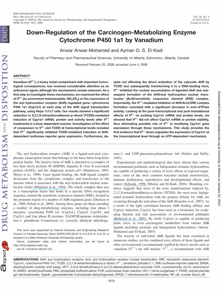
Effect of Coexposure to V5� and TCDD on Cell Viability. Todetermine the maximum nontoxic concentrations of V5� to be used inthe current study, Hepa 1c1c7 cells were exposed for 24 h to increas-ing concentrations of V5� (25–1000 �M) in the absence and presenceof 1 nM TCDD. Thereafter, cytotoxicity was assessed using MTT andCellTiter-Glo Luminescent assays.
Figure 1A shows that V5� alone at concentrations of 25 to 250 �Mdid not affect cell viability; however, the highest concentration, 1000�M, decreased cell viability to approximately 67%. Similarly coex-posure to V5� and TCDD produced a significant decrease in cellviability, at the highest concentration tested (1000 �M), to approxi-mately 70%. On the other hand, the concentration-dependent effect ofexposure to V5� in the absence and presence of 1 nM TCDD using theCellTiter-Glo Luminescent assay exhibited a pattern relatively similarto that observed with the MTT experiment (Fig. 1B). Therefore, allsubsequent studies were conducted using concentrations of 25 to 250�M in the absence and presence of 1 nM TCDD.
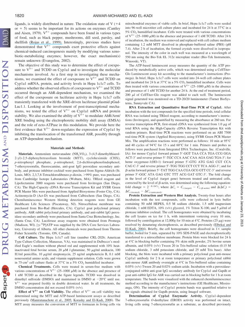
Concentration-Dependent Effect of Coexposure to V5� andTCDD on Inducible Cyp1a1 mRNA. To better understand the ki-netics of Cyp1a1 in response to coexposure to V5� and TCDD, Hepa1c1c7 cells were treated with various concentrations of V5� (Fig. 2).Thereafter, Cyp1a1 mRNA was assessed using real-time PCR. TCDDalone caused a 38-fold increase in Cyp1a1 mRNA that was inhibitedin a dose-dependent manner by V5�, starting at a concentration of 25�M and reaching maximum inhibition at the concentration of 250 �M(Fig. 2).
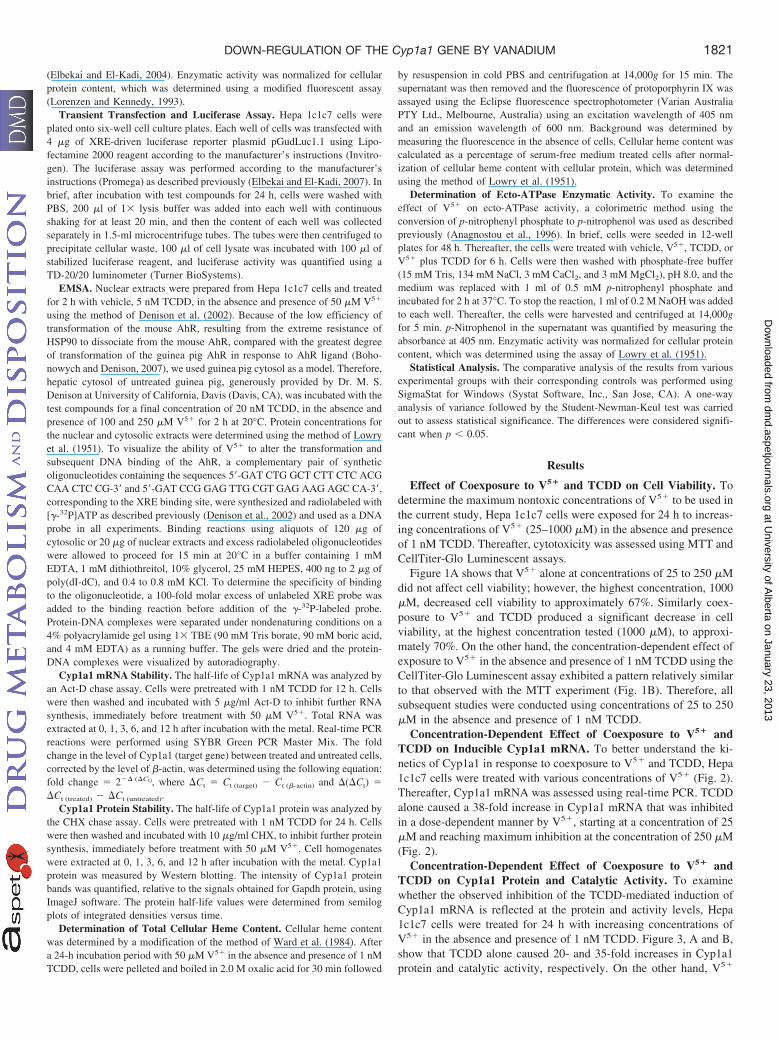
Concentration-Dependent Effect of Coexposure to V5� andTCDD on Cyp1a1 Protein and Catalytic Activity. To examinewhether the observed inhibition of the TCDD-mediated induction ofCyp1a1 mRNA is reflected at the protein and activity levels, Hepa1c1c7 cells were treated for 24 h with increasing concentrations ofV5� in the absence and presence of 1 nM TCDD. Figure 3, A and B,show that TCDD alone caused 20- and 35-fold increases in Cyp1a1protein and catalytic activity, respectively. On the other hand, V5�
1821DOWN-REGULATION OF THE Cyp1a1 GENE BY VANADIUM
at University of A
lberta on January 23, 2013dm
d.aspetjournals.orgD
ownloaded from
significantly reduced the TCDD-mediated induction of Cyp1a1 pro-tein and activity levels in a dose-dependent manner. This inhibitionpattern was consistent with that observed at mRNA levels, in whichthe initial significant inhibition took place with 50 �M V5�, andmaximal inhibition was reached at 250 �M (Fig. 3, A and B).
Transcriptional Inhibition of Cyp1a1 Gene Induction by V5�.To investigate whether the observed effect upon coexposure to V5�
and TCDD on Cyp1a1 is occurring through an AhR-dependent mech-anism, Hepa 1c1c7 cells were transiently transfected with the XRE-driven luciferase reporter gene to study the effect of V5� on theAhR-dependent transcriptional activation. Luciferase activity resultsshowed that 50 �M V5� alone did not alter the luciferase activity(Fig. 4). In contrast, 1 nM TCDD alone was capable of causing asignificant induction of the luciferase activity that reached up to 1200relative light units, compared with control. On the other hand, co-treatment with V5� and TCDD significantly decreased the luciferaseactivity by 3-fold compared with TCDD alone (Fig. 4).
In an effort to determine whether V5� interferes with the nuclearbinding of the transformed AhR to the XRE, we examined thepotential effect of V5� on TCDD-induced translocation of the AhR tothe nucleus and subsequent binding to XRE, the promoter sequence ofCyp1a1, by EMSA. For this purpose, Hepa 1c1c7 cells were treatedwith vehicle, V5�, TCDD, or V5� plus TCDD for 1 h, followed byextraction of nuclear extracts. Extracts from vehicle- and TCDD-treated cells were used as negative and positive controls, respectively.Figure 5A shows that V5� alone did not induce AhR/Arnt/XREcomplex formation, as shown by the intensity of the bands. In con-trast, TCDD significantly increased AhR/Arnt/XRE binding. In addi-tion, V5� completely abolished the TCDD-induced nuclear formation
FIG. 1. Effect of V5� on cell viability. Hepa 1c1c7 cells were treated for 24 h withV5� (0, 25, 50, 100, 250, and 1000 �M) in the absence and presence of 1 nMTCDD. Cell cytotoxicity was determined using MTT (A). and CellTiter-Glo Lumi-nescent (B) assays. Data are expressed as percentage of untreated control (which isset at 100%) S.E. (n � 8). �, P � 0.05, compared with control (concentration �0 �M); �, P � 0.05, compared with the respective TCDD treatment.
FIG. 2. Effect of V5� on Cyp1a1 mRNA using real-time PCR. Hepa 1c1c7 cellswere treated for 6 h with increasing concentrations of V5� in the presence of 1 nMTCDD. First-strand cDNA was synthesized from total RNA (1 �g) extracted fromHepa 1c1c7 cells. cDNA fragments were amplified and quantitated using an ABI7500 real-time PCR system as described under Materials and Methods. Duplicatereactions were performed for each experiment, and the values presented are themeans of three independent experiments. �, P � 0.05, compared with control (C)(concentration � 0 �M); �, P � 0.05, compared with the respective TCDD (T)treatment.
FIG. 3. Effect of V5� on inducible Cyp1a1 protein and EROD activity. Hepa 1c1c7cells were treated for 24 h with increasing concentrations of V5� in the presence of1 nM TCDD. A, protein (25 �g) was separated by 10% SDS-PAGE and transferredto a nitrocellulose membrane. Protein blots were then blocked overnight at 4°C andincubated with a primary Cyp1a1 antibody for 2 h at 4°C, followed by a 1-hincubation with secondary antibody at room temperature. Cyp1a1 protein wasdetected using the enhanced chemiluminescence method. The intensity of bands wasnormalized to Gapdh signals, which was used as a loading control. One of threerepresentative experiments is shown. B, EROD activity was measured in intactliving cells treated with increasing concentrations of V5�, in the absence andpresence of 1 nM TCDD for 24 h. Cyp1a1 activity was measured using7-ethoxyresorufin as a substrate. Values are presented as mean S.E. (n � 8). �,P � 0.05, compared with control (C); �, P � 0.05, compared with the respectiveTCDD (T) treatment.
1822 ANWAR-MOHAMED AND EL-KADI
at University of A
lberta on January 23, 2013dm
d.aspetjournals.orgD
ownloaded from
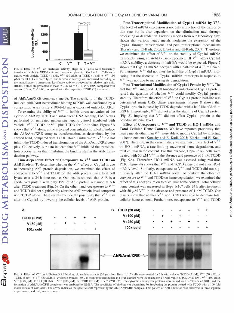
of AhR/Arnt/XRE complex (lane 3). The specificity of the TCDD-induced AhR/Arnt heterodimer binding to XRE was confirmed by acompetition assay using a 100-fold molar excess of unlabeled XRE.
To examine the ability of V5� to inhibit direct activation of thecytosolic AhR by TCDD and subsequent DNA binding, EMSA wasperformed on untreated guinea pig hepatic cytosol incubated withvehicle, V5�, TCDD, or V5� plus TCDD for 2 h in vitro. Figure 5Bshows that V5� alone, at the indicated concentrations, failed to inducethe AhR/Arnt/XRE complex transformation, as determined by theshifted band, compared with TCDD alone. Furthermore, V5� did notinhibit the TCDD-induced transformation of the AhR/Arnt/XRE com-plex. Collectively, our data indicate that V5� inhibited the transloca-tion process rather than inhibiting the binding step in the AhR trans-duction pathway.
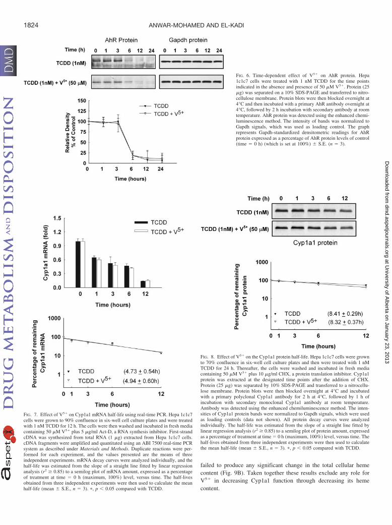
Time-Dependent Effect of Coexposure to V5� and TCDD onAhR Protein. To determine whether the V5� effect on Cyp1a1 is dueto increasing AhR protein degradation, we examined the effect ofcoexposure to V5� and TCDD on the AhR protein using total celllysate over a 24-h time course. Our results showed that AhR is ashort-lived protein with only 15% of AhR protein remained at 6 hafter TCDD treatment (Fig. 6). On the other hand, coexposure to V5�
and TCDD did not significantly alter the AhR protein level comparedwith TCDD alone. These results exclude the possibility that V5� mayalter the Cyp1a1 by lowering the cellular levels of AhR protein.
Post-Transcriptional Modification of Cyp1a1 mRNA by V5�.The level of mRNA expression is not only a function of the transcrip-tion rate but is also dependent on the elimination rate, throughprocessing or degradation. Previous reports from our laboratory haveshown that various heavy metals modulate the expression of theCyp1a1 through transcriptional and post-transcriptional mechanisms(Korashy and El-Kadi, 2005; Elbekai and El-Kadi, 2007). Therefore,we examined the effect of V5� on the stability of Cyp1a1 mRNAtranscripts, using an Act-D chase experiment. If V5� alters Cyp1a1mRNA stability, a decrease in half-life would be expected. Figure 7shows that Cyp1a1 mRNA decayed with a half-life of 4.73 0.54 h.Furthermore, V5� did not alter the half-life of Cyp1a1 mRNA, indi-cating that the decrease in Cyp1a1 mRNA transcripts in response toV5� was not due to increasing its degradation.
Post-Translational Modification of Cyp1a1 Protein by V5�. Thefact that V5� inhibited TCDD-mediated induction of Cyp1a1 proteinraised the question of whether V5� could modify Cyp1a1 proteinstability. Therefore, the effect of V5� on Cyp1a1 protein half-life wasdetermined using CHX chase experiments. Figure 8 shows thatCyp1a1 protein induced by TCDD degraded with a half-life of 8.41 0.29 h. Interestingly, V5� did not alter the stability of Cyp1a1 protein(Fig. 8), implying that V5� did not affect Cyp1a1 protein at thepost-translational level.
Effect of Coexposure to V5� and TCDD on HO-1 mRNA andTotal Cellular Heme Content. We have reported previously thatheavy metals other than V5� were able to modify Cyp1a1 by affectingits heme content (Korashy and El-Kadi, 2005; Elbekai and El-Kadi,2007). Therefore, in the current study we examined the effect of V5�
on HO-1 mRNA, a rate-limiting enzyme of heme degradation, andtotal cellular heme content. For this purpose, Hepa 1c1c7 cells weretreated with 50 �M V5� in the absence and presence of 1 nM TCDD(Fig. 9A). Thereafter, HO-1 mRNA was assessed using real-timePCR. Figure 9A shows that V5� and TCDD alone did not alter HO-1mRNA level. Similarly, coexposure to V5� and TCDD did not sig-nificantly alter the HO-1 mRNA level. To confirm the effect ofcoexposure to V5� and TCDD on heme degradation, we examined theeffect of this coexposure on total cellular heme content. Total cellularheme content was measured in Hepa 1c1c7 cells 24 h after treatmentwith 50 �M V5� in the absence and presence of 1 nM TCDD. Ourresults show that neither V5� nor TCDD was able to decrease totalcellular heme content. Furthermore, coexposure to V5� and TCDD
FIG. 5. Effect of V5� on AhR/Arnt/XRE binding. A, nuclear extracts (20 �g) from Hepa 1c1c7 cells were treated for 2 h with vehicle, TCDD (5 nM), V5� (50 �M), orTCDD (5 nM) � V5� (50 �M). B, cytosolic extracts (80 �g) from untreated guinea pig liver extracts were incubated for 2 h with vehicle, TCDD (20 nM), V5� (100 �M),V5� (250 �M), TCDD (20 nM) � V5� (100 �M), or TCDD (20 nM) � V5� (250 �M). The cytosolic and nuclear proteins were mixed with �-32P-labeled XRE, and theformation of AhR/Arnt/XRE complexes was analyzed by EMSA. The specificity of binding was determined by incubating the protein treated with TCDD with a 100-foldmolar excess of cold XRE. The arrow indicates the specific shift representing the AhR/Arnt/XRE complex. This pattern of AhR alteration was observed in three separateexperiments, and only one is shown.
FIG. 4. Effect of V5� on luciferase activity. Hepa 1c1c7 cells were transientlytransfected with the XRE-luciferase transporter plasmid pGudLuc1.1. Cells weretreated with vehicle, TCDD (1 nM), V5� (50 �M), or TCDD (1 nM) � V5� (50�M) for 24 h. Cells were lysed, and luciferase activity was measured according tothe manufacturer’s instruction. Luciferase activity is reported as relative light units(RLU). Values are presented as mean S.E. (n � 6). �, P � 0.05, compared withcontrol (C); �, P � 0.05, compared with the respective TCDD (T) treatment.
1823DOWN-REGULATION OF THE Cyp1a1 GENE BY VANADIUM
at University of A
lberta on January 23, 2013dm
d.aspetjournals.orgD
ownloaded from
failed to produce any significant change in the total cellular hemecontent (Fig. 9B). Taken together these results exclude any role forV5� in decreasing Cyp1a1 function through decreasing its hemecontent.
FIG. 7. Effect of V5� on Cyp1a1 mRNA half-life using real-time PCR. Hepa 1c1c7cells were grown to 90% confluence in six-well cell culture plates and were treatedwith 1 nM TCDD for 12 h. The cells were then washed and incubated in fresh mediacontaining 50 �M V5� plus 5 �g/ml Act-D, a RNA synthesis inhibitor. First-strandcDNA was synthesized from total RNA (1 �g) extracted from Hepa 1c1c7 cells.cDNA fragments were amplified and quantitated using an ABI 7500 real-time PCRsystem as described under Materials and Methods. Duplicate reactions were per-formed for each experiment, and the values presented are the means of threeindependent experiments. mRNA decay curves were analyzed individually, and thehalf-life was estimated from the slope of a straight line fitted by linear regressionanalysis (r2 � 0.85) to a semilog plot of mRNA amount, expressed as a percentageof treatment at time � 0 h (maximum, 100%) level, versus time. The half-livesobtained from three independent experiments were then used to calculate the meanhalf-life (mean S.E., n � 3). �, p � 0.05 compared with TCDD.
FIG. 6. Time-dependent effect of V5� on AhR protein. Hepa1c1c7 cells were treated with 1 nM TCDD for the time pointsindicated in the absence and presence of 50 �M V5�. Protein (25�g) was separated on a 10% SDS-PAGE and transferred to nitro-cellulose membrane. Protein blots were then blocked overnight at4°C and then incubated with a primary AhR antibody overnight at4°C, followed by 2 h incubation with secondary antibody at roomtemperature. AhR protein was detected using the enhanced chemi-luminescence method. The intensity of bands was normalized toGapdh signals, which was used as loading control. The graphrepresents Gapdh-standardized densitometric readings for AhRprotein expressed as a percentage of AhR protein levels of control(time � 0 h) (which is set at 100%) S.E. (n � 3).
FIG. 8. Effect of V5� on the Cyp1a1 protein half-life. Hepa 1c1c7 cells were grownto 70% confluence in six-well cell culture plates and then were treated with 1 nMTCDD for 24 h. Thereafter, the cells were washed and incubated in fresh mediacontaining 50 �M V5� plus 10 �g/ml CHX, a protein translation inhibitor. Cyp1a1protein was extracted at the designated time points after the addition of CHX.Protein (25 �g) was separated by 10% SDS-PAGE and transferred to a nitrocellu-lose membrane. Protein blots were then blocked overnight at 4°C and incubatedwith a primary polyclonal Cyp1a1 antibody for 2 h at 4°C, followed by 1 h ofincubation with secondary monoclonal Cyp1a1 antibody at room temperature.Antibody was detected using the enhanced chemiluminescence method. The inten-sities of Cyp1a1 protein bands were normalized to Gapdh signals, which were usedas loading controls (data not shown). All protein decay curves were analyzedindividually. The half-life was estimated from the slope of a straight line fitted bylinear regression analysis (r2 � 0.85) to a semilog plot of protein amount, expressedas a percentage of treatment at time � 0 h (maximum, 100%) level, versus time. Thehalf-lives obtained from three independent experiments were then used to calculatethe mean half-life (mean S.E., n � 3). �, p � 0.05 compared with TCDD.
1824 ANWAR-MOHAMED AND EL-KADI
at University of A
lberta on January 23, 2013dm
d.aspetjournals.orgD
ownloaded from
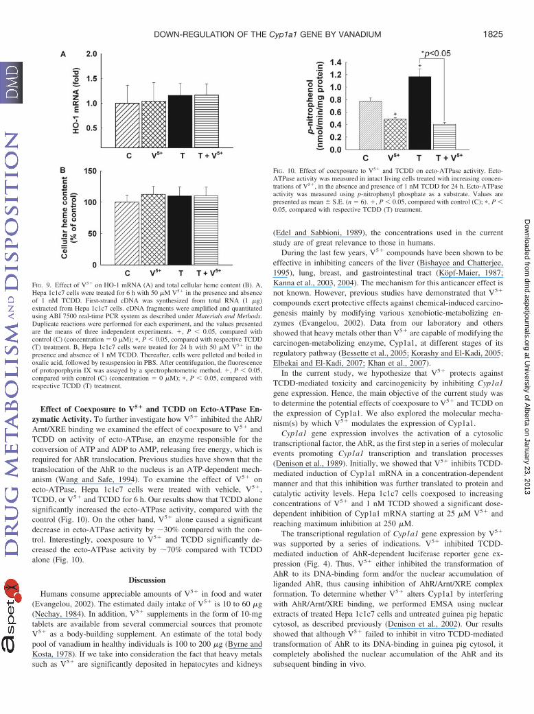
Effect of Coexposure to V5� and TCDD on Ecto-ATPase En-zymatic Activity. To further investigate how V5� inhibited the AhR/Arnt/XRE binding we examined the effect of coexposure to V5� andTCDD on activity of ecto-ATPase, an enzyme responsible for theconversion of ATP and ADP to AMP, releasing free energy, which isrequired for AhR translocation. Previous studies have shown that thetranslocation of the AhR to the nucleus is an ATP-dependent mech-anism (Wang and Safe, 1994). To examine the effect of V5� onecto-ATPase, Hepa 1c1c7 cells were treated with vehicle, V5�,TCDD, or V5� and TCDD for 6 h. Our results show that TCDD alonesignificantly increased the ecto-ATPase activity, compared with thecontrol (Fig. 10). On the other hand, V5� alone caused a significantdecrease in ecto-ATPase activity by 30% compared with the con-trol. Interestingly, coexposure to V5� and TCDD significantly de-creased the ecto-ATPase activity by 70% compared with TCDDalone (Fig. 10).
Discussion
Humans consume appreciable amounts of V5� in food and water(Evangelou, 2002). The estimated daily intake of V5� is 10 to 60 �g(Nechay, 1984). In addition, V5� supplements in the form of 10-mgtablets are available from several commercial sources that promoteV5� as a body-building supplement. An estimate of the total bodypool of vanadium in healthy individuals is 100 to 200 �g (Byrne andKosta, 1978). If we take into consideration the fact that heavy metalssuch as V5� are significantly deposited in hepatocytes and kidneys
(Edel and Sabbioni, 1989), the concentrations used in the currentstudy are of great relevance to those in humans.
During the last few years, V5� compounds have been shown to beeffective in inhibiting cancers of the liver (Bishayee and Chatterjee,1995), lung, breast, and gastrointestinal tract (Kopf-Maier, 1987;Kanna et al., 2003, 2004). The mechanism for this anticancer effect isnot known. However, previous studies have demonstrated that V5�
compounds exert protective effects against chemical-induced carcino-genesis mainly by modifying various xenobiotic-metabolizing en-zymes (Evangelou, 2002). Data from our laboratory and othersshowed that heavy metals other than V5� are capable of modifying thecarcinogen-metabolizing enzyme, Cyp1a1, at different stages of itsregulatory pathway (Bessette et al., 2005; Korashy and El-Kadi, 2005;Elbekai and El-Kadi, 2007; Khan et al., 2007).
In the current study, we hypothesize that V5� protects againstTCDD-mediated toxicity and carcinogenicity by inhibiting Cyp1a1gene expression. Hence, the main objective of the current study wasto determine the potential effects of coexposure to V5� and TCDD onthe expression of Cyp1a1. We also explored the molecular mecha-nism(s) by which V5� modulates the expression of Cyp1a1.
Cyp1a1 gene expression involves the activation of a cytosolictranscriptional factor, the AhR, as the first step in a series of molecularevents promoting Cyp1a1 transcription and translation processes(Denison et al., 1989). Initially, we showed that V5� inhibits TCDD-mediated induction of Cyp1a1 mRNA in a concentration-dependentmanner and that this inhibition was further translated to protein andcatalytic activity levels. Hepa 1c1c7 cells coexposed to increasingconcentrations of V5� and 1 nM TCDD showed a significant dose-dependent inhibition of Cyp1a1 mRNA starting at 25 �M V5� andreaching maximum inhibition at 250 �M.
The transcriptional regulation of Cyp1a1 gene expression by V5�
was supported by a series of indications. V5� inhibited TCDD-mediated induction of AhR-dependent luciferase reporter gene ex-pression (Fig. 4). Thus, V5� either inhibited the transformation ofAhR to its DNA-binding form and/or the nuclear accumulation ofliganded AhR, thus causing inhibition of AhR/Arnt/XRE complexformation. To determine whether V5� alters Cyp1a1 by interferingwith AhR/Arnt/XRE binding, we performed EMSA using nuclearextracts of treated Hepa 1c1c7 cells and untreated guinea pig hepaticcytosol, as described previously (Denison et al., 2002). Our resultsshowed that although V5� failed to inhibit in vitro TCDD-mediatedtransformation of AhR to its DNA-binding in guinea pig cytosol, itcompletely abolished the nuclear accumulation of the AhR and itssubsequent binding in vivo.
FIG. 9. Effect of V5� on HO-1 mRNA (A) and total cellular heme content (B). A,Hepa 1c1c7 cells were treated for 6 h with 50 �M V5� in the presence and absenceof 1 nM TCDD. First-strand cDNA was synthesized from total RNA (1 �g)extracted from Hepa 1c1c7 cells. cDNA fragments were amplified and quantitatedusing ABI 7500 real-time PCR system as described under Materials and Methods.Duplicate reactions were performed for each experiment, and the values presentedare the means of three independent experiments. �, P � 0.05, compared withcontrol (C) (concentration � 0 �M); �, P � 0.05, compared with respective TCDD(T) treatment. B, Hepa 1c1c7 cells were treated for 24 h with 50 �M V5� in thepresence and absence of 1 nM TCDD. Thereafter, cells were pelleted and boiled inoxalic acid, followed by resuspension in PBS. After centrifugation, the fluorescenceof protoporphyrin IX was assayed by a spectrophotometric method. �, P � 0.05,compared with control (C) (concentration � 0 �M); �, P � 0.05, compared withrespective TCDD (T) treatment.
FIG. 10. Effect of coexposure to V5� and TCDD on ecto-ATPase activity. Ecto-ATPase activity was measured in intact living cells treated with increasing concen-trations of V5�, in the absence and presence of 1 nM TCDD for 24 h. Ecto-ATPaseactivity was measured using p-nitrophenyl phosphate as a substrate. Values arepresented as mean S.E. (n � 6). �, P � 0.05, compared with control (C); �, P �0.05, compared with respective TCDD (T) treatment.
1825DOWN-REGULATION OF THE Cyp1a1 GENE BY VANADIUM
at University of A
lberta on January 23, 2013dm
d.aspetjournals.orgD
ownloaded from
Previous data have shown that liganded AhR is ubiquinated beforeits degradation by the 26S proteasomal pathway (Pollenz, 2002). Todetermine whether or not the decrease in AhR/Arnt/XRE binding isdue to an increase in the degradation of the AhR protein by V5�, weexamined the effect of V5� on AhR protein levels at different timepoints. Our results showed that AhR degrades rapidly after exposureto TCDD, and at 6 h the remaining AhR was 15% compared withcontrol (time � 0 h). Coexposure to V5� and TCDD did not signif-icantly alter the AhR protein levels compared with TCDD alone.These results suggest that the observed inhibitory effect of V5� onAhR/Arnt/XRE binding is not due to a decrease in the cellular level ofthe AhR protein.
We have shown previously that heavy metals modulate Cyp1a1through transcriptional, post-transcriptional, and post-translationalmechanisms (Korashy and El-Kadi, 2005; Elbekai and El-Kadi,2007). Thus, it was of great importance to determine the effect of V5�
on the post-transcriptional regulation of Cyp1a1. The cellular mRNAlevel at any time point is a function of the rate of its production,through a transcriptional mechanism, and the rate of its degradation.Therefore, we examined the effect of V5� on the stability of Cyp1a1mRNA using the Act-D chase experiment. Our results showed thatCyp1a1 mRNA induced by TCDD is short-lived, with an estimatedhalf-life of 4.73 0.54 h. Our results are in agreement with previousreports showing that the half-life of Cyp1a1 mRNA induced byTCDD in Hepa 1c1c7 cells ranges from 3 to 4.5 h (Miller et al., 1983;Chen et al., 1995). On the other hand, V5� did not significantly alterthe stability of Cyp1a1 mRNA, suggesting that a post-transcriptionalmechanism is not involved in the modulation of Cyp1a1 mRNAby V5�.
To examine the effect of coexposure to V5� and TCDD at thepost-translational level, a CHX chase experiment was performed. Ourresults showed that the Cyp1a1 protein induced by TCDD has anestimated half-life of 8.41 0.29 h. In contrast, V5� did not signif-icantly alter the stability of Cyp1a1 protein, inferring that a post-translational mechanism is not involved in the modulation of Cyp1a1protein by V5�.
Our previous studies have shown that heavy metals possess theability to decrease Cyp1a1 activity through an effect on its hemecontent (Korashy and El-Kadi, 2005; Elbekai and El-Kadi, 2007).These results prompted us to examine the effect of coexposure to V5�
and TCDD on HO-1 mRNA and total cellular heme content. In thecurrent study we showed that V5� did not significantly alter the HO-1mRNA level and total cellular heme content. Thus, these resultsexclude any possibility that V5� might have decreased Cyp1a1 activ-ity through affecting its heme content.
It has been reported previously that V5� is capable of activating theredox-sensitive transcription factor, nuclear factor-�B (NF-�B) (Chenet al., 2001). Of interest, it has been demonstrated that there is amutual inhibitory interaction between the AhR and the NF-�B sig-naling pathways (Ke et al., 2001). The possibility that NF-�B preventsAhR/Arnt binding to the XRE is excluded, because it has been shownthat NF-�B activation does not affect AhR/Arnt binding to XRE (Keet al., 2001). Therefore, the inhibitory effect of V5� on AhR/Arnt/XRE binding is NF-�B-independent.
Although V5� has been known since 1965 to inhibit ATPases(Nechay, 1984), few studies have been conducted to examine its effecton ecto-ATPase, the main enzyme responsible for releasing energyfrom ATP (Wood et al., 2002) that would be used by the ligandedAhR for nuclear translocation (Wang and Safe, 1994). Interestingly,previous studies using high concentrations of V5� (1 mM) showedthat V5� affects the translocation of the AhR to the nucleus byinhibiting ATPases (Wang and Safe, 1994). These findings prompted
us to investigate the possible role of ecto-ATPase in the modulation ofCyp1a1 by V5�. In this study we have shown that V5� decreasedecto-ATPase enzymatic activity. These results are in agreement withprevious studies (Wang and Safe, 1994) that have reported an ATP-dependent mechanism for the inhibition of AhR translocation by V5�.These results suggest that the ability of V5� to inhibit AhR transfor-mation and hence XRE binding is due to its effect on ecto-ATPasecatalytic activity.
In conclusion, the present study demonstrates that V5� down-regulates the bioactivating enzyme Cyp1a1 through a transcriptionalmechanism. The translocation of the transformed AhR was inhibitedby V5� probably by inhibiting ecto-ATPase activity. Thus, theseresults suggest that V5� may protect against TCDD-mediated toxicityby inhibiting Cyp1a1 gene expression. However, further studies areneeded to investigate the cytoprotective effect of V5� against TCDD-mediated toxicity.
Acknowledgments. We are grateful to Dr. M. S. Denison (Univer-sity of California, Davis, Davis, CA) for providing us with XRE-luciferase reporter plasmid pGudLuc1.1 and guinea pig hepaticcytosol.
References
Anagnostou F, Plas C, and Forest N (1996) Ecto-alkaline phosphatase considered as levamisole-sensitive phosphohydrolase at physiological pH range during mineralization in cultured fetalcalvaria cells. J Cell Biochem 60:484–494.
Anwar-Mohamed A and El-Kadi AO (2007) Induction of cytochrome P450 1a1 by the foodflavoring agent, maltol. Toxicol In Vitro 21:685–690.
Behnisch PA, Hosoe K, and Sakai S (2001) Bioanalytical screening methods for dioxins anddioxin-like compounds a review of bioassay/biomarker technology. Environ Int 27:413–439.
Bessette EE, Fasco MJ, Pentecost BT, and Kaminsky LS (2005) Mechanisms of arsenite-mediated decreases in benzo[k]fluoranthene-induced human cytochrome P4501A1 levels inHepG2 cells. Drug Metab Dispos 33:312–320.
Bishayee A and Chatterjee M (1995) Inhibitory effect of vanadium on rat liver carcinogenesisinitiated with diethylnitrosamine and promoted by phenobarbital. Br J Cancer 71:1214–1220.
Bohonowych JE, and Denison MS (2007) Persistent binding of ligands to the aryl hydrocarbonreceptor. Toxicol Sci 98:99–109.
Byrne AR and Kosta L (1978) Vanadium in foods and in human body fluids and tissues. Sci TotalEnviron 10:17–30.
Cantley LC Jr and Aisen P (1979) The fate of cytoplasmic vanadium: implications on (Na,K)-ATPase inhibition. J Biol Chem 254:1781–1784.
Chen F, Ding M, Castranova V, and Shi X (2001) Carcinogenic metals and NF-�B activation.Mol Cell Biochem 222:159–171.
Chen YH, Riby J, Srivastava P, Bartholomew J, Denison M, and Bjeldanes L (1995) Regulationof CYP1A1 by indolo[3,2-b]carbazole in murine hepatoma cells. J Biol Chem 270:22548–22555.
Denison MS, Fisher JM, and Whitlock JP Jr (1989) Protein-DNA interactions at recognition sitesfor the dioxin-Ah receptor complex. J Biol Chem 264:16478–16482.
Denison MS, Pandini A, Nagy SR, Baldwin EP, and Bonati L (2002) Ligand binding andactivation of the Ah receptor. Chem Biol Interact 141:3–24.
Edel J and Sabbioni E (1989) Vanadium transport across placenta and milk of rats to the fetusand newborn. Biol Trace Elem Res 22:265–275.
Elbekai RH and El-Kadi AO (2004) Modulation of aryl hydrocarbon receptor-regulated geneexpression by arsenite, cadmium, and chromium. Toxicology 202:249–269.
Elbekai RH and El-Kadi AO (2007) Transcriptional activation and posttranscriptional modifi-cation of Cyp1a1 by arsenite, cadmium, and chromium. Toxicol Lett 172:106–119.
Evangelou AM (2002) Vanadium in cancer treatment. Crit Rev Oncol Hematol 42:249–265.Hankinson O (1995) The aryl hydrocarbon receptor complex. Annu Rev Pharmacol Toxicol
35:307–340.Kanna PS, Mahendrakumar CB, Chatterjee M, Hemalatha P, Datta S, and Chakraborty P (2003)
Vanadium inhibits placental glutathione S-transferase (GST-P) positive foci in 1,2-dimethylhydrazine induced rat colon carcinogenesis. J Biochem Mol Toxicol 17:357–365.
Kanna PS, Mahendrakumar CB, Indira BN, Srivastawa S, Kalaiselvi K, Elayaraja T, andChatterjee M (2004) Chemopreventive effects of vanadium toward 1,2-dimethylhydrazine-induced genotoxicity and preneoplastic lesions in rat colon. Environ Mol Mutagen 44:113–118.
Ke S, Rabson AB, Germino JF, Gallo MA, and Tian Y (2001) Mechanism of suppression ofcytochrome P-450 1A1 expression by tumor necrosis factor-� and lipopolysaccharide. J BiolChem 276:39638–39644.
Khan S, Liu S, Stoner M, and Safe S (2007) Cobaltous chloride and hypoxia inhibit arylhydrocarbon receptor-mediated responses in breast cancer cells. Toxicol Appl Pharmacol223:28–38.
Kopf-Maier P (1987) Cytostatic non-platinum metal complexes: new perspectives for thetreatment of cancer? Naturwissenschaften 74:374–382.
Korashy HM and El-Kadi AO (2005) Regulatory mechanisms modulating the expression ofcytochrome P450 1A1 gene by heavy metals. Toxicol Sci 88:39–51.
Korashy HM and El-Kadi AO (2008) Modulation of TCDD-mediated induction of cytochromeP450 1A1 by mercury, lead, and copper in human HepG2 cell line. Toxicol In Vitro22:154–158.
Kransler KM, McGarrigle BP, and Olson JR (2007) Comparative developmental toxicity of
1826 ANWAR-MOHAMED AND EL-KADI
at University of A
lberta on January 23, 2013dm
d.aspetjournals.orgD
ownloaded from
2,3,7,8-tetrachlorodibenzo-p-dioxin in the hamster, rat and guinea pig. Toxicology 229:214–225.
Lorenzen A, and Kennedy SW (1993) A fluorescence-based protein assay for use with amicroplate reader. Anal Biochem 214:346–348.
Lowry OH, Rosebrough NJ, Farr AL, and Randall RJ (1951) Protein measurement with the Folinphenol reagent. J Biol Chem 193:265–275.
Maniratanachote R, Minami K, Katoh M, Nakajima M, and Yokoi T (2005) Chaperone proteinsinvolved in troglitazone-induced toxicity in human hepatoma cell lines. Toxicol Sci 83:293–302.
Meyer BK, Pray-Grant MG, Vanden Heuvel JP, and Perdew GH (1998) Hepatitis B virusX-associated protein 2 is a subunit of the unliganded aryl hydrocarbon receptor core complexand exhibits transcriptional enhancer activity. Mol Cell Biol 18:978–988.
Miller AG, Israel D, and Whitlock JP Jr (1983) Biochemical and genetic analysis of variantmouse hepatoma cells defective in the induction of benzo(a)pyrene-metabolizing enzymeactivity. J Biol Chem 258:3523–3527.
Nebert DW, Dalton TP, Okey AB, and Gonzalez FJ (2004) Role of aryl hydrocarbon receptor-mediated induction of the CYP1 enzymes in environmental toxicity and cancer. J Biol Chem279:23847–23850.
Nebert DW and Duffy JJ (1997) How knockout mouse lines will be used to study the role ofdrug-metabolizing enzymes and their receptors during reproduction and development, and inenvironmental toxicity, cancer, and oxidative stress. Biochem Pharmacol 53:249–254.
Nechay BR (1984) Mechanisms of action of vanadium. Annu Rev Pharmacol Toxicol 24:501–524.
Pollenz RS (2002) The mechanism of AH receptor protein down-regulation (degradation) and itsimpact on AH receptor-mediated gene regulation. Chem Biol Interact 141:41–61.
Rojas E, Herrera LA, Poirier LA, and Ostrosky-Wegman P (1999) Are metals dietary carcino-gens? Mutat Res 443:157–181.
Schrenk D (1998) Impact of dioxin-type induction of drug-metabolizing enzymes on themetabolism of endo- and xenobiotics. Biochem Pharmacol 55:1155–1162.
Wang X and Safe S (1994) Development of an in vitro model for investigating the formation ofthe nuclear Ah receptor complex in mouse Hepa 1c1c7 cells. Arch Biochem Biophys 315:285–292.
Ward JH, Jordan I, Kushner JP, and Kaplan J (1984) Heme regulation of HeLa cell transferrinreceptor number. J Biol Chem 259:13235–13240.
Whitelaw ML, Gustafsson JA, and Poellinger L (1994) Identification of transactivation andrepression functions of the dioxin receptor and its basic helix-loop-helix/PAS partner factorArnt: inducible versus constitutive modes of regulation. Mol Cell Biol 14:8343–8355.
Wood E, Johan Broekman M, Kirley TL, Diani-Moore S, Tickner M, Drosopoulos JH, Islam N,Park JI, Marcus AJ, and Rifkind AB (2002) Cell-type specificity of ectonucleotidase expres-sion and upregulation by 2,3,7,8-tetrachlorodibenzo-p-dioxin. Arch Biochem Biophys 407:49–62.
Address correspondence to: Dr. Ayman O. S. El-Kadi, Faculty of Pharmacy
and Pharmaceutical Sciences, 3126 Dentistry/Pharmacy Centre, University of
Alberta, Edmonton, AB, Canada T6G 2N8. E-mail: [email protected]
1827DOWN-REGULATION OF THE Cyp1a1 GENE BY VANADIUM
at University of A
lberta on January 23, 2013dm
d.aspetjournals.orgD
ownloaded from
Recommended


