
INFECrION AND IMMUNITY, June 1993, p. 2545-25520019-9567/93/062545-08$02.00/0Copyright C 1993, American Society for Microbiology
Effects of Listeria monocytogenes and Yersinia enterocoliticaon Cytokine Gene Expression and Release from Human
Polymorphonuclear Granulocytes and Epithelial (HEp-2) CellsR. ARNOLD, J. SCHEFFER, B. KONIG, AND W. KONIG*
Lehrstuhl fur Medizinische Mikrobiologie & Immunologie, Arbeitsgruppe fur Infektabwehrmechanismen,Ruhr-Universitat Bochum, 4630 Bochum, Germany
Received 9 November 1992/Accepted 19 March 1993
The gene expression and cytokine release of the proinflammatory cytokines interIeukin-113 (IL-110), IL-6, andtumor necrosis factor alpha (TNF-a) after infection of human epithelial cells (HEp-2 cells) and polymorpho-nuclear granulocytes (PMNs) were investigated by using isogenic pairs of Listeria monocytogenes and Yersiniaenterocolitica strains. By polymerase chain reaction-assisted mRNA amplification and RNA dot blot analysis,we showed that PMNs and HEp-2 cells expressed enhanced levels of mRNA encoding IL-1I, IL-6, and TNF-aafter bacterial infection. Concomitant with the enhanced mRNA level, an increased secretion rate of IL-1",IL-6, and TNF-a from PMNs as assessed by enzyme-linked immunosorbent assay was observed. HEp-2 cellsafter infection also released IL-6 and TNF-a into the cell supernatant, while no IL-11 release was detected.Cellular coincubation experiments were carried out with Transwell chambers. Our studies revealed that thecoculture of PMNs and HEp-2 cells led to an increased IL-1" and IL-6 release. In contrast, after infection withthe invasive bacteria, reduced levels of TNF-at were measured. Our data show that PMNs secrete theproinflammatory cytokines IL-11, IL-6, and TNF-a within some hours after infection with L. monocytogenesand Y. enterocolitica and that cellular interactions with epithelial cells alone via soluble mediators influence thenet amount of released proinflammatory cytokines.
During the onset of the acute-phase response, an influx ofinflammatory cells, including monocytes and polymorpho-nuclear granulocytes (PMNs), to the sites of inflammationcan be observed (29). Within the microenvironment of theinflamed tissues, these cells communicate with each other bydirect cell-cell contact or a complex array of cytokines andinflammatory mediators. In the course of inflammation ormicrobial invasion, the proinflammatory cytokines interleu-kin-1 (IL-1p), IL-6, and tumor necrosis factor alpha(TNF-a) are responsible for either local or systemic effects(reviewed in reference 1).The cytokines are functionally multipotent, with overlap-
ping biological activities, e.g., they induce fever (33), stim-ulate hepatocytes to synthesize a wide spectrum of acute-phase proteins (44), and express immunomodulatoryfunctions by increasing thymocyte proliferation (20). Smallamounts of these proinflammatory cytokines produced lo-cally within infected tissues play an important protectiverole in host defense prior to their secretion into the periph-eral circulation (48).Only few data exist for the expression of these cytokines
into the microenvironment of the gastrointestinal tract tissueafter infection with the facultatively intracellular bacteriaListeria monocytogenes and Yersinia enterocolitica. Allpathogenic L. monocytogenes and Y. enterocolitica strainsadhere to and penetrate the intestinal epithelium by means ofspecific gene products for invasion. The invasion process ofL. monocytogenes is mediated by internalin as well as by a
gene product called p60 (14, 28). Also, the inv gene locus,which encodes invasin and the attachment/invasion (ail)gene locus ofY enterocolitica, is responsible for an invasivephenotype (reviewed in reference 38).
* Corresponding author.
The purposes of our study were to investigate (i) whetherhuman epithelial (HEp-2) cells and PMNs transcribe thegenes encoding IL-1p, IL-6, and TNF-a and release themature cytokines after infection with L. monocytogenes andY enterocolitica; (ii) which role the invasion process playswith regard to cytokine gene expression and secretion; and(iii) whether paracrine cellular interactions between infectedepithelial cells and PMNs modulate the net amount ofreleased cytokines.For bacterial stimulation, we used isogenic pairs of L.
monocytogenes (invasive and noninvasive), Y enterocolit-ica (plasmidless and plasmid bearing), and Escherichia coli(invasive and noninvasive) (18, 22, 24) strains.
MATERIALS AND METHODS
Buffers. The medium used for washing the bacterial cellswas phosphate-buffered saline (PBS). Bacteria were grownin brain heart infusion (BHI) broth (Oxoid Ltd., London,England). The following electrophoresis buffers were usedfor gel electrophoresis: TBE buffer (Tris-borate) for analysisof amplified cDNAs and MOPS buffer [3-(N-morpholino-)propanesulfonic acid] for RNA gel electrophoresis.
Preparation of cells. (i) PMNs. PMNs were prepared fromheparinized venous blood from healthy donors on a Ficoll-metrizoate gradient; preparation was followed by dextransedimentation as described elsewhere (5). The contaminat-ing erythrocytes were removed by hypotonic lysis of theobtained cell suspension. This method led to >98% purePMNs (1 to 2% eosinophils). The cell preparations wereroutinely analyzed by morphological examination of Wright-stained smears and nonspecific esterase staining. Less than1% mononuclear cells were present.
(ii) HEp-2 cells. Cells of the human laryngeal epitheliumcell line HEp-2 were routinely grown in Dulbecco modified
2545
Vol. 61, No. 6
2546 ARNOLD ET AL.
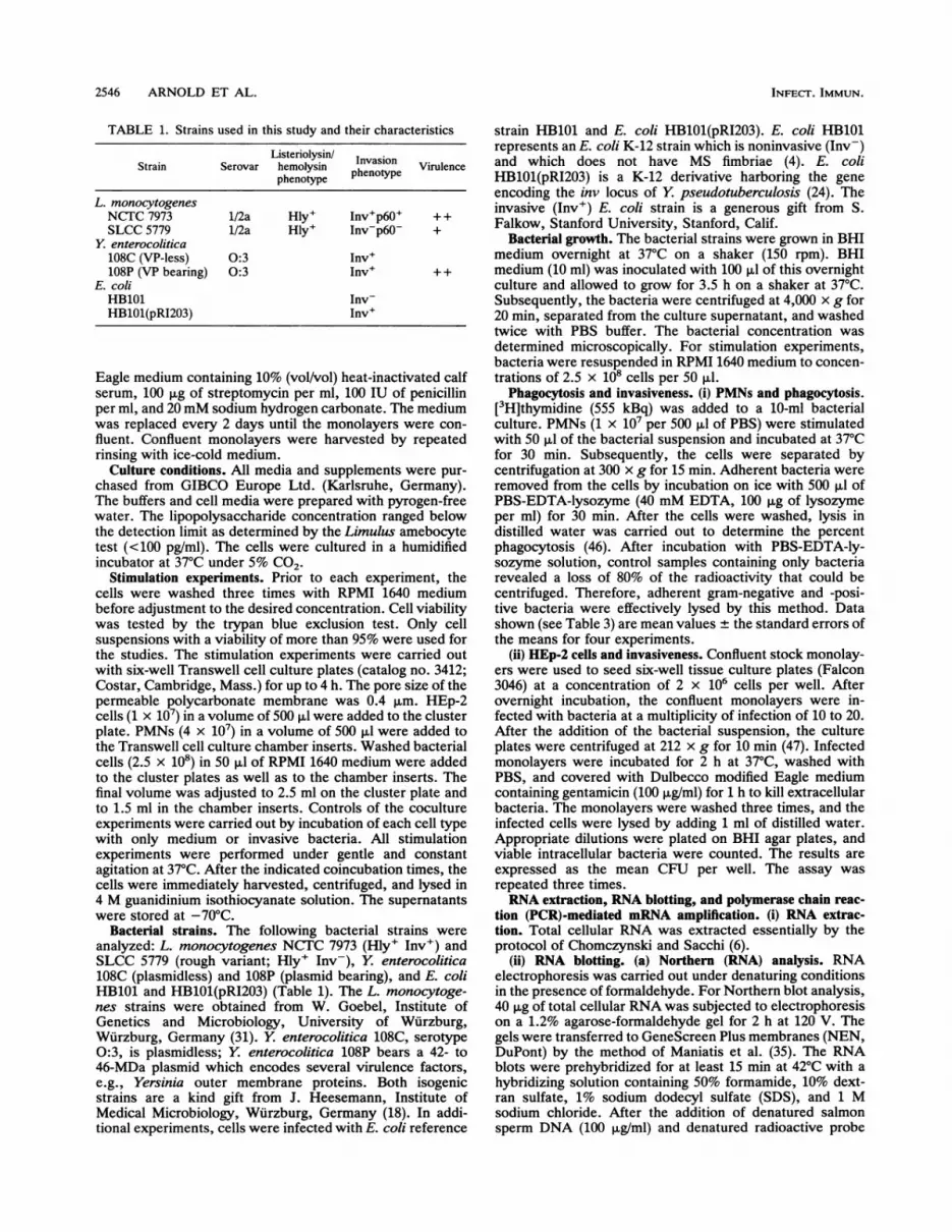
TABLE 1. Strains used in this study and their characteristics
Strain SerovarListeriolysin/ InvasionStrain Serovarhemolysin phenotypeViuecphenotype
L. monocytogenesNCTC 7973 1/2a Hly+ Inv+p6O0 + +SLCC 5779 1/2a Hly+ Inv-p60- +
Y. enterocolitica108C (VP-less) 0:3 Inv'108P (VP bearing) 0:3 Inv' ++
E. coliHB101 Inv-HB101(pRI203) Inv+
Eagle medium containing 10% (vol/vol) heat-inactivated calfserum, 100 jig of streptomycin per ml, 100 IU of penicillinper ml, and 20 mM sodium hydrogen carbonate. The mediumwas replaced every 2 days until the monolayers were con-fluent. Confluent monolayers were harvested by repeatedrinsing with ice-cold medium.
Culture conditions. All media and supplements were pur-chased from GIBCO Europe Ltd. (Karlsruhe, Germany).The buffers and cell media were prepared with pyrogen-freewater. The lipopolysaccharide concentration ranged belowthe detection limit as determined by the Limulus amebocytetest (<100 pg/ml). The cells were cultured in a humidifiedincubator at 37°C under 5% CO2.
Stimulation experiments. Prior to each experiment, thecells were washed three times with RPMI 1640 mediumbefore adjustment to the desired concentration. Cell viabilitywas tested by the trypan blue exclusion test. Only cellsuspensions with a viability of more than 95% were used forthe studies. The stimulation experiments were carried outwith six-well Transwell cell culture plates (catalog no. 3412;Costar, Cambridge, Mass.) for up to 4 h. The pore size of thepermeable polycarbonate membrane was 0.4 jim. HEp-2cells (1 x 10⁷) in a volume of 500 jil were added to the clusterplate. PMNs (4 x 10⁷) in a volume of 500 jil were added tothe Transwell cell culture chamber inserts. Washed bacterialcells (2.5 x 10⁸) in 50 jil of RPMI 1640 medium were addedto the cluster plates as well as to the chamber inserts. Thefinal volume was adjusted to 2.5 ml on the cluster plate andto 1.5 ml in the chamber inserts. Controls of the cocultureexperiments were carried out by incubation of each cell typewith only medium or invasive bacteria. All stimulationexperiments were performed under gentle and constantagitation at 37°C. After the indicated coincubation times, thecells were immediately harvested, centrifuged, and lysed in4 M guanidinium isothiocyanate solution. The supernatantswere stored at -70°C.
Bacterial strains. The following bacterial strains wereanalyzed: L. monocytogenes NCTC 7973 (Hly+ Inv') andSLCC 5779 (rough variant; Hly+ Inv-), Y enterocolitica108C (plasmidless) and 108P (plasmid bearing), and E. coliHB101 and HB101(pRI203) (Table 1). The L. monocytoge-nes strains were obtained from W. Goebel, Institute ofGenetics and Microbiology, University of Wurzburg,Wurzburg, Germany (31). Y enterocolitica 108C, serotype0:3, is plasmidless; Y enterocolitica 108P bears a 42- to46-MDa plasmid which encodes several virulence factors,e.g., Yersinia outer membrane proteins. Both isogenicstrains are a kind gift from J. Heesemann, Institute ofMedical Microbiology, Wurzburg, Germany (18). In addi-tional experiments, cells were infected with E. coli reference
strain HB101 and E. coli HB1O1(pRI203). E. coli HB101represents an E. coli K-12 strain which is noninvasive (Inv-)and which does not have MS fimbriae (4). E. coliHB101(pRI203) is a K-12 derivative harboring the geneencoding the inv locus of Y pseudotuberculosis (24). Theinvasive (Inv') E. coli strain is a generous gift from S.Falkow, Stanford University, Stanford, Calif.
Bacterial growth. The bacterial strains were grown in BHImedium overnight at 37°C on a shaker (150 rpm). BHImedium (10 ml) was inoculated with 100 ,ul of this overnightculture and allowed to grow for 3.5 h on a shaker at 37°C.Subsequently, the bacteria were centrifuged at 4,000 x g for20 min, separated from the culture supernatant, and washedtwice with PBS buffer. The bacterial concentration wasdetermined microscopically. For stimulation experiments,bacteria were resuspended in RPMI 1640 medium to concen-trations of 2.5 x 10⁸ cells per 50 ,ul.
Phagocytosis and invasiveness. (i) PMNs and phagocytosis.[3H]thymidine (555 kBq) was added to a 10-ml bacterialculture. PMNs (1 x 10⁷ per 500 ,ul of PBS) were stimulatedwith 50 pl of the bacterial suspension and incubated at 37°Cfor 30 min. Subsequently, the cells were separated bycentrifugation at 300 x g for 15 min. Adherent bacteria wereremoved from the cells by incubation on ice with 500 jil ofPBS-EDTA-lysozyme (40 mM EDTA, 100 jig of lysozymeper ml) for 30 min. After the cells were washed, lysis indistilled water was carried out to determine the percentphagocytosis (46). After incubation with PBS-EDTA-ly-sozyme solution, control samples containing only bacteriarevealed a loss of 80% of the radioactivity that could becentrifuged. Therefore, adherent gram-negative and -posi-tive bacteria were effectively lysed by this method. Datashown (see Table 3) are mean values ± the standard errors ofthe means for four experiments.
(ii) HEp-2 cells and invasiveness. Confluent stock monolay-ers were used to seed six-well tissue culture plates (Falcon3046) at a concentration of 2 x 10⁶ cells per well. Afterovernight incubation, the confluent monolayers were in-fected with bacteria at a multiplicity of infection of 10 to 20.After the addition of the bacterial suspension, the cultureplates were centrifuged at 212 x g for 10 min (47). Infectedmonolayers were incubated for 2 h at 37°C, washed withPBS, and covered with Dulbecco modified Eagle mediumcontaining gentamicin (100 ,ug/ml) for 1 h to kill extracellularbacteria. The monolayers were washed three times, and theinfected cells were lysed by adding 1 ml of distilled water.Appropriate dilutions were plated on BHI agar plates, andviable intracellular bacteria were counted. The results areexpressed as the mean CFU per well. The assay wasrepeated three times.RNA extraction, RNA blotting, and polymerase chain reac-
tion (PCR)-mediated mRNA amplification. (i) RNA extrac-tion. Total cellular RNA was extracted essentially by theprotocol of Chomczynski and Sacchi (6).
(ii) RNA blotting. (a) Northern (RNA) analysis. RNAelectrophoresis was carried out under denaturing conditionsin the presence of formaldehyde. For Northern blot analysis,40 jig of total cellular RNA was subjected to electrophoresison a 1.2% agarose-formaldehyde gel for 2 h at 120 V. Thegels were transferred to GeneScreen Plus membranes (NEN,DuPont) by the method of Maniatis et al. (35). The RNAblots were prehybridized for at least 15 min at 42°C with ahybridizing solution containing 50% formamide, 10% dext-ran sulfate, 1% sodium dodecyl sulfate (SDS), and 1 Msodium chloride. After the addition of denatured salmonsperm DNA (100 ,ug/ml) and denatured radioactive probe
INFECT. IMMUN.

L. MONOCYTOGENES, Y ENTEROCOLITICA, AND CYTOKINES 2547
TABLE 2. Specific primers synthesized for PCR
mRNA Size of PCR-amplified Synthesized primers (5' sense and 3' antisense)fragmnent (bp)
3-Actin 661 5'-TGA-CGG-GGT-CAC-CCA-CAC-TGT-GCC-CAT-CTA-3'5'-CTA-GAA-GCA-TTG-CGG-TGG-ACG-ATG-GAG-GG-3'
IL-6 628 5'-ATG-AAC-TCC-TCC-TCC-ACA-AGC-GC-3'5'-GAA-GAG-CCC-TCA-GGC-TGG-ACT-G-3'
IL-1~ 331 5'-CTT-CAT-CTT-TGA-AGA-AGA-ACC-TAT-CTT-CTT-3'5'-AAT-TTT-TGG-GAT-CTA-CAC-TCT-CCA-GCT-GTA-3'
TNF-a 325 5'-CAG-AGG-GAA-GAG-TTC-CCC-AG-3'5'-CCT-TGG-TCT-GGT-AGG-AGA-CG-3'
(<10 ng/ml), hybridization was performed overnight at 42°Cunder constant agitation. IL-6 mRNA was detected with anEcoRI cDNA fragment obtained from the clone pCSF309(American Type Culture Collection). The cDNA fragmentwas 32P radiolabeled by random priming (13). Standardiza-tion was performed with respect to 28S and 18S rRNA (21).
(b) RNA dot blot procedure. Total cellular RNA wasdissolved in an appropriate volume of 50% deionized forma-mide-6% formaldehyde solution. This RNA solution wasincubated for 60 min at 50°C to denature RNA. Up to 20 ,ugof total cellular RNA was distributed into wells of the usedmanifold. RNA dot blot analysis and Northern blot analysiswere performed under the following stringent washing con-ditions: (i) twice for 30 min each with 2x SSC (0.3 M sodiumchloride plus 0.03 M sodium citrate) and 1% SDS at 65°C; (ii)twice for 20 min each with 0.1 x SSC at room temperature.The membranes were exposed to Kodak XAR 5 films at-40°C to obtain autoradiographs. The bound radioactivity ofRNA dot blots representing 20 ,ug of total RNA was thendetermined (1 Rack 1209; LKB, Turku, Finland). Back-ground radioactivity was subtracted, and the specific bindingwas expressed as counts per minute.PCR-mediated mRNA amplification. (i) Reverse transcrip-
tion of total cellular RNA. RNA was reverse transcribed intocDNA as follows. Two micrograms of RNA was resus-pended in 20 ,ul of DEPC-double-distilled water (ddH20)containing 2.5 ,uM oligo(dT) (16-mer; GIBCO-BRL), 5 mMMgCl2, 50 mM KCl, 10 mM Tris-HCl (pH 8.3), 1 mM eachdeoxynucleoside triphosphate (GIBCO-BRL), 20 U of pla-cental RNase inhibitor (GIBCO-BRL), and 50 U of Moloneymurine leukemia virus reverse transcriptase (GIBCO-BRL).The reaction mixture was overlaid with mineral oil to pre-vent evaporation and incubated in a thermocycler progam-mable heating block (Perkin-Elmer Cetus Corp.) for 1 cyclefor up to 10 min at 18°C, for 60 min at 42°C, and for 5 min at95°C. The cDNA samples were stored at -20°C.
(ii) PCR amplification of cDNA. Ten microliters of reactionmixture was mixed with 40 ,ul of DEPC-ddH20 containing 2mM MgCl2, 50mM KCI, 10 mM Tris-HCI (pH 8.3), 1.25 U ofTaq polymerase (GIBCO-BRL), 150 nM downstreamprimer, and 150 nM upstream primer. The final mixturevolume of 50 ,ul was overlaid with 30 ,ul of mineral oil toprevent evaporation and then transcribed to double-strandedDNA in a DNA thermocycler in a three-temperature cyclewhich included 5 min of denaturation at 95°C, 60 s ofannealing at 60'C, and 180 s of transcription at 72°C. Unlessstated otherwise, the double-stranded DNA was amplified 25times in a repeated three-temperature cycle which included
60 s of denaturation at 95°C, 60 s of annealing at 60°C, and120 s of extension at 72°C.
(iii) Primer synthesis for PCR. Primer pairs specific forIL-1 3, IL-6, TNF-ax, and ,B-actin were synthesized on aDNA synthesizer (391 DNA synthesizer; Applied Biosys-tems). The specific primer sequences used are depicted inTable 2. The downstream and upstream primers are comple-mentary to sequences in the first and last exons, respec-tively. The synthesized oligonucleotide sequences spannedexon-exon connections, so they are mRNA specific. Thesequences of the primers specific for IL-1,, IL-6, and1-actin were recently described by Ehlers and Smith (12).
Analysis of IL-113, IL-6, and TNF-a release. In addition tomRNA analysis, the cell supernatants of infected and coc-ultured cells were analyzed by enzyme-linked immunosor-bent assay (ELISA) for secreted IL-11, IL-6, and TNF-a(Medgenix, Brussels, Belgium). The secretion rate of thecocultured cells was compared with the amount of thesecreted cytokines in the control experiments.
RESULTS
Invasiveness and phagocytosis of bacteria. Prior to thecytokine gene expression studies, we analyzed the bacterialstrains (Table 1) with respect to their invasiveness and theirphagocytosis pattern.
Isogenic pairs of L. monocytogenes, Y enterocolitica, andE. coli (Table 1) were studied with regard to their invasive-ness in cultured HEp-2 cells. As can be seen in Table 3, after2 h of infection all invasive bacteria entered the epithelialcells by means of their invasive gene products. E. coliHB101 and the noninvasive rough mutant strain of L.monocytogenes (SLCC 5779) were not able to invade thecultured epithelial cells. The phagocytosis data for thebacterial strains after 30 min of incubation with PMNs arealso shown in Table 3. The data demonstrate that all bacte-rial strains were phagocytosed by PMNs irrespective of theirvirulence factors. The degrees of phagocytosis differed from3.7 to 22.0%. Thus, the bacterial strain specificities were notable to inhibit the uptake by neutrophils; they only modu-lated the degree of uptake, resulting in an altered phagocy-tosis rate.
Cytokine gene expression and secretion by HEp-2 cells andPMNs after bacterial infection. (i) HEp-2 cells. In subsequentexperiments, HEp-2 cells were used as a model system forinfection. We investigated the release of TNF-ax, IL-6, andIL-1i and gene expression by analyzing the cytoplasmiclevels of mRNA encoding these cytokines.
VOL. 61, 1993

2548 ARNOLD ET AL.
TABLE 3. Rates of bacterial invasion and percentphagocytosis by PMNs
Invasion of HEp-2 %Strain cellsa (CFU) (106) Phagocytosis
byPMNs_L. monocytogenesNCTC 7973 11 + 4 3.7 0.6SLCC 5779 0 17.3 + 2.1
Y enterocolitica108C (VP-less) 9 ± 4 22.0 + 3.5108P (VP bearing) 14 ± 4 22.2 ± 4.1
E. coliHB101 0 20.7 ± 4.0HB101(pRI203) 13 ± 6 14.3 ± 3.8a Rates of invasion 2 h after infection. Values are means ± standard errors
of the means for three experiments.b Patterns of phagocytosis by PMNs after 30 min of incubation. Values are
means ± standard errors of the means for four experiments.
After infection of the epithelial HEp-2 cell line with L.monocytogenes (Hly+ Inv') and Y enterocolitica 108P,TNF-a in the cell supernatant was measured (Table 4). In theabsence of a bacterial stimulus, the HEp-2 cells released upto 30 pg of IL-6 per ml after 120 min of incubation. Thisconstitutive secretion was further enhanced after infectionwith the isogenic pairs of bacteria. The adhesion of thenoninvasive E. coli HB101 strain induced an elevated IL-6release, but, after invasion by E. coli HB101(pRI203), theIL-6 release increased to 235 pg/ml. This effect was alsoobserved after infection with the two L. monocytogenesstrains. The noninvasive mutant induced a lower rate of IL-6secretion than the invasive L. monocytogenes strain. Infec-tion of the cells with Y enterocolitica 108C and 108Presulted in similar rates of secretion of IL-6 (Table 4). Thesedata suggest that virulence factors encoded by the virulenceplasmid (VP) do not influence the pattern of IL-6 secretionby HEp-2 cells.IL-1 was not released from infected HEp-2 cells. This
cytokine secretion profile was also obtained 4 h after infec-tion. Prolonged incubation decreased the viability of thecells. Therefore, data for cytokine release presented in thisstudy were obtained 2 and 4 h after infection.
In order to elucidate whether the pattern of IL-6 cytokinesecretion by infected HEp-2 cells is accompanied withelevated cytoplasmic IL-6 mRNA levels, we performed IL-6RNA dot blot analysis. Figure 1 shows a characteristic RNA
1 2 3 4 5 6 71-- 1) 11 YFs 9 40 * 0 * 46
I ( ) I > T * 0 4* * * * -
- '~ !*
a I1, 4
cpm 81 176 232 152 116 201 172FIG. 1. RNA dot blot analysis of IL-6 mRNA expression in
HEp-2 cells after 60 min of infection. Total cellular RNA was
isolated from HEp-2 cells cultured in medium (lane 1) or in thepresence of E. coli HB101 (lane 2), E. coli HB101(pRI203) (lane 3),L. monocytogenes (Hly+ Inv') (lane 4), L. monocytogenes (Hly+Inv-) (lane 5), Y enterocolitica (108C) (lane 6), or Y enterocolitica(108P) (lane 7). Up to 20 ,ug of RNA was spotted onto GeneScreenPlus membranes. The row at the bottom shows the quantitation ofthe respective dot blots representing 20 ,ug of total RNA. Shown isan experiment representative of three performed, with similarresults.
dot blot probed with a radioactively labeled IL-6 cDNAfragment. After 60 min of incubation, HEp-2 cells expresseda detectable amount of IL-6 mRNA without any furthercellular stimulation, demonstrating a constitutive IL-6 geneexpression in HEp-2 cells (Fig. 1, lane 1). The infection ofcultured HEp-2 cells with all three isogenic bacterial strainpairs increased the IL-6 mRNA level compared with that ofHEp-2 cells cultured in medium alone (lanes 2 to 7). Whenthe radioactivity of the RNA dot blots representing 20 ,ug oftotal RNA was counted, it was shown that HEp-2 cellsexpressed an increased IL-6 mRNA level after infection withthe invasive E. coli or L. monocytogenes strain in compar-ison with the corresponding noninvasive isogenic bacterialstrains (lanes 2 to 5).
Infection of HEp-2 cells with the VP-less Y enterocoliticastrain 108C induced a more pronounced IL-6 mRNA accu-
mulation (lane 6) than infection with VP-harboring strain108P (lane 7). However, no differences in the IL-6 secretion
TABLE 4. Secretion of cytokines by HEp-2 cells and PMNsa
PMN secretion of: HEp-2 cell secretion of:Strain
TNF-a IL-6 IL-1,8 TNF-a IL-6
Control 123 31 9 4 10 5 0 30 23L. monocytogenesNCTC 7973 528 80 190 18 277 36 61 ± 12 110 15SLCC 5779 292 73 107 20 195 25 NDb 83 13
Y enterocolitica108C (VP-less) 2,286 ± 381 213 ± 30 382 ± 51 ND 187 ± 23108P (VP bearing) 1,508 ± 280 253 ± 24 267 ± 42 18 ± 4 198 ± 20
E. coliHB101 1,680 ± 243 208 ± 23 338 ± 50 ND 93 ± 21HB101 pRI203 2,631 ± 371 307 ± 32 483 ± 45 ND 235 ± 11
a The incubation time was 2 h. Cytokine release by 107 cell per ml was analyzed by ELISA. Values are means ± standard errors of the means for threeexperiments and are expressed as picograms per milliliter.
ND, not done.
INFECT. IMMUN.

L. MONOCYTOGENES, Y ENTEROCOLITICA, AND CYTOKINES 2549
1 2 3 4 5
3~~A738 bp-615 bp-
369 bp-
246 bp-
1f3 EL C.: t- iL "
.1- 1i
615bp-
369 bp-t
400 bP-
300 bp-
FIG. 2. PCR amplification of HEp-2 cells derived mRNA for,-actin, IL-13, IL-6, and TNF-ax. Total cellular RNA was reversetranscribed, and the resulting cDNAs were PCR amplified for 25cycles. HEp-2 cells were incubated with medium (lane 1), L.monocytogenes (Hly+ Inv') (lane 2), L. monocytogenes (Hly+Inv-) (lane 3), Y enterocolitica (108C) (lane 4), or Y enterocolitica(108P) (lane 5). Shown is an experiment representative of three.
rates after infection with these two strains were seen (Table4).Because of the fact that both the invasive and noninvasive
E. coli strains were able to enhance the IL-6 mRNA level,virulence factor invasiveness is not solely responsible for theincreased IL-6 mRNA accumulation. Therefore, additionalvirulence factors of L. monocytogenes and Y enterocoliticabesides the inv and ail loci and p60 led to IL-6 mRNAaccumulation.We examined IL-10, IL-6, and TNF-ax gene expression by
HEp-2 cells by PCR mRNA amplification. Figure 2 showsthat the genes encoding IL-6 and IL-1,B are constitutivelyexpressed in HEp-2 cells (Fig. 2, lane 1). In contrast, TNF-amRNA was not detected. After infection with the invasiveand noninvasive strains of L. monocytogenes (lane 2 and 3)and Y enterocolitica 108C (lane 4) or 108P (lane 5), anaccumulation of mRNA encoding IL-10, IL-6, and TNF-awas detected. The changes of the corresponding ,B-actinbands for every amplified RNA probe are minor in compar-ison to the observed accumulation of mRNA encodingIL-1p, IL-6, and TNF-a. This means that equal amounts ofRNA were amplified and assessed by gel electrophoresis.Furthermore, the pronounced IL-6 gene expression afterbacterial infection analyzed by PCR-amplified cDNA frag-ments was in good agreement with the results by the RNAdot blot technique (Fig. 1). Therefore, elevated TNF-ot andIL-6 secretion was accompanied by elevated cytoplasmiclevels of mRNA encoding these cytokines.
(ii) PMNs. It was shown that virulence factor invasivenessmodulates cytokine expression by HEp-2 cells. Therefore,we investigated the influence of this virulence factor oncytokine expression by human PMNs.
After phagocytosis of the above-mentioned bacterialstrains (Table 3), we observed that PMNs secreted TNF-a,IL-6, and IL-113 (Table 4). The noninvasive L. monocytoge-nes strain (SLCC 5779) induced a lower release of TNF-aL,IL-6, and IL-13 than the invasive L. monocytogenes strain(NCTC 7973). In addition, the invasive E. coli strain,HB1O1(pRI203), induced an elevated TNF-a, IL-6, andIL-lp release from PMNs in comparison to the noninvasiveE. coli strain. Thus, as was observed with epithelial cells, the
28 S ->
18 S R#
IL-6 -> ^ #
FIG. 3. Northern blot analysis of IL-6 mRNA in PMNs. Thirtymicrograms of total cellular RNA was subjected to gel electrophore-sis under denaturing conditions in the presence of formaldehyde.PMNs were stimulated with medium (lane 1), L. monocytogenes(Hly+ Inv') (lane 2), or Y enterocolitica (108P) (lane 3) for 120 min.
individual virulence factors which encode invasiveness trig-gered an increased cytokine release in granulocytes. Thenoninvasive L. monocytogenes strain tends to form cellchains and becomes more readily phagocytosed by PMNs(Table 3), but smaller amounts of cytokines were releasedafter phagocytosis compared with the amounts released inresponse to the invasive L. monocytogenes strain. There-fore, invasin receptor-mediated phagocytosis (uptake) iscarried out differently from the unspecific phagocytosis ofbacterial aggregates.The uptake of the two invasive Y enterocolitica strains
led to differences in the release of TNF-ax and IL-1,B. As isshown, VP-bearing Y enterocolitica strain 108P led to areduced cytokine release (Table 4). Therefore, the individualvirulence factors which encode invasiveness are not solelyresponsible for cytokine gene expression in granulocytes.Additional virulence factors, e.g., outer membrane compo-nents and adhesion and secretory factors specific for theindividual bacteria, may be responsible for the differences inTNF-a and IL-1,B release from PMNs. As was shown forHEp-2 cells, IL-6 release was not modulated by determi-nants encoded by the VP.Within 1 h after uptake of the bacterial strains, the TNF-a,
IL-6, and IL-13 mRNA levels of PMNs were increased asassessed by PCR (data not shown). The cytokine mRNAlevels remained elevated for up to 5 h. After this time period,the cell viability and therefore the cytoplasmic mRNAamounts decreased. An IL-6 Northern blot analysis of totalcellular RNA isolated from human PMNs (Fig. 3) is shown.A significant signal is apparent. The constitutive IL-6 mRNAlevel in uninfected PMNs may be a result of cell adherenceto the plastic wells and resulted in a constitutive IL-6 releaseof 10 pg/ml (Table 4). The size of the IL-6 gene transcriptswas in agreement with published results (21) demonstrating aspecific hybridization of IL-6 cDNA in the RNA dot blotstudies.TNF-4, IL-6, and IL-1l release from cocultured HEp-2
cells and PMNs after bacterial infection. In order to determinewhether cocultured PMNs and HEp-2 cells show differentreleases of cytokines, we performed coculture experimentsfor 2 and 4 h with Transwell chambers. We analyzed theTNF-a, IL-6, and IL-1l8 secretions of the cocultures andcompared the results with the cytokine amounts released inthe control experiments.
VOL. 61, 1993

2550 ARNOLD ET AL.
TABLE 5. Secretion of cytokines by PMNs and HEp-2 ceilsa
Amt (pg/ml) of cytokine secreted:Cytokine, expt type,
and incubation In In response to In response totime medium L. monocytogenes Y enterocolitica
NCTC 7973 108P
TNF-otControl, 2 h 123 ± 31 589 ± 78 1,526 ± 284Coincubation, 2 h 193 ± 24 427 ± 65 768 ± 94Control, 4 h 194 ± 19 3,095 ± 365 4,158 ± 401Coincubation, 4 h 362 ± 71 786 ± 163 1,026 + 205
IL-6Control, 2 h 32 ± 9 300 ± 21 450 ± 30Coincubation, 2 h 362 ± 28 637 ± 58 707 ± 47Control, 4 h 92 ± 22 1,205 ± 81 941 ± 42Coincubation, 4 h 942 ± 113 1,603 ± 139 1,412 ± 93
IL-1~Control, 2 h 10 ± 9 277 ± 36 267 ± 42Coincubation, 2 h 119 ± 48 373 ± 76 447 ± 81Control, 4 h 55 ± 20 1,185 ± 287 1,833 ± 252Coincubation, 4 h 955 ± 154 1,877 ± 360 2,433 + 351a PMNs and HEp-2 cells were cocultured in Transwell chambers. Cytokine
release was assessed by ELISA. Values are means ± standard errors of themeans for three experiments.
Table 5 shows the cytokine release in these cocultureexperiments. As can be seen, coincubation of PMNs andHEp-2 cells led to an increased IL-6 and IL-11 secretion.This increased cytokine release was also shown in cocultureexperiments performed without any further bacterial stimu-lation. For TNF-ot release, a different pattern was observed.The release was enhanced in coculture experiments withoutbacterial stimulation and decreased in coculture experimentsin which HEp-2 cells and PMNs were simultaneously in-fected with either L. monocytogenes (Hly+ Inv') or Y.enterocolitica 108P (Table 5).We wished to analyze whether the increased IL-6 secre-
tion was accompanied by elevated IL-6 mRNA levels inHEp-2 cells. Therefore, we performed RNA dot blot studieswith cocultured HEp-2 cells. Figure 4 shows the cytoplasmiclevel of IL-6 mRNA of HEp-2 cells cocultured with PMNs.
,A B J
1 2 3 4 5 6L-'0 * * *
, I, s * * *
cpm: 116 175 172 208 183 215-
FIG. 4. RNA dot blot analysis of IL-6 mRNA of HEp-2 cellscocultured with PMNs in Transwell coculture chambers for 60 min.Both HEp-2 cells and PMNs were incubated with medium (lanes 1and 2), L. monocytogenes (Hly+ Inv') (lanes 3 and 4), or Yenterocolitica (108P) (lanes 5 and 6) in each chamber compartment.HEp-2 cells were cocultured with PMNs (lanes 2, 4, and 6) orincubated alone (lanes 1, 3, and 5). The autoradiographs were
exposed for 4 (A), 3 (B), or 5 (C) days. The row at the bottom showsthe quantitation of the respective dot blots representing a total RNAamount of 20 ,ug.
HEp-2 cells and PMNs were incubated with medium alone(Fig. 4A) or infected with L. monocytogenes (Hly' Inv')(Fig. 4B) or Y enterocolitica 108P (Fig. 4C). As can be seenin this representative experiment, the RNA dot blot analysisdemonstrates the elevated IL-6 mRNA levels of HEp-2 cellscocultured with PMNs alone (116 versus 175 cpm), afterinfection with L. monocytogenes (Hly+ Inv') (172 versus208 cpm), and after infection with Y enterocolitica 108P (183versus 215 cpm). Therefore, coincubation of HEp-2 cellswith PMNs by itself already resulted in an increased cyto-plasmic level of IL-6 mRNA expression. Similarly, after theaddition of bacteria, e.g., L. monocytogenes (Hly+ Inv')and Y enterocolitica 108P, the cocultured HEp-2 cellsshowed an enhanced IL-6 mRNA level. No modulatoryeffect of PMNs cocultured with HEp-2 cells on the cytoplas-mic IL-1, and TNF-ot mRNA levels of HEp-2 cells wasdetected.
DISCUSSION
The results presented show that the facultatively intracel-lular bacteria L. monocytogenes and Y. enterocolitica in-duce a cytoplasmic mRNA accumulation and secretion ofthe proinflammatory cytokines TNF-ct, IL-10, and IL-6 inPMNs.
Epithelial (HEp-2) cells infected with L. monocytogenesor Y enterocolitica accumulate mRNA encoding TNF-ax,IL-1,, and IL-6 and release TNF-a and IL-6 into the cellsupernatant. No IL-11 release was observed.The fact that epidermal cells secrete not only cytokines
which regulate growth and differentiation, such as granulo-cyte macrophage colony-stimulating factor, but also proin-flammatory cytokines, e.g., IL-8 and IL-6 (8, 26), suggeststhat epithelial-cell-derived cytokines are obviously impor-tant for the regulation of inflammatory cell recruitment andactivation. The cytokines TNF-at and IL-1 activate PMNs(27, 45) and as a consequence of the induced IL-8 release arechemotactic for PMNs (36). The migration of PMNs throughfibroblast layers (39), which is highly dependent on thefunction of CD11b/CD18 (MAC-1) as was reported for themigration across intestinal epithelia (42), is further mediatedby TNF-a and IL-1,B.
Therefore, with respect to our in vitro data, one maysuggest that epithelial cells of the gastrointestinal tractinfected with L. monocytogenes or Y enterocolitica havethe ability to recruit and activate PMNs by the release ofproinflammatory cytokines. It appears that the induction ofmRNA accumulation and secretion from HEp-2 cells isobviously not solely dependent on the penetration step (25,32). Additional virulence factors which trigger the increasedaccumulation of IL-11, IL-6, and TNF-(x mRNA after stim-ulation with Listena spp. (10, 16, 37) and Yersinia spp. (3,15, 19, 43) must exist.
It is well documented that the presence of systemic TNF-aand IL-6 plays an important and protective role duringmurine listeriosis (17, 40, 41), and Iizawa et al. reported thecytokine mRNA response in the spleen of L. monocytoge-nes-infected mice during the first few hours of infection (23).They showed by qualitative PCR analysis that mRNA en-coding TNF-ao, IL-1,B, and IL-6 accumulated during the first4 h after infection with viable listeriae. Recently, it wasshown that PMNs are the dominant effector cells during theearly nonspecific phase of murine listeriosis in the liver andspleen (30) and that PMNs are able to synthesize IL-1, IL-6,and TNF-a after phagocytosis of different pathogens (2, 7, 9,11, 34).
INFECT. IMMUN.

L. MONOCYTOGENES, Y ENTEROCOLITICA, AND CYTOKINES 2551
In this study, we demonstrated the effect of invasivebacteria, e.g., L. monocytogenes and Y. enterocolitica, oncytokine synthesis and secretion by PMNs. Thus, PMNs arenot only endstage effector cells but they are also able tosynthesize and secrete proinflammatory cytokines, e.g.,TNF-cx, IL-10, and IL-6, after uptake of L. monocytogenesor Y enterocolitica. The fact that human PMNs are stimu-lated by these intracellular bacteria for cytokine releasesuggests that PMNs recruited to the sites of infection (liver,spleen, gastrointestinal tract) modulate the inflammatory andimmune response in vivo during the onset of infection.
In addition, our data obtained from coculture experimentsconfirmed that epithelial cells (HEp-2) and PMNs regulatetheir cytokine expression via paracrine cell communication.
Epithelial cells cocultured with PMNs expressed in-creased cytoplasmic levels of IL-6 mRNA resulting in anelevated IL-6 secretion. Furthermore, the IL-1, release ofcocultured PMNs is further enhanced. Since HEp-2 cells (i)did not release IL-lp into the cell supematant after bacterialinfection, perhaps because of the tumor cell status, and (ii)release only small amounts of TNF-a, it appears that solublefactors released from HEp-2 cells are responsible for theincreased IL-13 and diminished TNF-a release by infectedPMNs.The increased TNF-ot release by cocultured PMNs incu-
bated in medium without bacterial infection suggests that analtered pattern of soluble factors, e.g., cytokines or solublecytokine receptors, is spontaneously secreted from HEp-2cells, which then results in an elevated TNF-a release fromPMNs.
Conclusively, our coculture studies suggest that the re-dundancy of the cytokine release often observed in in vitroexperiments may be under the control of a paracrine regu-lation network. Obviously HEp-2 cells behave differentlyfrom gastrointestinal tract epithelial cells. The model, how-ever, provides a means to investigate the modulatory effectof cell-cell interaction via direct cell-cell contact or solublemediators. It is evident that the model system may be usefulin interpreting pathophysiological changes occurring duringinfection in vivo.
Further studies are needed to determine the soluble cellu-lar factors and the microbial virulence components of List-eria spp. and Yersinia spp., besides the virulence factorinvasiveness, which are involved in cytokine expression,secretion, and modulation.
ACKNOWLEDGMENTW. Konig was supported by the Deutsche Forschungsgemein-
schaft.
REFERENCES1. Akira, S., T. Hirano, T. Taga, and T. Kishimoto. 1990. Biology
of multifunctional cytokines: IL-6 and related molecules (IL-1and TNF). FASEB J. 4:2860-2867.
2. Bazzoni, F., M. A. Cassatella, C. Laudanna, and F. Rossi. 1991.Phagocytosis of opsonized yeast induces tumor necrosis fac-tor-a mRNA accumulation and protein release by human poly-morphonuclear leukocytes. J. Biol. Biol. 50:223-228.
3. Bliska, J. B., G. Kunliang, J. E. Dixon, and S. Falkow. 1991.Tyrosine phosphate hydrolysis of host proteins by an essentialYersinia virulence determinant. Proc. Natl. Acad. Sci. USA88:1187-1191.
4. Bolivar, F., and K. Backmann. 1979. Plasmids of Escherichiacoli as cloning vectors. Methods Enzymol. 68:245-267.
5. Boyum, A. 1968. A one stage procedure for isolation of granu-locytes and lymphocytes from human blood. General sedimen-tation properties of white blood cells in 1 g gravity field. Scand.
J. Clin. Lab. Invest. 21(Suppl. 97):51-76.6. Chomczynski, P., and N. Sacchi. 1987. Single step method forRNA isolation by acid guanidinium thiocyanate-phenol-chloro-form extraction. Anal. Biochem. 162:156-159.
7. Cicco, N. A., A. Lindemann, J. Content, P. Vandenbussche, M.Lubbert, J. Gauss, R. Mertelssmann, and F. Hermann. 1990.Inducible production of interleukin-6 by human polymorphonu-clear neutrophils: role of granulocyte-macrophage colony-stim-ulating factor and tumor necrosis factor-alpha. Blood 70:2049-2052.
8. Cromwell, O., Q. Hamid, C. J. Corrigan, J. Barkans, Q. Meng,P. D. Collins, and A. B. Kay. 1992. Expression and generation ofinterleukin-8, IL-6 and granulocyte-macrophage colony-stimu-lating factor by bronchial epithelial cells and enhancement byIL-11 and tumor necrosis factor-a. Immunology 77:330-337.
9. Djeu, J. Y., D. Serbousek, and D. K. Blanchard. 1990. Release oftumor necrosis factor by human polymorphonuclear leukocytes.Blood 76:1405-1409.
10. Domann, E., M. Leimeister-Wachter, W. Goebel, and T.Chakraborty. 1991. Molecular cloning, sequencing, and identi-fication of a metalloprotease gene from Listena monocytogenesthat is species specific and physically linked to the listeriolysingene. Infect. Immun. 59:65-72.
11. Dubravec, D. B., D. R. Springs, J. A. Mannick, and M. L.Rodrick. 1990. Circulating human peripheral blood granulocytessynthesize and secrete tumor necrosis factor a. Proc. Natl.Acad. Sci. USA 87:6758-6761.
12. Ehlers, S., and K. A. Smith. 1991. Differentiation of T celllymphokine gene expression: the in vitro acquisition of T cellmemory. J. Exp. Med. 173:25-36.
13. Feinberg, A. P., and B. Vogelstein. 1983. A technique forradiolabeling DNA restriction endonuclease fragments to highspecific activity. Anal. Biochem. 137:6-13.
14. Gaillard, J.-L., P. Berche, C. Frehel, E. Gouin, and P. Cossart.1991. Entry of L. monocytogenes into cells is mediated byinternalin, a repeat protein reminiscent of surface antigens fromgram-positive cocci. Cell 65:1127-1141.
15. Gemski, P., J. R. Lazere, and T. Casey. 1980. Plasmid associ-ated with pathogenicity and calcium dependency of Yersiniaenterocolitica. Infect. Immun. 27:682-685.
16. Geoffroy, C., J. Raveneau, J.-L. Beretti, A. Lecroisey, J.-A.Vazquez-Boland, J. E. Alouf, and P. Berche. 1991. Purificationand characterization of an extracellular 29-kilodalton phospho-lipase C from Listena monocytogenes. Infect. Immun. 59:2382-2388.
17. Havell, E. A., and P. B. Sehgal. 1991. Tumor necrosis factorindependent IL-6 production during murine listeriosis. J. Immu-nol. 146:756-761.
18. Heesemann, J., B. Algermissen, and R. Laufs. 1984. Geneticallymanipulated virulence of Yersinia enterocolitica. Infect. Im-mun. 46:105-110.
19. Heesemann, J., U. Gross, N. Schmidt, and R. Laufs. 1986.Immunochemical analysis of plasmid-encoded proteins releasedby enteropathogenic Yersinia sp. grown in calcium-deficientmedia. Infect. Immun. 54:561-567.
20. Helle, M., L. Boeije, and L. A. Aarden. 1989. IL-6 is anintermediate in IL-1-induced thymocyte proliferation. J. Immu-nol. 142:4335-4338.
21. Hirano, T., K. Yasukawa, H. Harada, T. Taga, Y. Watanabe, T.Matsuda, S.-I. Kashiwamura, K. Nakajima, K. Koyama, A.Iwamatsu, S. Tsunasawa, F. Sakiyama, H. Matsui, Y. Takahara,T. Taniguchi, and T. Kishimoto. 1986. Complementary DNA fora novel human interleukin (BSF-2) that induces B lymphocytesto produce immunoglobulin. Nature (London) 324:73-76.
22. Hof, H. 1984. Virulence of different strains of Listena monocy-togenes serovar 1/2a. Med. Microbiol. Immunol. 173:207-218.
23. Iizawa, Y., J. F. Brown, and C. J. Czuprynski. 1992. Earlyexpression of cytokine mRNA in mice infected with Listeriamonocytogenes. Infect. Immun. 60:4068-4073.
24. Isberg, R. R., and S. Falkow. 1985. A single genetic locusencoded by Yersinia pseudotuberculosis permits invasion ofcultured animal cells by Escherichia coli K-12. Nature (London)317:262-264.
VOL. 61, 1993

2552 ARNOLD ET AL.
25. Isberg, R. R., D. L. Voorhis, and S. Falkow. 1987. Identificationof invasin: a protein that allows enteric bacteria to penetratecultured mammalian cells. Cell 50:769-778.
26. Kirnbauer, R., A. Kock, T. Schwartz, A. Urbanski, J. Krut-mann, W. Borth, D. Damm, G. Shipley, J. C. Ansel, and T. A.Luger. 1989. IFN-P2, B cell differentiation factor 2, or hybri-doma growth factor (IL-6) is expressed and released by humanepidermal cells and epidermoid carcinoma cell lines. J. Immu-nol. 142:1922-1928.
27. Klempner, M. S., C. A. Dinarello, and J. Gallin. 1978. Humanleukocytic pyrogen induces release of specific granule contentsfrom human neutrophils. J. Clin. Invest. 61:1330-1337.
28. Kohler, S., M. Leimeister-Wachter, T. Chakraborty, F. Lottspe-ich, and W. Goebel. 1990. The gene coding for protein p60 ofListeria monocytogenes and its use as a specific probe forListeria monocytogenes. Infect. Immun. 58:1943-1950.
29. Konig, W., W. Schonfeld, M. Raulf, M. Koller, J. Knoller, J.Scheffer, and J. Brom. 1990. The neutrophil and leukotrienes-role in health and disease. Eicosanoids 3:1-22.
30. Kratz, S. S., and R. J. Kurlander. 1988. Characterization of thepattern of inflammatory cell influx and cytokine productionduring the murine host response to Listeria monocytogenes. J.Immunol. 141:598-606.
31. Kuhn, M., and W. Goebel. 1989. Identification of an extracellu-lar protein of Listeria monocytogenes possibly involved inintracellular uptake by mammalian cells. Infect. Immun. 57:55-61.
32. Leong, J. M., R. S. Fournier, and R. R. Isberg. 1990. Identifi-cation of the integrin binding domain of the Yersinia pseudotu-berculosis invasion protein. EMBO J. 9:1979-1989.
33. Lesnikow, V. A., 0. M. Efremov, E. A. Korneva, J. van Damme,and A. Billiau. 1991. Fever produced by intrahypothalamicinjection of interleukin-1 and interleukin-6. Cytokines 3:195-198.
34. Lord, P. C. W., L. M. G. Wilmoth, S. B. Mizel, and C. E.McCall. 1991. Expression of interleukin-la and P genes byhuman blood polymorphonuclear leukocytes. J. Clin. Invest.87:1312-1321.
35. Maniatis, T., E. F. Fritsch, and J. Sambrook. 1982. Molecularcloning: a laboratory manual. Cold Spring Harbor Laboratory,Cold Spring Harbor, N.Y.
36. Matsushima, K., and J. J. Oppenheim. 1989. Interleukin 8 andMCAF: novel inflammatory cytokines inducible by IL-1 andTNF. Cytokines 1:2-13.
37. Mengaud, J., M.-F. Vicente, J. Chenevert, J. M. Pereira, C.Geoffroy, B. Gicquel-Sanzey, F. Baquero, J.-C. Perez-Diaz, and
P. Cossart. 1988. Expression in Escherichia coli and sequenceanalysis of the listeriolysin 0 determinant of Listeria monocy-togenes. Infect. Immun. 56:766-772.
38. Miller, V. L. 1992. Yersinia invasion genes and their products.ASM News 58:26-33.
39. Morzycki, W., and A. C. Issekutz. 1991. Tumor necrosis factoralpha but not interleukin-1 induces polymorphonuclear leuko-cyte migration through fibroblast layers by a fibroblast-depen-dent mechanism. Immunology 74:107-113.
40. Nakane, A., T. Minagawa, and K. Kato. 1988. Endogenoustumor necrosis factor (cachectin) is essential to host resistanceagainst Listeria monocytogenes infection. Infect. Immun. 56:2563-2569.
41. Nakane, A., A. Numata, and T. Minagawa. 1991. Endogenoustumor necrosis factor, interleukin-6, and gamma interferonlevels during Listeria monocytogenes infection in mice. Infect.Immun. 60:523-528.
42. Parkos, C. A., C. Delp, M. A. Arnaout, and J. L. Madara. 1991.Neutrophil migration across a cultured intestinal epithelium. J.Clin. Invest. 88:1605-1612.
43. Portnoy, D. A., S. L. Moseley, and S. Falkow. 1981. Character-ization of plasmids and plasmid-associated determinants ofYersinia enterocolitica pathogenesis. Infect. Immun. 31:775-782.
44. Ramadori, G., J. van Damme, H. Rieder, and K.-H. M. zumBuschenfelde. 1988. Interleukin 6, the third mediator of acute-phase reaction, modulates hepatic protein synthesis in humanand mouse. Comparison with interleukin-1l3 and tumor necrosisfactor-a. Eur. J. Immunol. 18:1259-1264.
45. Shalaby, M. R., B. B. Aggarwal, E. Rinderknecht, L. P. Sve-dersky, B. S. Finkle, and M. A. Palladino. 1985. Activation ofpolymorphonuclear neutrophil functions by interferon-gammaand tumor necrosis factors. J. Immunol. 135:2069-2073.
46. Ventur, Y., J. Scheffer, J. Hacker, W. Goebel, and W. Konig.1990. Effects of adhesins from mannose-resistant Escherichiacoli on mediator release from human lymphocytes, monocytes,and basophils and from polymorphonuclear granulocytes. In-fect. Immun. 58:1500-1508.
47. Vesikari, T., J. Bromirska, and M. Miki. 1982. Enhancement ofinvasiveness of Yersinia enterocolitica and Eschenchia coli inHEp-2 cells by centrifugation. Infect. Immun. 36:834-836.
48. Waage, A., A. Halstensen, R. Shalaby, P. Brandtzaeg, P. Kierulf,and T. Espevik. 1989. Local production of tumor necrosis factora, interleukin 1, and interleukin 6 in meningococcal meningitis.J. Exp. Med. 170:1859-1867.
INFECT. IMMUN.
Recommended


