Endocrine Investigations
A/Prof Shane Hamblin
Head of Endocrinology & Diabetes
Western Health
Footer Text 2
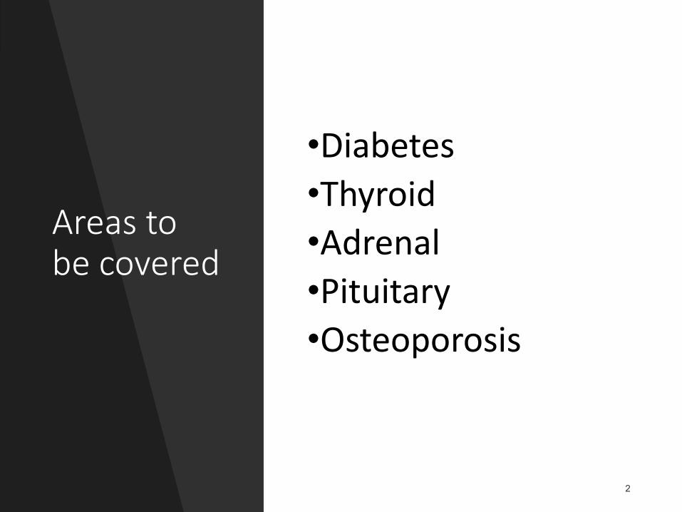
Areas to be covered
•Diabetes
•Thyroid
•Adrenal
•Pituitary
•Osteoporosis
Footer Text 3
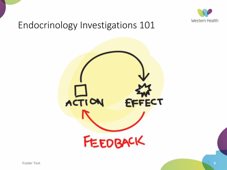
Endocrinology Investigations 101
3
Footer Text 4
Principles of endocrine investigations
•Clinical suspicion first
•Repeat abnormal tests to confirm
•Biochemical interference is not uncommon
•Speak to the lab if tests are unusual / confusing
•Only then proceed to localisation (e.g. MRI pituitary)
Diabetes
5
Footer Text 6
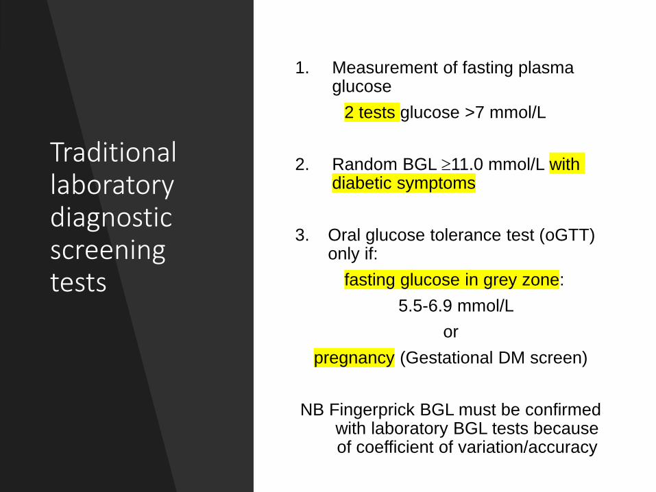
Traditional laboratory diagnostic screening tests
1. Measurement of fasting plasma glucose
2 tests glucose >7 mmol/L
2. Random BGL 11.0 mmol/L with diabetic symptoms
3. Oral glucose tolerance test (oGTT) only if:
fasting glucose in grey zone:
5.5-6.9 mmol/L
or
pregnancy (Gestational DM screen)
NB Fingerprick BGL must be confirmed with laboratory BGL tests because of coefficient of variation/accuracy
Footer Text 7
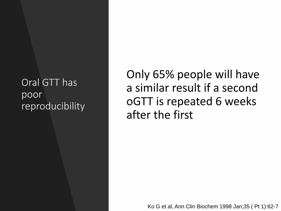
Oral GTT has poor reproducibility
Only 65% people will have a similar result if a second oGTT is repeated 6 weeks after the first
Ko G et al, Ann Clin Biochem 1998 Jan;35 ( Pt 1):62-7
Footer Text 8
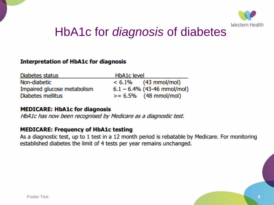
HbA1c for diagnosis of diabetes
Footer Text 9
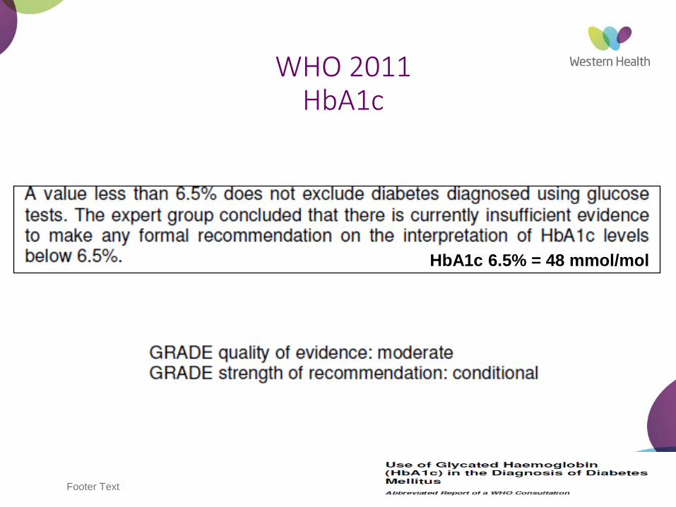
WHO 2011HbA1c
HbA1c 6.5% = 48 mmol/mol
10
Footer Text 11http://www.diabetes.org.uk/
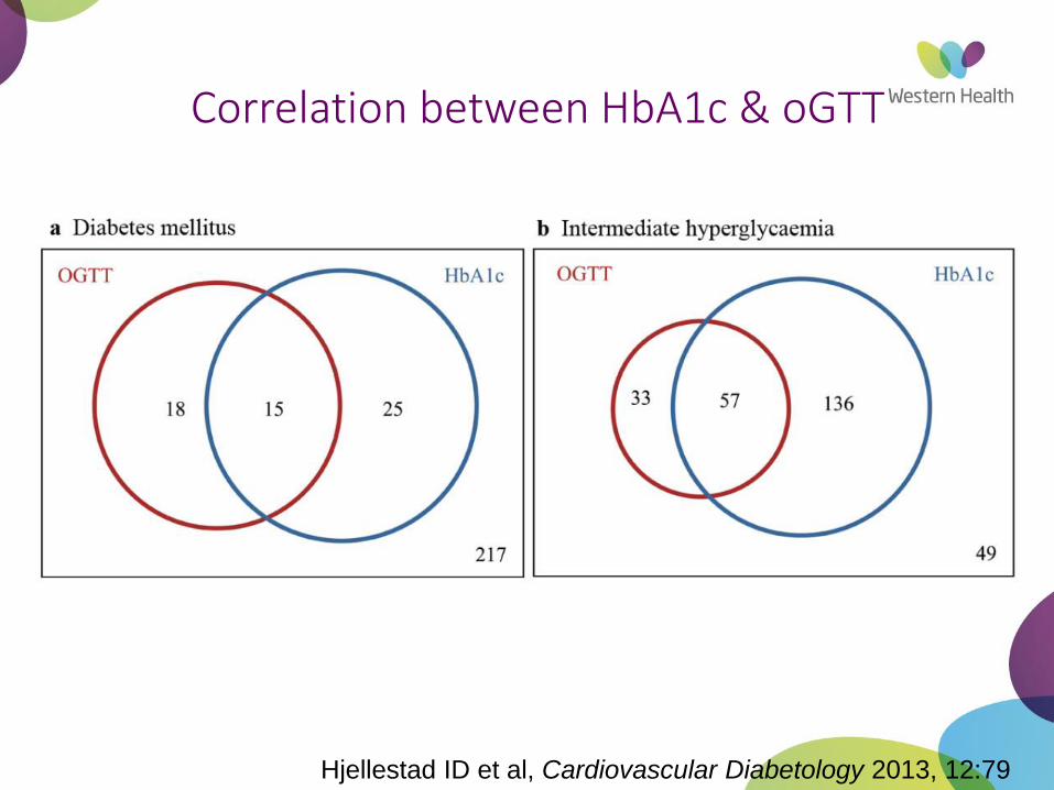
Hjellestad ID et al, Cardiovascular Diabetology 2013, 12:79
Correlation between HbA1c & oGTT
Footer Text 13
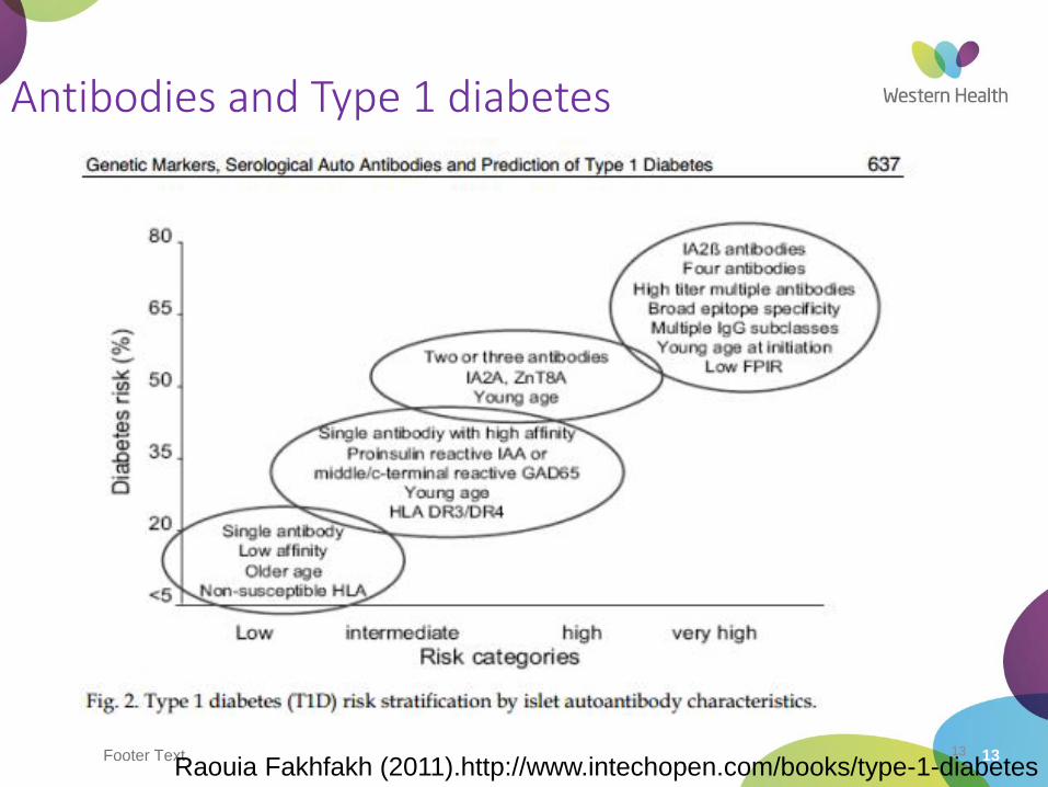
Antibodies and Type 1 diabetes
13
Raouia Fakhfakh (2011).http://www.intechopen.com/books/type-1-diabetes
Footer Text 14
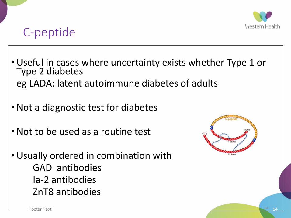
C-peptide
•Useful in cases where uncertainty exists whether Type 1 or Type 2 diabetes eg LADA: latent autoimmune diabetes of adults
•Not a diagnostic test for diabetes
•Not to be used as a routine test
•Usually ordered in combination with GAD antibodies Ia-2 antibodiesZnT8 antibodies
14
Footer Text 15
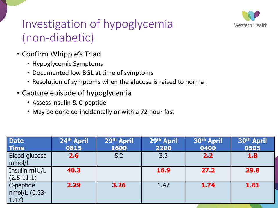
Investigation of hypoglycemia(non-diabetic)
• Confirm Whipple’s Triad• Hypoglycemic Symptoms
• Documented low BGL at time of symptoms
• Resolution of symptoms when the glucose is raised to normal
• Capture episode of hypoglycemia • Assess insulin & C-peptide
• May be done co-incidentally or with a 72 hour fast
DateTime
24th April 0815
29th April 1600
29th April 2200
30th April 0400
30th April0505
Blood glucose mmol/L
2.6 5.2 3.3 2.2 1.8
Insulin mIU/L (2.5-11.1)
40.3 16.9 27.2 29.8
C-peptide nmol/L (0.33-1.47)
2.29 3.26 1.47 1.74 1.81
Thyroid
16
Footer Text 17
Thyroid Investigations
1. Clinical examination still important (nodules, goitre, signs of obstruction, nodes, thyroid eye signs, reflexes etc etc)
2. CXR/CT Thoracic inlet
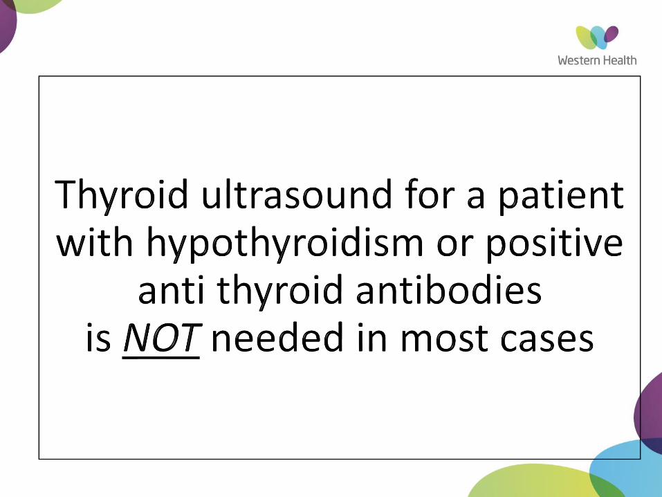
3. Thyroid Ultrasound
4. Technetium (Tc-99m) pertechnetate thyroid nuclear scan
5. Other: I131 scans and PET scans for thyroid cancer
6. TFTs, thyroid antibodies, TSH receptor antibodies(TRABs), Thyroid Stimulating Immunoglobulins (TSI)
7. FNA cytology
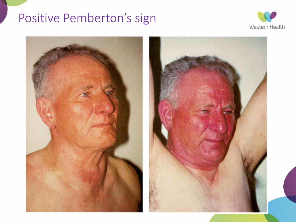
Positive Pemberton’s sign
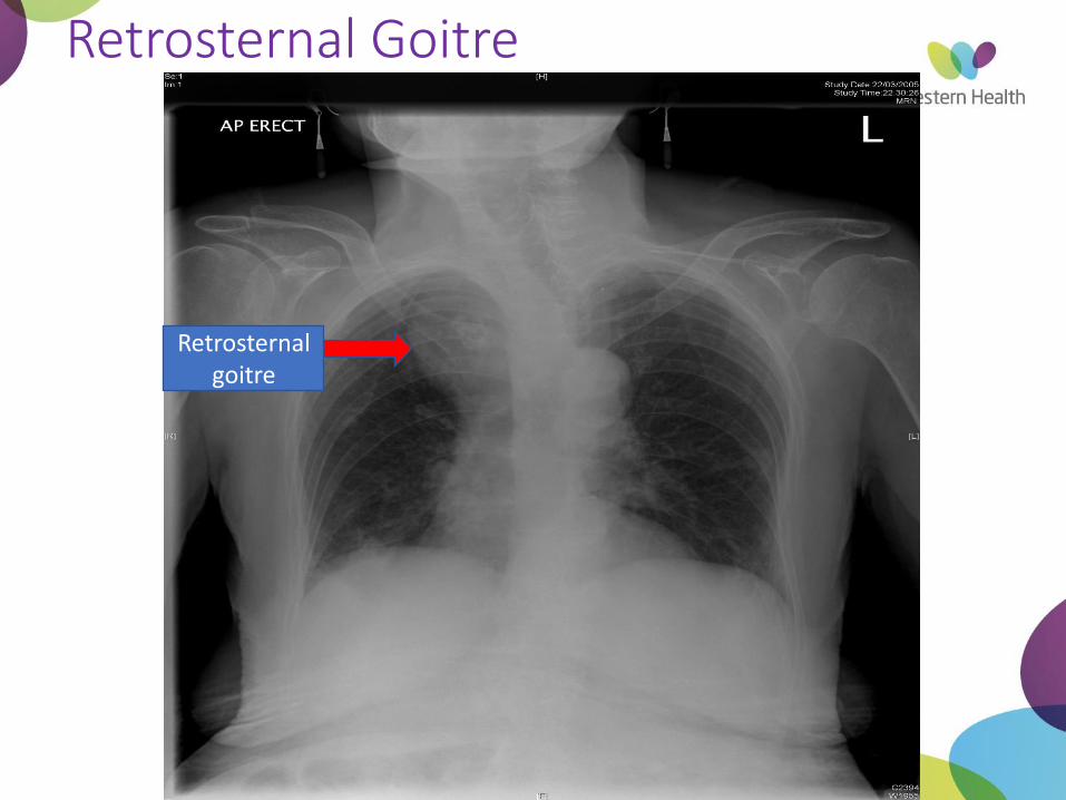
Retrosternal Goitre
Retrosternal goitre
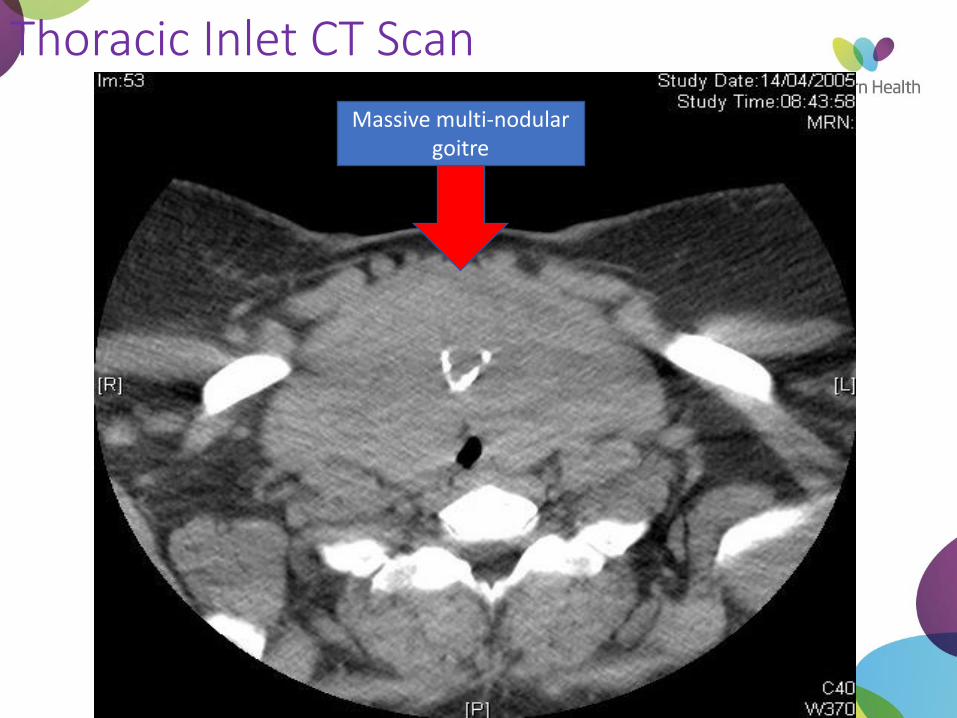
Thoracic Inlet CT Scan
Massive multi-nodular goitre
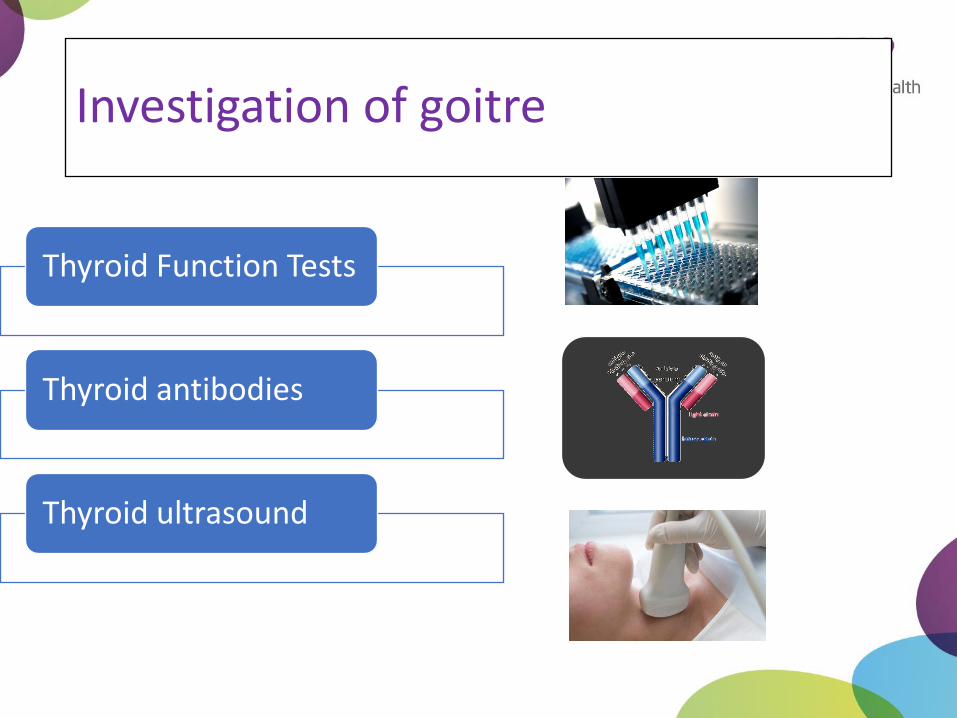
Investigation of goitre
Thyroid Function Tests
Thyroid antibodies
Thyroid ultrasound
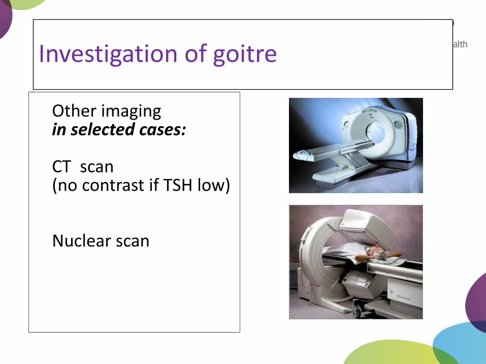
Investigation of goitre
Other imaging in selected cases:
CT scan(no contrast if TSH low)
Nuclear scan
Footer Text 23
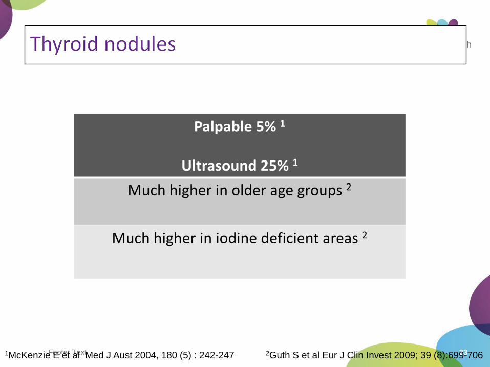
Palpable 5% 1
Ultrasound 25% 1
Much higher in older age groups 2
Much higher in iodine deficient areas 2
1McKenzie E et al Med J Aust 2004, 180 (5) : 242-247 2Guth S et al Eur J Clin Invest 2009; 39 (8):699-706
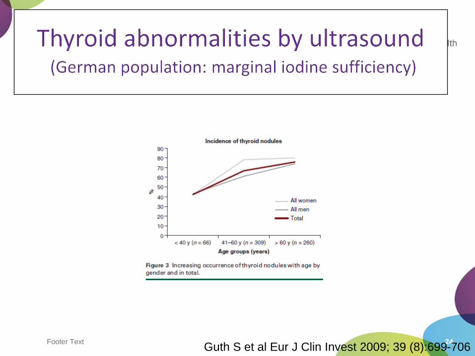
Footer Text 24Guth S et al Eur J Clin Invest 2009; 39 (8):699-706
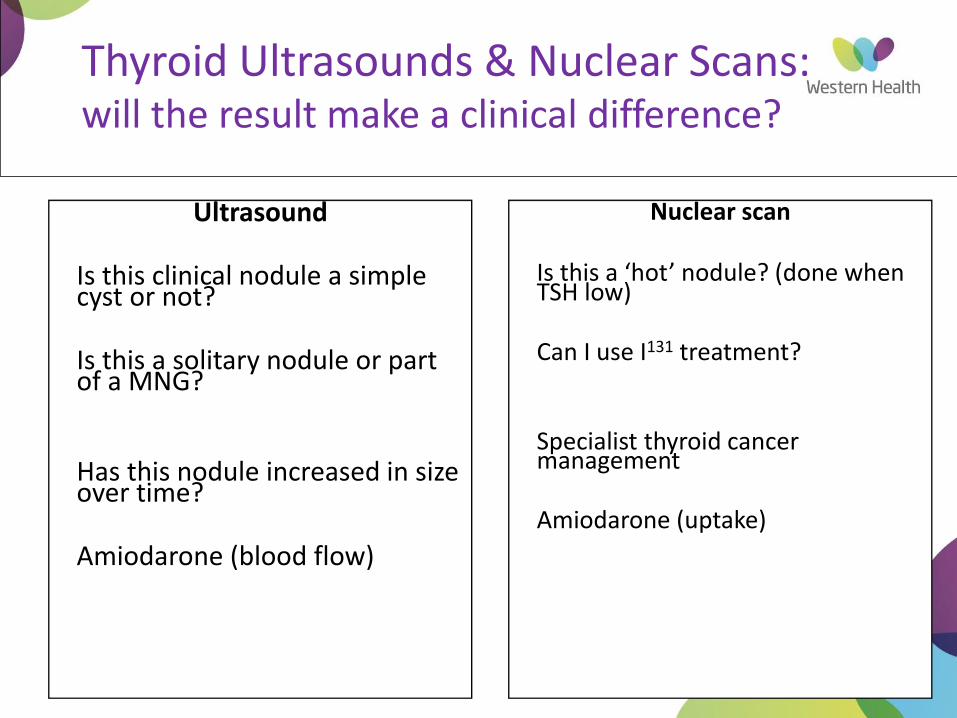
Thyroid Ultrasounds & Nuclear Scans: will the result make a clinical difference?
Ultrasound
Is this clinical nodule a simple cyst or not?
Is this a solitary nodule or part of a MNG?
Has this nodule increased in size over time?
Amiodarone (blood flow)
Nuclear scan
Is this a ‘hot’ nodule? (done when TSH low)
Can I use I131 treatment?
Specialist thyroid cancer management
Amiodarone (uptake)
Footer Text 26
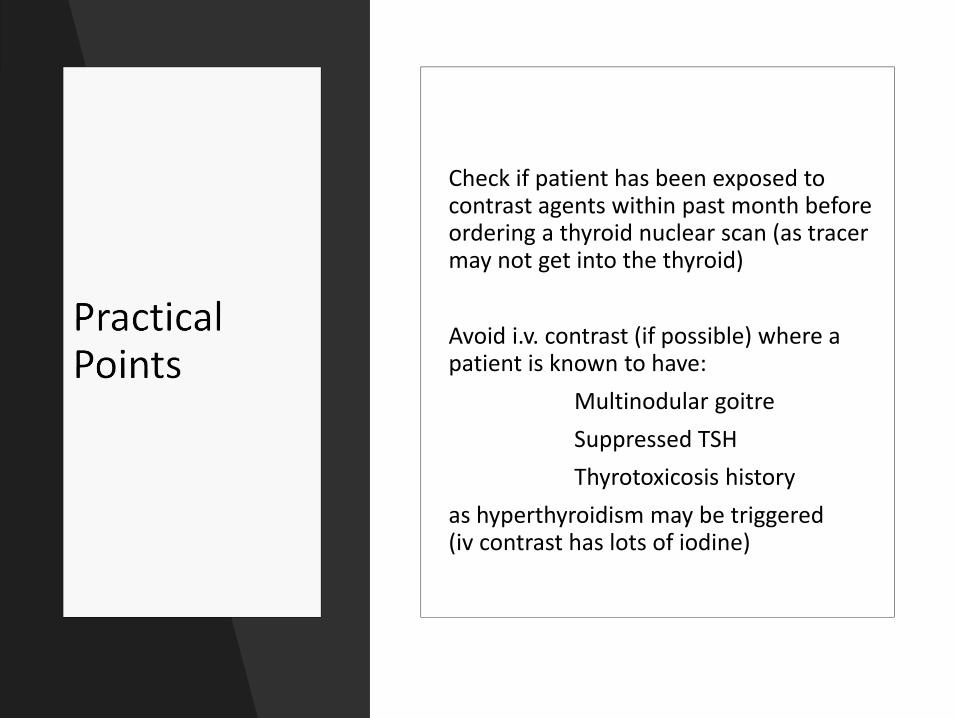
Check if patient has been exposed to contrast agents within past month before ordering a thyroid nuclear scan (as tracer may not get into the thyroid)
Avoid i.v. contrast (if possible) where a patient is known to have:
Multinodular goitre
Suppressed TSH
Thyrotoxicosis history
as hyperthyroidism may be triggered (iv contrast has lots of iodine)
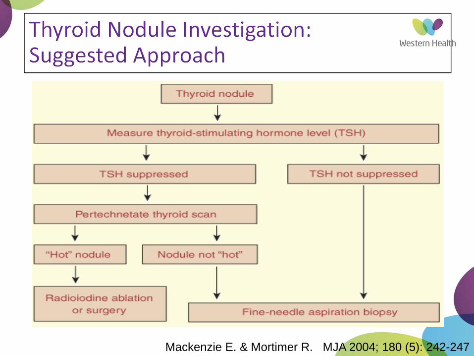
Mackenzie E. & Mortimer R. MJA 2004; 180 (5): 242-247
Footer Text 28
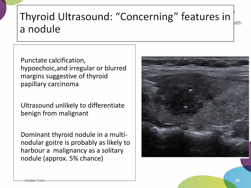
Punctate calcification, hypoechoic,and irregular or blurred margins suggestive of thyroid papillary carcinoma
Ultrasound unlikely to differentiate benign from malignant
Dominant thyroid nodule in a multi-nodular goitre is probably as likely to harbour a malignancy as a solitary nodule (approx. 5% chance)
29
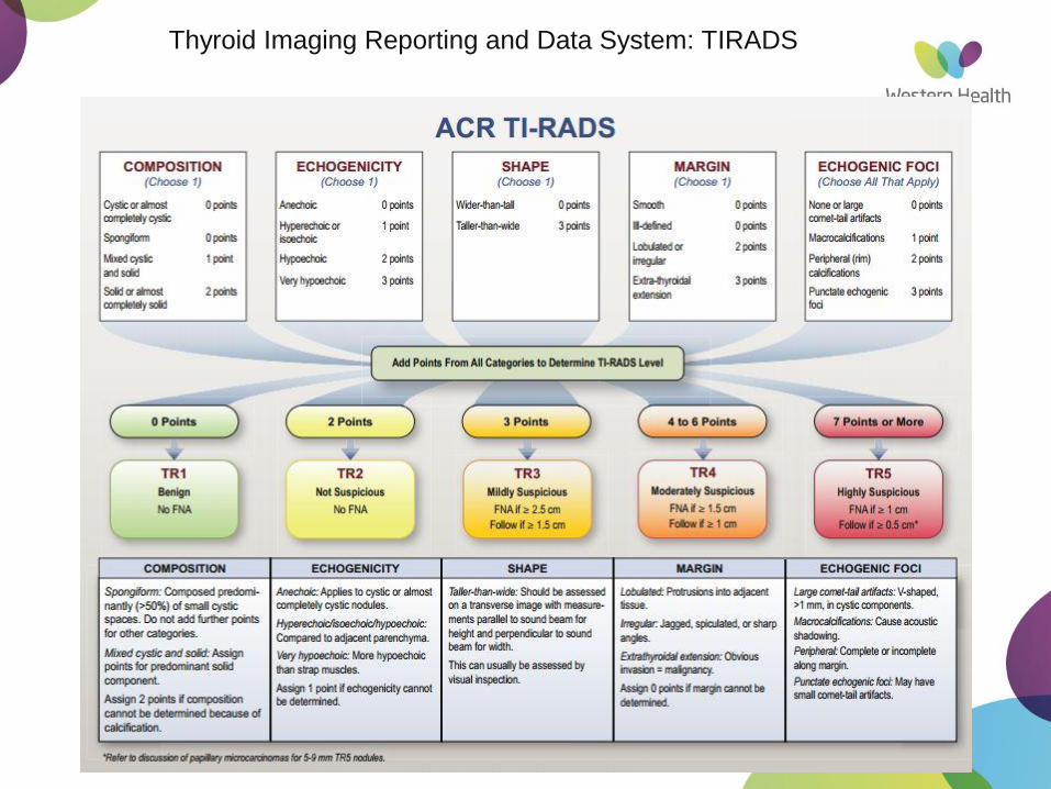
Thyroid Imaging Reporting and Data System: TIRADS
Footer Text 30
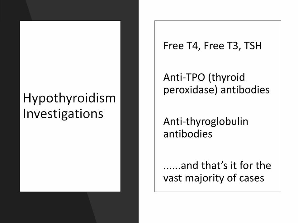
Free T4, Free T3, TSH
Anti-TPO (thyroid peroxidase) antibodies
Anti-thyroglobulin antibodies
......and that’s it for the vast majority of cases
Footer Text 32
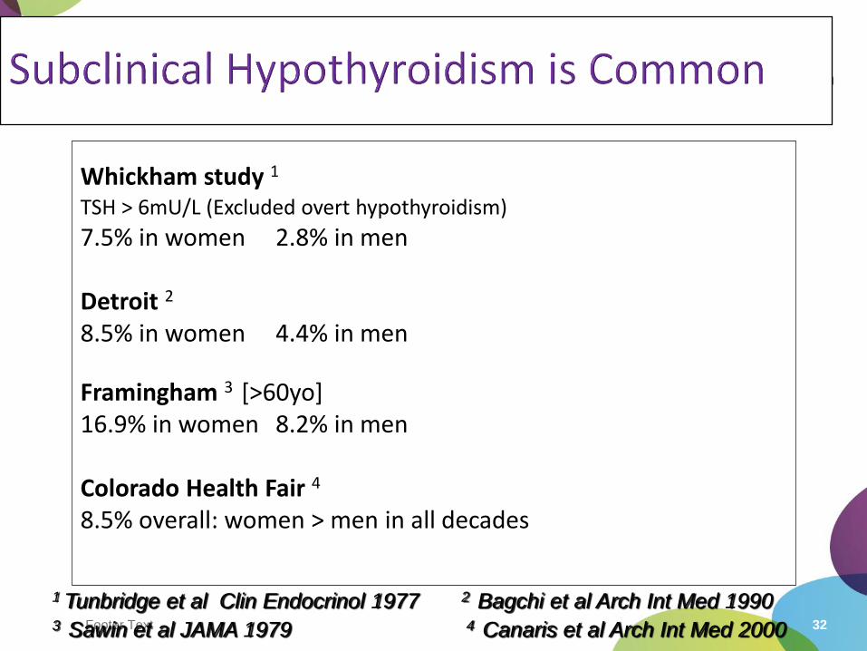
Whickham study 1
TSH > 6mU/L (Excluded overt hypothyroidism)
7.5% in women 2.8% in men
Detroit 2
8.5% in women 4.4% in men
Framingham 3 [>60yo]16.9% in women 8.2% in men
Colorado Health Fair 4
8.5% overall: women > men in all decades
1 Tunbridge et al Clin Endocrinol 1977 2 Bagchi et al Arch Int Med 19903 Sawin et al JAMA 1979 4 Canaris et al Arch Int Med 2000
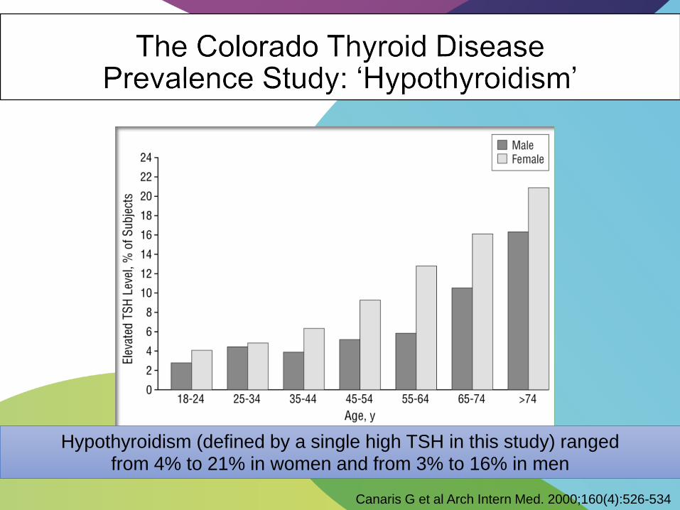
Canaris G et al Arch Intern Med. 2000;160(4):526-534
Hypothyroidism (defined by a single high TSH in this study) ranged from 4% to 21% in women and from 3% to 16% in men
Footer Text 34
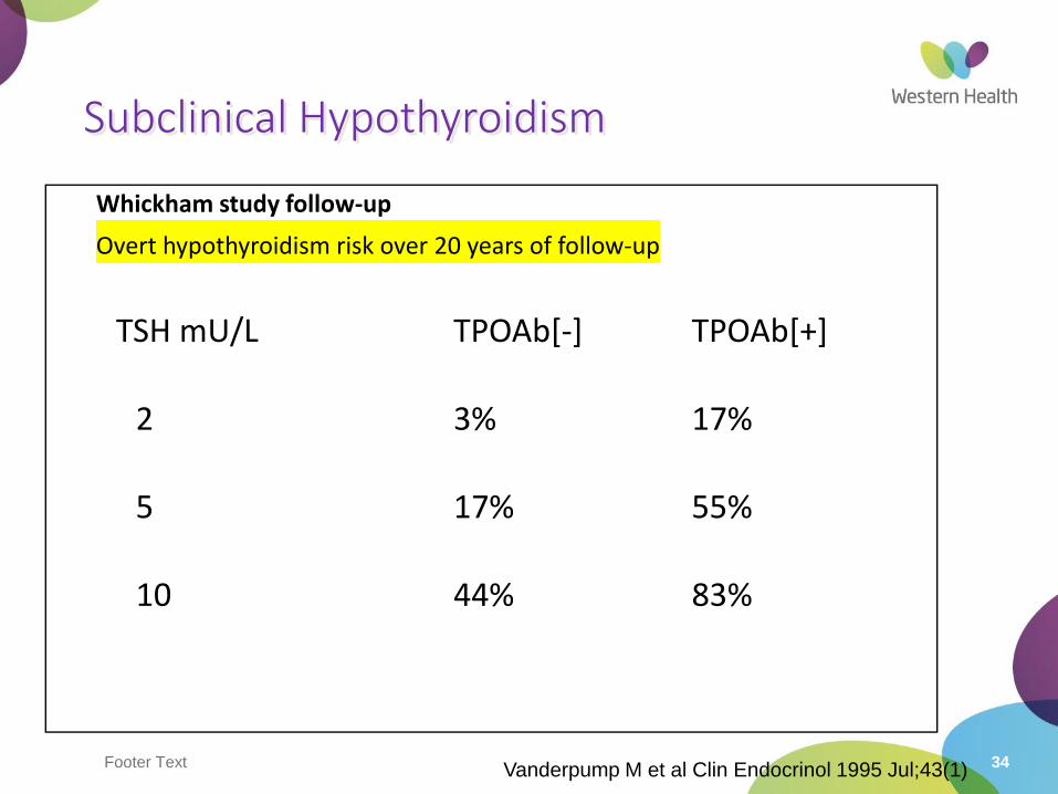
Subclinical Hypothyroidism
Whickham study follow-up
Overt hypothyroidism risk over 20 years of follow-up
TSH mU/L TPOAb[-] TPOAb[+]
2 3% 17%
5 17% 55%
10 44% 83%
Vanderpump M et al Clin Endocrinol 1995 Jul;43(1)
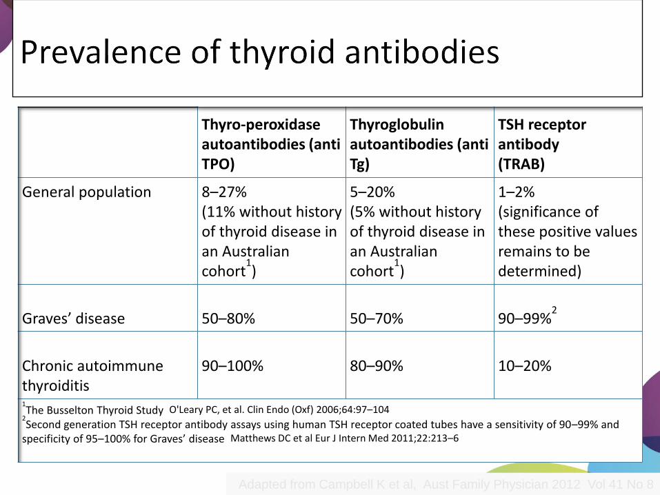
Thyro-peroxidase autoantibodies (anti TPO)
Thyroglobulin autoantibodies (anti Tg)
TSH receptor antibody (TRAB)
General population 8–27%(11% without history of thyroid disease in an Australian cohort
1)
5–20%(5% without history of thyroid disease in an Australian cohort
1)
1–2%(significance of these positive values remains to be determined)
Graves’ disease 50–80% 50–70% 90–99%2
Chronic autoimmune thyroiditis
90–100% 80–90% 10–20%
1The Busselton Thyroid Study O'Leary PC, et al. Clin Endo (Oxf) 2006;64:97–104
2Second generation TSH receptor antibody assays using human TSH receptor coated tubes have a sensitivity of 90–99% and
specificity of 95–100% for Graves’ disease Matthews DC et al Eur J Intern Med 2011;22:213–6
Adapted from Campbell K et al, Aust Family Physician 2012 Vol 41 No 8
Footer Text 36
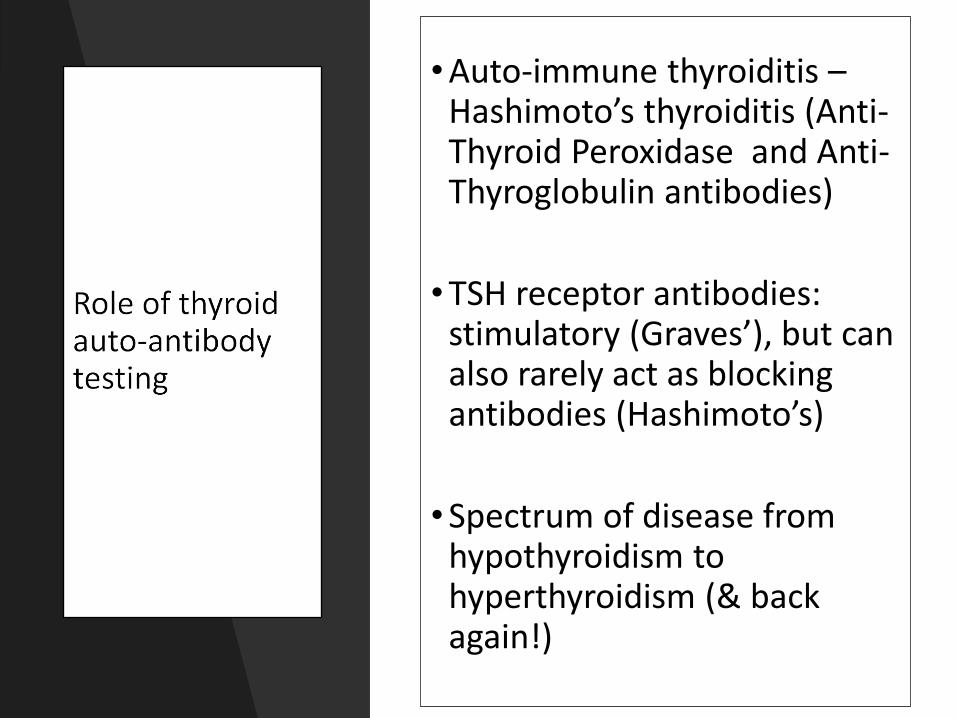
•Auto-immune thyroiditis –Hashimoto’s thyroiditis (Anti-Thyroid Peroxidase and Anti-Thyroglobulin antibodies)
•TSH receptor antibodies: stimulatory (Graves’), but can also rarely act as blocking antibodies (Hashimoto’s)
•Spectrum of disease from hypothyroidism to hyperthyroidism (& back again!)
Footer Text 37
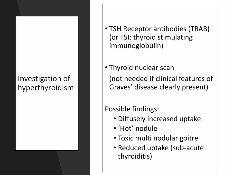
• TSH Receptor antibodies (TRAB) (or TSI: thyroid stimulating immunoglobulin)
• Thyroid nuclear scan
(not needed if clinical features of Graves’ disease clearly present)
Possible findings:• Diffusely increased uptake• ‘Hot’ nodule• Toxic multi nodular goitre
• Reduced uptake (sub-acute thyroiditis)
Therapeutic Guidelines
Therapeutic Guidelines Ltd (eTG July 2017 edition)
Therapeutic Guidelines
Therapeutic Guidelines
Therapeutic Guidelines Ltd (eTG July 2017 edition)
Therapeutic Guidelines
Footer Text 44
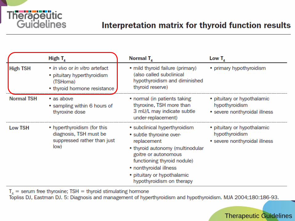
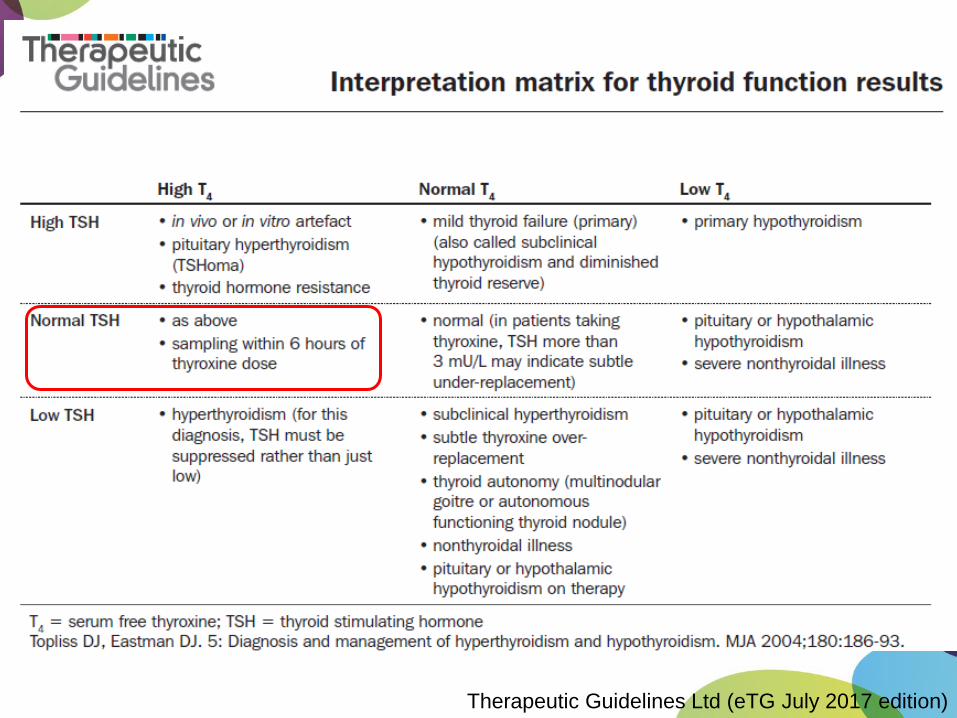
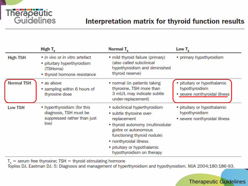
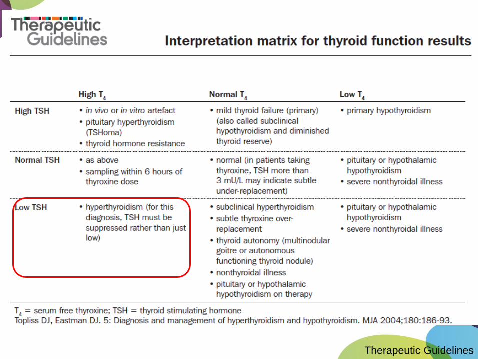
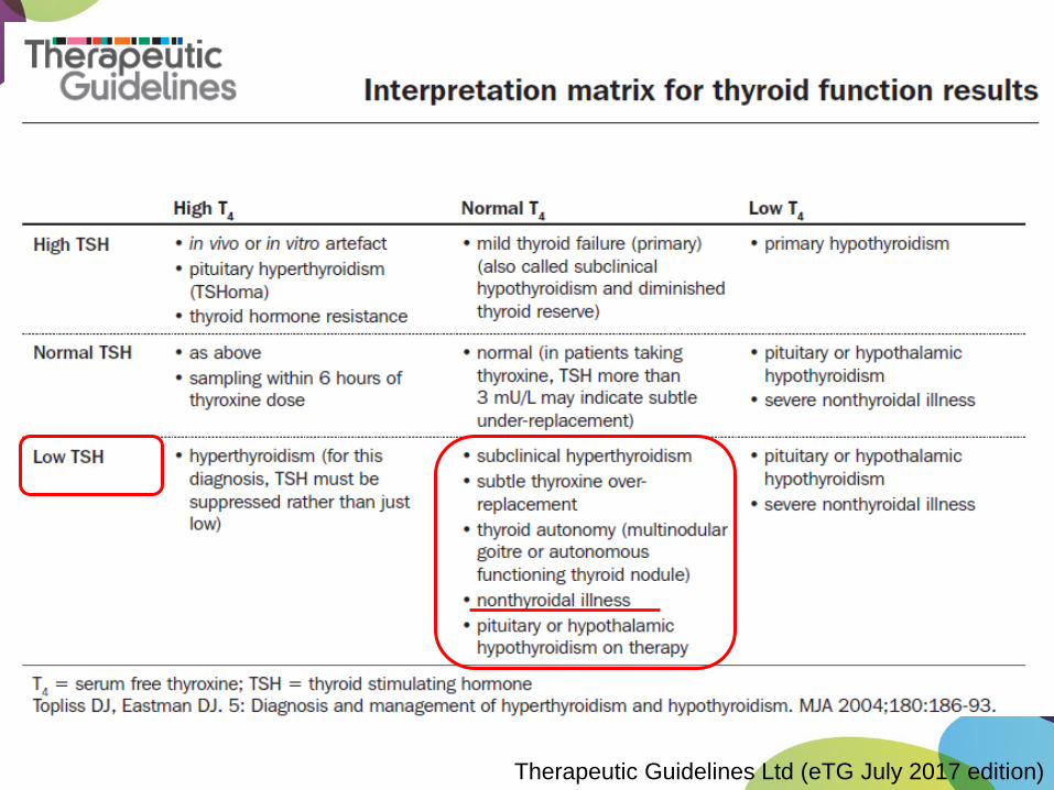
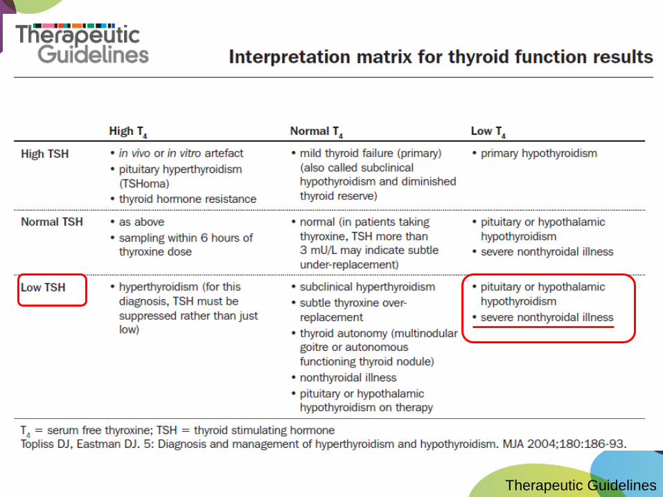
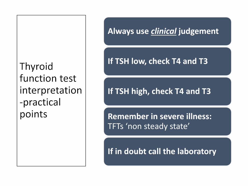
Always use clinical judgement
If TSH low, check T4 and T3
If TSH high, check T4 and T3
Remember in severe illness: TFTs ‘non steady state’
If in doubt call the laboratory
Footer Text 45
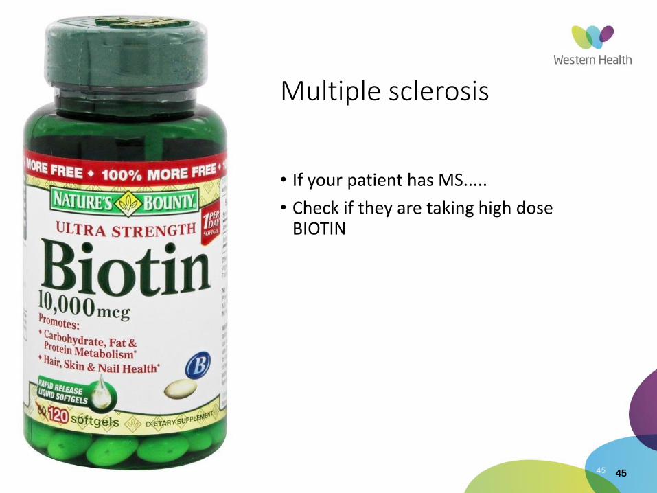
Multiple sclerosis
• If your patient has MS.....
• Check if they are taking high dose BIOTIN
Footer Text 46
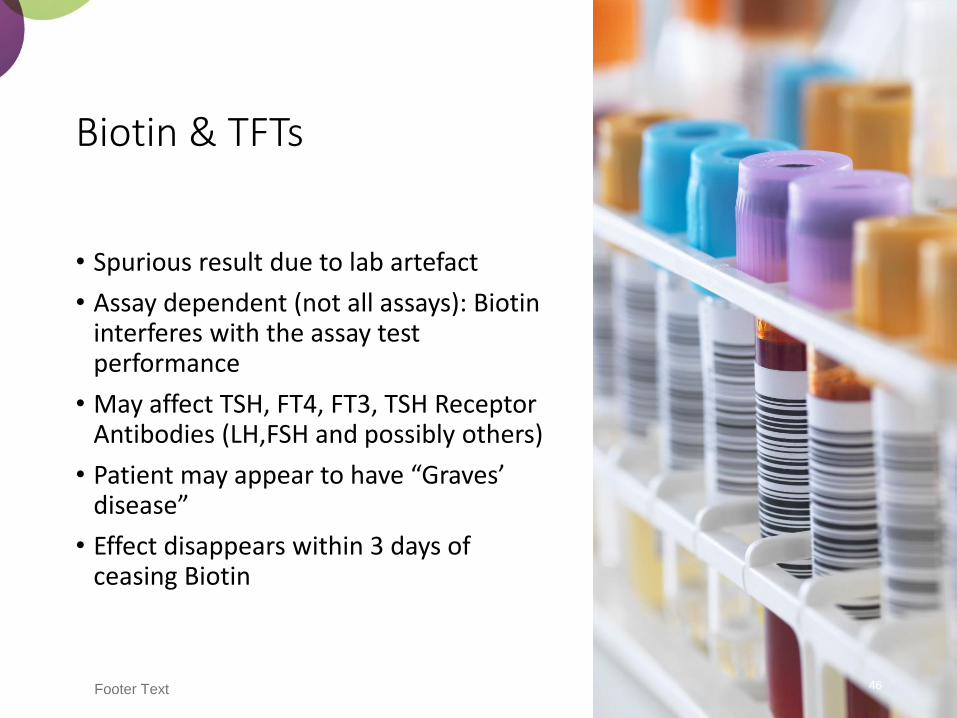
Biotin & TFTs
• Spurious result due to lab artefact
• Assay dependent (not all assays): Biotin interferes with the assay test performance
• May affect TSH, FT4, FT3, TSH Receptor Antibodies (LH,FSH and possibly others)
• Patient may appear to have “Graves’ disease”
• Effect disappears within 3 days of ceasing Biotin
46
Adrenal
47
Footer Text 48
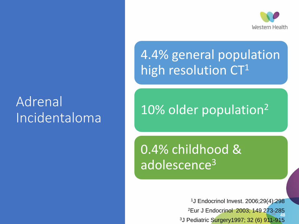
Adrenal Incidentaloma
48
1J Endocrinol Invest. 2006;29(4):298
2Eur J Endocrinol 2003; 149 273-285
3J Pediatric Surgery1997; 32 (6) 911-915
4.4% general population high resolution CT1
10% older population2
0.4% childhood & adolescence3
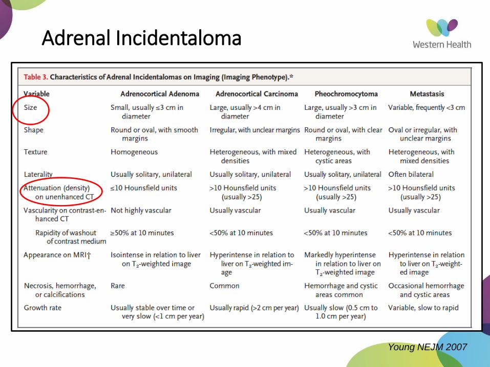
Young NEJM 2007
Adrenal Incidentaloma
Footer Text 50
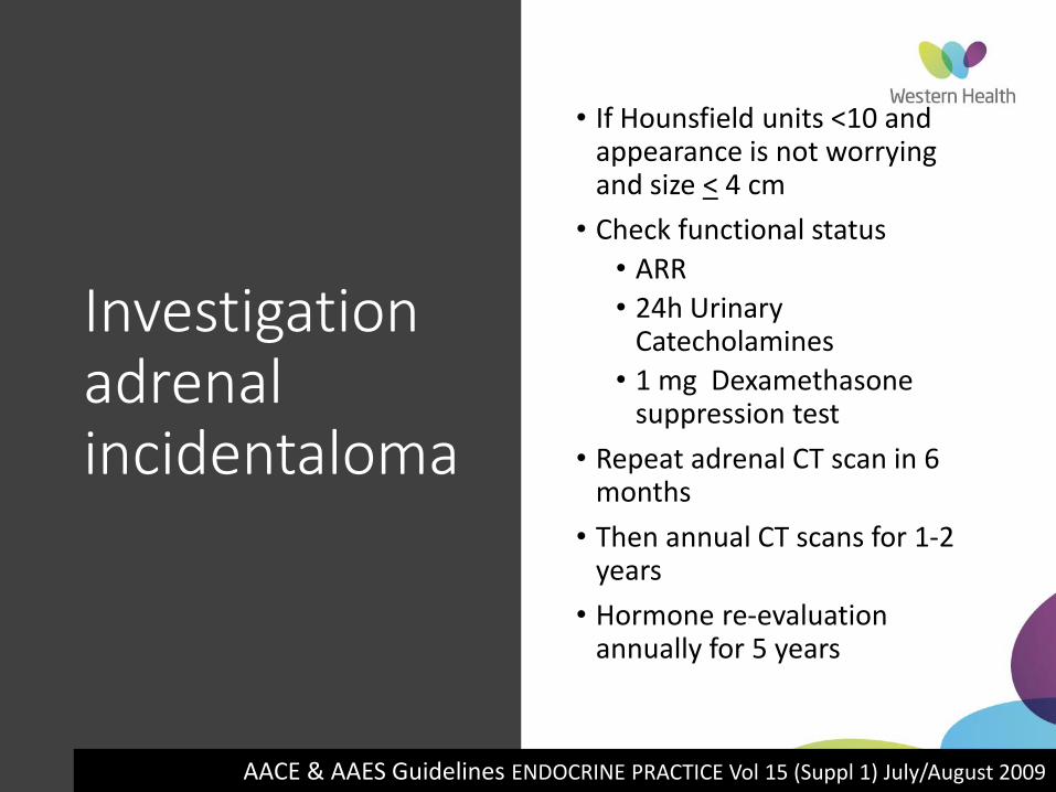
Investigation adrenal incidentaloma
• If Hounsfield units <10 and appearance is not worrying and size < 4 cm
• Check functional status
• ARR
• 24h Urinary Catecholamines
• 1 mg Dexamethasone suppression test
• Repeat adrenal CT scan in 6 months
• Then annual CT scans for 1-2 years
• Hormone re-evaluation annually for 5 years
50
AACE & AAES Guidelines ENDOCRINE PRACTICE Vol 15 (Suppl 1) July/August 2009
Footer Text 51
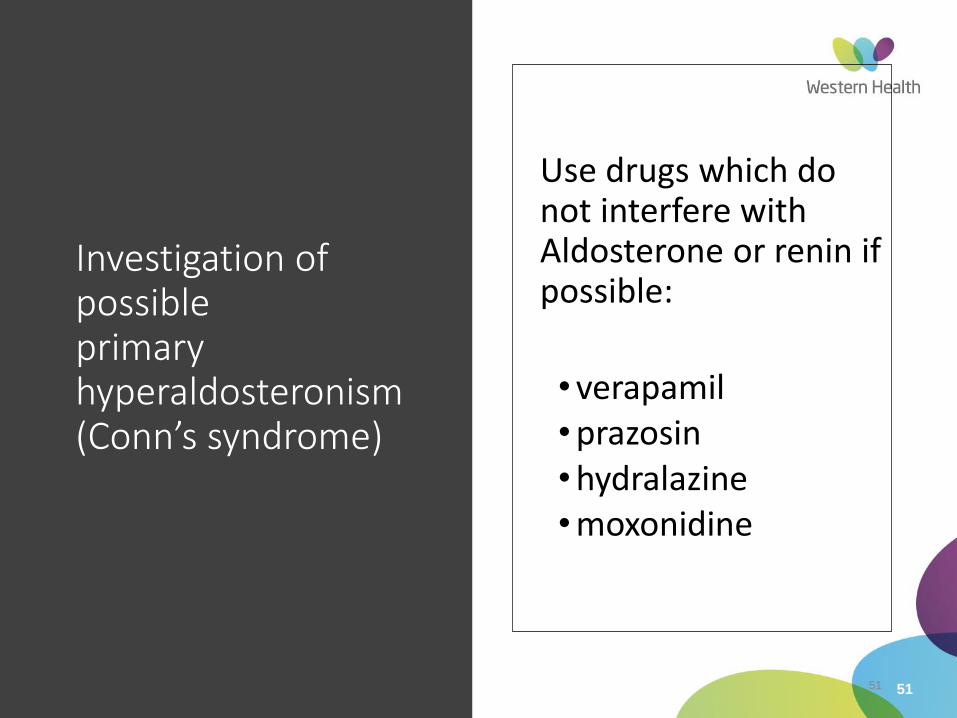
Investigation of possible primary hyperaldosteronism (Conn’s syndrome)
Use drugs which do not interfere with Aldosterone or renin if possible:
•verapamil•prazosin•hydralazine•moxonidine
51
Footer Text 52
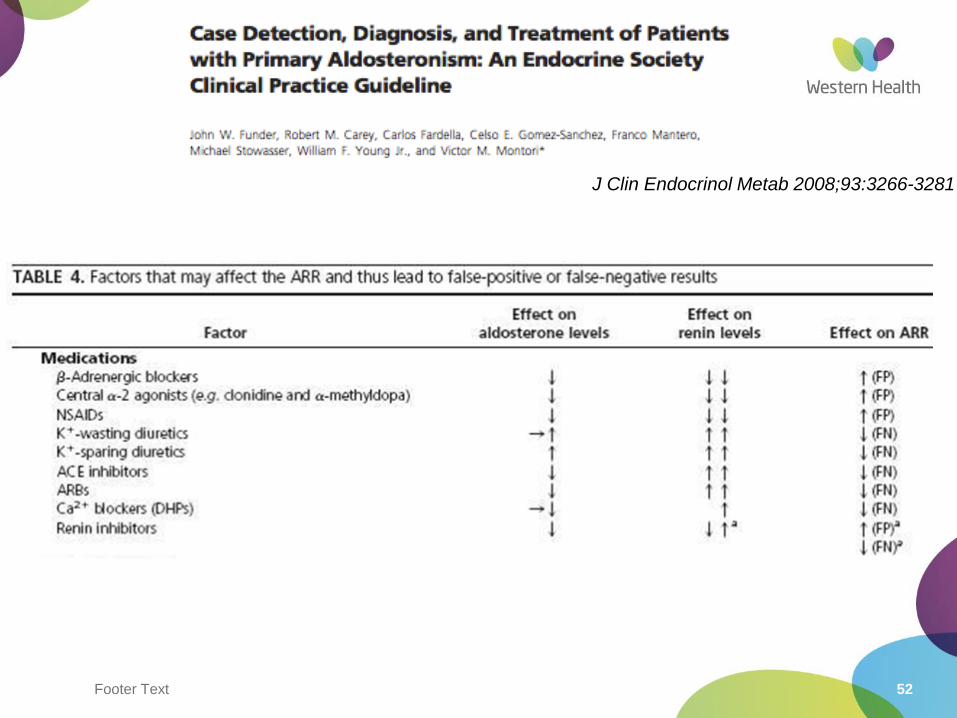
J Clin Endocrinol Metab 2008;93:3266-3281
53
J Clin Endocrinol Metab 2007;93:3266-3281
Footer Text 54
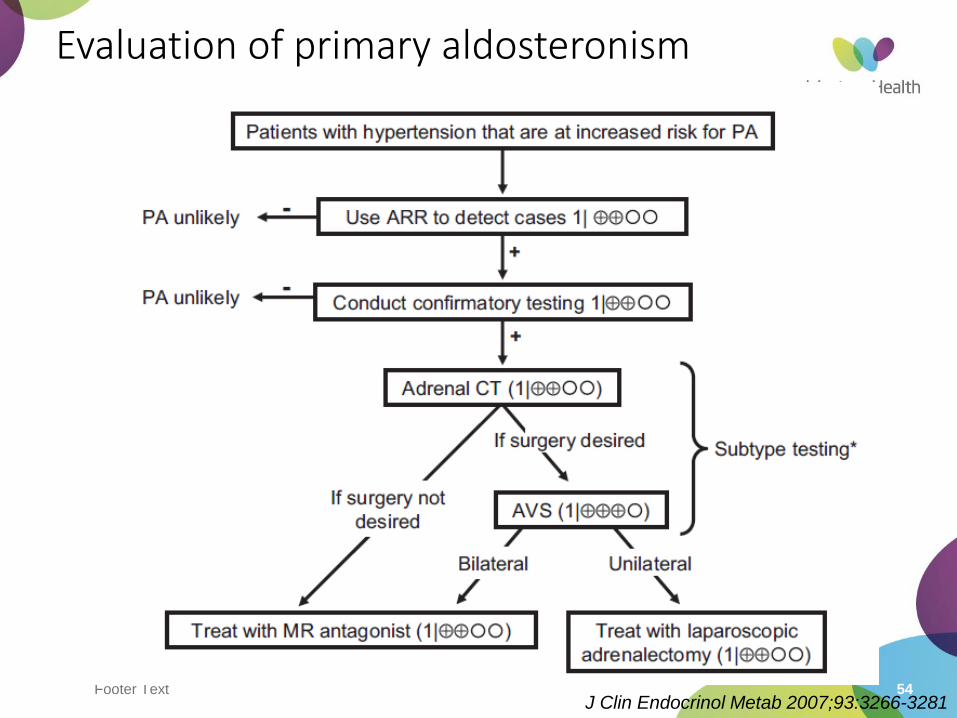
Evaluation of primary aldosteronism
J Clin Endocrinol Metab 2007;93:3266-3281
Footer Text 55
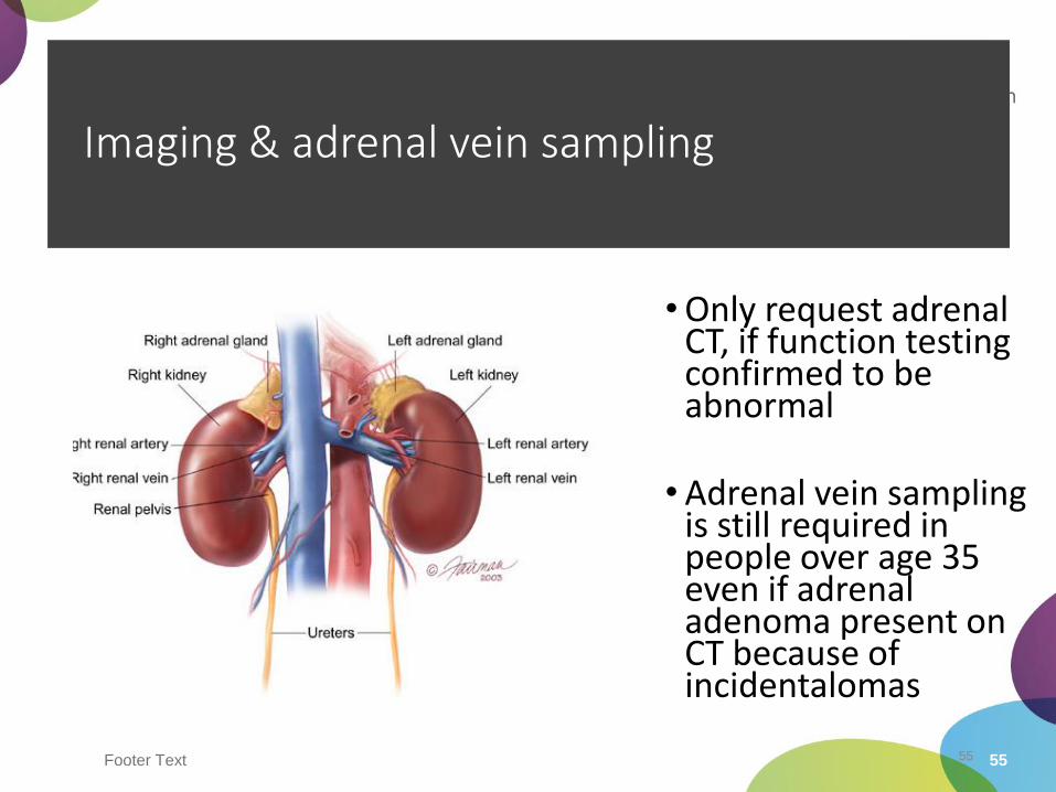
Imaging & adrenal vein sampling
•Only request adrenal CT, if function testing confirmed to be abnormal
•Adrenal vein sampling is still required in people over age 35 even if adrenal adenoma present on CT because of incidentalomas
55
Footer Text 56
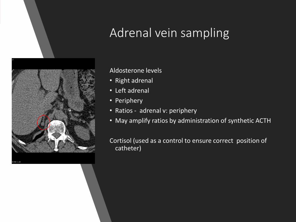
Adrenal vein sampling
Aldosterone levels
• Right adrenal
• Left adrenal
• Periphery
• Ratios - adrenal v: periphery
• May amplify ratios by administration of synthetic ACTH
Cortisol (used as a control to ensure correct position of catheter)

Footer Text 57
Adenoma
Carcinoma
Footer Text 58
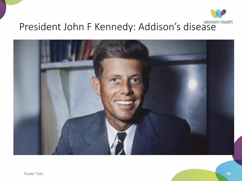
President John F Kennedy: Addison’s disease
58
59
Footer Text 60
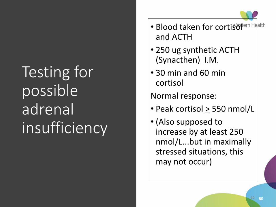
Testing for possible adrenal insufficiency
• Blood taken for cortisol and ACTH
• 250 ug synthetic ACTH (Synacthen) I.M.
• 30 min and 60 min cortisol
Normal response:
• Peak cortisol > 550 nmol/L
• (Also supposed to increase by at least 250 nmol/L...but in maximally stressed situations, this may not occur)
60
Footer Text 61
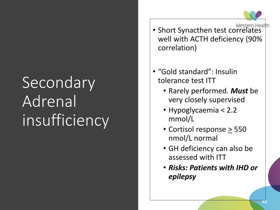
Secondary Adrenal insufficiency
• Short Synacthen test correlates well with ACTH deficiency (90% correlation)
• “Gold standard”: Insulin tolerance test ITT
• Rarely performed. Must be very closely supervised
• Hypoglycaemia < 2.2 mmol/L
• Cortisol response > 550 nmol/L normal
• GH deficiency can also be assessed with ITT
• Risks: Patients with IHD or epilepsy
61
Pituitary
62
Footer Text 63
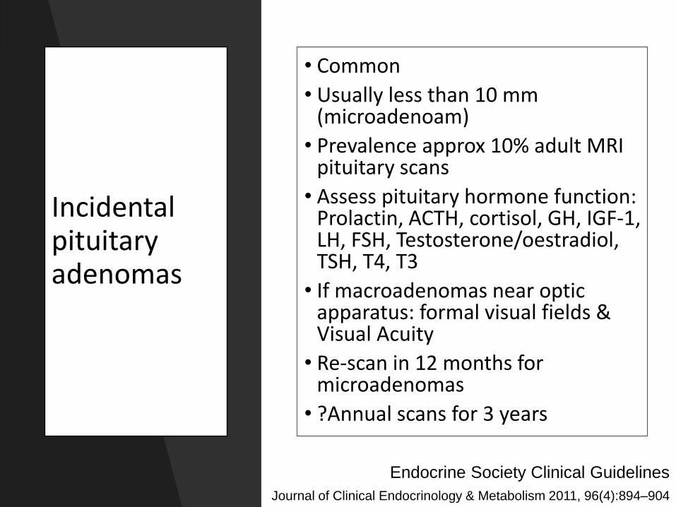
Incidental pituitary adenomas
• Common
• Usually less than 10 mm (microadenoam)
• Prevalence approx 10% adult MRI pituitary scans
• Assess pituitary hormone function: Prolactin, ACTH, cortisol, GH, IGF-1, LH, FSH, Testosterone/oestradiol, TSH, T4, T3
• If macroadenomas near optic apparatus: formal visual fields & Visual Acuity
• Re-scan in 12 months for microadenomas
• ?Annual scans for 3 years
Endocrine Society Clinical Guidelines
Journal of Clinical Endocrinology & Metabolism 2011, 96(4):894–904
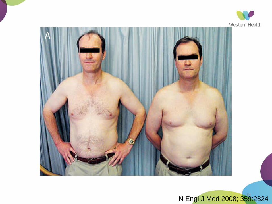
64
N Engl J Med 2008; 359:2824
Footer Text 65
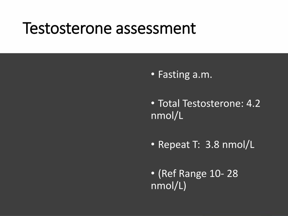
Testosterone assessment
• Fasting a.m.
• Total Testosterone: 4.2 nmol/L
• Repeat T: 3.8 nmol/L
• (Ref Range 10- 28 nmol/L)
Footer Text 66
Next investigation ?
A. MRI pituitary
B. Semen Analysis
C. Karyotype
D. LH/ FSH
E. SHBG/ calculated free testosterone

Footer Text 67
Next investigation ?
A. MRI pituitary
B. Semen Analysis
C. Karyotype
D. LH/ FSHE. SHBG/ calculated free testosterone
Footer Text 68
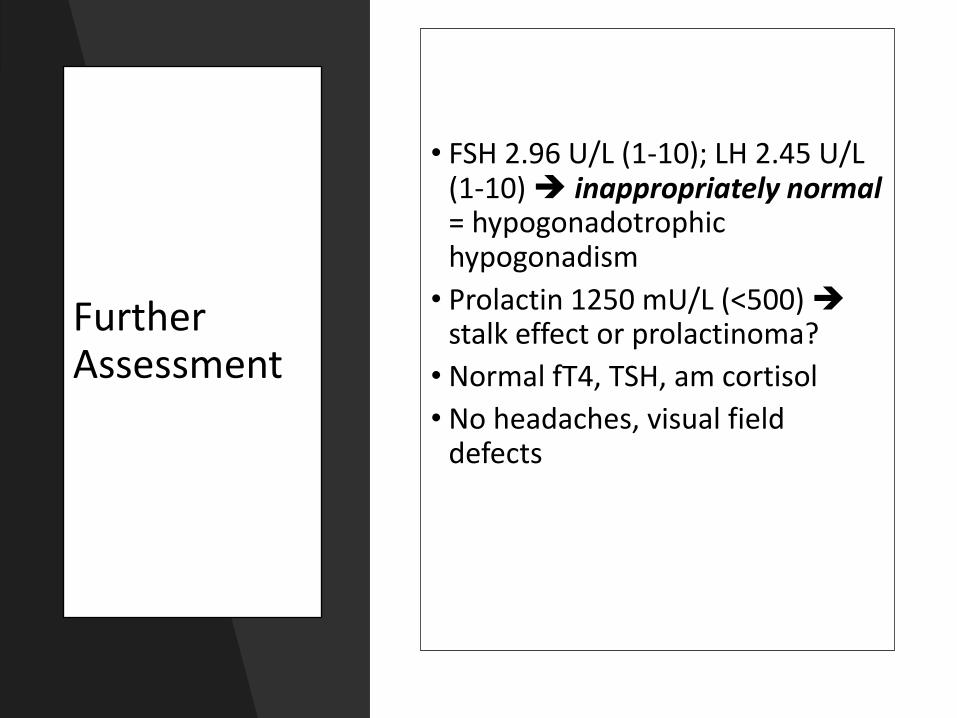
Further Assessment
• FSH 2.96 U/L (1-10); LH 2.45 U/L (1-10) ➔ inappropriately normal = hypogonadotrophichypogonadism
• Prolactin 1250 mU/L (<500) ➔stalk effect or prolactinoma?
• Normal fT4, TSH, am cortisol
• No headaches, visual field defects
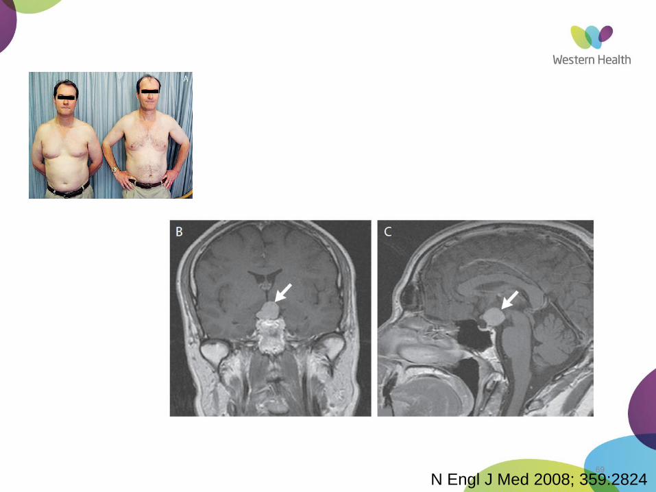
69
N Engl J Med 2008; 359:2824
Footer Text 70
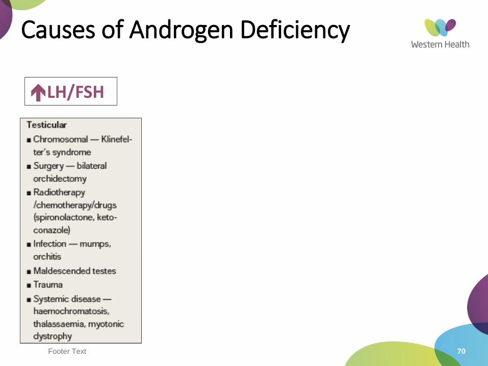
Causes of Androgen Deficiency
LH/FSH
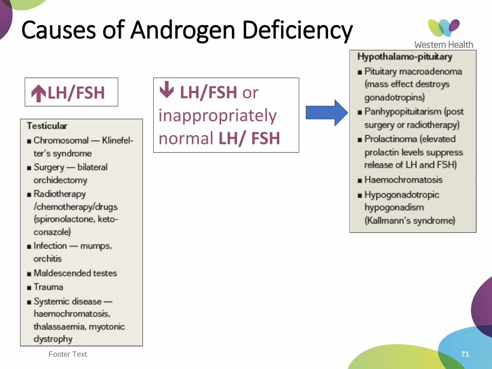
Footer Text 71
Causes of Androgen Deficiency
LH/FSH LH/FSH or inappropriately normal LH/ FSH
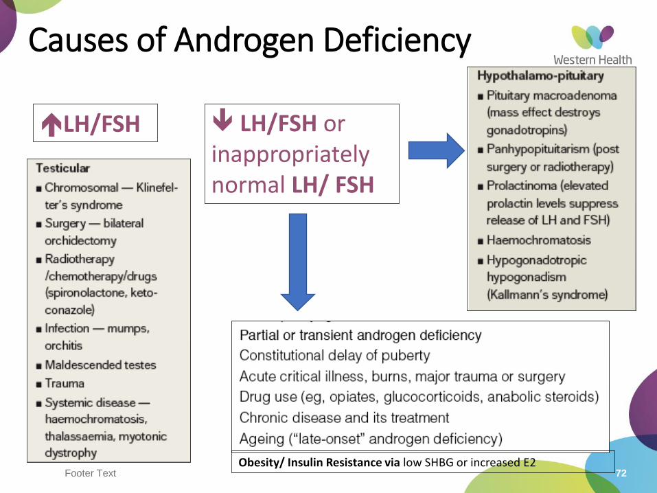
Footer Text 72
Causes of Androgen Deficiency
LH/FSH
Obesity/ Insulin Resistance via low SHBG or increased E2
LH/FSH or inappropriately normal LH/ FSH
Footer Text 73
Practical point
Do not request LH/FSH if a woman is taking OCP
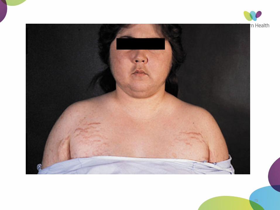
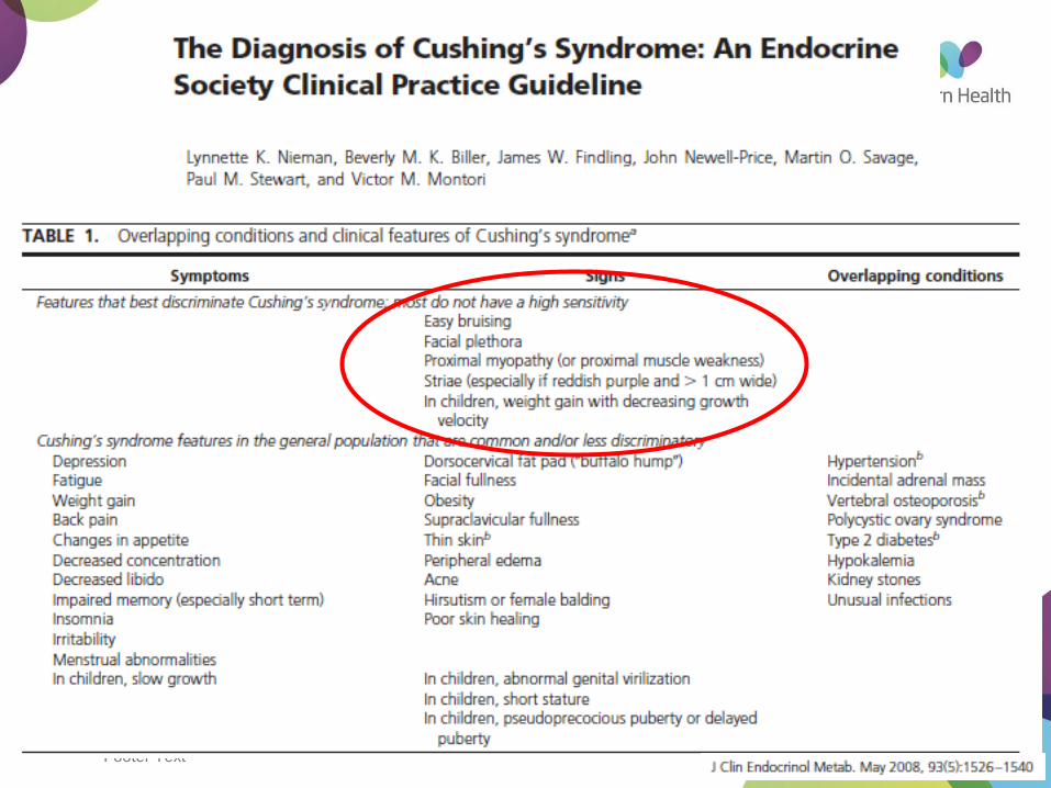
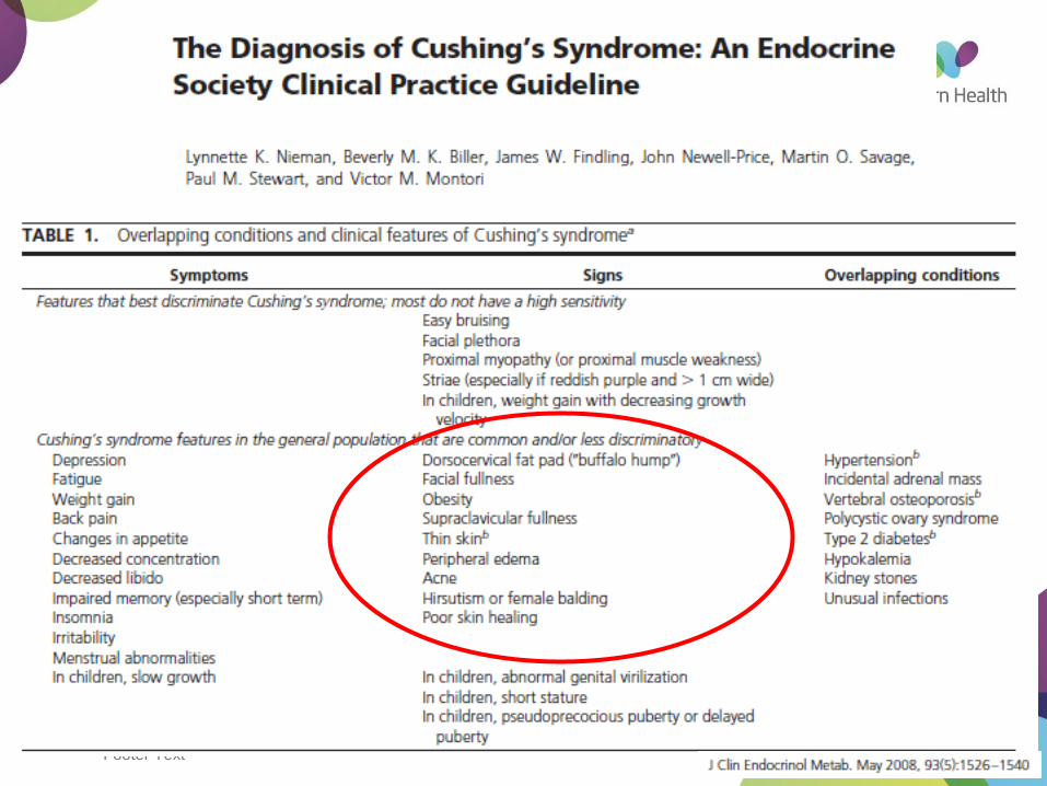
Cushing’s syndrome
74
75
Footer Text 76
Footer Text 77
Footer Text 78
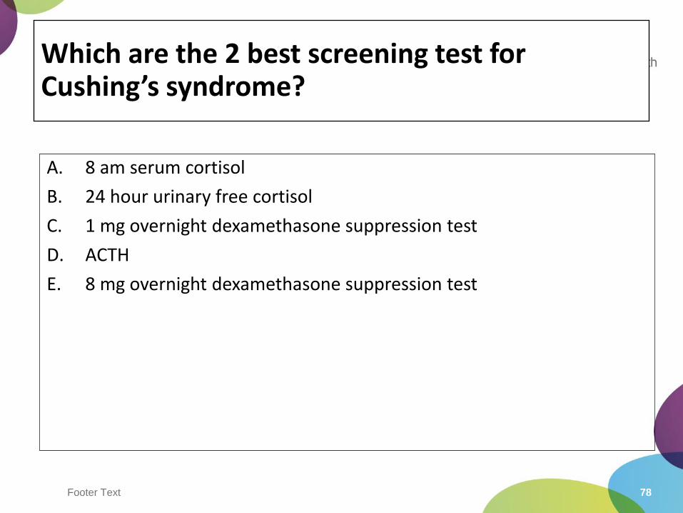
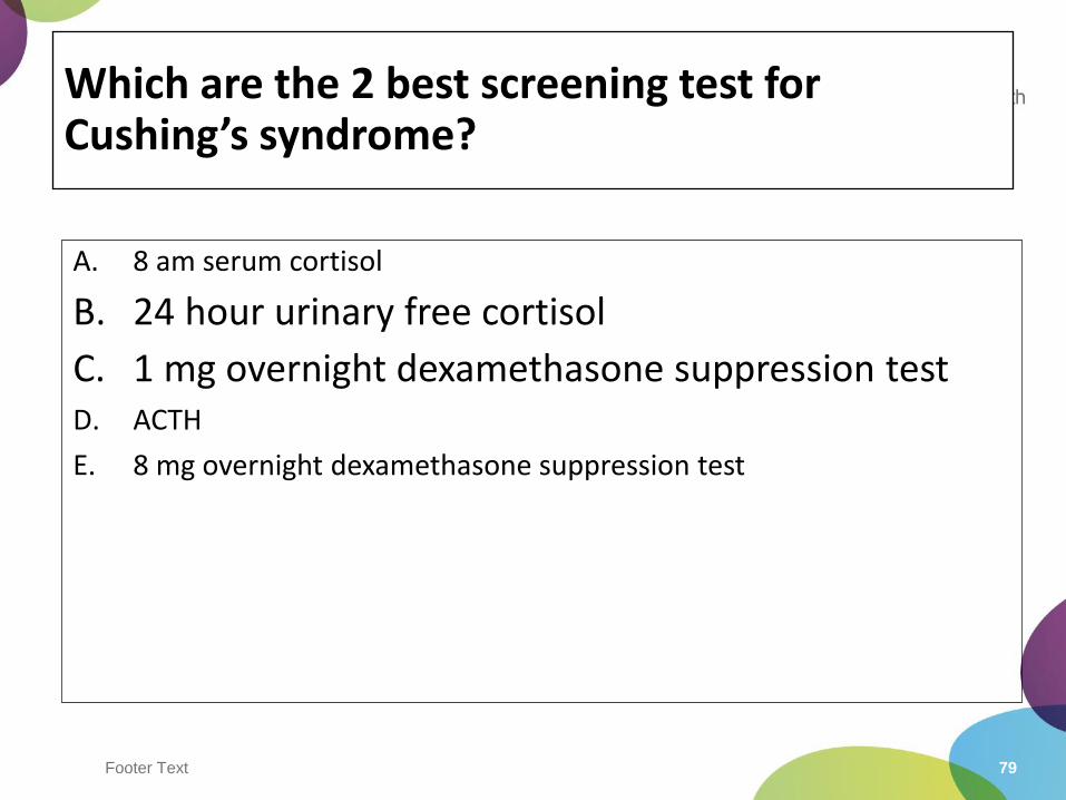
Which are the 2 best screening test for Cushing’s syndrome?
A. 8 am serum cortisol
B. 24 hour urinary free cortisol
C. 1 mg overnight dexamethasone suppression test
D. ACTH
E. 8 mg overnight dexamethasone suppression test
Footer Text 79
Which are the 2 best screening test for Cushing’s syndrome?
A. 8 am serum cortisol
B. 24 hour urinary free cortisol
C. 1 mg overnight dexamethasone suppression testD. ACTH
E. 8 mg overnight dexamethasone suppression test
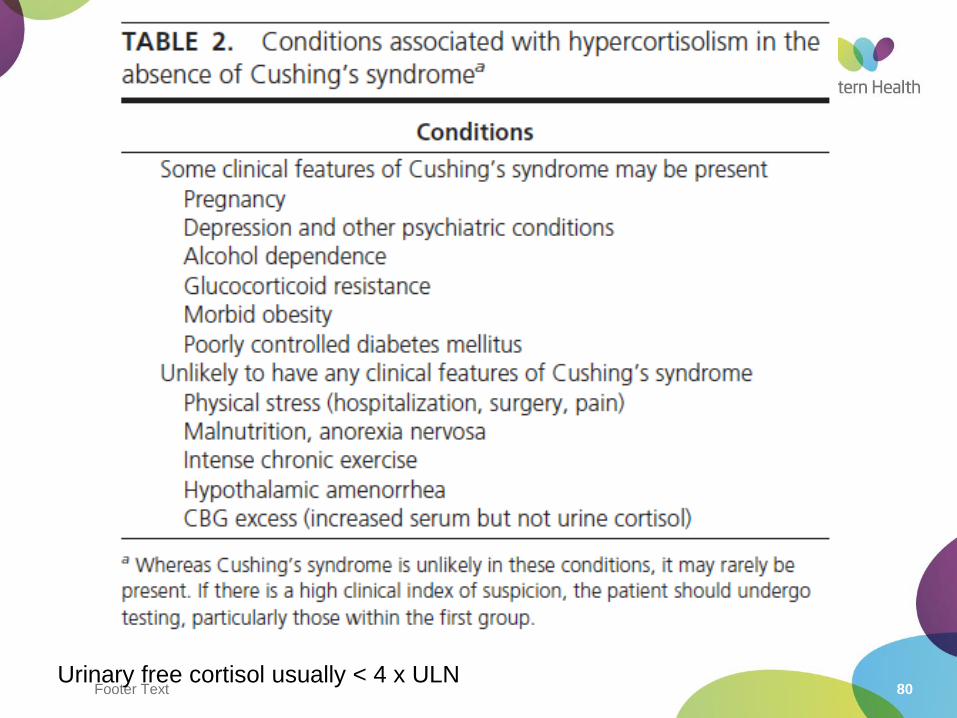
Footer Text 80Urinary free cortisol usually < 4 x ULN
Footer Text 81
Footer Text 82
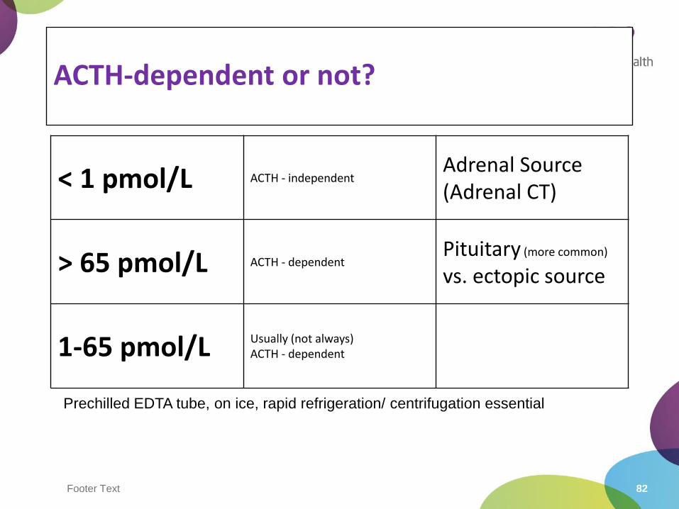
ACTH-dependent or not?
< 1 pmol/L ACTH - independentAdrenal Source(Adrenal CT)
> 65 pmol/L ACTH - dependentPituitary (more common)
vs. ectopic source
1-65 pmol/L Usually (not always)ACTH - dependent
Prechilled EDTA tube, on ice, rapid refrigeration/ centrifugation essential
Footer Text 83
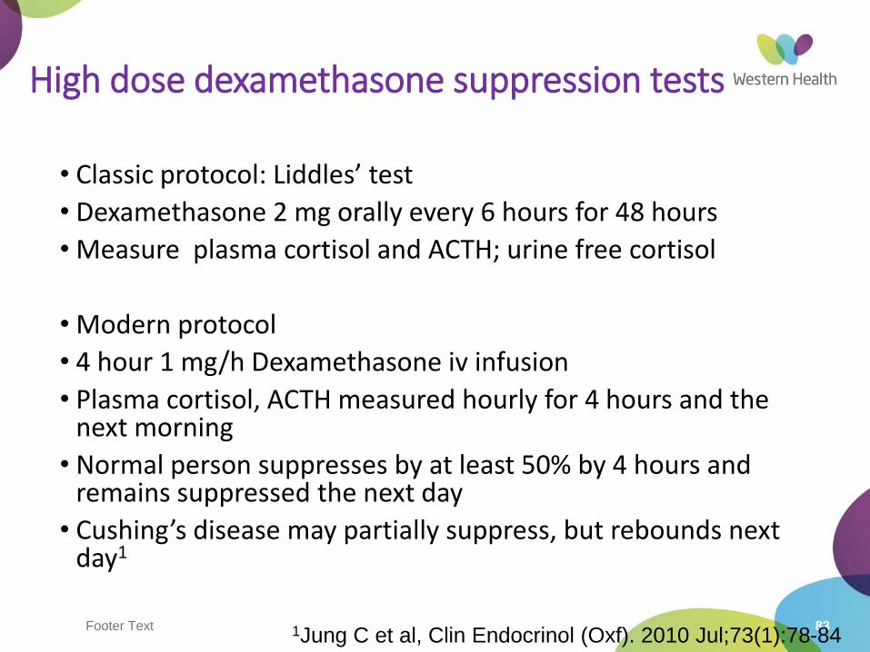
High dose dexamethasone suppression tests
• Classic protocol: Liddles’ test
• Dexamethasone 2 mg orally every 6 hours for 48 hours
• Measure plasma cortisol and ACTH; urine free cortisol
• Modern protocol
• 4 hour 1 mg/h Dexamethasone iv infusion
• Plasma cortisol, ACTH measured hourly for 4 hours and the next morning
• Normal person suppresses by at least 50% by 4 hours and remains suppressed the next day
• Cushing’s disease may partially suppress, but rebounds next day1
1Jung C et al, Clin Endocrinol (Oxf). 2010 Jul;73(1):78-84
Footer Text 84
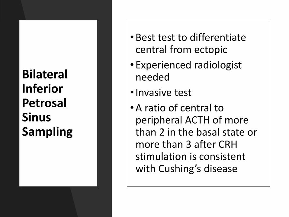
Bilateral Inferior PetrosalSinus Sampling
•Best test to differentiate central from ectopic
•Experienced radiologist needed
• Invasive test
•A ratio of central to peripheral ACTH of more than 2 in the basal state or more than 3 after CRH stimulation is consistent with Cushing’s disease
Osteoporosis
86
Footer Text 87
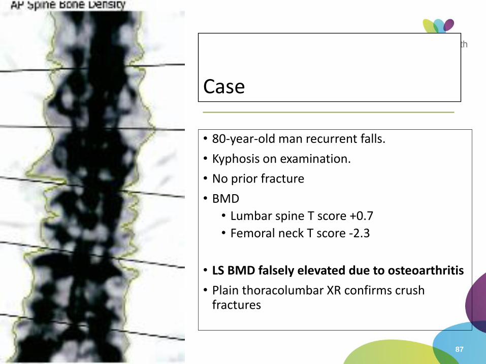
Case
• 80-year-old man recurrent falls.
• Kyphosis on examination.
• No prior fracture
• BMD
• Lumbar spine T score +0.7
• Femoral neck T score -2.3
• LS BMD falsely elevated due to osteoarthritis
• Plain thoracolumbar XR confirms crush fractures
Footer Text 88
Bone Mineral Density/DXA
• Osteoporosis T scores <-2.5
• Osteopenia T scores -2.4 to -1.0
• Look at the scout films
• When comparing scans, a significant change in BMD is only if:
• Lumbar spine D3% if normal BMD, 5% if osteopenia/OP
• Hip/Femur D7%
Footer Text 89
Bone turnover markers
• CTx (C-telopeptide) – bone resorption
• P1NP (Procollagen type 1 amino-terminal propeptide)–bone formation
• Fasting sample
• May be used to assess compliance with therapy• Expect CTx & P1NP to be
suppressed on anti-resorptive therapy
Recommended


