
Ephrin-B reverse signaling controls septation events at theembryonic midline through separate tyrosine phosphorylation-independent signaling avenues
Christopher Dravis and Mark Henkemeyer‡
Department of Developmental Biology, University of Texas Southwestern Medical Center, Dallas,TX 75390
AbstractWe report that the disruption of bidirectional signaling between ephrin-B2 and EphB receptorsimpairs morphogenetic cell-cell septation and closure events during development of the embryonicmidline. A novel role for reverse signaling is identified in tracheoesophageal foregut septation, asanimals lacking the cytoplasmic domain of ephrin-B2 present with laryngotracheoesophageal cleft(LTEC), while both EphB2/EphB3 forward signaling and ephrin-B2 reverse signaling are shownto be required for midline fusion of the palate. In a third midline event, EphB2/EphB3 are shownto mediate ventral abdominal wall closure by acting principally as ligands to stimulate ephrin-Breverse signaling. Analysis of new ephrin-B26YFΔV and ephrin-B2ΔV mutants that specificallyablate ephrin-B2 tyrosine phosphorylation- and/or PDZ domain-mediated signaling indicate thereare at least two distinct phosphorylation-independent components of reverse signaling. Theseinvolve both PDZ domain interactions and a non-canonical SH2/PDZ-independent form of reversesignaling that may utilize associations with claudin family tetraspan molecules, as EphB2 andactivated ephrin-B2 molecules are specifically co-localized with claudins in epithelia at the pointof septation. Finally, the developmental phenotypes described here mirror common human midlinebirth defects found with the VACTERL association, suggesting a molecular link to bidirectionalsignaling through B-subclass Ephs and ephrins.
Keywordsephrin-B2; EphB2; receptor tyrosine kinase; bidirectional signaling; claudin; VACTERL;tracheoesophageal septation; cleft palate; omphalocele
INTRODUCTIONVACTERL is used to denote a non-random association of human embryonic malformations.The defining features of VACTERL include vertebral defects (V), anorectal malformations(A), cardiac anomalies (C), tracheoesophageal septation defects (TE), renal dysplasia (R),and limb abnormalities (L) (Temtamy and Miller, 1974). Cases of VACTERL are defined aspresenting with two or more of these anomalies, and most of the 1 in 5,000 live birthsassociated with VACTERL have only diads or triads (Rittler et al., 1996). Additional
© 2011 Elsevier Inc. All rights reserved.‡Correspondence: Mark Henkemeyer, [email protected], tel: 214-645-5916, fax: 214-648-1960.Publisher's Disclaimer: This is a PDF file of an unedited manuscript that has been accepted for publication. As a service to ourcustomers we are providing this early version of the manuscript. The manuscript will undergo copyediting, typesetting, and review ofthe resulting proof before it is published in its final citable form. Please note that during the production process errors may bediscovered which could affect the content, and all legal disclaimers that apply to the journal pertain.
NIH Public AccessAuthor ManuscriptDev Biol. Author manuscript; available in PMC 2012 July 1.
Published in final edited form as:Dev Biol. 2011 July 1; 355(1): 138–151. doi:10.1016/j.ydbio.2011.04.020.
NIH
-PA Author Manuscript
NIH
-PA Author Manuscript
NIH
-PA Author Manuscript

congenital anomalies are also observed in VACTERL patients outside of the definingfeatures, including hypospadias, cleft palate, neural tube defects, and omphalocele (Botto etal., 1997; Rittler et al., 1996). Evidence has built for a genetic basis behind VACTERL,however a molecular understanding of the association remains rudimentary (Aynaci et al.,1996; Nezarati and McLeod, 1999). At present, the only VACTERL association animalmodels are mice impaired in Shh signaling (Shh, Gli deficient animals), knockouts forFoxf1, Noggin, or Sox2, or animals treated in utero with the anthracycline antibiotic,adriamycin (Kim et al., 2001; Shaw-Smith, 2009).
The Eph receptor family represents the largest collection of transmembrane receptor tyrosinekinases, while the ephrins are their membrane-bound ligands. A-subclass ephrins areattached to the outer leaflet of the plasma membrane by a GPI-linkage, while B-subclassephrins possess a single-pass transmembrane domain and a short cytoplasmic tail. Ingeneral, EphA receptors can promiscuously bind to A-subclass ephrins and EphB receptorscan similarly bind to B-subclass ephrins, although subclass crosstalk has been documented(Bergemann et al., 1998; Davis et al., 1994; Himanen et al., 2004). As both Ephs and ephrinsare membrane anchored, signaling by these molecules occurs at sites of cell-cell contact.Signal transduction between Ephs and ephrins is atypical from other receptor/ligand pairs inthat the potential exists for bidirectional signaling into both the Eph and ephrin expressingcells (Henkemeyer et al., 1996; Holland et al., 1996). Signaling into cells expressing Ephreceptors (forward signaling) occurs principally through activation of its intracellulartyrosine kinase catalytic domain which leads to autophosphorylation, coupling to Srchomology 2 (SH2) domain proteins, and phosphorylation of downstream substrates(Kullander and Klein, 2002). Eph receptors can also form other protein-protein associations,notably through their sterile-alpha (SAM) and C-terminal PDZ-binding motifs. Conversely,signaling into cells expressing B-subclass ephrins (reverse signaling) also leads to thephosphorylation of conserved tyrosine residues on the cytoplasmic tail of the ephrin-Bmolecule and the subsequent docking to SH2 adaptor proteins such as Grb4/Nck-β (Cowanand Henkemeyer, 2001; Xu and Henkemeyer, 2009). B-subclass ephrins also contain a C-terminal PDZ-binding motif. The bidirectional nature of Eph/ephrin signaling complicatesstudies of these molecules. Therefore, to precisely elucidate the physiological role of a givenEph or ephrin it is necessary to dissect both the forward and reverse signaling contributionsof that particular molecule.
Beyond their potential for bidirectional signaling, the Ephs and ephrins are further uniquewith respect to the targets of their signal transduction. Whereas most tyrosine kinase-mediated signaling events ultimately target the nucleus to regulate transcription, the Ephsand ephrins appear to principally exert control over cytoskeletal dynamics. This control isachieved through the activation of secondary molecules linked to cytoskeletal control, manyof which ultimately regulate small Rho family GTPases (Egea and Klein, 2007; Noren andPasquale, 2004). Given this ability to fundamentally alter the structure of the cell in responseto cell-cell contact, it is not surprising that Ephs and ephrins are utilized as key regulators ofcell migration/differentiation events throughout development, as typified by their well-defined physiological roles in axon pathfinding (Flanagan and Vanderhaeghen, 1998;Wilkinson, 2001), angiogenic remodeling (Cowan et al., 2004; Wang et al., 1998), andneural crest cell migration (Davy et al., 2004; Smith et al., 1997).
While the majority of studies implicate Eph-ephrin signaling in cell migration and axonpathfinding by inducing repulsion, there is some evidence to suggest these molecules mayalso function in the opposite manner to induce cell adhesion responses. This evidenceincludes the characterization of apparent cell adhesion defects in the palate, urethra, cloaca,and neural tube in Eph/ephrin mutant animals (Dravis et al., 2004; Holmberg et al., 2000;Orioli et al., 1996), Eph-ephrin mediated axon guidance events that appear to utilize
Dravis and Henkemeyer Page 2
Dev Biol. Author manuscript; available in PMC 2012 July 1.
NIH
-PA Author Manuscript
NIH
-PA Author Manuscript
NIH
-PA Author Manuscript

attraction outcomes following axon-cell contact instead of the canonical repulsion seen mostoften with the Ephs and ephrins (Eberhart et al., 2004; Hindges et al., 2002), and cell-basedstudies indicating positive outgrowth of cytoskeletal structures in response to Eph-ephrinsignaling activation instead of the more typical cytoskeletal collapse associated with thesemolecules (Hansen et al., 2004; Huynh-Do et al., 1999; Stein et al., 1998). Here we focus ondescribing new roles for these molecules that also appear consistent with cell-cell adhesionresponses.
Using a variety of EphB/ephrin-B mutant alleles, we identify requirements for ephrin-B2and EphB2 in septation and closure events at the embryonic midline in the developingforegut, palate, and ventral body wall. The LTEC, cleft palate, and omphalocele phenotypesmimic VACTERL association birth defects. Combined with previous studies reporting theinvolvement of the Ephs and ephrins in other VACTERL-like developmental malformations(Compagni et al., 2003; Cowan et al., 2004; Davy and Soriano, 2007; Dravis et al., 2004;Holmberg et al., 2000), we now find a near complete overlap between Eph/ephrin-associateddevelopmental defects and the malformations of VACTERL. This leads us to propose EphB/ephrin-B signaling as a molecular basis for VACTERL association and to highlight the useof EphB/ephrin-B deficient mice for VACTERL research.
RESULTSTo characterize roles for ephrin-B2 and EphB2 in midline development, we utilized 1)ephrin-B2lacZ mice, in which the cytoplasmic domain of ephrin-B2 has been replaced by β-galactosidase (β-gal) to abolish reverse signaling while leaving intact its ligand-like abilityto bind Eph receptors and activate forward signaling in the same fashion as native ephrin-B2(Supplemental Figure 1), and 2) EphB2lacZ mice, in which the majority of the EphB2cytoplasmic domain has been replaced by β-gal to block forward signaling by this moleculewhile leaving intact its ligand-like ability to bind ephrins and stimulate reverse signaling(Dravis et al., 2004; Henkemeyer et al., 1996). The ephrin-B2lacZ allele has been usedsuccessfully in the past to identify a distinct ligand-only role for ephrin-B2 in angiogenesis(Cowan et al., 2004) and axon guidance (Williams et al., 2003), as well as clear receptorroles in urorectal development (Dravis et al., 2004), anterior commissure axon guidance andcardiac development (Cowan et al., 2004), and inner ear fluid dynamics (Dravis et al.,2007). The ephrin-B2lacZ allele, which is homozygous viable throughout embryonicdevelopment, has proven to be very useful for understanding the functions of ephrin-B2reverse signaling, given that ephrin-B2−/− null mutants are early embryonic lethal due tovascular defects caused by the loss of EphB4 forward signaling that preclude analysis oflater development (Wang et al., 1998). Likewise, the EphB2lacZ allele has been used toidentify ligand-only roles for EphB2 in axon guidance (Birgbauer et al., 2000; Henkemeyeret al., 1996), and receptor roles for EphB2 in axon guidance (Cowan et al., 2000; Hindges etal., 2002), vestibular development (Cowan et al., 2000), and urorectal development (Draviset al., 2004).
Ephrin-B2 reverse signaling mediates midline septation of the foregutInitial studies of ephrin-B2lacZ mice indicated a loss of ephrin-B2 reverse signaling leads toneonatal lethality, with defects in cardiac development and midline adhesion of the urethraand anorectum (Cowan et al., 2004; Dravis et al., 2004). Because of the urorectal/hindgutdefects, ephrin-B2lacZ embryos were also examined to determine if septation of the foregutwas similarly affected. This revealed that 47% (n=15) of late stage embryonic day 18 (E18)ephrin-B2lacZ/lacZ mutants exhibited defects in tracheoesophageal (TE) septation asevidenced by common, unseptated foregut. The defects observed in the ephrin-B2lacZ/lacZ
embryos appeared with a phenotypic range of moderate, in which a septum was visible buthad failed to extend to the rostral apex, to severe, in which no septum was detected, and only
Dravis and Henkemeyer Page 3
Dev Biol. Author manuscript; available in PMC 2012 July 1.
NIH
-PA Author Manuscript
NIH
-PA Author Manuscript
NIH
-PA Author Manuscript

unseptated foregut was present rostral to the bronchi (Fig 1A). Sections taken of earlierE14.5 embryos showed the same defect in foregut septation; distinct trachea and esophaguswere visible in the wild-type (WT) embryo, while an ephrin-B2lacZ/lacZ littermate showedonly unseptated foregut (Fig 1B). The failure of foregut septation in ephrin-B2lacZ/lacZ
mutants results in morphological abnormalities in the unseptated trachea, as visualized bythe appearance of disorganized cartilage rings in the mutants (Fig 1C).
We next examined ephrin-B2lacZ embryos at earlier stages of development, and found thathomozygotes collected at E10.5 also showed defective TE septation. While ephrin-B2lacZ/+
embryos appeared normal and showed a common rostral foregut that septated into separatetracheal and esophageal endoderm caudally (Fig 2A), ephrin-B2lacZ/lacZ littermates showedno signs of TE septation all the way down to the terminal caudal point of the foregut, despiteotherwise normally patterned foregut endoderm (Fig 2B).
The discovery of TE fistula in ephrin-B2lacZ mutants suggests that reverse signaling throughthe cytoplasmic domain of ephrin-B2 is necessary for proper septation of the foregut.However, the ephrin-B2lacZ allele is not able to answer which Eph receptors are involved inactivating these signals. To address this, Eph receptor compound null animals weregenerated, as these genes can show redundant functions. However, analysis ofEphB2;EphB3, EphB2;EphB3;EphA4, and EphB1;EphB2;EphB3;EphA4 compoundknockouts did not reveal any defects in TE septation. This suggests at least one additionalEph receptor (i.e. EphB4 or EphB6) may be paired with ephrin-B2 in foregut septation.
X-gal stains on foregut sections from ephrin-B2lacZ embryos were carried out to documentexpression of the ephrin-B2-β-gal fusion protein, which provides a high signal-to-noise ratioreporter of protein expression (Fig 2). The data showed ephrin-B2 is expressed along thelength of the foregut endoderm, and appears to be most highly expressed in the epithelia ofthe foregut destined to become esophagus as well as in the mesenchyme surrounding theesophagus. Ephrin-B2 is notably present at the site of TE septation in ephrin-B2lacZ/+
specimens or where TE septation should be occurring in ephrin-B2lacZ/lacZ mutants (Fig 2,Supplemental Fig 2A). Indirect immunofluorescence (IF) using a pan-ephrin-B antibody onWT embryos shows a similar expression profile as the X-gal stains for the ephrin-B2-β-galfusion protein (Fig 2D).
While the Eph receptors involved in foregut septation are unknown, we reasoned that EphB3was likely involved based on its role in cell adhesion events during hindgut septation(Dravis et al., 2004). Unfortunately, conventional methods to examine EphB3 expression bymRNA in situ hybridization or immunohistochemistry have not provided satisfactory results,so a BAC transgenic animal was generated that expresses the reverse tetracyclinetransactivator, rtTA2S-M2 (Urlinger et al., 2000), under control of EphB3 promotersequences (Villasenor et al., in preparation). Tg-BAC-EphB3-rtTA transgenic mice werecrossed to a TRE-lacZ reporter line (Ludwig et al., 2004) and dox-induced embryoshemizygous for both transgenes were collected and stained with BluO-gal to reveal β-galexpressing cells. This showed that EphB3 is expressed in the mesenchymal cells cominginto contact with epithelial cells at the site of TE septation (Fig 2E). BluO-gal stains onforegut tissue from EphB2lacZ/+ animals indicated expression of EphB2 on epithelia at thepoint of septation (Supplemental Fig 2B). The expression data strongly indicates EphB2 andEphB3 likely participate in foregut septation.
EphB:ephrin-B bidirectional signaling in midline formation of the palateEphB2;EphB3 compound null mice exhibit a cleft palate phenotype that is not observed inEphB2 or EphB3 single mutants (Orioli et al., 1996; Risley et al., 2009). To determine ifEphB2 is acting as a receptor to transduce forward signals or as a ligand to activate reverse
Dravis and Henkemeyer Page 4
Dev Biol. Author manuscript; available in PMC 2012 July 1.
NIH
-PA Author Manuscript
NIH
-PA Author Manuscript
NIH
-PA Author Manuscript

signals important in palate development, both EphB2− null and EphB2lacZ Cterminaltruncation mutants were utilized. Histological analysis at E18.5 revealed that 41 % ofEphB2lacZ/lacZ;EphB3−/− and 15% of EphB2−/−EphB3−/− compound mutants exhibitedsevere cleft palates (Table 1). Finding cleft palates with the EphB2lacZ allele indicates thatEphB forward signaling is important in development of this midline structure. The increasein penetrance to 41% further suggests involvement of more than EphB2 and EphB3 in palatedevelopment, as the dominant negative effect seen here with the EphB2lacZ allele is bestexplained by the ability of the intracellular truncated EphB2-β-gal fusion protein to disruptforward signaling of other co-expressed Eph receptors (Cowan et al., 2000; Hindges et al.,2002).
To determine if ephrin-B2 is involved in palate formation, embryos carrying the ephrin-B2lacZ allele were examined. Histological analysis revealed that 26% of ephrin-B2lacZ/lacZ
homozygotes and 7% of ephrin-B2lacZ/+ heterozygotes showed a severe cleft palate (Table2, Fig 3A). This indicates that ephrin-B2 is involved in palate fusion and that reversesignaling through this molecule is important for this process, which is consistent with theconclusion from recent in vitro analyses of Eph-ephrin signaling in palatal shelf fusion (SanMiguel et al., 2011). Coupled with the aforementioned EphB2;EphB3 results, the dataindicate both forward signaling through EphB receptors and reverse signaling throughephrin-B2 are important for midline closure of the palate. This involvement of bidirectionalsignaling through both EphB and ephrin-B mirrors a similar requirement for both forwardand reverse signaling in urorectal/hindgut midline development (Dravis et al., 2004).
The expression profiles of ephrin-B2 and EphB2 were examined during palataldevelopment. X-gal stains of ephrin-B2lacZ/+ embryos revealed expression of ephrin-B2throughout the mesenchyme of the palatal shelf, as well as in the leading epithelia beforeadhesion and fusion take place (Fig 3B). Later, when adhesion has taken place, ephrin-B2 isclearly visible on the multilayer epithelial seam, as well as in the mesenchyme flanking themidline site of adhesion. IF of palatal shelves with an antibody against EphB2 revealedEphB2 expression in the mesenchyme and leading epithelia of the palatal shelf at E13.5 (Fig3C). No signal was detected in control EphB2−/− tissue (Fig 3C). X-gal stains ofEphB2lacZ/+ animals further showed that as palatal shelf closure nears and as it occursEphB2 expression becomes more restricted to the leading epithelia where adhesion takesplace (Fig 3C).
To investigate EphB3 expression, a BAC transgenic line was generated that expresses eYFPunder EphB3 promoter sequences, termed Tg-BAC-EphB3-YFP. IF for GFP to detectexpression of eYFP showed that EphB3 is expressed at the leading mesenchyme of thepalatal shelves (Fig 3D). EphB3 expression was further examined in X-gal stains of palatalshelves from dox treated embryos hemizygous for the Tg-BAC-EphB3-rtTA and TRE-lacZtransgenes. This also showed EphB3 expression in the mesenchyme of the palatal shelves atE13.5 and that this expression shifts from the mesenchyme to epithelia as palatal shelfclosure nears at E14.5 (Fig 3D). To summarize this expression data, remarkably the profilesfor EphB2, EphB3, and ephrin-B2 appear nearly identical to each other during formation ofthe palate.
Based on the individual expression studies taken for ephrin-B2 and the EphB receptors, wespeculated that these molecules were likely co-expressed at the site of cell-cell adhesion asthe palatal shelves meet and fuse at the midline. To address this, double IF analysis onephrin-B2lacZ/+ embryos using antibodies against EphB2 and β-gal were performed. Thisrevealed that EphB2 and ephrin-B2 are co-expressed on the midline epithelia where palatalshelf closure occurs (Fig 3E). Interestingly, this mimics the previous finding of co-
Dravis and Henkemeyer Page 5
Dev Biol. Author manuscript; available in PMC 2012 July 1.
NIH
-PA Author Manuscript
NIH
-PA Author Manuscript
NIH
-PA Author Manuscript

expression in adhering midline epithelial cells at the site of urorectal/hindgut septation(Dravis et al., 2004).
EphB2/EphB3 activate reverse signaling for midline closure of the ventral body wallWhile analyzing palate and foregut defects in the EphB receptor mutant mice, it was noticedthat 40% of EphB2−/−;EphB3−/− compound nulls exhibited a failure in midline closure ofthe abdominal wall (Fig 4A, Table 3), as briefly reported before (Orioli et al., 1996). Theventral midline defect in these mice resembles the birth defect omphalocele, in whichvisceral organs are herniated. This defect appears to be dependent on the genetic backgroundof the mouse, as omphalocele was detected in the inbred 129 strain, but not in mice of aCD1 background. To determine where EphB2 is expressed during ventral midline closure ofthe abdominal wall, BluO-gal stains on whole-mounted EphB2lacZ/+ embryos wereperformed (Fig 4B). Expression of EphB2 was detected at E13.5 in cells at the ventralmidline where abdominal closure takes place and at E15.5 was localized to the terminalclosure spot of the umbilical ring. To determine if forward signaling through EphB2participates in ventral body wall closure, the penetrance of this phenotype was investigatedusing the EphB2lacZ allele, revealing that only 9% of EphB2lacZ/lacZ;EphB3−/− animalsexhibited omphalocele (Table 3). Given this data, and taking into account the reports thatephrin-B1 knockouts also have incompletely-penetrant omphalocele (Compagni et al., 2003;Davy et al., 2004), it appears that EphB2 and EphB3 act primarily as ligands to stimulateephrin-B1 reverse signaling during midline closure of the ventral body wall.
Ephrin reverse signaling is activated at sites of septationEph-ephrin bidirectional signaling occurs at sites of cell-cell contact, which leads toclustering of the molecules into tetramers and higher order aggregates, along with tyrosinephosphorylation of their intracellular domains. We therefore sought to address where ephrin-B reverse signaling was activated during these midline events by using two differentantibodies that recognize phosphorylated tyrosine residues within the conserved ephrin-Bcytoplasmic domain. IF on sections of septating foregut at E10.5 showed that while someephrin-B is tyrosine phosphorylated in the mesenchyme flanking the point of septation, themost striking tyrosine phosphorylation of ephrin-B is detected along the septating endoderm(Fig 5A). Adjacent sections treated with lambda phosphatase confirmed the phospho-specific activity of the antibodies. This data localizes ephrin-B tyrosine phosphorylation tomidline septation in the foregut.
Adjacent palatal shelf sections from an E14 ephrin-B2lacZ/+ embryo were similarly probedwith an antibody against β-gal to detect ephrin-B2 and phospho-ephrin-B antibodies todetermine where ephrin-B2 was phosphorylated. While the β-gal antibody labeled theleading epithelia of the palatal shelf as well as the flanking mesoderm, the phospho-ephrinantibodies indicated that only ephrin-B2 in the leading epithelia of the palatal shelf wastyrosine phosphorylated (Fig 5B). Double IF with EphB2 and phospho-ephrin antibodies co-localized EphB2 with tyrosine phosphorylated ephrin-B2 (Fig 5C). This data importantlylocalizes ephrin-B2 reverse signaling to epithelia at the point of cell adhesion and closure inthe developing palate.
Ephrin-B2 PDZ domain interactions contribute to midline septationHaving characterized roles for ephrin-B2 reverse signaling in midline septation, we nextsought to determine which signaling avenues of the ephrin-B2 cytoplasmic tail are utilizedfor these morphogenetic events. To address this, two new ephrin-B2 alleles were created,ephrin-B2ΔV (Fig 6, Supp. Fig 2) and ephrin-B26YFΔV (Thakar et al., in preparation). Inephrin-B2ΔV mice, the codon for the C-terminal valine residue of ephrin-B2 was deleted toprevent the interaction of ephrin-B2 with PDZ domain-containing proteins. In ephrin-
Dravis and Henkemeyer Page 6
Dev Biol. Author manuscript; available in PMC 2012 July 1.
NIH
-PA Author Manuscript
NIH
-PA Author Manuscript
NIH
-PA Author Manuscript

B26YFΔV mice, the six tyrosines of the ephrin-B2 cytoplasmic domain were changed tophenylalanine to prevent tyrosine phosphorylation of ephrin-B2 and the same C-terminalvaline was deleted, resulting in the loss of interactions with both SH2 and PDZ domain-containing proteins (Fig 6B). The cell surface localization of these molecules and theirinability to interact with PDZ domain-containing proteins have been demonstrated (Thakaret al., in preparation; Makinen et al., 2005), while proper expression of ephrin-B2ΔV wasconfirmed by immunoblot analysis (Supplemental Fig 3).
Interestingly, neither ephrin-B2ΔV/ΔV nor ephrin-B26YFΔV/6YFΔV mutant mice present withdefects in urorectal, tracheoesophageal, or palatal development. Remarkably, while 100% ofephrin-B2lacZ/lacZ animals die within hours of birth due to VACTERL defects, ephrin-B2ΔV/ΔV and ephrin-B26YFΔV/6YFΔV mice can be recovered as adults, although many diebefore adulthood, presumably due to lung and lymphatic defects (Makinen et al., 2005;Wilkinson et al, 2008). This indicates that tyrosine phosphorylation of ephrin-B2 and thetransduction of ephrin-B2 reverse signaling through SH2 and PDZ domain interactions arenot necessary for the development and septation of these midline structures.
A role for ephrin-B2 reverse signaling through PDZ domain interactions in midlinedevelopment did become apparent however when ephrin-B2lacZ/+ and ephrin-B26YFΔV/+
mice were crossed to generate ephrin-B2lacZ/6YFΔV mutant animals (Fig 6C). The incidenceof hypospadias was significantly greater in ephrin-B2lacZ/6YFΔV mutants (86%) compared tothat seen in ephrin-B2lacZ/+ littermates (16%) (P<0.0001,***). A similar increase in theincidence of hypospadias was found in parallel crosses generating ephrin-B2lacZ/ΔV mutants(71%) (P=0.0031,**). Because both ephrin-B2lacZ/6YFΔV and ephrin-B2lacZ/ΔV mice presentwith increased hypospadias, it appears that PDZ domain interactions through the C-terminalvaline represent an important component of ephrin-B2 reverse signaling in midline closure.
EphB2 forward signaling in midline septation does not require receptor tyrosine kinaseactivity or PDZ domain interactions
Having also characterized roles for EphB2 forward signaling in palate and urorectaldevelopment, we similarly attempted to identify what elements of EphB2 forward signalingmight be involved at the midline by analyzing recently generated EphB2 forward signalingmutant animals. These mice include EphB2VEV mutants, in which the codons for theterminal three amino acids, valine-glutamate-valine (VEV), were deleted to abolishinteractions between EphB2 and PDZ domain-containing proteins, EphB2K661R mutants, inwhich the lysine residue at amino acid 661 was mutated to arginine to abolish the tyrosinekinase catalytic activity of the EphB2 receptor, and EphB2KVEV mutants, which combineboth mutations to simultaneously lose PDZ domain interactions as well as catalytic tyrosinekinase activity (Genander et al., 2009). No urorectal malformation was found in any of theEphB2VEV/VEV (n=107), EphB2K661R/K661R (n=102), or EphB2KVEV/KVEV (n=202) mutantmice in an EphB3−/− background. Further, all compound mutants were recovered as adultsat expected Mendelian ratios, indicating no neonatal lethality, and thus no cleft palatephenotype. The data indicates EphB2 forward signaling in these midline structures does notrequire the phosphorylation of secondary molecules by the receptor tyrosine kinase domainor the interaction of EphB2 with PDZ domain-containing proteins.
Ephrin-B2 reverse signaling through claudin at the midlineThat ephrin-B2ΔV and ephrin-B26YFΔV mice do not phenocopy ephrin-B2lacZ micesuggests: 1) there is more to ephrin-B2 reverse signaling than just SH2 domain interactionsthrough phosphorylated tyrosines and PDZ domain interactions through the C-terminus, and2) these non-canonical signaling avenue(s) are critical elements of the reverse signalemployed for midline septation or closure. B-subclass ephrins have been shown to interact
Dravis and Henkemeyer Page 7
Dev Biol. Author manuscript; available in PMC 2012 July 1.
NIH
-PA Author Manuscript
NIH
-PA Author Manuscript
NIH
-PA Author Manuscript

with the claudins, a class of tetraspan proteins known to directly mediate cell-cell adhesion(Tanaka et al., 2005). To examine a potential role for the claudins in ephrin-B2 reversesignaling, the expression of claudin-1 and claudin-4 (the two claudin molecules shown toassociate with ephrin-B) was investigated at the point of adhesion in the palate. Strikingly,IF shows that both claudin-1 and claudin-4 are highly expressed in epithelia directly beforeand right at midline cell-cell adhesion in the palate of WT embryos (Fig 7). This expressionmimics the profile of tyrosine-phosphorylated ephrin-B, and indeed double IF shows thatboth claudin and activated ephrin co-localize in adherent epithelia. This data supports atyrosine phosphorylation-independent role for ephrin-B2 reverse signaling involving theclaudins in midline septation.
DISCUSSIONThe data presented here show that ephrin-B2lacZ/lacZ homozygotes present with LTEC andcleft palate phenotypes, indicating novel roles for ephrin-B2 reverse signaling in foregutseptation and palatal shelf closure. Moreover, we have shown that EphB2lacZ/lacZ;EphB3−/−
animals also present with cleft palate, indicating a role for EphB forward signaling inmidline fusion of the palate as well. The roles identified here for both ephrin-B2 reversesignaling and EphB2 forward signaling in midline formation of the palate are remarkablysimilar to what we previously reported for the tubularization of the urethra and septation ofthe hindgut/cloaca into distinct urogenital and anorectal compartments (Dravis et al., 2004).We also report here that EphB2−/−; EphB3−/−compound null animals, but less so forwardsignaling deficient EphB2lacZ/lacZ; EphB3−/− animals present with omphalocele. Thisindicates that closure of the ventral abdominal wall occurs through EphB2/B3 actingprimarily as ligands to stimulate ephrin-B reverse signaling. Surprisingly, deconstruction ofthe ephrin-B2 reverse signal and the EphB2 forward signal in these midline structures usingselect point mutant mice indicates that these bidirectional signals do not appear to requiretyrosine kinase phosphorylation events. Instead, the ephrin-B2 reverse signal can be brokendown into distinct tyrosine phosphorylation-independent elements, one of which involvesPDZ domain interactions, and the other we propose involves interaction with claudin familytetraspan molecules.
Fidelity of the lacZ allelesA question to address in considering the data is the reliability of lacZ alleles that result in thesynthesis of C-terminally truncated proteins that lack their cytoplasmic domains andcovalently attach β-gal. This has been controversial given that similar animal models haveproduced divergent claims about the physiological roles of forward and reverse signalingevents (Adams et al., 2001; Cowan et al., 2004). Therefore, we would like to point out that:1) the ephrin-B2- β gal fusion protein encoded by the ephrin-B2lacZ allele localizes properlyto the cell surface and clusters into higher order oligomers, unlike other ephrin-B2unconjugated C-terminal cytoplasmic truncations which become trapped in the trans-Golginetwork and do not traffic to the plasma membrane (Cowan et al., 2004), and can properlyactivate forward signaling through EphB receptors (Supplemental Figure 1); 2) the successof the lacZ alleles to properly signal non-cell autonomously has been demonstrated in vivonow several times over, as the ephrin-B3lacZ/lacZ mutant does not present with locomotordefects in the spinal cord associated with the ephrin-B3−/− protein null (Yokoyama et al.,2001), the ephrin-B2lacZ/lacZ mutant does not result in early embryonic lethality due tovascular angiogenic failures associated with either ephrin-B2−/− protein null or ephrin-B2unconjugated C-terminal truncation mice (Adams et al., 2001; Cowan et al., 2004), andEphB2lacZ/lacZ mutants do not exhibit axon pathfinding defects associated with EphB2−/−
protein nulls (Birgbauer et al., 2000; Cowan et al., 2004; Henkemeyer et al., 1996); 3) thelacZ alleles do not hyperactivate forward signaling, as genetic assays have shown (Dravis et
Dravis and Henkemeyer Page 8
Dev Biol. Author manuscript; available in PMC 2012 July 1.
NIH
-PA Author Manuscript
NIH
-PA Author Manuscript
NIH
-PA Author Manuscript

al., 2004), and as is suggested by the fact that EphB2F620D mice with hyperactive EphB2forward signals (Holmberg et al., 2006) do not present with any midline defects (data notshown); and 4) the cell autonomous defects seen with the lacZ alleles have also been foundin traditional germline knockout animals for EphB and as well as the targeted mutations ofephrin-B26YFΔV and ephrin-B2ΔV now, further confirming the midline defects characterizedin lacZ mice are not the result of hypermorphic or neomorphic activity.
Midline EphB:ephrin-B bidirectional signaling operates independently of tyrosinephosphorylation
One of the most interesting findings in our data is that ephrin-B26YFΔV/6YFΔV mice do notphenocopy ephrin-B2lacZ/lacZ mice, which is surprising because both alleles block canonicalsignaling structures of the ephrin-B cytoplasmic domain: phosphorylated tyrosines that serveas a substrate for SH2 domain docking and a C-terminal valine that interacts with PDZdomains. This indicates that signaling through these two elements are not necessary in themidline septation events we have linked to ephrin-B reverse signaling. However, bycombining the ephrin-B26YFΔV and ephrin-B2ΔV alleles with ephrin-B2lacZ, to produceephrin-B2lacZ/6YFΔV and ephrin-B2lacZ/ΔV mice, and comparing those animals to ephrin-B2lacZ/+ animals (Fig 6C), it becomes apparent that PDZ domain interactions through the C-terminus of ephrin-B2 do contribute to these midline events. The disconnect between theephrin-B2lacZ and ephrin-B26YFΔV or ephrin-B2ΔV alleles suggests there are alternateavenues of reverse signaling through ephrin-B that are independent of SH2/PDZ domaininteractions, but nonetheless still necessary for proper midline development.
Other groups have identified potential non-canonical signaling avenues that may be ofrelevance here. These include the discovery that the ephrin-B cytoplasmic tail is serinephosphorylated, that a P-P-Q-S-P motif on the cytoplasmic tail can mediate SH3 domaininteractions, that Dishevelled can constitutively interact with the ephrin-B cytoplasmic tailin Xenopus, and that ephrin-Bs can interact with claudins (Essmann et al., 2008; Segura etal., 2007; Tanaka et al., 2005; Tanaka et al., 2003). The identification of the SH3 bindingsite is particularly interesting, as it could explain why our genetic studies indicate noapparent role for ephrin-B2 tyrosine phosphorylation in midline septation, even though ourexpression data clearly demonstrates that ephrin-B is tyrosine phosphorylated precisely atthe adhesion sites where septation takes place. Reverse signaling might still occur in theabsence of ephrin-B tyrosine phosphorylation with our ephrin-B26YFΔV mice, if a keyadaptor protein like Grb4 could simply find itself recruited to the ephrin cytoplasmic tailthrough one of its three SH3 domains, instead of its SH2 domain, to produce similarphysiological outcome at the midline. The ephrin-B SH3 and SH2 domain binding motifsmight therefore possess some redundancy that masks their importance in midline signaling.
However, given the nature of the defects in midline septation and closure that arecharacterized in our mice here, it is the interaction between the ephrins and claudins that wefind of particular interest. The claudins have been characterized to directly mediate adhesionbetween cells through high affinity interactions involving their extracellular loops, and theinteraction between ephrin-B and claudin has been shown to modulate this epithelialadhesion, albeit in regulating paracellular flow (Tanaka et al., 2005) . This suggests that theinteraction between ephrin-B2 and claudins might regulate the adhesiveness of epitheliacoming into contact at the midline, and indeed we find that both claudin-1 and claudin-4 arehighly co-localized with ephrin-B2 and EphB2 on the cell surface of adhering epithelial cellsin the palate. Of note, claudin expression does not appear restricted to its more canonicaltight junction localization, which visualize as distinct puncta, but instead appears to beexpressed across the breadth of the epithelial cell surface. Based on this expression, westrongly suspect that ephrin-claudin interactions represent a tyrosine phosphorylation-
Dravis and Henkemeyer Page 9
Dev Biol. Author manuscript; available in PMC 2012 July 1.
NIH
-PA Author Manuscript
NIH
-PA Author Manuscript
NIH
-PA Author Manuscript

independent portion of ephrin-B reverse signaling in these midline septation events, perhapsin mediating the actual cell-cell adhesion of these lateral structures.
On the other side of the bidirectional signal, it is also interesting that forward signalingthrough the EphB2 receptor appears to require neither the tyrosine kinase catalytic activityof the receptor nor the C-terminal PDZ-binding motif in these midline structures. This is lesssurprising than our findings with reverse signaling, however. Even without tyrosine kinaseactivity, EphB2 can still be phosphorylated on critical tyrosine residues by intracellularmolecules such a Abl, Src family kinases, or co-expressed Eph receptors in much the sameway the kinase-dead Eph receptor EphB6 activates forward signaling (Freywald et al., 2002;Vindis et al., 2003; Yu et al., 2001). Further, EphB2 is a large molecule with numerousprotein interaction sites that were untouched by our targeted mutations (notably includingthe juxtamembrane tyrosines and SAM domain), which could play important roles in theforward signaling element involved in midline development.
EphB/ephrin-B signaling in midline cell-cell adhesionOur genetic analyses indicate critical roles for ephrin-B reverse signaling in multipleseptation events at the embryonic midline. What then is the ephrin-B reverse signal doing inthese structures? Our analysis of where ephrin-B reverse signaling is localized by utilizingIF is very useful here, as it demonstrates that both ephrin-B tyrosine phosphorylation(representing activated ephrin-B) and claudin specifically localize to the leading epithelia atthe sites where cell adhesion initiates septation and closure. In addition, EphB2 and EphB3are also expressed and likely actively signaling in the same midline epithelia. Theseepithelial cells are not necessarily proliferative or migratory; their principal function is toadhere to adjacent epithelia at the point of septation. When we couple the failure of midlinestructures to properly adhere in forward and reverse signaling deficient animals with thisexpression data localizing EphB, ephrin-B, and the adhesion regulatory molecule claudin tosites of cell adhesion, it appears that these bidirectional signals might be responsible forproducing a cell adhesion outcome in these midline septation events. This is an excitinghypothesis that will need to be addressed through future analyses of the behavior of thesecells in vivo and in cell culture assays.
How then might the Ephs and ephrins produce a cell-cell adhesion response? This continuesto be a confounding question, trying to determine how in one instance these molecules canprovoke a cell repulsion response and in others lead to cell adhesion. An emerging answer isthat Eph-ephrin reverse signaling produces a cell adhesion response the same way itproduces a cell repulsion response—through modulating the balance of activity in the Rhofamily GTPases Rho, Rac, and Cdc42. Both forward and reverse signal transductionfunction to regulate these Rho-family GTPases, and subtle shifts in the relative activity ofthese molecules (through differential activation or antagonistic crosstalk) can producedifferent cytoskeletal behavior consistent with either cell adhesion or cell repulsionoutcomes (Burridge and Doughman, 2006; Jaffe and Hall, 2005).
The link between ephrin reverse signaling and Rho family GTPases is potentially interestinggiven that the activation of these molecules has been linked to the production of filopodia-like extensions, which facilitate midline adhesion in Drosophila dorsal closure, a paradigmfor the epithelial adhesion events discussed here (Jacinto et al., 2000). This presents theintriguing hypothesis that ephrin-B reverse signaling might become activated in theseadherent epithelia, producing activated combinations of Rho/Rac/Cdc42 that then elicitfilopodia-like extensions in these cells; these filopodia then extend across the midline toestablish initial sites of contact between adjacent cells that rapidly mature into adherensjunctions to zip these midline structures closed. Eph-ephrin regulation of filopodiageneration and motility has previously been shown to be important in synaptic development,
Dravis and Henkemeyer Page 10
Dev Biol. Author manuscript; available in PMC 2012 July 1.
NIH
-PA Author Manuscript
NIH
-PA Author Manuscript
NIH
-PA Author Manuscript

particularly for the generation of dendritic spines, suggesting some potential functionalredundancy in these very different structures (Klein, 2009). For the PDZ-dependent aspectof midline ephrin signaling identified in our new ephrin-B2ΔV mice, this might involve thedocumented interaction of ephrin-B with PAR-3, which targets Rho family moleculesthrough its complex with PAR-6 and aPKC (Lin et al., 1999; Nishimura et al., 2005). For thetyrosine phosphorylation- and PDZ-independent signaling avenues suggested by the mice,this might involve Grb4 docking via the ephrin-B SH3 domain or the association of ephrin-B with Dishevelled, both of which are linked to Rho family GTPase activity (Segura et al.,2007; Tanaka et al., 2003).
It is less clear how ephrin and claudin might work together to mediate midline adhesion.This interaction is not thought to require the cytoplasmic tail of ephrin, so it is possible thatdifferential clustering of WT ephrin-B2 and the ephrin-B2-β-gal fusion protein mightsomehow alter the respective interactions of the ephrin with claudin, resulting incompromised midline adhesion in ephrin-B2lacZ animals. Future biochemical analyses mustaim to address the nature of this ephrin-claudin interaction, how it might function to controlcell adhesion, and how this process may fail in our mutant animals.
Our data differs with other studies proposing how Eph-ephrin signaling can produce celladhesion. It has been suggested that cell adhesion is a more passive outcome that takes placethrough the inhibition of canonical repulsive Eph-ephrin signaling, either through cis-inhibition on co-expressing cells (Carvalho et al., 2006; Yin et al., 2004), or throughinhibitory splice variants (Holmberg et al., 2000). Our work demonstrates that midlineepithelia are actively transducing reverse signals, suggesting that B-subclass ephrins areeliciting an active adhesion response, not just inhibiting cell repulsion to permit adhesion. Ithas also been suggested that the Ephs and ephrins are themselves adhesion molecules, andcell repulsion occurs when the interaction of these cell-surface receptors is broken, either byendocytosis or by a protease (Marston et al., 2003; Zimmer et al., 2003). This model is notconsistent with our observations because the ectodomains of both Eph and ephrin in the lacZtruncation alleles are left intact and are able to traffic to the plasma membrane, where theyshould be able to still perform as non-signaling cell adhesion molecules. And yet, as the datashows, mice with these truncated alleles present with the same, if not more severe, adhesiondefects as the null mutants. This further supports an active signaling role for the Ephs andephrins in promoting cell adhesion.
EphB/ephrin-B signaling and the VACTERL associationVACTERL refers to an association of embryonic malformations comprised of vertebral (V),anorectal (A), cardiac (C), tracheoesophageal (TE), renal (R), and limb (L) abnormalities,that often presents with additional anomalies such as hypospadias, cleft palate, neural tubedefects, and omphalocele. Here we find that animals with EphB/ephrin-B signaling ablationspresent with LTEC, cleft palate, and omphalocele, which represent one of the definingfeatures of VACTERL as well as two associated anomalies. This data extends upon previousstudies demonstrating that Eph/ephrin signaling disruptions cause anorectal malformationsand hypospadias (Dravis et al., 2004), cardiac defects (Cowan et al., 2004), limb and skeletalabnormalities (Compagni et al., 2003), and neural tube defects (Holmberg et al., 2000). Wehave also noticed a distended kidney phenotype in ephrin-B2lacZ mice, due to a defect inureter-bladder integration (unpublished data), suggesting links to renal development as well.This striking overlap between EphB/ephrin-B malformations and VACTERL association(Table 4) leads us to propose Eph/ephrin signaling as a molecular basis for the association.This extends our knowledge of VACTERL from morphogens like Shh to cytoskeletalregulators like the Ephs and ephrins, which are likely directly responsible for guiding thecell-cell interactions, movements, and adhesion events that are disrupted in affectedindividuals.
Dravis and Henkemeyer Page 11
Dev Biol. Author manuscript; available in PMC 2012 July 1.
NIH
-PA Author Manuscript
NIH
-PA Author Manuscript
NIH
-PA Author Manuscript

METHODSAnimals
EphB2 (Henkemeyer et al., 1996), EphB3 (Orioli et al., 1996), EphB2VEV, EphB2K661R andEphB2KVEV (Genander et al., 2009), ephrin-B26YFΔV (Thakar et al., submitted), and ephrin-B2lacZ (Dravis et al., 2004) mice have been described. The EphB2 and EphB3 mutationshave been maintained on an inbred 129 background and have also been backcrossed to theCD1 strain. All ephrin-B2 mutants were maintained on a mixed 129/CD1 background.Induction of the tet-on system was achieved by supplying pregnant females with drinkingwater containing 0.5 mg/ml doxycycline and 5% sucrose for 16–24 hours. Statisticalcomparisons utilized the two-tail Fischer’s extract test.
Generation of Tg-BAC-EphB3-YFP and ephrin-B2ΔV animalsTg-BAC-EphB3-YFP mice contain a destabilized variant of eYFP knocked into the ORF ofBAC EphB3 sequences. Transgenic mice were generated from BAC clone RP23-213M14, a208 kb BAC from a C57BL/6J background containing 42 kb upstream of the EphB3 startcodon, that was purchased from the BACPAC resources center at the Children’s HospitalOakland. D2eYFP-1 (Clontech) was targeted into the EphB3 ORF in RP23-213M14 usingbacterial homologous recombination(http://web.ncifcrf.gov/research/brb/recombineeringInformation.aspx). Targeting wasconfirmed by PCR and restriction digests. Pronuclear injection was performed byTransgenic Core facilities at the UTSW Department of Developmental Biology. Toconstruct the targeting vector for ephrin-B2ΔV, RP23-328F4 was purchased from the sameBACPAC resources center. This BAC contains ephrin-B2 exon 5, which has the sequencescoding for the cytoplasmic tail. Through bacterial homologous recombination, the ephrin-B2exon 5 was targeted to delete the GTC codon for valine at the ephrin-B2 C-terminus andinsert a LoxP-flanked NeoR cassette 27 bp downstream of the stop codon. Insertion of theLoxP site results in a 53 bp deletion of 3′ non-coding sequence downstream of the stopcodon, GACCTGC.…GGACTGC. Bacterial homologous recombination was thenperformed on the modified BAC with a capture vector containing 5′ Diphtheria toxin and 3′HSV-tk selection markers to produce a targeting vector with a 6.1 kb 5′ homology arm andan 11.9 kb 3′ homology arm. The targeting vector was linearized through AscI digestion andelectroporated into ES cells, with cell lines screened through Southern blot analysis with 5′and 3′ external probes to detect proper homologous recombination. Blastocyst injection wasperformed with two of the positive cell lines to generate chimeric mice through the UTSWTransgenic Core Facility, and germline transmission was confirmed. The LoxP-flankedcassette was removed with a germline Krox20-Cre line. Protein expression was confirmedby immunoblotting protein lysates from E12.5 embryos with an antibody recognizing the N-terminus of ephrin-B2 (Novus Biologicals)
ImmunofluorescenceImmunofluorescence was performed as described (Dravis et al., 2004). As aphosphotyrosine control sections were treated with λ-protein phosphatase (NEB) for 45'prior to primary application. Primary antibodies used: goat anti-EphB2 (R&D Systems),rabbit anti-β-Gal (Cappel), rabbit anti-phospho-ephrin-B (Cell Signaling Technology), rabbitanti-phospho-ephrin-B [Tyr298] recognizing mouse Y317 (Novus Biologicals), rabbit anti-ephrin-B1 (C-18, Santa Cruz), rabbit anti-GFP (Molecular Probes), rabbit anti-claudin-1(Invitrogen) and Alexa Fluor 488-conjugated claudin-4 (Invitrogen). Secondary antibodieswere Cy2 donkey anti-goat, Cy3 donkey anti-rabbit, and Cy2 donkey anti-rabbit (JacksonImmunoResearch). Images were visualized on a Zeiss 510 LSM confocal microscope.
Dravis and Henkemeyer Page 12
Dev Biol. Author manuscript; available in PMC 2012 July 1.
NIH
-PA Author Manuscript
NIH
-PA Author Manuscript
NIH
-PA Author Manuscript

Histochemical β-gal stainingX-gal and BluO-gal stains were performed as described (Dravis et al., 2004) andcounterstained with nuclear fast red. Whole-mount embryos were dehydrated throughsequential ethanol washes and cleared in methyl salicylate.
EphB2 tyrosine phosphorylation detectionProtein lysates from the head of E14.5 embryos were dissociated mechanically in lysisbuffer (30 mM Hepes pH 7.5, 150 mM NaCl, 10% glycerol, 1.5% Triton-X 100, 1 mMEGTA, 10 mM sodium pyrophosphate, 10 mM NaF, 1 mM sodium orthovanadate, 1 mMPMSF, and protease inhibitor cocktail), immunoprecipitated with an antibody against EphB2(R&D Systems), separated by SDS-PAGE, and immunoblotted with antibodies againstEphB2 or phosphotyrosine (Upstate Biotechnology).
Supplementary MaterialRefer to Web version on PubMed Central for supplementary material.
AcknowledgmentsWe thank Jennifer Shay, Tracey Bowdler, and Jan La for their histological and genotyping assistance in thelaboratory. C.D. was supported by the Division of Cell and Molecular Biology Training Program (T32 GM08203).This work was funded in part by the NIH (R01 EY017434 and 2R01 MH66332).
REFERENCESAdams RH, Diella F, Hennig S, Helmbacher F, Deutsch U, Klein R. The cytoplasmic domain of the
ligand ephrinB2 is required for vascular morphogenesis but not cranial neural crest migration. Cell.2001; 104:57–69. [PubMed: 11163240]
Aynaci FM, Celep F, Karaguzel A, Baki A, Yildiran A. A case of VATER association associated with9qh+ Genet Couns. 1996; 7:321–322. [PubMed: 8985737]
Bergemann AD, Zhang L, Chiang MK, Brambilla R, Klein R, Flanagan JG. Ephrin-B3, a ligand forthe receptor EphB3, expressed at the midline of the developing neural tube. Oncogene. 1998;16:471–480. [PubMed: 9484836]
Birgbauer E, Cowan CA, Sretavan DW, Henkemeyer M. Kinase independent function of EphBreceptors in retinal axon pathfinding to the optic disc from dorsal but not ventral retina.Development. 2000; 127:1231–1241. [PubMed: 10683176]
Botto LD, Khoury MJ, Mastroiacovo P, Castilla EE, Moore CA, Skjaerven R, Mutchinick OM,Borman B, Cocchi G, Czeizel AE, Goujard J, Irgens LM, Lancaster PA, Martinez-Frias ML,Merlob P, Ruusinen A, Stoll C, Sumiyoshi Y. The spectrum of congenital anomalies of the VATERassociation: an international study. Am J Med Genet. 1997; 71:8–15. [PubMed: 9215761]
Burridge K, Doughman R. Front and back by Rho and Rac. Nat Cell Biol. 2006; 8:781–782. [PubMed:16880807]
Carvalho RF, Beutler M, Marler KJ, Knoll B, Becker-Barroso E, Heintzmann R, Ng T, Drescher U.Silencing of EphA3 through a cis interaction with ephrinA5. Nat Neurosci. 2006; 9:322–330.[PubMed: 16491080]
Compagni A, Logan M, Klein R, Adams RH. Control of skeletal patterning by ephrinB1-EphBinteractions. Dev Cell. 2003; 5:217–230. [PubMed: 12919674]
Cowan CA, Henkemeyer M. The SH2/SH3 adaptor Grb4 transduces B-ephrin reverse signals. Nature.2001; 413:174–179. [PubMed: 11557983]
Cowan CA, Yokoyama N, Bianchi LM, Henkemeyer M, Fritzsch B. EphB2 guides axons at themidline and is necessary for normal vestibular function. Neuron. 2000; 26:417–430. [PubMed:10839360]
Dravis and Henkemeyer Page 13
Dev Biol. Author manuscript; available in PMC 2012 July 1.
NIH
-PA Author Manuscript
NIH
-PA Author Manuscript
NIH
-PA Author Manuscript

Cowan CA, Yokoyama N, Saxena A, Chumley MJ, Silvany RE, Baker LA, Srivastava D, HenkemeyerM. Ephrin-B2 reverse signaling is required for axon pathfinding and cardiac valve formation butnot early vascular development. Dev Biol. 2004; 271:263–271. [PubMed: 15223333]
Davis S, Gale NW, Aldrich TH, Maisonpierre PC, Lhotak V, Pawson T, Goldfarb M, YancopoulosGD. Ligands for EPH-related receptor tyrosine kinases that require membrane attachment orclustering for activity. Science. 1994; 266:816–819. [PubMed: 7973638]
Davy A, Aubin J, Soriano P. Ephrin-B1 forward and reverse signaling are required during mousedevelopment. Genes Dev. 2004; 18:572–583. [PubMed: 15037550]
Davy A, Soriano P. Ephrin-B2 forward signaling regulates somite patterning and neural crest celldevelopment. Dev Biol. 2007; 304:182–193. [PubMed: 17223098]
Dravis C, Wu T, Chumley MJ, Yokoyama N, Wei S, Wu DK, Marcus DC, Henkemeyer M. EphB2and ephrin-B2 regulate the ionic homeostasis of vestibular endolymph. Hear Res. 2007; 223:93–104. [PubMed: 17158005]
Dravis C, Yokoyama N, Chumley MJ, Cowan CA, Silvany RE, Shay J, Baker LA, Henkemeyer M.Bidirectional signaling mediated by ephrin-B2 and EphB2 controls urorectal development. DevBiol. 2004; 271:272–290. [PubMed: 15223334]
Eberhart J, Barr J, O'Connell S, Flagg A, Swartz ME, Cramer KS, Tosney KW, Pasquale EB, KrullCE. Ephrin-A5 exerts positive or inhibitory effects on distinct subsets of EphA4-positive motorneurons. J Neurosci. 2004; 24:1070–1078. [PubMed: 14762125]
Egea J, Klein R. Bidirectional Eph-ephrin signaling during axon guidance. Trends Cell Biol. 2007Essmann CL, Martinez E, Geiger JC, Zimmer M, Traut MH, Stein V, Klein R, Acker-Palmer A. Serine
phosphorylation of ephrinB2 regulates trafficking of synaptic AMPA receptors. Nat Neurosci.2008; 11:1035–1043. [PubMed: 19160501]
Flanagan JG, Vanderhaeghen P. The ephrins and Eph receptors in neural development. Annu RevNeurosci. 1998; 21:309–345. [PubMed: 9530499]
Freywald A, Sharfe N, Roifman CM. The kinase-null EphB6 receptor undergoes transphosphorylationin a complex with EphB1. J Biol Chem. 2002; 277:3823–3828. [PubMed: 11713248]
Genander M, Halford MM, Xu NJ, Eriksson M, Yu Z, Qiu Z, Martling A, Greicius G, Thakar S,Catchpole T, Chumley MJ, Zdunek S, Wang C, Holm T, Goff SP, Pettersson S, Pestell RG,Henkemeyer M, Frisen J. Dissociation of EphB2 signaling pathways mediating progenitor cellproliferation and tumor suppression. Cell. 2009; 139:679–692. [PubMed: 19914164]
Hansen MJ, Dallal GE, Flanagan JG. Retinal axon response to ephrin-as shows a graded,concentration-dependent transition from growth promotion to inhibition. Neuron. 2004; 42:717–730. [PubMed: 15182713]
Henkemeyer M, Orioli D, Henderson JT, Saxton TM, Roder J, Pawson T, Klein R. Nuk controlspathfinding of commissural axons in the mammalian central nervous system. Cell. 1996; 86:35–46. [PubMed: 8689685]
Himanen JP, Chumley MJ, Lackmann M, Li C, Barton WA, Jeffrey PD, Vearing C, Geleick D,Feldheim DA, Boyd AW, Henkemeyer M, Nikolov DB. Repelling class discrimination: ephrin-A5binds to and activates EphB2 receptor signaling. Nat Neurosci. 2004; 7:501–509. [PubMed:15107857]
Hindges R, McLaughlin T, Genoud N, Henkemeyer M, O'Leary DD. EphB forward signaling controlsdirectional branch extension and arborization required for dorsal-ventral retinotopic mapping.Neuron. 2002; 35:475–487. [PubMed: 12165470]
Holland SJ, Gale NW, Mbamalu G, Yancopoulos GD, Henkemeyer M, Pawson T. Bidirectionalsignalling through the EPH-family receptor Nuk and its transmembrane ligands. Nature. 1996;383:722–725. [PubMed: 8878483]
Holmberg J, Clarke DL, Frisen J. Regulation of repulsion versus adhesion by different splice forms ofan Eph receptor. Nature. 2000; 408:203–206. [PubMed: 11089974]
Holmberg J, Genander M, Halford MM, Anneren C, Sondell M, Chumley MJ, Silvany RE,Henkemeyer M, Frisen J. EphB receptors coordinate migration and proliferation in the intestinalstem cell niche. Cell. 2006; 125:1151–1163. [PubMed: 16777604]
Dravis and Henkemeyer Page 14
Dev Biol. Author manuscript; available in PMC 2012 July 1.
NIH
-PA Author Manuscript
NIH
-PA Author Manuscript
NIH
-PA Author Manuscript

Huynh-Do U, Stein E, Lane AA, Liu H, Cerretti DP, Daniel TO. Surface densities of ephrin-B1determine EphB1-coupled activation of cell attachment through alphavbeta3 and alpha5beta1integrins. Embo J. 1999; 18:2165–2173. [PubMed: 10205170]
Jacinto A, Wood W, Balayo T, Turmaine M, Martinez-Arias A, Martin P. Dynamic actin-basedepithelial adhesion and cell matching during Drosophila dorsal closure. Curr Biol. 2000; 10:1420–1426. [PubMed: 11102803]
Jaffe AB, Hall A. Rho GTPases: biochemistry and biology. Annu Rev Cell Dev Biol. 2005; 21:247–269. [PubMed: 16212495]
Kim J, Kim P, Hui CC. The VACTERL association: lessons from the Sonic hedgehog pathway. ClinGenet. 2001; 59:306–315. [PubMed: 11359461]
Klein R. Bidirectional modulation of synaptic functions by Eph/ephrin signaling. Nat Neurosci. 2009;12:15–20. [PubMed: 19029886]
Kullander K, Klein R. Mechanisms and functions of Eph and ephrin signalling. Nat Rev Mol Cell Biol.2002; 3:475–486. [PubMed: 12094214]
Lin D, Gish GD, Songyang Z, Pawson T. The carboxyl terminus of B class ephrins constitutes a PDZdomain binding motif. J Biol Chem. 1999; 274:3726–3733. [PubMed: 9920925]
Ludwig A, Schlierf B, Schardt A, Nave KA, Wegner M. Sox10-rtTA mouse line for tetracycline-inducible expression of transgenes in neural crest cells and oligodendrocytes. Genesis. 2004;40:171–175. [PubMed: 15493017]
Makinen T, Adams RH, Bailey J, Lu Q, Ziemiecki A, Alitalo K, Klein R, Wilkinson GA. PDZinteraction site in ephrinB2 is required for the remodeling of lymphatic vasculature. Genes Dev.2005; 19:397–410. [PubMed: 15687262]
Marston DJ, Dickinson S, Nobes CD. Rac-dependent trans-endocytosis of ephrinBs regulates Eph-ephrin contact repulsion. Nat Cell Biol. 2003; 5:879–888. [PubMed: 12973357]
Nezarati MM, McLeod DR. VACTERL manifestations in two generations of a family. Am J MedGenet. 1999; 82:40–42. [PubMed: 9916841]
Nishimura T, Yamaguchi T, Kato K, Yoshizawa M, Nabeshima Y, Ohno S, Hoshino M, Kaibuchi K.PAR-6-PAR-3 mediates Cdc42-induced Rac activation through the Rac GEFs STEF/Tiam1. NatCell Biol. 2005; 7:270–277. [PubMed: 15723051]
Noren NK, Pasquale EB. Eph receptor-ephrin bidirectional signals that target Ras and Rho proteins.Cell Signal. 2004; 16:655–666. [PubMed: 15093606]
Orioli D, Henkemeyer M, Lemke G, Klein R, Pawson T. Sek4 and Nuk receptors cooperate inguidance of commissural axons and in palate formation. Embo J. 1996; 15:6035–6049. [PubMed:8947026]
Risley M, Garrod D, Henkemeyer M, McLean W. EphB2 and EphB3 forward signalling are requiredfor palate development. Mech Dev. 2009; 126:230–239. [PubMed: 19032981]
Rittler M, Paz JE, Castilla EE. VACTERL association, epidemiologic definition and delineation. Am JMed Genet. 1996; 63:529–536. [PubMed: 8826430]
San Miguel S, Serrano MJ, Sachar A, Henkemeyer M, Svoboda KK, Benson MD. Ephrin reversesignaling controls palate fusion via a PI3 kinase-dependent mechanism. Dev Dyn. 2011; 240:357–364. [PubMed: 21246652]
Segura I, Essmann CL, Weinges S, Acker-Palmer A. Grb4 and GIT1 transduce ephrinB reverse signalsmodulating spine morphogenesis and synapse formation. Nat Neurosci. 2007; 10:301–310.[PubMed: 17310244]
Shaw-Smith C. Genetic factors in esophageal atresia, tracheo-esophageal fistula and the VACTERLassociation: roles for FOXF1 and the 16q24.1 FOX transcription factor gene cluster, and review ofthe literature. Eur J Med Genet. 2009; 53:6–13. [PubMed: 19822228]
Smith A, Robinson V, Patel K, Wilkinson DG. The EphA4 and EphB1 receptor tyrosine kinases andephrin-B2 ligand regulate targeted migration of branchial neural crest cells. Curr Biol. 1997;7:561–570. [PubMed: 9259557]
Stein E, Lane AA, Cerretti DP, Schoecklmann HO, Schroff AD, Van Etten RL, Daniel TO. Ephreceptors discriminate specific ligand oligomers to determine alternative signaling complexes,attachment, and assembly responses. Genes Dev. 1998; 12:667–678. [PubMed: 9499402]
Dravis and Henkemeyer Page 15
Dev Biol. Author manuscript; available in PMC 2012 July 1.
NIH
-PA Author Manuscript
NIH
-PA Author Manuscript
NIH
-PA Author Manuscript

Tanaka M, Kamata R, Sakai R. Phosphorylation of ephrin-B1 via the interaction with claudinfollowing cell-cell contact formation. Embo J. 2005; 24:3700–3711. [PubMed: 16211011]
Tanaka M, Kamo T, Ota S, Sugimura H. Association of Dishevelled with Eph tyrosine kinase receptorand ephrin mediates cell repulsion. Embo J. 2003; 22:847–858. [PubMed: 12574121]
Temtamy SA, Miller JD. Extending the scope of the VATER association: definition of the VATERsyndrome. J Pediatr. 1974; 85:345–349. [PubMed: 4372554]
Urlinger S, Baron U, Thellmann M, Hasan MT, Bujard H, Hillen W. Exploring the sequence space fortetracycline-dependent transcriptional activators: novel mutations yield expanded range andsensitivity. Proc Natl Acad Sci U S A. 2000; 97:7963–7968. [PubMed: 10859354]
Vindis C, Cerretti DP, Daniel TO, Huynh-Do U. EphB1 recruits c-Src and p52Shc to activate MAPK/ERK and promote chemotaxis. J Cell Biol. 2003; 162:661–671. [PubMed: 12925710]
Wang HU, Chen ZF, Anderson DJ. Molecular distinction and angiogenic interaction betweenembryonic arteries and veins revealed by ephrin-B2 and its receptor Eph-B4. Cell. 1998; 93:741–753. [PubMed: 9630219]
Wilkinson DG. Multiple roles of EPH receptors and ephrins in neural development. Nat Rev Neurosci.2001; 2:155–164. [PubMed: 11256076]
Wilkinson GA, Schittny JC, Reinhardt DP, Klein R. Role for ephrinB2 in postnatal lung alveolardevelopment and elastic matrix integrity. Dev Dyn. 2008; 237:2220–2234. [PubMed: 18651661]
Williams SE, Mann F, Erskine L, Sakurai T, Wei S, Rossi DJ, Gale NW, Holt CE, Mason CA,Henkemeyer M. Ephrin-B2 and EphB1 mediate retinal axon divergence at the optic chiasm.Neuron. 2003; 39:919–935. [PubMed: 12971893]
Xu NJ, Henkemeyer M. Ephrin-B3 reverse signaling through Grb4 and cytoskeletal regulatorsmediates axon pruning. Nat Neurosci. 2009; 12:268–276. [PubMed: 19182796]
Yin Y, Yamashita Y, Noda H, Okafuji T, Go MJ, Tanaka H. EphA receptor tyrosine kinases interactwith co-expressed ephrin-A ligands in cis. Neurosci Res. 2004; 48:285–296. [PubMed: 15154674]
Yokoyama N, Romero MI, Cowan CA, Galvan P, Helmbacher F, Charnay P, Parada LF, HenkemeyerM. Forward signaling mediated by ephrin-B3 prevents contralateral corticospinal axons fromrecrossing the spinal cord midline. Neuron. 2001; 29:85–97. [PubMed: 11182083]
Yu HH, Zisch AH, Dodelet VC, Pasquale EB. Multiple signaling interactions of Abl and Arg kinaseswith the EphB2 receptor. Oncogene. 2001; 20:3995–4006. [PubMed: 11494128]
Zimmer M, Palmer A, Kohler J, Klein R. EphB-ephrinB bi-directional endocytosis terminatesadhesion allowing contact mediated repulsion. Nat Cell Biol. 2003; 5:869–878. [PubMed:12973358]
Dravis and Henkemeyer Page 16
Dev Biol. Author manuscript; available in PMC 2012 July 1.
NIH
-PA Author Manuscript
NIH
-PA Author Manuscript
NIH
-PA Author Manuscript
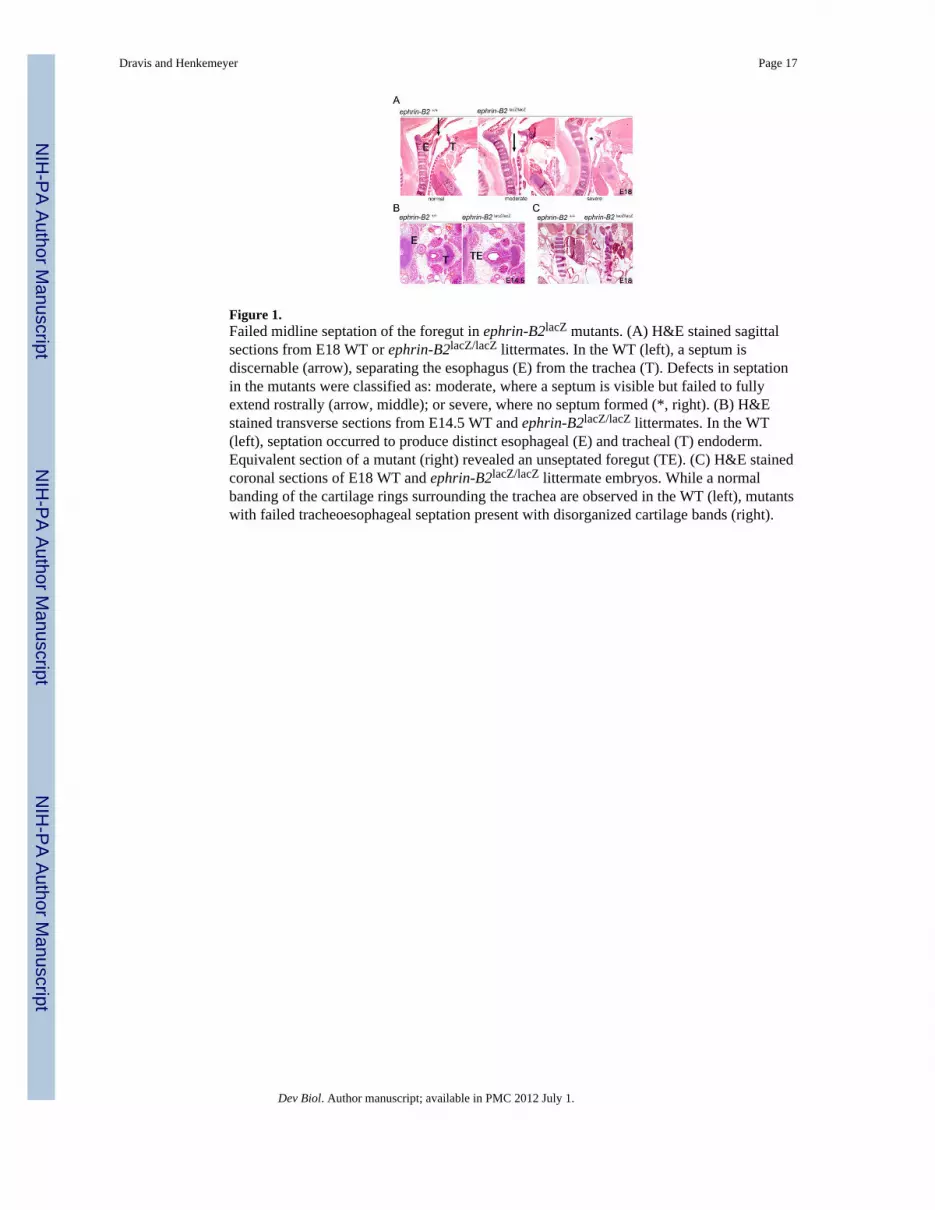
Figure 1.Failed midline septation of the foregut in ephrin-B2lacZ mutants. (A) H&E stained sagittalsections from E18 WT or ephrin-B2lacZ/lacZ littermates. In the WT (left), a septum isdiscernable (arrow), separating the esophagus (E) from the trachea (T). Defects in septationin the mutants were classified as: moderate, where a septum is visible but failed to fullyextend rostrally (arrow, middle); or severe, where no septum formed (*, right). (B) H&Estained transverse sections from E14.5 WT and ephrin-B2lacZ/lacZ littermates. In the WT(left), septation occurred to produce distinct esophageal (E) and tracheal (T) endoderm.Equivalent section of a mutant (right) revealed an unseptated foregut (TE). (C) H&E stainedcoronal sections of E18 WT and ephrin-B2lacZ/lacZ littermate embryos. While a normalbanding of the cartilage rings surrounding the trachea are observed in the WT (left), mutantswith failed tracheoesophageal septation present with disorganized cartilage bands (right).
Dravis and Henkemeyer Page 17
Dev Biol. Author manuscript; available in PMC 2012 July 1.
NIH
-PA Author Manuscript
NIH
-PA Author Manuscript
NIH
-PA Author Manuscript

Figure 2.Ephrin-B2 and EphB3 expression during foregut septation. (A, B) Serial X-gal stainedtransverse sections from E10.5 ephrin-B2lacZ/+ and ephrin-B2lacZ/lacZ littermates. Normalseptation in the ephrin-B2lacZ/+ embryo proceeds from common foregut (left), to septatingforegut (middle), to septated foregut showing separated esophagus (E) and trachea (T)(right). Septation of the foregut in the homozygous mutant failed to occur (left), caudal towhich only unseptated foregut remains (middle). A fistula (TE) between esophagus and theemerging bronchi is present at the very caudal end of the foregut (right). X-gal stain showsexpression of the ephrin-B2-β-gal fusion protein (blue) in the endoderm of the foregut atseptation, with higher levels of expression detected at the future esophageal pole of theforegut. Ephrin-B2 is also found in the mesenchyme surrounding the future esophagus. (C)Serial X-gal stained sections from an E11.5 ephrin-B2lacZ/+ embryo. Similar to E10.5,ephrin-B2 is highly expressed in the mesenchyme and epithelia associated with futureesophageal endoderm. Ephrin-B2 is also present on the endoderm and flanking mesenchymeat the point of septation (arrows). (D) Coronal sections capturing foregut septation from WTembryo stained at E10.5 and E11.5 with a pan-ephrin-B antibody. Ephrin-B is present in themesenchyme associated with the future esophageal endoderm and in the endoderm of theforegut. (E) BluO-gal stained coronal sections from a Tg-BAC-EphB3-rtTA; TRE-lacZembryo treated with dox to visualize EphB3 expression (blue). Like ephrin-B2, EphB3 ishighly expressed in the mesenchyme flanking the future point of septation (*, left), wherelateral folds will invaginate into the foregut and meet at the midline as seen in an adjacentcaudal section (right).
Dravis and Henkemeyer Page 18
Dev Biol. Author manuscript; available in PMC 2012 July 1.
NIH
-PA Author Manuscript
NIH
-PA Author Manuscript
NIH
-PA Author Manuscript

Figure 3.Cleft palate in ephrin-B2lacZ mutants. (A) H&E stained coronal sections of WT and ephrin-B2lacZ/lacZ E18 littermates. The ephrin-B2lacZ homozygote presents with a cleft palate (*).(B) X-gal stained coronal sections from ephrin-B2lacZ/+ embryos at E12.5, E13.5, and E14.5detect expression of the ephrin-B2-β-gal fusion protein (blue). Ephrin-B2 is expressed in themesenchyme and leading epithelia of the palatal shelves at E12.5 and E13.5 (*, left andmiddle) before palatal shelf closure. After adhesion, ephrin-B2 expression is strongest in themidline epithelial seam (arrow, right). (C) Coronal sections at E13.5 from WT andEphB2−/− embryos treated with anti-EphB2 antibody (top). EphB2 is expressed in themesenchyme of the palatal shelf in the WT (*, left), while no signal is detected in the mutantcontrol. Coronal sections from an E14.5 EphB2lacZ/+ embryo stained with X-gal to showexpression of the EphB2-β-gal fusion protein (bottom) reveal EphB2 becomes specificallyexpressed in the epithelia of the palatal shelves, both immediately preceding (left) and at thesite of adhesion (right). (D) Coronal section from an E13.5 Tg-BAC-EphB3-YFP embryotreated with anti-GFP antibody shows specific expression of EphB3 in the mesenchyme ofthe palatal shelf (left). X-gal stained coronal sections from E13.5 and E14.5 Tg-BAC-EphB3-rtTA; TRE-lacZ embryos treated with dox show EphB3 is initially highly expressed in themesenchyme of the palatal shelf at E13.5 (*, middle) but then becomes more preferentiallyexpressed in the leading epithelia as midline adhesion becomes imminent (*, right). Theexpression of EphB3 is very similar to EphB2 and ephrin-B2. (E) Coronal section from anE14 ephrin-B2lacZ/+ embryo treated with anti-EphB2 (left) and anti-β-gal (middle)antibodies. The merged image (right) shows EphB2 and ephrin-B2-β-gal are co-expressedon the midline epithelial seam where adhesion has occurred.
Dravis and Henkemeyer Page 19
Dev Biol. Author manuscript; available in PMC 2012 July 1.
NIH
-PA Author Manuscript
NIH
-PA Author Manuscript
NIH
-PA Author Manuscript

Figure 4.Failed closure of the ventral abdominal wall in EphB2; EphB3 compound null embryos. (A)Whole-mount image of an E18 EphB2−/−;EphB3−/− compound null shows herniated visceralorgans (*) due to failed abdominal body wall closure (left). Transverse H&E stained sectionof an EphB2−/−;EphB3−/− E18 embryo similarly shows failed midline closure of the ventralbody wall and herniated visceral organs (*, right). (B) Whole-mount BluO-gal stainedEphB2lacZ/+ and EphB2+/+ embryos at E13.5 and E15.5 to detect expression of the EphB2-β-gal fusion protein. EphB2 is expressed at the ventral midline at E13.5 (arrows, top left) andat leading edges of the closing umbilical ring at E15.5 (arrows, top right). No BluO-galactivity is detected in the WT embryos that do not express the EphB2-β-gal fusion protein(bottom).
Dravis and Henkemeyer Page 20
Dev Biol. Author manuscript; available in PMC 2012 July 1.
NIH
-PA Author Manuscript
NIH
-PA Author Manuscript
NIH
-PA Author Manuscript

Figure 5.Ephrin-B is tyrosine phosphorylated at the point of midline septation and closure in theforegut and palate. (A) Coronal sections from E10.5 WT embryos stained with an antibodythat recognizes specific phosphorylation of tyrosines 324 and 329 (top) or of tyrosine 317(bottom) on the conserved cytoplasmic tail of ephrin-B molecules to detect reverse signalingactivity in vivo. Both antibodies detect tyrosine phosphorylated ephrin-B on the endodermbefore (left) and during septation (middle). Activated ephrin-B also appears in themesenchyme flanking the site of septation (*). Adjacent sections pre-treated with λ-phosphatase confirm antibody specificity (right). (B) Adjacent coronal sections from an E14ephrin-B2lacZ/+ embryo treated with anti-β-gal (left) or anti-phospho-ephrin-BY324/Y329
Dravis and Henkemeyer Page 21
Dev Biol. Author manuscript; available in PMC 2012 July 1.
NIH
-PA Author Manuscript
NIH
-PA Author Manuscript
NIH
-PA Author Manuscript

(right) antibodies. While ephrin-B2 is expressed in both the mesenchyme and leadingepithelia of the palatal shelf (left), it is only the ephrin in the leading epithelia that is tyrosinephosphorylated (right). (C) Coronal section from a WT E14 embryo treated with anti-EphB2(left) and anti-phospho-ephrin-BY324/329 (middle) antibodies. EphB2 is co-expressed withtyrosine-phosphorylated ephrin-B in leading epithelia of the palatal shelves.
Dravis and Henkemeyer Page 22
Dev Biol. Author manuscript; available in PMC 2012 July 1.
NIH
-PA Author Manuscript
NIH
-PA Author Manuscript
NIH
-PA Author Manuscript

Figure 6.New ephrin-B2 reverse signaling point mutant mice identify separate tyrosinephosphorylation-independent components of ephrin-B reverse signaling at the midline. (A)Targeting strategy for the generation of the ephrin-B2ΔV allele. (N= NcoI). (B) Summary ofthe expected consequences on signaling for the ephrin-B2lacZephrin-B26yfΔv and ephrin-B2ΔV alleles. (C) Incidence of urorectal malformation in male progeny following crossesbetween ephrin-B2lacZ/+ males and either ephrin-B26YFV/+ or ephrin-B2ΔV/+ females,compared to the 100% incidence found in ephrin-B2lacZ/lacZ animals.
Dravis and Henkemeyer Page 23
Dev Biol. Author manuscript; available in PMC 2012 July 1.
NIH
-PA Author Manuscript
NIH
-PA Author Manuscript
NIH
-PA Author Manuscript

Figure 7.Claudin-1 and claudin-4 are co-expressed with activated ephrin-B and EphB2 at the point ofmidline adhesion in the palate. (A) Coronal section from a WT E14 embryo treated withanti-claudin-4 (green) and phospho-ephrin-BY324/Y329 (red) antibodies reveal bothmolecules are expressed in adherent epithelia of the palatal shelves. (B) Coronal sectionfrom a WT E14 embryo treated with anti-claudin-4 (green) and phospho-ephrin-BY317 (red)reveals the same co-expression after adhesion has been initiated. (C) Adjacent sections froma WT E14 embryo treated with anti-claudin-1 (left) and phospho-ephrin-BY317 (right) revealclaudin-1 is also present in the adherent palatal epithelia with tyrosine phosphorylatedephrin-B.
Dravis and Henkemeyer Page 24
Dev Biol. Author manuscript; available in PMC 2012 July 1.
NIH
-PA Author Manuscript
NIH
-PA Author Manuscript
NIH
-PA Author Manuscript

NIH
-PA Author Manuscript
NIH
-PA Author Manuscript
NIH
-PA Author Manuscript
Dravis and Henkemeyer Page 25
Table 1
Incidence of cleft palate in EphB2;EphB3 compound mutants
EphB2 mutation background B2/B2;B3/B3 B2/+;B3/B3 +/+;B3/B3
EphB2− CD1 27 (4) 32 (1) NA
EphB21acZ CD1 29 (12) 17 (2) NA
EphB2−/−;EphB3−/− or (EphB2lacZ/lacZ; EphB3−/−) males were intercrossed with EphB2−/+; EphB3−/− females (or
EphB2lacZ/+;EphB3−/−) and offspring collected at E18.5. Animals with cleft palate are in parentheses.
Dev Biol. Author manuscript; available in PMC 2012 July 1.

NIH
-PA Author Manuscript
NIH
-PA Author Manuscript
NIH
-PA Author Manuscript
Dravis and Henkemeyer Page 26
Table 2
Incidence of cleft palate in ephrin-B2lacZ mutants
background ephrin-B21acZ/lacZ ephrin-B21acZ/+ ephrin-B2+/+
129/CD1 35 (9) 59 (4) >40 (0)
Ephrin-B2lacZ/+ heterozygotes were intercrossed and offspring collected at E18.5. Animals with cleft palate are in parentheses.
Dev Biol. Author manuscript; available in PMC 2012 July 1.
NIH
-PA Author Manuscript
NIH
-PA Author Manuscript
NIH
-PA Author Manuscript
Dravis and Henkemeyer Page 27
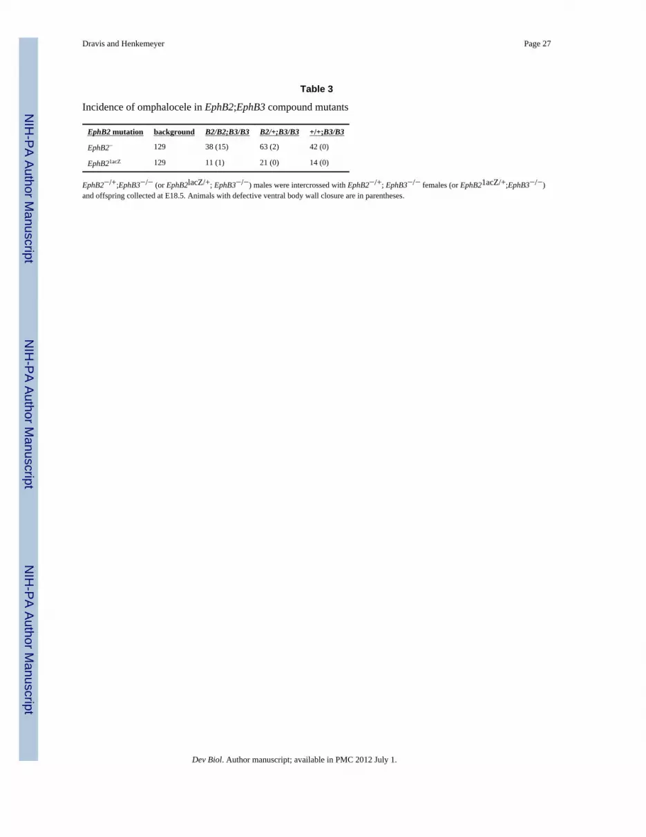
Table 3
Incidence of omphalocele in EphB2;EphB3 compound mutants
EphB2 mutation background B2/B2;B3/B3 B2/+;B3/B3 +/+;B3/B3
EphB2− 129 38 (15) 63 (2) 42 (0)
EphB21acZ 129 11 (1) 21 (0) 14 (0)
EphB2−/+;EphB3−/− (or EphB2lacZ/+; EphB3−/−) males were intercrossed with EphB2−/+; EphB3−/− females (or EphB21acZ/+;EphB3−/−)and offspring collected at E18.5. Animals with defective ventral body wall closure are in parentheses.
Dev Biol. Author manuscript; available in PMC 2012 July 1.

NIH
-PA Author Manuscript
NIH
-PA Author Manuscript
NIH
-PA Author Manuscript
Dravis and Henkemeyer Page 28
Table 4
VACTERLAssociation Defects
Eph/ephrininvolved
Defect References
Vertebral ephrin-B1 Asymmetric rib attachment (Compagni et al., 2003)
ephrin-B2 Abnormal somite patterning (Davy and Soriano, 2007)
Anorectal EphB2,EphB3 ephrin-B2 Failed cloacal septation leading to persistentcloaca with GI fistula
(Dravis et al., 2004)
Cardiac ephrin-B2 Enlarged cardiac valves (Cowan et al., 2004)
Tracheo-Esophageal ephrin-B2 Tracheoesophageal fistula with esophagealatresia
(this manuscript)
Renal ephrin-B2 Hydronephrosis due to failed ureter-bladderintegration
(unpublished data)
Limb ephrin-B1 polydactyly (Compagni et al., 2003) (Davy et al.,2004)
PeripheralVACTERL Defects
Eph/ephrininvolved
References
Hypospadias EphB2,EphB3 ephrin-B2 (Dravis et al., 2004)
Cleft Palate EphB2,EphB3 ephrin-B1 ephrin-B2 (Orioli et al., 1996) (Compagni et al., 2003) (Davy et al., 2004) (this manuscript)
Omphalocele EphB2,EphB3 ephrin-B1 (Compagni et al., 2003) (Orioli et al., 1996) (this manuscript)
Neural Tube Closure EphA7 ephrin-A5 (Holmberg at al., 2000)
Dev Biol. Author manuscript; available in PMC 2012 July 1.
Recommended


