
EXPERIMENTAL NEUROLOGY 57, 132-141 (1977)
Facilitation of Secondary-Site Amygdaloid Kindling Following
Bisection of the Corpus Callosum and Hippocampal Commissure in Rats
JAMES A. MCCAUGHRAN, JR., MICHAEL E. CORCORAN,
ANDJUHN A. WADA=
Division of Neurological Sciences, UGevsity of British Columbia, 207.5 Wesbrook Place, Vancouver, British Columbia V6T 1 WS Canada
Received March 14,1977
The effects of forebrain bisection on the development of secondary-site amygdaloid kindling in rats were examined. Bisection of the corpus callosum and hippocampal commissure after primary-site kindling had a facilitative effect on the subsequent rate of secondary-site kindling. Although the rate of secondary-site kindling was significantly facilitated after forebrain bisec- tion, bisected rats failed to develop typical bisymmetrical seizures. The bi- sected rats instead displayed asymmetrical, and in many cases hemiconvulsive, clinical manifestations. Seizure duration and afterdischarge duration were not affected by the forebrain bisection. These data suggest that seizure duration, afterdischarge duration, and the morphology of the seizure are independent aspects of the kindling phenomenon. Furthermore, because the facilitation of secondary-site kindling could not be adequately explained by denervation supersensitivity, we conclude that the callosal and commissural pathways are capable of mediating an interhemispheric interaction that is in turn able to interfere with the rate of seizure development in the intact rat.
INTRODUCTION
Repetitive electrical stimulation of various cortical and subcortical loci in the brain has been shown to result in the progressive development of seizures in a number of species (5, 15-18). This phenomenon is referred to
as kindling and was first described in detail by Goddard (4) and Goddard etal. (5).
Previous studies have examined the role of the forebrain commissures in the development of kindled amygdaloid convulsions. Wada and Sato
1 Supported by grants from the Medical Research Council of Canada and the U.S.- N.I.N.C.D.S. M. E. Corcoran was Killam Research Scholar of the Canada Council.
132
All rights of reproduction in any form reserved. Copyright 0 1977 by Academic Press. Inc.
ISSN 0014 -4886
FOREBRAIN BISECTION AND KIXDLING 133
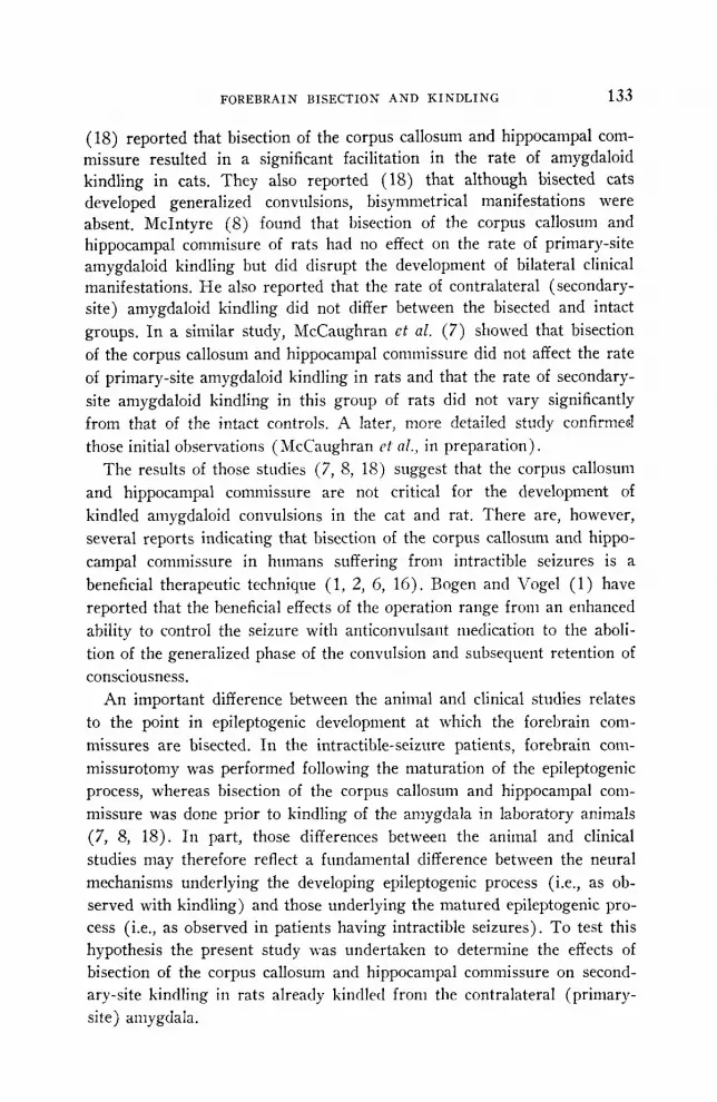
(IS) reported that bisection of the corpus callosum and hippocampal com- missure resulted in a significant facilitation in the rate of amygdaloid kindling in cats. They also reported (18) that although bisected cats developed generalized convulsions, bisymmetrical manifestations were absent. McIntyre (8) found that bisection of the corpus callosum and hippocampal commisure of rats had no effect on the rate of primary-site amygdaloid kindling but did disrupt the development of bilateral clinical manifestations. He also reported that the rate of contralateral (secondary- site) amygdaloid kindling did not differ between the bisected and intact groups. In a similar study, McCaughran et al. (7) showed that bisection of the corpus callosum and hippocampal commissure did not affect the rate of primary-site amygdaloid kindling in rats and that the rate of secondary- site amygdaloid kindling in this group of rats did not vary significantly from that of the intact controls. A later, more detailed study confirmed those initial observations ( McCaughran ct al., in preparation).
The results of those studies (7, 8, 18) suggest that the corpus callosum and hippocampal commissure are not critical for the development of kindled amygdaloid convulsions in the cat and rat. There are, however, several reports indicating that bisection of the corpus callosum and hippo- campal commissure in humans suffering from intractible seizures is a beneficial therapeutic technique (1, 2, 6, 16). Bogen and Vogel (1) have reported that the beneficial effects of the operation range from an enhanced ability to control the seizure with anticonvulsant medication to the aboli- tion of the generalized phase of the convulsion and subsequent retention of consciousness.
An important difference between the animal and clinical studies relates to the point in epileptogenic development at which the forebrain com- missures are bisected. In the intractible-seizure patients, forebrain com-
missurotomy was performed following the maturation of the epileptogenic process, whereas bisection of the corpus callosum and hippocampal com- missure was done prior to kindling of the amygdala in laboratory animals (7, 8, 18). In part, those differences between the animal and clinical studies may therefore reflect a fundamental difference between the neural mechanisms underlying the developing epileptogenic process (i.e., as ob- served with kindling) and those underlying the matured epileptogenic pro- cess (i.e., as observed in patients having intractible seizures). To test this hypothesis the present study was undertaken to determine the effects of bisection of the corpus callosum and hippocampal commissure on second- ary-site kindling in rats already kindled from the contralateral (primary- site) amygdala.

134 MCCAUGHRAN, CORCORAN AND WADA
METHODS
Seventeen male hooded rats of the Long-Evans strain (Canadian Breeding Farms) weighing between 300 and 3.50 g were used. Rats were housed individually in a temperature-controlled colony room with a 12-h: 12-h light-dark cycle. They had free access to food and water except during the testing and had 10 to 14 days to adjust to the colony environment be- fore undergoing surgery.
Rats were anesthetized with sodium pentobarbital (60.0 mg/kg, intra- peritoneally) and bipolar electrodes made from twisted Nichrome wire (127 pm in diameter) were implanted bilaterally in the basolateral nucleus of the amygdala using conventional stereotaxic techniques. The electrodes were fixed to the skull with dental acrylic and jeweler’s screws and a 5.0- mm separation was left between the left and right electrode pairs to main- tain access to the midline. Seven or more days after the operation, kindling of the primary site began. Stimulation consisted of a 1.0-s train of constant current 60-Hz sine waves at an intensity of 160 PA (rms), applied to either the right or left amygdala of each rat once daily between 1000 and 1300 h. Kindled seizure development was classified according to the method of Racine (11) : C-l, mouth and facial twitching ; C-2, the above plus head nodding ; C-3, the above plus forelimb clonus ; C-4, the above plus rearing onto the hindlegs; C-5, the above plus rearing and then falling over. Rats were considered kindled after reaching the C-5 stage in seizure develop- ment .
Eight C-5 seizures were evoked from the amygdala (primary site). After the eighth C-5 seizure, rats were anesthetized with sodium pento- baribital (60.0 mg/kg, intraperitoneally), and a midline craniotomy was done using stereotaxic techniques. The dura mater was exposed and dampened periodically with normal saline, and overlying chips of bone were removed. The sagittal sinus was visualized and the dura mater im- mediately latera to it reflected. Two fine sewing needles, with diameters of approximately 0.45 mm, were each mounted in an electrode carrier. The eyes of the needles were oriented downward. One needle was then posi- tioned over the anterior extreme of the corpus callosum and the other was placed over the posterior extreme. A single piece of 6-O surgical silk was passed between the eyes of the needles, and the needles were lowered to points slightly ventral to the extremes of the callosum. The silk was then pulled taut. In this manner, all tissue lying between the needles was transected, including the corpus callosum and hippocampal commissure. After the bisection, the needles were retracted from the brain and the thread was extracted. Excessive bleeding was readily controlled by gently apply- ing pressure with a cotton swab, and the exposed cortex w;ts covered by a thin strip of sterile Gelfoam soaked in normal saline. The skull was then
FOREBRAIN BISECTION AND KINDLING 135
TABLE 1
Mean Number (and Ranges) of Sessions to Kindle at Each Site
Group Primary-site kindling
Secondary-site kindling
Primary-site rekindling
Secondary-site rekindling
Bisected N
14.6 (1 l-20) 4.0,’ (2-9) 1.6 (l-4) 1.3* (l-2)
8 8 8 7
Control 15.9 (8-26) 7.0 (6-9) 2.4 (l-4) 2.4 (2-3) N 7 7 7 7
a Signiticantly different from the rate of secondary-site kindling of the controls (P < 0.05).
*Significantly different from the rate of secondary-site rekindling of the controls (I’ < 0.01).
covered with a thin layer of dental acrylic. Rats in the control group were prepared in a manner identical to the above except that only the cortical tissue down to the dorsal surface of the callosum was transected.
All rats had a minimum of 10 days to recover from surgery before being stimulated. After recovery, the site of the stimulation was switched to the secondary site. Rats were again kindled and allowed to have eight C-5 seizures. The site of stimulation was then switched back to the primary site. After eight C-5 seizures were evoked by rekindling the primary site, the site of stimulation was again switched for secondary-site rekindling. After the eighth C-5 seizure, the experiment was terminated and the position of the electrodes and the extent of the bisection were determined histologically.
RESULTS
Because no record was kept concerning which rats had undergone bi- section of the commissures, the bisected and control groups were not assembled until a histological analysis of the brains had been made.
Table 1 shows the mean rate of kindling observed at each site in each group. As expected, no significant difference was observed between the bisected and control groups during primary-site kindling. IIowever, rats in which the corpus callosum and hippocampal commissure had been bi- sected after primary-site kindling kindled significantly faster than the control group [F(l, 13) = 8.83, p < O.O.S]. There was no difference be- tween the groups in the rate of primary-site rekindling. During secondary- site rekindling, however, the bisected group kindled significantly faster than the control group [F( 1, 12) = 17.65, P < 0.011.
Although the rate of secondary-site kindling was significantly acceler- ated by forebrain bisection, the morphology of the C-5 seizures in the

136 MCCAUGHRAN, CORCORAN AND WADA
TABLE 2
Effect of Forebrain Bisection on the Development of Bisymmetrical C-5 Motor Seizures
Primary Secondary Primary Secondary retest retest
Bisected Number showing
bisymmetrical C-5 seizure
N 7 lb 4 3a 8 8 7 7
Control Number showing
bisymmetrical C-S seizure
N 6 7 7 7 7 7 7 7
a Significantly different from controls (P < 0.05). b Significantly different from controls (P < 0.01).
bisected animals differed from that of the controls. Stage-5 seizures in the control rats were characterized by generalized bisymmetrical clonic activity, whereas the C-5 seizure in the bisected rats typically consisted of asym- metrical clonus involving the extremities contralateral to the site of stimu- lation. Some ipsilateral clonic activity was observed in a few of those rats, but it failed to persist and only appeared briefly at the end of the seizure. Only one of eight rats in the bisected group developed the typical C-5 seizure during secondary-site kindling, as compared to seven of seven rats in the control group (Table 2). The difference between the groups was highly significant [x2 (1) = 8.24, P < O.Ol]. Most rats in the bisected group continued to show the asymmetrical seizure pattern throughout the subsequent phases of the study. Some, however, did eventually develop the typical bisymmetrical C-5 seizure. During rekindling of the primary site, seven of seven rats in the control group displayed the bisymmetrical pat- tern, whereas this pattern was observed in only four of seven in the bi- sected group (Table 2). This difference, however, failed to reach the level of significance. Rekindling of the secondary site failed to elicit any further development toward the bisymmetrical C-5 seizure in the bisected group. Only three of seven rats in this group displayed C-5 bisymmetry, as com- pared to seven of seven control rats during this phase of the study (Table 2). The difference between the groups was significant (x2( 1) = 3.22, P < 0.05). In addition, the three rats in the bisected group displaying a bisymmetrical C-5 seizure had also done so during the previous phase of the study (i.e., rekindling of the primary site).

FOREBRAIN BISECTION AND KINDLIKG 137
FIG. 1. Tracings of sections indicating tile cstcnt of the fur&rain bisection in a representative rat from each group. Lightly shaded areas represent ventricular spaces. Abbreviations : C, control ; CC, bisected.

138 MCCAUGHRAN, CORCORAN AND WADA
In contrast to its effects on the rate of seizure development and seizure morphology, bisection of the corpus callosum and hippocampal commissure had no significant effect on the duration of the clinical seizure or on the propagation of afterdischarge during any phase of the study.
Histology. All electrode tips were found in or immediately adjacent to the basolateral nucleus of the amygdala. Bisection of the corpus callosum in the bisected group (N = 8) was relatively constant between animals, with most rats possessing a complete bisection of the corpus callosum and hippo- campal commissure (Fig. 1). Extracallosal damage in this group was minimal ; in most rats it was confined to the tissue overlying the callosum (i.e., cingulate cortex). Likewise, subcortical damage also appeared negligi- ble. The hippocampus was relatively free of damage, although some rats displayed minor damage to the medial aspects of Ammon’s horn. No evi- dence of midline thalamic damage was observed in any of the rats.
There were no signs of callosal damage in the control group (N = 7). The transection in this group was confined to a small strip of cingulate cortex, adjacent to the midline (Fig. 1). In a few rats the location and ex- tent of the transection were similar to the above except that some “under- cutting” of the cortex was noted, apparently resulting from the deflection of the surgical silk by the fibers of the callosum.
DISCUSSION
The results of the present study indicate that the corpus callosum and hippocampal commissure in the rat are not essential pathways for the development of kindled amygdaloid seizures. Bisection of these structures after primary-site kindling resulted in a significant facilitation in the rate of secondary-site kindling. These results suggest that in the intact rat those commissural connections may interfere with the interhemispheric transfer of kindled seizure susceptibility. This further suggests that structures send- ing efferents via these pathways are able to interfere with the development of epileptiform activity in the contralateral hemisphere. Alternatively, the facilitation in secondary-site kindling may have been due to the develop- ment of denervation supersensitivity caused by the bisection of the com- missures. Stavraky (14) reported an increase in the susceptibility of rats and cats to the convulsant effects of pentylenetetrazol after bisection of the corpus callosum. Similarly, Sharpless (13) indicated that denervation supersensitivity is capable of producing increases in seizure susceptibility. However, this explanation for the present results is difficult to support. For example, bisection of the corpus callosum and hippocampal com- missure prior to kindling does not produce a facilitation in primary-site or secondary-site kindling (7, S) . A hypothesis of denervation supersensitivity would predict that bisection of those structures prior to kindling would

FOREBRAIN BISECTION AND KINDLING 139
also result in facilitation of seizure development. Furthermore, in both the present and previous studies (7, S), the durations of the clinical seizure and afterdischarge in the bisected rats were not significantly different from controls. Wada and Sato (18) also reported that the seizure-triggering threshold in callosum-bisected cats did not differ significantly from that of controls. If denervation supersensitivity were involved in the present re- sults, one might expect to also see marked changes in these other parame- ters of kindled epileptogenicity.
A hypothesis suggesting that structures in one hemisphere are able to interfere with the development of epileptiform activity in the other is supported by a number of studies showing that changes in spike discharges of secondary foci can occur following the inactivation of primary foci (3, 9, 12). Mutani et al. (10) showed that the activity of bilateral cortical foci in chronic cats was enhanced following bisection of the corpus callosum and reduced after isolation of the cortex and corpus callosum from the sub- cortex. They suggested that transcallosal pathways were responsible for the transmission of an interhemispheric inhibitory influence and that brain stem pathways mediated an interhemispheric facilitatory effect. The results of the present study, therefore, may be due to a facilitative effect through this hypothetical brain stem pathway.
The present results also indicate that although the corpus callosum and hippocampal commissure are not essential for amygdaloid kindling, they do participate in the clinical development of bisymmetrical C-5 seizures. Bisection of these structures following primary-site kindling caused a sig- nificant reduction in the number of rats displaying bisymmetrical C-S seizures during secondary-site kindling. Bisection of these structures prior to kindling has been shown to retard but not stop the development of bi- symmetrical C-5 seizures (7, 8). This, perhaps, suggests that alternate pathways able to mediate the development of bisymmetrical C-5 seizures may develop if the commissures are transected prior to kindling. The eventual establishment of alternate routes may also be indicated by the pres- ent finding that the number of bisected rats displaying bisymmetrical C-5 seizures increased during primary-site and secondary-site rekindling. The establishment of these routes may be more difficult in a rat that has under- gone forebrain bisection after primary-site kindling because these potential alternate routes may by this time be involved in some other aspect of kindling.
Previous reports have indicated that bisection of the corpus callosum in humans suffering from intractible seizures has had a beneficial therapeutic effect (1, 2, 6). None of the authors, to our knowledge, has reported an in- crease in seizure activity in bisected patients as might be suggested by the present study. However, it may be important to note that although bisected

140 MCCAUGHRAN, CORCORAN AND WADA
rats in this study displayed a significant facilitation in secondary-site kin- dling, there was a significant reduction in the occurrence of fullblown bi- symmetrical C-S seizures, indicating that the ictal response had been some- what attenuated. In addition, comparison between the present study and human clinical studies is further confounded by the fact that the patients were maintained on a regimen of anticonvulsant medication after surgery. Finally, because there is a marked difficulty in assessing the precise extent of the epileptogenic process in human patients, it is not possible to be certain that amygdaloid kindling in rats represents a similar situation.
REFERENCES
1. BOGEN, J. E., AND P. J. VOGEL. 1963. Treatment of generalized seizures by cerebral commissurotomy. Surg. Forum 14 : 431.
2. BOCEN, J. E., E. D. FISHER, AND P. J. VOGEL. 1965. Cerebral commissurotomy : A
second case report. JAMA. 194: 1328. 3. COCEANI, F., 1. LIBMAN, AND P. GLOOR. 1966. The effect of intracarotid amobarbi-
tal injections upon experimentally induced epileptiform activity. Electroence- phalogr. Clin. Neurophysiol. 20 : 542-558.
4. GODDARD, G. V. 1967. Development of epileptic seizures through brain stimulation at low intensity. Nature (Lo&) 214: 1020-1021.
5. GODDARD, G. V., D. C. MCINTYRE, AND C. K. LEECH. 1969. A permanent change in brain function resulting from daily electrical stimulation. Exp. Neural. 25: 295330.
6. LUESSENHOP, A. J. 1970. Interhemispheric commissurotomy : (The split brain operation) as an alternate to hemispherectomy for control of intractible sei- zures. Amer. Surg. May : 265-268.
7. MCCAUGHRAN, J. A., JR., M. E. CORCORAN, AND J. A. WADA. 1976. Development of kindled seizures after section of the forebrain commissures in rats. Folia Psychiatr. Neural. Japan 30 : 65-71.
8. MCINTYRE, D. C. 1975. Split-brain rat: Transfer and interference of kindled amygdala convulsions. Canad. J. Neural. Sci. 2: 429-437.
9. MORRELL, F. 1960. Secondary epileptogenic lesions. Epilepsia 1: 535-560. 10. MUTANI, R., L. BERGAMINI, R. FARIELLO, AND G. QUATTROCOLO. 1972. An experi-
mental investigation on the mechanisms of interaction of asymmetrical acute epileptic foci. Epilepsiu 13 : 597-608.
11. RACINE, R. J. 1972. Modification of seizure activity by electrical stimulation: II. Motor Seizure. Electroencephalogr. Clin. Neurophysiol. 32 : 281-294.
12. ROVIT, R. L., J. HARDY, AND P. GLOOR. 1960. Electroencephalographic effects of intracarotid amobarbital on epileptic activity. An experimental study using peni- cillin-induced epileptic foci in rabbits. Arch. Neural. 3: 642-655.
13. SHARPLESS, S. K. 1969. Isolated and deafferented neurons : Disuse supersensitivity. Pages 329-348 in H. JASPER, A. WARD, AND A. POPE, Eds., “Basic Mechanisms of the Epilepsies.” Little, Brown and Company, Boston.
14. STAVRAKY, G. W. 1961. Neuronal isolation of the cerebral cortex and con- vulsibility in animals and man. A section of the corpus callosum. Pages 33-38 in G. W. STAVRAKY, Ed., Supersetwititity Following Lesions of the Nervous System. University of Toronto Press, Toronto.

FOREBRAIN BISECTION AND KINDLING 141
15. TANAKA, A. 1972. Progressive changes of behavioral and electroencephalographic responses to daily amygdaloid stimulations in rabbits. Fukuoka Acfa Med. 63: 152-164.
16. VAN WAGENEN, W. P., AND R. Y. HERREN. 1940. Surgical division of commissural pathways in the corpus callosum. Arch. Neural. Psychiatry 44: 740-759.
17. WADA, J. A., AND T. OSAWA. 1976. Spontaneous recurrent seizure state induced by daily electrical amygdaloid stimulation Senegalese baboons, Papio papio. Neurology 26 : 273-286.
18. WADA, J. A., AND M. SATO. 1974. Generalized convulsive seizures induced by daily electrical stimulation of the amygdala in cats: Correlative electrographic and behavioral features. Nezrrology 24 : 564-574.
19. WADA, J. A., AND M. SATO. 1975. The generalized convulsive seizure state induced by daily electrical stimulation of the amygdala in split-brain cats. Epilepsiu 16: 417-430.
Recommended


