
Chemico-Biological Interactions 174 (2008) 126–133
Contents lists available at ScienceDirect
Chemico-Biological Interactions
journa l homepage: www.e lsev ier .com/ locate /chembio int
Food-associated estrogenic compounds induce estrogenreceptor-mediated luciferase gene expression intransgenic male mice
Marcel G.R. ter Velda,1, E. Zawadzkaa,2, J.H.J. van den Berga, Paul T. van der Saagb,Ivonne M.C.M. Rietjensa, Albertinka J. Murka,∗
a Toxicology Section, Wageningen University, Postbox 8000,6700 EA, Wageningen, The Netherlandsb Hubrecht Laboratory/NIOB, Uppsalalaan 8, 3584 CT Utrecht, The Netherlands
a r t i c l e i n f o
Article history:Received 13 February 2008Received in revised form 28 March 2008Accepted 31 March 2008Available online 10 April 2008
Keywords:Estrogen receptorReporter gene miceEndocrine disrupterPhthalatesBisphenol ANonylphenolp,p′-DDEFood contaminants
a b s t r a c t
The present paper aims at clarifying to what extent seven food-associated compounds,shown before to be estrogenic in vitro, can induce estrogenic effects in male mice with anestrogen receptor (ER)-mediated luciferase (luc) reporter gene system. The luc inductionwas determined in different tissues 8 h after dosing the ER-luc male mice intraperitoneally(IP) or 14 h after oral dosing. Estradiol-propionate (EP) was used as a positive control at0.3 and 1 mg/kg bodyweight (bw), DMSO as solvent control. The food-associated estrogeniccompounds tested at non-toxic doses were bisphenol A (BPA) and nonylphenol (NP) (both at10 and 50 mg/kg bw), dichlorodiphenyldichloroethylene (p,p′-DDE; at 5 and 25 mg/kg bw),quercetin (at 1.66 and 16.6 mg/kg bw), di-isoheptyl phthalate (DIHP), di-(2-ethylhexyl)phthalate (DEHP) and di-(2-ethylhexyl) adipate (DEHA) all at 30 and 100 mg/kg bw. In gen-eral IP dosing resulted in higher luc inductions than oral dosing. EP induced luc activity inthe liver in a statistically significant dose-related way with the highest induction of all com-pounds tested which was 20,000 times higher than the induction by the DMSO-control. NP,DDE, DEHA and DIHP did not induce luc activity in any of the tissues tested. BPA induced lucin the liver up to 420 times via both exposure routes. BPA, DEHP and quercetin induced lucactivity in the liver after oral exposure. BPA (50 mg/kg bw IP) also induced luc activity in the
testis, kidneys and tibia. The current study reveals that biomarker-responses in ER-luc malemice occur after a single oral exposure to food-associated estrogenic model compounds atexposure levels 10 to 104 times higher than the established TDI’s for some of these com-pounds. Given the facts that (i) the present study did not include chronic exposure and that(ii) simultaneous exposure to multiple estrogenic compounds may be a realistic exposurescenario, it remains to beAbbreviations: DDE, 1,1-dichloro-2,2-bis(4-chlorophenyl)ethylene; EC50, the cequivalency factor; EEQ, estradiol equivalent; ER, estrogen receptor; ER-Luc miceIP, intraperitoneal; RLU, relative light units; TDI, tolerable daily intake.∗ Corresponding author. Tel.: +31 317483233; fax: +31 317484931.
E-mail address: [email protected] (A.J. Murk).1 Current address: Department of Toxicology and Drug Disposition, Organon N2 Current address: Department of Andrology and Reproductive Endocrinology,
0009-2797/$ – see front matter © 2008 Elsevier Ireland Ltd. All rights reserved.doi:10.1016/j.cbi.2008.03.019
seen whether this margin is sufficiently high.
© 2008 Elsevier Ireland Ltd. All rights reserved.oncentration that induces 50% of the maximal response; EEF, estradiol, estrogen receptor reporter gene (luciferase) mice; IF, induction factor;
.V., Postbox 20, 5340 BH, Oss, The Netherlands.Medical University of Lodz, 91-425 Lodz, Sterlinga 5, Poland.

ological
1
i[eVrcahglDz
wsegtapvgglo[wcmutccbwswatIusegbrceTmaecmviws
M.G.R. ter Veld et al. / Chemico-Bi
. Introduction
Estrogenic compounds are studied intensively, as theres concern about their possible endocrine disrupting effects1]. For the quantification of the estrogenicity of pseudo-strogens several in vitro assays have been developed.ery specific and sensitive in vitro assays are estrogeneceptor (ER)-mediated reporter gene assays using geneti-ally modified cell lines [2,3]. In general pseudo-estrogensre 104 to 108 less estrogenic than the natural femaleormone estradiol (E2) [2,3]. The in vivo relative estro-enicity (compared to E2), however, can be much higher orower as has been shown respectively for example for o,p′-DT, ethynylestradiol, and nonylphenol (NP) in transgenicebrafish [4].
For human risk assessment it is necessary to knowhether food-associated compounds which have been
hown to be estrogenic in vitro [3], are also able to inducestrogenic effects in vivo. For instance, the in vitro estro-enicity for phthalates appeared not to be predictive forheir in vivo estrogenicity [5]. The current research projectims at clarifying to what extent food-associated com-ounds, shown to be estrogenic in ER�- and ER�-specific initro ER-mediated reporter gene cell lines, can induce estro-enic effects in vivo in male ER-luciferase (ER-luc) reporterene mice. The ER-luc mice contain a 3xERE-tata-Luc insu-ated reporter gene construct, which reveals the activationf the endogenous estrogen receptor, as has been described6,7]. In these studies dosing was intraperitoneally (IP)hich has the advantage that the compound has a high
hance of reaching the circulation and tissues [6,7]. Thisay not be the case upon oral exposure. Low intestinal
ptake and first pass metabolism could strongly reducehe systemic concentration, resulting in differences whenomparing the effects of orally dosed compared to IP-dosedompounds. Therefore in this study the mice were exposedoth IP as well as orally and the ER-mediated luc inductionas determined in several tissues of the animals. It has been
hown that the in vivo luc response is very fast, as only 8 here needed after IP exposure to estradiol-propionate (EP)
nd diethylstilbestrol (DES) for maximum luc induction inhe liver [6]. In this study we determine luc activity 8 h afterP-exposure and 14 h after oral exposure to allow time forptake and response. Exposure periods are expected to behort enough to prevent feedback mechanisms and indirectffects, such as down-regulation of receptors and relevantenes, to become too important. Male mice were chosenecause of their relative low background levels of natu-al estrogens. Seven, in vitro estrogenic, food-associatedompounds were tested for their in vivo estrogenicity, 17�-stradiol 3,17-dipropionate was used as a positive control.he main aim of the current experiment was to deter-ine whether the in vitro estrogenic compounds would
lso be estrogenic in vivo via either the more relevant oralxposure route and the worst-case IP exposure which isommonly used for experiments in ER-luc reporter gene
ice. The lowest dose inducing an estrogenic response inivo in the present study is compared to the tolerable dailyntake for estrogenic compounds in food as far as these
ere available for the model compounds of the presenttudy.
Interactions 174 (2008) 126–133 127
2. Animals, materials and methods
2.1. Chemicals and reagents
The estrogenic compounds tested were 17�-estradiol3,17-dipropionate (EP, >98%, CAS: 113-38-2), bisphenol A(BPA, >99%, CAS: 80-05-7), p,p′-DDE (>99%, CAS: 72-55-9), obtained from Sigma (Zwijndrecht, The Netherlands),quercetin dihydrate (99%, CAS: 6151-25-3) from AcrosOrganics (Geel, Belgium), nonylphenol (NP, ∼85% technicalmixture, CAS: 104-40-5) from Fluka (Buchs, Germany), di-(2-ethylhexyl) phthalate (DEHP) (CAS: 117-81-7) from TCI(Tokyo, Japan). Di-isoheptyl phthatale (DIHP, CAS: 41451-28-9) and di-(2-ethylhexyl) adipate (DEHA, CAS: 103-23-1)were technical grade and obtained from Agrotechnologyand Food Innovations (Wageningen, The Netherlands).
Further chemicals used were tricine (>99%),glycylglycine, ethylene glycol-bis(2-aminoethylether)-N,N,N′,N′-tetra-acetic acid (EGTA), Triton X-100, bovineserum albumin (BSA, >97%) and corn-oil all fromSigma (Zwijndrecht, The Netherlands). Sodium hydrox-ide (NaOH, >99%), EDTA·2H2O (>99%), MgSO4·7H2O(>99,5%) and 1,4-dithiothreitol (>99%) were obtainedfrom Merck (Darmstadt, Germany). d-Luciferin (>99.5%)was obtained from Duchefa (Haarlem, The Netherlands).(MgCO3)4Mg(OH)2·5H2O was obtained from Aldrich(Milwaukee, WI, USA). ATP was obtained from Roche(Mannheim, Germany).
All chemicals were dissolved and diluted in dimethylsulfoxide (DMSO >99.9%) purchased from Acros Organics(Geel, Belgium).
2.2. Animal experiment
The male ER-luc reporter gene mice originated from thestable reporter line INS7 [6] and were randomly bred withinour colony. The mice were fed a phyto-estrogen-free dietthroughout their life (AM-II diet, Hope Farms, The Nether-lands). Water was provided in glass bottles. Water andfood were provided ad libitum. The animal experiment wasapproved by the Animal Ethics Committee of WageningenUniversity. Six animals were used per group for IP expo-sure. Eight animals were used per group for oral exposure.Exceptions in number of animals were for oral exposureonly: the DMSO control group consisted of nine mice, thegroups exposed to 5 or 25 mg/kg bw DDE consisted of fourmice each, and the groups exposed to 100 mg/kg bw DEHAor 1.66 mg/kg bw quercetin consisted of seven mice. Theweight of the animals ranged from 21.8 to 57.9 g for themice in the oral exposure groups and from 24.2 to 49.4 gfor the mice in the IP exposed groups.
For IP exposure DMSO stock solutions of the compoundswere diluted to 5% in corn-oil and 6.66 �l/g bodyweight(bw) of this solution was injected IP once. Eight hoursafter IP dosing, comparable to Lemmen et al. [6,7], malemice were euthanized by cervical dislocation and tissues
were collected, weighed, immediately frozen on dry ice andstored at −80 ◦C.Oral exposure was performed in one dose of 0.5 mlvanilla custard (of organic origin, Demeter, The Nether-lands) to avoid discomfort due to oral gavage. The

128 M.G.R. ter Veld et al. / Chemico-Biological Interactions 174 (2008) 126–133
Table 1The low and high dosages of estrogenic compounds ER-luc male mice received once, either orally in vanilla custard or IP in corn-oil
Dose (mg/kg bw) TDI (mg/kg bw) EEF EEQ high dose (mg/kg bw) TDI, as EEQ (mg/kg bw)
Low High
DMSO (�l/kg) 0.33 – 0 0 –EP 0.3 1 – >1 [50] >1 –BPA 10 50 0.01 [51] 2.5 × 10⁻⁵ [3] 1.3 × 10⁻³ 2.5 × 10⁻⁷
NP 10 50 – 4.6 × 10⁻⁵ [3] 2.3 × 10⁻³ –DDE 5 25 0.002 [52] 2.3 × 10⁻⁶ [22] 5.7 × 10⁻⁵ 1.1 × 10⁻⁷
DEHP 30 100 0.05 [53,54] 2.2 × 10⁻⁷ [3] 2.2 × 10⁻⁵ 1.1 × 10⁻⁸
DEHA 30 100 0.3 [55] 4.9 × 10⁻⁸ [3] 4.9 × 10⁻⁶ 1.5 × 10⁻⁸
DIHP 30 100 – 3.1 × 10⁻⁷ [3] 3.1 × 10⁻⁵ –Quercetin 1.66 16.6 – 2 × 10⁻⁷ [10] 3.3 × 10⁻⁶ –
alents (EFood an
to prevent an excess of Triton X-100, which would interferewith the Pierce protein BCA assay. Of this dilution 10 �l wasanalyzed in duplicate in a 96 well flat bottom Greiner plate(Alphen a/d Rijn, The Netherlands). On each plate a full cal-ibration curve from 0 to 1.5 mg/ml bovine serum albumin
Table 2Amount of lysis buffer added to the tissues before homogenization withmicro-pestles and the fold dilution of the samples with PBS to preventinterference of Triton X-100 in the protein measurement
Tissue �l lysis buffer Fold dilution in PBS
Pituitary 100 10Brain per part 500 25Tibia 200 10
The high dosage of each compound also is expressed in estradiol equivvitro studies. When available from the European Commission (EC) or thecompounds is given as well; (–) not available.
compounds were dissolved in DMSO and added to the cus-tard at a dose of 0.33 �l DMSO/g bw. Four days before theactual exposure, mice were accustomed to eat 0.5 ml vanillacustard daily. All animals consumed their portion of 0.5 mlvanilla custard within 5 min. The period between oral expo-sure and sacrifice of the animals was 14 h, 6 h longer thanafter IP exposure to allow extra time for internal uptake.
The low and high exposure concentrations (Table 1) forEP and BPA were based on Lemmen et al. [6,7]. Since NPand BPA are more or less equipotent in vitro [3], the samedose was chosen for NP. For the phthalates, concentrationswere chosen based on maximal human exposure to DEHPin medical procedures, which was reported to be about0.01–140 mg/kg bw [8]. Based on these values a high doseof 100 mg/kg bw and a lower dose of 30 mg/kg bw, one logscale lower, were chosen. For DEHA the same concentra-tions as for DEHP were tested, as these two compounds areplasticizers used in a comparable way. The dose of DDE waschosen based on the concentrations used in the study of Youet al. [9], which dosed 10 and 100 mg/kg bw. The highestdose of quercetin was derived from the human intake withdietary supplements which can amount up to 1 g/day [10],which is equivalent to 16.6 mg/(kg bw day) for a 60 kg per-son. All concentrations were below levels of known toxicityand the same for both routes of exposures. The doses alsowere expressed as estradiol equivalents (EEQs) by multipli-cation with the in vitro-based estradiol equivalency factors(EEFs) for the compounds (Table 1).
Based on a pilot study with three animals per groupexposed IP to EP it was decided not to collect lung, prostate,esophagus, heart and intestines as in these tissues no lucactivity was found 8 h after IP exposure (data not shown),and the tissues analyzed in the present study are listed inTable 2.
2.3. Measurement of luciferase activity
Tissues were homogenized manually with an Eppendorfmicro-pestle in 1.5-ml vials (Eppendorf, Hamburg, Ger-
many) in lysis buffer (25 mM glycylglycine, 15 mM MgSO4,4 mM EGTA, 1 mM DTT and 1% Triton X-100; pH 7.8). Liv-ers were cut in four equal pieces of which three wereanalyzed, brain was cut in half, all other tissues wereanalyzed in full. The amount of lysis buffer added per tis-EQs) using estradiol equivalency factors (EEFs) based on ER�-based ind Agricultural Organization (FAO), the tolerable daily intake (TDI) for the
sue (Table 2) was sufficient to homogenize the samplewithout diluting the luciferase activity too much. Tibiaand femur were homogenized as good as possible withthe micro-pestle. The samples were kept on ice duringhomogenization, after which the Eppendorf vials were cen-trifuged for 15 min at 13,000 × g at 4 ◦C. After centrifugationthe supernatant was quantitatively diluted in lysis bufferto approximately 1 mg/ml protein, 25 �l was pipetted induplicate into a white 96 well assay plate (Costar, Corning,NY, USA). Light emission was measured at room tempera-ture in a luminometer (Labsystems, Luminoskan RS) during2 s, before and after automated addition of 100 �l ‘flash-mix’ (20 mM tricine, 1.07 mM (MgCO3)4Mg(OH)2·5H2O,2.67 mM MgSO4, 0.1 mM EDTA·2H2O, 2 mM DTT, 0.47 mMd-luciferin, 5 mM ATP; pH 7.8), subsequently light emissionwas extinguished with 50 �l 0.2 M NaOH before the nextwell was measured to prevent cross-scattering of light.
2.4. Protein analysis
The protein content of the supernatant was determinedusing the Pierce protein BCA assay according to manufac-turers’ instructions (Pierce, Rockford, IL) with some minormodifications regarding pH, since in the present study pH ofthe samples was adjusted to 7.8 before analysis. The sam-ples were diluted in PBS (Table 2) before protein analysis
Femur 200 10Liver per part 500 100Testis 500 10Adrenal 100 10Kidney 500 50

M.G.R. ter Veld et al. / Chemico-Biological Interactions 174 (2008) 126–133 129
Table 3Luciferase activity expressed as relative light units/mg protein (±S.E.M.)in tissues from ER-luc transgenic male mice, 14 or 8 h after, respectively asingle oral or IP dosage for mice exposed to the solvent control (DMSO) orthe positive control dosed at 0.3 and 1 mg/kg bw (EP 0.3 and EP1)
Oral exposure DMSO (n = 9) EP 0.3 (n = 8) EP 1 (n = 8)
Liver 0.8 ± 0.1 13,270 ± 9636 16,961 ± 4790*Testis 20 ± 3.8 19 ± 3.4 44 ± 7.1@*Kidney 0.3 ± 0.1 3.9 ± 1.0* 57 ± 20@*Adrenal 0.1 ± 0 67 ± 34 440 ± 146@*Brain 6.4 ± 1.6 8.2 ± 0.8* 16 ± 1.9@*Pituitary 144 ± 35 586 ± 240 355 ± 248Femur 0.5 ± 0.1 8.0 ± 4.3 34 ± 4.7@*Tibia 1.7 ± 0.9 7.1 ± 1.1* 54 ± 2.7@*
IP exposure DMSO (n = 6) EP 0.3 (n = 6) EP 1 (n = 6)
Liver 0.3 ± 0.1 3324 ± 1058* 6760 ± 2039*Testis 17 ± 2.8 47 ± 12* 88 ± 13@*Kidney 0.3 ± 0.1 116 ± 31* 332 ± 58@*Adrenal 0.4 ± 0.2 132 ± 52* 277 ± 181Brain 6.7 ± 0.5 12 ± 2.0 20 ± 3.3*Pituitary 235 ± 84 746 ± 132* 441 ± 80Femur 1.8 ± 1.7 84 ± 25* 148 ± 33*Tibia 0.8 ± 0.3 66 ± 29* 121 ± 34*
neb
it
2
stlinpfacaDtlltp
3
l6etttspw
Fig. 1. Graphic presentation of the ER-mediated tissue-specific inductionfactor of luciferase activity (relative light units (RLU)/mg protein) rela-tive to control induction in the same tissue 14 or 8 h after, respectively asingle oral (black) or IP dosage (grey) in male ER-Luc mice for the liver.
: the number of animals per exposure group. *Indicates significant differ-nces with the DMSO-exposed animals, @indicates significant differenceetween dosage groups.
n Triton X-100 in the same dilution in PBS was included inriplicate.
.5. Data analysis
The relative light units (RLUs) of the supernatant of allamples, prepared as described above, were corrected forhe background RLU from the sample before addition ofuciferin using MS Excel 2003. For comparison of the lucnduction between tissues and animals, the luc activity wasormalized for the protein level and expressed as RLU/mgrotein. Statistical analysis was performed with SPSS 12.0.1or Windows. Differences between tissues from controlnd exposed animals were tested for statistical signifi-ance using a Student’s t-test (p < 0.05). A dose–responsenalysis was performed by testing correlations betweenMSO, low and high doses using the cross-tabular statis-
ics for correlations in SPSS 12.0.1. To be able to compare theuc inductions between tissues with different backgrounduc activities induction factors (IFs) were used. Induc-ion factors were calculated using the formula: (RLU/mgroteinaverage exposed)/(RLU/mgaverage DMSO).
. Results
The mice were responsive to EP exposure and theuc activity (RLU/mg tissue) in the liver was more than000 RLU/mg protein upon dosing 1 mg/kg bw via bothxposure routes (Table 3). The luc activity in the liver ofhe orally EP exposed mice, was almost four times higher
han the luc activity in the livers of IP-exposed mice, whilehe orally DMSO exposed mice had a 2.7 times, but notignificant, higher background luc activity in the liver com-ared to the IP DMSO exposed mice (Table 3). Tissues inhich oral exposure to both concentrations of EP inducedAverage induction factors ± S.E.M. are shown. *indicates significant differ-ences from control (Student’s t-test, p < 0.05). EP-bars use the right y-axis,1000 times higher than the left y-axis. The dotted line indicates the lucinduction factor (of 1) in control animals.
luc activity were kidney, brain and tibia. Tissues in whichonly one of the two concentrations of EP induced luc activ-ity after oral exposure were liver, testis, adrenal and thefemur (Table 3).
After IP exposure both dosages of EP induced luc activityin liver, testis, kidney, femur and tibia. One of the two con-centrations induced luc activity after IP exposure of EP inthe adrenal, brain and pituitary (Table 3). Dose-related lucinduction upon IP and oral exposure to EP was observed forliver, testis, kidney, adrenal, brain, femur and tibia (Table 3).Responses for pituitary, femur, tibia and adrenals (Table 3)have high standard deviations and are less often statisti-cally significant than the other tissues. This probably wasdue to the very small tissue size or difficulty in homogeniz-ing these tissues.
Table 4 presents the results obtained for ER-receptor-mediated tissue-specific induction of luciferase activitywhen mice were dosed orally or IP with the different food-associated estrogenic compounds including data for testis,kidney, tibia and femur. The results obtained for the liverare presented in Fig. 1. From the results it can be concludedthat, especially in the kidney, and to a lower extent alsoin other tissues except for the liver, induction levels werehigher upon IP than upon oral dosing (Table 4).
The IF in the liver of mice exposed to 1 mg/kg bw EP wasmore than 10,000 for both exposure routes (Fig. 1). NP, DDE,DEHA, DIHP and quercetin did not significantly induce lucactivity in the testis, kidney, tibia or femur after oral and IPdosing (Table 4).
BPA induced luc activity in the liver (significant afteroral exposure only), testis, kidney and tibia (after IP expo-
sure only) in a dose-related manner which was statisticallysignificant. DEHP induced luc activity in the liver after oralexposure and reduced the luc activity in the testis after IPexposure (Table 4). Quercetin significantly induced the lucactivity in the liver after oral exposure to both concentra-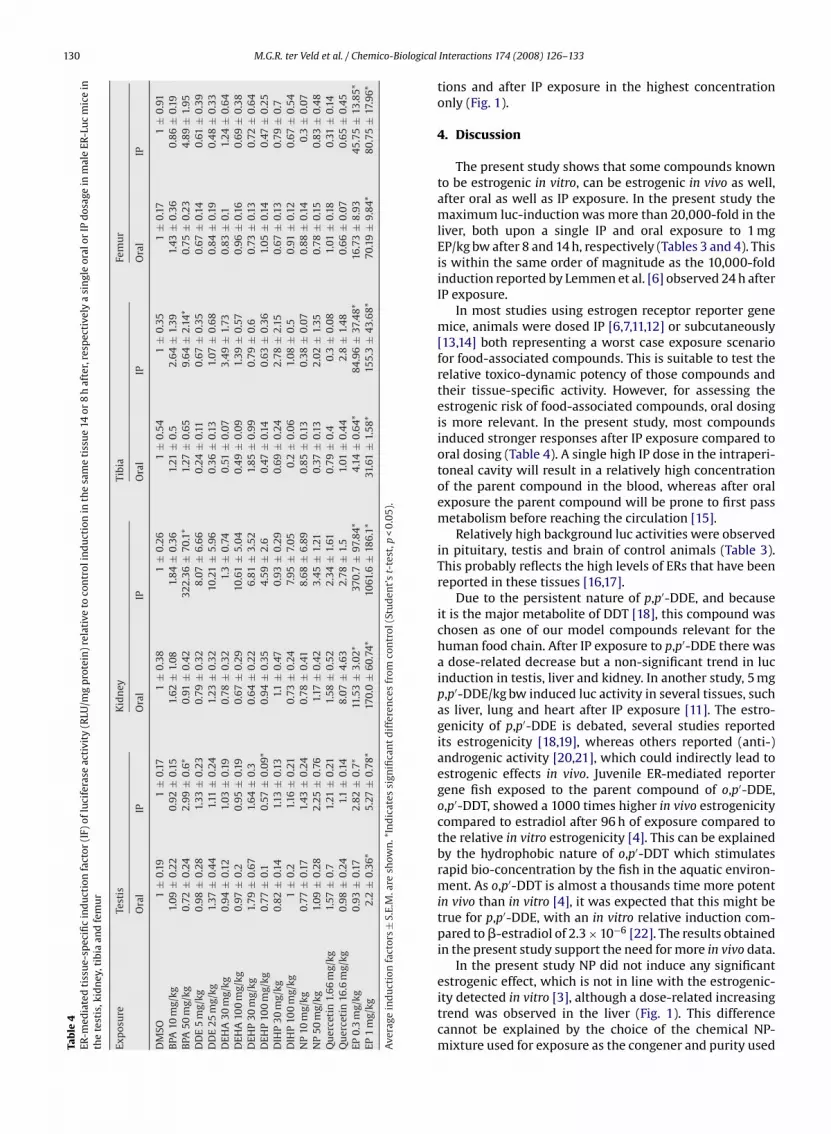
130 M.G.R. ter Veld et al. / Chemico-BiologicalTa
ble
4ER
-med
iate
dti
ssu
e-sp
ecifi
cin
du
ctio
nfa
ctor
(IF)
oflu
cife
rase
acti
vity
(RLU
/mg
pro
tein
)re
lati
veto
con
trol
ind
uct
ion
inth
esa
me
tiss
ue
14or
8h
afte
r,re
spec
tive
lya
sin
gle
oral
orIP
dos
age
inm
ale
ER-L
uc
mic
ein
the
test
is,k
idn
ey,t
ibia
and
fem
ur
Exp
osu
reTe
stis
Kid
ney
Tibi
aFe
mu
r
Ora
lIP
Ora
lIP
Ora
lIP
Ora
lIP
DM
SO1
±0.
191
±0.
171
±0.
381
±0.
261
±0.
541
±0.
351
±0.
171
±0.
91B
PA10
mg/
kg1.
09±
0.22
0.92
±0.
151.
62±
1.08
1.84
±0.
361.
21±
0.5
2.6
4±
1.39
1.43
±0.
360.
86±
0.19
BPA
50m
g/kg
0.72
±0.
242.
99±
0.6*
0.91
±0.
4232
2.36
±70
.1*
1.27
±0.
659.
64
±2.
14*
0.75
±0.
234.
89±
1.95
DD
E5
mg/
kg0.
98±
0.28
1.33
±0.
230.
79±
0.32
8.07
±6.
660.
24±
0.11
0.67
±0.
350.
67±
0.14
0.61
±0.
39D
DE
25m
g/kg
1.37
±0.
44
1.11
±0.
241.
23±
0.32
10.2
1±
5.96
0.36
±0.
131.
07±
0.6
80.
84±
0.19
0.4
8±
0.33
DEH
A30
mg/
kg0.
94±
0.12
1.03
±0.
190.
78±
0.32
1.3
±0.
740.
51±
0.07
3.4
9±
1.73
0.83
±0.
11.
24±
0.6
4D
EHA
100
mg/
kg0.
97±
0.2
0.95
±0.
190.
67±
0.29
10.6
1±
5.04
0.4
9±
0.09
1.39
±0.
570.
96±
0.16
0.69
±0.
38D
EHP
30m
g/kg
1.79
±0.
671.
64
±0.
30.
64
±0.
226.
81±
3.52
1.85
±0.
990.
79±
0.6
0.73
±0.
130.
72±
0.6
4D
EHP
100
mg/
kg0.
77±
0.1
0.57
±0.
09*
0.94
±0.
354.
59±
2.6
0.47
±0.
140.
63±
0.36
1.05
±0.
140.
47±
0.25
DIH
P30
mg/
kg0.
82±
0.14
1.13
±0.
131.
1±
0.47
0.93
±0.
290.
69±
0.24
2.78
±2.
150.
67±
0.13
0.79
±0.
7D
IHP
100
mg/
kg1
±0.
21.
16±
0.21
0.73
±0.
247.
95±
7.05
0.2
±0.
061.
08±
0.5
0.91
±0.
120.
67±
0.54
NP
10m
g/kg
0.77
±0.
171.
43±
0.24
0.78
±0.
418.
68
±6.
890.
85±
0.13
0.38
±0.
070.
88±
0.14
0.3
±0.
07N
P50
mg/
kg1.
09±
0.28
2.25
±0.
761.
17±
0.42
3.45
±1.
210.
37±
0.13
2.02
±1.
350.
78±
0.15
0.83
±0.
48
Qu
erce
tin
1.66
mg/
kg1.
57±
0.7
1.21
±0.
211.
58±
0.52
2.34
±1.
610.
79±
0.4
0.3
±0.
081.
01±
0.18
0.31
±0.
14Q
uer
ceti
n16
.6m
g/kg
0.98
±0.
241.
1±
0.14
8.07
±4.
632.
78±
1.5
1.01
±0.
44
2.8
±1.
48
0.66
±0.
070.
65±
0.45
EP0.
3m
g/kg
0.93
±0.
172.
82±
0.7*
11.5
3±
3.02
*37
0.7
±97
.84*
4.14
±0.
64*
84.9
6±
37.4
8*16
.73
±8.
9345
.75
±13
.85*
EP1
mg/
kg2.
2±
0.36
*5.
27±
0.78
*17
0.0
±60
.74*
1061
.6±
186.
1*31
.61
±1.
58*
155.
3±
43.6
8*70
.19
±9.
84*
80.7
5±
17.9
6*
Ave
rage
ind
uct
ion
fact
ors±
S.E.
M.a
resh
own
.*In
dic
ates
sign
ifica
nt
dif
fere
nce
sfr
omco
ntr
ol(S
tud
ent’
st-
test
,p<
0.05
).
Interactions 174 (2008) 126–133
tions and after IP exposure in the highest concentrationonly (Fig. 1).
4. Discussion
The present study shows that some compounds knownto be estrogenic in vitro, can be estrogenic in vivo as well,after oral as well as IP exposure. In the present study themaximum luc-induction was more than 20,000-fold in theliver, both upon a single IP and oral exposure to 1 mgEP/kg bw after 8 and 14 h, respectively (Tables 3 and 4). Thisis within the same order of magnitude as the 10,000-foldinduction reported by Lemmen et al. [6] observed 24 h afterIP exposure.
In most studies using estrogen receptor reporter genemice, animals were dosed IP [6,7,11,12] or subcutaneously[13,14] both representing a worst case exposure scenariofor food-associated compounds. This is suitable to test therelative toxico-dynamic potency of those compounds andtheir tissue-specific activity. However, for assessing theestrogenic risk of food-associated compounds, oral dosingis more relevant. In the present study, most compoundsinduced stronger responses after IP exposure compared tooral dosing (Table 4). A single high IP dose in the intraperi-toneal cavity will result in a relatively high concentrationof the parent compound in the blood, whereas after oralexposure the parent compound will be prone to first passmetabolism before reaching the circulation [15].
Relatively high background luc activities were observedin pituitary, testis and brain of control animals (Table 3).This probably reflects the high levels of ERs that have beenreported in these tissues [16,17].
Due to the persistent nature of p,p′-DDE, and becauseit is the major metabolite of DDT [18], this compound waschosen as one of our model compounds relevant for thehuman food chain. After IP exposure to p,p′-DDE there wasa dose-related decrease but a non-significant trend in lucinduction in testis, liver and kidney. In another study, 5 mgp,p′-DDE/kg bw induced luc activity in several tissues, suchas liver, lung and heart after IP exposure [11]. The estro-genicity of p,p′-DDE is debated, several studies reportedits estrogenicity [18,19], whereas others reported (anti-)androgenic activity [20,21], which could indirectly lead toestrogenic effects in vivo. Juvenile ER-mediated reportergene fish exposed to the parent compound of o,p′-DDE,o,p′-DDT, showed a 1000 times higher in vivo estrogenicitycompared to estradiol after 96 h of exposure compared tothe relative in vitro estrogenicity [4]. This can be explainedby the hydrophobic nature of o,p′-DDT which stimulatesrapid bio-concentration by the fish in the aquatic environ-ment. As o,p′-DDT is almost a thousands time more potentin vivo than in vitro [4], it was expected that this might betrue for p,p′-DDE, with an in vitro relative induction com-pared to �-estradiol of 2.3 × 10⁻⁶ [22]. The results obtainedin the present study support the need for more in vivo data.
In the present study NP did not induce any significant
estrogenic effect, which is not in line with the estrogenic-ity detected in vitro [3], although a dose-related increasingtrend was observed in the liver (Fig. 1). This differencecannot be explained by the choice of the chemical NP-mixture used for exposure as the congener and purity used
ological
([abdIhi1[dwFNwuhdaarbanawSstabTroiNgltoNaae
picrDpgshtivDi[i
M.G.R. ter Veld et al. / Chemico-Bi
85% of 4-n-nonylphenol from Fluka) is the estrogenic form23]. One plausible explanation would be the rapid clear-nce of NP in vivo. The half-life of NP after oral gavage haseen reported to be maximal 3.5 h [24] which is in accor-ance with the t1/2 values reported by Green et al. [25].
n addition to this, the uptake of NP after oral exposureas been reported to be low, in humans the bioavailabil-
ty only was 20% [26]. Only after repeated doses up tog/kg bw NP has been shown to be estrogenic in rodents
27]. Taking into account only experiments with non-toxicoses of NP thereby excluding nephrotoxicity [28] andeight loss [29,30], some estrogenic effects were reported.
or example ovariectomized prepubertal Long-Evans andoble rats dosed for 3 days either orally or subcutaneouslyith 75–200 mg NP/(kg bw day), had over 100% increasedterine wet weights. In pre-pubertal Long-Evans rats, theighest increase in uterine wet weight was seen after oralosing compared to subcutaneous dosing [31]. The aget vaginal opening was decreased significantly with 5.3nd 6.8 days after exposure to 50 and 100 mg NP/kg bw,espectively starting from PND21 to PND35, effects onody weight were lacking [31]. Increased uterine weightslso were observed in female CD-1 mice exposed subcuta-eously from postnatal day 17 till 20 to NP to 10 mg/kg bwnd higher (200%) increase based on uterine weight/bodyeight ratio [27]. In a three-generational experiment with
prague–Dawley rats the absolute ovary weight in theecond generation was decreased with 13.7 and 20.7% inhe high dose groups (more or less equivalent to 30–100nd 100–350 mg/(kg bw day)), however when corrected forody weight, these changes were both around 12% [29].hese results suggest that NP is estrogenic in vivo, only afterepeated high doses during a longer time, probably becausef its fast degradation. Even in our experiment where lucnduction is a quite sensitive biomarker for ER-activation,P was not able to significantly induce luc activity after sin-le dosing (Fig. 1, Table 4). This is in accordance with theack of induction reported from an in vivo experiment withransgenic ER-mediated reporter gene zebrafish continu-usly exposed to nominal water concentrations up to 1 �MP during 96 h of exposure [4]. When exposure was longernd to very high concentrations, amounting up to 3 weeksnd 2.27 �M NP, vitellogenin, a biomarker for estrogenicxposure, was induced in zebrafish [32].
In in vitro studies DIHP was a weak estrogenic com-ound [3,33]. The reported estrogenicity of DIHP, however,
s so low (EEF of 3 × 10⁻⁷, Table 1) that the effect con-entrations are close to the solubility limits. Our presentesults are in accordance with other in vivo studies whereIHP was estrogenic in extremely high doses. For exam-le ovariectomized Sprague–Dawley rats exposed via oralavage to 200–2000 mg/kg bw for 4 consecutive days hadignificantly increased uterine wet weights. This effect,owever, could not be reproduced in later experiments byhe same authors [33]. As with p,p′-DDE, the estrogenic-ty of DIHP is under debate and the results obtained in in
itro studies on the estrogenicity of DIHP are not consistent.IHP was estrogenic in Chinese Hamster Ovary (CHO) andn human osteoblastic (U2-OS) ER reporter gene cell lines3,34], but not in ER ligand-binding assays performed withmmature Sprague–Dawley uterine cytosol and in human
Interactions 174 (2008) 126–133 131
reporter gene assays in MCF-7 and HeLa cell lines [33]. Dueto the nature of DIHP, being estrogenic in vivo and in vitroonly at very high concentrations [3,35], it is not surprisingthat DIHP only showed a slight non-significant trend in lucinduction in ER-luc mice.
The slightly more estrogenic phthalate, DEHP signifi-cantly induced luc activity in the liver with a factor of 2 inthe highest dosage group (Fig. 1) and reduced the luc activ-ity in the testis at the high dose (Table 4). The estrogenicityof DEHP was shown before, with an induction of the uter-ine wet weight of 54% after a single oral gavage of 2 g/kg bwDEHP in ovariectomized Sprague–Dawley rats. This effect,however, was not reproducible in a second experiment with15–22% heavier rats as had been the case for DIHP [33]. Thesignificant twofold reduction of luc activity compared tocontrol in the testis in the DEHP high dose group might beexplained by the anti-androgenicity [36]. Phthalates havebeen shown to be either estrogenic or anti-androgenic andare rapidly metabolized both in experimental animals aswell as in humans [37]. More effects are therefore to beexpected upon chronic exposure to high concentrations.
BPA induced luc activity in the liver after oral exposure(Fig. 1), and in the testis, kidney and tibia after IP dosage(Table 4). In pregnant females BPA induced an estrogenicresponse in fetuses 2 h after a single maternal IP exposureof 1 mg/kg bw on day 13.5 post-coitum [7]. A lower dose upto 0.2 mg BPA/(kg day) via oral gavage or subcutaneouslyfor at least 6 subsequent days given to male C57black/6Nmice, did not have an effect on the male reproductiveorgans irrespective of the life stage they were exposed in[38]. In adult C57black ER reporter gene mice, luc activ-ity was significantly induced in the uterus 20–24 h after asingle subcutaneous injection with doses between 0.8 and25 mg BPA/kg bw, whereas the uterine wet weight was onlyslightly significantly increased [39]. This was to be expectedas gene induction usually is a much faster response, withinhours, compared to a physiological response such as uterineweight, which takes 3 days [40]. When BPA was adminis-tered in the diet, intraperitoneally or subcutaneous in levelsup to almost 1 g/(kg day) up to 68 days to Wistar, HoltzmanSD rats and CD-1 and C57black mice effects were mainlypresent in rats, not in mice. A significant increase of ster-ile seminiferous tubules, causing a decrease in prostateand preputial weight, was seen after oral and subcuta-neous dosing of 20 mg to 1 g BPA/(kg bw day) for at least28 days [41]. The relative weak estrogenic effects of BPAin vivo compared to in vitro could be due to its efficientmetabolic clearance. Already 1.5 h after oral administra-tion of 100 mg BPA/kg bw maximum blood concentrationof BPA were reached in DA/Han rats. After 24 h BPA wasnot detectable in the blood anymore [42]. In humans, BPAis extensively glucuronidated and the BPA-glucuronide isexcreted in urine [43]. From our results (Table 4) it is clearthat an oral dose of BPA usually results in much lower lucinduction than an IP dose (Table 4). This is in accordancewith results obtained in Fischer 344 rats, in which IP dosing
resulted in a two times higher maximal blood concentra-tion after dosing for 7 consecutive days [44]. In the samestudy, the resulting blood concentration of BPA given IP wasup to 164 times greater compared to blood BPA levels inorally dosed animals. In rats and monkeys, the toxicokinet-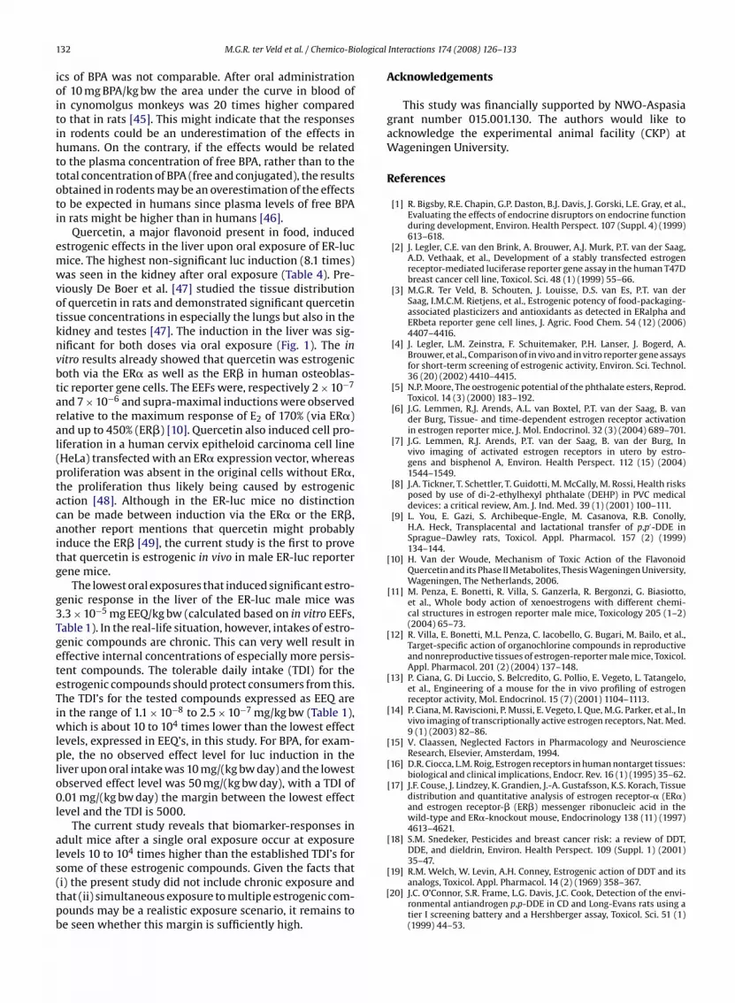
ological
132 M.G.R. ter Veld et al. / Chemico-Biics of BPA was not comparable. After oral administrationof 10 mg BPA/kg bw the area under the curve in blood ofin cynomolgus monkeys was 20 times higher comparedto that in rats [45]. This might indicate that the responsesin rodents could be an underestimation of the effects inhumans. On the contrary, if the effects would be relatedto the plasma concentration of free BPA, rather than to thetotal concentration of BPA (free and conjugated), the resultsobtained in rodents may be an overestimation of the effectsto be expected in humans since plasma levels of free BPAin rats might be higher than in humans [46].
Quercetin, a major flavonoid present in food, inducedestrogenic effects in the liver upon oral exposure of ER-lucmice. The highest non-significant luc induction (8.1 times)was seen in the kidney after oral exposure (Table 4). Pre-viously De Boer et al. [47] studied the tissue distributionof quercetin in rats and demonstrated significant quercetintissue concentrations in especially the lungs but also in thekidney and testes [47]. The induction in the liver was sig-nificant for both doses via oral exposure (Fig. 1). The invitro results already showed that quercetin was estrogenicboth via the ER� as well as the ER� in human osteoblas-tic reporter gene cells. The EEFs were, respectively 2 × 10⁻⁷
and 7 × 10⁻⁶ and supra-maximal inductions were observedrelative to the maximum response of E2 of 170% (via ER�)and up to 450% (ER�) [10]. Quercetin also induced cell pro-liferation in a human cervix epitheloid carcinoma cell line(HeLa) transfected with an ER� expression vector, whereasproliferation was absent in the original cells without ER�,the proliferation thus likely being caused by estrogenicaction [48]. Although in the ER-luc mice no distinctioncan be made between induction via the ER� or the ER�,another report mentions that quercetin might probablyinduce the ER� [49], the current study is the first to provethat quercetin is estrogenic in vivo in male ER-luc reportergene mice.
The lowest oral exposures that induced significant estro-genic response in the liver of the ER-luc male mice was3.3 × 10−5 mg EEQ/kg bw (calculated based on in vitro EEFs,Table 1). In the real-life situation, however, intakes of estro-genic compounds are chronic. This can very well result ineffective internal concentrations of especially more persis-tent compounds. The tolerable daily intake (TDI) for theestrogenic compounds should protect consumers from this.The TDI’s for the tested compounds expressed as EEQ arein the range of 1.1 × 10⁻⁸ to 2.5 × 10⁻⁷ mg/kg bw (Table 1),which is about 10 to 104 times lower than the lowest effectlevels, expressed in EEQ’s, in this study. For BPA, for exam-ple, the no observed effect level for luc induction in theliver upon oral intake was 10 mg/(kg bw day) and the lowestobserved effect level was 50 mg/(kg bw day), with a TDI of0.01 mg/(kg bw day) the margin between the lowest effectlevel and the TDI is 5000.
The current study reveals that biomarker-responses inadult mice after a single oral exposure occur at exposurelevels 10 to 104 times higher than the established TDI’s for
some of these estrogenic compounds. Given the facts that(i) the present study did not include chronic exposure andthat (ii) simultaneous exposure to multiple estrogenic com-pounds may be a realistic exposure scenario, it remains tobe seen whether this margin is sufficiently high.[
Interactions 174 (2008) 126–133
Acknowledgements
This study was financially supported by NWO-Aspasiagrant number 015.001.130. The authors would like toacknowledge the experimental animal facility (CKP) atWageningen University.
References
[1] R. Bigsby, R.E. Chapin, G.P. Daston, B.J. Davis, J. Gorski, L.E. Gray, et al.,Evaluating the effects of endocrine disruptors on endocrine functionduring development, Environ. Health Perspect. 107 (Suppl. 4) (1999)613–618.
[2] J. Legler, C.E. van den Brink, A. Brouwer, A.J. Murk, P.T. van der Saag,A.D. Vethaak, et al., Development of a stably transfected estrogenreceptor-mediated luciferase reporter gene assay in the human T47Dbreast cancer cell line, Toxicol. Sci. 48 (1) (1999) 55–66.
[3] M.G.R. Ter Veld, B. Schouten, J. Louisse, D.S. van Es, P.T. van derSaag, I.M.C.M. Rietjens, et al., Estrogenic potency of food-packaging-associated plasticizers and antioxidants as detected in ERalpha andERbeta reporter gene cell lines, J. Agric. Food Chem. 54 (12) (2006)4407–4416.
[4] J. Legler, L.M. Zeinstra, F. Schuitemaker, P.H. Lanser, J. Bogerd, A.Brouwer, et al., Comparison of in vivo and in vitro reporter gene assaysfor short-term screening of estrogenic activity, Environ. Sci. Technol.36 (20) (2002) 4410–4415.
[5] N.P. Moore, The oestrogenic potential of the phthalate esters, Reprod.Toxicol. 14 (3) (2000) 183–192.
[6] J.G. Lemmen, R.J. Arends, A.L. van Boxtel, P.T. van der Saag, B. vander Burg, Tissue- and time-dependent estrogen receptor activationin estrogen reporter mice, J. Mol. Endocrinol. 32 (3) (2004) 689–701.
[7] J.G. Lemmen, R.J. Arends, P.T. van der Saag, B. van der Burg, Invivo imaging of activated estrogen receptors in utero by estro-gens and bisphenol A, Environ. Health Perspect. 112 (15) (2004)1544–1549.
[8] J.A. Tickner, T. Schettler, T. Guidotti, M. McCally, M. Rossi, Health risksposed by use of di-2-ethylhexyl phthalate (DEHP) in PVC medicaldevices: a critical review, Am. J. Ind. Med. 39 (1) (2001) 100–111.
[9] L. You, E. Gazi, S. Archibeque-Engle, M. Casanova, R.B. Conolly,H.A. Heck, Transplacental and lactational transfer of p,p′-DDE inSprague–Dawley rats, Toxicol. Appl. Pharmacol. 157 (2) (1999)134–144.
[10] H. Van der Woude, Mechanism of Toxic Action of the FlavonoidQuercetin and its Phase II Metabolites, Thesis Wageningen University,Wageningen, The Netherlands, 2006.
[11] M. Penza, E. Bonetti, R. Villa, S. Ganzerla, R. Bergonzi, G. Biasiotto,et al., Whole body action of xenoestrogens with different chemi-cal structures in estrogen reporter male mice, Toxicology 205 (1–2)(2004) 65–73.
[12] R. Villa, E. Bonetti, M.L. Penza, C. Iacobello, G. Bugari, M. Bailo, et al.,Target-specific action of organochlorine compounds in reproductiveand nonreproductive tissues of estrogen-reporter male mice, Toxicol.Appl. Pharmacol. 201 (2) (2004) 137–148.
[13] P. Ciana, G. Di Luccio, S. Belcredito, G. Pollio, E. Vegeto, L. Tatangelo,et al., Engineering of a mouse for the in vivo profiling of estrogenreceptor activity, Mol. Endocrinol. 15 (7) (2001) 1104–1113.
[14] P. Ciana, M. Raviscioni, P. Mussi, E. Vegeto, I. Que, M.G. Parker, et al., Invivo imaging of transcriptionally active estrogen receptors, Nat. Med.9 (1) (2003) 82–86.
[15] V. Claassen, Neglected Factors in Pharmacology and NeuroscienceResearch, Elsevier, Amsterdam, 1994.
[16] D.R. Ciocca, L.M. Roig, Estrogen receptors in human nontarget tissues:biological and clinical implications, Endocr. Rev. 16 (1) (1995) 35–62.
[17] J.F. Couse, J. Lindzey, K. Grandien, J.-A. Gustafsson, K.S. Korach, Tissuedistribution and quantitative analysis of estrogen receptor-� (ER�)and estrogen receptor-� (ER�) messenger ribonucleic acid in thewild-type and ER�-knockout mouse, Endocrinology 138 (11) (1997)4613–4621.
[18] S.M. Snedeker, Pesticides and breast cancer risk: a review of DDT,DDE, and dieldrin, Environ. Health Perspect. 109 (Suppl. 1) (2001)35–47.
[19] R.M. Welch, W. Levin, A.H. Conney, Estrogenic action of DDT and itsanalogs, Toxicol. Appl. Pharmacol. 14 (2) (1969) 358–367.
20] J.C. O’Connor, S.R. Frame, L.G. Davis, J.C. Cook, Detection of the envi-ronmental antiandrogen p,p-DDE in CD and Long-Evans rats using atier I screening battery and a Hershberger assay, Toxicol. Sci. 51 (1)(1999) 44–53.

ological
[
[
[
[
[
[
[
[
[
[
[
[
[
[
[
[
[
[
[
[
[
[
[
[
[
[
[
[
[
[
[
[
M.G.R. ter Veld et al. / Chemico-Bi
21] L.E. Gray, J. Ostby, J. Furr, C.J. Wolf, C. Lambright, L. Parks, et al.,Effects of environmental anti-androgens on reproductive develop-ment in experimental animals, Hum. Reprod. Update 7 (3) (2001)248–264.
22] J.D. Gordon, A.C. Chu, M.D. Chu, M.S. Denison, G.C. Clark, Detec-tion of estrogen receptor endocrine disruptor potency of commonlyused organochlorine pesticides using the LUMI-CELLTM bioassay,Organohalogen Compd. 66 (2004) 169–174.
23] T.F.H. Bovee, R.J.M. Helsdingen, I.M.C.M. Rietjens, J. Keijer, R.L.A.P.Hoogenboom, Rapid yeast estrogen bioassays stably expressinghuman estrogen receptors alpha and beta, and green fluorescent pro-tein: a comparison of different compounds with both receptor types,J. Steroid. Biochem. Mol. Biol. 91 (3) (2004) 99–109.
24] D.R. Doerge, N.C. Twaddle, M.I. Churchwell, H.C. Chang, R.R. Newbold,K.B. Delclos, Mass spectrometric determination of p-nonylphenolmetabolism and disposition following oral administration toSprague–Dawley rats, Reprod. Toxicol. 16 (1) (2002) 45–56.
25] T. Green, C. Swain, J.P. Van Miller, R.L. Joiner, Absorption, bioavailabil-ity, and metabolism of para-nonylphenol in the rat, Regul. Toxicol.Pharmacol. 38 (1) (2003) 43–51.
26] S. Muller, P. Schmid, C. Schlatter, Pharmacokinetic behavior of 4-nonylphenol in humans, Environ. Toxicol. Pharmacol. 5 (4) (1998)257–265.
27] M.D. Shelby, R.R. Newbold, D.B. Tully, K. Chae, V.L. Davis, Assessingenvironmental chemicals for estrogenicity using a combination of invitro and in vivo assays, Environ. Health Perspect. 104 (12) (1996)1296–1300.
28] J. Odum, I.T. Pyrah, J.R. Foster, J.P. Van Miller, R.L. Joiner, J. Ashby, Com-parative activities of p-nonylphenol and diethylstilbestrol in noble ratmammary gland and uterotrophic assays, Regul. Toxicol. Pharmacol.29 (2 Pt 1) (1999) 184–195.
29] R.E. Chapin, J. Delaney, Y. Wang, L. Lanning, B. Davis, B. Collins, et al.,The effects of 4-nonylphenol in rats: a multigeneration reproductionstudy, Toxicol. Sci. 52 (1) (1999) 80–91.
30] A. Hossaini, M. Dalgaard, A.M. Vinggaard, H. Frandsen, J.J. Larsen, Inutero reproductive study in rats exposed to nonylphenol, Reprod.Toxicol. 15 (5) (2001) 537–543.
31] S.C. Laws, S.A. Carey, J.M. Ferrell, G.J. Bodman, R.L. Cooper, Estrogenicactivity of octylphenol, nonylphenol, bisphenol A and methoxychlorin rats, Toxicol. Sci. 54 (1) (2000) 154–167.
32] K. Van den Belt, P. Berckmans, C. Vangenechten, R. Verheyen, H. Wit-ters, Comparative study on the in vitro/in vivo estrogenic potenciesof 17beta-estradiol, estrone, 17alpha-ethynylestradiol and nonylphe-nol, Aquat. Toxicol. 66 (2) (2004) 183–195.
33] T.R. Zacharewski, M.D. Meek, J.H. Clemons, Z.F. Wu, M.R. Fielden, J.B.Matthews, Examination of the in vitro and in vivo estrogenic activi-ties of eight commercial phthalate esters, Toxicol. Sci. 46 (2) (1998)282–293.
34] S. Takeuchi, M. Iida, S. Kobayashi, K. Jin, T. Matsuda, H. Kojima, Dif-ferential effects of phthalate esters on transcriptional activities viahuman estrogen receptors [alpha] and [beta], and androgen receptor,Toxicology 210 (2–3) (2005) 223–233.
35] R.H. McKee, K.L. Pavkov, G.W. Trimmer, L.H. Keller, D.G. Stump, Anassessment of the potential developmental and reproductive toxic-ity of di-isoheptyl phthalate in rodents, Reprod. Toxicol. 21 (2006)241–252.
36] R.W. Moore, T.A. Rudy, T.M. Lin, K. Ko, R.E. Peterson, Abnormalities
of sexual development in male rats with in utero and lactationalexposure to the antiandrogenic plasticizer di(2-ethylhexyl) phtha-late, Environ. Health Perspect. 109 (3) (2001) 229–237.37] IARC. World Health Organization International Agency For Researchon Cancer, Some Industrial Chemicals, vol. 77, IARC Monographs onthe Evaluation of Carcinogenic Risks to Humans, 2000.
[
Interactions 174 (2008) 126–133 133
38] T. Nagao, Y. Saito, K. Usumi, S. Yoshimura, H. Ono, Low-dose bisphenolA does not affect reproductive organs in estrogen-sensitive C57BL/6Nmice exposed at the sexually mature, juvenile, or embryonic stage,Reprod. Toxicol. 16 (2) (2002) 123–130.
39] S.C. Nagel, J.L. Hagelbarger, D.P. McDonnell, Development of an ERaction indicator mouse for the study of estrogens, selective ER mod-ulators (SERMs), and xenobiotics, Endocrinology 142 (11) (2001)4721–4728.
40] OECD. Detailed background review of the uterotrophic assay. OECDseries on testing and assessment, number 38, 2003.
[41] O. Takahashi, S. Oishi, Testicular toxicity of dietarily or parenterallyadministered bisphenol A in rats and mice, Food Chem. Toxicol. 41(7) (2003) 1035–1044.
42] A. Upmeier, G.H. Degen, P. Diel, H. Michna, H.M. Bolt, Toxicokinet-ics of bisphenol A in female DA/Han rats after a single i.v. and oraladministration, Arch. Toxicol. 74 (8) (2000) 431–436.
43] W. Volkel, T. Colnot, G.A. Csanady, J.G. Filser, W. Dekant, Metabolismand kinetics of bisphenol a in humans at low doses following oraladministration, Chem. Res. Toxicol. 15 (10) (2002) 1281–1287.
44] L.H. Pottenger, J.Y. Domoradzki, D.A. Markham, S.C. Hansen, S.Z.Cagen, J.M. Waechter Jr., The relative bioavailability and metabolismof bisphenol A in rats is dependent upon the route of administration,Toxicol. Sci. 54 (1) (2000) 3–18.
45] T. Tominaga, T. Negishi, H. Hirooka, A. Miyachi, A. Inoue, I. Hayasaka, Y.Yoshikawa, Toxicokinetics of bisphenol A in rats, monkeys and chim-panzees by the LC–MS/MS method, Toxicology 226 (2006) 208–217.
46] EC EFSA, Opinion of the scientific panel on food additives, flavour-ings, processing aids and materials in contact with food on a requestfrom the commission related to 2,2-bis(4-hydroxyphenyl)propane(Bisphenol A), EFSA J. 428 (2006) 1–75.
47] V.C.J. De Boer, A.A. Dihal, H. Van der Woude, I.C.W. Arts, S. Wolffram,G.M. Alink, I.M.C.M. Rietjens, J. Keijer, P.C.H. Hollman, Tissue distri-bution of quercetin in rats and pigs, J. Nutr. 135 (7) (2005) 1617–1618.
48] F. Virgili, F. Acconcia, R. Ambra, A. Rinna, P. Totta, M. Marino, Nutri-tional flavonoids modulate estrogen receptor alpha signaling, IUBMBLife 56 (3) (2004) 145–151.
49] G.G. Kuiper, J.G. Lemmen, B. Carlsson, J.C. Corton, S.H. Safe, P.T. vander Saag, et al., Interaction of estrogenic chemicals and phytoestro-gens with estrogen receptor beta, Endocrinology 139 (10) (1998)4252–4263.
50] J.G. Lemmen, C.E. Van Den Brink, J. Legler, P.T. Van Der Saag, B. VanDer Burg, Detection of oestrogenic activity of steroids present duringmammalian gestation using oestrogen receptor alpha- and oestrogenreceptor beta- specific in vitro assays, J. Endocrinol. 174 (3) (2002)435–446.
[51] EC, SCF. Opinion of the scientific committee on food on bisphenolA. SCF/CS/PM/3936 (2002) Final. Brussels: European commission,Directorate C—Scientific Opinions.
52] FAO. Food Agriculture Organization of the United Nations, PesticideResidues in Food, Food and Agriculture Organization of the UnitedNations, Rome, Italy, 1996.
53] EC, CSTEE. Opinion on the results of a second risk assess-ment of bis(2-ethylhexyl) phthalate (DEHP) human health partC7/GF/csteeop/dehp/080104 D. Brussels, European commission,2004.
54] EC EFSA, Opinion of the Scientific panel on food additives, flavourings,processing aids and materials in contact with food (AFC) on a request
from the commission related to bis(2-ethylhexyl)phthalate (DEHP)for use in food contact materials, EFSA J. 243 (2005) 1–20.55] EC, SCF. Opinion of the Scientific Committee on Food on a survey ondietary intake of the food contact material di-2-(ethylhexyl) adipate(DEHA), Brussels, European commission, Directorate C—ScientificOpinions, 2000.
Recommended


