
-E JOURNAL zyxwvutsrqponmlkjihgfedcbaZYXWVUTSRQPONMLKJIHGFEDCBAOF BIOLOGICAL CHEMISTRY zyxwvutsrqponmlkjihgfedcbaZYXWVUTSRQPONMLKJIHGFEDCBA0 1986 zyxwvutsrqponmlkjihgfedcbaZYXWVUTSRQPONMLKJIHGFEDCBAby The American Society of Biological Chemists, Inc.
Vol. 261, No. 15, Issue of May 25, pp. 6904-6911 1986 zyxwvutsrqponmlkjihgfedcbaZYXWVUTSRQPONMLKJIHGFEDCBAPrinted in c. zyxwvutsrqponmlkjihgfedcbaZYXWVUTSRQPONMLKJIHGFEDCBAS. A.
G-protein-mediated Interconversions of Cell-surface cAMP Recentors and Their Involvement in Excitation and Desensitization of Guanylate Cyclase in Dictyostelium discoideum”
~
(Received for publication, April 29,1985)
Peter J. M. van Haastertl, Rene J. W. de Wit$, Pim M. W. Janssenss, Fanja Kesbeket, and Jacob DeGoedell zyxwvutsrqponmlkjihgfedcbaZYXWVUTSRQPONMLKJIHGFEDCBAFrom the $Cell Biology and Morphgenesis Unit, Zoological Laboratory, and the qDepartment of Physiology and Physiological Physics, University zyxwvutsrqponmlkjihgfedcbaZYXWVUTSRQPONMLKJIHGFEDCBAof Leiden, 2300 RA Leiden and the §Laboratory of Biochemistry, University of Amsterdam, 1000 BH Amsterdam, The Nether1and.s
In Dictyostelium discoideum cells, extracellular cAMP induces the rapid (within zyxwvutsrqponmlkjihgfedcbaZYXWVUTSRQPONMLKJIHGFEDCBA2 s) activation of guan- ylate cyclase, which is followed by complete desensiti- zation after about 10 s. cAMP binding to these cells is heterogeneous, showing a subclass of fast dissociating sites coupled to adenylate cyclase (A-sites) and a sub- class of slowly dissociating sites coupled to guanylate cyclase (B-sites). The kinetics of the B-sites were fur- ther investigated on a seconds time scale.
Statistical analysis of the association of 13H]cAMP to the B-sites and dissociation of the complex revealed that the receptor can exist in three states which inter- convert according to the following scheme.
BP- BS- Bss 3 s
cAMP binds to the BF-state (off-rate zyxwvutsrqponmlkjihgfedcbaZYXWVUTSRQPONMLKJIHGFEDCBA2.5 s) which rap- idly (tH = 3 s) converts to the Bs-state (off-rate 15 s) and subsequently (without a detectable delay) into the Bss-state (off-rate 150 s). In membranes, both the BS- and Bss-states are converted to the BF-state by GTP and GDP, suggesting the involvement of a G-protein.
Desensitized cells show a 80% reduction of the for- mation of the Bss-state, but no reduction of the BF- or Bs-state.
These data are combined into a model in which the transitions of the B-sites are mediated by a G-protein; activation of the G-protein and guanylate cyclase is associated with the transition of the BS- to the Bss- state of the receptor, whereas desensitization is asso- ciated with the inhibition of this transition.
~~~~~
The eukaryotic microorganism Dictyostelium discoideum is a suitable organism to study signal transduction. In this organism, the hormone-like substance is cAMP which is detected by cell-surface receptors. Extracellular cAMP in- duces the rapid activation of guanylate cyclase (1) and the slower activation of adenylate cyclase (2). Intracellular cGMP reaches a peak after 10 s, declines to prestimulated levels
* This work was supported by the Foundation for Fundamental Biological Research (Biologisch Onderzoek Nederland) and the C . and C. Huygens Fund, which are subsidized by the Netherlands Organization for the Advancement of Pure Scientific Research (Zuiver Wetenschappelijk Onderzoek). The costs of publication of this article were defrayed in part by the payment of page charges. This article must therefore be hereby marked “advertisement” in accordance with 18 U.S.C. Section 1734 solely to indicate this fact.
reached at about 30 s (3), and is supposed to be involved in the CAMP-induced chemotactic reaction (4). Intracellular cAMP reaches maximal levels after 60-120 s and is secreted, thus acting as an autocatalytic feedback loop (5).
Prolonged stimulation of D. discoideum cells with constant cAMP concentrations induces desensitization by at least two mechanisms: (i) down-regulation of cAMP binding activity after a long incubation (5-30 min) with high cAMP concen- trations (1-100 pM) (after removal of CAMP, cells resensitize with a half-life of about 60 min (6, 7)); (ii) a rapid desensiti- zation of the CAMP-mediated activation of the cyclases by nanomolar cAMP concentrations (8, 9) without a loss of CAMP-binding sites (after removal of CAMP, cells resensitize with a half-life of 1-2 min for the guanylate cyclase (9) and 3-4 min for the adenylate cyclase (10)). Desensitization of the CAMP-mediated cGMP accumulation is completed within about 10 s (91, whereas desensitization of the cAMP accu- mulation is completed only after about 5 min (11). The mechanism of excitation and desensitization of the cGMP response is the subject of the present report.
A second chemotactic compound in D. discoideum is folate (12). Binding of cAMP or folate to sensitive cells is hetero- geneous in respect to kinetic properties (13-15). Since binding of both compounds shows essentially identical complexity, we will unify the previously used notations for the different forms of the receptor. The major portion (-96%) of binding sites are fast dissociating (& = 1-2 s) and are designated as A- sites. The A-sites may exist in two states, AH and AL, with high and low affinity, respectively. cAMP induces a time- and concentration-dependent transition of AH to A‘, a transition which can be induced in membranes by guanine nucleotides, suggesting the involvement of a guanine nucleotide regulatory protein (G-protein) in this transition (16, 17). The A-sites appear to be responsible for the activation of adenylate cyclase in D. discoideum (18-20).
A second class of binding sites releases bound cAMP more slowly. This class of binding sites is designated B-sites and may exist in at least two states, BS and Bss, which have a half-life of dissociation equal to 15 and 150 s, respectively (13-15). The B-sites are also sensitive to guanine nucleotides (16, 21) and are most likely involved in the activation of guanylate cyclase (18,201.
We have analyzed the kinetics of the B-sites in more detail and present evidence for a CAMP-induced interconversion between three states of the receptor that are related to the interaction of receptor with a G-protein. Cells subjected to homologous desensitization of guanylate cyclase (22) show a strongly reduced binding to only one of these states, whereas
6904
cAMP Receptors, G-protein, zyxwvutsrqponmlkjihgfedcbaZYXWVUTSRQPONMLKJIHGFEDCBAand Guanylate Cyclase in D. discoideum zyxwvutsrqponmlkjihgfedcbaZYXWVUTSRQPONMLKJIHGFEDCBA6905 zyxwvutsrqponmlkjihgfedcbaZYXWVUTSRQPONMLKJIHGFEDCBAbinding to the others is not altered. A model on the interaction between B-sites and G-protein is presented.
EXPERIMENTAL PROCEDURES
Material~-[2,8-~H]cAMP (1.5 TBq/mmol) was obtainedfrom The Radiochemical Centre, Amersham, United Kingdom. CAMP, GTP, GDP, GMP, and dithiothreitol were from Sigma, GppNHp: ATP, ADP, and AppNHp were purchased from Boehringer Mannheim. GTPrS was a generous gift of Dr. F. Eckstein (Max-Planck-Institut fur experimentelle Medizin, Gottingen, West Germany). Silicon oils (AR 20 and AR 200) were obtained from Wacker Chemie.
Culture Conditions-D. discoideum NC-4(H) cells were grown with Escherichia coli 281 on a solid medium containing 3.3 g of peptone, 3.3 g of glucose, 4.5 g of KHzP04, 1.5 g of NazHP04.2Hz0, and 15 g of agar/liter. Cells were harvested in the late logarithmic phase with 10 mM sodium/potassium phosphate buffer, pH 6.5 (Pb buffer), and freed from bacteria by repeated centrifugations at zyxwvutsrqponmlkjihgfedcbaZYXWVUTSRQPONMLKJIHGFEDCBA100 zyxwvutsrqponmlkjihgfedcbaZYXWVUTSRQPONMLKJIHGFEDCBAX g for 4 min.
Preparation zyxwvutsrqponmlkjihgfedcbaZYXWVUTSRQPONMLKJIHGFEDCBAof Membranes (23)-Cells were starved for 4.5 h by shaking in Pb buffer, centrifuged, washed twice, and resuspended in this buffer at 2 zyxwvutsrqponmlkjihgfedcbaZYXWVUTSRQPONMLKJIHGFEDCBAX 108cells/ml. Air was bubbled through the suspension for about 10 min at 20 “C. The cells were then transferred to 0 “C and broken by passage through a Nucleopore filter (diameter, 25 mm; pore size, 3 pm). The filter was washed with an equal volume of Pb buffer, and the combined homogenates were centrifuged at 10,000 X g for 5 min at 0 “C. The supernatant was removed, and the pellet was resuspended in Pb buffer at a concentration equivalent to 1.25 X 10’ cells/ml. Membranes were kept on ice during the entire experiment, which did not last longer than 1 h.
CAMP Binding Assays (13)-All binding experiments have been performed at 20 “C except when indicated. The incubation mixture was in a total volume of 100 pl of Pb buffer with 5 mM dithiothreitol (an inhibitor of phosphodiesterase in D. discoideum (24)), different concentrations of [3H]cAMP, and lo7 cells or membranes derived from lo7 cells. Binding of cAMP to all sites was measured by centrif- ugation of 95 pl of the incubation mixture through silicon oil (AR 200AR 20 = 1:2). Binding to the slowly dissociating sites was meas- ured by a squirt of 1 ml of Pb buffer with 0.1 mM cAMP and 5 mM dithiothreitol into the incubation mixture, followed by centrifugation of 1 ml of the mixture through silicon oil. Cells were centrifuged for 15 s, and membranes for 30 s. The efficiency of the centrifugation step was investigated by measuring the protein content of the pellet. The amount of protein was recovered quantitatively in the pellet below the silicon oil when concentrated cells, 11-fold diluted cells, or concentrated membranes were used. However, when 10-fold diluted membranes (equivalent to 10’ cells/ml) are centrifuged, only 25% of the protein is recovered in the pellet after a 30-s centrifugation period. Therefore, binding experiments with membranes have been per- formed only with concentrated membrane preparations.
Identification of Cell-associated Radioactivity-Tubes contained 10 pl of 20% (w/v) sucrose in 3.5% (v/v) perchloric acid and 200 pl of silicon oil. Cells were incubated with 10 nM [3H]cAMP as described above and were centrifuged through silicon oil at the moment of binding equilibrium, at 10 s, or at 2 min after the onset of dissociation. The tip of the tubes containing the cell-associated radioactivity was cut, and the content of four tips was added to 200 pl of Pb buffer. The lysate was neutralized with 20 p1 of KHC03 (50% saturated at 20 “C) and centrifuged a t 10,000 X g for 2 min, and the supernatant was analyzed by high pressure liquid chromatography (25). The column was LiChrosorp RP-18; the mobile phase was 25 mM tribu- tylammonium formate, 15% methanol, pH 3.0; the flow rate was 1 ml/min. The retention times of cAMP and its derivatives are: dead time, 2.5 min; adenosine, 3.24 min; 5’-AMP, 4.58 min; cIMP, 5.18 min; CAMP, 8.31 min. At least 95% of the radioactivity bound to cells at equilibrium, at 10 s, or at 2 min after dissociation co-eluted with authentic CAMP.
Data Analysis of the Release of Bound PHJcAMP-Cells or mem- branes were incubated with [3H]cAMP until equilibrium. The release
The abbreviations used are: GppNHp, guanyl-5’-yl imidodiphos- phate; GTP-@, guanosine 5’-0-(3-thiotriphosphate); AppNHp, ad- enyl-5’-yl imidodiphosphate; AIC, Akaike information criterion.
It should be noted that cAMP does accelerate the release of bound [3H]cAMP when cells (or membranes prepared from them) were used which were starved in suspension for an unphysiologically long period (7-9 h). Cells starved on a solid support for the same period do not show this effect.
of bound [3H]cAMP was initiated by the addition of excess cAMP or by dilution of the incubation mixture. The residual [3H]cAMP bound was measured at time ti, with n; as the number of replicas and N as the number of time points.
The release of bound [3H]cAMP as a function of time was fitted to sums-of-exponentials models.
a
y(t) = Aje-”jt a = 1, 2, 3, 4 (1)
To this end, the mean (2:) of the replicas at time t i was used as the single value, and the sample variance zyxwvutsrqponmlkjihgfedcbaZYXWVUTSRQPONMLKJIHGFEDCBA(3) of the replicas was divided by their number ni as weights. To find the optimal parameter set, the minimum of the weighted residual sum of squares (WRSS) at the N time points (26) was searched.
j=l
WRSS = (Z(ti) - zyxwvutsrqponmlkjihgfedcbaZYXWVUTSRQPONMLKJIHGFEDCBAy(ti))’ N
(2)
The minimum was found by using a nonlinear Marquardt-type min- imalization procedure.
To estimate the model order (26, 27) (Le. the number of exponen- tials in Equation l ) , we determined the minimum value of the Akaike information criterion (AIC),
i=1 ai
AIC = WRSS + 2p (3)
in which p is the number of free parameters in the model. The order of the model was also estimated by using the F test (26, 27),
where the subscripts 1 and 2 denote the smaller and larger model, respectively. The degrees of freedom of F are (pz - p1, N - pz) .
RESULTS
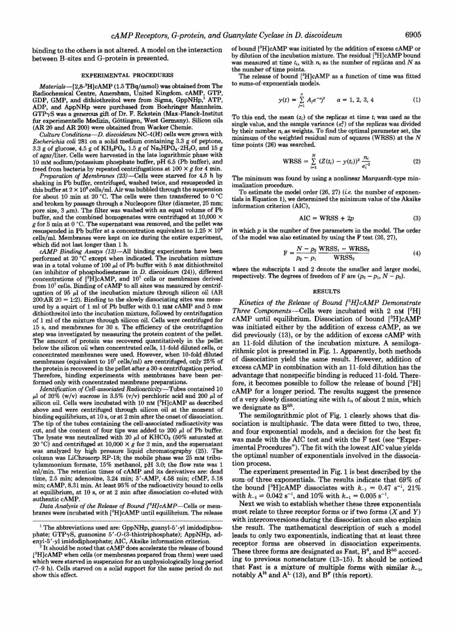
Kinetics of the Release of Bound PHICAMP Demonstrate Three Components-Cells were incubated with 2 nM [3H] cAMP until equilibrium. Dissociation of bound [3H]cAMP was initiated either by the addition of excess CAMP, as we did previously (13), or by the addition of excess cAMP with an 11-fold dilution of the incubation mixture. A semiloga- rithmic plot is presented in Fig. 1. Apparently, both methods of dissociation yield the same result. However, addition of excess cAMP in combination with an 11-fold dilution has the advantage that nonspecific binding is reduced 11-fold. There- fore, it becomes possible to follow the release of bound [3H] cAMP for a longer period. The results suggest the presence of a very slowly dissociating site with tlh of about 2 min, which we designate as BSS.
The semilogarithmic plot of Fig. 1 clearly shows that dis- sociation is multiphasic. The data were fitted to two, three, and four exponential models, and a decision for the best fit was made with the AIC test and with the F test (see “Exper- imental Procedures”). The fit with the lowest AIC value yields the optimal number of exponentials involved in the dissocia- tion process.
The experiment presented in Fig. 1 is best described by the sum.of three exponentials. The results indicate that 69% of the bound [3H]cAMP dissociates with k l = 0.47 s-l, 21% with = 0.042 s-l, and 10% with k 1 = 0.005 s-l.
Next we wish to establish whether these three exponentials must relate to three receptor forms or if two forms (X and Y) with interconversions during the dissociation can also explain the result. The mathematical description of such a model leads to only two exponentials, indicating that at least three receptor forms are observed in dissociation experiments. These three forms are designated as Fast, BS, and Bss accord- ing to previous nomenclature (13-15). It should be noticed that Fast is a mixture of multiple forms with similar K1,
notably A”’ and AL (13), and BF (this report).

6906 zyxwvutsrqponmlkjihgfedcbaZYXWVUTSRQPONMLKJIHGFEDCBACAMP Receptors, G-protein, and Guanylate Cyclase in D. discoideum
I zyxwvutsrqponmlkjihgfedcbaZYXWVUTSRQPONMLKJIHGFEDCBA0 zyxwvutsrqponmlkjihgfedcbaZYXWVUTSRQPONMLKJIHGFEDCBA1 zyxwvutsrqponmlkjihgfedcbaZYXWVUTSRQPONMLKJIHGFEDCBA2 3 4 6
I
minutes
FIG. 1. Semilogarithmic plot of the release of bound [3H] cAMP from cells. zyxwvutsrqponmlkjihgfedcbaZYXWVUTSRQPONMLKJIHGFEDCBAD. discoideum cells were equilibrated with 2 nM [3H]cAMP at 20 "C for 45 s in a total volume of 100 pl. At t zyxwvutsrqponmlkjihgfedcbaZYXWVUTSRQPONMLKJIHGFEDCBA= 0 s, either 2 zyxwvutsrqponmlkjihgfedcbaZYXWVUTSRQPONMLKJIHGFEDCBApl of 10 mM cAMP was added and 95 pl of the incubation mixture was centrifuged through silicon oil at the times indicated zyxwvutsrqponmlkjihgfedcbaZYXWVUTSRQPONMLKJIHGFEDCBA(0) or 1 ml of buffer with 0.1 mM CAMP was added and 1 ml of the mixture was centrifuged at the times indicated (0). b( t ) is the specific binding at t min after the onset of dissociation. b(0) stands for the specific binding at t = 0 s and was 1275 f 51 dpm/107 cells. Nonspe- cific binding was measured by including 0.1 mM cAMP in the prein- cubation mixture and was determined as 153 f 8 dpm/107 cells (0) and 16 -t 1 dpm/107 cells (0). The results shown are the means and standard deviations of the logarithm of quadruplicate determinations of a typical experiment reproduced four times. The data were sub- jected to weighted, least squares curve fitting using the sum of two, three, or four exponentials (see "Experimental Procedures"). Statis- tical analysis indicates that the curve is described better (lower AIC values) by the sum of three exponentials than by the zyxwvutsrqponmlkjihgfedcbaZYXWVUTSRQPONMLKJIHGFEDCBAsum of two or four exponentials: AIC = 119 for two exponentials, AIC = 19.5 for three exponentials, and AIC = 22.3 for four exponentials. The same conclusion is reached when an F test is used. The values of the parameters for the three exponentials are mentioned in the text.
Finally, we would like to establish whether these forms interconvert during the dissociation experiment or whether they dissociate independently of each other. Unfortunately, discrimination between these possibilities is not possible from single dissociation experiments.
Dissociation of Bound r3H]cAMP at Different Conditions- The conditions include dissociation of [3H]cAMP bound to cells after equilibration with different concentrations of ra- dioligand, dissociation in the absence or presence of free CAMP, and dissociation of [3H]cAMP bound to membranes (Table I). The statistical analysis (AIC values) reveals that at all conditions the dissociation curves are best described by three components. The F test at the level of significance of 0.05 leads to the same model order.
The apparent off-rates of BS and Bss are not statistically different when cells are equilibrated with 2, 10, 30, or 100 nM [3H]cAMP. Furthermore, essentially the same off-rates were observed for the dissociation of [3H]cAMP that was bound to membranes. In the previous experiments, dissociation of [3H] cAMP was observed while all receptors were occupied with [3H]cAMP or CAMP. The detection of the dissociation kinet- ics while only a small minority of the receptors are occupied requires the equilibration of cells with a low concentration of [3H]cAMP (e.g. only 2% of the receptors are occupied at 2 nM CAMP). Dissociation is induced by dilution of the incubation mixture, which prevents reassociation of [3H]cAMP. This experiment cannot be performed at 20 "C because cells start to secrete cAMP at about 60-90 s after addition of [3H]cAMP (8,13, 28,29). However, this secretion is delayed until at least 5 min if the temperature is reduced to 0 "C (29). Thus, cells were equilibrated at 0 "C with 2 nM [3H]cAMP, which was followed by an 11-fold dilution of the incubation mixture with
buffer or with buffer and excess CAMP. The results (Table I) reveal no statistically relevant difference between these two methods. The reduction of the off-rates at 0 "C compared to 20 "C is most likely due to the lowered temperature.'
The statistical analyses of the exponential curve fitting of the dissociation of bound [3H]cAMP (Table I) can be sum- marized as follows. Under all conditions, we observed three forms, Fast, BS, and Bss. Their rate constant of dissociation was not affected by the condition used. If these three forms would interconvert during the process of dissociation, then these interconversions are not affected by the concentration of [3H]~AMP present during equilibration, the concentration of free cAMP present during dissociation, nor a cytosolic component (e.g. ATP).
Number and Affinity of BS and BSS-Table I contains the results of dissociation experiments in which cells were equil- ibrated at 20 "C with different concentrations of [3H]cAMP. The number of binding sites that were occupied with [3H] cAMP at the onset of dissociation was obtained for the three components, Fast, BS, and Bss. These binding data are pre- sented in Fig. 2 as a Scatchard plot (open symbols).
Since we observed no interconversions of the three forms during the course of the dissociation, it should be possible to calculate the occupancy of Fast, BS, and Bss at equilibrium by a more simple procedure. The rate constants of dissociation (kl) are known from Table I for cells at 20 "C: Fast, k 1 =
f 0.4) X s-'. Therefore, it can be calculated that binding of [3H]cAMP to Fast is lost after a dissociation period of 10 s, whereas binding of BS is reduced to 65.1 f 2.8% and binding of Bss is reduced to 95.4 k 0.4%. After a dissociation period of 120 s, binding to both Fast and BS has been lost; binding to Bss is reduced to 55.6 zyxwvutsrqponmlkjihgfedcbaZYXWVUTSRQPONMLKJIHGFEDCBAf 2.7%.
0.43-0.73 s-'; BS, K - 1 = (4.3 f 0.4) X lo-' S-'; Bss, K-1 = (4.75
This is described by the following set of equations
b(0) = Fast(0) + Bs(0) + Bss(0) (5a)
b(10) = (0.651 -t 0.028) Bs(0) + (0.954 f 0.0004) Bss(0) (5b)
b(120) = (0.556 f 0.027) Bss(0) (54
Thus, by observing the binding at equilibrium, b(O), and at 10 and 120 s after the onset of dissociation, it is possible to calculate the occupancy of the three components at equilib- rium (Fast(O), Bs(0), and Bss(0)).
This "back-calculation" was done for experiments using different concentrations of [3H]cAMP. The results are also presented in the Scatchard plot of Fig. 2 (closed symbols). We emphasize that the data points obtained by experimental curve fitting and by back-calculation lie close together. This further supports the validity of the back-calculation. A cell contains about 77,000 fast dissociating sites; the curve is convex upward, in agreement with previous observations (13) that this class is composed of high affinity (AH, Kd = 60 nM) and low affinity (AL, Kd = 450 nM) sites. In addition, a cell contains about 2300 Bs-sites and about 1100 Bss-sites; curves are approximately linear, yielding an apparent Kd of 13 nM for both receptor forms.
In this and the previous sections, we did not find any evidence for a transition among the B-sites during their dissociation. When these transitions are absent, it is possible to calculate the occupancy of BS and Bss at the onset of dissociation by differential dissociation using Equation 5 (a- c). This method is preferred above exponential curve fitting because it is less time-consuming and requires at least 5-fold less data points than exponential curve fitting to obtain the same accuracy.
Kinetics of Association of PHICAMP to BS and BSS-In the

CAMP Receptors, G-protein, and Guanylate Cyclase in D. discoideum zyxwvutsrqponmlkjihgfedcbaZYXWVUTSRQPONMLKJIHGFEDCBA6907 zyxwvutsrqponmlkjihgfedcbaZYXWVUTSRQPONMLKJIHGFEDCBATABLE zyxwvutsrqponmlkjihgfedcbaZYXWVUTSRQPONMLKJIHGFEDCBAI
Statistical analysis zyxwvutsrqponmlkjihgfedcbaZYXWVUTSRQPONMLKJIHGFEDCBAof the release zyxwvutsrqponmlkjihgfedcbaZYXWVUTSRQPONMLKJIHGFEDCBAof bound PHICAMP under different conditions Fitted parameters for a zyxwvutsrqponmlkjihgfedcbaZYXWVUTSRQPONMLKJIHGFEDCBA= 3‘
Condition“ AIC values’
kl Occupied sites/cells
a = 2 a = 3 a = 4 Fast BS B= Fast BS BSS zyxwvutsrqponmlkjihgfedcbaZYXWVUTSRQPONMLKJIHGFEDCBAs-1 x IO s-1 x zyxwvutsrqponmlkjihgfedcbaZYXWVUTSRQPONMLKJIHGFEDCBA100 s-1 x 1,000
Cells 2 nM (15, 7) 103 17.4 19.6 4.7 f 0.3 4.2 f 0.7 4.9 zyxwvutsrqponmlkjihgfedcbaZYXWVUTSRQPONMLKJIHGFEDCBA2 0.4 955 f 36 300 f 25 143 2 13
30 nM (15, 5 ) 128 17.3 21.0 6.6 f 0.2 4.1 2 0.6 4.6 f 0.5 8,720 f 167 1,600 f 107 750 f 8 2 100 nM (15,s) 167 15.3 18.1 7.3 f 0.3 4 . 7 2 0.5 4.7 20.5 20,6002 400 1,910 f 70 910 f 64
10 nM (15, 5) 114 14.8 18.8 5.8 f 0.3 4.2 f 0.4 4.8 & 0.3 3,880 f 66 980 f 52 460 f 35
Membranes (16, 7) 84 26.5 32.5 3.6 f 0.4 4.3 -+ 0.6 3.9 f 0.9 2,380 f 130 1,060 f 110 275 f 45
Cells, 0 “C Chase (16, 4) 277 43.5 47.4 2.1 f 0.4 3.1 f 0.5 1.7 2 0.6 837 2 72 440 2 61 73 f 14 Dilution (14, 4) 86 29.1 35.0 2.1 f 0.3 2.8 f 0.5 1 . 8 f 1.1 848 f 62 424 2 49 79 f 23
a Unless specified, the experiments were performed at 20 “C, dissociation was started by excess CAMP, and the concentration of [3H]cAMP was 2 nM. The values in parentheses indicate the number of time points and the number of replicas at these time points, respectively; their multiplication yields the total number of data points used for the fit.
* The experimental data were fitted by using the sum of two, three, or four exponentials (indicated by a values). AIC values were calculated with Eauation 3: their lowest value yields the preferred model (see “Experimental Procedures”).
e Values are the means and standard deviations.
previous section, it was shown that the different forms prob- ably do not interconvert during dissociation. Michaelis-Men- ten kinetics should be applicable if they also do not intercon- vert during association. The following equations should then be valid
b(t) = b(m)( l - (6a)
or
-ln(l - b(t)/b(m)) = t , . t (6b)
where
kn = CAMP] 4- k-1 (6c)
In these Equations, b( t ) is cAMP bound at time t, b(m) is cAMP bound at equilibrium, and k,, is the apparent on-rate.
Thus, the association of [3H]cAMP to BS and Bss was determined (Fig. 3, A and B ) . The data were replotted accord- ing to Equation 6b, yielding a slope equal to the apparent on- rate, kon (Fig. 3C). The kn was determined for different concentrations of [3H]cAMP. Finally, the kOn was presented against the concentration of [3H]cAMP (Fig. 3D), which should yield a straight line when Michaelis-Menten kinetics are applicable (Equation 6c). The apparent k-l of BS and Bss and their apparent K d are known from Table I and Fig. 2. The predicted curve for BS and Bss is quite different from observed kinetics. Thus, we must reject the hypothesis that association follows Michaelis-Menten kinetics.
First, cAMP associates to Bss about 10-fold faster than predicted using known values of k 1 and Kd. At 2 nM [3H] CAMP, the half-time of association should be about 150 s; however, equilibrium is reached within 60 s, and half-maximal occupancy is obtained after about 10 s. This suggests that the occupied Bss-form does not arise from association of cAMP to the empty Bss-site, but by the conversion of another occupied site which associates with faster kinetics. Second, association of [3H]cAMP to BS and Bss at high [CAMP] is independent of the cAMP concentration. Apparently, associ- ation of cAMP to BS and Bss cannot exceed an apparent kon of 0.22 s-’. This implies that also the occupied Bs-form arises from a conversion of a form with faster association kinetics and that the rate of conversion is 0.22 s-’.
These results may suggest the following scheme of inter- actions during association of cAMP to the B-sites (see also “Discussion”). Initially, cAMP binds to a fast dissociating form; subsequently this site converts into BS. This conversion shows a half-life of 3 s ( k = 0.22 s-’). Finally, BS converts to Bss without a detectable delay.
Binding zyxwvutsrqponmlkjihgfedcbaZYXWVUTSRQPONMLKJIHGFEDCBAof PHJcAMP to Desensitized Cells-CAMP induces the rapid desensitization of guanylate cyclase, which is com- pleted after about 10-20 s (9). Fig. 4 presents the binding of 2 nM [3H]cAMP to the Fast, BS, and Bss forms at two conditions: either 2 nM cAMP and 2 nM [3H]cAMP were added simultaneously (closed symbols) or cells were desensi- tized by 2 nM cAMP given at t = 0 s and binding was measured with 2 nM [3H]cAMP given at t = 20 s (open symbols). Thus, the binding of 2 nM [3H]cAMP to regular and desensitized cells was measured. Fig. 4 shows that binding of [3H]cAMP to Fast and BS in control and desensitized cells is essentially the same. In contrast, binding to Bss is reduced about 80% upon the preincubation with CAMP.
Guanine Nucleotides Modulate BS and Bss in Membranes- Previously, we have demonstrated that guanine nucleotides interfere with cAMP binding to membranes from D. discoi- deum (16, 21); the number of BS and Bss-sites was reduced by GTP or GDP, whereas the total number of binding sites was not altered.
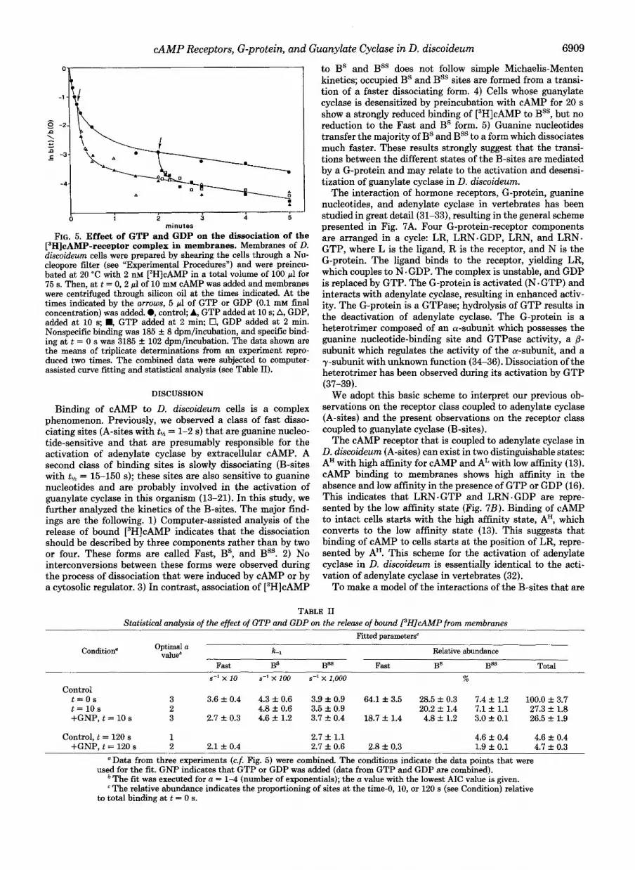
The experiment presented in Fig. 5 was set up to test whether both BS and Bss are sensitive to guanine nucleotides and especially to test to which form they interconvert upon addition of guanine nucleotides. Membranes were incubated with [3H]cAMP until equilibrium was reached. Then, at t = 0 s, excess cAMP was added, and GTP or GDP was added at 10 or 120 s. The results were analyzed by computer-assisted curve fitting (Table 11).
Membranes possess three binding forms which dissociate with similar rate constants if compared to cells. Their pro- portioning at 2 nM [3H]cAMP is 65% Fast, 29% BS, and 7% Bss. When only the data points at 10 s and later are taken into account, further dissociation should be described by only two components, BS and Bss, because all Fast sites have dissociated. The distribution of residual bound [3H]cAMP at t = 10 s is 0 Fast, 20% BS, and 7% Bss. When GDP or GTP

6908 zyxwvutsrqponmlkjihgfedcbaZYXWVUTSRQPONMLKJIHGFEDCBACAMP Receptors, G-protein, and Guanylate Cyclase in D. discoideum zyxwvutsrqponmlkjihgfedcbaZYXWVUTSRQPONMLKJIHGFEDCBA
bound, zyxwvutsrqponmlkjihgfedcbaZYXWVUTSRQPONMLKJIHGFEDCBArnolec. cell" x
FIG. zyxwvutsrqponmlkjihgfedcbaZYXWVUTSRQPONMLKJIHGFEDCBA2. Scatchard plots of fast dissociating sites and Bs and Bss. This figure consists of data obtained from exponential curve fitting in Table I (0, A) and data obtained by back-calculation zyxwvutsrqponmlkjihgfedcbaZYXWVUTSRQPONMLKJIHGFEDCBA(0, A). For the latter method, cells were incubated with different concen- trations of [3H]cAMP (2-2000 nM) for 45 zyxwvutsrqponmlkjihgfedcbaZYXWVUTSRQPONMLKJIHGFEDCBAs. Then cells were centri- fuged through silicon oil directly (yielding b(0)) or at 10 or 120 s after addition of excess cAMP (yielding b(10) and b(120), respectively). Binding to Fast, BS, and Bss at equilibrium was calculated using Equation 5 (a-c). The results of three experiments with triplicate incubations were combined. The data on Fast that were published previously (Fig. 6 of Ref. 13) are indicated by the asterisks.
is added at 10 s, the dissociation is optimally described again by three components: 19% Fast with k-l = 0.3 s-l, 5% BS, and 3% Bss. This suggests that the majority of BS is transferred by GTP or GDP to a fast dissociating form. Bss is also affected by guanine nucleotides, but a decision about its transfer to BS or Fast cannot yet be made. The distribution of bound [3H] cAMP at 2 min after the onset of dissociation is 0 Fast, 0 BS, and 5% Bss. The addition of GTP or GDP at this moment results in the enhanced release of bound [3H]cAMP. The analysis indicates only two components: 3% Fast with k 1 = 0.21 s" and 2% Bss. BS is not present, indicating that also the Bss is transferred to a fast dissociating site by guanine nucleotides. It should be noticed that GTP and GDP and the nonhydrolyzable analog GTPyS (data not shown) have simi- lar effects.
Specificity of Guanyl Nucleotides-Membranes were incu- bated with 2 nM [3H]cAMP in the presence of nucleotides until equilibrium, which is followed by a 10-s dissociation with excess cAMP (Fig. 6). Guanyl nucleotides inhibit the thus measured cAMP binding by maximally 80%. A half- maximal effect is observed at about 2.5 PM GTP, GDP, and
seconds (CAMPI, nM
FIG. 3. Kinetics of association of ['HICAMP to BS and Bss. Cells were incubated with [3H]cAMP for the times indicated in A-C in a volume of 100 pl. Then 1 ml of buffer with 0.1 mM cAMP was added, and 1 ml of the mixture was layered on top of silicon oil. Tubes were centrifuged at 10 s (open symbols) or at 2 min (closed symbols) after the cAMP chase. A and zyxwvutsrqponmlkjihgfedcbaZYXWVUTSRQPONMLKJIHGFEDCBAB, the CAMP concentrations used were 2 nM (A: zyxwvutsrqponmlkjihgfedcbaZYXWVUTSRQPONMLKJIHGFEDCBA0, O), 30 nM (B: A, A), and 100 nM (B: 0). Data are the means and standard deviations of quadruplicate incubations. Nonspecific binding, which is subtracted from all data, was 11.1 f 0.6 dpm (0, O), 165 f 6 dpm (A, A), and 491 f 15 dpm (O), all with n = 6. C, replot of the data of A and B, where b( t ) is the binding at time t and b(m) is the binding at equilibrium (45-60 s). zyxwvutsrqponmlkjihgfedcbaZYXWVUTSRQPONMLKJIHGFEDCBAD, association kinetics were measured at different [3H]cAMP concentrations as shown in A and B. The slope in the logarithmic plot (C) equals the apparent zyxwvutsrqponmlkjihgfedcbaZYXWVUTSRQPONMLKJIHGFEDCBAk,, 0, kOn of BS + Bss (10-s dissociation); 0, k,. of Bss (2- min dissociation); A, 8-s chase (data from Ref. 13). The theoretical curves for BS and Bss (dashed lines) are based on the observed dissociation constants (Fig. 2) and off-rate ( L l , Table I).
umndr
FIG. 4. Binding of [3H]cAMP to desensitized cells. The in- cubation mixture was composed 2 X 10' cells in 180 pl, 20 pl of 20 nM CAMP, and 200 pl of 2 nM [3H]cAMP. For control cells (O), all components were mixed simultaneously at t = 0; for desensitized cells (0), cells and CAMP were mixed at t = 0, and [3H]cAMP was added at t = 20 s. Thus, the concentration of cyclic nucleotide remained 2 nM during the entire experiment. At the times indicated binding of 13H]cAMP was measured directly or at 10 or 120 s after a chase with excess CAMP. Binding to the Fast-, BS- and Bss-sites was calculated using Equation 5 (ax) . Nonspecific binding has been subtracted from the data. Shown are the means and standard deviations of quadru- plicate determinations reproduced three times.
GTPyS and at about 16 MM GppNHp; GMP is about 1000- fold less active than GTP. This specificity is consistent with the action of a guanine nucleotide regulatory protein (G- protein). Interestingly, ATP strongly stimulates cAMP bind- ing measured at 10 s after a cAMP chase; ADP is without effect, and AppNHp slightly inhibits cAMP binding. Recent results suggest that this effect of ATP is mediated by protein kinase C (30).
cAMP Receptors, G-protein, and Guanylate Cyclase in D. discoideum zyxwvutsrqponmlkjihgfedcbaZYXWVUTSRQPONMLKJIHGFEDCBA6909 zyxwvutsrqponmlkjihgfedcbaZYXWVUTSRQPONMLKJIHGFEDCBAc I zyxwvutsrqponmlkjihgfedcbaZYXWVUTSRQPONMLKJIHGFEDCBA0 zyxwvutsrqponmlkjihgfedcbaZYXWVUTSRQPONMLKJIHGFEDCBA1 2 3 4 5
minutes zyxwvutsrqponmlkjihgfedcbaZYXWVUTSRQPONMLKJIHGFEDCBAFIG. 5. Effect of GTP and GDP on the dissociation of the
['HICAMP-receptor complex in membranes. zyxwvutsrqponmlkjihgfedcbaZYXWVUTSRQPONMLKJIHGFEDCBAMembranes of zyxwvutsrqponmlkjihgfedcbaZYXWVUTSRQPONMLKJIHGFEDCBAD. diseoideum cells were prepared by shearing the cells through a Nu- cleopore filter (see "Experimental Procedures") and were preincu- bated at 20 "C with 2 nM [3H]cAMP in a total volume of 100 pl for 75 s. Then, at t = 0, 2 pl of 10 mM cAMP was added and membranes were centrifuged through silicon oil at the times indicated. At the times indicated by the arrows, 5 pl of GTP or GDP (0.1 mM final concentration) was added. 0, control; A, GTP added at 10 s; A, GDP, added at 10 s; ., GTP added at 2 min; zyxwvutsrqponmlkjihgfedcbaZYXWVUTSRQPONMLKJIHGFEDCBA0, GDP added at 2 min. Nonspecific binding was 185 f 8 dpm/incubation, and specific bind- ing at t = 0 s was 3185 +. 102 dpm/incubation. The data shown are the means of triplicate determinations from an experiment repro- duced two times. The combined data were subjected to computer- assisted curve fitting and statistical analysis (see Table 11).
DISCUSSION
Binding of cAMP to D. discoideum cells is a complex phenomenon. Previously, we observed a class of fast disso- ciating sites (A-sites with tM = 1-2 s) that are guanine nucleo- tide-sensitive and that are presumably responsible for the activation of adenylate cyclase by extracellular CAMP. A second class of binding sites is slowly dissociating (B-sites with txh = 15-150 s); these sites are also sensitive to guanine nucleotides and are probably involved in the activation of guanylate cyclase in this organism (13-21). In this study, we further analyzed the kinetics of the B-sites. The major find- ings are the following. 1) Computer-assisted analysis of the release of bound [3H]cAMP indicates that the dissociation should be described by three components rather than by two or four. These forms are called Fast, BS, and Bss. 2) No interconversions between these forms were observed during the process of dissociation that were induced by cAMP or by a cytosolic regulator. 3) In contrast, association of [3H]cAMP
to BS and Bss does not follow simple Michaelis-Menten kinetics; occupied B S and Bss sites are formed from a transi- tion of a faster dissociating form. 4) Cells whose guanylate cyclase is desensitized by preincubation with cAMP for 20 s show a strongly reduced binding of [3H]cAMP to Bss, but no reduction to the Fast and BS form. 5) Guanine nucleotides transfer the majority of BS and Bss to a form which dissociates much faster. These results strongly suggest that the transi- tions between the different states of the B-sites are mediated by a G-protein and may relate to the activation and desensi- tization of guanylate cyclase in D. discoideum.
The interaction of hormone receptors, G-protein, guanine nucleotides, and adenylate cyclase in vertebrates has been studied in great detail (31-33), resulting in the general scheme presented in Fig. 7A. Four G-protein-receptor components are arranged in a cycle: LR, LRN-GDP, LRN, and LRN- GTP, where L is the ligand, R is the receptor, and N is the G-protein. The ligand binds to the receptor, yielding LR, which couples to N . GDP. The complex is unstable, and GDP is replaced by GTP. The G-protein is activated (N + GTP) and interacts with adenylate cyclase, resulting in enhanced activ- ity. The G-protein is a GTPase; hydrolysis of GTP results in the deactivation of adenylate cyclase. The G-protein is a heterotrimer composed of an a-subunit which possesses the guanine nucleotide-binding site and GTPase activity, a p- subunit which regulates the activity of the a-subunit, and a y-subunit with unknown function (34-36). Dissociation of the heterotrimer has been observed during its activation by GTP (37-39).
We adopt this basic scheme to interpret our previous ob- servations on the receptor class coupled to adenylate cyclase (A-sites) and the present observations on the receptor class coupled to guanylate cyclase (B-sites).
The CAMP receptor that is coupled to adenylate cyclase in D. discoideum (A-sites) can exist in two distinguishable states: AH with high affinity for cAMP and AL with low affinity (13). cAMP binding to membranes shows high affinity in the absence and low affinity in the presence of GTP or GDP (16). This indicates that LRN. GTP and LRN. GDP are repre- sented by the low affinity state (Fig. 7B). Binding of cAMP to intact cells starts with the high affinity state, AH, which converts to the low affinity state (13). This suggests that binding of cAMP to cells starts at the position of LR, repre- sented by AH. This scheme for the activation of adenylate cyclase in D. discoideum is essentially identical to the acti- vation of adenylate cyclase in vertebrates (32).
To make a model of the interactions of the B-sites that are
TABLE I1 Statistical zyxwvutsrqponmlkjihgfedcbaZYXWVUTSRQPONMLKJIHGFEDCBAanalysis of the effect of GTP and GDP on the release of bound PHJeAMP from membranes
Fitted parameters'
k-1 Relative abundance
Fast BS BB Fast BS B= Total
Condition" Optimal a valueb
s-' x 10 s-1 x 100 s-1 x 1,000 %
Control t = O s 3 3.6 f 0.4 4.3 f 0.6 3.9 f 0.9 64.1 f 3.5 28.5 f 0.3 7.4f 1.2 100.0 f 3 . 7 t = 1 0 s 2 4.8 +. 0.6 3.5 f 0.9 20.2 +. 1.4 7.1 f 1.1 27.3 f 1.8 +GNP, t = 10 s 3 2.7 f 0.3 4.6f 1.2 3.7 f0 .4 18.7f 1.4 4.8f 1.2 3.0f 0.1 26.5 f 1.9
Control, t = 120 s 1 2.7 f 1.1 4.6 f 0.4 4.6 f 0.4 +GNP, t = 120 s 2 2.1 f 0.4 2.7 f 0.6 2.8 f 0.3 1.9 f 0.1 4.7 f 0.3
"Data from three experiments (c.f. Fig. 5) were combined. The conditions indicate the data points that were used for the fit. GNP indicates that GTP or GDP was added (data from GTP and GDP are combined).
The fit was executed for a = 1-4 (number of exponentials); the a value with the lowest AIC value is given. e The relative abundance indicates the proportioning of sites at the time-0, 10, or 120 s (see Condition) relative
to total binding at t = 0 s.

6910 zyxwvutsrqponmlkjihgfedcbaZYXWVUTSRQPONMLKJIHGFEDCBAcAMP Receptors, G-protein, and Guunylate Cyclase zyxwvutsrqponmlkjihgfedcbaZYXWVUTSRQPONMLKJIHGFEDCBAin D. discoideum zyxwvutsrqponmlkjihgfedcbaZYXWVUTSRQPONMLKJIHGFEDCBA180 zyxwvutsrqponmlkjihgfedcbaZYXWVUTSRQPONMLKJIHGFEDCBA
-
$ 140 zyxwvutsrqponmlkjihgfedcbaZYXWVUTSRQPONMLKJIHGFEDCBAe- zyxwvutsrqponmlkjihgfedcbaZYXWVUTSRQPONMLKJIHGFEDCBA0 c
8
6 e n zyxwvutsrqponmlkjihgfedcbaZYXWVUTSRQPONMLKJIHGFEDCBAd. zyxwvutsrqponmlkjihgfedcbaZYXWVUTSRQPONMLKJIHGFEDCBA60 E
100
._
2 20
0 I
16-7 ' 16-5 ' 16-3 nucleotide concentration. M. zyxwvutsrqponmlkjihgfedcbaZYXWVUTSRQPONMLKJIHGFEDCBA
FIG. 6. Nucleotide specificity of the alteration of 13H]cAMP binding to BS and BSs in membranes from D. discoideum. Membranes (derived from lo7 cells) were incubated at 20 "C with 2 nM [3H]cAMP and different concentrations of nucleotides in a volume of 100 pl for 75 s. cAMP (2 pl of 10 mM) was added, and 10 s later, membranes were centrifuged through silicon oil. The nucleotides are GTP (O), GDP (A), GppNHp (m), GMP (V), ATP (O), ADP (A), and AppNHp (0). Nonspecific binding was measured by including 0.1 mM cAMP in the entire incubation period; it was 155 f 14 dpm/ sample zyxwvutsrqponmlkjihgfedcbaZYXWVUTSRQPONMLKJIHGFEDCBA(n = 4) without the nucleotides and was changed by less than 15% in the presence of 1 mM indicated nucleotides. Specific binding was 1512 f 52 dpm/sample (n = 3) for the control.
coupled to guanylate cyclase, we combine the general scheme with the kinetics of formation of BS and Bss in cells, with the effects of guanine nucleotides on these states in membranes, and with the observations made in desensitized cells. In membranes, the major parts of BS and Bss are converted by GTP or GDP to a fast dissociating site, indicating that LRN. GDP and LRN. GTP are fast dissociating, designated as BF in Fig. 7C. In cells, the rate of formation of the Bs-state at high cAMP concentrations does not depend on the cAMP concentration, indicating that this state arises by transition from a faster dissociating state; LRN-GDP is the obvious candidate. The maximal on-rate of the Bs-state is 0.22 s-l; apparently, this figure represents the transition rate constant of the step LRN . GDP 4 LRN. At low cAMP concentrations, the formation of the Bs-state follows Michaelis-Menten ki- netics; in contrast, the formation of Bss never obeys this law, but follows the kinetics of BS. This suggests that, in the course of events, BS is formed before Bss. This view is supported by the observation that, in desensitized cells, BS is still formed, but binding to Bss is strongly reduced. The simplest expla-
Vertebrate
FIG. 7. Models on the interaction between surface receptor (R), li- gand (L), and guanine nucleotide- binding protein (N). A, general scheme of hormone-induced activation of adenylate cyclase in vertebrates; B, scheme for the interactions of cAMP with the A-sites leading to the activation of adenylate cyclase in D. discoideum; C, scheme for the interactions of cAMP with the B-sites leading to the activation of guanylate cyclase in D. discoideum. See text for details.
adenylate cyclase GTPase, cyclase
adenylate
activation deactivation
t 1 N - F L R 1 3 P
LRN-GTP
GTP LLRNJGDP GDP
A
nation for these observations is that LRN is associated with BS, and LR with Bss. This would imply that activation of guanylate cyclase in D. discoideum is proportional to the transition of BS to Bss, a transition which no longer occurs in desensitized cells. One observation seems to be incompatible with the proposed model, i.e. the formation of Bss in mem- branes. The absence of GTP would exclude the transition of BS to Bss. However, we observed that binding of [3H]cAMP to Bss is much slower in membranes than in cells (21). This could mean that in membranes Bss is not reached via a transition of the Bs-state, but arises from direct association of cAMP to the unoccupied receptor not associated with a G- protein.
A comparison of the schemes for the activation of adenylate cyclase (Fig. 7 B ) and guanylate cyclase (Fig. 7 C ) in D. discoi- deum reveals two main differences, which could be relevant for their functioning. First, CAMP enters the A-cycle at the position of LR and the B-cycle at LRN. GDP. This difference between coupling and precoupling of receptor and G-protein could explain the effects of agents that immobilize proteins at the cell surface; these agents inhibit the CAMP-induced activation of adenylate cyclase, whereas they potentiate the activation of guanylate cyclase (40, 41). Second, the rate- limiting step in the activation of adenylate cyclase is the transition LR +. LRN . GDP (Fig. 7B), i.e. the coupling of receptor with G-protein. In contrast, the rate-limiting step in the activation of guanylate cyclase is the dissociation of GDP from the LRN.GDP complex (Fig. 7C). This seems to be a rather unusual situation if compared to the interactions of GTP and GDP with the vertebrate G-protein (33, 42,43). It should be noted, however, that this rate-limiting step is still very fast, showing a half-life of about 3 s. This constant is not very different from that of hormone-induced dissociation of GDP from the vertebrate G-protein (33, 44, 45). By pre- coupling of receptor and G-protein, less steps in the activation of a cyclase are required. This may explain why guanylate cyclase activity increases within 2 s after cAMP addition, whereas adenylate cyclase activity increases only after about 30 s.
The response induced by the ligand is proportional to the rate of passage through the cycle. The ligand may still bind to the receptor in the absence of any transition, which implies that the cell is desensitized. Indeed, this was observed in D. discoideurn (Fig. 4). Fast (AH, AL, and B") and BS were still occupied with cAMP in desensitized cells, but the transition of BS to Bss no longer occurred. This suggests either that GTP no longer binds to the G-protein-receptor complex in desensitized cells or that the G-protein is not activated by
D. discoideum
IigandkAMP)
cyclase adenylate
activation I p\ mLRL:LL ,-;
LRN]
B
cyclase guanylate
activation t a ligand(cAMP)

CAMP Receptors, G-protein, and Guanylate Cyclase in zyxwvutsrqponmlkjihgfedcbaZYXWVUTSRQPONMLKJIHGFEDCBAD. discoideum zyxwvutsrqponmlkjihgfedcbaZYXWVUTSRQPONMLKJIHGFEDCBA691 zyxwvutsrqponmlkjihgfedcbaZYXWVUTSRQPONMLKJIHGFEDCBA1
GTP. In the former situation, the cycle would arrest in LRN (Bs), whereas in the latter case, in LRN. GTP. We observe significant amounts of LRN in cells. This would favor the former explanation.
What could be the molecular mechanism of desensitization? In vertebrates, it has been shown that phosphorylation of the receptor is associated with desensitization of the hormone- induced activation of adenylate cyclase (46-48). Phosphoryla- tion of the receptor could be mediated by different protein kinases, including CAMP-dependent protein kinase and pro- tein kinase C (49), and resulted in a reduced interaction of receptor and G-protein (45,50, 51). Recently, CAMP-induced phosphorylation of the receptor has been established in D. discoideum (52-54) and was associated with the CAMP-in- duced desensitization of adenylate cyclase (55). It should be noted, however, that desensitization of guanylate cyclase is much faster than desensitization of adenylate cyclase; at 10 s after CAMP addition, guanylate cyclase is already desensi- tized, whereas adenylate cyclase still has to be activated (9- zyxwvutsrqponmlkjihgfedcbaZYXWVUTSRQPONMLKJIHGFEDCBA11). This suggests that desensitization of adenylate and guan- ylate cyclase does not proceed via an identical mechanism. Recently, we observed a Ca" and phorbol ester-dependent effect of ATP on the B-sites that are coupled to guanylate cyclase. The A-sites that are coupled to adenylate cyclase were not affected (30). This may indicate that the possible activation of protein kinase C in D. discoideum is associated with desensitization of guanylate rather than of adenylate cyclase activity. This possibility is presently being investi- gated.
Acknowledgments-We gratefully acknowledge The0 M. Konijn and Roe1 van Driel for stimulating discussions.
1. 2. 3.
4.
5.
6. 7. zyxwvutsrqponmlkjihgfedcbaZYXWVUTSRQPONMLKJIHGFEDCBA8.
9.
10.
11.
12.
13.
14.
15.
16.
17.
18.
REFERENCES
Mato, J. M., and Malchow, D. (1978) FEBS Lett. 9 0 , 119-122 Roos, W., and Gerisch, G. (1976) FEBS Lett. 68,170-172 Mato, J. M., Krens, F. A., Van Haastert, P. J. M., and Konijn,
Van Haastert, P. J. M., Van Lookeren Campagne, M. M., and
Gerisch, G., and Wick, U. (1975) Biochem. Biophys. Res. Com-
Klein, C., and Juliani, M. H. (1977) Cell 10, 329-335 Klein, C. (1979) J. Biol. Chem. 254 , 12573-12578 Devreotes, P. N., and Steck, T. L. (1979) J. Cell Biol. 8 0 , 300-
Van Haastert, P. J. M., and Van der Heijden, P. R. (1983) J. Cell
Dinauer, M. C., Steck, T. L., and Devreotes, P. N. (1980) J. Cell
Dinauer, M. C., Steck, T. L., and Devreotes, P. N. (1980) J. Cell
Pan, P., Hall, E. M., and Bonner, J. T. (1972) Nature New Biol.
Van Haastert, P. J. M., and De Wit, R. J. W. (1984) J. Biol.
De Wit, R. J. W., and Van Haastert, P. J. M. (1985) Biochim.
Van Haastert, P. J. M. (1985) Biochim. Biophys. Acta 845,254-
Van Haastert, P. J. M. (1984) Biochem. Biophys. Res. Commun.
De Wit, R. J. W., and Bulgakov, R. (1985) FEBSLett. 179,257-
Van Haastert, P. J. M. (1985) Biochim. Biophys. Acta 846 ,
T. M. (1977) Proc. Natl. Acad. Sci. U. S. A. 74,2348-2352
ROSS, F. M. (1982) FEBS Lett. 147,149-152
mun. 65,364-370
309
Biol. 96,347-353
Biol. 86,545-553
Biol. 86,554-561
237,181-182
Chem. 259,13321-13328
Biophy~. Acta 814 , 199-213
260
124,597-604
261
324-333
19.
20.
21.
22.
23.
24.
25. 26.
27.
28.
29. 30.
31.
32.
33.
34.
35.
36.
37.
38.
39.
40.
41.
42.
43.
44.
45.
46.
47.
48.
49.
50.
51.
52.
53.
54.
55.
Kesbeke, F., and Van Haastert, P. J. M. (1985) Biochim. Biophys.
De Wit, R. J. W., Bulgakov, R., Pinas, J. E., and Konijn, T. M.
Janssens, P. M. W., Van der Geer, P. L. J., Arents, J. C., and
Van Haastert, P. J. M. (1983) Biochem. Biophys. Res. Commun.
Das, zyxwvutsrqponmlkjihgfedcbaZYXWVUTSRQPONMLKJIHGFEDCBA0. P., and Henderson, E. J. (1983) Biochim. Biophys. Acta
Pannbacker, R. G., and Bravard, J. L. (1972) Science 175,1014-
Van Haastert, P. J. M. (1981) J. Chromatogr. 210, 229-240 Landaw, E. M., and DiStefano, J. J., I11 (1984) Am. J. Physiol.
Spriet, J. A., and Vansteenkiste, G. C. (1982) Computer-aided Modelling and Simulation, Academic Press Inc., Ltd., London
Dinauer, M. C., MacKay, S. A., and Devreotes, P. N. (1980) J. Cell Biol. 86 , 537-544
Van Haastert, P. J. M. (1984) J. Gen. Microbiol. 130,2559-2564 Van Haastert, P. J. M., De Wit, R. J. W., and Van Lookeren
Campagne, M. M. (1985) Biochem. Biophys. Res. Commun.
Cassel, D., and Selinger, Z. (1978) Proc. Natl. Acad. Sei. U. S. A.
Lefkowitz, R. J., Stadel, J. M., and Caron, M. G. (1983) Annu.
Tolkowsky, A. M., Braun, S., and Levitzki, A. L. (1982) Proc.
Northup, J. K., Smigel, M. D., Sternweis, P. C., and Gilman, A.
Northup, J. K., Sternweis, P. C., and Gilman, A. G. (1983) J.
Hildebrandt, J. D., Codina, J., Resinger, R., and Birnbaumer, L. (1984) J. Biol. Chem. 259,2039-2042
Katada, T., Northup, J. K., Bokock, G. M., Ui, M., and Gilman, A. G. (1984) J. Biol. Chem. 259,3578-3585
Codina, J., Hildebrandt, J. D., Birnbaumer, L., and Sekura, R. D. (1984) J. Biol. Chem. 259, 11408-11418
Brandt, D. R., and Ross, E. M. (1985) J. Biol. Chem. 260 , 266- 272
Mato, J. M., Van Haastert, P. J. M., Krens, F. A., and Konijn, T. M. (1978) Cell Biol. Int. Rep. 2 , 163-170
Fontana, D. R., and Devreotes, P. N. (1984) Deu. Biol. 106, 76- 82
Northup, J. K., Smigel, M. D., and Gilman, A. G. (1982) J. Biol. Chem. 257,11416-11423
Birnbaumer, L., Swartz, T. L., Abramowitz, J., Mintz, P. W., and Iyengar, R. (1980) J. Biol. Chem. 255,3542-3551
Pike, L. J., andLefkowitz, R. J. (1981) J. Biol. Chem. 256,2207- 2212
Briggs, M. M., Stadel, J. M., Iyengar, R., and Lefkowitz, R. J. (1983) Arch. Biochem. Biophys. 224 , 142-151
Stadel, J. M., Nambi, P., Shorr, R. G. L., Sawyer, D. R., Caron, M. G.. and Lefkowitz. R. J. 11983) Proc. Natl. Acad. Sci. U. S.
Acta 847,33-39
(1985) Biochim. Biophys. Acta 814,214-226
Van Driel, R. (1985) Mol. Cell. Biochem. 67, 119-124
115,130-136
736,45-56
1016
246 , R665-R677
128,185-192
75,4155-4159
Reu. Biochem. 52,159-186
Natl. Acad. Sci. U. S. A. 79, 213-217
G. (1983) J. Biol. Chem. 258,11369-11376
Biol. Chem. 258,11361-11368
A. 80; 3171-3177 zyxwvutsrqponmlkjihgfedcbaZYXWVUTSRQPONMLKJIHGFEDCBA'
. ,
Siblev. D. R.. Peters. J. R.. Nambi. P.. Caron. M. G.. and Lefkow- itz,"R. J. (1984) J.'Biol. Chem. 259,9742-9749 '
(1984) Proc. Natl. Acad. Sci. U. S. A. 81 , 4316-4320
J. Biol. Chem. 260 , 2165-2171
Kelleher, D. J., Pessin, J. E., Ruoho, A. E., and Johnson, G.
Nambi, P., Peters, J. R., Sibley, D. R., and Lefkowitz, R. J. (1985)
Kirchick, H. J., Iyengar, R., and Birnbaumer, L. (1983) Endocri-
Benovic, J. L., Pike, L. J., Cerione, R. A., Staniszewski, C., Voshimasa, T., Codina, J., Caron, M. G., and Lefkowitz, R. J. (1985) J. Biol. Chem. 260, 7094-7101
nology 113,1638-1648
Theibert, A., Klein, P., and Devreotes, P. N. (1984) J. Biol. Chem.
Klein, P.. Theibert. A.. Fontana. D.. and Devreotes. P. N. (1985) 259,12318-12321
J. Biol.' Chem. 260,1757-1764 '
~ ~~,
Klein, C., Lubs-Haukeness, J., and Simons, S. (1985) J. Cell Biol.
Devreotes, P. N., and Sherring, J. A. (1985) J. Biol. Chem. 260, 100,715-720
6378-6384
Recommended


