Histology lectures
First class of clinical laboratory
science
Pharmacy College .University of
Anbar
D. Nahidah Ibrahim Hammadi

Histology is a science which study the
microstructure and the relationship
between the structure and function of
human being.

Tissues

Tissues
Cells work together in functionally related groups called tissues
How is this done?
Attachments
communication
Types of tissues:1. Epithelial – lining and covering
2. Connective – support
3. Muscle – movement
4. Nervous – control

Epithelial Tissue –
General Characteristics & Functions
Covers a body surface or lines a body cavity
Forms most glands
Functions of epithelium
Protection
Absorption, secretion, and diffusion
Filtration
Forms slippery surfaces (mucus secretion)
Special Characteristics of
Epithelia Cellularity
cells are in close contact with each other with little or no intercellular space between them
Specialized contacts may have junctions for both attachment and communication
Polarity epithelial tissues always have an apical and basal surface
Support by connective tissue at the basal surface, both the epithelial tissue and the connective tissue
contribute to the basement membrane
Avascular nutrients must diffuse from basal layer
Innervated
Regenerative epithelial tissues are highly mitotic

Special Characteristics of
Epithelia

First name of tissue indicates number of layers
Simple – one layer of cells
Stratified – more than one layer of cells
Classifications of Epithelia

Classifications of Epithelia
Last name of tissue describes shape of cells
Squamous – cells wider than
tall (plate or “scale” like)
Cuboidal – cells are as wide as
tall, as in cubes
Columnar – cells are taller than
they are wide, like columns

Naming Epithelia
Naming the epithelia includes both the layers (first) and
the shape of the cells (second)
i.e. stratified cuboidal epithelium
The name may also include any accessory structures
Goblet cells
Cilia
Keratin
Special epithelial tissues (don’t follow naming
convention)
Psuedostratified
Transitional

Simple Squamous Epithelium
Description
single layer of flat cells with disc-shaped nuclei
Special types
Endothelium (inner covering)
slick lining of hollow organs
Mesothelium (middle covering)
Lines peritoneal, pleural, and pericardial cavities
Covers visceral organs of those cavities
Simple Squamous Epithelium
Function
Passage of materials by passive diffusion and filtration
Secretes lubricating substances in serous membranes
Location
Renal corpuscles (kidneys)
Alveoli of lungs
Lining of heart, blood and lymphatic vessels
Lining of ventral body cavity (serosae/serous memb.)

Simple Squamous Epithelium
Simple squamous
lining the walls of
the capillary
If it’s from a
mesothelial lining

Simple Cuboidal Epithelium
Description single layer of cube-like cells with large, spherical central
nuclei
Function secretion and absorption
Location kidney tubules, secretory portions of small glands, ovary
surface
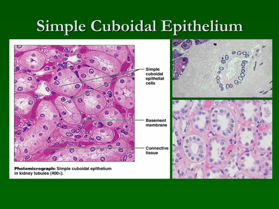
Simple Cuboidal Epithelium

Simple Columnar Epithelium
Description
single layer of column-shaped (rectangular) cells with oval nuclei
Some bear cilia at their apical surface
May contain goblet cells
Function
Absorption; secretion of mucus, enzymes, and other substances
Ciliated type propels mucus or reproductive cells by ciliary action

Simple Columnar Epithelium
Location
Non-ciliated form
Lines digestive tract, gallbladder, ducts of some glands
Ciliated form
Lines small bronchi,
uterine tubes, and uterus

Pseudostratified Columnar
Epithelium
Description
All cells originate at basement membrane
Only tall cells reach the apical surface
May contain goblet cells and bear cilia
Nuclei lie at varying heights within cells
Gives false impression of stratification
Function
secretion of mucus; propulsion of mucus by cilia
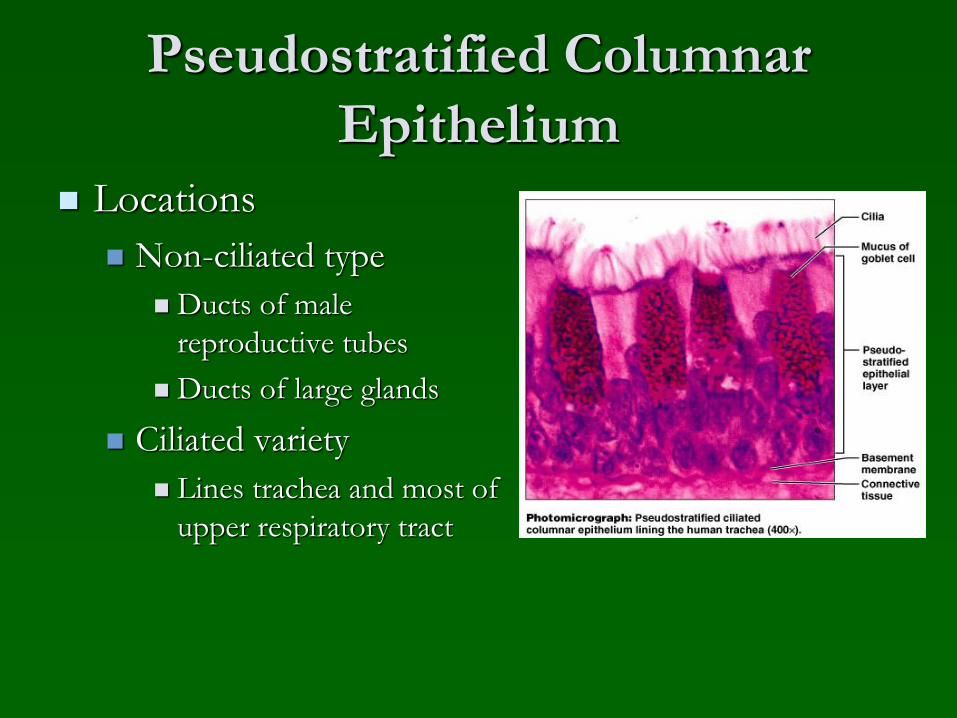
Pseudostratified Columnar
Epithelium
Locations
Non-ciliated type
Ducts of male
reproductive tubes
Ducts of large glands
Ciliated variety
Lines trachea and most of
upper respiratory tract

Stratified Epithelia
Contain two or more layers of cells
Regenerate from below
Major role is protection
Are named according to the shape of cells at
apical layer

Stratified Squamous Epithelium
Description
Many layers of cells – squamous in shape
Deeper layers of cells appear cuboidal or columnar
Thickest epithelial tissue – adapted for protection

Stratified Squamous Epithelium
Specific types
Keratinized – contain the protective protein keratin
Surface cells are dead and full of keratin
Non-keratinized – forms moist lining of body openings
Function
Protects underlying tissues in areas subject to abrasion
Location
Keratinized – forms epidermis
Non-keratinized – forms lining of esophagus, mouth, and
vagina

Stratified Squamous Epithelium
Non-keratinized vs. Keratinized

Transitional Epithelium
Description
Basal cells usually cuboidal or columnar
Superficial cells dome-shaped or squamous
Function
stretches and permits distension of urinary bladder
Location
Lines ureters, urinary bladder and part of urethra

Transitional EpitheliumRelaxed state
Stretched state

Epithelial Surface Features
Apical surface features
Microvilli – finger-like extensions of plasma
membrane
Abundant in epithelia of small intestine and kidney
Maximize surface area across which small molecules
enter or leave
Act as stiff knobs that resist abrasion

Epithelial Surface Features
Apical surface features
Cilia – whip-like, highly motile extensions of
apical surface membranes
Contains a core of nine pairs of microtubules
encircling one middle pair
Axoneme – a set of microtubules
Each pair of microtubules – arranged in a doublet
Microtubules in cilia – arranged similarly to
cytoplasmic organelles called centrioles
Movement of cilia – in coordinated waves

A Cilium
Recommended