Imaging the cell entry of the anthrax oedema andlethal toxins with fluorescent protein chimerascmi_1480 1435..1445
Irene Zornetta,1 Lucia Brandi,1 Blythe Janowiak,2
Federica Dal Molin,1 Fiorella Tonello,1*R. John Collier2 and Cesare Montecucco1
1Dipartimento di Scienze Biomediche dell’Università diPadova and Istituto di Neuroscienze del CNR, Via G.Colombo 3, 35100 Padova, Italy.2Department of Microbiology and Molecular Genetics,Harvard Medical School, Boston, MA 02115, USA.
Summary
To investigate the cell entry and intracellular traf-ficking of anthrax oedema factor (EF) and lethalfactor (LF), they were C-terminally fused to theenhanced green fluorescent protein (EGFP) andmonomeric Cherry (mCherry) fluorescent proteins.Both chimeras bound to the surface of BHK cellstreated with protective antigen (PA) in a patchymode. Binding was followed by rapid internaliza-tion, and the two anthrax factors were found totraffic along the same endocytic route and withidentical kinetics, indicating that their intracellularpath is essentially dictated by PA. Colocaliza-tion studies indicated that anthrax toxins entercaveolin-1 containing compartments and thenendosomes marked by phoshatidylinositol 3-phoshate and Rab5, but not by early endosomeantigen 1 and transferrin. After 40 min, both EF andLF chimeras were observed to localize within latecompartments. Eventually, LF and EF appeared inthe cytosol with a time-course consistent withtranslocation from late endosomes. Only the EGFPderivatives reached the cytosol because they aretranslocated by the PA channel, while the mCherryderivatives are not. This difference is attributed toa higher resistance of mCherry to unfolding. Aftertranslocation, LF disperses in the cytosol, while EFlocalizes on the cytosolic face of late endosomes.
Introduction
Anthrax is a zoonosis caused by infection with toxigenicstrains of Bacillus anthracis, a spore-forming Gram-
positive bacterium widespread in the environment (Beyerand Turnbull, 2009; Koehler, 2009). Depending on the siteof entry of spores or bacteria, humans may develop threeforms of anthrax: cutaneous, gastrointestinal or inhala-tional anthrax, the latter form being the most dangerous(Dixon et al., 1999). Virulent B. anthracis strains are char-acterized by the expression of three major virulencefactors: an anti-phagocytic polyglutamic capsule, whichlargely prevents phagocytosis by neutrophils and mac-rophages (Fouet, 2009), and two A-B type toxins. Thetoxins are capable of binding and entering almost any celltype, where they derange two major cell signalling path-ways (Ascenzi et al., 2002; Moayeri and Leppla, 2009).The two toxins were termed oedema toxin (EdTx)because, when injected sub-cutaneously, it causedoedema, and lethal toxin (LeTx) because it led to rapiddeath in guinea pigs and in Fischer 344 rats (Smith andKeppie, 1954; Smith et al., 1955; 1956; Beall et al., 1962).These two toxins share the same B binding component, aprotein of 83 kDa, named protective antigen (PA) from thefact that it is an immunogen that provides effective pro-tection against B. anthracis infection in several animalspecies, including humans (Cybulski et al., 2009). The Apart of LeTx is lethal factor (LF, 90 kDa) and that of EdTxis oedema factor (EF, 89 kDa). PA binds to two cell surfacereceptors: Tumor Endothelial Marker 8 (TEM8) and Cap-illary Morphogenesis Protein 2 (CMG2), both widelyexpressed in different tissues and cell types. The removalof a 20 kDa N-terminal domain converts PA into the PA63form, which self-associates into heptamers (PA63)7
capable of binding LF and EF (Young and Collier, 2007).The (PA63)7 + LF complex (LeTx) promotes its ownendocytosis by entering surface rafts and inducing spe-cific signalling events on the cytosolic face of the mem-brane. Similar data are not available for the (PA63)7 + EF(EdTx) complex, and it remains to be determined if thistoxin follows the same binding and intracellular traffickingroute followed by LeTx. After entering early endosomalcompartments, LeTx reaches late endosomes, wherefromLF is delivered into the cytosol (Abrami et al., 2005 andPuhar and Montecucco, 2007).
Lethal factor is a zinc metalloprotease that cleavesmitogen-activated protein kinase kinases (MAPKKs orMEKs) (Duesbery et al., 1998, Vitale et al., 1998 andTonello and Montecucco, 2009), thereby interfering withthe MAPK cascade, a major signalling event triggered by
Received 17 December, 2009; revised 9 April, 2010; accepted 13April, 2010. *For correspondence. E-mail [email protected];Tel. (+39) 049 8276077; Fax (+39) 049 8276049.
Cellular Microbiology (2010) 12(10), 1435–1445 doi:10.1111/j.1462-5822.2010.01480.xFirst published online 18 May 2010
© 2010 Blackwell Publishing Ltd
cellular microbiology
surface receptors and controlling cell proliferation andsurvival (Gaestel, 2006). As this signalling plays a majorrole in the activation of immune cells, this inhibitionaccounts for the immuno-suppressive activity of LF (Pel-lizzari et al., 1999 and Baldari et al., 2006). EF is acalcium/calmodulin-activated adenylate cyclase, whichcatalyses the formation of cAMP (Drum et al., 2002 andShen et al., 2005), thus altering cell signalling and tissueion fluxes; the ensuing oedema causes failure of differentorgans leading to rapid death (Firoved et al., 2005;Moayeri and Leppla, 2009). Another consequence is thatEF is a strong inhibitor of the activation of differentimmune cells (Puhar et al., 2008; Tournier et al., 2009). Itis noteworthy that EF and LF cause a strong synergisticsuppression of the activation of dendritic cells and T cells(Paccani Rossi et al., 2005; Tournier et al., 2005).
The process of cell entry of EF and LF has been studiedusing cell fractionation and electron microscopic methods,and in several cases a construct between the N-terminusof LF and diphtheria toxin (for recent review see Collier,2009; Moayeri and Leppla, 2009; Van Der Goot andYoung, 2009). We have attempted to improve the knowl-edge of this essential process by producing chimerasconsisting of the full-length LF and EF molecules fused attheir C-termini to different fluorescent proteins: enhancedgreen fluorescent protein (EGFP) or monomeric Cherryprotein (mCherry) (Shaner et al., 2004; 2005). Fusion at
the N-terminus was avoided because this part of the mol-ecule is essential for membrane translocation (Collier,2009). We have found that the two toxins follow exactlythe same pathway of entry with the same time-course.Both EF and LF enter the cytosol from late endosomalcompartments only when coupled to EGFP, but not to themCherry protein. The two toxins reach different cytosoliclocations: LF diffuses in the cytosol while EF appears toremain bound to the cytosolic face of the limiting mem-brane of late endosomes.
Results
Fluorescent chimeras of EF and LF
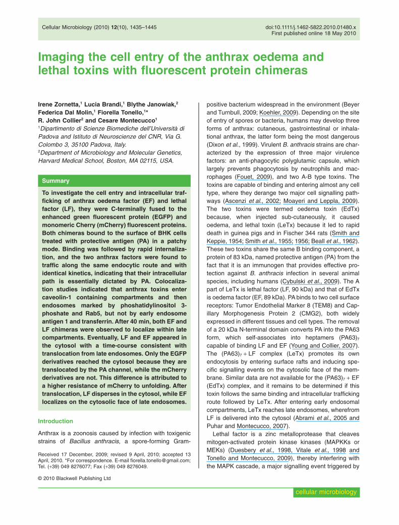
The four chimeras EF-EGFP, EF-mCherry, LF-EGFP andLF-mCherry were produced as recombinant moleculeswith an N-terminal 6xHis tag, which allows easier purifica-tion (Fig. S1) and was shown to confer to LF an increasedcapacity to enter and translocate across the (PA63)7 trans-membrane channel (Neumeyer et al., 2006). The two LFderivatives and the two EF derivatives were shown toretain the ability to perform their respective biochemicalactivities in cell culture. BHK cells were used because oftheir flattened shape, which favours fluorescence micros-copy, and because they express both PA receptors, as themajority of sensitive animal cells do. Figure 1 shows thatthe EGFP derivatives of both LF and EF are active in the
Fig. 1. Fluorescent chimeric anthrax edema factor and lethal factor are enzymatically active in the cytosol of cells in culture. cAMPfluorescence resonance energy transfer imaging in transfected cells expressing the catalytic PKA subunit-YFP and the regulatory PKAsubunit-CFP in the cytosol. BHK cells were imaged after treatment (time zero) with PA (400 nM) in combination with 200 nM of EF (A) orEF-EGFP (B) and EF-mCherry (C). Arrows indicate addition of forskolin (25 mM) as control at the end of experiments. The time course of MEK3 cleavage in BHK cells treated with PA (400 nM) and LF (200 nM), or LF-EGFP (200 nM) or LF-mCherry (200 nM) is given in panel D. Cellextracts were made at the given time, and after SDS-PAGE the proteins were blotted onto nitro-cellulose paper and stained with an anti-MEK3 antibody (for further details see Experimental procedures section). Arrows indicate the full-length MEK3b and its cleaved form. Panel Eshows the corresponding densitometric quantification of the bands expressed as ratio between the MEK3b band and the lower molecularweight band intensities, normalizing the result against the control. Notice that the two mCherry fusion proteins do not display activity.
1436 I. Zornetta et al.
© 2010 Blackwell Publishing Ltd, Cellular Microbiology, 12, 1435–1445
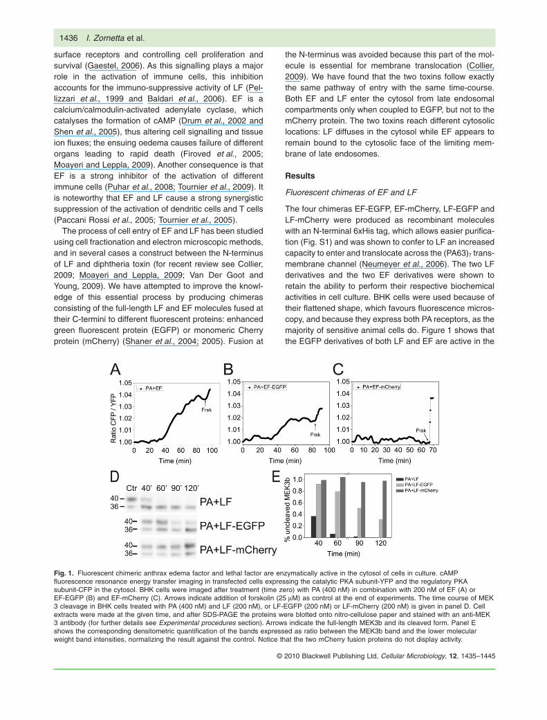
cell cytoplasm, though with lower levels of activity withrespect to their nonconjugated counterparts, as if a smallernumber of molecules is capable of reaching the cytosolwhen EGFP is present. However, these chimeric toxins doreach the cytosol and therefore can report on each step ofthe cell entry process. On the contrary, the two mCherryderivatives displayed no intracellular activity. The mostlikely explanation was that the two mCherry chimeras wereunable to unfold and translocate into the cytosol across the(PA63)7 channel (Collier, 2009). To test this possibility, wemeasured translocation of the chimeric proteins throughpores formed by PA63 in planar phospholipid bilayers(Fig. 2). The EGFP derivatives of both LF and EF translo-cated with slightly lower efficiency and time-course thanthe wild-type proteins, whereas the mCherry derivativesdid not translocate at all. We have not investigated in detailthe reason for the difference between mCherry and EGFP,but this is likely to be due to a different energy requirementfor their unfolding, as shown by their different resistance toguanidinium-induced unfolding (Fig. S2). Protein unfoldinghas been documented before to be necessary for thetranslocation across the PA63 channel and this result pro-vides a further example (Collier, 2009). Accordingly, only
EF-EGFP and LF-EGFP were used in the study of the finalstage of cell intoxication.
Cell binding of the EGFP derivatives of EF and LF
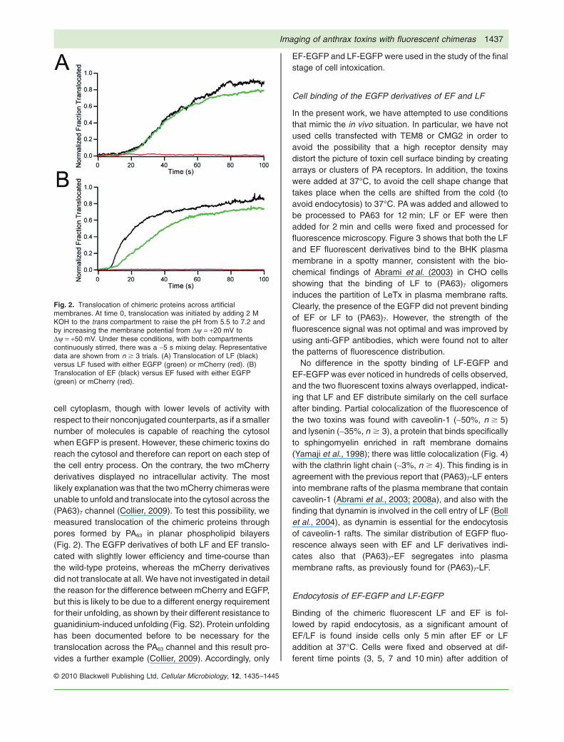
In the present work, we have attempted to use conditionsthat mimic the in vivo situation. In particular, we have notused cells transfected with TEM8 or CMG2 in order toavoid the possibility that a high receptor density maydistort the picture of toxin cell surface binding by creatingarrays or clusters of PA receptors. In addition, the toxinswere added at 37°C, to avoid the cell shape change thattakes place when the cells are shifted from the cold (toavoid endocytosis) to 37°C. PA was added and allowed tobe processed to PA63 for 12 min; LF or EF were thenadded for 2 min and cells were fixed and processed forfluorescence microscopy. Figure 3 shows that both the LFand EF fluorescent derivatives bind to the BHK plasmamembrane in a spotty manner, consistent with the bio-chemical findings of Abrami et al. (2003) in CHO cellsshowing that the binding of LF to (PA63)7 oligomersinduces the partition of LeTx in plasma membrane rafts.Clearly, the presence of the EGFP did not prevent bindingof EF or LF to (PA63)7. However, the strength of thefluorescence signal was not optimal and was improved byusing anti-GFP antibodies, which were found not to alterthe patterns of fluorescence distribution.
No difference in the spotty binding of LF-EGFP andEF-EGFP was ever noticed in hundreds of cells observed,and the two fluorescent toxins always overlapped, indicat-ing that LF and EF distribute similarly on the cell surfaceafter binding. Partial colocalization of the fluorescence ofthe two toxins was found with caveolin-1 (~50%, n � 5)and lysenin (~35%, n � 3), a protein that binds specificallyto sphingomyelin enriched in raft membrane domains(Yamaji et al., 1998); there was little colocalization (Fig. 4)with the clathrin light chain (~3%, n � 4). This finding is inagreement with the previous report that (PA63)7-LF entersinto membrane rafts of the plasma membrane that containcaveolin-1 (Abrami et al., 2003; 2008a), and also with thefinding that dynamin is involved in the cell entry of LF (Bollet al., 2004), as dynamin is essential for the endocytosisof caveolin-1 rafts. The similar distribution of EGFP fluo-rescence always seen with EF and LF derivatives indi-cates also that (PA63)7-EF segregates into plasmamembrane rafts, as previously found for (PA63)7-LF.
Endocytosis of EF-EGFP and LF-EGFP
Binding of the chimeric fluorescent LF and EF is fol-lowed by rapid endocytosis, as a significant amount ofEF/LF is found inside cells only 5 min after EF or LFaddition at 37°C. Cells were fixed and observed at dif-ferent time points (3, 5, 7 and 10 min) after addition of
Fig. 2. Translocation of chimeric proteins across artificialmembranes. At time 0, translocation was initiated by adding 2 MKOH to the trans compartment to raise the pH from 5.5 to 7.2 andby increasing the membrane potential from Dy = +20 mV toDy = +50 mV. Under these conditions, with both compartmentscontinuously stirred, there was a ~5 s mixing delay. Representativedata are shown from n � 3 trials. (A) Translocation of LF (black)versus LF fused with either EGFP (green) or mCherry (red). (B)Translocation of EF (black) versus EF fused with either EGFP(green) or mCherry (red).
Imaging of anthrax toxins with fluorescent chimeras 1437
© 2010 Blackwell Publishing Ltd, Cellular Microbiology, 12, 1435–1445
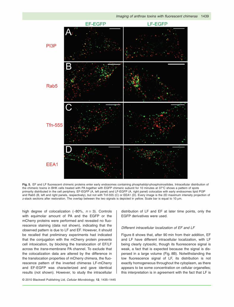
PA and LF/EF. Figure 5 shows the images obtained afteran incubation of 10 min at 37°C; the two toxins arewithin early endosomal compartments that contain phos-phatidylinositol 3-phosphate (PI3P) (~30%, n � 3) andRab5 (~40%, n � 3), but apparently not transferrin (Tfn)or early endosomal antigen 1 (EEA1) (13% and 12%,respectively, n � 4). This is not unprecedented, as it hasbeen reported that cholera toxin B- and Simian virus40-containing organelles are distinct from classicalEEA1- and Tfn-positive endosomes, but communicatewith early endosomes via a pathway regulated by Rab5(Pelkmans et al., 2001; Nichols, 2002; Parton andSimons, 2007). These data show that the endocytosis ofLF/EF mediated by the binding of PA to its receptors inBHK cells is rapid and efficient, with undetectable LF/EFremaining on the cell surface after 10 min. This kineticsreflects the fact that the (PA63)7-LF triggers its ownendocytosis via modifications of the PA receptor, whichtake place on the cytosolic side of the protein andincludes ubiquitination, palmitoylation and induction ofspecific phosphorylation of the receptors associated pro-teins (Abrami et al., 2004, 2006, 2008b).
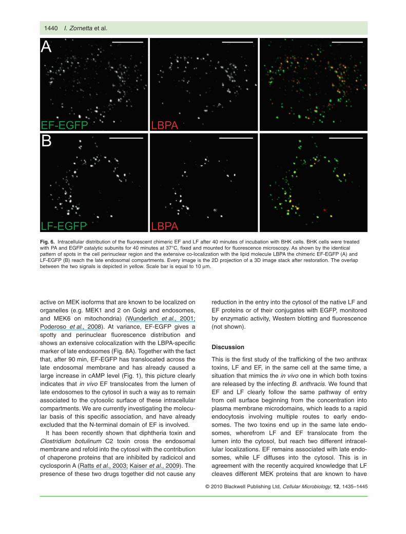
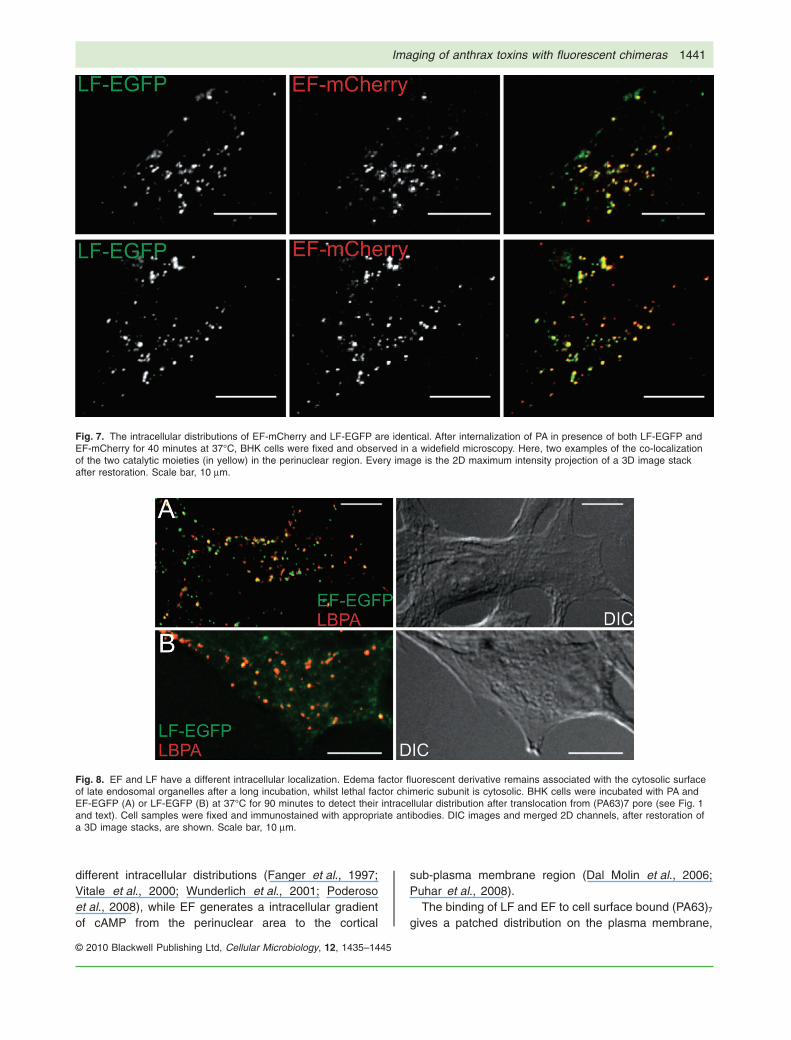
After 40 min, both EF and LF are within late endosomalcompartments, as indicated by the extensive colocaliza-tion (~94%, n � 3) with the lipid molecule lysobisphospha-tidic acid (LBPA) (Fig. 6), a marker of these compartments(Kobayashi et al., 1998). The fluorescence patterns ofEF-mCherry and LF-EGFP intracellular distribution areidentical up to this time point (Fig. 7), as indicated by a
Fig. 3. Binding of EF and LF fluorescent chimeric proteins to BHK cells. EF-EGFP (A) and LF-EGFP (D) colocalize with caveolin1 and lysenin(G and J, respectively) in peripheral punctuate structures. Cells were pre-treated with PA83 to allow its binding and processing, and for 2minutes at 37°C with chimeric subunits, after which they were fixed and stained with antibodies to caveolin and lysenin. Boxes define theareas from which the corresponding insets (B, E, H and K) were generated. Reconstruction of the z-axis profile was performed (C, F, I and Linsets). Every image is the 2D projection of a 3D image stack after restoration. The overlap between the two signals is depicted in yellow.Scale bar is equal to 10 mm.
Fig. 4. As in figure 3, EF-EGFP (A) and LF-EGFP (B) show loweror no colocalization with clathrin in cells transfected withmRFP-clathrin light chain. Every image is the 2D projection of a 3Dimage stack after restoration. The overlap between the two signalsis depicted in yellow. Scale bar is equal to 10 mm.
1438 I. Zornetta et al.
© 2010 Blackwell Publishing Ltd, Cellular Microbiology, 12, 1435–1445
high degree of colocalization (~80%, n � 3). Controlswith equimolar amount of PA and the EGFP or themCherry proteins were performed and revealed no fluo-rescence staining (data not shown), indicating that theobserved pattern is due to LF and EF. However, it shouldbe recalled that preliminary experiments had indicatedthat the conjugation with the mCherry protein preventscell intoxication, by blocking the translocation of EF/LFacross the trans-membrane PA channel. To exclude thatthe colocalization data are altered by the difference inthe translocation properties of mCherry chimera, the fluo-rescence pattern of the inverted chimeras LF-mCherryand EF-EGFP was characterized and gave identicalresults (not shown). However, to study the intracellular
distribution of LF and EF at later time points, only theEGFP derivatives were used.
Different intracellular localization of EF and LF
Figure 8 shows that, after 90 min from their addition, EFand LF have different intracellular localization, with LFbeing clearly cytosolic, though its fluorescence signal isweak, a fact that is expected because the signal is dis-persed in a large volume (Fig. 8B). Notwithstanding thelow fluorescence signal of LF, its distribution is notexactly homogeneous throughout the cytoplasm, as thereappears to be some concentration on cellular organelles;this interpretation is in agreement with the fact that LF is
Fig. 5. EF and LF fluorescent chimeric proteins enter early endosomes containing phosphatidyl-phosphoinositides. Intracellular distribution ofthe chimeric toxins in BHK cells treated with PA together with EGFP chimeric subunit for 10 minutes at 37°C shows a pattern of spotsprimarily distributed in the cell periphery. EF-EGFP (A, left panel) and LF-EGFP (A, right panel) colocalize with early endosomes lipid PI3Pand Rab5 (B, left and right panels, respectively), but not with Tnf-555 (C) or EEA1 (D). Every image is the 2D maximum intensity projection ofz-stack sections after restoration. The overlap between the two signals is depicted in yellow. Scale bar is equal to 10 mm.
Imaging of anthrax toxins with fluorescent chimeras 1439
© 2010 Blackwell Publishing Ltd, Cellular Microbiology, 12, 1435–1445
active on MEK isoforms that are known to be localized onorganelles (e.g. MEK1 and 2 on Golgi and endosomes,and MEK6 on mitochondria) (Wunderlich et al., 2001;Poderoso et al., 2008). At variance, EF-EGFP gives aspotty and perinuclear fluorescence distribution andshows an extensive colocalization with the LBPA-specificmarker of late endosomes (Fig. 8A). Together with the factthat, after 90 min, EF-EGFP has translocated across thelate endosomal membrane and has already caused alarge increase in cAMP level (Fig. 1), this picture clearlyindicates that in vivo EF translocates from the lumen oflate endosomes to the cytosol in such a way as to remainassociated to the cytosolic surface of these intracellularcompartments. We are currently investigating the molecu-lar basis of this specific association, and have alreadyexcluded that the N-terminal domain of EF is involved.
It has been recently shown that diphtheria toxin andClostridium botulinum C2 toxin cross the endosomalmembrane and refold into the cytosol with the contributionof chaperone proteins that are inhibited by radicicol andcyclosporin A (Ratts et al., 2003; Kaiser et al., 2009). Thepresence of these two drugs together did not cause any
reduction in the entry into the cytosol of the native LF andEF proteins or of their conjugates with EGFP, monitoredby enzymatic activity, Western blotting and fluorescence(not shown).
Discussion
This is the first study of the trafficking of the two anthraxtoxins, LF and EF, in the same cell at the same time, asituation that mimics the in vivo one in which both toxinsare released by the infecting B. anthracis. We found thatEF and LF clearly follow the same pathway of entryfrom cell surface beginning from the concentration intoplasma membrane microdomains, which leads to a rapidendocytosis involving multiple routes to early endo-somes. The two toxins end up in the same late endo-somes, wherefrom LF and EF translocate from thelumen into the cytosol, but reach two different intracel-lular localizations. EF remains associated with late endo-somes, while LF diffuses into the cytosol. This is inagreement with the recently acquired knowledge that LFcleaves different MEK proteins that are known to have
Fig. 6. Intracellular distribution of the fluorescent chimeric EF and LF after 40 minutes of incubation with BHK cells. BHK cells were treatedwith PA and EGFP catalytic subunits for 40 minutes at 37°C, fixed and mounted for fluorescence microscopy. As shown by the identicalpattern of spots in the cell perinuclear region and the extensive co-localization with the lipid molecule LBPA the chimeric EF-EGFP (A) andLF-EGFP (B) reach the late endosomal compartments. Every image is the 2D projection of a 3D image stack after restoration. The overlapbetween the two signals is depicted in yellow. Scale bar is equal to 10 mm.
1440 I. Zornetta et al.
© 2010 Blackwell Publishing Ltd, Cellular Microbiology, 12, 1435–1445
different intracellular distributions (Fanger et al., 1997;Vitale et al., 2000; Wunderlich et al., 2001; Poderosoet al., 2008), while EF generates a intracellular gradientof cAMP from the perinuclear area to the cortical
sub-plasma membrane region (Dal Molin et al., 2006;Puhar et al., 2008).
The binding of LF and EF to cell surface bound (PA63)7
gives a patched distribution on the plasma membrane,
Fig. 7. The intracellular distributions of EF-mCherry and LF-EGFP are identical. After internalization of PA in presence of both LF-EGFP andEF-mCherry for 40 minutes at 37°C, BHK cells were fixed and observed in a widefield microscopy. Here, two examples of the co-localizationof the two catalytic moieties (in yellow) in the perinuclear region. Every image is the 2D maximum intensity projection of a 3D image stackafter restoration. Scale bar, 10 mm.
Fig. 8. EF and LF have a different intracellular localization. Edema factor fluorescent derivative remains associated with the cytosolic surfaceof late endosomal organelles after a long incubation, whilst lethal factor chimeric subunit is cytosolic. BHK cells were incubated with PA andEF-EGFP (A) or LF-EGFP (B) at 37°C for 90 minutes to detect their intracellular distribution after translocation from (PA63)7 pore (see Fig. 1and text). Cell samples were fixed and immunostained with appropriate antibodies. DIC images and merged 2D channels, after restoration ofa 3D image stacks, are shown. Scale bar, 10 mm.
Imaging of anthrax toxins with fluorescent chimeras 1441
© 2010 Blackwell Publishing Ltd, Cellular Microbiology, 12, 1435–1445
which is fully consistent with the two toxins enteringcholesterol-enriched microdomains. This was establishedpreviously by Abrami et al. (2003) for LF and it isextended here to EF. Here, we also found that several ofthese microdomains are enriched in sphingomyelin, asthere is colocalization with lysenin, a sphingomyelinbinding protein (Yamaji et al., 1998). Cell surface bindingwas found to be rapidly followed by endocytosis.From the present study, it appears that the (PA63)7-LFand (PA63)7-EF complexes may enter various types ofendocytic vesicles to reach early endosomal compart-ments, most of them marked by the presence of Rab5 andPI3P. It should be noted that, here, we have deliberatelychosen not to use any method that may alter the physi-ological process of cell entry of these toxins, such ascholesterol depletion, cross-linking, inhibitors, siRNA,overexpression of different proteins, etc. The only ‘cellmanipulation’ procedure used was that of expressing thelight chain of clathrin coupled to mRFP, but this proteinwas not found to colocalize substantially with the twotoxins. This result is at variance with the previous report ofa clathrin-mediated endocytosis of LF in CHO cells(Abrami et al., 2003), and we have no satisfactory expla-nation for such a difference. However, different cell lineswere used and fibroblasts are known to have multiplepathways of endocytosis (Doherty and McMahon, 2009).On the other hand, the present result is not surprising inlight of the fact that other toxins that bind via an oligomericbinding protomer enter cells via nonclathrin-dependenttrafficking (Sandvig et al., 2008) and that the majority ofligands that bind to raft microdomains are preferentiallytaken up by clathrin-independent endocytosis (Nichols,2003).
No matter which initial traffic route is taken by LF andEF to early endosomes, eventually they reach late endo-somal compartments, wherefrom they translocate into thecytosol. This translocation is not assisted by chaperonesinhibited by radicicol or cyclosporin A, as was found to bethe case for diphtheria toxin and Clostridium botulinum C2toxin (Kaiser et al., 2009).
A remarkable finding presented here is that both LFand EF are capable of pulling the conjugated EGFPthrough the PA channel with high efficiency, while this isnot the case of the mCherry fluorescent protein, whichis more resistant to unfolding than the EGFP protein(Fig. S2). Taken together, these data provide further evi-dence in favour of the model of translocation of LF thatenvisages a low pH driven unfolding of the polypeptidechain to enter the PA channel and translocation of theunfolded chain that refolds in the neutral pH of thecytosol (Krantz et al., 2005; Collier, 2009). At the sametime, the present report shows that mCherry may not bean appropriate choice as reporter of intracellularly actingtoxins.
Experimental procedures
Cells, antibodies and reagents
BHK cells were maintained in DMEM (Gibco) containing 10%heat-inactivated foetal calf serum (FCS, Euroclone), penicillin(100 U ml-1) and streptomycin (100 mg ml-1). Antibodies wereobtained from the following sources: anti-His tag monoclonalantibody from Novagen, monoclonal and polyclonal anti-GFP,-RFP, -EF and -LF polyclonal antibodies from Abcam, anti-caveolin and anti-EEA1 antibodies from BD Transduction Labo-ratories; anti-PI3P from Echelon, anti-Rab5 from SynapticSystem, anti-LBPA (6C4) was a kind gift of J. Gruenberg (Uni-versity of Geneva, CH), Tfn-Alexa555 and fluorescently labelledsecondary antibodies from Molecular Probes. Lysenin andlysenin antiserum were from Peptide Institute; FuGENE HD fromRoche Diagnostics Corporation. The plasmid encoding mRFP-Clathrin Light Chain was from Addgene. Reagents were Sigma-Aldrich and Calbiochem.
Cloning, expression and purification of chimeric proteins
The EGFP gene was PCR-amplified from pEGFP-N1 (Clontech)using the following primers: forward 5′-AAAGAGCTCATGGTGAGCAAGGGCG-3′ and reverse 5′-AAAGAATTCCTTGTACAGCTCGTCCAT-3′. The mCherry gene was PCR-amplified frompREST B, a generous gift from RY Tsien, using the followingprimers: forward 5′-AAAGAGCTCATGGCCACTGGTGGACAG-3′ and reverse 5′-AAAGAATTCTAGGCGCCGGYGGAGT-3′.Both fragments were digested with SacI and EcoRI and insertedin pRSET A (Invitrogen) containing LF or EF, respectively, aspreviously described (Dal Molin et al., 2006), downstream froman N-terminal His-tag coding region. The sequences were con-firmed by DNA sequencing. LF chimeric derivatives wereexpressed in Escherichia coli BL21(DE3) and EF chimeric pro-teins in E. coli BL21 (DE3)-Codon Plus-RIL (Stratagene) grownat 37°C in LB broth containing 100 mg ml-1 ampicillin or100 mg ml-1 ampicillin and 34 mg ml-1 chloramphenicol. After 4 hof induction with 1 mM isopropyl-1-thio-a-D-galactopyranoside at30°C, the pellet was resuspended in buffer A (50 mM Na2HPO4,500 mM NaCl, pH 8) and lysozyme (0.1 mg ml-1). Bacterial cellswere disrupted by ultrasonic dispersion and centrifuged, and thesupernatant was loaded onto a Hi-trap column charged withCu2SO4 and equilibrated with buffer A. The column was washedwith buffer A, the protein was eluted with a 0–100 mM imidazolegradient, and the fractions containing chimeric proteins werepooled and dialysed against binding buffer (50 mM Tris, 20 mMNaCl and 1 mM EDTA, pH 7.5) to remove imidazole and NaCl.The identities of chimeric proteins were assessed by immunob-lotting with anti-His tag, anti-LF, anti-EF and anti-GFP or anti-mRFP antibodies. We used this antibody, which recognizes aconserved epitope on mCherry, because no other was availablefor this red variant.
FRET imaging of cAMP intracellular dynamics
BHK cells (2.0 ¥ 105) were co-transfected with 1 mg of twopcDNA3.1 plasmids, one carrying the catalytic (C) subunit of PKAfused to YFP (C-YFP) and one carrying the regulatory (R) subunitof PKA fused to CFP (RII-CFP) (Lissandron et al., 2005) usingFuGENE HD reagent, following manufacturer’s instructions.
1442 I. Zornetta et al.
© 2010 Blackwell Publishing Ltd, Cellular Microbiology, 12, 1435–1445
Forty-eight hours after transfection, cells were incubated in abalanced salt solution (NaCl 135 mM, KCl 5 mM, KH2PO40.4 mM, MgSO4 1 mM, HEPES 20 mM, CaCl2 1.8 mM, glucose5.4 mM, pH 7.4) in a microscope-adapted micro-incubatorequipped with a temperature controller (HTC, Italy) at 37°C andconstant 5% CO2 pressure. Toxins were added after about 15 minof imaging, and images were taken every 20 s for the indicatedtime periods. Integration time was 200 ms. At each time point, theintracellular cAMP level was estimated by measuring the ratiobetween the background-subtracted CFP emission image andthe YFP emission image upon excitation of CFP (R CFP/YFP)(Mongillo et al., 2005). Images were acquired using an oil immer-sion 40¥ PlanApo 1.4 NA objective on a Leica DMI6000 micro-scope. FRET images were collected through a BP 436/20-nmexcitation filter and a custom-made optical beam splitter built witha 515 nm dichroic mirror and ET 480/40-nm and ET 535/30-nmemission filters. All optical filters were obtained from ChromaTechnologies. A cooled camera from OES (Padova, Italy) with a1,4 Megapixel CCD and a sensor resolution of 1360 ¥ 1024 Pixelwas used. The acquisition software was from OES (Padova,Italy). Recorded images were processed with WCIF ImageJv1.40 (http://rsb.info.nih.gov/ij).
MEK3 cleavage by chimeric lethal factor
BHK cells (1.5 ¥ 104) were incubated with PA 400 nM and 200 nMLF or LF-EGFP in DMEM plus 1% BSA at 37°C for different timeperiods in a 96-well plate. After removal of the culture medium,the cells were lysed, subjected to SDS-PAGE, and immunoblot-ted for the isoform 3 of MEK with a specific polyclonal antibodyfrom Santa Cruz Biotechnology (USA). Samples were developedwith ECL plus detection system (Amersham Biosciences), andchemiluminescence emission was detected with ChemiDoc™XRS (Biorad). The antibody detects two splicing isoforms ofMEK3, MEK3b and MEK3a, a variant lacking of the first 29residues. Only the MEK3b is cleaved by LF and the percentageof cleavage was quantified considering the ratio between theMEK3b band and the band intensity at lower molecular weight,normalizing the result against the control. Band intensities werequantified with the Quantity One software from Biorad.
Fluorescence microscopy
Sub-confluent BHK cells grown on glass coverslips were rinsedtwo times with DMEM plus 2% w/v BSA and treated for differentperiods of time with EF-mCherry and/or LF-EGFP (200 nM) andPA (600 nM). Then cells were washed with PBS, fixed with ice-cold acetone for 5 min at room temperature to localize the toxinsalong the endocytic pathway or with PFA 4% (10 min at roomtemperature) to mark endocytic lipids and incubated sequentiallywith a mixture of primary antibodies and a mixture of fluorescentsecondary antibodies. To monitor cell surface binding, cells werefirst treated with PA83 for 12 min, washed, incubated with LF orEF derivatives for 2 min at 37°C and immediately washed andfixed. All antibody incubations were performed for 1 h at roomtemperature. Images were acquired sequentially with a FITC andTexas Red® filter set (Chroma Technology corp., USA) with250 ms or longer integration times by using an oil immersion 63¥PlanApo 1.40 NA objective on a Leica DMIRE3 widefield invertedmicroscope equipped with a DC 500 digital camera with
1300 ¥ 1030 pixels resolution from Leica. The acquisition soft-ware was FW4000 (Leica). Images were processed with ImageJv1.40 (http://rsb.info.nih.gov/ij) and colocalization analysis onraw images was performed using the JaCoP plugin (Bolte andCordelières, 2006) and OBCOL plugin (Woodcroft et al., 2009)under ImageJ.
Colocalization studies were performed with an object basedanalysis with plugins which autonomously detect objects withinan image stack (z-planes � 4) and analyse them as individualobjects. Before segmentation, each channel was preprocessedto remove background fluorescence below a given intensity cut-off. Cut-offs between 40 and 50 and 50–60 intensity units wereapplied to the red and the green channels respectively. Anoverlap of three pixels was required to join objects and theobjects of dimensions < 10 pixel were excluded. Images weresegmented to automatically define discrete objects, at least 120objects were defined for each examined cell. Colocalization wasexpressed as the percentage of objects containing green and redpixels on the objects that contain only red pixels.
Translocation of chimeric proteins acrossartificial membranes
Planar phospholipid bilayers were formed by standard methods(Janowiak et al., 2009). Once a membrane was formed, WT PA63
prepore (25 pM) was added to the cis compartment, held at aDy = +20 mV with respect to the trans compartment. Free PA63
not inserted into the membrane was removed by perfusion.Binding cargo (LF, EF, or chimeric proteins) was added to the ciscompartment (1 mg ml-1), and the progress of binding to PA chan-nels was monitored by the decrease in conductance. Free wasremoved by buffer exchange. Translocation was initiated byraising the pH of the trans compartment to pH 7.2 with 2 M KOH,while maintaining the cis compartment pH at 5.5. At the sametime, we increased the membrane potential from Dy = +20 mV toDy = +50 mV. Experiments were normalized to controls lackingcargo protein (n � 3). All planar phospholipid experiments wereperformed in a Warner Instruments Planar Lipid Bilayer Worksta-tion (BC 525D, Hamden, CT).
Acknowledgements
This work has been supported by the MIUR programme FIRBInternazionalizzazione and the Istituto Superiore di Sanità andNIH grants 1F32 AI077280 to BEJ and 5R01 AI022021 to RJC,who holds equity in PharmAthene.
References
Abrami, L., Liu, S., Cosson, P., Leppla, S.H., and van derGoot, F.G. (2003) Anthrax toxin triggers endocytosis ofits receptor via a lipid raft-mediated clathrin-dependentprocess. J Cell Biol 160: 321–328.
Abrami, L., Lindsay, M., Parton, R.G., Leppla, S.H., and vander Goot, F.G. (2004) Membrane insertion of anthrax pro-tective antigen and cytoplasmic delivery of lethal factoroccur at different stages of the endocytic pathway. J CellBiol 166: 645–651.
Abrami, L., Reig, N., and van der Goot, F.G. (2005) Anthraxtoxin: the long and winding road that leads to the kill.Trends Microbiol 13: 72–78.
Imaging of anthrax toxins with fluorescent chimeras 1443
© 2010 Blackwell Publishing Ltd, Cellular Microbiology, 12, 1435–1445
Abrami, L., Leppla, S.H., and van der Goot, F.G. (2006)Receptor palmitoylation and ubiquitination regulate anthraxtoxin endocytosis. J Cell Biol 172: 309–320.
Abrami, L., Kunz, B., Deuquet, J., Bafico, A., Davidson, G.,and van der Goot, F.G. (2008a) Functional interactionsbetween anthrax toxin receptors and the WNT signallingprotein LRP6. Cell Microbiol 10: 2509–2519.
Abrami, L., Kunz, B., Iacovache, I., and van der Goot, F.G.(2008b) Palmitoylation and ubiquitination regulate exit ofthe Wnt signaling protein LRP6 from the endoplasmicreticulum. PNAS 105: 5384–5389.
Ascenzi, P., Visca, P., Ippolito, G., Spallarossa, A., Bolognesi,M., and Montecucco, C. (2002) Anthrax toxin: a tripartitelethal combination. FEBS Lett 531: 384–388.
Baldari, C.T., Tonello, F., Paccani Rossi, S., and Montecucco,C. (2006) Anthrax toxins: a paradigm of bacterial immunesuppression. Trends Immunol 27: 434–440.
Beall, F.A., Taylor, M.J., and Thorne, C.B. (1962) Rapid lethaleffect in rats of a third component found upon fractionatingthe toxin of Bacillus anthracis. J Bacteriol 83: 1274–1280.
Beyer, W., and Turnbull, P.C.B. (2009) Anthrax in animals.Mol Aspects Med 30: 481–489.
Boll, W., Ehrlich, M., Collier, R.J., and Kirchhausen, T. (2004)Effects of dynamin inactivation on pathways of anthraxtoxin uptake. Eur J Cell Biol 83: 281–288.
Bolte, S., and Cordelières, F.P. (2006) A guided tour intosubcellular colocalization analysis in light microscopy.J Microsc 224: 213–232.
Collier, R.J. (2009) Membrane translocation by anthrax toxin.Mol Aspects Med 30: 413–422.
Cybulski, R.J.J., Sanz, P., and O’Brien, A.D. (2009) Anthraxvaccination strategies. Mol Aspects Med 30: 490–502.
Dal Molin, F., Tonello, F., Ladant, D., Zornetta, I., Zamparo, I.,Di Benedetto, G., et al. (2006) Cell entry and cAMPimaging of anthrax edema toxin. EMBO J 25: 5405–5413.
Dixon, T.C., Meselson, M., Guillemin, J., and Hanna, P.(1999) Anthrax. N Engl J Med 341: 815–826.
Doherty, G.J., and McMahon, H.T. (2009) Mechanisms ofendocytosis. Annu Rev Biochem 78: 857–902.
Drum, C.L., Yan, S., Bard, J., Shen, Y., Lu, D., Soelaiman, S.,et al. (2002) Structural basis for the activation of anthraxadenylyl cyclase exotoxin by calmodulin. Nature 415: 396–402.
Duesbery, N.S., Webb, C.P., Leppla, S.H., Gordon, V.M.,Klimpel, K.R., Copeland, T.D., et al. (1998) Proteolyticinactivation of MAP-kinase-kinase by anthrax lethal factor.Science 280: 734–737.
Fanger, G.R., Johnson, N.L., and Johnson, G.L. (1997) MEKkinases are regulated by EGF and selectively interact withRac/Cdc42. EMBO J 16: 4961–4972.
Firoved, A.M., Miller, G.F., Moayeri, M., Kakkar, R., Shen, Y.,Wiggins, J.F., et al. (2005) Bacillus anthracis edema toxincauses extensive tissue lesions and rapid lethality in mice.Am J Pathol 167: 1309–1320.
Fouet, A. (2009) The surface of Bacillus anthracis. MolAspects Med 30: 374–385.
Gaestel, M. (2006) MAPKAP kinases – MKs – two’scompany, three’s a crowd. Nat Rev Mol Cell Biol 7: 120–130.
Janowiak, B.E., Finkelstein, A., and Collier, R.J. (2009)An approach to characterizing single-subunit mutations in
multimeric prepores and pores of anthrax protectiveantigen. Protein Sci 18: 348–358.
Kaiser, E., Pust, S., Kroll, C., and Barth, H. (2009) CyclophilinA facilitates translocation of the Clostridium botulinum C2toxin across membranes of acidified endosomes into thecytosol of mammalian cells. Cell Microbiol 11: 780–795.
Kobayashi, T., Stang, E., Fang, K.S., de Moerloose, P.,Parton, R.G., and Gruenberg, J. (1998) A lipid associatedwith the antiphospholipid syndrome regulates endosomestructure and function. Nature 392: 193–197.
Koehler, T.M. (2009) Bacillus anthracis physiology and genet-ics. Mol Aspects Med 30: 386–396.
Krantz, B.A., Melnyk, R.A., Zhang, S., Juris, S.J., Lacy, D.B.,Wu, Z., et al. (2005) A phenylalanine clamp catalyzesprotein translocation through the anthrax toxin pore.Science 309: 777–781.
Lissandron, V., Terrin, A., Collini, M., D’Alfonso, L., Chirico,G., Pantano, S., et al. (2005) Improvement of a FRET-based indicator for cAMP by linker design and stabilizationof donor-acceptor interaction. J Mol Biol 354: 546–555.
Moayeri, M., and Leppla, S.H. (2009) Cellular and systemiceffects of anthrax lethal toxin and edema toxin. MolAspects Med 30: 439–455.
Mongillo, M., Terrin, A., Evellin, S., Lissandron, V., andZaccolo, M. (2005) Study of cyclic adenosine monophos-phate microdomains in cells. Methods Mol Biol 307: 1–13.
Neumeyer, T., Tonello, F., Dal Molin, F., Schiffler, B., andBenz, R. (2006) Anthrax edema factor, voltage-dependentbinding to the protective antigen ion channel and compari-son to LF binding. J Biol Chem 281: 32335–32343.
Nichols, B. (2003) Caveosomes and endocytosis of lipid rafts.J Cell Sci 116: 4707–4714.
Nichols, B.J. (2002) A distinct class of endosome mediatesclathrin-independent endocytosis to the Golgi complex. NatCell Biol 4: 374–378.
Paccani Rossi, S., Tonello, F., Ghittoni, R., Natale, M.,Muraro, L., D’Elios, M.M., et al. (2005) Anthrax toxins sup-press T lymphocyte activation by disrupting antigen recep-tor signaling. J Exp Med 201: 325–331.
Parton, R.G., and Simons, K. (2007) The multiple faces ofcaveolae. Nat Rev Mol Cell Biol 8: 185–194.
Pelkmans, L., Kartenbeck, J., and Helenius, A. (2001)Caveolar endocytosis of simian virus 40 reveals a newtwo-step vesicular-transport pathway to the ER. Nat CellBiol 3: 473–483.
Pellizzari, R., Guidi-Rontani, C., Vitale, G., Mock, M., andMontecucco, C. (1999) Anthrax lethal factor cleaves MKK3in macrophages and inhibits the LPS/IFNgamma-inducedrelease of NO and TNFalpha. FEBS Lett 462: 199–204.
Poderoso, C., Converso, D.P., Maloberti, P., Duarte, A.,Neuman, I., Galli, S., et al. (2008) A mitochondrial kinasecomplex is essential to mediate an ERK1/2-dependentphosphorylation of a key regulatory protein in steroid bio-synthesis. PLoS One 3: e1443.
Puhar, A., and Montecucco, C. (2007) Where and how doanthrax toxins exit endosomes to intoxicate host cells?Trends Microbiol 15: 477–482.
Puhar, A., Dal Molin, F., Horvath, S., Ladants, D., and Mon-tecucco, C. (2008) Anthrax edema toxin modulates PKA-and CREB-dependent signaling in two phases. PLoS One3: e3564.
1444 I. Zornetta et al.
© 2010 Blackwell Publishing Ltd, Cellular Microbiology, 12, 1435–1445
Ratts, R., Zeng, H., Berg, E.A., Blue, C., McComb, M.E.,Costello, C.E., et al. (2003) The cytosolic entry of diphthe-ria toxin catalytic domain requires a host cell cytosolictranslocation factor complex. J Cell Biol 160: 1139–1150.
Sandvig, K., Torgersen, M.L., Raa, H.A., and van Deurs, B.(2008) Clathrin-independent endocytosis: from nonexistingto an extreme degree of complexity. Histochem Cell Biol129: 267–276.
Shaner, N.C., Campbell, R.E., Steinbach, P.A., Giepmans,B.N.G., Palmer, A.E., and Tsien, R.Y. (2004) Improvedmonomeric red, orange and yellow fluorescent proteinsderived from Discosoma sp. red fluorescent protein. NatBiotechnol 22: 1567–1572.
Shaner, N.C., Steinbach, P.A., and Tsien, R.Y. (2005) A guideto choosing fluorescent proteins. Nat Methods 2: 905–909.
Shen, Y., Zhukovskaya, N.L., Guo, Q., Florián, J., and Tang,W. (2005) Calcium-independent calmodulin binding andtwo-metal-ion catalytic mechanism of anthrax edemafactor. EMBO J 24: 929–941.
Smith, H., and Keppie, J. (1954) Observations on experimen-tal anthrax; demonstration of a specific lethal factor pro-duced in vivo by Bacillus anthracis. Nature 173: 869–870.
Smith, H., Keppie, J., Stanley, J.L., and Harris-Smith, P.W.(1955) The chemical basis of the virulence of Bacillusanthracis. IV. Secondary shock as the major factor in deathof guinea-pigs from anthrax. Br J Exp Pathol 36: 323–335.
Smith, H., Tempest, D.W., Stanley, J.L., Harris-Smith, P.W.,and Gallop, R.C. (1956) The chemical basis of the viru-lence of Bacillus anthracis. VII. Two components of theanthrax toxin: their relationship to known immunisingaggressins. Br J Exp Pathol 37: 263–271.
Tonello, F., and Montecucco, C. (2009) The anthrax lethalfactor and its MAPK kinase-specific metalloprotease activ-ity. Mol Aspects Med 30: 431–438.
Tournier, J., Quesnel-Hellmann, A., Mathieu, J., Montecucco,C., Tang, W., Mock, M., et al. (2005) Anthrax edema toxincooperates with lethal toxin to impair cytokine secretionduring infection of dendritic cells. J Immunol 174: 4934–4941.
Tournier, J., Ulrich, R.G., Quesnel-Hellmann, A., Moham-adzadeh, M., and Stiles, B.G. (2009) Anthrax, toxins andvaccines: a 125-year journey targeting Bacillus anthracis.Expert Rev Anti Infect Ther 7: 219–236.
Van Der Goot, G., and Young, J.A.T. (2009) Receptors ofanthrax toxin and cell entry. Mol Aspects Med 30: 406–412.
Vitale, G., Pellizzari, R., Recchi, C., Napolitani, G., Mock, M.,and Montecucco, C. (1998) Anthrax lethal factor cleaves
the N-terminus of MAPKKs and induces tyrosine/threoninephosphorylation of MAPKs in cultured macrophages.Biochem Biophys Res Commun 248: 706–711.
Vitale, G., Bernardi, L., Napolitani, G., Mock, M., and Mon-tecucco, C. (2000) Susceptibility of mitogen-activatedprotein kinase kinase family members to proteolysis byanthrax lethal factor. Biochem J 352: 739–745.
Woodcroft, B.J., Hammond, L., Stow, J.L., and Hamilton, N.A.(2009) Automated organelle-based colocalization in whole-cell imaging. Cytometry 75: 941–950.
Wunderlich, W., Fialka, I., Teis, D., Alpi, A., Pfeifer, A., Parton,R.G., et al. (2001) A novel 14-kilodalton protein interactswith the mitogen-activated protein kinase scaffold mp1 ona late endosomal/lysosomal compartment. J Cell Biol 152:765–776.
Yamaji, A., Sekizawa, Y., Emoto, K., Sakuraba, H., Inoue, K.,Kobayashi, H., et al. (1998) Lysenin, a novelsphingomyelin-specific binding protein. J Biol Chem 273:5300–5306.
Young, J.A.T., and Collier, R.J. (2007) Anthrax toxin: receptorbinding, internalization, pore formation, and translocation.Annu Rev Biochem 76: 243–265.
Supporting information
Additional Supporting Information may be found in the onlineversion of this article:
Fig. S1. Immunoblot analysis of EF and LF chimeric derivatives.Immunoblot analysis of SDS-PAGE separated, fluorescent chi-meric proteins (0.1 mg) performed with antibodies against the6xHis, EF or LF, and fluorescent protein mCherry or EGFP. Weused an antibody against mRFP that recognizes a conservedepitope on mCherry because no antibody was available for thelatter red variant. WB analysis of EGFP derivatives of EF (A) andLF (B) and of EF-mCherry (C) and LF-mCherry (D).Fig. S2. Fluorescence changes for EGFP and mCherry accom-panying protein unfolding in different concentration of guanidinehydrochloride. Protein samples (0.050 mg ml-1) were incubatedfor 20 h at 25 °C in the presence of various concentrations ofguanidine hydrochloride. Unfolding curves were determined byexciting the samples at 365 nm and detecting the emitted fluo-rescence at appropriate wavelenght for EGFP and mCherry (seesupplementary experimental procedures).Appendix S1. Supplementary experimental procedures.
Please note: Wiley-Blackwell are not responsible for the contentor functionality of any supporting materials supplied by theauthors. Any queries (other than missing material) should bedirected to the corresponding author for the article.
Imaging of anthrax toxins with fluorescent chimeras 1445
© 2010 Blackwell Publishing Ltd, Cellular Microbiology, 12, 1435–1445
Recommended


