
Intestinal lschemia-Reperfusion InjuryCauses Pulmonary EndothelialCell ATP DepletionTodd M. Gerkin, M.D.,t Keith T. Oldham, M.D.,t Karen S. Guice, M.D.,*Daniel B. Hinshaw, M.D.,t and Una S. Ryan, Ph.D.§
From the Divisions of General Surgery* and Pediatric Surgery,t Department of Surgery, DukeUniversity Medical Center, Durham, North Carolina; the Section of General Surgery,tDepartment of Surgery, University of Michigan Medical School, Ann Arbor, Michigan; and theDepartment of Surgery,§ Washington University School of Medicine, St. Louis, Missouri
Intestinal ischemia-reperfusion is a common clinical event associated with both clinical andexperimental distant organ injury. In particular, the pulmonary microvasculature appears to besusceptible to injury resulting from systemic inflammatory mediator activation. This study wasdesigned to evaluate the hypothesis that noncellular humoral factors associated with intestinalischemia-reperfusion result in pulmonary endothelial cell adenosine triphosphate (ATP) depletion.Male Sprague-Dawley rats had intestinal ischemia induced by microvascular clip occlusion ofthe superior mesenteric artery (SMA) for 120 minutes. Reperfusion resulted from superiormesenteric artery clip removal. After reperfusion for 0, 15, or 30 minutes, plasma samples wereobtained from the portal vein. Monolayers of cultured rat pulmonary artery endothelial cells thenwere incubated with the plasma samples. Adenosine triphosphate levels were determined usinga luciferin-luciferase assay. A 5'Cr-release assay using labeled endothelial cells was performedunder identical conditions to assess cytotoxicity. Potential mechanisms of ATP depletion wereevaluated by analysis of cellular energy charge and assessment of microfilament architecture.Endothelial cell ATP levels decreased from 2.23 ± 0.16 X 10⁻¹¹ moles/,ug DNA in shampreparations to 1.23 ± 0.09 X 10`1 moles/,ug DNA (p < 0.001) after 4 hours in plasma fromanimals undergoing 120 minutes of intestinal ischemia. For plasma obtained after 15 minutesof reperfusion, the decrease in cellular ATP concentration persisted (1.23 ± 0.27 X 10-"moles/,ug DNA, p < 0.001 vs. sham). After 30 minutes' reperfusion, cellular ATP levels increasedonly slightly after the 4-hour incubation (1.39 ± 0.26 X 10-" moles/,ug DNA, p < 0.005 vs.sham). No significant cytotoxic injury occurred in any group when compared with controls.Cellular energy charge was unchanged, and microfilament architecture was preserved. Thesedata confirm the hypothesis that humoral factors, independent of the neutrophil, result inendothelial cell ATP depletion without metabolic inhibition or cell death. Depletion of energystores by noncellular humoral factors may represent an early event that predisposes the cell tomore severe injury by other mediators of the endogenous inflammatory response.
Multiple organ failure, or MOF, is a leading cause of sive care unit.' Clinically, MOF may be defined as a pro-death in surgical and posttrauma patients in the inten- gressive deterioration in the ability ofthe visceral organs
to perform normal physiologic functions. The patho-... . ~genic events that initiate MOF include many commonAddress reprint requests to Keith T. Oldham, M.D., Division ofPediat- genicaevnt iti aTherma incude many common
ric Surgery, Duke University Medical Center, PO Box 3815, Dur- clinical condltons. Thermal injury,t sepsis,"4n5hemor-ham, NC 27710. rhagic shock,6'7 trauma,8 acute pancreatitis,9"0 and isch-Accepted for publication April 17, 1992. emia-reperfusion events"'3 are all known precipitants
48

Endothelial ATP Depletion After Ischemia-Reperfusion 49
of MOF. Each stimulus is apparently unique; however,all are associated with activation of the endogenous in-flammatory response. Some ofthe inflammatory media-tors appear to be part of a common pathway resulting indistant organ injury.Acute lung injury associated with the MOF syndrome
is a subject of particular interest and clinical impor-tance.0'" l A substantial portion of the morbidity andmortality associated with MOF is directly attributable tothe adult respiratory distress syndrome (ARDS).'5 Adultrespiratory distress syndrome is associated with an over-all mortality rate higher than 50% in the context ofMOF '5"6 and may account for 100,000 deaths annuallyin the United States.'7 Therapeutic interventions havebeen directed primarily toward the symptoms ofpulmo-nary dysfunction, because the underlying pathogenicmechanisms ofthis clinical entity are poorly understood.Current evidence supports the concept that activation ofa systemic inflammatory process leads to "targeting" ofthe alveolar endothelial cell, acute lung injury, and clini-cal respiratory failure.An ARDS-like acute lung injury induced by an isch-
emia-reperfusion injury to the rat intestine has been de-scribed previously." This experimental lung injury wascharacterized by histologic evidence ofalveolar capillaryendothelial cell injury, increased microvascular perme-ability, reduced lung tissue adenosine triphosphate(ATP) levels, and pulmonary sequestration of neutro-phils. The current study was designed to evaluate thehypothesis that humoral factors, independent of the cel-lular constituents of blood (primarily the neutrophil),mediate pulmonary endothelial cell injury in this clini-cally relevant setting. Because the in vivo preparationdoes not allow for independent manipulation of the in-testinal and pulmonary injuries, an in vitro pulmonaryendothelial cell preparation was developed. A prelimi-nary assessment of this combined model of pulmonaryendothelial cell injury after an in vivo MOF stimulus andalso of the pathogenic mechanisms leading to endothe-lial cell injury is provided.
METHODSAnimal Model
Pathogen-free male Sprague-Dawley rats (150-200 g,Charles River Laboratory Inc., Portage, MI) were usedfor all experiments. All experimental protocols were ap-proved by the University of Michigan Committee on
Use and Care of Laboratory Animals. Animals were
fasted for 12 hours immediately before experiments. An-esthesia was administered by intramuscular injection of100 mg/kg ketamine hydrochloride (Aveco Co. Inc.,Fort Dodge, IA). Midline laparotomy was performed,
and the superior mesenteric artery was occluded by ap-plication of a noncrushing microvascular clip. Reperfu-sion was achieved by removal of the microvascular clipat a second laparotomy after a second anesthetic (50 mg/kg intramuscular ketamine hydrochloride). Intestinalischemia was maintained by superior mesenteric arteryocclusion for 120 minutes, followed by microvascularclip removal and reperfusion for 0, 15, or 30 minutes.This procedure provided a profound reversible reduc-tion in intestinal blood flow confirmed by laser-Dopplerand microsphere analysis. l ' l8In addition, relative hemo-dynamic stability was maintained during the experimen-tal periods. " Sham-operated control animals underwentidentical preparation except that the superior mesentericartery clip was not applied (no ischemic injury resulted).Blood samples were obtained from the portal vein afterdesired ischemia and reperfusion time and after 180 min-utes in sham-operated animals. Death was by exsangui-nation after the portal venous phlebotomy. Sampleswere drawn into cold heparin-coated plastic syringes andpooled for each experimental group. The pooled bloodsamples then were centrifuged at 3000g x 15 minutes at3 C. Plasma from each sample was decanted into coldsterile plastic conicals and held on ice until use.
Endothelial Cell PreparationThe rat pulmonary artery endothelial cell line was pro-
vided by U. Ryan; the isolation and characterization ofthese cells has been described previously.'9 Cells weremaintained in culture using minimal essential mediumof Eagle with Earle's balanced salt solution (WhittakerBioproducts Inc., Walkersville, MD) supplemented with10% fetal bovine serum, 0.1 mmol/L nonessentialamino acids (Gibco Laboratories Inc., Grand Island,NY), 2.0 mmol/L L-glutamine (Irvine Scientific, SantaAna, CA), penicillin G 100 U/mL, streptomycin sulfate100 ,ug/mL, and fungizone 0.25 ,ug/mL. The cells weregrown at 37 C in a humidified 5% CO2 atmosphere. Cellswere subcultured by trypsinization when confluentmonolayers were obtained.
Cellular ATP DeterminationEndothelial cells were seeded into a 24-well culture
plate in 1 mL/well culture media. Cells were incubateduntil near-confluent monolayers were achieved (80% to90% confluency) representing approximately 2 x 10⁵cells/well. Each monolayer then was washed twice withDulbecco's modified Eagle medium (DMEM, GibcoLaboratories Inc., Grand Island, NY). Plasma from eachexperimental ischemia-reperfusion group, as well as thesham group, then was diluted with DMEM to 25% (vol/vol) and 1.0 mL placed onto the endothelial cell mono-

50 Gerkin and Others
layers. The 24-well plate then was incubated at 37 C in ahumidified 5% CO2 atmosphere for 4 hours. At the con-clusion ofthe incubation period, the entire 24-well platewas snap-frozen in a liquid nitrogen bath and stored at-70 C.At the time of assay, the 24-well plate was slowly
warmed to the melting point and then held on ice. Eachindividual well was sonicated for 15 seconds to lift anddisrupt any intact or adherent endothelial cells (Kontes,Micro Ultrasonic Cell Disrupter, Model ASI). CellularATP levels were determined by the method of Stanleyand Williams20 as adapted for use with endothelialcells21'22 with modifications as described. Luciferin-lucif-erase in glycine salt (Sigma Chemical Co., St. Louis,MO) was prepared 30 minutes before use by mixing withsterile H20 to a final concentration of 4.0 mg/mL. Astandard stock solution of 10 mmol/L adenosine 5'-tri-phosphate (ATP) disodium salt (Sigma Chemical Co.,St. Louis, MO) was prepared in phosphate buffer (10mmol/L KH2PO4, 4 mmol/L MgSO4, pH 7.75) andstored at -70 C. Eight standard curve solutions wereprepared by serially diluting this stock solution for an[ATP] range of I0-4 to 10-9 mol/L. Using a dry heatingblock, 1.5-mL aliquots ofthe phosphate buffer were dis-pensed into test tubes and preheated to 100 C. Aliquotsof 250 uL were taken of standards and samples andadded to the boiling phosphate buffer. After vortexing,the solutions were heated for 5 minutes at 100 C andthen placed on ice. To eliminate any artifactual enzy-matic ATP hydrolysis during the assay, 1.0 mL of eachsample or standard solution was mixed with 1.5 mL arse-nic buffer (10 mmol/L Na2HASO4, 4 mmol/L MgSO4,pH 7.75). A 500-,L aliquot of the samples or standardsin arsenic buffer then was transferred to a luminometercuvette. Luminescence intensity was measured by inte-gration for 10 seconds after a 2-second delay on additionof 100 ,uL of the luciferin-luciferase reagent using anLKB-Wallac 1251 Luminometer and 1291 Dispenser(LKB-Wallac, Turku, Finland).
DNA DeterminationAfter sampling ofthe 24-well plate forATP determina-
tion, DNA analysis was carried out using the method ofLabarca and Paigen23 as modified below. Hoechst 33258reagent (Sigma Chemical Co., St. Louis, MO) was pre-pared by diluting in phosphate-buffered saline (pH 7.4)to a final concentration of 20 ,ug/mL. A standard stocksolution of highly polymerized calf thymus DNA so-dium salt (Sigma Chemical Co., St. Louis, MO) was pre-pared by dissolving 1.0 mg DNA in 10.0 mL phosphate-buffered saline. Standard curve solutions were preparedby serially diluting the stock solution to a range of con-centrations from 5 - 100 ,ug/mL. The final assay solu-
tion consisted of 100 gL standard solution or sample(directly from 24-well plate), 100,uL Hoechst 33258 re-agent solution, and 1.8 mL DNA buffer (50 mmol/LNaH2PO4, 50 mmol/L Na2HPO4, 2.0 mol/L NaCl, pH7.4). After vigorous vortexing, fluorescence was mea-sured at 356 nm excitation and 458 nm emission on aPerkin-Elmer LS-5B Fluorimeter.
High-performance Liquid ChromatographyAdenine Nucleotide Analysis
Experiments for high-performance liquid chromatog-raphy analysis were performed with rat pulmonary ar-tery endothelial cell line in monolayers of 6 x 10⁶ cells.Cells were incubated in 25% plasma samples under con-ditions identical to those for ATP determinations. At theconclusion of the 4-hour incubation period, nucleotideswere extracted by scraping cells into 80% methanol(Mallinckrodt Inc., Paris, KY) heated to 75 C as de-scribed by Shryock et al.24 Samples were centrifuged,evaporated to dryness, reconstituted in distilled H20,and filtered through a Millipore type HA micropartitionsystem (Millipore Corp., Bedford, MA) to remove insolu-ble material. Nucleotides then were separated by themethod of Tekkanat and Fox,25 with modifications asnoted. Standard curves were generated for ATP, adeno-sine diphosphate (ADP), adenosine monophosphate(AMP), and inosine-5'-monophosphate (IMP) daily. Nu-cleotides were separated on a C- 18 reverse-phase column(Waters ,uBondapak C-18, 3.9 x 300 mm). Separationwas by isocratic elution using the following reagents:0.86% acetonitrile/water, 1 mmol/L tetrabutylammo-nium phosphate (pairing agent), and 65 mmol/LKH2PO4 at a flow rate of 1.2 mL/minute, pH adjusted to2.9. A Waters 712 WISP injector in conjunction with aWaters 484 Tunable Absorbance Detector (Waters As-soc., Milford, MA) set at 254 nm was used for all mea-surements.
Fluorescent Staining of MicrofilamentsEndothelial cells were grown in 6-well plates to an ap-
proximate density of 1 to 2 x 10⁵ cells / cm2. Cells wereincubated in 25% plasma for 4 hours as in the ATP deter-mination experiments. Staining of adherent endothelialcells with fluorescent phallotoxin26'27 was performed asfollows. Cells were fixed with 2% paraformaldehyde at25 C and held on ice for 1 hour. After washing, the cellswere permeabilized with 0.2% Triton X-100 for 5 min-utes. After a second washing, 165 nmol/L rhodaminephalloidin was added to the monolayer and incubatedfor 20 minutes at room temperature in the dark (to pre-vent photobleaching). After another wash step, a glass

Endothelial ATP Depletion After Ischemia-Reperfusion 51
coverslip was sealed to the monolayer with a drop (5 ,gL)of90% glycerol. Stained samples then were viewed with aNikon optiphot fluorescence microscope. Fluorescencemicrographs at either 400x or IOOOX magnificationwere taken, using Tmax film (Eastman Kodak, Roches-ter, NY).
Cytotoxicity Assay
Cytotoxicity was measured using a 5'Cr-release as-say.28 Endothelial cells were seeded into a 24-well cultureplate. Each well received 2 ,uCi Na51CrO4 (New EnglandNuclear, Boston, MA). The plate was incubated for 24hours until monolayers reached near-confluency (ap-proximately 2 x I05 cells/well). Immediately before use,monolayers were washed twice with DMEM to removeunincorporated radioactivity. Cells then were incubatedin 25% plasma as described for ATP determinations.After a 4-hour incubation period, culture media was re-moved from each well and centrifuged at 3000g X 5 min-utes. The supernatants (0.5 mL) were aspirated and ana-lyzed on a gamma counter (Auto-Gamma 5000, Pack-ard Instrument Co., Downers Grove, IL). Spontaneousrelease was measured in wells receiving DMEM only(controls). Maximal release was obtained in wells receiv-ing 0.2% Triton X-100. Spontaneous release measuredless than 10% in all experiments. Cytotoxicity then wascalculated using the following formula:
% Cytotoxicity
= (CPMexperimental- CPMcontrol/CPMmaximal- CPMcontrol) X 100%
Statistical Methods
All data are expressed as the mean plus or minus thestandard error of the mean. Statistical analysis was per-formed using analysis ofvariance and a post hoc compar-ison among groups with Fisher's protected least squaresdifference test (Statview II, Abacus Concepts Inc., Cala-basas, CA). Significance was assigned when the F test formulti-comparison was significant and p < 0.05 wasachieved.
RESULTS
This animal model of intestinal ischemia and reperfu-sion yields a time-dependent, progressive intestinal in-jury that provides a consistent stimulus for MOF. The
25J
x
I-I-a
0E
20o-
1.5 -
1.0
O.5-
o0o
--q- _ r T
Sham 120/0 120/15 120/30
TIME (minutes)Figure 1. Endothelial cell ATP levels after 4-hour incubation with intes-tinal ischemia-reperfusion plasma. Data are pooled from five individualexperiments with sample size indicated. Time points on the x-axis repre-sent minutes of intestinal ischemia/minutes of intestinal reperfusion; forexample, 120/0 represents 120 minutes of ischemia and no reperfusion.This nomenclature is used in all subsequent figures. (*p < 0.001 vs. sham;#p < 0.005 vs. sham).
acute pulmonary microvascular injury was previouslycharacterized in vivo."
Cellular Adenosine TriphosphateDepletion by lschemia-ReperfusionPlasmaMeasurement of endothelial cell ATP levels after in-
cubation with intestinal ischemia-reperfusion plasmawas performed based on the hypothesis that depletion ofcellular energy stores is consistent with cellular injury ormetabolic stress. As shown in Figure 1, endothelial cellsexposed to portal venous plasma from injured animalsdemonstrated a significant decrease in cellular ATP lev-els. Cells incubated in plasma from sham-operated ani-mals had a stable ATP level of 2.23 ± 0.16 x 10⁻¹¹moles/,ug DNA. Incubation with plasma from animalssubjected to 120 minutes of ischemia, however, resultedin a significant decrease in endothelial cell ATP contentto 1.23 ± 0.09 x 10-" moles/,ag DNA (p < 0.001). Cellsincubated with plasma obtained after 15 and 30 minutesof reperfusion contained 1.23 ± 0.27 x 10⁻¹" molesATP/,ug DNA (p < 0.001 vs. sham) and 1.39 ± 0.26x 10⁻¹¹ moles ATP/,ug DNA (p < 0.005 vs. sham), re-spectively. Data shown are endothelial cell ATP levelsafter 4 hours of incubation with ischemia-reperfusionplasmas. A significant decrease in cellular ATP contentwas noted as early as 2 hours and persisted for at least 8hours of incubation (Fig. 2).
52 Gerkin and Others
125
Ea1aIs
1.0a
0 I100
o Shamplasma0 120/0 plasma
0.5 1.0 2.0 4.0 8.0INCUBATION TIME (hours)
Figure 2. Endothelial cell ATP levels after various lengths of incubationwith intestinal ischemia-reperfusion plasma. (*p < 0.05 vs. sham).
Stability of Cellular Energy Charge
Analysis of nucleotide pools in the endothelial cellswas carried out by high-performance liquid chromatogra-phy as above. Three experimental groups were analyzed:a control group consisting of cells incubated in DMEMgrowth medium only, a sham group consisting of cellsincubated in 25% plasma from sham-operated animals,and an injured group consisting ofcells incubated in 25%plasma from animals sustaining 120 minutes of intes-tinal ischemia. As shown in Figure 3, nucleotide poolsremained relatively constant between the three groups.Only insignificant changes were noted in the relative lev-els of ADP, AMP, and IMP when compared with ATPlevels (ATP normalized to 100% in each group for com-parison purposes). Thus there appeared to be no accu-
120
100.
I-c
la
3 ATPEJ ADPQ AMP[I IMP
Control Sham 120/0
Figure 3. Endothelial cell nucleotide pools after 4 hours' incubation witheither normal growth medium (control), plasma from sham-operated ani-mals (sham), or intestinal ischemia-reperfusion plasma (120/0). Data stan-dardized for comparison purposes by defining ATP concentrations as
100% in each group.
0.6 -
0.4-
Ml
0
zML
0.2
0.0Control Sham 120/0
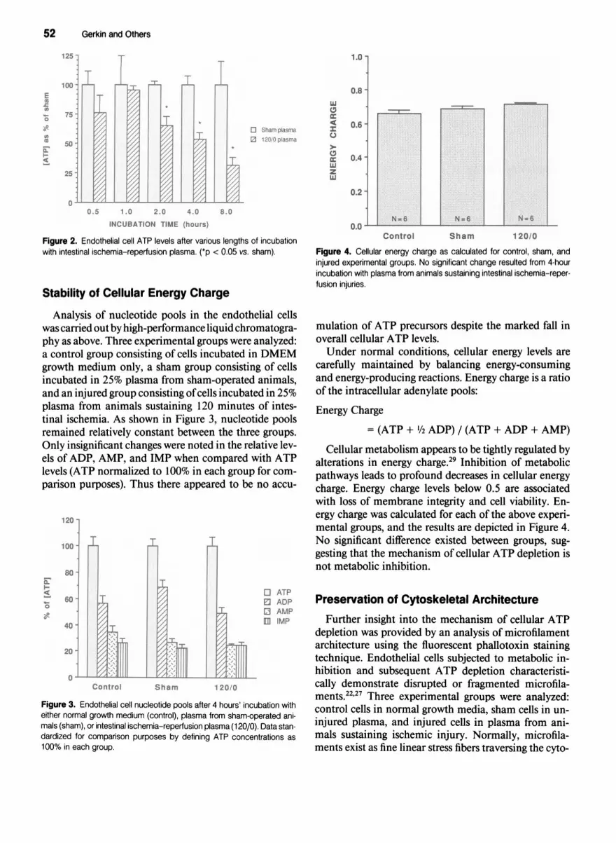
Figure 4. Cellular energy charge as calculated for control, sham, andinjured experimental groups. No significant change resulted from 4-hourincubation with plasma from animals sustaining intestinal ischemia-reper-fusion injuries.
mulation of ATP precursors despite the marked fall inoverall cellular ATP levels.Under normal conditions, cellular energy levels are
carefully maintained by balancing energy-consumingand energy-producing reactions. Energy charge is a ratioof the intracellular adenylate pools:
Energy Charge
= (ATP + 1/2 ADP) / (ATP + ADP + AMP)
Cellular metabolism appears to be tightly regulated byalterations in energy charge.29 Inhibition of metabolicpathways leads to profound decreases in cellular energycharge. Energy charge levels below 0.5 are associatedwith loss of membrane integrity and cell viability. En-ergy charge was calculated for each of the above experi-mental groups, and the results are depicted in Figure 4.No significant difference existed between groups, sug-gesting that the mechanism of cellular ATP depletion isnot metabolic inhibition.
Preservation of Cytoskeletal ArchitectureFurther insight into the mechanism of cellular ATP
depletion was provided by an analysis of microfilamentarchitecture using the fluorescent phallotoxin stainingtechnique. Endothelial cells subjected to metabolic in-hibition and subsequent ATP depletion characteristi-cally demonstrate disrupted or fragmented microfila-ments.2227 Three experimental groups were analyzed:control cells in normal growth media, sham cells in un-injured plasma, and injured cells in plasma from ani-mals sustaining ischemic injury. Normally, microfila-ments exist as fine linear stress fibers traversing the cyto-

Endothelial ATP Depletion After Ischemia-Reperfusion 53
B CFigure 5. Endothelial cells after fluorescent staining with rhodamine phalloidin. (A, left) Control group consist-ing of cells incubated 4 hours in normal growth medium. Note the fine linear stress fibers traversing thecytoplasm and concentrated at the cell periphery. (B, center) Cells incubated 4 hours with plasma fromsham-operated animals. Microfilament architecture is unchanged. (C, right) Cells after 4 hours in plasmaobtained from the portal vein after 120 minutes' intestinal ischemia (resulted in 45% decrease in cellular ATPlevels). Again, microfilaments remain intact and are essentially unchanged from controls (magnification X400on Kodak Tmax film).
plasm and concentrated at the periphery of the cell. Ascan be seen in Figure 5 a through c, this pattern persistsunder all three experimental conditions. Despite signifi-cant decreases in cellular ATP levels, microfilament ar-chitecture was preserved, suggesting that the mechanismof ATP depletion may be one of enhanced metabolicactivity with consumption of energy stores and not oneof metabolic inhibition.
Cytotoxic Injury to Endothelial CellsThe 51Cr-release assay is a reliable and quantitative
method ofanalyzing cytotoxic injury to endothelial cellsresulting in membrane disruption.30 In view ofthe obser-vation that endothelial cell ATP levels were significantlydepleted by ischemia-reperfusion plasmas yet the energycharge maintained, the issue of whether the injury was
cytotoxic was assessed. As shown in Figure 6, no signifi-cant cytotoxicity resulted from incubation ofendothelialcells with plasma from either sham or injured animals as
compared with normal growth medium controls. Hy-drogen peroxide exposure at a concentration of 500nmol/mL served as an internal control and consistentlyproduced a cytotoxic injury of 85% ± 2.5%.
DISCUSSION
This model of an in vivo stimulus of MOF combinedwith an in vitro preparation ofcultured pulmonary endo-thelial cells provides a straightforward, reproducible, andclinically relevant format in which to study the patho-genic mechanisms associated with distant organ injuryand ARDS. Important characteristics of the experimen-tal model are: (1) it is a consistent, reproducible stimulusof MOF; (2) the determinants of endothelial cell injurycan be assessed independently from the intestinal injury;and (3) the role of cellular elements (specifically the neu-
trophil) in the pathogenesis of acute lung injury may bestudied both separately and in conjunction with hu-moral factors.
Vascular endothelium appears to be a critical earlytarget in the pathogenesis of distant organ injury asso-
ciated with activation ofthe inflammatory system. Intactendothelial cell metabolic and functional activities are
required to maintain the vascular compartment.3' Lossof the integrity of the endothelial monolayer has beenimplicated as a primary event in the development ofacute edema in various vascular beds, particularly thelung.3233 Evidence continues to accumulate that the pul-
A

54 Gerkin and Others
100
*
80
x
0
> 60
00
40
20
_ E ZE~~c 0 E cm. 04 o o c
o x (n _ N N N
o _~ o
Figure 6. 51Cr-release assay to assess cytotoxic injury to endothelialcells by intestinal ischemia-reperfusion plasma after 4-hour incubationperiod. Controls were incubated in DMEM only. Maximal release was mea-sured with Triton X-100. H202 injury served as internal control. (*p < 0.001vs. control).
monary endothelial cell is a primary and particularlyvulnerable site for injury initiated by many differentclinical events and mediated by cytokines, neutrophils,proteases, toxic oxygen metabolites, and other fac-tors.8" 5'34-37 The model described in this study was de-signed to evaluate endothelial cell injury by humoral in-flammatory mediators in the context of MOF. The pul-monary microvascular injury may be the critical step inthe development of ARDS.Humoral factors associated with intestinal ischemia-
reperfusion injury result in depletion ofpulmonary endo-thelial cell energy stores. As noted in other studies,32'38'39depletion ofcellular ATP levels serves as an early markerof endothelial cell dysfunction. Whether the cell con-sumes ATP at an accelerated rate to maintain integralcell functions (such as stabilization of membranes) inresponse to exposure to I/R plasma constituents is yet tobe demonstrated. Maintenance of energy charge in cellsexposed to injured plasma, however, argues that meta-bolic inhibition of ATP synthetic pathways is not theunderlying mechanism of the observed cellular energy
store depletion. Further evidence against a metabolic in-hibitory mechanism is provided by the assessment ofmi-crofilament architecture. Metabolic inhibition of ATPsynthetic pathways has been shown to cause disruptionof microfilaments in the face of significant ATP deple-tion.2227 In the current study, microfilament architec-ture was preserved despite a45% fall in ATP levels. Accel-
erated consumption exceeding synthetic capacity ap-pears to be a more likely mechanism for depletion ofcellular energy stores in this setting. Further studies todefine the precise humoral factors responsible for theaforementioned observations are ongoing.
Depletion of ATP in the vascular endothelium as anisolated event appears unlikely to be sufficient to lead tothe devastating clinical scenario ofARDS. As shown bythe 51Cr-release data, humoral factors do not result incytotoxic injury to the endothelial monolayer. Other me-diators associated with the inflammatory system and in-testinal ischemia-reperfusion injury are likely to pro-mote the injury of the already compromised endothelialcell. Although these additional mediators have not beencharacterized, candidates include the neutrophil, 40'41various cytokines (tumor necrosis factor, platelet-acti-vating factor, others),35'42 endotoxin,4345 proteases,34,46and toxic oxygen metabolites.37'47
Limitations ofthe model are inherent in its design as a"transfer-type" experiment. Certainly very short-livedmediators, such as oxygen radicals, are not likely to havea significant effect and cannot be evaluated appropri-ately. Oxygen radicals generated in the gut and releasedinto the portal circulation, however, are unlikely to con-tribute to the in vivo pulmonary injury as observed inour model. The neutrophil-endothelium microenviron-ment is the more likely circumstance for injury mediatedby oxygen radicals.
In addition to the cellular injury hypothesis, an alter-native explanation for the depletion of energy stores inendothelial cells after intestinal ischemia-reperfusionshould be considered. Activation of vascular endothelialcells has been shown to occur both in vivo and in vitro inassociation with inflammation and with a variety of in-flammatory mediators.48 Activated endothelial cells ap-pear to play a role in enhanced adhesion of neutrophils,lymphocytes, and monocytes to vascular endotheliumafter exposure to various cytokines. Interleukin- 1, tumornecrosis factor, and lymphotoxin all have been shown toincrease endothelial cell adhesiveness for leukocytes andto modulate procoagulant activity.49 Studies suggest thatthe mechanism of adhesion involves expression of cellsurface adhesion molecules such as ELAM-1, MEL-14antigen, and H-CAM.4849 The process is time depen-dent, requiring protein and RNA synthesis. As demon-strated by Bevilacqua and Gimbrone,49 the time fromactivation of endothelial cells (activated with tumor ne-crosis factor or interleukin- 1) to peak cell surface adhe-sion molecule expression, neutrophil binding, and peakprocoagulant activity is 4 hours.49 In view of the timecourse of ATP depletion observed in the current study,the potential association between endothelial cell activa-tion and depletion of cellular energy stores deserves fur-ther investigation.

Endothelial ATP Depletion After Ischemia-Reperfusion 55
In summary, these studies describe a combined invivo/in vitro model designed for the investigation of thepathogenesis of acute pulmonary injury after a stimulusof MOF. Humoral factors, independent of the neutro-phil, result in endothelial cell ATP depletion withoutmetabolic inhibition or cell death. Depletion of energystores by noncellular humoral factors may represent theearly event that predisposes the cell to more severe injuryby other mediators of the endogenous inflammatory re-sponse.
Acknowledgments
The authors thank T. Welling, A. Jeffers, M. Imlay, and K. Childs fortechnical assistance.
References
1. Goris RJA, teBoekhorst TPA, Nuytinck JKS, Gimbrere JSF. Mul-tiple-organ failure: generalized autodestructive inflammation?Arch Surg 1985; 120:1109-1115.
2. Oldham KT, Guice KS, Till GO, Ward PA. Activation ofcomple-ment by hydroxyl radical in thermal injury. Surgery 1988;104:272-279.
3. Till GO, Beauchamp C, Menapace D, et al. Oxygen radical depen-dent lung damage following thermal injury of rat skin. J Trauma1983; 23:269-276.
4. Bell RC, Coalson JJ, Smith JD, Johanson WG. Multiple organsystem failure and infection in adult respiratory distress syndrome.Ann Intern Med 1983; 99:293-298.
5. Hersch M, Gnidec AA, Bersten AD, et al. Histologic and ultrastruc-tural changes in nonpulmonary organs during early hyperdynamicsepsis. Surgery 1990; 107:397-410.
6. Demling RH, Niehaus G, Will JA. Pulmonary microvascular re-sponse to hemorrhagic shock, resuscitation, and recovery. J ApplPhysiol 1979; 46:498-503.
7. Vedder NB, Fouty BW, Winn RK, et al. Role of neutrophils ingeneralized reperfusion injury associated with resuscitation fromshock. Surgery 1989; 106:509-516.
8. Rivkind Al, Siegel JH, Guadalupi P, Littleton M. Sequential pat-terns of eicosanoid, platelet, and neutrophil interactions in theevolution of the fulminant post-traumatic adult respiratory dis-tress syndrome. Ann Surg 1989; 210:355-373.
9. Guice KS, Oldham KT, Johnson KJ, et al. Pancreatitis-inducedacute lung injury: an ARDS model. Ann Surg 1988; 208:71-77.
10. Guice KS, Oldham KT, Caty MG, et al. Neutrophil-dependent,oxygen-radical mediated lung injury associated with acute pancre-atitis. Ann Surg 1989; 210:740-747.
11. Schmeling DJ, Caty MG, Oldham KT, et al. Evidence for neutro-phil-related acute lung injury after intestinal ischemia-reperfusion.Surgery 1989; 106:195-202.
12. Otamiri T: Oxygen radicals, lipid peroxidation, and neutrophilinfiltration after small-intestinal ischemia and reperfusion. Sur-gery 1989; 105:593-597.
13. Klausner JM, Paterson IS, Kobzik L, et al. Leukotrienes but notcomplement mediate limb ischemia-induced lung injury. AnnSurg 1989; 209:462-470.
14. Abdalla EK, Caty MG, Guice KS, et al. Arterial levels of oxidized
glutathione (GSSG) reflect oxidant stress in vivo. J Surg Res 1990;48:291-296.
15. Demling RH. Current concepts on the adult respiratory distresssyndrome. Circ Shock 1990; 30:297-309.
16. Andreadis N, Petty TL. Adult respiratory distress syndrome: prob-lems and progress. Am Rev Respir Dis 1985; 132:1344-1346.
17. National Heart and Lung Institutes. Respiratory Diseases: TaskForce Report on Problems, Research Approaches, Needs. Wash-ington DC, US Government Printing Office, pg. 167-180, 1972.DHEW Pub. NIH 74-432.
18. Turnage RH, Abdalla EK, Gerkin TM, et al. Regional blood flowafter intestinal ischemia-reperfusion injury [Abstract]. Circ Shock1991; 34:64.
19. Ryan US, Clements E, Habliston D, Ryan JW. Isolation and cul-ture of pulmonary artery endothelial cells. Tissue Cell 1978;10:535-554.
20. Stanley PE, Williams SG. Use ofthe liquid scintillation spectrome-ter for determining adenosine triphosphate by the luciferase en-zyme. Anal Biochem 1969; 29:281-292.
21. Spragg RG, Hinshaw DB, Hyslop PA, et al. Alterations in adeno-sine triphosphate and energy charge in cultured endothelial andP388D, cells after oxidant injury. J Clin Invest 1985; 76:1471-1476.
22. Hinshaw DB, Armstrong BC, Burger JM, et al. ATP and microfila-ments in cellular oxidant injury. Am J Pathol 1988; 132:479-488.
23. Labarca C, Paigen K. A simple, rapid, and sensitive DNA assayprocedure. Anal Biochem 1980; 102:344-353.
24. Shryock JC, Rubio R, Berne RM. Extraction of adenine nucleo-tides from cultured endothelial cells. Anal Biochem 1986; 159:73-81.
25. Tekkanat KK, Fox IH. Isocratic separation ofATP and its degrada-tion products from biological fluids by automated liquid chroma-tography. Clin Chem 1988; 34:925-932.
26. Hinshaw DB, Sklar LA, Bohl B, et al. Cytoskeletal and morpho-logic impact of cellular oxidant injury. Am J Pathol 1986;123:454-464.
27. Hinshaw DB, Burger JM, Armstrong BC, Hyslop PA. Mechanismofendothelial cell shape change in oxidant injury. J Surg Res 1989;46:339-349.
28. Varani J, Fligiel SEG, Till GO, et al. Pulmonary endothelial cellkilling by human neutrophils: possible involvement of hydroxylradical. Lab Invest 1985; 53:656-663.
29. Atkinson DE. Cellular Energy Metabolism and Its Regulation.New York: Academic Press, 1977, pp 85-107, 201-224.
30. Freshney RI. Culture of Animal Cells-A Manual of Basic Tech-nique. New York: Liss Inc., 1987, pp 247.
31. Weiss SJ, Young J, LoBuglio AF, et al. Role of hydrogen peroxidein neutrophil-mediated destruction of cultured endothelial cells. JClin Invest 1981; 68:714-721.
32. Wysolmerski RB, LagunoffD. Inhibition ofendothelial cell retrac-tion by ATP depletion. Am J Pathol 1988; 132:28-37.
33. Grosso MA, Brown JM, Viders DE, et al. Xanthine oxidase-de-rived oxygen radicals induce pulmonary edema via direct endothe-lial cell injury. J Surg Res 1989; 46:355-360.
34. Schraufstatter IU, Revak SD, Cochrane CG. Proteases and oxi-dants in experimental pulmonary inflammatory injury. J Clin In-vest 1984; 73:1175-1184.
35. Varani J, Bendelow MJ, Sealey DE, et al. Tumor necrosis factorenhances susceptibility of vascular endothelial cells to neutrophil-mediated killing. Lab Invest 1988; 59:292-295.
36. Demling RH. The role of mediators in human ARDS. J Crit Care1988; 3:56-72.
37. Fantone JC, Ward PA. Role of oxygen-derived free radicals and

56 Gerkin and Others
metabolites in leukocyte-dependent inflammatory reactions. Am JPathol 1982; 107:397-418.
38. Holmsen H, Robkin L. Hydrogen peroxide lowers ATP levels inplatelets without altering adenylate energy charge and plateletfunction. J Biol Chem 1977; 252:1752-1757.
39. Berger NA. Oxidant-induced cytotoxicity: a challenge for meta-bolic modulation. Am J Respir Cell Mol Biol 1991; 4:1-3.
40. Weiss SJ. Tissue destruction by neutrophils. N Engl J Med 1989;320:365-376.
41. Horgan MJ, Wright SD, Malik AB. Antibody against leukocyteintegrin (CD1 8) prevents reperfusion-induced lung vascular in-jury. Am J Physiol 1990; 259:L315-L319.
42. Hsueh W, Gonzalez-Crussi F, Arroyave JL. Platelet-activating fac-tor: an endogenous mediator for bowel necrosis in endotoxemia.FASEBJ 1987; 1:403-405.
43. Mayoral JL, Dunn DL. Cross-reactive murine monoclonal anti-bodies directed against the core/lipid A region ofendotoxin inhibitproduction oftumor necrosis factor. J Surg Res 1990; 49:287-292.
44. Dunn DL, Priest BP, Condie RM. Protective capacity of polyclo-
nal and monoclonal antibodies directed against endotoxin duringexperimental sepsis. Arch Surg 1988; 123:1389-1393.
45. Schoffel U, Shiga J, Mittermayer C. The proliferation-inhibitingeffect of endotoxin on human endothelial cells in culture and itspossible implication in states of shock. Circ Shock 1982; 9:499-508.
46. LeRoy EC, Ager A, Gordon JL. Effects of neutrophil elastase andother proteases on porcine aortic endothelial prostaglandin l2 pro-duction, adenine nucleotide release, and responses to vasoactiveagents. J Clin Invest 1984; 74:1003-1010.
47. Sacks T, Moldow CF, Craddock PR, et al. Oxygen radicals mediateendothelial cell damage by complement-stimulated granulocytes.J Clin Invest 1978; 61:1161-1167.
48. Jutila MA, Berg EL, Kishimoto TK, et al. Inflammation-inducedendothelial cell adhesion to lymphocytes, neutrophils, and mono-cytes. Transplantation 1989; 48:727-731.
49. Bevilacqua MP, Gimbrone MA. Inducible endothelial functionsin inflammation and coagulation. Semin Thromb Hemost 1987;13:425-433.
Recommended


