
Isolates of ‘Candidatus Nostocoida limicola’Blackall et al. 2000 should be described as threenovel species of the genus Tetrasphaera, asTetrasphaera jenkinsii sp. nov., Tetrasphaeravanveenii sp. nov. and Tetrasphaera veronensis sp.nov.
C. M. McKenzie,1 E. M. Seviour,1 P. Schumann,2 A. M. Maszenan,13J.-R. Liu,14 R. I. Webb,3 P. Monis,4 C. P. Saint,4 U. Steiner2 and R. J. Seviour1
Correspondence
R. J. Seviour
1Biotechnology Research Centre, La Trobe University, Bendigo, Victoria 3552, Australia
2DSMZ – Deutsche Sammlung von Mikroorganismen und Zellkulturen GmbH,Mascheroder Weg 1b, D-38124 Braunschweig, Germany
3Centre for Microscopy and Microanalysis, Department of Microbiology, The University ofQueensland, St Lucia, Brisbane, Queensland 4072, Australia
4Australian Water Quality Centre, Bolivar, South Australia 5108, Australia
Despite differences in their morphologies, comparative analyses of 16S rRNA gene sequences
revealed high levels of similarity (>94 %) between strains of the filamentous bacterium ‘Candidatus
Nostocoida limicola’ and the cocci Tetrasphaera australiensis and Tetrasphaera japonica and the
rod Tetrasphaera elongata, all isolated from activated sludge. These sequence data and their
chemotaxonomic characters, including cell wall, menaquinone and lipid compositions and
fingerprints of their 16S–23S rRNA intergenic regions, support the proposition that these isolates
should be combined into a single genus containing six species, in the family Intrasporangiaceae in
the Actinobacteria. This suggestion receives additional support from DNA–DNA hybridization data
and when partial sequences of the rpoC1 gene are compared between these strains. Even though
few phenotypic characterization data were obtained for these slowly growing isolates, it is
proposed, on the basis of the extensive chemotaxonomic and molecular evidence presented here,
that ‘Candidatus N. limicola’ strains Ben 17, Ben 18, Ben 67, Ben 68 and Ben 74 all be placed into
the species Tetrasphaera jenkinsii sp. nov. (type strain Ben 74T=DSM 17519T=NCIMB 14128T),
‘Candidatus N. limicola’ strain Ben 70 into Tetrasphaera vanveenii sp. nov. (type strain Ben
70T=DSM 17518T=NCIMB 14127T) and ‘Candidatus N. limicola’ strains Ver 1 and Ver 2 into
Tetrasphaera veronensis sp. nov. (type strain Ver 1T=DSM 17520T=NCIMB 14129T).
INTRODUCTION
The application of molecular techniques has begun toresolve the taxonomic status of some of the bacteria presentin activated sludge, especially those morphotypes which mayoften dominate the activated sludge bacterial community(Seviour et al., 2000; Martins et al., 2004). For example,some of the Gram-positive and Gram-negative filamentousbacteria responsible for the operational disorders of bulkingand foaming have now been grown in pure culture and, onthe basis of 16S rRNA gene sequence data, their phylogenyhas been elucidated (Kampfer & Wagner, 2002; Martinset al., 2004). Some, like ‘Microthrix parvicella’ (Blackall et al.,1994; Rossetti et al., 2005) and Eikelboom type 1851 (Beeret al., 2002), are novel bacteria, while others, like type 1863
3Present address: Environmental Engineering Research Centre, Schoolof Civil and Environmental Engineering, Nanyang TechnologicalUniversity, Singapore.
4Present address: Department of Chemistry and Biology, RyersonUniversity, Toronto, Ontario M5B 2K3, Canada.
The GenBank/EMBL/DDBJ accession numbers for the 16S rRNAgene sequences of strains Ben 68, Ben 70 and Ben 74 areDQ007319–DQ007321, respectively, and the accession numbers forthe rpoC1 gene sequences determined in this study are DQ007322–DQ007334, as detailed in Fig. 4.
Fatty acid profiles of selected strains of ‘Candidatus N. limicola’ andrelated strains are available in IJSEM Online.
Abbreviation: TFO, tetrad-forming organism.
63978 G 2006 IUMS Printed in Great Britain 2279
International Journal of Systematic and Evolutionary Microbiology (2006), 56, 2279–2290 DOI 10.1099/ijs.0.63978-0

(Seviour et al., 1997), are close relatives of previouslydescribed non-filamentous bacteria. Of these filaments,‘Nostocoida limicola’ is particularly interesting. Threemorphotypes of this filament have been recognized, i.e.‘N. limicola’ I, II and III, separated solely on the basis ofdifferences in their cell dimensions (Eikelboom & vanBuijsen, 1983). It is now known that these morphotypes arein fact a collection of unrelated bacteria, being members ofthe Firmicutes (Liu et al., 2002), the Actinobacteria (Blackallet al., 2000), the Alphaproteobacteria (Snaidr et al., 2002;Levantesi et al., 2004; Kragelund et al., 2005) or theChloroflexi (Schade et al., 2002) and the Planctomycetales(Liu et al., 2001). In the case of actinobacterial ‘N. limicola’II, no chemotaxonomic data were available to help todelineate its taxonomic status precisely and that of theindividual isolates obtained in pure culture and so, while thesuggestion was made that this filament represented a newgenus, it was given Candidatus status (Blackall et al., 2000).
The tetrad-forming organism (TFO) morphotype (Tsai &Liu, 2002), appearing in activated sludge and pure culturesas cocci in tetrads and clusters, can dominate some activatedsludge communities, and some of these TFO have beencultured and characterized. They include several novelGram-positive and Gram-negative cocci (Seviour et al.,2000, 2003). Among these are members of the genusTetrasphaera, now thought to be important populationsresponsible for the microbiological removal of phosphorusin some activated sludge processes (Kong et al., 2005). When16S rRNA gene sequences of cultured actinobacterial‘Candidatus Nostocoida limicola’ isolates were comparedwith those from isolates of Tetrasphaera australiensis andTetrasphaera japonica, Gram-positive TFO (Maszenan et al.,2000), these bacteria all clustered together at a highsimilarity. Although not possessing a TFO morphology,Tetrasphaera elongata (Hanada et al., 2002), which grows asrods, was also phylogenetically very similar. Therefore,attempts were made to clarify their taxonomic interrelation-ships using both 16S rRNA and rpoC1 gene sequences(Morse et al., 1996; Wilson et al., 2000) and detailed
chemotaxonomic characterization. On the basis of all theavailable evidence, it is proposed that these ‘Candidatus N.limicola’ isolates should all be placed into the genusTetrasphaera and represent three novel species.
METHODS
Strains used in study. Isolates of actinobacterial ‘Candidatus N.limicola’ (identified and cultured as ‘N. limicola’ II) andTetrasphaera species, obtained and maintained as described byBlackall et al. (2000) and Maszenan et al. (2000), respectively(Table 1), were used in this study. They were grown on either GS orR2A agar (Blackall et al., 2000). Additional strains of ‘Candidatus N.limicola’ (Ben 70 and Ben 74) were also included in this study; thesewere isolated in our laboratory from other activated sludge biomasssamples, by micromanipulation onto the same media used in theearlier study, and their sources are also listed in Table 1. The follow-ing cultures were also included in the rpoC1-based phylogenetic stu-dies: Terrabacter tumescens DSM 20308T (Collins et al., 1989),Terracoccus luteus DSM 44267T (Prauser et al., 1997) and Janibacterlimosus DSM 11140T (Martin et al., 1997).
Characterization of isolates. Selected substrate utilization pat-terns for strains Ben 70 and Ben 74 were obtained with the methodsof Blackall et al. (2000), who had reported these for strains Ben 17,Ben 18, Ben 67 and Ver 1. The methods used for preparing speci-mens for scanning and transmission electron microscopy were thosedescribed by Maszenan et al. (1997, 1999) and Butler et al. (2002).The medium used to prepare the inoculum for each strain for char-acterization was that which supported its growth, since some isolatesof ‘Candidatus N. limicola’ would grow only on R2A agar and notGS agar (Blackall et al., 2000). Biomass for chemotaxonomic charac-terization of selected strains was obtained by inoculating 200 platesof freshly prepared R2A agar (Reasoner & Geldreich, 1985) for eachisolate and incubating these at 26 uC for exactly 4 weeks. Cells wereharvested by gently scraping them from the agar surface into steriledistilled water, centrifuging and washing (36). These were finallyfreeze-dried. The 16S rRNA genes of the newly acquired isolates of‘Candidatus N. limicola’ and Tetrasphaera were amplified, clonedand sequenced according to the methods of Maszenan et al. (2000)and Liu et al. (2002).
Sequences for the rpoC1 genes from isolates were obtained as follows.Degenerate primers were designed after all publicly available rpoCsequences in GenBank were aligned and edited with CLUSTAL W.
Table 1. Source of isolates used in this study
Isolate Source Isolation medium Reference
‘Candidatus N. limicola’
Ben 17 Sunbury, Australia GS agar Blackall et al. (2000)
Ben 18 Sunbury, Australia GS agar Blackall et al. (2000)
Ben 67 Verona, Italy R2A agar Blackall et al. (2000)
Ben 68 Verona, Italy R2A agar Blackall et al. (2000)
Ben 70 Carrum, Australia GS agar R. J. Seviour (unpublished)
Ben 74 Glenelg, Australia GS agar R. J. Seviour (unpublished)
Tetrasphaera australiensis
Ben 109T Glenelg, Australia GS agar Maszenan et al. (2000)
Ben 110 Carrum, Australia GS agar Maszenan et al. (2000)
Tetrasphaera japonica
T1-X7T Tokyo, Japan Cell extract agar Kataoka et al. (1996)
2280 International Journal of Systematic and Evolutionary Microbiology 56
C. M. McKenzie and others

Primers eventually selected for PCR amplification of the rpoC1 genewere the forward primer 59-TAYCGYCGKGTHATYAAYCG-39 andthe reverse primer 59-TGRTCACCRTCRAARTCRGC-39 (synthesizedby Geneworks Pty Ltd, Adelaide, Australia); these primers gave a DNAfragment after PCR of approx. 600 bp. PCR was performed asdescribed by Wilson et al. (2000). Thermal cycling conditions were ahot start at 95 uC for 10 min followed by 30 cycles of 92 uC for 1 min,42 uC for 1 min and 72 uC for 3 min, followed by 72 uC for 10 min anda final hold at 4 uC. The PCR product was purified with QIAprep(Qiagen) and ligated into pGEM-T Easy vector (Promega). Cloneswere screened by restriction digestion with EcoRI and both DNAstrands were sequenced with primers M13F and M13R. Sequencing wasperformed with Big Dye technology on a model 373A sequencer(Applied Biosystems). The 16S rRNA gene sequences for the other‘Candidatus N. limicola’ isolates used in this study were thosedetermined and reported by Blackall et al. (2000) and Maszenan et al.(2000), and sequences were also used that were reported in the originalpublications for Terracoccus luteus (Prauser et al., 1997), Terrabactertumescens (Collins et al., 1989), Knoellia subterranea and Knoelliasinensis (Groth et al., 2002), Janibacter limosus (Martin et al., 1997),Janibacter anophelis (Kampfer et al., 2006), Janibacter terrae (Yoonet al., 2000) and Janibacter melonis (Yoon et al., 2004), their closestphylogenetic relatives.
For both phylogenetic markers, the sequences were aligned usingCLUSTAL X version 1.81 (Thompson et al., 1997). For rpoC1 analysis,550 nucleotides equivalent to positions 302–852 of the Escherichia colirpoC1 sequence were included in the alignment. For 16S rRNA geneanalysis, 1426 nucleotides equivalent to positions 37–1463 of the E. coli16S rRNA sequence were included in the alignment. Phylogeneticanalyses were conducted using MEGA2 version 2.1 (Kumar et al., 2001).Alignment files from CLUSTAL X were imported into MEGA2 and thedata were analysed by distance-based and parsimony methods. Fordistance-based analyses, relationships were inferred using neighbour-joining analysis of Tamura–Nei distances that were calculated with thepairwise deletion option in effect. The robustness of the resultingphylogenies was tested using bootstrap analysis of 1000 replicates. Formaximum-parsimony analyses, relationships were inferred using theclose-neighbour-interchange search option with a search level of 3 andrandom addition of trees using 10 replications. All sites were includedin each analysis and standard weighting was used for scoring changes.The robustness of the resulting phylogenies was tested using bootstrapanalysis of 1000 replicates.
Chemotaxonomic characterization. The diastereoisomer of dia-minopimelic acid in cell-wall hydrolysates of these isolates wasdetermined with the HPLC method of McKerrow et al. (2000).Menaquinones were analysed by reversed-phase HPLC (Groth et al.,1999) and cellular fatty acid methyl esters and polar lipids were ana-lysed using the methods of Schumann et al. (1997).
16S–23S rRNA intergenic spacer region fingerprinting. DNAwas extracted from cultures as described by Liu et al. (2002) and thePCR primers selected to amplify this region were the primers 2 for-ward and 7 reverse of Gurtler & Stanisich (1996). PCR conditionsused were detailed by Liu et al. (2002). Restriction enzymes MspIand BstUI (New England Biolabs) were selected for digestion, andthe digestion products were separated on agarose gels as describedpreviously (Liu et al., 2002). Patterns were analysed using the SSM
coefficient and UPGMA algorithm.
DNA–DNA hybridizations. It was not possible to carry out DNA–DNA hybridization analysis with all these strains because of pro-blems in obtaining sufficient quantities of biomass. Those analysesthat were possible were performed following the methods used byMartin et al. (1997).
RESULTS
Phenotypic properties of isolates
As reported earlier (Blackall et al., 2000), all the isolates of‘Candidatus N. limicola’ used in this study that could growon GS agar grew as filamentous bacteria on this medium.Swollen and flattened discoid irregular cells within thesefilaments were common, as shown with strains Ben 67 andBen 68 (Fig. 1e, f), and filaments were very similar to theirappearance in biomass samples in activated sludge plants.Extensive irregular septation of some individual cells wasalso apparent (Fig. 1e, f), similar to that seen withTetrasphaera elongata Lp2T (Hanada et al., 2002). WithR2A agar and broth, only rudimentary filamentous growthwas seen. Instead, both strains Ben 17 and Ben 70 grewmainly as swollen and irregular cocci in very short chains orclusters (Fig. 1a, b). Budding was also visible, especially withstrain Ben 70 (e.g. Fig. 1c). Intermediate forms withoccasional swollen cocci in the filaments could also oftenbe seen on GS medium (Fig. 1d–f).
The ultrastructural features of strains Ben 70 and Ben 74grown on these two media are shown in Fig. 2. On R2Amedium, both grew as irregular clusters of cells, althoughoccasional short chains were seen (Fig. 2a–d). Cell shape washighly varied because of the erratic septation. This producesdifferently shaped and sized cells, which could be anythingfrom coccoid to discoid. With strain Ben 74, the cells wereoften flattened and appeared more compressed than those ofstrain Ben 70 (Fig. 2a–d). Vesicles were commonly seen inboth strains. Small vesicles appeared in tight clusters in cells.In Ben 74, several large electron-lucent vesicles were alsopresent (Fig. 2c, d), while, in Ben 70, a single large vesicle thatalways contained electron-dense material was seen (Fig. 2a,b). Granules (30–40 nm diameter) were regularly seen in thecytoplasm of both strains. These often formed such denseaggregates that they led to the division of the cytoplasm intotwo distinct regions: electron-lucent (granule-filled) andelectron-dense (containing all other cytoplasmic structures)(Fig. 2a–d). On GS agar, there appeared to be more emptycells in the chains than on R2A (Fig. 2h). Septation was notas random and hence the cells were largely in chains, thoughsome irregular division caused chains of cells to split andclump (Fig. 2g, h). Narrow, compressed cells were present inboth, but were seen more commonly with strain Ben 74,where flattened cells were routinely visible. Vesicles were notas common as in cells grown on R2A. In both strains, therewere smaller vesicles of variable size clustered in thecytoplasm. In Ben 74, larger vesicles were also seen, andtheir size was often sufficient to distort the shape of the cells(Fig. 2h). Small, electron-dense granules were sparselydistributed through the cytoplasm of cells of both Ben 70and Ben 74 grown on GS agar (Fig. 2g).
All cells showed a Gram-positive wall. This cell wallconsisted of three visible layers: an inner electron-denselayer, which was closely associated with the cytoplasmicmembrane, an electron-lucent layer and, on the outside,
http://ijs.sgmjournals.org 2281
Reclassification of ‘Ca. Nostocoida limicola’ isolates

another electron-dense layer (Fig. 2f). There were twolayers of capsular material, an electron-lucent layer thatsurrounded each cell and an electron-dense layer thatsurrounded the entire cluster of cells (Fig. 2f). A thin strandof material from this outer capsule continued into the centreof the septum separating the cells. The dense capsule wasmuch better developed in cells of Ben 70 and Ben 74 grownon GS agar than those on R2A.
In contrast to these ‘Candidatus N. limicola’ isolates,all three strains of Tetrasphaera examined, Tetrasphaera
australiensis strains Ben 109T and Ben 110 and Tetrasphaerajaponica T1-X7T, always grew as cocci in tetrads or clusterson all the media examined, and filamentous growth wasnever seen with them. On the other hand, Tetrasphaeraelongata Lp2T was reported to grow as rods or oval unicells(Hanada et al., 2002).
Because of their very slow growth rates in pure culture(Blackall et al., 2000), extensive phenotypic characterizationof these ‘Candidatus N. limicola’ isolates was not alwayspossible, experiences that agree with those reported earlier
(a) (b)
(c) (d)
(e) (f)
Fig. 1. Scanning electron micrographs of ‘Candidatus N. limicola’ strains grown on R2A agar (a–d) and GS agar (e, f), showingmedium-dependent morphological variations. On R2A agar, strains Ben 17 (a) and Ben 70 (b) appear as regular cocci in clustersand short chains, although in Ben 70 (c) and Ben 74 (d), these cocci can appear inflated and in some cases appear to bud. On GSagar, strains Ben 67 (e) and Ben 68 (f) show typical features of all these strains on this medium of irregular flattened discoid cells inchains with occasional terminal swollen coccoid cells seen. Bars, 3 mm (a–d) and 4 mm (e, f).
2282 International Journal of Systematic and Evolutionary Microbiology 56
C. M. McKenzie and others

(a) (b) (c) (d)
(e) (f) (g) (h)
Fig. 2. Transmission electron micrographs of cell ultrastructure of strains of ‘Candidatus N. limicola’. (a, b) Strain Ben 70 grown on R2A agar shows capsular materialsurrounding short chains (a) or irregular clusters of cells (b). Randomly orientated septation is clearly apparent. Cells contain tight groups of small vesicles and a single largevesicle with electron-dense material. Aggregates of electron-lucent granules appear in the cytoplasm. Bars, 500 nm. (c, d) Strain Ben 74 grown on R2A shows manyfeatures described for strain Ben 70, except that cells in groups are more compressed. Larger, clear vesicles are present. Bars, 1 mm. (e, f) Strain Ben 70 grown on GS agarshows most cells in irregular chains. Vesicles of varying sizes are generally distributed more randomly through the cytoplasm (e). The arrangement of the three-layeredGram-positive wall (W) lying outside the cytoplasmic membrane (CM) is visible in (f). The capsular material is divided into two distinct regions, the inner (IC) closelyassociated with the cells and the outer electron-dense (OC) that surrounds the clumps and is also seen as a strand in the centre of the dividing septum. Bars, 200 nm (e)and 50 nm (f). (g, h) Strain Ben 74 grown on GS agar shows most of the features of strain Ben 70, especially the growth in chains and the same random septation, but cellsappear more compressed. Bars, 500 nm (g) and 1 mm (h).
http://ijs.sgm
journals.org2
28
3
Reclassification
of‘C
a.N
ostocoidalim
icola’isolates

by Blackall et al. (2000). However, some biochemicalcharacteristics of strains Ben 17, Ben 67 and Ver 1 werereported by Blackall et al. (2000). They were very similar inall three strains, except that Ver 1 alone could not grow withnitrite as sole nitrogen source. When Ben 70 and Ben 74 werecharacterized in the same way, both showed very similarpatterns to these other isolates. Thus all grew on acetate,pyruvate, propionate, glucose, fructose, lactose, mannose,Tween 80 and glycerol as carbon sources and nitrate, nitriteand urea as nitrogen sources. Like the other strainsexamined, neither could utilize lactate, ethanol, oleic acidor oleate. All contained both poly-b-hydroxyalkanoates andpolyphosphate granules by staining. All were catalase- andoxidase-positive, as are Tetrasphaera australiensis Ben 109T
and Tetrasphaera japonica T1-X7T. However, Tetrasphaeraaustraliensis Ben 110 and Tetrasphaera elongata Lp2T areboth oxidase-negative (Maszenan et al., 2000; Hanada et al.,2002), which may be an artefact reflecting their generally lowlevels of metabolic activity.
Chemotaxonomic features of isolates
These characters are summarized in Table 2, and could bedetermined only for selected strains of ‘Candidatus N.limicola’, where sufficient biomass for the analyses could beobtained. Cell walls of all the isolates examined (strains Ben17, Ben 70, Ben 74 and Ver 1) had meso-diaminopimelic acidas the dibasic amino acid (i.e. type A1c peptidoglycan), asdid Tetrasphaera australiensis strains Ben 109T and Ben 110and Tetrasphaera japonica T1-X7T (Maszenan et al., 2000)and Tetrasphaera elongata Lp2T (Hanada et al., 2002). Forboth strains Ben 17 and Ben 74, the menaquinones wereMK-8(H4), MK-8(H2), MK-8 and MK-6(H4), in ratios of87 : 5 : 3 : 2 and 80 : 3 : 4 : 5, respectively. However, severalattempts failed to detect menaquinones or ubiquinones inVer 1 by HPLC/mass spectrometry, although similarquantities of biomass as for the other strains were usedfor their extraction. Strain Ben 70 was distinct in containingMK-8, MK-8(H2) and MK-8(H4), in the ratio of 39 : 29 : 6.Polar lipid profiles of these strains were dominated bydiphosphatidylglycerol, phosphatidylglycerol and phospha-tidylinositol, except Ben 17 and Ben 74, which didnot possess diphosphatidylglycerol (Table 2). Phospha-tidylethanolamine was detected only in Ben 110 andTetrasphaera elongata Lp2T, but an unidentified aminopho-spholipid was present in Ben 17, Ben 70, Ben 74, Ver 1 andTetrasphaera japonica T1-X7T.
Fatty acid profiles of strains Ben 17, Ben 70, Ben 74 and Ver1, together with those from Tetrasphaera australiensis strainsBen 109T and Ben 110, Tetrasphaera japonica T1-X7T andTetrasphaera elongata Lp2T (Maszenan et al., 2000; Hanadaet al., 2002), are given in Supplementary Table S1 in IJSEMOnline. 14-Methylpentadecanoic acid (i-C16 : 0) dominatedin all these strains, including the Tetrasphaera strains exceptfor Tetrasphaera elongata Lp2T (Hanada et al., 2002). Thisstrain alone possessed ai-C15 : 0 as the major fatty acid. StrainBen 17 contained more 14-methylhexadecanoic acid (ai-C17 : 0) than any of the other strains (Supplementary
Table S1). Qualitative and quantitative differences wereapparent among the ‘Candidatus N. limicola’ strains. Onlystrain Ben 74 possessed small amounts of octadecanoicacid (C18 : 0), 9-heptadecenoic acid (C17 : 1) and 16-methylheptadecanoic acid (i-C18 : 0), while strain Ver1 alone lacked 9-octadecenoic acid (C18 : 1), 15-methylhexadecanoic acid (i-C17 : 0) and 15-methyl-9-hexadecenoic acid (i-C17 : 1). These fatty acid profiles werealso markedly different quantitatively from those of thecharacterized Knoellia and Janibacter strains (as given inSupplementary Table S1 in IJSEM Online), especially sincei-C16 : 0 is not as prominent, adding weight to the view thatthe strains described here are all members of the one genus,Tetrasphaera.
Phylogenetic analyses
Almost-complete sequences of the 16S rRNA genes from‘Candidatus N. limicola’ strains Ben 70 (1473 bp) and Ben74 (1477 bp) were obtained and analysed with those fromthe other ‘Candidatus N. limicola’ and Tetrasphaera isolatesdescribed earlier by Blackall et al. (2000), Maszenan et al.(2000) and Hanada et al. (2002). Parsimony analysisidentified three trees of equal length, one of which had anidentical topology to that of the distance-based phylogenetictree illustrated in Fig. 3. The other two trees differed only inthe branch orders of species outside the clade containing the‘Candidatus N. limicola’ isolates (data not shown).Sequences from strains Ben 17, Ben 18, Ben 67, Ben 68and Ben 74 were all >99 % similar to each other and formeda well-defined and separate cluster supported by highbootstrap values (¢98 %) for both analyses. Theirsequences were all 97–98 % similar to those of strains Ver1 and Ben 70, which in turn were 97 % similar to each other.Strain Ben 70 and Tetrasphaera australiensis strains Ben 109T
and Ben 110 were more closely related to each other thaneither was to Tetrasphaera japonica T1-X7T andTetrasphaera elongata Lp2T. Only strains Ver 1 and Ver 2of these ‘Candidatus N. limicola’ strains had the distinctiveV6 region sequence variation noted by Blackall et al. (2000)for this filament. None of Tetrasphaera japonica T1-X7T,Tetrasphaera australiensis strains Ben 109T and Ben 110 orTetrasphaera elongata Lp2T possessed it.
The rpoC1 analysis (Fig. 4) did not provide the same level ofresolution as the 16S rRNA gene analysis. The distance-based analysis (Fig. 4) produced a tree with a similartopology to the 16S rRNA tree for the ‘Candidatus N.limicola’ and Tetrasphaera australiensis isolates. ‘CandidatusN. limicola’ isolates Ben 17, Ben 18, Ben 67, Ben 68 and Ben74 again formed a single cluster that received strongbootstrap support. However, the internal branch order,which received poor bootstrap support, was different fromthat seen with the 16S rRNA gene sequence results.‘Candidatus N. limicola’ isolate Ben 70 also clustered withTetrasphaera australiensis with moderate bootstrap supportand ‘Candidatus N. limicola’ strain Ver 1 was placed as asister taxon to this group, which is different from thetopology of the 16S rRNA gene tree. The clustering of Ben 70
2284 International Journal of Systematic and Evolutionary Microbiology 56
C. M. McKenzie and others
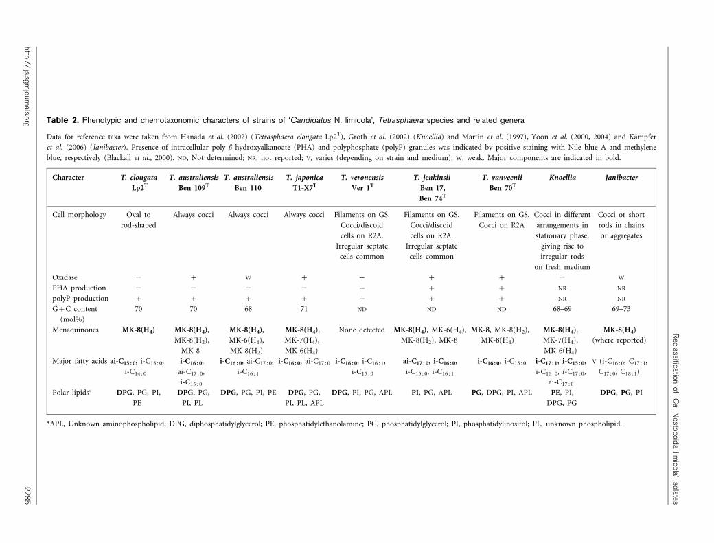
Table 2. Phenotypic and chemotaxonomic characters of strains of ‘Candidatus N. limicola’, Tetrasphaera species and related genera
Data for reference taxa were taken from Hanada et al. (2002) (Tetrasphaera elongata Lp2T), Groth et al. (2002) (Knoellia) and Martin et al. (1997), Yoon et al. (2000, 2004) and Kampfer
et al. (2006) (Janibacter). Presence of intracellular poly-b-hydroxyalkanoate (PHA) and polyphosphate (polyP) granules was indicated by positive staining with Nile blue A and methylene
blue, respectively (Blackall et al., 2000). ND, Not determined; NR, not reported; V, varies (depending on strain and medium); W, weak. Major components are indicated in bold.
Character T. elongata
Lp2T
T. australiensis
Ben 109T
T. australiensis
Ben 110
T. japonica
T1-X7T
T. veronensis
Ver 1T
T. jenkinsii
Ben 17,
Ben 74T
T. vanveenii
Ben 70T
Knoellia Janibacter
Cell morphology Oval to
rod-shaped
Always cocci Always cocci Always cocci Filaments on GS.
Cocci/discoid
cells on R2A.
Irregular septate
cells common
Filaments on GS.
Cocci/discoid
cells on R2A.
Irregular septate
cells common
Filaments on GS.
Cocci on R2A
Cocci in different
arrangements in
stationary phase,
giving rise to
irregular rods
on fresh medium
Cocci or short
rods in chains
or aggregates
Oxidase 2 + W + + + + 2 W
PHA production 2 2 2 2 + + + NR NR
polyP production + + + + + + + NR NR
G+C content
(mol%)
70 70 68 71 ND ND ND 68–69 69–73
Menaquinones MK-8(H4) MK-8(H4),
MK-8(H2),
MK-8
MK-8(H4),
MK-6(H4),
MK-8(H2)
MK-8(H4),
MK-7(H4),
MK-6(H4)
None detected MK-8(H4), MK-6(H4),
MK-8(H2), MK-8
MK-8, MK-8(H2),
MK-8(H4)
MK-8(H4),
MK-7(H4),
MK-6(H4)
MK-8(H4)
(where reported)
Major fatty acids ai-C15 : 0, i-C15 : 0,
i-C14 : 0
i-C16 : 0,
ai-C17 : 0,
i-C15 : 0
i-C16 : 0, ai-C17 : 0,
i-C16 : 1
i-C16 : 0, ai-C17 : 0 i-C16 : 0, i-C16 : 1,
i-C15 : 0
ai-C17 : 0, i-C16 : 0,
i-C15 : 0, i-C16 : 1
i-C16 : 0, i-C15 : 0 i-C17 : 1, i-C15 : 0,
i-C16 : 0, i-C17 : 0,
ai-C17 : 0
V (i-C16 : 0, C17 : 1,
C17 : 0, C18 : 1)
Polar lipids* DPG, PG, PI,
PE
DPG, PG,
PI, PL
DPG, PG, PI, PE DPG, PG,
PI, PL, APL
DPG, PI, PG, APL PI, PG, APL PG, DPG, PI, APL PE, PI,
DPG, PG
DPG, PG, PI
*APL, Unknown aminophospholipid; DPG, diphosphatidylglycerol; PE, phosphatidylethanolamine; PG, phosphatidylglycerol; PI, phosphatidylinositol; PL, unknown phospholipid.
http://ijs.sgm
journals.org2
28
5
Reclassification
of‘C
a.N
ostocoidalim
icola’isolates

with the Tetrasphaera australiensis strains was recovered innine of the parsimony trees (data not shown). The‘Candidatus N. limicola’ and Tetrasphaera australiensisisolates formed a cluster consistent with that in the 16SrRNA gene sequence results, but in this case there was nobootstrap support for the grouping. The rest of the treetopology (with the exception of the placement of theoutgroups) received poor bootstrap support, and wasdifferent from the 16S rRNA gene tree topology.Tetrasphaera elongata Lp2T was not included in this analysis.
16S–23S rRNA intergenic fingerprinting
The fingerprints obtained after restriction digestion of thisregion with the two enzymes showed that strains Ben 17, Ben18, Ben 67, Ben 68 and Ben 74 gave identical patterns afterdigestion with MspI (Fig. 5a), while strain Ben 70 was quite
different, sharing only two bands with these other strains.Strain Ver 1 was also different, having two bands in commonwith the ‘strain Ben 17’ group and one band in commonwith strain Ben 70, as well as possessing a single band not
Fig. 3. Phylogenetic analysis of 16S rRNAgene sequences of ‘Candidatus N. limicola’strains (in bold) and their nearest relativesbased on 1420 bp, after neighbour-joininganalysis using Tamura–Nei distance esti-mates. Nodes receiving ¢50 % bootstrapsupport (1000 replicates) are indicated. Bar,0?01 nucleotide substitutions per site.
Fig. 4. Phylogenetic analysis of rpoC1 DNA sequences of‘Candidatus N. limicola’ strains (in bold) and their closest rela-tives based on 600 bp and neighbour-joining analysis usingTamura–Nei distance estimates. Nodes receiving ¢50 % boot-strap support (1000 replicates) are indicated. Bar, 0?05 nucleo-tide substitutions per site.
(a)
(b)
1 2 3 4 5 6 7 8 9 10 11
1 2 3 4 5 6 7 8 9 10 11
Fig. 5. Fingerprints of the 16S–23S rRNA intergenic spacerregion of strains of ‘Candidatus N. limicola’ and Tetrasphaera
species after digestion with the restriction endonucleases MspI(a) and BstUI (b) and separation by agarose gel electro-phoresis. Lanes: 1, 100 bp molecular marker; 2, Ben 17; 3,Ben 18; 4, Ben 67; 5, Ben 68; 6, Ben 70; 7, Ben 74; 8, Ver1; 9, Tetrasphaera japonica T1-X7T; 10, Tetrasphaera austra-
liensis Ben 109T; 11, Tetrasphaera australiensis Ben 110.
2286 International Journal of Systematic and Evolutionary Microbiology 56
C. M. McKenzie and others

seen in any of the other isolates. The patterns fromTetrasphaera australiensis strains Ben 109T and 110 wereidentical to each other, possessing a unique band. They hadtwo bands in common with all the other strains exceptTetrasphaera japonica T1-X7T (where only one commonband could be seen). This strain also shared this band withall other strains and had two bands in common with strainBen 70, while one was unique to it.
The patterns generated with the enzyme BstUI suggestedslightly different relationships between these strains(Fig. 5b). Thus, even though strains Ben 17 and Ben 18again gave identical patterns with this enzyme, strains Ben67, Ben 68 and Ben 74, although identical to each other, nowshared only two of four bands with them. Strain Ben 70 wasagain quite different, possessing three bands not seen in theothers, although two of its other bands were common to allother strains. The pattern of strain Ver 1 was also distinctive,sharing only two bands with strains Ben 17 and Ben 18 andone with strains Ben 67, Ben 68, Ben 70 and Ben 74. Again,Tetrasphaera australiensis strains Ben 109T and Ben 110 gaveidentical patterns, possessing two unique bands and twobands in common with all other strains except strain Ver 1,with which it shared only a single band. Tetrasphaerajaponica T1-X7T gave a distinctive pattern, with one uniqueband, one band shared with all the other strains, another incommon with all except strain Ver 1 and another sharedonly with strains Ben 67, Ben 68 and Ben 74. A dendrogram(not shown) obtained after numerical analysis of 23characters from these patterns revealed that strains Ben17, Ben 18, Ben 67, Ben 68 and Ben 74 formed a single clusterat 0?86 SSM, while the patterns of the other strains and thoseof Tetrasphaera australiensis and Tetrasphaera japonica T1-X7T emerged as quite different from each other. Thus, strainVer 1 was closer to Tetrasphaera australiensis than it was tothe other ‘N. limicola’ II isolates analysed.
DNA–DNA hybridization data
Previous DNA–DNA hybridization data showed thatTetrasphaera australiensis strains Ben 109T and 110 andT. japonica T1-X7T represent two separate genomic species(Maszenan et al., 2000). DNA–DNA hybridization valuescould be obtained between only some of the strainsexamined in this present study. Strains Ben 17 and Ben 74had a DNA–DNA relatedness value of 74 %, Ben 74 and Ben67 were 70 % related, while the DNA–DNA hybridizationvalue between Ben 74 and Ver 1 was only 17 %. The DNA–DNA hybridization values determined betweenTetrasphaera australiensis Ben 109T and strains Ben 70,Ver 1, Ben 67 and Ben 74 were respectively 46?9 % (mean ofduplicates), 39?7, 33?1 and 14?2 %.
DISCUSSION
The chemotaxonomic and phylogenetic data presented heresuggest that all of the ‘Candidatus N. limicola’ strainsexamined belong to a single genus. The question thenremains as to whether these ‘Candidatus N. limicola’ isolates
represent a new genus or are members of any of the existinggenera Tetrasphaera, Knoellia or Janibacter, their closestphylogenetic relatives (Fig. 3). No taxonomic decisions werepossible in the earlier study with ‘Candidatus N. limicola’(Blackall et al., 2000) in the absence of importantchemotaxonomic data, because of their very slow growthrates and the consequent problems of obtaining sufficientbiomass for analyses. The 16S rRNA gene sequence data(Fig. 3) show that the Tetrasphaera and ‘Candidatus N.limicola’ isolates form clusters supported by high bootstrapvalues, which are quite separate from the type strains ofJ. limosus, J. terrae, J. melonis, J. anophelis, K. sinensis,K. subterranea, Terrabacter tumescens and Terracoccus luteus(Fig. 3). The phylogenetic closeness of all these ‘CandidatusN. limicola’ strains (especially strains Ben 17, Ben 18, Ben67, Ben 68 and Ben 74) to the existing Tetrasphaera species issubstantiated when rpoC1 gene sequence data are analysedfrom the same group of bacteria (Fig. 4), and these too aregenerally consistent with them all belonging to a singlegenus. Some differences were seen compared with thegroupings that emerged from 16S rRNA gene sequenceanalyses (most notably with strains Ben 70 and Ver 1 nowclustering most closely), despite claims that rpoC1 sequencedata generally provide phylogenetic support for groupingsfrom 16S rRNA gene sequence data (Morse et al., 1996; Seo& Yokota, 2003). It has also been claimed, at least with thecyanobacteria, that rpoC1 sequence data are able to resolvegenus–species relationships better than 16S rRNA genesequence data can (Wilson et al., 2000; Fergusson & Saint,2000).
Chemotaxonomic data obtained from strains Ben 17, Ben74, Ben 70 and Ver 1 (chosen for analysis as representingmembers of the main clusters revealed after the 16S rRNAgene sequence analyses) showed them all to have the A1c
cell-wall type, and all except Ver 1 contained MK-8(H4) as amajor menaquinone. These chemical features are shared bymembers of the genera Tetrasphaera (Maszenan et al., 2000),Janibacter (Martin et al., 1997; Yoon et al., 2000, 2004;Kampfer et al., 2006) and Knoellia (Groth et al., 2002) anddistinguish them from all other currently described closerelatives, including members of the genus Terrabacter andTerracoccus, both of which possess the A3c cell-wall type(Maszenan et al., 2000). Comparisons of their polar lipidand fatty acid compositions (Supplementary Table S1)suggest that the ‘Candidatus N. limicola’ isolates are alsomore similar to Tetrasphaera australiensis strains,Tetrasphaera japonica T1-X7T and Tetrasphaera elongataLp2T (Maszenan et al., 2000; Hanada et al., 2002) than tospecies of either Janibacter or Knoellia (SupplementaryTable S1).
The problem of speciation then needs to be addressed. The16S rRNA gene, rpoC1 and 16S–23S rRNA intergenic spacerregion fingerprints together suggest that six species arepresent in this group of bacteria, i.e. the pre-existingTetrasphaera australiensis, Tetrasphaera japonica (Maszenanet al., 2000) and Tetrasphaera elongata (Hanada et al., 2002)
http://ijs.sgmjournals.org 2287
Reclassification of ‘Ca. Nostocoida limicola’ isolates

and three novel ones consisting of strains Ben 17, Ben 18,Ben 67 and Ben 74, strain Ben 70 and strain Ver 1. Theavailable DNA–DNA hybridization data support this.Furthermore, strain Ver 1 was the only ‘Candidatus N.limicola’ isolate among these strains to possess thedistinctive nucleotide sequence variation in the V6 regionof the 16S rRNA gene that was noticed by Blackall et al.(2000). Ben 70 is different from the other isolates in havingMK-8 and MK-8(H2) as its major menaquinones, while nomenaquinones could be detected in strain Ver 1, despiterepeated analyses. Thus, when these menaquinone profilesare considered, strains Ben 70 and Ver 1 appear to belong toseparate species. Strains Ben 17, Ben 18, Ben 67, Ben 68 andBen 74 share nearly identical 16S rRNA gene and rpoC1sequences, and their 16S–23S rRNA intergenic fingerprintsare either identical or very similar, depending on therestriction endonuclease used. The DNA–DNA hybridiza-tion values of 74 and 70 %, respectively, for Ben 17 versusBen 74 and Ben 67 versus Ben 74 are consistent with theseother observations. Thus, by association, these strains andBen 18 and Ben 68 are all considered to belong to the samegenomic species. Although not subjected to some of thecharacterizations described here, Tetrasphaera elongataLp2T is considered a separate species on the basis of itsprevious description (Hanada et al., 2002), especially itsfatty acid composition, which contains distinctively highlevels of ai-C15 : 0 and low levels of i-C16 : 0 (Hanada et al.,2002).
Therefore, we propose that these isolates of ‘Candidatus N.limicola’ (Ben 17, Ben 18, Ben 67, Ben 68, Ben 70, Ben 74 andVer 1) be combined with Tetrasphaera elongata,Tetrasphaera australiensis and Tetrasphaera japonica in thegenus Tetrasphaera. On the basis of the characterization datapresented here, we propose that strains Ben 17, Ben 18, Ben67, Ben 68 and Ben 74T be assigned to a single species asTetrasphaera jenkinsii sp. nov. These data also support thedesignation of strain Ben 70T to a novel species Tetrasphaeravanveenii sp. nov. and strains Ver 1T and Ver 2 toTetrasphaera veronensis sp. nov.
Description of Tetrasphaera jenkinsii sp. nov.
Tetrasphaera jenkinsii (jen.kin9si.i. N.L. gen. n. jenkinsii ofJenkins, referring to David Jenkins, a contemporaryAmerican environmental engineer, who has made aconsiderable contribution to our understanding of thefilamentous bacteria causing bulking and foaming inactivated sludge processes).
The phenotypic features of this species are those given byBlackall et al. (2000) for ‘Candidatus N. limicola’ strains Ben17, Ben 18, Ben 67, Ben 68 and Ben 74, supplemented by theresults of this study as follows. Poly-b-hydroxyalkanoateand polyphosphate production is positive. Major menaqui-none is MK-8(H4), while MK-6(H4), MK-8 and MK-8(H2)occur in minor amounts. Present members of the specieswere isolated from activated sludge systems in Australia.
The type strain is Ben 74T (=DSM 17519T=NCIMB14128T).
Description of Tetrasphaera vanveenii sp. nov.
Tetrasphaera vanveenii (van.vee9ni.i. N.L. gen. n. vanveeniiof van Veen, referring to the late Dutch microbiologist W. L.van Veen, who originally isolated this filamentous bacter-ium from activated sludge).
The phenotypic features of this species are those given byBlackall et al. (2000) for ‘Candidatus N. limicola’ strain Ben70, supplemented by the results of this study as follows.Poly-b-hydroxyalkanoate and polyphosphate production ispositive. Menaquinones are MK-8, MK-8(H2) and MK-8(H4). The polar lipid pattern contains diphosphatidylgly-cerol and an unidentified aminophospholipid.
The type strain, Ben 70T (=DSM 17518T=NCIMB14127T), was isolated from an activated sludge plant inCarrum, Victoria, Australia.
Description of Tetrasphaera veronensis sp. nov.
Tetrasphaera veronensis (ve.ro.nen9sis. L. nom. fem. adj.veronensis of Verona, Italy, from where the first isolatesoriginated).
The phenotypic features of this species are those given byBlackall et al. (2000) for ‘Candidatus N. limicola’ strain Ver1, supplemented by the results of this study as follows.Catalase- and oxidase-positive. Poly-b-hydroxyalkanoateand polyphosphate production is positive. Isoprenoidquinones can not be detected in the type strain. The polarlipid pattern contains diphosphatidylglycerol and anunidentified phospholipid. Major cellular fatty acids containiso-C16 : 1.
The type strain, Ver 1T (=DSM 17520T=NCIMB 14129T),was isolated from an activated sludge plant in Verona, Italy.
ACKNOWLEDGEMENTS
R. J. S. wishes to acknowledge the financial support of an AustralianResearch Council Large Grant to support this work, and J.-R. L.received a La Trobe University PhD scholarship.
REFERENCES
Beer, M., Seviour, E. M., Kong, Y., Cunningham, M., Blackall, L. L. &Seviour, R. J. (2002). Phylogeny of the filamentous bacteriumEikelboom type 1851, and design and application of a 16S rRNAtargeted oligonucleotide probe for its fluorescence in situ identifica-tion in activated sludge. FEMS Microbiol Lett 207, 179–183.
Blackall, L. L., Seviour, E. M., Cunningham, M. A., Seviour, R. J. &Hugenholtz, P. (1994). ‘‘Microthrix parvicella’’ is a novel, deepbranching member of the actinomycetes subphylum. Syst ApplMicrobiol 17, 513–518.
2288 International Journal of Systematic and Evolutionary Microbiology 56
C. M. McKenzie and others

Blackall, L. L., Seviour, E. M., Bradford, D., Rossetti, S., Tandoi, V. &Seviour, R. J. (2000). ‘Candidatus Nostocoida limicola’, a filamen-tous bacterium from activated sludge. Int J Syst Evol Microbiol 50,
703–709.
Butler, M. K., Wang, J., Webb, R. I. & Fuerst, J. A. (2002). Molecularand ultrastructural confirmation of classification of ATCC 35122 as a
strain of Pirellula staleyi. Int J Syst Evol Microbiol 52, 1663–1667.
Collins, M. D., Dorsch, M. & Stackebrandt, E. (1989). Transfer of
Pimelobacter tumescens to Terrabacter gen. nov. as Terrabactertumescens comb. nov. and of Pimelobacter jensenii to Nocardioides as
Nocardioides jensenii comb. nov. Int J Syst Bacteriol 39, 1–6.
Eikelboom, D. H. & van Buijsen, H. J. J. (1983). Microscopic SludgeInvestigation Manual, 2nd edn. Delft: TNO Research Institute of
Environmental Hygiene.
Fergusson, K. M. & Saint, C. P. (2000). Molecular phylogeny of
Anabaena circinalis and its identification in environmental samplesby PCR. Appl Environ Microbiol 66, 4145–4148.
Groth, I., Schumann, P., Martin, K., Schuetze, B., Augsten, K.,Kramer, I. & Stackebrandt, E. (1999). Ornithinicoccus hortensis gen.nov., sp. nov., a soil actinomycete which contains L-ornithine. Int
J Syst Bacteriol 49, 1717–1724.
Groth, I., Schumann, P., Schutze, B., Augsten, K. & Stackebrandt, E.(2002). Knoellia sinensis gen. nov., sp. nov. and Knoellia subterraneasp. nov., two novel actinobacteria isolated from a cave. Int J Syst Evol
Microbiol 52, 77–84.
Gurtler, V. & Stanisich, V. A. (1996). New approaches to typing andidentification of bacteria using the 16S–23S rDNA spacer region.
Microbiology 142, 3–16.
Hanada, S., Liu, W.-T., Shintani, T., Kamagata, Y. & Nakamura, K.(2002). Tetrasphaera elongata sp. nov., a polyphosphate-accumulat-ing bacterium isolated from activated sludge. Int J Syst Evol Microbiol
52, 883–887.
Kampfer, P. & Wagner, M. (2002). Filamentous bacteria in activatedsludge: current taxonomic status and ecology. In Encyclopedia of
Environmental Microbiology, pp. 1287–1306. Edited by G. Bitton.New York: Wiley-Interscience.
Kampfer, P., Terenius, O., Lindh, J. M. & Faye, I. (2006). Janibacteranophelis sp. nov., isolated from the midgut of Anopheles arabiensis.
Int J Syst Evol Microbiol 56, 389–392.
Kataoka, N., Tokiwa, Y., Tanaka, Y., Takeda, K. & Suzuki, T. (1996).Enrichment culture and isolation of slow-growing bacteria. Appl
Microbiol Biotechnol 45, 771–777.
Kong, Y., Nielsen, J. L. & Nielsen, P. H. (2005). Identity and
ecophysiology of uncultured actinobacterial polyphosphate-accumu-lating organisms in full-scale enhanced biological phosphorus
removal plants. Appl Environ Microbiol 71, 4076–4085.
Kragelund, C., Nielsen, J. L., Thomsen, T. R. & Nielsen, P. H. (2005).Ecophysiology of the filamentous Alphaproteobacterium Meganema
perideroedes in activated sludge. FEMS Microbiol Ecol 54, 111–122.
Kumar, S., Tamura, K., Jakobsen, I.-B. & Nei, M. (2001). MEGA2:
molecular evolutionary genetics analysis software. Bioinformatics 17,1244–1245.
Levantesi, C., Beimfohr, C., Geurkink, B., Rossetti, S., Thelen, K.,Kroonman, J., Snaidr, J., van der Waarde, J. & Tandoi, V. (2004).Filamentous Alphaproteobacteria associated with bulking in industrial
wastewater treatment plants. Syst Appl Microbiol 27, 716–727.
Liu, J. R., McKenzie, C. A., Seviour, E. M., Webb, R. I., Blackall, L. L.,Saint, C. P. & Seviour, R. J. (2001). Phylogeny of the filamentous
bacterium ‘Nostocoida limicola’ III from activated sludge. Int J SystEvol Microbiol 51, 195–202.
Liu, J. R., Tanner, R. S., Schumann, P. & 7 other authors (2002).Emended description of the genus Trichococcus, description of
Trichococcus collinsii sp. nov., and reclassification of
Lactosphaera pasteurii as Trichococcus pasteurii comb. nov. and of
Ruminococcus palustris as Trichococcus palustris comb. nov. in
the low-G+C Gram-positive bacteria. Int J Syst Evol Microbiol 52,
1113–1126.
Martin, K., Schumann, P., Rainey, F. A., Schuetze, B. & Groth, I.(1997). Janibacter limosus gen. nov., sp. nov., a new actinomycete
with meso-diaminopimelic acid in the cell wall. Int J Syst Bacteriol
47, 529–534.
Martins, A. M. P., Pagilla, K., Heijnen, J. J. & van Loosdrecht, M. C. M.(2004). Filamentous bulking sludge – a critical review. Water Res 38,
793–817.
Maszenan, A. M., Seviour, R. J., Patel, B. K. C., Rees, G. N. &McDougall, B. M. (1997). Amaricoccus gen. nov., a gram-negative
coccus occurring in regular packages and tetrads, isolated from
activated sludge biomass, and descriptions of Amaricoccus veronensis
sp. nov., Amaricoccus tamworthensis sp. nov., Amaricoccus macauensis
sp. nov., and Amaricoccus kaplicensis sp. nov. Int J Syst Bacteriol 47,727–734.
Maszenan, A. M., Seviour, R. J., Patel, B. K. C., Schumann, P. &Rees, G. N. (1999). Tessaracoccus bendigoensis gen. nov., sp. nov., a
Gram-positive coccus occurring in regular packages or tetrads,
isolated from activated sludge biomass. Int J Syst Bacteriol 49,
459–468.
Maszenan, A. M., Seviour, R. J., Patel, B. K. C., Schumann, P.,Burghardt, J., Tokiwa, Y. & Stratton, H. M. (2000). Three isolates ofnovel polyphosphate-accumulating Gram-positive cocci, obtained
from activated sludge, belong to a new genus, Tetrasphaera gen. nov.,
and description of two new species, Tetrasphaera japonica sp. nov.
and Tetrasphaera australiensis sp. nov. Int J Syst Evol Microbiol 50,
593–603.
McKerrow, J., Vagg, S., McKinney, T., Seviour, E. M., Maszenan,A. M., Brooks, P. & Seviour, R. J. (2000). A simple HPLC method for
analysing diaminopimelic acid diastereoisomers in cell walls of
Gram-positive bacteria. Lett Appl Microbiol 30, 178–182.
Morse, R., Collins, M. D., O’Hanlon, K., Wallbanks, S. & Richardson,P. T. (1996). Analysis of the b9 subunit of the DNA-dependent
RNA polymerase does not support the hypothesis inferred from
16S rRNA analysis that Oenococcus oeni (formerly Leuconostoc
oenos) is a tachytelic (fast-evolving) bacterium. Int J Syst Bacteriol 46,
1004–1009.
Prauser, H., Schumann, P., Rainey, F. A., Kroppenstedt, R. M. &Stackebrandt, E. (1997). Terracoccus luteus gen. nov., sp. nov., an
LL-diaminopimelic acid-containing coccoid actinomycete from soil.
Int J Syst Bacteriol 47, 1218–1224.
Reasoner, D. J. & Geldreich, E. E. (1985). A new medium for the
enumeration and subculture of bacteria from potable water. Appl
Environ Microbiol 49, 1–7.
Rossetti, S., Tomei, M. C., Nielsen, P. H. & Tandoi, V. (2005).‘‘Microthrix parvicella’’, a filamentous bacterium causing bulking and
foaming in activated sludge systems: a review of current knowledge.
FEMS Microbiol Rev 29, 49–64.
Schade, M., Beimfohr, C. & Lemmer, H. (2002). Phylogenetic and
physiological characterization of a ‘‘Nostocoida limicola’’-like
organism isolated from activated sludge. Water Sci Technol 46
(1–2), 91–97.
Schumann, P., Prauser, H., Rainey, F. A., Stackebrandt, E. &Hirsch, P. (1997). Friedmanniella antarctica gen. nov., sp. nov., an
LL-diaminopimelic acid-containing actinomycete from Antarctic
sandstone. Int J Syst Bacteriol 47, 278–283.
Seo, P. S. & Yokota, A. (2003). The phylogenetic relationships of
cyanobacteria inferred from 16S rRNA, gyrB, rpoC1 and rpoD1 gene
sequences. J Gen Appl Microbiol 49, 191–203.
http://ijs.sgmjournals.org 2289
Reclassification of ‘Ca. Nostocoida limicola’ isolates

Seviour, E. M., Blackall, L. L., Christenson, C., Hugenholtz, P.,Cunningham, M. A., Bradford, D., Stratton, H. M. & Seviour, R. J.(1997). The filamentous morphotype Eikelboom type 1863 is not asingle genetic entity. J Appl Microbiol 82, 411–421.
Seviour, R. J., Maszenan, A. M., Soddell, J. A., Tandoi, V., Patel,B. K. C., Kong, Y. & Schumann, P. (2000). Microbiology of the ‘G-bacteria’ in activated sludge. Environ Microbiol 2, 581–593.
Seviour, R. J., Mino, T. & Onuki, M. (2003). The microbiology ofbiological phosphorus removal in activated sludge systems. FEMSMicrobiol Rev 27, 99–127.
Snaidr, J., Beimfohr, C., Levantesi, C., Rossetti, C., van derWaarde, J., Geurkink, B., Eikelboom, D., Lemaitre, M. & Tandoi, V.(2002). Phylogenetic analysis and in situ identification of ‘‘Nostocoidalimicola’’-like filamentous bacteria in activated sludge from industrialwastewater treatment plants. Water Sci Technol 46 (1–2), 99–104.
Thompson, J. D., Gibson, T. J., Plewniak, F., Jeanmougin, F. &Higgins, D. G. (1997). The CLUSTAL_X windows interface: flexible
strategies for multiple sequence alignment aided by quality analysistools. Nucleic Acids Res 25, 4876–4882.
Tsai, C.-S. & Liu, W.-T. (2002). Phylogenetic and physiologicaldiversity of tetrad-forming organisms in deterioratedbiological phosphorus removal systems. Water Sci Technol 46(1–2), 179–184.
Wilson, K. M., Schembri, M. A., Baker, P. D. & Saint, C. P. (2000).Molecular characterization of the toxic cyanobacteriumCylindrospermopsis raciborskii and design of a species-specific PCR.Appl Environ Microbiol 66, 332–338.
Yoon, J.-H., Lee, K.-C., Kang, S.-S., Kho, Y. H., Kang, K. H. & Park,Y.-H. (2000). Janibacter terrae sp. nov., a bacterium isolated from soilaround a wastewater treatment plant. Int J Syst Evol Microbiol 50,1821–1827.
Yoon, J.-H., Lee, H. B., Yeo, S.-H. & Choi, J.-E. (2004). Janibactermelonis sp. nov., isolated from abnormally spoiled oriental melon inKorea. Int J Syst Evol Microbiol 54, 1975–1980.
2290 International Journal of Systematic and Evolutionary Microbiology 56
C. M. McKenzie and others
Recommended


