Isolation and functional characterization of murineprostate stem cellsDevon A. Lawson*, Li Xin*, Rita U. Lukacs*, Donghui Cheng†, and Owen N. Witte*†‡§
*Departments of Microbiology, Immunology, and Molecular Genetics and ‡Molecular and Medical Pharmacology, David Geffen School of Medicine,and †Howard Hughes Medical Institute, University of California, Los Angeles, CA 90095
Contributed by Owen N. Witte, November 3, 2006 (sent for review October 19, 2006)
The ability to isolate prostate stem cells is essential to explore theirrole in prostate development and disease. In vitro prostate colony-and sphere-forming assays were used to quantitatively measuremurine prostate stem/progenitor cell enrichment and self-renewal.Cell surface markers were screened for their ability to positively ornegatively enrich for cells with enhanced growth potential in theseassays. Immunohistochemical and FACS analyses demonstrate thatspecific cell surface markers can be used to discriminate prostatestromal (CD34�), luminal epithelial (CD24�CD49f�), basal epithelial(CD24�CD49f�), hematopoietic (CD45�, Ter119�), and endothelial(CD31�) lineages. Sorting for cells with a CD45�CD31�Ter119�Sca-1�CD49f� antigenic profile results in a 60-fold enrichment forcolony- and sphere-forming cells. These cells can self-renew andexpand to form spheres for many generations and can differenti-ate to produce prostatic tubule structures containing both basaland luminal cells in vivo. These cells also localize to the basal celllayer within the region of the gland that is proximal to the urethra,which has been identified as the prostate stem cell niche. Prostatestem cells can be isolated to a purity of up to 1 in 35 by using thisantigenic profile. The remarkable similarity in cell surface profilebetween prostate and mammary gland stem cells suggests thesemarkers may be conserved among epithelial stem cell populations.
CD49f � integrin �6 � Sca-1 � CD24 heat-stable antigen � stem cell niche
S tem cells are of interest clinically because of their potentialto repair damaged tissues, treat degenerative diseases, and
because of their purported role in tumor initiation. The abilityto identify and isolate stem cells is necessary to study theirspecialized biology. Enrichment for many types of tissue stemcells has been achieved by using cell surface markers. Murinehematopoietic stem cells can be enriched by sorting Lin�Thy-1loSca-1�ckit� cells from the bone marrow (1). Recent studiessuggest that even better purity can be achieved by further sortingbased on expression of the SLAM family receptors CD150 andCD48 (2). Bronchioavelolar stem cells can be isolated from theirniche at the bronchioalveolar duct junction (BADJ) by sortingcells with a CD45�CD31�Sca-1�CD34� profile (3). Data fromtwo recent reports show that mouse mammary stem cells possessa Lin�Sca-1�CD140a�CD24�CD49f�CD29� cell surface pro-file and can be isolated to a purity of up to 1 in 20 by using subsetsof these markers (4, 5).
The presence of stem cells in the prostate first was proposedto explain the seemingly inexhaustible capacity of the organ toregenerate during androgen cycling experiments (6). The iden-tification of side-population cells and replication quiescent BrdUlabel-retaining cells further suggests that stem cells exist in thegland (7, 8). Several studies have enriched for primitive prostatecells by using cell surface markers. Richardson et al. (9) dem-onstrated that the CD44�/�2�1hi/CD133� human prostate cellsubpopulation is 10-fold enriched over CD44�/�2�1hi/CD133�
cells for colony-forming activity in vitro. Sca-1 also has been usedto enrich for murine prostate cells with enhanced prostate tubuleforming capacity in vivo (10, 11). Sca-1� cells possess severalproperties of stem cells, including replication quiescence, mul-tilineage differentiation capacity, and localization in the region
of prostatic ducts proximal to the urethra that has been identifiedas the prostate stem cell niche. Sca-1 also is expressed in fetalprostate epithelium, demonstrating it is expressed by prostatestem cells from very early stages of prostate development (12).Here, we demonstrate that the CD45�CD31�Ter119�Sca-1�CD49f� prostate cell subpopulation is enriched for cellscapable of both colony and sphere formation in vitro. These cellscan self-renew to form spheres for multiple generations and candifferentiate to produce prostatic tubule structures containingboth basal and luminal cells in vivo.
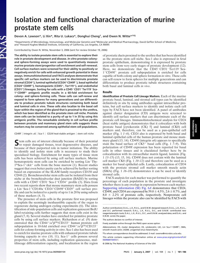
ResultsIdentification of Prostate Cell Lineage Markers. Each of the murineprostate basal, luminal, and stromal cell types can be identifieddefinitively in situ by using antibodies against intracellular pro-teins, but cell surface markers to identify and isolate each celltype by FACS have not been identified. A panel of antibodiesagainst cluster designation (CD) antigens were screened toidentify cell surface markers that can discriminate each of theprostate cell lineages. Immunohistochemical analysis for CD24(heat stable antigen) demonstrates that it colocalizes with bothprostate basal [cytokeratin (CK) 5] and luminal (CK8) cellmarkers and, therefore, can be used as a pan-epithelial cellmarker (Fig. 1 1–6). CD24 also is expressed by both basal andluminal epithelial cells of the human prostate and mouse mam-mary gland (13, 14). CD49f (integrin �6) appears to specificallystain the basal surface of CK5� basal cells (Fig. 1 7–9). Thispolarization of CD49f expression has been reported for basalcells in other tissues and is elucidated further here by itscolocalization with its extracellular matrix ligand, laminin (Fig.1 13–15) (13, 15, 16). CD49f does not costain with the luminalcell marker CK8 (Fig. 1 10–12) and therefore can be used as amarker for basal epithelial cells. Lastly, colocalization of CD34with the prostate stromal cell marker smooth muscle actin(SMA) (Fig. 1 16–18) demonstrates it can be used to identifystromal cells.
FACS analysis for each marker was performed to quantify thepercentage of each population in the prostate and investigatewhether there is any overlap in expression between each marker.Supporting information (SI) Fig. 6A demonstrates that CD24,CD49f, and CD34 are expressed by 59.0 � 4.5%, 2.6 � 0.7%, and8.0 � 2.2% of prostate cells, respectively. Nonprostate celllineages within the prostate also can be identified by FACS with
Author contributions: D.A.L., L.X., R.U.L., and O.N.W. designed research; D.A.L., L.X., R.U.L.,and D.C. performed research; D.A.L., L.X., R.U.L., D.C., and O.N.W. contributed newreagents/analytic tools; D.A.L., L.X., R.U.L., D.C., and O.N.W. analyzed data; and D.A.L. andO.N.W. wrote the paper.
The authors declare no conflict of interest.
Freely available online through the PNAS open access option.
Abbreviations: CD, cluster designation; CK, cytokeratin; LSC, Lin�Sca-1�CD49f�; SMA,smooth muscle actin; UGSM, urogenital sinus mesenchyme.
§To whom correspondence should be addressed. E-mail: [email protected].
This article contains supporting information online at www.pnas.org/cgi/content/full/0609684104/DC1.
© 2006 by The National Academy of Sciences of the USA
www.pnas.org�cgi�doi�10.1073�pnas.0609684104 PNAS � January 2, 2007 � vol. 104 � no. 1 � 181–186
DEV
ELO
PMEN
TAL
BIO
LOG
Y
antibodies against CD45 (hematopoietic), Ter119 (red bloodcell), and CD31 (endothelial). Pairwise analysis for all markersshows that the stromal cell marker CD34 does not significantlyoverlap with either epithelial marker CD24 or CD49f. Nearly allCD49f� cells express CD24 as predicted based on in situ analysis.Small populations of CD24� and CD49f� cells also express thenonprostate cell lineage markers (CD45, Ter119, and CD31,collectively called ‘‘Lin’’), which is consistent with studies show-ing hematopoietic and endothelial cells express CD24 and CD49f(17, 18). These data suggest that it should be possible to isolatebasal and luminal cells by sorting the CD24�CD49f� andCD24�CD49f� fractions of the prostate, respectively. CK5 andCK8 staining of cytospins prepared from each fraction demon-strates that �70% of CD24�CD49f� cells express CK5 (SI Fig.6B Left). Conversely, the majority of CD24�CD49f� cells areCK8� (SI Fig. 6B Right), and �4% of these cells express CK5(data not shown).
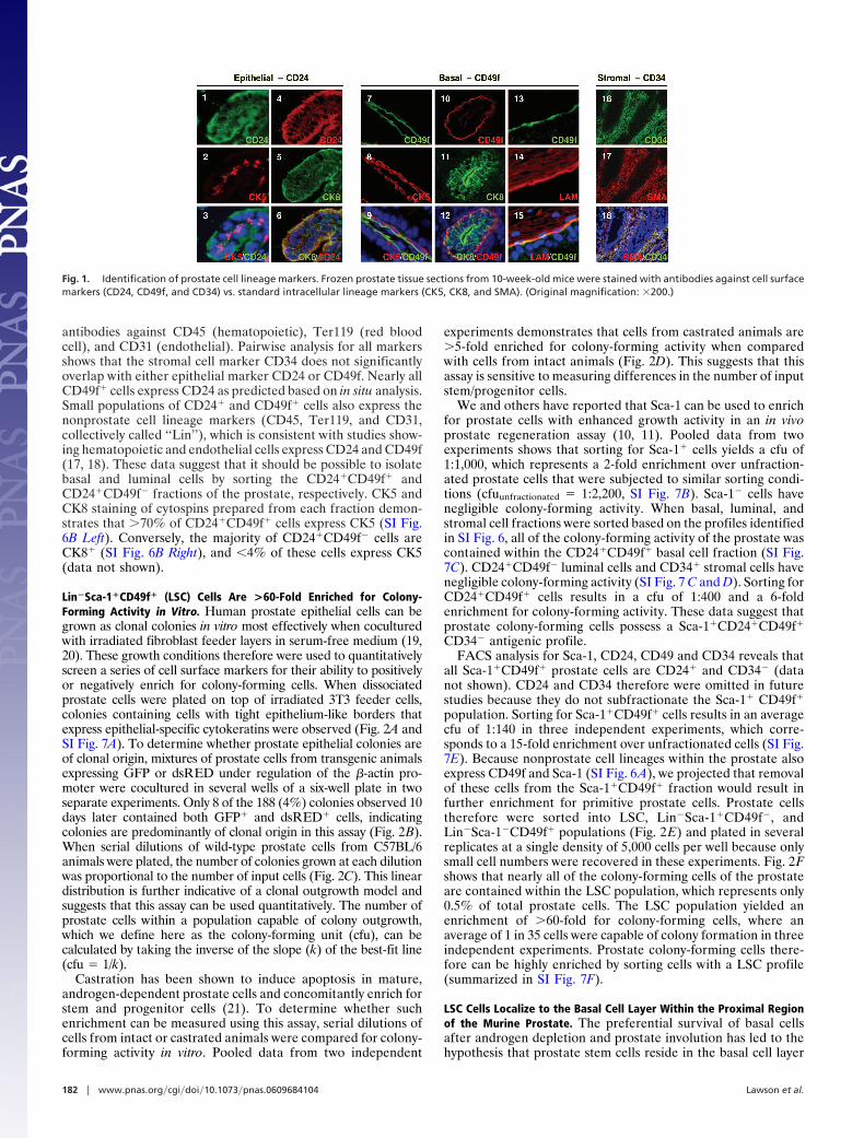
Lin�Sca-1�CD49f� (LSC) Cells Are >60-Fold Enriched for Colony-Forming Activity in Vitro. Human prostate epithelial cells can begrown as clonal colonies in vitro most effectively when coculturedwith irradiated fibroblast feeder layers in serum-free medium (19,20). These growth conditions therefore were used to quantitativelyscreen a series of cell surface markers for their ability to positivelyor negatively enrich for colony-forming cells. When dissociatedprostate cells were plated on top of irradiated 3T3 feeder cells,colonies containing cells with tight epithelium-like borders thatexpress epithelial-specific cytokeratins were observed (Fig. 2A andSI Fig. 7A). To determine whether prostate epithelial colonies areof clonal origin, mixtures of prostate cells from transgenic animalsexpressing GFP or dsRED under regulation of the �-actin pro-moter were cocultured in several wells of a six-well plate in twoseparate experiments. Only 8 of the 188 (4%) colonies observed 10days later contained both GFP� and dsRED� cells, indicatingcolonies are predominantly of clonal origin in this assay (Fig. 2B).When serial dilutions of wild-type prostate cells from C57BL/6animals were plated, the number of colonies grown at each dilutionwas proportional to the number of input cells (Fig. 2C). This lineardistribution is further indicative of a clonal outgrowth model andsuggests that this assay can be used quantitatively. The number ofprostate cells within a population capable of colony outgrowth,which we define here as the colony-forming unit (cfu), can becalculated by taking the inverse of the slope (k) of the best-fit line(cfu � 1/k).
Castration has been shown to induce apoptosis in mature,androgen-dependent prostate cells and concomitantly enrich forstem and progenitor cells (21). To determine whether suchenrichment can be measured using this assay, serial dilutions ofcells from intact or castrated animals were compared for colony-forming activity in vitro. Pooled data from two independent
experiments demonstrates that cells from castrated animals are�5-fold enriched for colony-forming activity when comparedwith cells from intact animals (Fig. 2D). This suggests that thisassay is sensitive to measuring differences in the number of inputstem/progenitor cells.
We and others have reported that Sca-1 can be used to enrichfor prostate cells with enhanced growth activity in an in vivoprostate regeneration assay (10, 11). Pooled data from twoexperiments shows that sorting for Sca-1� cells yields a cfu of1:1,000, which represents a 2-fold enrichment over unfraction-ated prostate cells that were subjected to similar sorting condi-tions (cfuunfractionated � 1:2,200, SI Fig. 7B). Sca-1� cells havenegligible colony-forming activity. When basal, luminal, andstromal cell fractions were sorted based on the profiles identifiedin SI Fig. 6, all of the colony-forming activity of the prostate wascontained within the CD24�CD49f� basal cell fraction (SI Fig.7C). CD24�CD49f� luminal cells and CD34� stromal cells havenegligible colony-forming activity (SI Fig. 7 C and D). Sorting forCD24�CD49f� cells results in a cfu of 1:400 and a 6-foldenrichment for colony-forming activity. These data suggest thatprostate colony-forming cells possess a Sca-1�CD24�CD49f�
CD34� antigenic profile.FACS analysis for Sca-1, CD24, CD49 and CD34 reveals that
all Sca-1�CD49f� prostate cells are CD24� and CD34� (datanot shown). CD24 and CD34 therefore were omitted in futurestudies because they do not subfractionate the Sca-1� CD49f�
population. Sorting for Sca-1�CD49f� cells results in an averagecfu of 1:140 in three independent experiments, which corre-sponds to a 15-fold enrichment over unfractionated cells (SI Fig.7E). Because nonprostate cell lineages within the prostate alsoexpress CD49f and Sca-1 (SI Fig. 6A), we projected that removalof these cells from the Sca-1�CD49f� fraction would result infurther enrichment for primitive prostate cells. Prostate cellstherefore were sorted into LSC, Lin�Sca-1�CD49f�, andLin�Sca-1�CD49f� populations (Fig. 2E) and plated in severalreplicates at a single density of 5,000 cells per well because onlysmall cell numbers were recovered in these experiments. Fig. 2Fshows that nearly all of the colony-forming cells of the prostateare contained within the LSC population, which represents only0.5% of total prostate cells. The LSC population yielded anenrichment of �60-fold for colony-forming cells, where anaverage of 1 in 35 cells were capable of colony formation in threeindependent experiments. Prostate colony-forming cells there-fore can be highly enriched by sorting cells with a LSC profile(summarized in SI Fig. 7F).
LSC Cells Localize to the Basal Cell Layer Within the Proximal Regionof the Murine Prostate. The preferential survival of basal cellsafter androgen depletion and prostate involution has led to thehypothesis that prostate stem cells reside in the basal cell layer
Fig. 1. Identification of prostate cell lineage markers. Frozen prostate tissue sections from 10-week-old mice were stained with antibodies against cell surfacemarkers (CD24, CD49f, and CD34) vs. standard intracellular lineage markers (CK5, CK8, and SMA). (Original magnification: �200.)
182 � www.pnas.org�cgi�doi�10.1073�pnas.0609684104 Lawson et al.
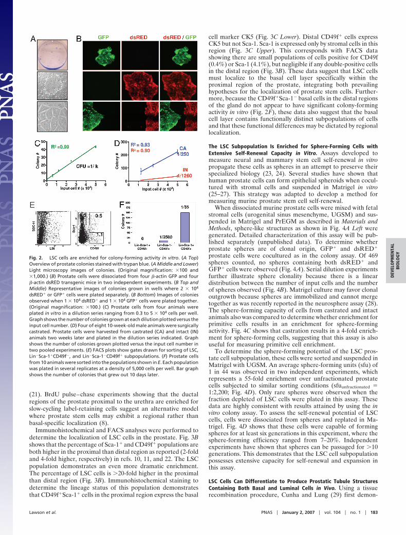
(21). BrdU pulse–chase experiments showing that the ductalregions of the prostate proximal to the urethra are enriched forslow-cycling label-retaining cells suggest an alternative modelwhere prostate stem cells may exhibit a regional rather thanbasal-specific localization (8).
Immunohistochemical and FACS analyses were performed todetermine the localization of LSC cells in the prostate. Fig. 3Bshows that the percentage of Sca-1� and CD49f� populations areboth higher in the proximal than distal region as reported (2-foldand 4-fold higher, respectively) in refs. 10, 11, and 22. The LSCpopulation demonstrates an even more dramatic enrichment.The percentage of LSC cells is �20-fold higher in the proximalthan distal region (Fig. 3B). Immunohistochemical staining todetermine the lineage status of this population demonstratesthat CD49f�Sca-1� cells in the proximal region express the basal
cell marker CK5 (Fig. 3C Lower). Distal CD49f� cells expressCK5 but not Sca-1. Sca-1 is expressed only by stromal cells in thisregion (Fig. 3C Upper). This corresponds with FACS datashowing there are small populations of cells positive for CD49f(0.4%) or Sca-1 (4.1%), but negligible if any double-positive cellsin the distal region (Fig. 3B). These data suggest that LSC cellsmust localize to the basal cell layer specifically within theproximal region of the prostate, integrating both prevailinghypotheses for the localization of prostate stem cells. Further-more, because the CD49f�Sca-1� basal cells in the distal regionsof the gland do not appear to have significant colony-formingactivity in vitro (Fig. 2F), these data also suggest that the basalcell layer contains functionally distinct subpopulations of cellsand that these functional differences may be dictated by regionallocalization.
The LSC Subpopulation Is Enriched for Sphere-Forming Cells withExtensive Self-Renewal Capacity in Vitro. Assays developed tomeasure neural and mammary stem cell self-renewal in vitropropagate these cells as spheres in an attempt to preserve theirspecialized biology (23, 24). Several studies have shown thathuman prostate cells can form epithelial spheroids when cocul-tured with stromal cells and suspended in Matrigel in vitro(25–27). This strategy was adapted to develop a method formeasuring murine prostate stem cell self-renewal.
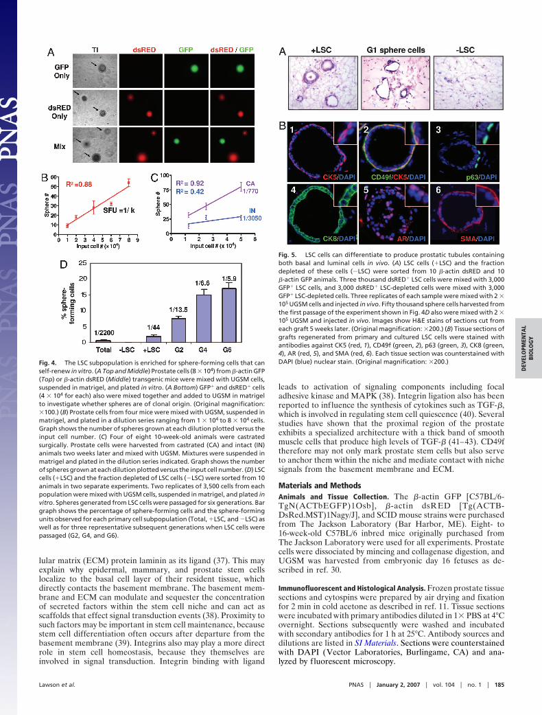
When dissociated murine prostate cells were mixed with fetalstromal cells (urogenital sinus mesenchyme, UGSM) and sus-pended in Matrigel and PrEGM as described in Materials andMethods, sphere-like structures as shown in Fig. 4A Left weregenerated. Detailed characterization of this assay will be pub-lished separately (unpublished data). To determine whetherprostate spheres are of clonal origin, GFP� and dsRED�
prostate cells were cocultured as in the colony assay. Of 469spheres counted, no spheres containing both dsRED� andGFP� cells were observed (Fig. 4A). Serial dilution experimentsfurther illustrate sphere clonality because there is a lineardistribution between the number of input cells and the numberof spheres observed (Fig. 4B). Matrigel culture may favor clonaloutgrowth because spheres are immobilized and cannot mergetogether as was recently reported in the neurosphere assay (28).The sphere-forming capacity of cells from castrated and intactanimals also was compared to determine whether enrichment forprimitive cells results in an enrichment for sphere-formingactivity. Fig. 4C shows that castration results in a 4-fold enrich-ment for sphere-forming cells, suggesting that this assay is alsouseful for measuring primitive cell enrichment.
To determine the sphere-forming potential of the LSC pros-tate cell subpopulation, these cells were sorted and suspended inMatrigel with UGSM. An average sphere-forming units (sfu) of1 in 44 was observed in two independent experiments, whichrepresents a 55-fold enrichment over unfractionated prostatecells subjected to similar sorting conditions (sfuunfractionated �1:2,200; Fig. 4D). Only rare spheres were observed when thefraction depleted of LSC cells were plated in this assay. Thesedata are highly consistent with results attained by using the invitro colony assay. To assess the self-renewal potential of LSCcells, cells were dissociated from spheres and replated in Ma-trigel. Fig. 4D shows that these cells were capable of formingspheres for at least six generations in this experiment, where thesphere-forming efficiency ranged from 7–20%. Independentexperiments have shown that spheres can be passaged for �10generations. This demonstrates that the LSC cell subpopulationpossesses extensive capacity for self-renewal and expansion inthis assay.
LSC Cells Can Differentiate to Produce Prostatic Tubule StructuresContaining Both Basal and Luminal Cells in Vivo. Using a tissuerecombination procedure, Cunha and Lung (29) first demon-
Fig. 2. LSC cells are enriched for colony-forming activity in vitro. (A Top)Overview of prostate colonies stained with trypan blue. (A Middle and Lower)Light microscopy images of colonies. (Original magnification: �100 and�1,000.) (B) Prostate cells were dissociated from four �-actin GFP and four�-actin dsRED transgenic mice in two independent experiments. (B Top andMiddle) Representative images of colonies grown in wells where 2 � 104
dsRED� or GFP� cells were plated separately. (B Bottom) Images of coloniesobserved when 1 � 104 dsRED� and 1 � 104 GFP� cells were plated together.(Original magnification: �100.) (C) Prostate cells from four animals wereplated in vitro in a dilution series ranging from 0.3 to 5 � 104 cells per well.Graph shows the number of colonies grown at each dilution plotted versus theinput cell number. (D) Four of eight 10-week-old male animals were surgicallycastrated. Prostate cells were harvested from castrated (CA) and intact (IN)animals two weeks later and plated in the dilution series indicated. Graphshows the number of colonies grown plotted versus the input cell number intwo pooled experiments. (E) FACS plots show gates drawn for sorting of LSC,Lin�Sca-1�CD49f�, and Lin�Sca-1�CD49f� subpopulations. (F) Prostate cellsfrom 10 animals were sorted into the populations shown in E. Each populationwas plated in several replicates at a density of 5,000 cells per well. Bar graphshows the number of colonies that grew out 10 days later.
Lawson et al. PNAS � January 2, 2007 � vol. 104 � no. 1 � 183
DEV
ELO
PMEN
TAL
BIO
LOG
Y
strated that prostate tissue can be grown de novo when fragmentsof adult rodent prostate tissue are combined with fragments ofUGSM and implanted under the kidney capsule of immunode-ficient mice. We have since modified this system to use disso-ciated cells rather than tissue fragments (30), and others haveshown that prostate ducts will regenerate when using a shorterincubation period and a simpler method of injecting cells s.c. inMatrigel (31, 32).
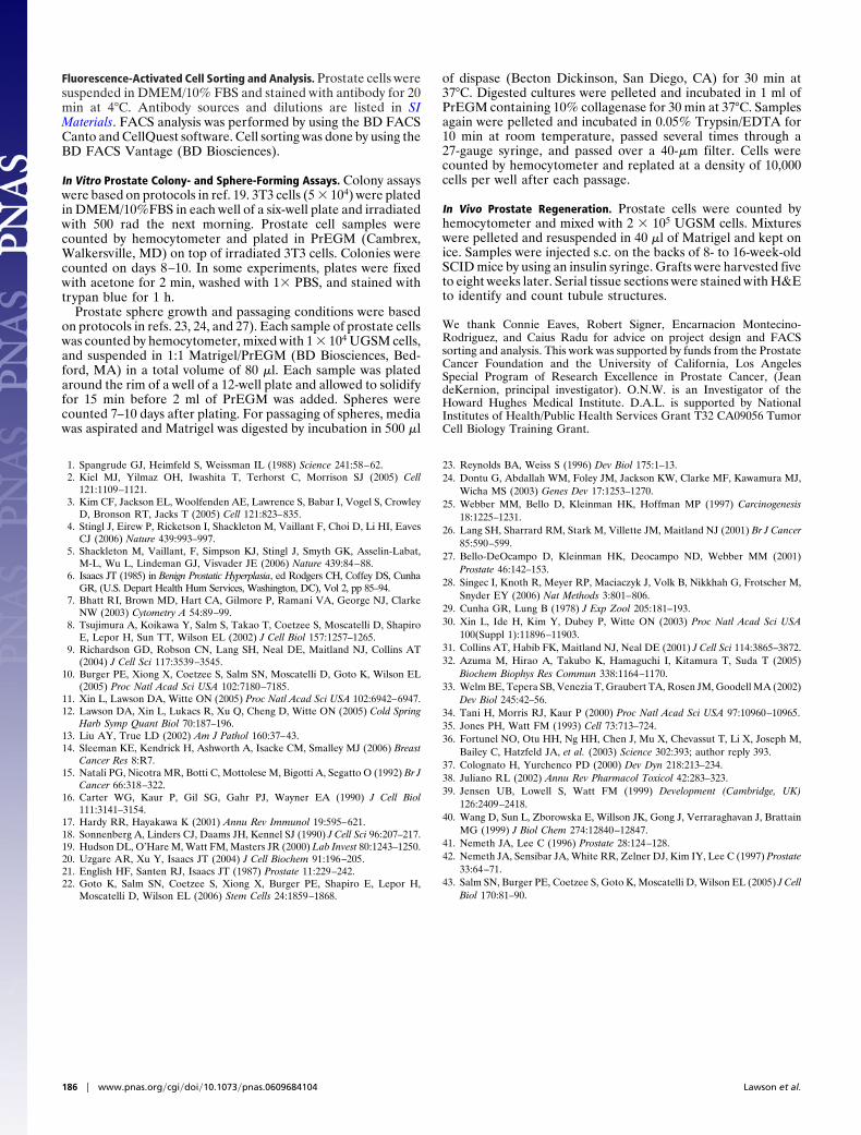
Primary and cultured LSC cells were evaluated for theircapacity to form prostate ductal structures by using this in vivoprostate regeneration method. LSC cells and the fraction de-pleted of these cells were sorted from �-actin dsRED and �-actinGFP animals as in the colony and sphere assays. Three thousanddsRED� LSC cells were mixed with 3,000 GFP� LSC cells, and3,000 dsRED� LSC-depleted cells were mixed with 3,000 GFP�
LSC-depleted cells. Each sample was mixed with UGSM andinjected in vivo. Sphere cells from the first generation of theexperiment shown in Fig. 4D also were mixed with UGSM andinjected in vivo. Fig. 5A shows that primary LSC cells as well asthe sphere cells they produced in vitro are capable of regener-ating prostatic tubule structures in vivo. No tubules were ob-served in grafts grown from the fraction depleted of LSC cells.This suggests that all of the in vivo regenerative activity of theprostate is contained within the LSC subpopulation and that thispopulation can be expanded in vitro without loss of regenerativeactivity.
Experiments in which mixtures of differentially marked cellswere implanted in the in vivo prostate regeneration system havedemonstrated that regenerated tubules are of clonal originbecause chimeric tubules are observed rarely (11, 12, 32).Likewise, no chimeric tubules containing both dsRED� andGFP� cells were observed in these experiments (SI Fig. 8). Fig.5B shows that tubules regenerated from both primary andcultured LSC cells possess normal lineage marker expressionpatterns. Regenerated tubules contain populations of basal cellsthat express CK5 (Fig. 5B1), CD49f (Fig. 5B2) and p63 (Fig.5B3). Each tubule also contains CK8� luminal cells (Fig. 5B4) as
well as cells that express low levels of the androgen receptor (Fig.5B5). A layer of SMA� stromal cells also surrounds each ductalstructure (Fig. 5B6). Because regenerated tubules are clonal, thepresence of both basal and luminal cells in each tubule indicatesthat LSC cells are capable of multilineage differentiation, whichis a defining property of stem cells.
DiscussionStem cells are defined by their unique capacity for self-renewaland multilineage differentiation. We find that CD45�CD31�Ter119�Sca-1�CD49f� prostate cells can self-renew to formspheres for many generations and can differentiate to produceprostatic tubule structures containing both basal and luminalcells in vivo. These cells also localize to the putative prostate stemcell niche in the proximal region of the gland. We thereforeconclude that prostate stem cells can be highly purified by usingthis antigenic profile.
An interesting implication of this study is the remarkable con-servation of antigenic profile between prostate stem cells and othertypes of tissue stem cells. Sca-1 is present on hematopoietic, lung,and mammary stem cells (1, 3, 33). CD49f/integrin �6 shows aneven wider distribution amongst stem cell populations. Enrichmentfor skin stem cells can be achieved by sorting cells that express highlevels of CD49f or its subunit pair CD29/integrin �1 (34, 35). Arecent study by Stingl et al. (4) showed that mammary stemcells express a CD45�Ter119�CD31�CD140a�CD24med
Sca-1loCD49fhi cell surface phenotype, which is very similar to theCD45�Ter119�CD31�CD34�CD24�Sca-1�CD49f� profile iden-tified here for the prostate stem cell, because CD34 is the stromalmarker counterpart for CD140a in the prostate. Microarray exper-iments performed by several independent groups also have shownCD49f is overexpressed consistently in hematopoietic, neural, andembryonic stem cells (36).
The conservation of these markers on stem cells suggests theyalso may function in maintaining the stem cell phenotype.Integrin �6 pairs with either �1 or �4 to form an integrinreceptor that recognizes the basement membrane and extracel-
Fig. 3. LSC cells localize to the basal cell layer within the proximal region of the prostate. (A) H&E stain of a longitudinal section from a 10-week-old mouseprostate. Arrows denote regions of the anterior prostate proximal and distal to the urethra (URE). (Original magnification: �10.) (B) Tissue from the proximaland distal regions of four mouse prostates was microdissected and digested to make dissociated cell suspensions. (B Left and Center) FACS analysis was performedto compare Sca-1 (Left) and CD49f (Center) expression in each region. (B Right) Gated on Lin� cells and indicate the percentage of LSC, Lin�Sca-1�CD49f� andLin�Sca-1�CD49f� populations in each region of the prostate. (C) Fluorescence microscopy images show Sca-1 (green), CD49f (green), CK5 (red), and DAPI nuclearcounterstain (blue) in the proximal and distal regions of the prostate. (C Insets) Magnified views of cells within each image. (Original magnification: �200.)
184 � www.pnas.org�cgi�doi�10.1073�pnas.0609684104 Lawson et al.
lular matrix (ECM) protein laminin as its ligand (37). This mayexplain why epidermal, mammary, and prostate stem cellslocalize to the basal cell layer of their resident tissue, whichdirectly contacts the basement membrane. The basement mem-brane and ECM can modulate and sequester the concentrationof secreted factors within the stem cell niche and can act asscaffolds that effect signal transduction events (38). Proximity tosuch factors may be important in stem cell maintenance, becausestem cell differentiation often occurs after departure from thebasement membrane (39). Integrins also may play a more directrole in stem cell homeostasis, because they themselves areinvolved in signal transduction. Integrin binding with ligand
leads to activation of signaling components including focaladhesive kinase and MAPK (38). Integrin ligation also has beenreported to influence the synthesis of cytokines such as TGF-�,which is involved in regulating stem cell quiescence (40). Severalstudies have shown that the proximal region of the prostateexhibits a specialized architecture with a thick band of smoothmuscle cells that produce high levels of TGF-� (41–43). CD49ftherefore may not only mark prostate stem cells but also serveto anchor them within the niche and mediate contact with nichesignals from the basement membrane and ECM.
Materials and MethodsAnimals and Tissue Collection. The �-actin GFP [C57BL/6-TgN(ACTbEGFP)1Osb], �-actin dsRED [Tg(ACTB-DsRed.MST)1Nagy/J], and SCID mouse strains were purchasedfrom The Jackson Laboratory (Bar Harbor, ME). Eight- to16-week-old C57BL/6 inbred mice originally purchased fromThe Jackson Laboratory were used for all experiments. Prostatecells were dissociated by mincing and collagenase digestion, andUGSM was harvested from embryonic day 16 fetuses as de-scribed in ref. 30.
Immunofluorescent and Histological Analysis. Frozen prostate tissuesections and cytospins were prepared by air drying and fixationfor 2 min in cold acetone as described in ref. 11. Tissue sectionswere incubated with primary antibodies diluted in 1� PBS at 4°Covernight. Sections subsequently were washed and incubatedwith secondary antibodies for 1 h at 25°C. Antibody sources anddilutions are listed in SI Materials. Sections were counterstainedwith DAPI (Vector Laboratories, Burlingame, CA) and ana-lyzed by fluorescent microscopy.
Fig. 4. The LSC subpopulation is enriched for sphere-forming cells that canself-renew in vitro. (A Top and Middle) Prostate cells (8 � 104) from �-actin GFP(Top) or �-actin dsRED (Middle) transgenic mice were mixed with UGSM cells,suspended in matrigel, and plated in vitro. (A Bottom) GFP� and dsRED� cells(4 � 104 for each) also were mixed together and added to UGSM in matrigelto investigate whether spheres are of clonal origin. (Original magnification:�100.) (B) Prostate cells from four mice were mixed with UGSM, suspended inmatrigel, and plated in a dilution series ranging from 1 � 104 to 8 � 104 cells.Graph shows the number of spheres grown at each dilution plotted versus theinput cell number. (C) Four of eight 10-week-old animals were castratedsurgically. Prostate cells were harvested from castrated (CA) and intact (IN)animals two weeks later and mixed with UGSM. Mixtures were suspended inmatrigel and plated in the dilution series indicated. Graph shows the numberof spheres grown at each dilution plotted versus the input cell number. (D) LSCcells (�LSC) and the fraction depleted of LSC cells (�LSC) were sorted from 10animals in two separate experiments. Two replicates of 3,500 cells from eachpopulation were mixed with UGSM cells, suspended in matrigel, and plated invitro. Spheres generated from LSC cells were passaged for six generations. Bargraph shows the percentage of sphere-forming cells and the sphere-formingunits observed for each primary cell subpopulation (Total, �LSC, and �LSC) aswell as for three representative subsequent generations when LSC cells werepassaged (G2, G4, and G6).
Fig. 5. LSC cells can differentiate to produce prostatic tubules containingboth basal and luminal cells in vivo. (A) LSC cells (�LSC) and the fractiondepleted of these cells (�LSC) were sorted from 10 �-actin dsRED and 10�-actin GFP animals. Three thousand dsRED� LSC cells were mixed with 3,000GFP� LSC cells, and 3,000 dsRED� LSC-depleted cells were mixed with 3,000GFP� LSC-depleted cells. Three replicates of each sample were mixed with 2 �105 UGSM cells and injected in vivo. Fifty thousand sphere cells harvested fromthe first passage of the experiment shown in Fig. 4D also were mixed with 2 �105 UGSM and injected in vivo. Images show H&E stains of sections cut fromeach graft 5 weeks later. (Original magnification: �200.) (B) Tissue sections ofgrafts regenerated from primary and cultured LSC cells were stained withantibodies against CK5 (red, 1), CD49f (green, 2), p63 (green, 3), CK8 (green,4), AR (red, 5), and SMA (red, 6). Each tissue section was counterstained withDAPI (blue) nuclear stain. (Original magnification: �200.)
Lawson et al. PNAS � January 2, 2007 � vol. 104 � no. 1 � 185
DEV
ELO
PMEN
TAL
BIO
LOG
Y
Fluorescence-Activated Cell Sorting and Analysis. Prostate cells weresuspended in DMEM/10% FBS and stained with antibody for 20min at 4°C. Antibody sources and dilutions are listed in SIMaterials. FACS analysis was performed by using the BD FACSCanto and CellQuest software. Cell sorting was done by using theBD FACS Vantage (BD Biosciences).
In Vitro Prostate Colony- and Sphere-Forming Assays. Colony assayswere based on protocols in ref. 19. 3T3 cells (5 � 104) were platedin DMEM/10%FBS in each well of a six-well plate and irradiatedwith 500 rad the next morning. Prostate cell samples werecounted by hemocytometer and plated in PrEGM (Cambrex,Walkersville, MD) on top of irradiated 3T3 cells. Colonies werecounted on days 8–10. In some experiments, plates were fixedwith acetone for 2 min, washed with 1� PBS, and stained withtrypan blue for 1 h.
Prostate sphere growth and passaging conditions were basedon protocols in refs. 23, 24, and 27). Each sample of prostate cellswas counted by hemocytometer, mixed with 1 � 104 UGSM cells,and suspended in 1:1 Matrigel/PrEGM (BD Biosciences, Bed-ford, MA) in a total volume of 80 �l. Each sample was platedaround the rim of a well of a 12-well plate and allowed to solidifyfor 15 min before 2 ml of PrEGM was added. Spheres werecounted 7–10 days after plating. For passaging of spheres, mediawas aspirated and Matrigel was digested by incubation in 500 �l
of dispase (Becton Dickinson, San Diego, CA) for 30 min at37°C. Digested cultures were pelleted and incubated in 1 ml ofPrEGM containing 10% collagenase for 30 min at 37°C. Samplesagain were pelleted and incubated in 0.05% Trypsin/EDTA for10 min at room temperature, passed several times through a27-gauge syringe, and passed over a 40-�m filter. Cells werecounted by hemocytometer and replated at a density of 10,000cells per well after each passage.
In Vivo Prostate Regeneration. Prostate cells were counted byhemocytometer and mixed with 2 � 105 UGSM cells. Mixtureswere pelleted and resuspended in 40 �l of Matrigel and kept onice. Samples were injected s.c. on the backs of 8- to 16-week-oldSCID mice by using an insulin syringe. Grafts were harvested fiveto eight weeks later. Serial tissue sections were stained with H&Eto identify and count tubule structures.
We thank Connie Eaves, Robert Signer, Encarnacion Montecino-Rodriguez, and Caius Radu for advice on project design and FACSsorting and analysis. This work was supported by funds from the ProstateCancer Foundation and the University of California, Los AngelesSpecial Program of Research Excellence in Prostate Cancer, (JeandeKernion, principal investigator). O.N.W. is an Investigator of theHoward Hughes Medical Institute. D.A.L. is supported by NationalInstitutes of Health/Public Health Services Grant T32 CA09056 TumorCell Biology Training Grant.
1. Spangrude GJ, Heimfeld S, Weissman IL (1988) Science 241:58–62.2. Kiel MJ, Yilmaz OH, Iwashita T, Terhorst C, Morrison SJ (2005) Cell
121:1109–1121.3. Kim CF, Jackson EL, Woolfenden AE, Lawrence S, Babar I, Vogel S, Crowley
D, Bronson RT, Jacks T (2005) Cell 121:823–835.4. Stingl J, Eirew P, Ricketson I, Shackleton M, Vaillant F, Choi D, Li HI, Eaves
CJ (2006) Nature 439:993–997.5. Shackleton M, Vaillant, F, Simpson KJ, Stingl J, Smyth GK, Asselin-Labat,
M-L, Wu L, Lindeman GJ, Visvader JE (2006) Nature 439:84–88.6. Isaacs JT (1985) in Benign Prostatic Hyperplasia, ed Rodgers CH, Coffey DS, Cunha
GR, (U.S. Depart Health Hum Services, Washington, DC), Vol 2, pp 85–94.7. Bhatt RI, Brown MD, Hart CA, Gilmore P, Ramani VA, George NJ, Clarke
NW (2003) Cytometry A 54:89–99.8. Tsujimura A, Koikawa Y, Salm S, Takao T, Coetzee S, Moscatelli D, Shapiro
E, Lepor H, Sun TT, Wilson EL (2002) J Cell Biol 157:1257–1265.9. Richardson GD, Robson CN, Lang SH, Neal DE, Maitland NJ, Collins AT
(2004) J Cell Sci 117:3539–3545.10. Burger PE, Xiong X, Coetzee S, Salm SN, Moscatelli D, Goto K, Wilson EL
(2005) Proc Natl Acad Sci USA 102:7180–7185.11. Xin L, Lawson DA, Witte ON (2005) Proc Natl Acad Sci USA 102:6942–6947.12. Lawson DA, Xin L, Lukacs R, Xu Q, Cheng D, Witte ON (2005) Cold Spring
Harb Symp Quant Biol 70:187–196.13. Liu AY, True LD (2002) Am J Pathol 160:37–43.14. Sleeman KE, Kendrick H, Ashworth A, Isacke CM, Smalley MJ (2006) Breast
Cancer Res 8:R7.15. Natali PG, Nicotra MR, Botti C, Mottolese M, Bigotti A, Segatto O (1992) Br J
Cancer 66:318–322.16. Carter WG, Kaur P, Gil SG, Gahr PJ, Wayner EA (1990) J Cell Biol
111:3141–3154.17. Hardy RR, Hayakawa K (2001) Annu Rev Immunol 19:595–621.18. Sonnenberg A, Linders CJ, Daams JH, Kennel SJ (1990) J Cell Sci 96:207–217.19. Hudson DL, O’Hare M, Watt FM, Masters JR (2000) Lab Invest 80:1243–1250.20. Uzgare AR, Xu Y, Isaacs JT (2004) J Cell Biochem 91:196–205.21. English HF, Santen RJ, Isaacs JT (1987) Prostate 11:229–242.22. Goto K, Salm SN, Coetzee S, Xiong X, Burger PE, Shapiro E, Lepor H,
Moscatelli D, Wilson EL (2006) Stem Cells 24:1859–1868.
23. Reynolds BA, Weiss S (1996) Dev Biol 175:1–13.24. Dontu G, Abdallah WM, Foley JM, Jackson KW, Clarke MF, Kawamura MJ,
Wicha MS (2003) Genes Dev 17:1253–1270.25. Webber MM, Bello D, Kleinman HK, Hoffman MP (1997) Carcinogenesis
18:1225–1231.26. Lang SH, Sharrard RM, Stark M, Villette JM, Maitland NJ (2001) Br J Cancer
85:590–599.27. Bello-DeOcampo D, Kleinman HK, Deocampo ND, Webber MM (2001)
Prostate 46:142–153.28. Singec I, Knoth R, Meyer RP, Maciaczyk J, Volk B, Nikkhah G, Frotscher M,
Snyder EY (2006) Nat Methods 3:801–806.29. Cunha GR, Lung B (1978) J Exp Zool 205:181–193.30. Xin L, Ide H, Kim Y, Dubey P, Witte ON (2003) Proc Natl Acad Sci USA
100(Suppl 1):11896–11903.31. Collins AT, Habib FK, Maitland NJ, Neal DE (2001) J Cell Sci 114:3865–3872.32. Azuma M, Hirao A, Takubo K, Hamaguchi I, Kitamura T, Suda T (2005)
Biochem Biophys Res Commun 338:1164–1170.33. Welm BE, Tepera SB, Venezia T, Graubert TA, Rosen JM, Goodell MA (2002)
Dev Biol 245:42–56.34. Tani H, Morris RJ, Kaur P (2000) Proc Natl Acad Sci USA 97:10960–10965.35. Jones PH, Watt FM (1993) Cell 73:713–724.36. Fortunel NO, Otu HH, Ng HH, Chen J, Mu X, Chevassut T, Li X, Joseph M,
Bailey C, Hatzfeld JA, et al. (2003) Science 302:393; author reply 393.37. Colognato H, Yurchenco PD (2000) Dev Dyn 218:213–234.38. Juliano RL (2002) Annu Rev Pharmacol Toxicol 42:283–323.39. Jensen UB, Lowell S, Watt FM (1999) Development (Cambridge, UK)
126:2409–2418.40. Wang D, Sun L, Zborowska E, Willson JK, Gong J, Verraraghavan J, Brattain
MG (1999) J Biol Chem 274:12840–12847.41. Nemeth JA, Lee C (1996) Prostate 28:124–128.42. Nemeth JA, Sensibar JA, White RR, Zelner DJ, Kim IY, Lee C (1997) Prostate
33:64–71.43. Salm SN, Burger PE, Coetzee S, Goto K, Moscatelli D, Wilson EL (2005) J Cell
Biol 170:81–90.
186 � www.pnas.org�cgi�doi�10.1073�pnas.0609684104 Lawson et al.
Recommended


![Self-compatibility in peach [Prunus persica (L.) Batsch]](https://img.pdfslide.net/doc/110x75/633c28d92c5c765259043bcb/self-compatibility-in-peach-prunus-persica-l-batsch.jpg)


