
Solid State Sciences 9 (2007) 267e273www.elsevier.com/locate/ssscie
Magnetic and spectroscopic properties of gadoliniumtripodal Schiff base complex
G. Leniec a,*, S.M. Kaczmarek a, J. Typek a, B. Ko1odziej b, E. Grech b, W. Schilf c
a Institute of Physics, Szczecin University of Technology, Al. Piastow 17, 70-310 Szczecin, Polandb Department of Inorganic and Analytical Chemistry, Szczecin University of Technology, Al. Piastow 42, 70-310 Szczecin, Poland
c Institute of Organic Chemistry PAS, Kasprzaka 44/52, 01-224 Warsaw, Poland
Received 23 November 2006; received in revised form 6 February 2007; accepted 6 February 2007
Available online 20 February 2007
Abstract
Gadolinium(III) tripodal Schiff base (tris(((5-chlorosalicylidene)amino)ethyl)amine) complex has been obtained and investigated by infraredspectroscopy (IR), magnetic susceptibility, and electron paramagnetic resonance (EPR) methods. Comparison of IR bands in ligand and gado-linium complex confirmed the formation of the gadolinium complex and allowed to propose its structure. Both electron ionization and electronspray molecular spectroscopy spectra confirmed the [1:1] proportion of a ligand to metal in gadolinium tripodal Schiff base complex sample. IRspectroscopy and TGeDTA excluded the presence of water molecule in the metal coordination sphere. X-ray powder analysis applying Fullprofcomputer program has shown that the investigated sample was monophase with the monoclinic symmetry of the unit cell having the lattice con-stants: a¼ 10.028(4) A, b¼ 13.282(5) A, c¼ 21.20(1) A and b¼ 101.58(4)�. Space group P21/c, Z¼ 4. EPR spectra of the complex have beenregistered in the 4e300 K temperature range. Each spectrum has been fitted using EPReNMR computer program and the values of the spin-Hamiltonian parameters at each temperature have been calculated. Temperature dependence of the integrated intensity of the EPR spectrumallowed revealing the magnetic interactions in the spin system of this compound. Comparison of the temperature dependence of dc magneticsusceptibility (c) and EPR susceptibility (cEPR) showed significant differences between these quantities due to the presence of short-lived clus-ters with a non-magnetic ground state.� 2007 Elsevier Masson SAS. All rights reserved.
Keywords: Gadolinium complexes; Schiff base; Podand; IR spectroscopy; TGeDTA; XRD; EPR; Magnetic susceptibility
1. Introduction
The coordination chemistry of lanthanide(III) ions has beenextensively studied in the recent years because the resultingcomplexes can be conveniently employed as useful devicesor probes in a variety of fields, ranging from solid state to an-alytical chemistry, hydrometallurgy, biology, medicine, etc.[1e3]. The great interest in synthetic macrocycles and mac-roacycles and their corresponding metal complexes is relatedto the fact that they can mimic naturally occurring macromol-ecules in their structural features. The formation of tripodal
* Corresponding author.
E-mail address: [email protected] (G. Leniec).
1293-2558/$ - see front matter � 2007 Elsevier Masson SAS. All rights reserve
doi:10.1016/j.solidstatesciences.2007.02.002
complexes depends significantly on the dimension of the inter-nal cavity, on the rigidity of the ligand, on the nature of its do-nor atoms and on the complexing properties of the counterion.For tripodal compounds, like podands, higher flexibility ofarms and complexation ability grows with the ionic radius ofrare-earth ions. These compounds have been designed toform [1:1], [1:2] and more metal ions complexes [4e6].
The tripodal Schiff base ligands have gained favor due toboth their relatively straightforward synthesis and their multi-dentate nature which results in very high binding constants formany d- and f-metals [7e9]. Schematic structure of the ligandused in our investigations, designated as 33T, is presented inFig. 1.
Over the last decade, the sustained research activity de-voted to the lanthanide ions and their complexes has stemmed
d.

268 G. Leniec et al. / Solid State Sciences 9 (2007) 267e273
in part from the successful applications of these compounds inmedicine and in biology. Because of its high magnetic momentand its long relaxation times, gadolinium is used to improvethe contrast of magnetic resonance images [10,11]. Moreover,the long lifetimes of excited states of some lanthanides allowtime-gated measurements of the luminescence of ions withoutinterferences from biological molecules. Finally, lanthanideions are effective catalysts for the hydrolytic cleavage ofRNA [12]. All these applications require that the lanthanideions are used as stable chelates and the ligands featuring an-chor groups are preferred to ensure that the metal ions alwaysremain tethered to a biologically active macromolecule. Inmost cases, tripodal ligands and their metal complexes arevery stable and kinetically very inert [13].
Fig 1. Schematic representation of the structure of 33T ligand (without hydro-
gen atoms).
In this paper, we report synthesis of the gadolinium tripodalSchiff base complex (designated as 33TGd) and study its spec-troscopic properties using infrared spectroscopy (IR), massspectroscopy (MS), electron paramagnetic resonance (EPR),and dc magnetic susceptibility methods. Analysis of the ob-tained EPR spectra in a wide temperature range would allowus to draw conclusions on the structure and magnetic proper-ties of the investigated complex.
2. Experimental
Tris(((5-chlorosalicylidene)amino)ethyl)amine ligand wasobtained based on method presented in Refs. [7,8]. Tris(((5-chlorosalicylidene)amino)ethyl)amine gadolinium complex(33TGd) was obtained according to a known method [6].Tris-(2-aminoethyl)amine (tren) was added to a solution ofGd(CF3SO3)3 in hot methanol (70 cm3) and refluxed for10 min. Then 5-chlorosalicylaldehyde in methanol (30 cm3)was added to this solution and refluxed for 2 min. The molarratio of tren, Gd(CF3SO3)3, 5-chlorosalicylaldehyde was2:1:3. A yellow solid was precipitated upon cooling for 6 h.The obtained crystalline powder was separated by filtration.Yield: 76%. Anal. Calcd for C27H24N4O3Cl3 (716.13): C,45.29; H, 3.38; N, 7.82. Found: C, 44.37; H, 3.34; N, 7.46and C, 44.21; H, 3.39; N, 7.64.
The IR spectra of a solid compound were recorded ona Bruker FT-IR IFS 113v spectrophotometer in the region of3500e400 cm�1 applying 33T complex powder mixed withKBr and pressed into pellets and complex powder immersedin poly(chlorotrifluoroethylene) emulsion placed betweenCaF2 plates. The spectra of the Schiff base ligand and gadoli-nium complex were recorded at room temperature (RT) withthe resolution of 2 cm�1.
Static magnetic susceptibility measurements were per-formed on a Quantum Design MPMS SQUID magnetometer
Fig 2. RT IR spectra of 33TGd complex immersed in poly(chlorotrifluoroethylene) emulsion placed between CaF2 plates.

269G. Leniec et al. / Solid State Sciences 9 (2007) 267e273
in the temperature range of 2e300 K and magnetic fields up to50 kOe in zero-field-cooled (ZFC) mode. The diamagneticcontribution of the compound was estimated using Pascal’s co-efficients, while the contribution of the support cell was inde-pendently measured and subtracted.
MS analysis was performed using the following devices: theelectron ionization (EI) mass spectrum of 33TGd sample hasbeen registered using AMD 604 system (EI 70 eV, T¼ 346 K);for electron spray (ES) mass spectrum the Mariner of PE Biosys-tems spectrometer, operating with TOF detection system has beenused (MeOH solution, positive ions, NP¼ 400).
The DTA measurements were conducted by using the F.PaulikeL. PaulikeL. Erdey derivatograph (MOM, Budapest,Hungary). TGeDTA was performed in N2 atmosphere witha heating rate of 10�/min in the range of 20e500 �C. Themass of the investigated powder form sample was 21 mg.
The XRD examination was performed by using the DRON-3 diffractometer (Bourevestnik, Sankt Petersburg, Russia) ap-plying CoKa/Fe radiation. The identification of individualphases was carried out on the ground of the consistence ofthe obtained diffraction patterns with the data supplied byJC PDF files [6]. Exact positions of diffraction lines were de-termined by the internal standard method. The crystallo-graphic parameters were determined by using the FullProfcomputer program (pattern matching).
The EPR spectra were recorded using Bruker E 500 spec-trometer operating at X-band microwave frequency equippedwith TE102 cavity and 100 kHz field modulation. The investi-gated sample in the form of loose powder was used and duringthe measurements it was placed in quartz tube 4 mm in diam-eter. As usual, the first derivative of powder absorption hasbeen determined as a function of applied magnetic field. Tem-perature dependence of the EPR spectrum was registered usingan Oxford Instruments ESP helium-flow cryostat working inthe 3e300 K temperature range. Determination and optimiza-tion of the spin-Hamiltonian parameters and EPR data simula-tion have been done using the software package EPReNMR[14].
3. Results and discussion
In Fig. 2 IR absorption spectrum of the gadolinium Schiffbase complex (33TGd) is presented. The most characteristicvibrational wave numbers of gadolinium podante mixed withKBr had been compared with those of a free ligand mixedwith KBr and are listed in Table 1.
The n(C]N) stretching bond in the ligand was observed at1635 cm�1. In the Gd(III) complex, this bond was shifted to-wards lower energy region and observed at 1625 cm�1. Thisindicates that the nitrogen of the azomethine group is coordi-nated to the rare earth ion. The phenolic n(OH) stretching bondin the ligand was observed at 3436 cm�1. The n(OH) stretch-ing bond in Gd(III) complex was observed at 3431 cm�1 incase of KBr pellets, whereas using CaF2 plates this bondwas not observed (see Fig. 2, bottom panel). This indicatesthat oxygen of the phenolic OH group is coordinated to therare earth ion. Consequently, the connection between metal
and ligand takes place between nitrogen of the azomethinegroup and oxygen of the phenolic group. The coordinationnumber of the gadolinium is seven, it means that the gadoli-nium ion is involved in coordination with three oxygen atomsas donor atoms and four nitrogen atoms. Moreover, this con-firms the formation of a complex.
TGeDTA has revealed that up to 699 K there is not a signif-icant loss of a mass of the sample. Thus the TGeDTA profileconfirms no water coordination of the metal ion in the inves-tigated complex.
In the EI spectrum of 33TGd complex the molecular peakof m/e¼ 717 was observed that corresponds to a compoundin which three protons have been replaced by one gadoliniumatom. The isotope pattern of molecular peak is typical for thecompound with one gadolinium atom. In the electron sprayspectrum the main peak, with the isotope pattern similar tothe above mentioned molecular peak, was observed at m/e¼ 718 and assigned to the single-protonated species contain-ing one gadolinium atom. Both MS spectra confirm the [1:1]proportion of ligand to metal in the 33TGd sample.
X-ray powder diffraction measurements have shown thatthe investigated 33TGd sample has a monoclinic symmetrywith the lattice parameters: a¼ 10.028(4) A, b¼ 13.282(5) A,c¼ 21.20(1) A and b¼ 101.58(4)� and cell volume V¼2751.9(2) A3, space group P21/c, Z¼ 4. The diffraction patternof this sample revealed the absence of any additional diffractionlines from other phases.
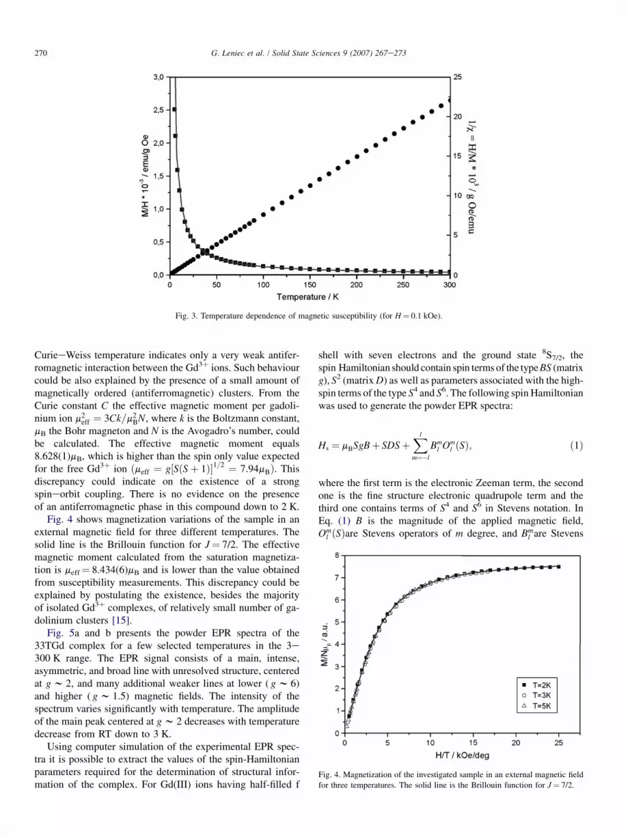
Results of the dc magnetic susceptibility measurements arepresented in Fig. 3. For the whole investigated temperaturerange the experimental data could be fitted very well to theCurieeWeiss law, c¼ C/(T� q), with the CurieeWeiss tem-perature q¼�0.191(1) K. The solid line in Fig. 3 representsthe best fit to the experimental data according to that law.The magnetic data were also plotted as 1/c versus T, yieldinglinear behaviour. A small negative value of the paramagnetic
Table 1
Characteristic absorption bands observed in both Schiff base ligand and gado-
linium podand mixed with KBr
Assignment Schiff base
ligand (cm�1)
Gadolinium
podand (cm�1)
dring 648
705
797
g(CeH)aromatic 832
815
n(CeC) 1089
1034
1172
n(CeO)phenolic 1200
1270
1326
ds(CH2) 1367
n(CeN)aliphatic 1474 1463
n(C]C)aromatic 1523
1608 Superposition
n(C]N) 1635 1625
n(CH2) 2950e2816 2967e2853
n, Stretching; d, in-plane bending; g, out of plane bending; subscript s,
scissoring.
270 G. Leniec et al. / Solid State Sciences 9 (2007) 267e273
Fig. 3. Temperature dependence of magnetic susceptibility (for H¼ 0.1 kOe).
CurieeWeiss temperature indicates only a very weak antifer-romagnetic interaction between the Gd3þ ions. Such behaviourcould be also explained by the presence of a small amount ofmagnetically ordered (antiferromagnetic) clusters. From theCurie constant C the effective magnetic moment per gadoli-nium ion m2
eff ¼ 3Ck=m2BN, where k is the Boltzmann constant,
mB the Bohr magneton and N is the Avogadro’s number, couldbe calculated. The effective magnetic moment equals8.628(1)mB, which is higher than the spin only value expectedfor the free Gd3þ ion ðmeff ¼ g½SðSþ 1Þ�1=2 ¼ 7:94mBÞ. Thisdiscrepancy could indicate on the existence of a strongspineorbit coupling. There is no evidence on the presenceof an antiferromagnetic phase in this compound down to 2 K.
Fig. 4 shows magnetization variations of the sample in anexternal magnetic field for three different temperatures. Thesolid line is the Brillouin function for J¼ 7/2. The effectivemagnetic moment calculated from the saturation magnetiza-tion is meff¼ 8.434(6)mB and is lower than the value obtainedfrom susceptibility measurements. This discrepancy could beexplained by postulating the existence, besides the majorityof isolated Gd3þ complexes, of relatively small number of ga-dolinium clusters [15].
Fig. 5a and b presents the powder EPR spectra of the33TGd complex for a few selected temperatures in the 3e300 K range. The EPR signal consists of a main, intense,asymmetric, and broad line with unresolved structure, centeredat g w 2, and many additional weaker lines at lower ( g w 6)and higher ( g w 1.5) magnetic fields. The intensity of thespectrum varies significantly with temperature. The amplitudeof the main peak centered at g w 2 decreases with temperaturedecrease from RT down to 3 K.
Using computer simulation of the experimental EPR spec-tra it is possible to extract the values of the spin-Hamiltonianparameters required for the determination of structural infor-mation of the complex. For Gd(III) ions having half-filled f
shell with seven electrons and the ground state 8S7/2, thespin Hamiltonian should contain spin terms of the type BS (matrixg), S2 (matrix D) as well as parameters associated with the high-spin terms of the type S4 and S6. The following spin Hamiltonianwas used to generate the powder EPR spectra:
Hs ¼ mBSgBþ SDSþXl
m¼�l
Bml Om
l ðSÞ; ð1Þ
where the first term is the electronic Zeeman term, the secondone is the fine structure electronic quadrupole term and thethird one contains terms of S4 and S6 in Stevens notation. InEq. (1) B is the magnitude of the applied magnetic field,Om
l ðSÞare Stevens operators of m degree, and Bml are Stevens
Fig. 4. Magnetization of the investigated sample in an external magnetic field
for three temperatures. The solid line is the Brillouin function for J¼ 7/2.

271G. Leniec et al. / Solid State Sciences 9 (2007) 267e273
Fig. 5. EPR spectra of 33TGd complex at selected temperatures: (a) high-temperature range (T> 100 K); (b) low-temperature range (T< 100 K).
parameters [16]. The number of non-zero Bml parameters de-
pends on the site symmetry of paramagnetic centre [17].All registered EPR spectra of the investigated complex at
different temperatures have been simulated using EPReNMRcomputer program in order to study thermal changes of thespin-Hamiltonian parameters. As an example, Fig. 6 presentssimulated (upper panel) and experimental (middle panel) spec-tra of the 33TGd complex at 288 K. The accordance betweenthese two spectra is satisfactory, what can be evidenced bythe lowest panel in Fig. 6, which shows their difference, with
no apparent structure discernable. In Table 2 the calculatedspin-Hamiltonian parameters for T¼ 3 K and at RT, are given.The presence of three different gi and non-zero values of B0
2, B22,
B04, B2
4, B�24 , B4
4, B�44 and B0
6 indicates monoclinic symmetry ofthe crystal field at the Gd(III) site. The accuracy of the calcu-lated parameters has been estimated by varying the values ofparameters and observing changes in the simulated spectrum.If the difference between the simulated and experimental spec-trum revealed a significant spectral feature then that set ofparameters was judged as not appropriate. This method is not

272 G. Leniec et al. / Solid State Sciences 9 (2007) 267e273
entirely reliable (as in case of other methods used for powderanalysis), but the relative error of the estimated non-zerospin-Hamiltonian parameters should not exceed 20%. For theseparameters, which were calculated to have zero value, the erroris lower than 1 Gs for the B2 parameters, 0.5 Gs for the B4 pa-rameters and 0.03 Gs for the B6 parameters.
Thermal dependence study of the spin-Hamiltonian param-eters showed that gi-factors do not change with temperature,while many Bm
l (l¼ 2, 4, 6) parameters displayed rather signif-icant temperature dependence. The calculated spin-Hamilto-nian parameters Bm
l (l¼ 2, 4, 6) showed significant changeswith temperature decrease from RT. For low and high temper-atures the values of these parameters are listed in Table 2. Allthe other Bm
l parameters have zero value and didn’t changewith temperature.
A very important spectroscopic parameter that can be cal-culated from the EPR spectrum is the integrated intensity. Itis defined as the area under the absorption resonance line(not the first derivative of absorption line usually registered)and is proportional to the magnetic susceptibility of the inves-tigated spin system. The integrated intensity of the EPR spec-trum will be designated as cEPR. The study of the temperature
Fig. 6. Simulated (top panel), experimental (middle panel) and the difference
(lower panel) of the EPR spectra of 33TGd complex at T¼ 288 K.
dependence of cEPR could yield information on magnetic in-teractions between the spin species. Fig. 7 displays the resultsof the temperature studies of cEPR for the 33TGd complex.The upper panel in Fig. 7 shows the temperature dependenceof cEPR, the middle panel reveals the reciprocal integrated in-tensity cEPR
�1 , and the lower one presents the product TcEPR
that is proportional to the square root of the effective magneticmoment. Inspection of Fig. 7 indicates that there is no magnet-ically ordered state in the 33TGd complex in this temperaturerange. As temperature is decreased from RT, Iint decreasesslightly down to w190 K, and on further cooling starts to in-crease at accelerated rate. It is clear that the thermal changesof Iint could not be described by a simple CurieeWeiss rela-tion, in the whole investigated temperature range. Below130 K there is a linear dependence of Iint
�1 (Fig. 7, middlepanel), and in that range the CurieeWeiss relation holdswith the CurieeWeiss temperature qEPR¼ 10 K. It suggeststhere is a ferromagnetic interaction between 33TGd complexesbelow 130 K. On the other hand, in the high-temperaturerange, T> 190 K, the situation is quite different. The lowerpanel in Fig. 7 indicates that the magnetic moment decreasessignificantly as the temperature is lowered from RT. Thus, athigh temperatures, there is a strong antiferromagnetic interac-tion between at least part of the Gd(III) complexes. EPR mea-surements confirm that there is no long-range magnetic orderin the investigated compound, but only magnetic interactionsbetween the paramagnetic centers. To explain the observed be-haviour of the temperature EPR spectra at least two optionshave to be considered. The first one is the existence of thehigh-spin low-spin transition and the occurrence of the excitedlow-spin form at room temperature. Such a transition wouldrequire a very strong change of the crystal field acting onthe paramagnetic complex. This has not been observed inthe thermal dependence of crystal field parameters. Besides,the observed thermal changes of the Bm
l parameters do not cor-relate with the changes of the effective magnetic moment. An-other possibility that should be considered is the existence ofthe short-lived clusters. The antiferromagnetic interaction be-tween neighboring complexes might favor formation of a cer-tain number of short-lived (on the time scale of the EPR
Table 2
Values of the spin-Hamiltonian parameters for the 33TGd complex at 3 and 288 K
g Tensor Stevens parameters (Gs)
m Bm2 m Bm
4 m Bm6
gxx¼ 1.989 T¼ 3 K T¼ 288 K T¼ 3 K T¼ 288 K T¼ 3 K T¼ 288 K
gyy¼ 1.999 0 �86 �86.5 0 0.2 �0.12 0 0.07 0.06
gzz¼ 2.000 1 0.0 0.0 1 0.0 0.00 1 0.00 0.00
2 6.5 26.5 2 3.0 1.00 2 0.00 0.00
�2 0.0 0.0 3 0.0 0.00 3 0.00 0.00
�1 0.0 0.0 4 1.5 3.00 4 0.00 0.00
�4 2.0 2.00 5 0.00 0.00
�3 0.0 0.00 6 0.00 0.00
�2 3.5 3.00 �6 0.00 0.00
�1 0.0 0.00 �5 0.00 0.00
�4 0.00 0.00
�3 0.00 0.00
�2 0.00 0.00
�1 0.00 0.00

273G. Leniec et al. / Solid State Sciences 9 (2007) 267e273
Fig. 7. Temperature dependence of the EPR integrated intensity for the 33TGd complex (upper panel), of the reciprocal integrated intensity (middle panel) and the
product TIint (lower panel).
spectroscopy, t w 10�10 s) clusters with non-magnetic S¼ 0ground state and magnetic excited states. As the temperaturedecreases from RT, the population of the excited states de-creases and the population of the non-magnetic state increases,decreasing in consequence the total magnetic moment of thesystem. Static magnetic susceptibility measurements in thattemperature range didn’t reveal any contribution from theseshort-lived clusters what is understandable as that method reg-isters only static magnetic entities.
4. Conclusions
IR measurements have evidenced the formation of the33TGd complex, in which the nearest neighborhood of Gd(III)consists of seven atoms: three oxygen and four nitrogen. Thelocal symmetry at the Gd(III) site is rather low (C1) as provedby the presence of many non-zero parameters in the spin Ham-iltonian. Temperature dependence of the EPR integrated inten-sity indicates that there is no magnetically ordered state in thiscompound down to temperature of 3 K. In the high-tempera-ture range, T> 190 K, a strong antiferromagnetic interactionsbetween Gd(III) complexes are evidenced by decreasing mag-netic moment with the temperature decrease. This interactionmight favor formation of short-lived clusters with a non-magnetic ground state.
Acknowledgements
Authors deeply acknowledge Prof. R. Szymczak andDr. M. Baran from the Institute of Physics, Polish Academyof Sciences, Warsaw, for magnetic susceptibility measurements.
References
[1] B. Dietrich, et al., Macrocyclic Chemistry, Aspects of Organic and Inor-
ganic Supramolecular Chemistry, VCH, Weinheim, 1993.
[2] D. Parker, Macrocyclic Synthesis, Oxford University Press, Oxford,
1996.
[3] W. Radecka-Paryzek, V. Patroniak-Krzyminiewska, Wiad. Chem. 50
(1996) 171 (in polish).
[4] P.A. Vigato, S. Tamburini, Coord. Chem. Rev. 248 (2004) 1717.
[5] N. Brianese, U. Casellato, S. Tamburini, P. Tomasin, P.A. Vigato, Inorg.
Chim. Acta 272 (1998) 235.
[6] M. Kanesato, F.N. Ngassapa, T. Yokoyama, Anal. Sci. 17 (2001)
1359.
[7] S. Liu, L. Gelmini, S.J. Rettig, R.C. Thompson, C. Orvig, J. Am. Chem.
Soc. 114 (1992) 6081.
[8] W. Schilf, B. Kamienski, B. Ko1odziej, E. Grech, J. Mol. Struct. 708
(2004) 33.
[9] W. Schilf, B. Kamienski, B. Ko1odziej, E. Grech, Z. Rozwadowski,
T. Dziembowska, J. Mol. Struct. 615 (2002) 141.
[10] H. Kobayashi, S. Kawamoto, S.K. Jo, H.L. Bryant Jr., M.W. Brechbiel,
R.A. Star, Bioconjugate Chem. 14 (2003) 388.
[11] K.N. Raymond, V.C. Pierre, Bioconjugate Chem. 16 (2005) 3.
[12] J. Torres, M. Brusoni, F. Peluffo, C. Kremer, S. Dominguez, A. Mederos,
E. Kremer, Inorg. Chim. Acta 358 (2005) 3320.
[13] W. Radecka-Paryzek, V. Patroniak, J. Lisowski, Coord. Chem. Rev. 249
(2005) 2156.
[14] M.J. Mombourquette, J.A. Weil, D.G. McGavin, EPReNMR User’s
Manual, Department of Chemistry, University of Saskatchewan, Saska-
toon, SK, Canada, 1999.
[15] J. Kliava, I. Edelman, A. Potseluyko, E. Petrakovskaja, R. Berger,
I. Bruckental, Y. Yeshurun, A. Malakhovskii, T. Zarubina, J. Magn.
Magn. Mater. 272 (2004) 1647.
[16] A. Abragam, B. Bleaney, Electron Paramagnetic Resonance of Transition
Ions, Clarendon Press, Oxford, 1970.
[17] Guillaume Morin, Dominique Bonnin, J. Magn. Reson. 136 (1999)
176.
Recommended


