ABSTRACT
Background and Objective: There is paucity of data on the outcome of combined VPS
insertion and myelomeningocoele repair, and whether this will reduce morbidity and
mortality. This study was designed to address this research question.
Method: Prospective descriptive interventional study was used and patients were
recruited between January and October 2009, giving a total of 22. Information on
sociodemographic, referral status, preoperative and postoperative outcome was
documented and analysed.
Result: Males constituted 54.7% and females 45.2% of cases. The youngest age at
presentation was 1 week, the oldest was 32 weeks. The majority (90%) were referred
from clinics and hospitals outside Lusaka, and most hail from poor socioeconomic
backgrounds (64%). The malformation occurred in the lumbar and sacral regions in
77.4%. Most of the patients presented with normal and mild forms of neurological
impairment. Ultrasound examination showed that 90% had mild findings and 9% had
moderate form of hydrocephalus. All patients were shunted and had surgical closure of
the sac. Postoperative complications were seen in patients who had oedamatous (59%)
and infected wounds (9%). One patient had CSF leakage and later died of meningitis.
Average hospital stay for all patients was 25days.
Conclusion: Being the only referral hospital and the only centre with neurosurgical unit
for all public institutions, children born with such defects outside Lusaka will continue
being seen late.
Although the sample size was small, it could show that even when they come late, a
combined surgical approach can still be recommended in such patients. Most of the
patients operated on recovered well postoperatively, but period of hospital stay was too
short to be assessed neurologically.
1
INTRODUCTION
1. Background
Spinal dysraphism encompasses a group of congenital malformations that include pseudo
tails, notomelia, lipoma, spinal bifida and anterior sacral meningocoele. Of these, spina
bifida and its variations are the commonest, (1, 2, and 5).
Myelomeningocoele results from a teratogenic process causing failed closure and
abnormal differentiation of the embryonic neural tube during the first 28 days of
gestation. Abnormal development of the posterior caudal neural tube produces spinal
cord lesion roughly correlating with the patient’s neurological, motor and sensory
deficits. The Anorld Chiari II malformation impedes the flow and absorption of
cerebrospinal fluid and causes hydrocephalus, which occurs in more than 90% infants
with myelomeningocoele. (2, 5).
In a significant proportion of patients with Spina bifida Aperta, hydrocephalus is absent
at birth but develops in the first few weeks or months of life. Hydrocephalus occurs 15 –
25% of children with open myelomeningocoele at birth; however, in most surgical series,
the proportion of patients with the lesion who require shunting reaches 80 -90% (4.8).
Myelomeningocoele is the commonest form in many African countries, while
meningocoele is the simplest form with no associated limb paralysis or sphincter
disturbance (1, 2).
2. Prevalence
Available data from the developed countries show that outcome of patients with spinal
dysraphism has improved. In one cohort study, of the patients treated in the 1970s, 52%
were alive 20 years after treatment. Most deaths observed occurred in the first year of
life, mostly due to renal and respiratory problems associated with spina bifida. A few
deaths were related to hydrocephalus (3, 5).
In a similar but recent study, review of children treated in the 1980s, only 27% died, most
of them in the first year of life from causes related to spina bifida than hydrocephalus. (5)
2
The incidence of myelomeningocoele, the commonest form of spinal dysraphism, ranges
from 0.2 to 2 per 1000 live births in most developed countries. Overall incidence has
significantly declined in the last 2 decades because of improved maternal nutrition during
pregnancy, including folic acid addition, a wider availability of prenatal diagnostic
wakeup and therapeutic termination of pregnancy. (6, 8, 9).
For the United States, spina bifida occurs in 7 out of 10,000 live births. In Arkansas, the
Centre for Birth Defects Prevention (ACBDP) calculated a rate of 5.1 per 10,000 live
births for the years 1998 – 2000. For this time period, ACBDP indicated that 57 persons
were born with spina bifida in Arkansas. (10)
Most cases of myelomeningocoele are managed prenatally with surgical correction, or
within 48 hours of delivery to prevent infection and protect exposed neural tissue from
trauma and drying. Neurological deficit depends on the level of lesion and initial
condition of spinal cord/nerves. Possible outcomes range from normal development to
various losses of muscle function and or bladder control. Deterioration is faster in the
absence of surgery. (7, 8).
Mutuszczan et al showed that a higher spinal cord lesion, hydrocephalus and
complications due to its surgical management had a negative influence on the
development of children with myelomeningocoele. In the absence of treatment, mortality
was estimated at 50%. (10).
In recent retrospective case reviews, shunt placement has been shown to vary based on
the level of lesion, with a greater number of patients with thoracic lesions requiring
shunts than those with lumber or sacral lesions. Lesions at levels of T12 and above have
also been associated with increased incidence of brain abnormalities and lower scores on
psychometric testing than lesions at L1 or below. (2, 4, 13).
3
3. Outcome of surgery
Surgery is commonly staged, with shunt placement for hydrocephalus followed by
definitive surgery a few weeks later to repair the lesion. A few cases had shunt
placement and repair of lesion in one setting. (9, 11, 13).
Miller et al reviewed results obtained in the study to compare simultaneous and delayed
ventriculoperitoneal shunt insertion in children undergoing myelomeningocoele repair at
the University Of Pittsburgh School Of Medicine. Sixty-nine patients were followed up
between 1987 and 1993, were they found that neither the overall frequency of
complications nor the frequency of CSF infection, shunt malfunction, or symptomatic
Chiari malformation differed significantly between the two groups. In contrast, there was
a significant higher rate of myelomeningocoele wound leak (eight versus zero; P= 05)
and longer mean hospital stay in the sequential group versus the simultaneous group (22
days versus 13 days; P= 05). (17).
Hubballah and Hoffman followed up patients between 1975 and 1985 at the Hospital of
the Sick children in Toronto, Canada. In the 10 patients who underwent simultaneous
myelomeningocoele repair and VPS insertion, none developed shunt infections or wound
repair breakdown in the follow-up period of 1 to 9 years. (18).
In Brazil, Machado and Santos de Oliveira followed 28 patients between 1998 and 2001
that underwent closure of neural tube defects at Paediatric Neurosurgical division of
University of Sao Paulo. Of the 11 patients that had concomitant surgery performed after
birth, they concluded that simultaneous VPS insertion and correction of
myelomeningocoele did not pose an additional risk to the child and do have some
advantages, facilitating healing of the back without CSF leakage and protecting the brain
from effects of progressive ventricular dilatation. (19).
Studies done in Africa show that management is selective due to difficulties with
multidisciplinary care and rehabilitation. Furthermore, late presentation, preoperative
infection and poor care of patients with functional handicap remain a challenge. (1, 12,
15, 16).
4
Default rate was found to be high in cases were parents failed to get expected hospital
treatment. The type of surgery performed also depended on the extent and size of lesion
and age of the baby. (14).
4. The problem
In Zambia, the incidence of spinal dysraphism has not being documented, but
epidemiological survey showed most patients came from outside Lusaka, most affected
the sacral region with a female preponderance and characteristically low socioeconomic
status. Folic acid consumption was less in more than 50% study subjects. (20)
Furthermore, no intervention measures have being outlined to properly manage such
patients. Luck of proper anaesthetic equipment means surgery cannot be done in newborn
babies. Being the only public referral hospital for such cases, late patient presentation is
inevitable.
5. The question
Would combined ventriculoperitoneal shunting and myelomeningocoele repair improve
the postoperative outcome and reduce morbidity in paediatric neurosurgical patients
presenting to UTH?
II STUDY JUSTIFICATION
Since early closure of the sac is said to decrease morbidity/mortality and prevent
deteriorating neurological symptoms, late presentation and poor patient follow up will
necessitate a combined surgical approach.
This will reduce on total hospital stay and reduce theatre time for the patients.
Early and appropriate intervention is likely to prevent recurrent infections, facilitate
nursing care and handling of these patients. In addition and depending on surgical
outcome, multidisciplinary management will be instituted with referral to specific
subspecialty when need arises.
5
Hypothesis
H1 combined venriculoperitoneal shunting and repair of meningomyelocoele reduces
risks of meningitis, has less hospital stay and contributes to significant neurological
outcome.
III OBJECTIVES
1. General
To determine the short-term outcome of simultaneous VPS insertion and repair of
myelomeningocoele in paediatric neurosurgical patients at UTH.
2. Secondary
a. To find out the socio-demographic factors associated
hydrocephalus/myelomeningocoele
b. To determine the age at presentation, size and site of lesion
c. To document and evaluate the post operative outcome
6
IV METHODOLOGY AND RESULTS
Study site and setting
The study was conducted at university teaching hospital in Lusaka, in the paediatric
neurosurgical wards
Study Population
All paediatric patients admitted with spinal dysraphism and hydrocephalus meeting
inclusion criteria were recruited.
Study design
Prospective descriptive interventional study was used and patient recruitment between
January and October 2009. A total of 22 patients were enrolled.
A case control study would not be done due to late patient presentation, poor patient
compliance and small sample size.
Case Definition
A case was defined as a patient with spinal dysraphism complicated by hydrocephalus.
Inclusion criteria
All patients with meningomyelocoele complicated by hydrocephalus and who had given
consent were included.
7
Exclusion criteria
Infected and/or ulcerated sac
Severe hydrocephalus
Severe neurological and/or kyphoscoliosis
Prematurity
Refusal of consent
Entry and analysis
Entry was done on epi data sheet and analysis based on descriptions and possible
associations.
Ethical Considerations
The University of Zambia Biomedical Research Ethics Committee approved the
study, and consent for inclusion into the study was obtained from the
parents/guardians.
8
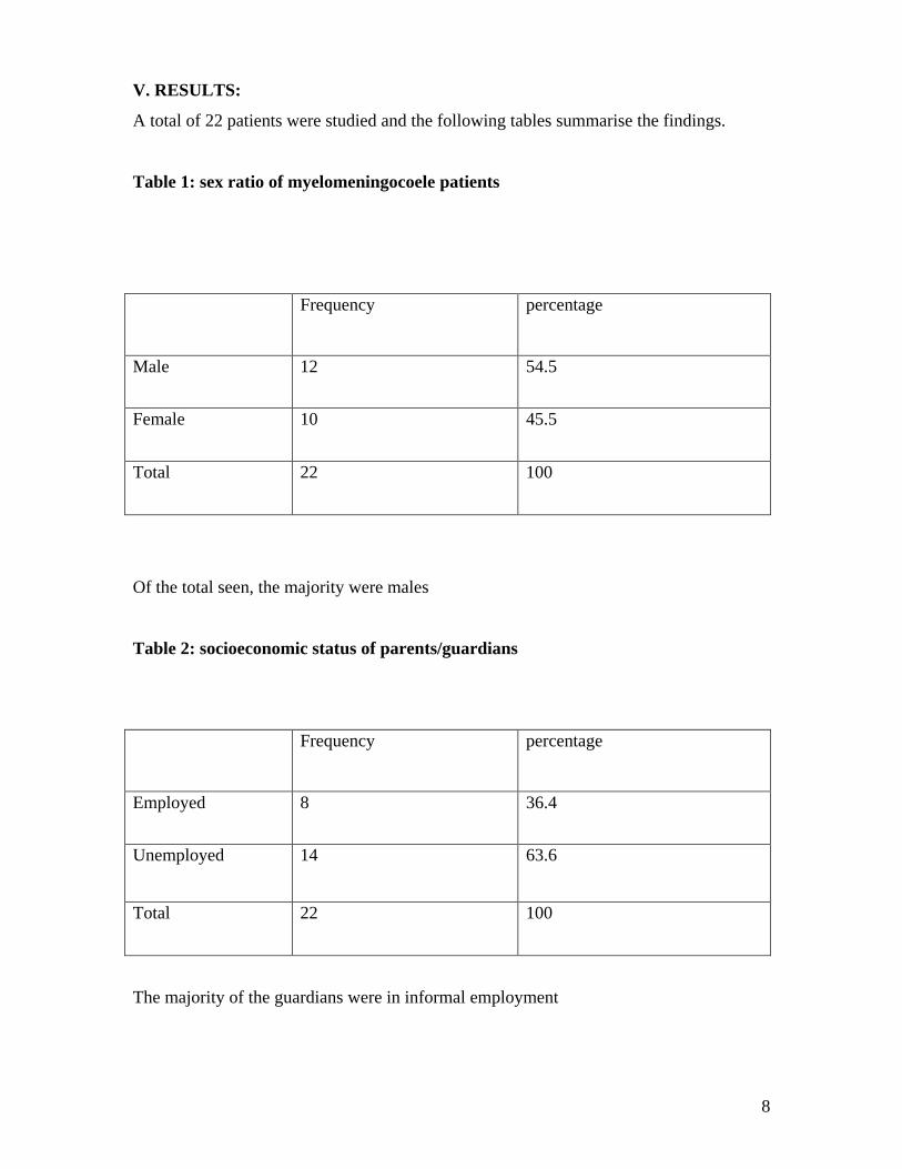
V. RESULTS:
A total of 22 patients were studied and the following tables summarise the findings.
Table 1: sex ratio of myelomeningocoele patients
Frequency percentage
Male 12 54.5
Female 10 45.5
Total 22 100
Of the total seen, the majority were males
Table 2: socioeconomic status of parents/guardians
Frequency percentage
Employed 8 36.4
Unemployed 14 63.6
Total 22 100
The majority of the guardians were in informal employment
9
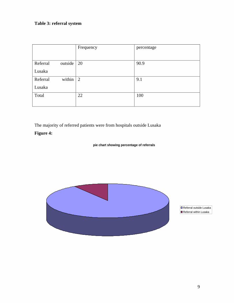
Table 3: referral system
Frequency percentage
Referral outside
Lusaka
20 90.9
Referral within
Lusaka
2 9.1
Total 22 100
The majority of referred patients were from hospitals outside Lusaka
Figure 4:
pie chart showing percentage of referrals
Referral outside Lusaka
Referral within Lusaka
10
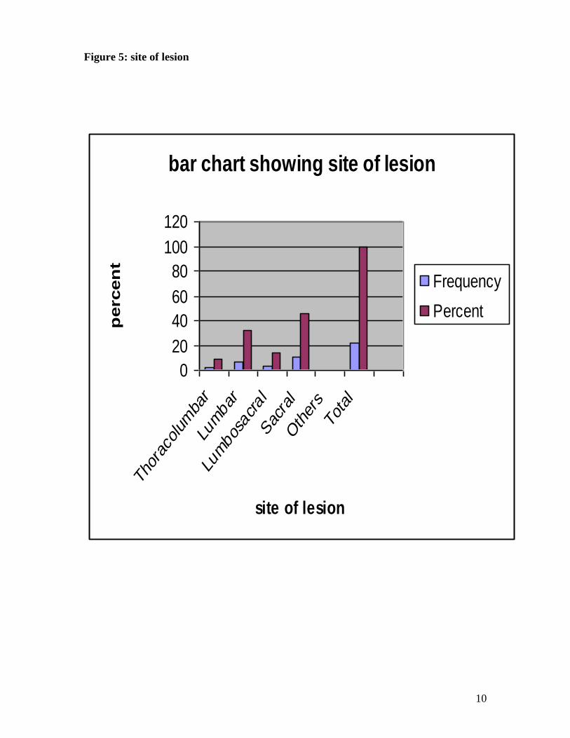
Figure 5: site of lesion
bar chart showing site of lesion
0
20
40
60
80
100
120
Tho
raco
lumba
r
Lumbar
Lumbos
acral
Sac
ral
Other
s
Total
site of lesion
percen
t
Frequency
Percent
11
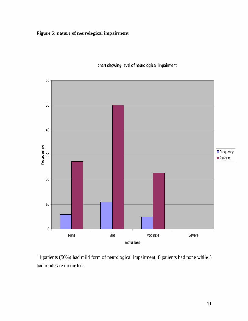
Figure 6: nature of neurological impairment
chart showing level of neurological impairment
0
10
20
30
40
50
60
None Mild Moderate Severe
motor loss
freq
uen
cy
Frequency
Percent
11 patients (50%) had mild form of neurological impairment, 8 patients had none while 3
had moderate motor loss.
12
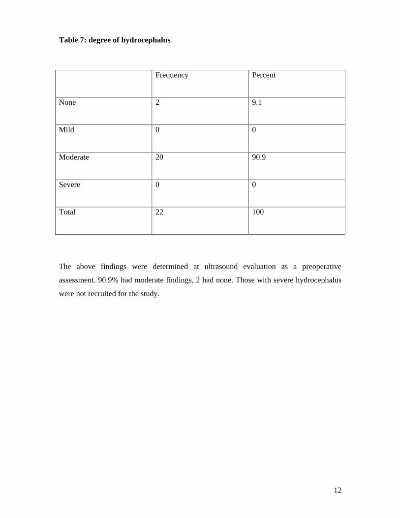
Table 7: degree of hydrocephalus
The above findings were determined at ultrasound evaluation as a preoperative
assessment. 90.9% had moderate findings, 2 had none. Those with severe hydrocephalus
were not recruited for the study.
Frequency Percent
None
2 9.1
Mild
0 0
Moderate
20 90.9
Severe
0 0
Total
22 100
13
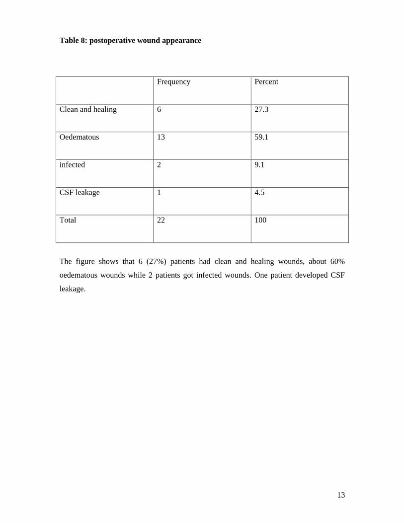
Table 8: postoperative wound appearance
Frequency Percent
Clean and healing
6 27.3
Oedematous
13 59.1
infected
2 9.1
CSF leakage
1 4.5
Total
22 100
The figure shows that 6 (27%) patients had clean and healing wounds, about 60%
oedematous wounds while 2 patients got infected wounds. One patient developed CSF
leakage.
14
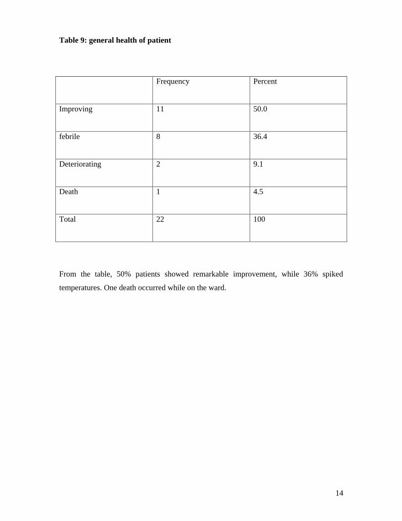
Table 9: general health of patient
Frequency Percent
Improving
11 50.0
febrile
8 36.4
Deteriorating
2 9.1
Death
1 4.5
Total
22 100
From the table, 50% patients showed remarkable improvement, while 36% spiked
temperatures. One death occurred while on the ward.
15
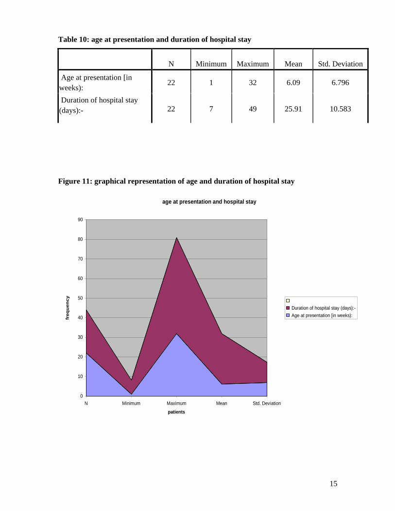
Table 10: age at presentation and duration of hospital stay
N Minimum Maximum Mean Std. Deviation
Age at presentation [in
weeks): 22 1 32 6.09 6.796
Duration of hospital stay
(days):- 22 7 49 25.91 10.583
Figure 11: graphical representation of age and duration of hospital stay
age at presentation and hospital stay
0
10
20
30
40
50
60
70
80
90
N Minimum Maximum Mean Std. Deviation
patients
freq
uen
cy
Duration of hospital stay (days):-
Age at presentation [in weeks):
16
VI. DISCUSSION
Demographic characteristics
From the study, both sexes were equally represented, albeit a small sample size.
Available data shows a female preponderance, and that the majority of patients are a
referral from hospitals outside Lusaka. The study reveals that about 90% patients are
referrals and most were from economically disadvantaged parents/guardians. This has a
bearing on total patient care and hospital stay.
Preoperative assessment
Table 4 shows that about 46% had myelomeningocoele occurring in the sacral region,
followed by lumbar area. This conforms to studies done elsewhere, which show that the
higher the lesion, the more likelihood of finding severe cranial malformation {2, 4, 13,
20).
The actual sizes of the lesions were not measured but there appearance assessed for
leakage and /or rupture. Over 90% of the patients had intact sac, though the older ones
showed varied degrees of bruising.
Figure 5 summarises the degree of motor dysfunction at presentation. The majority of
those less than 4 weeks presented with normal or mild form of dysfunction (27 and 50%
respectively). The rest had moderate degree of neurological impairment, and none had
any bladder or anal sphincter dysfunction.
For the purpose of this survey, the degree of paralysis has been divided into three grades:
Mild-The muscle weakness was slight and confined to muscles below the knee, denoting
paralysis below the fifth lumbar or first sacral neural segments.
17
Moderate-There was severe or complete paralysis of muscles below the knee, but
sufficient action in hip flexors, quadriceps, hamstrings and glutei so that control of the
hip and knee might be expected: paralysis below the fourth lumbar neural segment.
Severe-All muscles of the limb except possibly the iliopsoas and hip adductors were
paralysed so that control of the hip and knee were poor-paralysis proximal to the fourth
lumbar neural segment.
The parents/guardians were counselled on possible unfavourable postoperative outcome,
in view of late presentation and the nature of neurological impairment. The duration of
postoperative review was too short to document notable changes neurologically.
Ultrasound evaluation revealed that over 90% had moderate findings and only 9% had
none. These were recruited for study and shunted routinely as previous hospital reviews
showed that such presented with hydrocephalus following repair of myelomeningocoele
alone. In the quoted literature, hydrocephalus was seen in 15 to 25% of patients with
open MMC, of which 80 to 90% required shunting. (4, 8) Those with severe
hydrocephalus were shunted as an emergency for immediate relief and repair was
rescheduled for later date.
Postoperative assessment
The postoperative period was centred mainly on wound appearance and general health of
the patient, as the recovery from any neurological impairment was considered too short a
time.
18
From figure 8 on postoperative wound appearance, 27% were clean and healing well.
59% had oedematous wounds mostly in the younger population, but recovered well too.
Only 2 patients had wound infection and were noted to have bruised sac preoperatively.
They recovered after antibiotics treatment and wound care. In the initial postoperative
days, the sterile dressings were kept for 5days and change of dressing done by attending
doctor. The parents/guardians were not allowed to touch the wounds till they were
comfortable with nursing care. No special dressings were applied as cost was a factor in
the management. In more advanced setups, no postoperative complications were recorded
between 1 to 9 year follow-up. (18)
One patient died following wound dehiscence complicating into meningitis. CSF culture
revealed gram positive diplococcal.
Table 9 shows results on general health of patients while on the ward. All patients did
well except for the mortality and the 2 who had fever but responded to treatment. The
babies were nursed prone till the wounds were dry and less oedematous.
Some studies done does show that the combined approach pose no additional risk to the
child, but facilitate wound healing and protect the brain from ventricular dilatation. (19)
The rationale of shunting patients with no hydrocephalus is prophylactic, as they tend to
develop the condition late on.
As most of the patients were referrals from peri-urban and hospitals outside Lusaka, the
period of hospital stay was extended so as to remove stitches and adequately teach
guardians how to care for their patients. This was also preventing poor patient
compliance and follow up due to low socioeconomic status. Being a small sample size,
extended hospital stay ensured that a good number were retained at the end of the study
period. Most western centres discharge patients after 14 days of hospital stay in the
absence of complications.
19
The average hospital stay for this study was 25 days, due to different healing times and
presence of oedema on the surgical wounds.
CONCLUSION AND RECOMMENDATIONS
Most literature quoted show that the number of patients with myelomeningocoele is
declining in the west due to various interventions being put in place to prevent
occurrence. However, countries like Zambia still lag behind in folic acid fortification,
improving peoples living conditions and expanding tertiary level hospitals.
Being the only referral hospital and the only centre with neurosurgical unit for all public
institutions, children born with such defects outside Lusaka will continue being seen late.
Although the sample size was small, it could show that even when they come late, a
combined surgical approach can still be recommended in such patients. The duration of
hospital stay may not compare with what most literature show, but given the low
socioeconomic status and distant referral centres, patients can be kept in till wounds heal
completely.
The postoperative review for the study needs to be extended so that neurological recovery
and /or deterioration can be documented.
20
REFERENCES
1. Adeloye A Neurosurgery in Africa 1989: Ibadan University Press, Nigeria
2. Kramer L.C, Azarow K. Management of Spina bifida, hydrocephalus and
shunts; eMedicine.com
3. Davis B.E., Daley C.M., Shurtleff D.B. et al. Long-term survival of individuals
with myelomeningocoele. Paediatr. Neurosurg Jul-Aug 2005; 49(4): 186-191
4. Dias M.S, Mclone D.G. Hydrocephalus in the child with dysraphism.
Neurosurg Clin N AM. Oct 1993; 4 (4): 715-726
5. Mitchell L.E, Adzick N.S, Melchianne J, Pasquanello P.S, Sutton N.L,
Whithead A.S. Spina bifida. Lancet 2004; 364:1885-95
6. Merkins M.J Care of the Newborn with a Neural Tube Defect; Genetics
Northwest 1996; X:4
7. Bruner J.P, Tulipan N, Ray L, Pascal et al. Fetal surgery for
myelomeningocoele and the incidence of shunt dependent hydrocephalus.
JAMA 1999; 282 (19): 1819-25
8. Muir C, Kuschel C, Law et al. Management of infants with
myelomeningocoele. NW Newborn Clinical Guidelines-Neural Tube Defects
2005, New Zealand
9. Morley et al, A new look at myelomeningocoele, Paediatric 2003; 111:1494-98
10. Rucker J. Spina bifida. Arkansas Centre for Birth Defects Prevention 2004
11. Matuszczak E, Leukiewicz T. Results of surgical treatment of
myelomeningocoele. Eur J Paediatr Surg 2006 Oct; 16(5):337-42
21
12. Ugwu R.O, Eneh A.U, Oruamabo R.S. Neural tube defects in a university
teaching hospital in southern Nigeria: trends and outcomes: Niger J Med 2007
Oct; 16 (4): 368-71
13. Salome et al. myelomeningocoele: Surgical treatment and results. J Pediatr
(RIOJ) 1995; 71(6): 317-21
14. Oyewole A, Adeloyi A, Adeyokunnu A.A: Psychosocial and cultural factors
associated with management of spina bifida cystica in Nigeria: Dev Med Child
in Nigeria: Dev Med Child Neuro 1985; 27(4): 498-503
15. Shelu B.D., Ameh E.A, Ismail N.J. Spina bifida cystica: selective management
in Zaire, Nigeria: Ann Trop Paediatr 2000 Sep; 20(3): 239-42
16. Alitise et al. Pattern and factors affecting management outcome of spina bifida
cystica in lle-Ife, Nigeria; Paediatric Neurosurg 2006; 42(5): 277-83
17. Miller P.D, Pollack I.F, Pang D, Albright A.L: Comparison of simultaneous
versus delayed ventriculoperitoneal shunt insertion inchildren undergoing
myelomeningocoele repair; J Child Neurol. 1996 Sep: 11(5): 370-2
18. Hubballah M.Y. Hoffman H.J. Early repair of myelomeningocoele and
simultaneous insertion and of ventriculoperitoneal shunt: technique and results;
Neurosurgery. 1987 Jan; 20(1): 21-3
19. Machado H.R. Santos de Oliveira R. Simulteneous repair of
myelomeningocoele and shunt insertion; Childs Nerv Syst. 2004 Feb; 20(2):107-
9
20. Lungu M.M; Epidemiological characteristics of patients with
myelomeningocoele presenting to university teaching hospital- Lusaka (2004).
University of Zambia publishing.
22
CONSENT FORM
I …..………………………………………………………………………….. (Name of
parent/guardian) of ………………………………………………………… (Residence)
have agreed to have my child
……………………………………………………………………………….. (Name of
patient) participate in the study being conducted on ‘determining the outcome of
combined VPs insertion and repair of myelomeningocoele at the University Teaching
Hospital of Lusaka’.
I am fully aware of the implications of being part of this study. I have been assisted that
all necessary information regarding the study will be made available to me and my child.
I understand that I may withdraw from the study at any time, and will not be under
obligation to continue with the study.
I now give consent to have my child be a part of the above mentioned study.
………………………. ……………………… …………………………
Signed Witness Researcher
23
PATIENT INFORMATION SHEET
(To be handed to patient/parent/guardian)
1. PURPOSE OF THE STUDY AND EXPLANATION OF PROCEDURES
We are asking you and your child to be in a research study because you want him/her to
be operated on. This study is about inserting a tube in head to drain excess water and at
the same time remove and close the growth at the back of child. This is done separately
within the first two days of birth. In this study we are trying to learn whether combining
putting a tube and removing the growth will reduce time spent in hospital and number of
operating times in an older child. We also want to see if the wounds would heal fast
especially that the child is older. This will reduce chances of the child harming
him/herself with the growth on the back, which maybe infected.
The operation will be done by a doctor at University Teaching Hospital’s children’s
surgical ward.
Before you decide if you want your child to be in the study, you need to know its
purpose, the possible risks and benefits, and what is expected of you. Then, you can
decide whether or not you want him/her to be in the study. This process is called
informed consent. This consent form gives you information about the study. If you
agree for your son to take part in the study, you will be asked to sign this consent form.
You will be offered a copy to keep. You may choose to read this consent or have the
consent read to you.
It is important that you know certain things:
Taking part in the study is entirely voluntary. It is up to you to decide whether or not
you and your son will be in the study.
You may decide not to take part or to leave the study at any time.
Inserting the tube and removing the growth Procedures
After you agree to the procedure, your child will have the operation done. It usually takes
about 2hours 30minutes. Your child will be made to sleep while being operated, so there
24
will be no pain. After the procedure, your child will remain on the ward to check on the
wounds and size of the head. Any problems arising will be addressed and the child will
continue receiving treatment. Before you go home, we will advise you on how to care for
your child and the wound.
Follow-up Visits
We will ask you to return with your child for at least two follow-up visits. As your child
will be on the ward for at least a week, the first follow up visit at one month is to check
that the tube is working well and there are no problems. The next visits will not be part of
the study but to continue looking after the child as he/she will require regular checkups.
At each visit your child will have a physical exam. We will also ask you some questions
about your experiences with the procedure and the results.
If you and your child miss a follow up visit, we would like to be able to contact you. If
you agree, we will ask you to provide contact information for you and a family member
or friend who could help us find you in case you move or change your phone number. If
you decide at any time that you do not want to be contacted by the study, we will no
longer contact you.
If you have questions or concerns at any time during the study please let us know.
2. RISKS OR DISCOMFORTS
Risks of the operation include bleeding and infection. When the operation is done
under sterile conditions in a hospital, these risks are small. Bleeding is usually
stopped with a small amount of pressure or other safe methods available in
theatre. When an infection occurs, it may involve just the skin or the brain when
severe. The infection can be treated with antibiotic medicines, if needed.
Very rarely, more than normal fluid can be drained from the head causing it to
shrink. However this is prevented because the tubes inserted have a valve that
allows a little fluid at a time.
25
Your child will be given an injection of numbing medicine to decrease any pain
he/she may feel after the operation. This will cause the child to appear sleepy
once injected.
Your child will have a tube put in theatre for passing urine, and may cause some
discomfort. It should not be removed as nurses will want to record the amount of
urine he/she is passing in a day.
There may be other risks and discomforts that are not known at this time.
3. POTENTIAL BENEFITS
Your child may benefit from taking part in this study if he is operated. However, we
cannot guarantee that this will happen. It is possible that your son may not benefit from
being in this study. Information learned from this study may help other parents with such
children to have a combined operation of putting a tube and removing the growth on the
back.
4. ALTERNATIVES TO PARTICIPATION
This study is entirely voluntary. Your decision about whether or not you and your child
will be in the study will not affect the care that you or your child receives. If you do not
choose to take part in this study, your child will still be operated.
5. CONFIDENTIALITY
If you agree for you and your child to be in the study, you agree to allow the information
we collect to be used for scientific purposes. We will not use any names of participants
when we publish or share results of the study. Your records and your child’s records will
be kept confidential. You and your child will be identified in our records by a code.
However, your doctor and their staff, and study personnel will be able to look at your
medical records and have access to confidential information that identifies you by name.
26
6. WITHDRAWAL FROM THE STUDY / REASONS WHY YOU MAY BE
WITHDRAWN FROM THE STUDY WITHOUT YOUR CONSENT
At any time during the study, you are free to withdraw your son without any
discrimination against you. You may be removed from the study without your consent if
the study doctor decides that remaining in the study would be harmful to your son.
7. PERSONS TO CONTACT FOR PROBLEMS OR QUESTIONS
If you ever have questions about this study or in case you are injured because of
participation in this study, you should contact:
Dr. Daniel Makawa
University Teaching Hospital
Department of Surgery
P/B RW IX
Nationalist Road
Lusaka, Zambia
260-977100044, 0955765000
If you ever have questions about your rights as a research subject you may call:
Dr. E.M. Nkandu
Chair, Research and Ethics Committee
Department of Physiotherapy
Lusaka, Zambia

27
EVALUATION FORM
1. Demographic Data
a) Date…………………………… b). File No:…………………………….
b) Age: …………………………. c). Sex:………………………………..
c) Age at presentation:…………………………………………………………..
d) Prematurity:………………........................................................................
Yes [ ] No [ ]
e) Tribe …………………………………………………………………………..
f) Education level of parent/guardian………………………………………..
g) Occupation of parent/guardian……………………………………………..
h) Referral patient………………………………………………………………
2. Preoperative assessment
2.1 Physical signs
a) Sun setting eyes Yes [ ] No [ ]
b) Patent anterior fontanelle Yes [ ] No [ ]
c) Head circumference in cm Yes [ ] No [ ]
28
3. Ultrasound cranial ventricular dimensions in mm
a) Bifrontal diameter……………………………………………………………
b) Bicaudate diameter…………………………………………………………..
c) Maximum transverse diameter of lateral ventricle-----------------
4. Level of Lesion:
a) Thoracolumbar…………………………………………………………………
b) Lumbar…………………………………………………………………………
c) Lumbosacral……………………………………………………………………
d) Sacral…………………………………………………………………………..
e) Others…………………………………………………………………………..
5. Size of Lesion
5.1 Appearance of sac:
Yes = [ 1 ] No = [ 2 ]
a) Intact { } { }
b) Ruptured { } { }
c) Infected { } { }
29
6. Type of neurological impairment:
6.1 Loss of motor function
Yes = [ 1 ] No = [ 2 ]
a) None { } { }
b) Mild { } { }
c) Moderate { } { }
d) Severe { } { }
e) Loss of bladder control { } { }
f) Loss of anal sphincter control { } { }
7. Ultra findings of hydrocephalus:
Yes = [ 1 ] No = [ 2 ]
a) None { } { }
b) Mild { } { }
b) Moderate { } { }
c) Severe { } { }
8. Post operative assessment:
Yes = [ 1 ] No = [ 2 ]
a) CSF Leakage { } { }
b) Wound infection { } { }
c) Meningitis { } { }
d) Blocked Shunt { } { }
30
9. Wound appearance:
Yes = [ 1 ] No = [ 2 ]
a) Clean and healing well { } { }
b) Oedematous { } { }
c) Infected { } { }
d) CSF Leakage { } { }
10. General Health of Patient:
Yes = [ 1 ] No = [ 2 ]
a) Improving { } { }
b) Febrile { } { }
c) Deteriorating { } { }
11. Duration of hospital stay:-
………………………………………………………………………………………………..
Recommended

![Pembangunan Pertanian[12140].pdf - Unmul Repository Home](https://img.pdfslide.net/doc/110x75/63299be0eedc98f54f0146d4/pembangunan-pertanian12140pdf-unmul-repository-home.jpg)


