Maternal dietary uridine causes, and deoxyuridine prevents,neural tube closure defects in a mouse model of folate-responsiveneural tube defects1–5
Lucia Martiniova, Martha S Field, Julia L Finkelstein, Cheryll A Perry, and Patrick J Stover
ABSTRACTBackground: Folic acid prevents neural tube closure defects(NTDs), but the causal metabolic pathways have not been estab-lished. Serine hydroxymethyltransferase 1 (SHMT1) is an essentialscaffold protein in folate-dependent de novo thymidylate synthesisin the nucleus. SHMT1-deficient mice provide a model to investi-gate folic acid–responsive NTDs wherein disruption of de novothymidylate synthesis impairs neural tube closure.Objective: We examined the effects of maternal supplementationwith the pyrimidine nucleosides uridine, thymidine, or deoxyuridinewith and without folate deficiency on NTD incidence in the Shmt1mouse model.Design: Shmt1+/+ and Shmt12/2 female mice fed folate-replete orfolate-deficient diets and supplemented with uridine, thymidine, ordeoxyuridine were bred, and litters (n = 10–23 per group) wereexamined for the presence of NTDs. Biomarkers of impaired folatestatus and metabolism were measured, including plasma nucleo-sides, hepatic uracil content, maternal plasma folate concentrations,and incorporation of nucleoside precursors into DNA.Results: Shmt1+/2 and Shmt12/2 embryos from dams fed the folate-deficient diet were susceptible to NTDs. No NTDs were observed inlitters from dams fed the folate-deficient diet supplemented withdeoxyuridine. Surprisingly, uridine supplementation increasedNTD incidence, independent of embryo genotype and dietaryfolic acid. These dietary nucleosides did not affect maternalhepatic uracil accumulation in DNA but did affect plasma folateconcentrations.Conclusions: Maternal deoxyuridine supplementation preventedNTDs in dams fed the folate-deficient diet, whereas maternal uri-dine supplementation increased NTD incidence, independent offolate and embryo genotype. These findings provide new insightsinto the metabolic impairments and mechanisms of folate-responsiveNTDs resulting from decreased Shmt1 expression. Am JClin Nutr 2015;101:860–9.
Keywords: SHMT1, deoxyuridine, folic acid, neural tube defects,uridine
INTRODUCTION
Low maternal folate status is among the strongest environ-mental determinants of neural tube closure defects (NTDs)6 inhuman populations (1) and interacts with specific gene variantsto confer risk for an NTD-affected pregnancy (2–5). Althoughit known that genetic variants interact with folate status to in-
fluence NTD risk, most of the genetic risk has yet to be iden-tified (6). Clinical trials have established that periconceptionalmaternal folic acid supplementation prevents the occurrenceand recurrence of NTDs by up to 70% (7, 8). Folic acid forti-fication of enriched grains has been introduced in more than 60countries (9) and has significantly reduced the rates of neuraltube defects (7, 8). However, most but not all NTDs are re-sponsive to dietary folic acid (10). Other risk factors for NTD-affected pregnancies include environmental and food-basedtoxins (11, 12), obesity (13), and maternal diabetes (14). De-ficiencies in other nutrients that biochemically interact withfolate may also contribute to NTD risk (15, 16), although theirrole has not been established.
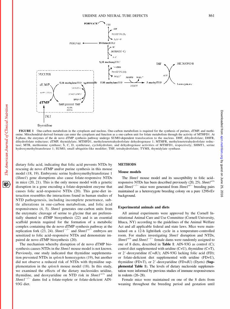
Folates function as enzyme cofactors that carry and chem-ically activate one-carbon units for a network of pathwayscollectively known as one-carbon metabolism. One-carbonmetabolism is essential for de novo purine and thymidylate(dTMP) biosynthesis and for the remethylation of homocysteineto methionine (Figure 1). Responsiveness to maternal folicacid supplementation has been determined in only a few ofthe many mouse models (17) that exhibit NTDs (17–19). TwoNTD models, Pax3 and Shmt1 loss of function, exhibit folicacid–responsive NTDs and impairments in de novo dTMPbiosynthesis. The splotch mutant (Pax3Sp) demonstrates im-paired de novo dTMP (19, 20) and purine nucleotide bio-synthesis (20). NTDs in the splotch mutant can be rescued with
1 From the Division of Nutritional Sciences, Cornell University, Ithaca,
NY.2 Supported by grants HD059120 and DK58144 to PJS from the US Public
Health Service.3 TheUS Public Health Service had no role in the study design, imple-
mentation data collection, data analysis, data interpretation, or writing of the
manuscript.4 Supplemental Table 1 is available from the “Supplemental data” link in
the online posting of the article and from the same link in the online table of
contents at http://ajcn.nutrition.org.5 Address correspondence to PJ Stover, Division of Nutritional Sciences,
Cornell University, 127 Savage Hall, Ithaca, NY 14850. E-mail: pjs13@
cornell.edu.6 Abbreviations used: C, AIN-93G control diet; DMEM, defined minimal
essential medium; dTMP, thymidylate; dU, deoxyuridine; FBS, fetal bovine
serum; FD, AIN-93G diet lacking folic acid; NTD, neural tube closure de-
fect; Shmt1, serine hydroxymethyltransferase gene; T, thymidine; U, uridine.
ReceivedAugust 6, 2014. Accepted for publication December 22, 2014.
First published online January 28, 2015; doi: 10.3945/ajcn.114.097279.
860 Am J Clin Nutr 2015;101:860–9. Printed in USA. � 2015 American Society for Nutrition
by guest on June 25, 2016ajcn.nutrition.org
Dow
nloaded from
97279.DCSupplemental.html http://ajcn.nutrition.org/content/suppl/2015/01/28/ajcn.114.0Supplemental Material can be found at:
dietary folic acid, indicating that folic acid prevents NTDs byrescuing de novo dTMP and/or purine synthesis in this mousemodel (18, 19). Embryonic serine hydroxymethyltransferase 1(Shmt1) gene disruptions also cause folate-responsive NTDsin mice (20, 21). This is the only mouse model with a geneticdisruption in a gene encoding a folate-dependent enzyme thatcauses folic acid–responsive NTDs (20). This gene-diet in-teraction resembles the interactions found in human studies ofNTD pathogenesis, including incomplete penetrance, sub-tle alterations in one-carbon metabolism, and folic acidresponsiveness (4, 5). Shmt1 generates one-carbon units fromthe enzymatic cleavage of serine to glycine that are preferen-tially shunted to dTMP biosynthesis (22) and is an essentialscaffold protein required for the formation of a metaboliccomplex containing the de novo dTMP synthesis pathway at thereplication fork (23, 24). Shmt12/2 and Shmt1+/2 embryos aresensitized to folic acid–responsive NTDs and demonstrate im-paired de novo dTMP biosynthesis (20).
The mechanism whereby disruption of de novo dTMP bio-synthesis causes NTDs in the Shmt1mouse model is not known.Previously, one study indicated that thymidine supplementa-tion prevented NTDs in splotch homozygotes (19), but anotherdid not observe a reduced risk of NTDs with thymidine sup-plementation in the splotch mouse model (18). In this study,we examined the effects of the dietary nucleosides uridine,thymidine, and deoxyuridine on NTD risk in Shmt1+/+ andShmt12/2 dams fed a folate-replete or folate-deficient AIN-93G diet.
METHODS
Mouse models
The Shmt1 mouse model and its susceptibility to folic acid–responsive NTDs has been described previously (20, 25). Shmt1+/+
and Shmt12/2 mice were generated from Shmt1+/2 breeding pairsmaintained as a heterozygote breeding colony on a pure 129SvEvbackground.
Experimental animals and diets
All animal experiments were approved by the Cornell In-stitutional Animal Care and Use Committee (Cornell University,Ithaca, NY) according to the guidelines of the Animal WelfareAct and all applicable federal and state laws. Mice were main-tained on a 12-h light/dark cycle in a temperature-controlledroom. For studies investigating Shmt1 disruption and NTDs,Shmt1+/+ and Shmt12/2 female dams were randomly assigned toone of 8 diets, described in Table 1: AIN-93G as control (C);control diet supplemented with uridine (C+U), thymidine (C+T),or 2#-deoxyuridine (C+dU); AIN-93G lacking folic acid (FD);or folate-deficient diet supplemented with uridine (FD+U),thymidine (FD+T), or 2#-deoxyuridine (FD+dU) (Dyets) (Sup-plemental Table 1). The levels of dietary nucleoside supplemen-tation were informed by previous studies of immune responsivenessin rodents (26–28).
Female mice were maintained on one of the 8 diets fromweaning throughout the breeding period and gestation until
FIGURE 1 One-carbon metabolism in the cytoplasm and nucleus. One-carbon metabolism is required for the synthesis of purines, dTMP, and methi-onine. Mitochondrial-derived formate can enter the cytoplasm and function as a one-carbon unit for folate metabolism through the activity of MTHFD1. AtS-phase, the enzymes of the de novo dTMP synthesis pathway undergo SUMO-dependent translocation to the nucleus. DHF, dihydrofolate; DHFR,dihydrofolate reductase; dTMP, thymidylate; MTHFD1, methylenetetrahydrofolate dehydrogenase 1; MTHFR, methylenetetrahydrofolate reduc-tase; MTR, methionine synthase; S, C, D, synthetase, cyclohydrolate, and dehydrogenase activities of MTHFD1, respectively; SHMT1, serinehydroxymethyltransferase 1; SUMO, small ubiquitin-like modifier; THF, tetrahydrofolate; TYMS, thymidylate synthase.
URIDINE AND NEURAL TUBE DEFECTS 861
by guest on June 25, 2016ajcn.nutrition.org
Dow
nloaded from
killed. Virgin female mice aged 70–120 d were housed overnightwith males. The following morning, females were examined forthe presence of a vaginal plug. Gestational day 0.5 (E0.5) wasdesignated at 0900 on the day of the plug appearance. Pregnantfemales were sacrificed by cervical dislocation at E11.5, andblood was collected by cardiac puncture. Gravid uteri were re-moved, and all implants and resorption sites were recorded.Embryos were examined for NTDs and morphologic abnor-malities at E11.5 and measured for crown-rump length. All yolksacs were collected for subsequent genotyping. Once analyzed
and recorded, embryos were randomly divided into one of 2groups. One group was fixed in 10% neutral buffered formalinfor preservation, and the other group was frozen in liquid ni-trogen (and stored at or below2808C) for biochemical analyses.
Genotype analysis
Genotyping for embryo sex was performed by using establishedprotocols (29–31). Genotyping for Shmt1+/+, Shmt1+/2, and Shmt12/2
alleles was performed by using a previously described protocol (25).
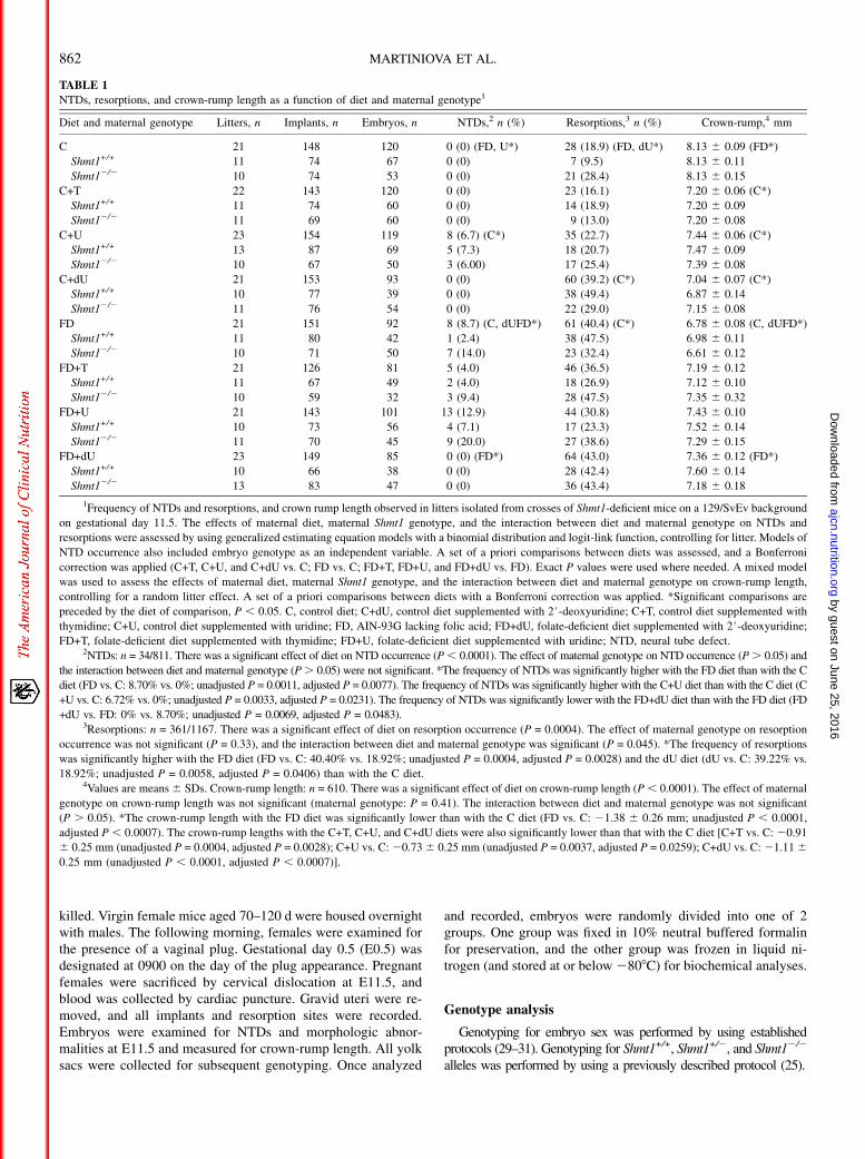
TABLE 1
NTDs, resorptions, and crown-rump length as a function of diet and maternal genotype1
Diet and maternal genotype Litters, n Implants, n Embryos, n NTDs,2 n (%) Resorptions,3 n (%) Crown-rump,4 mm
C 21 148 120 0 (0) (FD, U*) 28 (18.9) (FD, dU*) 8.13 6 0.09 (FD*)
Shmt1+/+ 11 74 67 0 (0) 7 (9.5) 8.13 6 0.11
Shmt12/2 10 74 53 0 (0) 21 (28.4) 8.13 6 0.15
C+T 22 143 120 0 (0) 23 (16.1) 7.20 6 0.06 (C*)
Shmt1+/+ 11 74 60 0 (0) 14 (18.9) 7.20 6 0.09
Shmt12/2 11 69 60 0 (0) 9 (13.0) 7.20 6 0.08
C+U 23 154 119 8 (6.7) (C*) 35 (22.7) 7.44 6 0.06 (C*)
Shmt1+/+ 13 87 69 5 (7.3) 18 (20.7) 7.47 6 0.09
Shmt12/2 10 67 50 3 (6.00) 17 (25.4) 7.39 6 0.08
C+dU 21 153 93 0 (0) 60 (39.2) (C*) 7.04 6 0.07 (C*)
Shmt1+/+ 10 77 39 0 (0) 38 (49.4) 6.87 6 0.14
Shmt12/2 11 76 54 0 (0) 22 (29.0) 7.15 6 0.08
FD 21 151 92 8 (8.7) (C, dUFD*) 61 (40.4) (C*) 6.78 6 0.08 (C, dUFD*)
Shmt1+/+ 11 80 42 1 (2.4) 38 (47.5) 6.98 6 0.11
Shmt12/2 10 71 50 7 (14.0) 23 (32.4) 6.61 6 0.12
FD+T 21 126 81 5 (4.0) 46 (36.5) 7.19 6 0.12
Shmt1+/+ 11 67 49 2 (4.0) 18 (26.9) 7.12 6 0.10
Shmt12/2 10 59 32 3 (9.4) 28 (47.5) 7.35 6 0.32
FD+U 21 143 101 13 (12.9) 44 (30.8) 7.43 6 0.10
Shmt1+/+ 10 73 56 4 (7.1) 17 (23.3) 7.52 6 0.14
Shmt12/2 11 70 45 9 (20.0) 27 (38.6) 7.29 6 0.15
FD+dU 23 149 85 0 (0) (FD*) 64 (43.0) 7.36 6 0.12 (FD*)
Shmt1+/+ 10 66 38 0 (0) 28 (42.4) 7.60 6 0.14
Shmt12/2 13 83 47 0 (0) 36 (43.4) 7.18 6 0.18
1Frequency of NTDs and resorptions, and crown rump length observed in litters isolated from crosses of Shmt1-deficient mice on a 129/SvEv background
on gestational day 11.5. The effects of maternal diet, maternal Shmt1 genotype, and the interaction between diet and maternal genotype on NTDs and
resorptions were assessed by using generalized estimating equation models with a binomial distribution and logit-link function, controlling for litter. Models of
NTD occurrence also included embryo genotype as an independent variable. A set of a priori comparisons between diets was assessed, and a Bonferroni
correction was applied (C+T, C+U, and C+dU vs. C; FD vs. C; FD+T, FD+U, and FD+dU vs. FD). Exact P values were used where needed. A mixed model
was used to assess the effects of maternal diet, maternal Shmt1 genotype, and the interaction between diet and maternal genotype on crown-rump length,
controlling for a random litter effect. A set of a priori comparisons between diets with a Bonferroni correction was applied. *Significant comparisons are
preceded by the diet of comparison, P , 0.05. C, control diet; C+dU, control diet supplemented with 2#-deoxyuridine; C+T, control diet supplemented with
thymidine; C+U, control diet supplemented with uridine; FD, AIN-93G lacking folic acid; FD+dU, folate-deficient diet supplemented with 2#-deoxyuridine;FD+T, folate-deficient diet supplemented with thymidine; FD+U, folate-deficient diet supplemented with uridine; NTD, neural tube defect.
2NTDs: n = 34/811. There was a significant effect of diet on NTD occurrence (P, 0.0001). The effect of maternal genotype on NTD occurrence (P. 0.05) and
the interaction between diet and maternal genotype (P. 0.05) were not significant. *The frequency of NTDs was significantly higher with the FD diet than with the C
diet (FD vs. C: 8.70% vs. 0%; unadjusted P = 0.0011, adjusted P = 0.0077). The frequency of NTDs was significantly higher with the C+U diet than with the C diet (C
+U vs. C: 6.72% vs. 0%; unadjusted P = 0.0033, adjusted P = 0.0231). The frequency of NTDs was significantly lower with the FD+dU diet than with the FD diet (FD
+dU vs. FD: 0% vs. 8.70%; unadjusted P = 0.0069, adjusted P = 0.0483).3Resorptions: n = 361/1167. There was a significant effect of diet on resorption occurrence (P = 0.0004). The effect of maternal genotype on resorption
occurrence was not significant (P = 0.33), and the interaction between diet and maternal genotype was significant (P = 0.045). *The frequency of resorptions
was significantly higher with the FD diet (FD vs. C: 40.40% vs. 18.92%; unadjusted P = 0.0004, adjusted P = 0.0028) and the dU diet (dU vs. C: 39.22% vs.
18.92%; unadjusted P = 0.0058, adjusted P = 0.0406) than with the C diet.4Values are means6 SDs. Crown-rump length: n = 610. There was a significant effect of diet on crown-rump length (P, 0.0001). The effect of maternal
genotype on crown-rump length was not significant (maternal genotype: P = 0.41). The interaction between diet and maternal genotype was not significant
(P . 0.05). *The crown-rump length with the FD diet was significantly lower than with the C diet (FD vs. C: 21.38 6 0.26 mm; unadjusted P , 0.0001,
adjusted P , 0.0007). The crown-rump lengths with the C+T, C+U, and C+dU diets were also significantly lower than that with the C diet [C+T vs. C: 20.91
6 0.25 mm (unadjusted P = 0.0004, adjusted P = 0.0028); C+U vs. C:20.736 0.25 mm (unadjusted P = 0.0037, adjusted P = 0.0259); C+dU vs. C:21.1160.25 mm (unadjusted P , 0.0001, adjusted P , 0.0007)].
862 MARTINIOVA ET AL.
by guest on June 25, 2016ajcn.nutrition.org
Dow
nloaded from
Analysis of plasma folate concentrations
Folate concentrations in plasma samples were quantifiedusing a Lactobacillus casei microbiological assay as previouslydescribed (32).
Uracil content in hepatic nuclear DNA and HeLa cells
HeLa cells were maintained and routinely passaged in minimalessential media, a modification (Hyclone) supplemented with10% fetal bovine serum (FBS; Hyclone). To measure uracil innuclear DNA, we plated cells in triplicate in 150-mmol/L platesand allowed them to grow for 4 doublings in defined minimalessential medium (DMEM, formulated without glycine, serine,methionine, pyridoxine, folic acid, and all nucleosides/nucleo-tides; Hyclone) supplemented with 10% dialyzed FBS and200 mmol/L methionine and 1 mg/L pyridoxine. DMEM wasalso supplemented with 50 mmol/L uridine or 50 mmol/Ldeoxyuridine, both with and without 1 mg/L folic acid. DNAwas extracted from either HeLa cell pellets or 25–50 mg flash-frozen maternal liver tissue by using a DNeasy Kit (Qiagen),including an incubation with RNase A (Sigma) and RNase T1(Ambion) for 30 min at 378C, and then treated with 1 U uracilDNA glycosylase (Epicentre) for 1 h at 378C. Immediatelyfollowing incubation, 10 pg [15N2]-uracil (Cambridge Isotopes)was added to each sample as an internal standard, and the samplewas dried completely in a desiccator. Then, 50 mL acetonitrile,10 mL triethylamine, and 1 mL 3,5-bis(trifluormethyl)benzylbromide were added to each sample and incubated for 25 min at308C with shaking at 500 rpm. Next, 50 mL water, followed by100 mL isooctane, was added to each sample. Following ex-traction of the organic phase, uracil content in nuclear DNAwasanalyzed by gas chromatography–mass spectrometry, as pre-viously described (25).
Determination of plasma uridine, deoxyuridine, andthymidine
Plasma from mice was collected, flash frozen, and stored at orbelow 2808C until analyses were conducted by HPLC withultraviolet detection. Then, 50 mL plasma was diluted with anequal volume of 50 mmol/L ammonium acetate (pH 5.6) andspiked with 10 mmol/L 5-fluoruridine as an internal standard.The diluted plasma was clarified by using an Amicon Ultracentrifugal filter with a molecular weight cutoff of 3000 kDa andcentrifuged at 14,000 3 g for 30 min at 48C. The flowthroughwas collected, and 20 mL was injected into a Supelco SupelcosilLC 18-T 25 cm 3 4.6–mm 5-mm column by using a binarybuffer system at 1 mL/min. Buffer A consisted of 100 mmol/Lammonium acetate (pH 5.6), and buffer B was 100 mmol/Lammonium acetate and 20% methanol. The nucleosides wereeluted with a linear gradient from 1 to 30 min starting with 0%buffer B to 75% at 30 min, followed by a linear gradient from30 to 35 min, decreasing buffer B from 75% to 0%. Uridine,deoxyuridine, and thymidine concentrations were quantified byusing a Shimadzu diode array detector, with a starting wave-length of 240 nm and ending at 300 nm, and analyzed by usinga 5-point standard curve for each analyte. All values were cor-rected for sample loss by using the internal standard.
Pyrimidine incorporation assay
HeLa cells were maintained and routinely passaged in minimalessential media, a modification (Hyclone) supplemented with10% FBS (Hyclone). Mouse embryonic fibroblast cells weregenerated from embryos isolated 10–14 d after coitus fromShmt1+/2 intercrosses and were maintained and routinely pas-saged in minimal essential media, a modification (Hyclone)supplemented with 10% FBS (Hyclone). During the pyrimidineincorporation assay, cells were plated in triplicate in 100-mmplates and allowed to grow for 4 doublings in DMEM (formu-lated without glycine, serine, methionine, pyridoxine, folic acid,and all nucleosides/nucleotides; Hyclone) supplemented with10% dialyzed FBS and 200 mmol/L methionine, 1 mg/L pyri-doxine, 2.35 nM [2,8-3H]-2#-deoxyadenosine (Moravek), andeither 33 nM [5-3H]-uridine (American Radiochemicals) or33 nM [5-3H]-2#-deoxyuridine (American Radiochemicals).Nuclear DNAwas isolated by using a DNeasy kit (Qiagen) withRNase A treatment per the manufacturer’s instructions. DNAwas digested to nucleosides, as previously described (33), and25 mL was injected into a Phenomenex Synergi 4-mm Fusion-RP 80A 150 3 4.6–mm column by using a binary buffer systemwith a flow rate of 1 mL/min. Buffer A consisted of 20 mmol/Lammonium acetate (pH 4.5), and buffer B was acetonitrile.Buffer B concentration increased from 5% to 11% over the 0- to4-min runtime, to 11–13% between 4 and 8.5 min, and finally to35% between 8.5 and 10 min. Nucleotides were detected byusing a Shimadzu SPD-M20A diode array detector, monitoringwavelengths from 220 to 300 nm. Then, 100-mL fractions werecollected by using a Shimadzu FRC-10A fraction collector;elution times were verified by using tritiated nucleoside stan-dards. Fractions were mixed with 4 mL Ecoscint (NationalDiagnostics) scintillation fluid and radioactivity quantified ona Beckman LS-6500 liquid scintillation counter. Data are shown ofthe ratio of the 3H counts per minute in the dT fraction to the 3Hcounts per minute in the dA fraction, and the means and SDs of 3biological replicates are presented.
Statistical analyses
The effects of maternal diet, maternal Shmt1 genotype, and theinteraction between diet and maternal genotype on the incidenceof NTDs and resorptions were assessed by using generalizedestimating equation models with a binomial distribution andlogit-link function, controlling for litter. Models of NTD oc-currence included embryo genotype as an independent variable.The effect of embryonic Shmt1 genotype was evaluated by usingan exact test. Mixed models were used to evaluate the effects ofmaternal diet, maternal Shmt1 genotype, and the interactionbetween diet and maternal genotype on embryonic crown-rumplength. General linear models were used to model the effects ofmaternal diet, maternal Shmt1 genotype, and the interactionbetween diet and maternal genotype on plasma folate, uridine,thymidine, and deoxyuridine and hepatic uracil concentrations.A set of a priori comparisons between diets was assessed (C+T,C+U, and C+dU vs. C; FD vs. C; FD+T, FD+U, and FD+dU vs.FD), and a Bonferroni correction for multiple comparisons wasapplied (Supplemental Table 1). The x2 test was used to evaluatepotential deviation from Mendelian ratios for embryo genotype.For HeLa cell analyses, statistical significance was determined
URIDINE AND NEURAL TUBE DEFECTS 863
by guest on June 25, 2016ajcn.nutrition.org
Dow
nloaded from
by using a Student’s t test with a Bonferroni correction for multiplecomparisons. Statistical analyses were performed with SAS soft-ware, version 9.4 (SAS Institute).
RESULTS
Dietary nucleosides modify risk of folic acid–responsiveNTDs
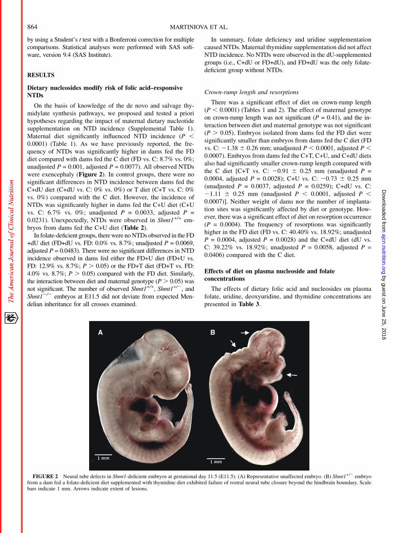
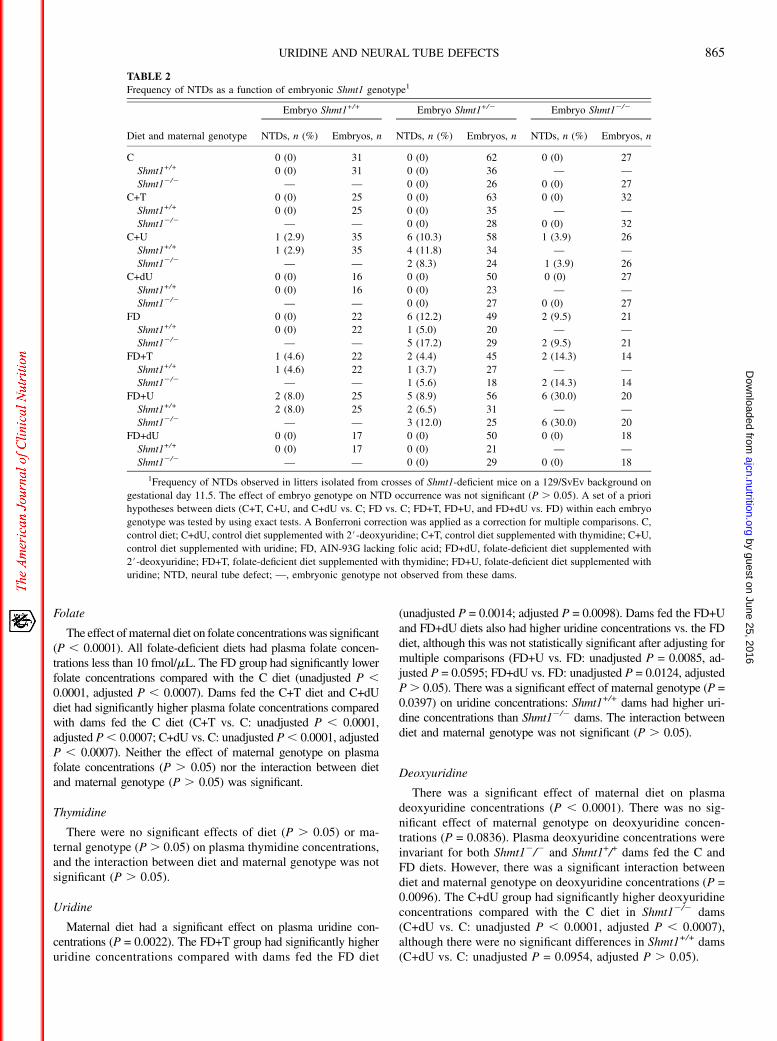
On the basis of knowledge of the de novo and salvage thy-midylate synthesis pathways, we proposed and tested a priorihypotheses regarding the impact of maternal dietary nucleotidesupplementation on NTD incidence (Supplemental Table 1).Maternal diet significantly influenced NTD incidence (P ,0.0001) (Table 1). As we have previously reported, the fre-quency of NTDs was significantly higher in dams fed the FDdiet compared with dams fed the C diet (FD vs. C: 8.7% vs. 0%;unadjusted P = 0.001, adjusted P = 0.0077). All observed NTDswere exencephaly (Figure 2). In control groups, there were nosignificant differences in NTD incidence between dams fed theC+dU diet (C+dU vs. C: 0% vs. 0%) or T diet (C+T vs. C: 0%vs. 0%) compared with the C diet. However, the incidence ofNTDs was significantly higher in dams fed the C+U diet (C+Uvs. C: 6.7% vs. 0%; unadjusted P = 0.0033, adjusted P =0.0231). Unexpectedly, NTDs were observed in Shmt1+/+ em-bryos from dams fed the C+U diet (Table 2).
In folate-deficient groups, therewere noNTDs observed in the FD+dU diet (FD+dU vs. FD: 0.0% vs. 8.7%; unadjusted P = 0.0069,adjusted P = 0.0483). There were no significant differences in NTDincidence observed in dams fed either the FD+U diet (FD+U vs.FD: 12.9% vs. 8.7%; P . 0.05) or the FD+T diet (FD+T vs. FD:4.0% vs. 8.7%; P . 0.05) compared with the FD diet. Similarly,the interaction between diet and maternal genotype (P. 0.05) wasnot significant. The number of observed Shmt1+/+, Shmt1+/2, andShmt12/2 embryos at E11.5 did not deviate from expected Men-delian inheritance for all crosses examined.
In summary, folate deficiency and uridine supplementationcaused NTDs.Maternal thymidine supplementation did not affectNTD incidence. No NTDs were observed in the dU-supplementedgroups (i.e., C+dU or FD+dU), and FD+dU was the only folate-deficient group without NTDs.
Crown-rump length and resorptions
There was a significant effect of diet on crown-rump length(P , 0.0001) (Tables 1 and 2). The effect of maternal genotypeon crown-rump length was not significant (P = 0.41), and the in-teraction between diet and maternal genotype was not significant(P . 0.05). Embryos isolated from dams fed the FD diet weresignificantly smaller than embryos from dams fed the C diet (FDvs. C: 21.38 6 0.26 mm; unadjusted P , 0.0001, adjusted P ,0.0007). Embryos from dams fed the C+T, C+U, and C+dU dietsalso had significantly smaller crown-rump length compared withthe C diet [C+T vs. C: 20.91 6 0.25 mm (unadjusted P =0.0004, adjusted P = 0.0028); C+U vs. C: 20.73 6 0.25 mm(unadjusted P = 0.0037, adjusted P = 0.0259); C+dU vs. C:21.11 6 0.25 mm (unadjusted P , 0.0001, adjusted P ,0.0007)]. Neither weight of dams nor the number of implanta-tion sites was significantly affected by diet or genotype. How-ever, there was a significant effect of diet on resorption occurrence(P = 0.0004). The frequency of resorptions was significantlyhigher in the FD diet (FD vs. C: 40.40% vs. 18.92%; unadjustedP = 0.0004, adjusted P = 0.0028) and the C+dU diet (dU vs.C: 39.22% vs. 18.92%; unadjusted P = 0.0058, adjusted P =0.0406) compared with the C diet.
Effects of diet on plasma nucleoside and folateconcentrations
The effects of dietary folic acid and nucleosides on plasmafolate, uridine, deoxyuridine, and thymidine concentrations arepresented in Table 3.
FIGURE 2 Neural tube defects in Shmt1-deficient embryos at gestational day 11.5 (E11.5). (A) Representative unaffected embryo. (B) Shmt1+/2 embryofrom a dam fed a folate-deficient diet supplemented with thymidine diet exhibited failure of rostral neural tube closure beyond the hindbrain boundary. Scalebars indicate 1 mm. Arrows indicate extent of lesions.
864 MARTINIOVA ET AL.
by guest on June 25, 2016ajcn.nutrition.org
Dow
nloaded from
Folate
The effect ofmaternal diet on folate concentrationswas significant(P , 0.0001). All folate-deficient diets had plasma folate concen-trations less than 10 fmol/mL. The FD group had significantly lowerfolate concentrations compared with the C diet (unadjusted P ,0.0001, adjusted P , 0.0007). Dams fed the C+T diet and C+dUdiet had significantly higher plasma folate concentrations comparedwith dams fed the C diet (C+T vs. C: unadjusted P , 0.0001,adjusted P, 0.0007; C+dU vs. C: unadjusted P, 0.0001, adjustedP , 0.0007). Neither the effect of maternal genotype on plasmafolate concentrations (P . 0.05) nor the interaction between dietand maternal genotype (P . 0.05) was significant.
Thymidine
There were no significant effects of diet (P . 0.05) or ma-ternal genotype (P . 0.05) on plasma thymidine concentrations,and the interaction between diet and maternal genotype was notsignificant (P . 0.05).
Uridine
Maternal diet had a significant effect on plasma uridine con-centrations (P = 0.0022). The FD+T group had significantly higheruridine concentrations compared with dams fed the FD diet
(unadjusted P = 0.0014; adjusted P = 0.0098). Dams fed the FD+Uand FD+dU diets also had higher uridine concentrations vs. the FDdiet, although this was not statistically significant after adjusting formultiple comparisons (FD+U vs. FD: unadjusted P = 0.0085, ad-justed P = 0.0595; FD+dU vs. FD: unadjusted P = 0.0124, adjustedP. 0.05). There was a significant effect of maternal genotype (P =0.0397) on uridine concentrations: Shmt1+/+ dams had higher uri-dine concentrations than Shmt12/2 dams. The interaction betweendiet and maternal genotype was not significant (P . 0.05).
Deoxyuridine
There was a significant effect of maternal diet on plasmadeoxyuridine concentrations (P , 0.0001). There was no sig-nificant effect of maternal genotype on deoxyuridine concen-trations (P = 0.0836). Plasma deoxyuridine concentrations wereinvariant for both Shmt12/2 and Shmt1+/+ dams fed the C andFD diets. However, there was a significant interaction betweendiet and maternal genotype on deoxyuridine concentrations (P =0.0096). The C+dU group had significantly higher deoxyuridineconcentrations compared with the C diet in Shmt12/2 dams(C+dU vs. C: unadjusted P , 0.0001, adjusted P , 0.0007),although there were no significant differences in Shmt1+/+ dams(C+dU vs. C: unadjusted P = 0.0954, adjusted P . 0.05).
TABLE 2
Frequency of NTDs as a function of embryonic Shmt1 genotype1
Diet and maternal genotype
Embryo Shmt1+/+ Embryo Shmt1+/2 Embryo Shmt12/2
NTDs, n (%) Embryos, n NTDs, n (%) Embryos, n NTDs, n (%) Embryos, n
C 0 (0) 31 0 (0) 62 0 (0) 27
Shmt1+/+ 0 (0) 31 0 (0) 36 — —
Shmt12/2 — — 0 (0) 26 0 (0) 27
C+T 0 (0) 25 0 (0) 63 0 (0) 32
Shmt1+/+ 0 (0) 25 0 (0) 35 — —
Shmt12/2 — — 0 (0) 28 0 (0) 32
C+U 1 (2.9) 35 6 (10.3) 58 1 (3.9) 26
Shmt1+/+ 1 (2.9) 35 4 (11.8) 34 — —
Shmt12/2 — — 2 (8.3) 24 1 (3.9) 26
C+dU 0 (0) 16 0 (0) 50 0 (0) 27
Shmt1+/+ 0 (0) 16 0 (0) 23 — —
Shmt12/2 — — 0 (0) 27 0 (0) 27
FD 0 (0) 22 6 (12.2) 49 2 (9.5) 21
Shmt1+/+ 0 (0) 22 1 (5.0) 20 — —
Shmt12/2 — — 5 (17.2) 29 2 (9.5) 21
FD+T 1 (4.6) 22 2 (4.4) 45 2 (14.3) 14
Shmt1+/+ 1 (4.6) 22 1 (3.7) 27 — —
Shmt12/2 — — 1 (5.6) 18 2 (14.3) 14
FD+U 2 (8.0) 25 5 (8.9) 56 6 (30.0) 20
Shmt1+/+ 2 (8.0) 25 2 (6.5) 31 — —
Shmt12/2 — — 3 (12.0) 25 6 (30.0) 20
FD+dU 0 (0) 17 0 (0) 50 0 (0) 18
Shmt1+/+ 0 (0) 17 0 (0) 21 — —
Shmt12/2 — — 0 (0) 29 0 (0) 18
1Frequency of NTDs observed in litters isolated from crosses of Shmt1-deficient mice on a 129/SvEv background on
gestational day 11.5. The effect of embryo genotype on NTD occurrence was not significant (P . 0.05). A set of a priori
hypotheses between diets (C+T, C+U, and C+dU vs. C; FD vs. C; FD+T, FD+U, and FD+dU vs. FD) within each embryo
genotype was tested by using exact tests. A Bonferroni correction was applied as a correction for multiple comparisons. C,
control diet; C+dU, control diet supplemented with 2#-deoxyuridine; C+T, control diet supplemented with thymidine; C+U,
control diet supplemented with uridine; FD, AIN-93G lacking folic acid; FD+dU, folate-deficient diet supplemented with
2#-deoxyuridine; FD+T, folate-deficient diet supplemented with thymidine; FD+U, folate-deficient diet supplemented with
uridine; NTD, neural tube defect; —, embryonic genotype not observed from these dams.
URIDINE AND NEURAL TUBE DEFECTS 865
by guest on June 25, 2016ajcn.nutrition.org
Dow
nloaded from
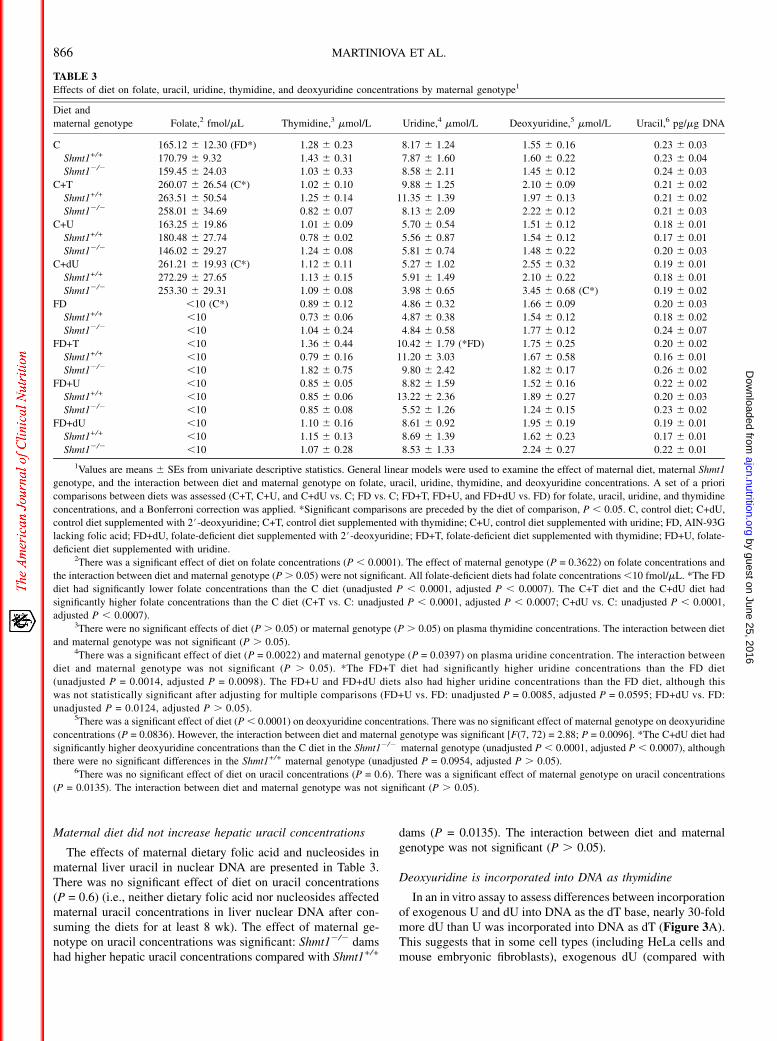
Maternal diet did not increase hepatic uracil concentrations
The effects of maternal dietary folic acid and nucleosides inmaternal liver uracil in nuclear DNA are presented in Table 3.There was no significant effect of diet on uracil concentrations(P = 0.6) (i.e., neither dietary folic acid nor nucleosides affectedmaternal uracil concentrations in liver nuclear DNA after con-suming the diets for at least 8 wk). The effect of maternal ge-notype on uracil concentrations was significant: Shmt12/2 damshad higher hepatic uracil concentrations compared with Shmt1+/+
dams (P = 0.0135). The interaction between diet and maternalgenotype was not significant (P . 0.05).
Deoxyuridine is incorporated into DNA as thymidine
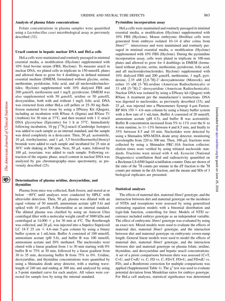
In an in vitro assay to assess differences between incorporationof exogenous U and dU into DNA as the dT base, nearly 30-foldmore dU than U was incorporated into DNA as dT (Figure 3A).This suggests that in some cell types (including HeLa cells andmouse embryonic fibroblasts), exogenous dU (compared with
TABLE 3
Effects of diet on folate, uracil, uridine, thymidine, and deoxyuridine concentrations by maternal genotype1
Diet and
maternal genotype Folate,2 fmol/mL Thymidine,3 mmol/L Uridine,4 mmol/L Deoxyuridine,5 mmol/L Uracil,6 pg/mg DNA
C 165.12 6 12.30 (FD*) 1.28 6 0.23 8.17 6 1.24 1.55 6 0.16 0.23 6 0.03
Shmt1+/+ 170.79 6 9.32 1.43 6 0.31 7.87 6 1.60 1.60 6 0.22 0.23 6 0.04
Shmt12/2 159.45 6 24.03 1.03 6 0.33 8.58 6 2.11 1.45 6 0.12 0.24 6 0.03
C+T 260.07 6 26.54 (C*) 1.02 6 0.10 9.88 6 1.25 2.10 6 0.09 0.21 6 0.02
Shmt1+/+ 263.51 6 50.54 1.25 6 0.14 11.35 6 1.39 1.97 6 0.13 0.21 6 0.02
Shmt12/2 258.01 6 34.69 0.82 6 0.07 8.13 6 2.09 2.22 6 0.12 0.21 6 0.03
C+U 163.25 6 19.86 1.01 6 0.09 5.70 6 0.54 1.51 6 0.12 0.18 6 0.01
Shmt1+/+ 180.48 6 27.74 0.78 6 0.02 5.56 6 0.87 1.54 6 0.12 0.17 6 0.01
Shmt12/2 146.02 6 29.27 1.24 6 0.08 5.81 6 0.74 1.48 6 0.22 0.20 6 0.03
C+dU 261.21 6 19.93 (C*) 1.12 6 0.11 5.27 6 1.02 2.55 6 0.32 0.19 6 0.01
Shmt1+/+ 272.29 6 27.65 1.13 6 0.15 5.91 6 1.49 2.10 6 0.22 0.18 6 0.01
Shmt12/2 253.30 6 29.31 1.09 6 0.08 3.98 6 0.65 3.45 6 0.68 (C*) 0.19 6 0.02
FD ,10 (C*) 0.89 6 0.12 4.86 6 0.32 1.66 6 0.09 0.20 6 0.03
Shmt1+/+ ,10 0.73 6 0.06 4.87 6 0.38 1.54 6 0.12 0.18 6 0.02
Shmt12/2 ,10 1.04 6 0.24 4.84 6 0.58 1.77 6 0.12 0.24 6 0.07
FD+T ,10 1.36 6 0.44 10.42 6 1.79 (*FD) 1.75 6 0.25 0.20 6 0.02
Shmt1+/+ ,10 0.79 6 0.16 11.20 6 3.03 1.67 6 0.58 0.16 6 0.01
Shmt12/2 ,10 1.82 6 0.75 9.80 6 2.42 1.82 6 0.17 0.26 6 0.02
FD+U ,10 0.85 6 0.05 8.82 6 1.59 1.52 6 0.16 0.22 6 0.02
Shmt1+/+ ,10 0.85 6 0.06 13.22 6 2.36 1.89 6 0.27 0.20 6 0.03
Shmt12/2 ,10 0.85 6 0.08 5.52 6 1.26 1.24 6 0.15 0.23 6 0.02
FD+dU ,10 1.10 6 0.16 8.61 6 0.92 1.95 6 0.19 0.19 6 0.01
Shmt1+/+ ,10 1.15 6 0.13 8.69 6 1.39 1.62 6 0.23 0.17 6 0.01
Shmt12/2 ,10 1.07 6 0.28 8.53 6 1.33 2.24 6 0.27 0.22 6 0.01
1Values are means 6 SEs from univariate descriptive statistics. General linear models were used to examine the effect of maternal diet, maternal Shmt1
genotype, and the interaction between diet and maternal genotype on folate, uracil, uridine, thymidine, and deoxyuridine concentrations. A set of a priori
comparisons between diets was assessed (C+T, C+U, and C+dU vs. C; FD vs. C; FD+T, FD+U, and FD+dU vs. FD) for folate, uracil, uridine, and thymidine
concentrations, and a Bonferroni correction was applied. *Significant comparisons are preceded by the diet of comparison, P , 0.05. C, control diet; C+dU,
control diet supplemented with 2#-deoxyuridine; C+T, control diet supplemented with thymidine; C+U, control diet supplemented with uridine; FD, AIN-93G
lacking folic acid; FD+dU, folate-deficient diet supplemented with 2#-deoxyuridine; FD+T, folate-deficient diet supplemented with thymidine; FD+U, folate-
deficient diet supplemented with uridine.2There was a significant effect of diet on folate concentrations (P , 0.0001). The effect of maternal genotype (P = 0.3622) on folate concentrations and
the interaction between diet and maternal genotype (P. 0.05) were not significant. All folate-deficient diets had folate concentrations,10 fmol/mL. *The FD
diet had significantly lower folate concentrations than the C diet (unadjusted P , 0.0001, adjusted P , 0.0007). The C+T diet and the C+dU diet had
significantly higher folate concentrations than the C diet (C+T vs. C: unadjusted P , 0.0001, adjusted P , 0.0007; C+dU vs. C: unadjusted P , 0.0001,
adjusted P , 0.0007).3There were no significant effects of diet (P . 0.05) or maternal genotype (P . 0.05) on plasma thymidine concentrations. The interaction between diet
and maternal genotype was not significant (P . 0.05).4There was a significant effect of diet (P = 0.0022) and maternal genotype (P = 0.0397) on plasma uridine concentration. The interaction between
diet and maternal genotype was not significant (P . 0.05). *The FD+T diet had significantly higher uridine concentrations than the FD diet
(unadjusted P = 0.0014, adjusted P = 0.0098). The FD+U and FD+dU diets also had higher uridine concentrations than the FD diet, although this
was not statistically significant after adjusting for multiple comparisons (FD+U vs. FD: unadjusted P = 0.0085, adjusted P = 0.0595; FD+dU vs. FD:
unadjusted P = 0.0124, adjusted P . 0.05).5There was a significant effect of diet (P, 0.0001) on deoxyuridine concentrations. There was no significant effect of maternal genotype on deoxyuridine
concentrations (P = 0.0836). However, the interaction between diet and maternal genotype was significant [F(7, 72) = 2.88; P = 0.0096]. *The C+dU diet had
significantly higher deoxyuridine concentrations than the C diet in the Shmt12/2 maternal genotype (unadjusted P , 0.0001, adjusted P , 0.0007), although
there were no significant differences in the Shmt1+/+ maternal genotype (unadjusted P = 0.0954, adjusted P . 0.05).6There was no significant effect of diet on uracil concentrations (P = 0.6). There was a significant effect of maternal genotype on uracil concentrations
(P = 0.0135). The interaction between diet and maternal genotype was not significant (P . 0.05).
866 MARTINIOVA ET AL.
by guest on June 25, 2016ajcn.nutrition.org
Dow
nloaded from
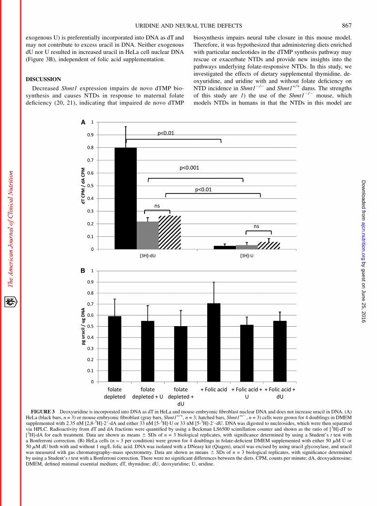
exogenous U) is preferentially incorporated into DNA as dT andmay not contribute to excess uracil in DNA. Neither exogenousdU nor U resulted in increased uracil in HeLa cell nuclear DNA(Figure 3B), independent of folic acid supplementation.
DISCUSSION
Decreased Shmt1 expression impairs de novo dTMP bio-synthesis and causes NTDs in response to maternal folatedeficiency (20, 21), indicating that impaired de novo dTMP
biosynthesis impairs neural tube closure in this mouse model.Therefore, it was hypothesized that administering diets enrichedwith particular nucleotides in the dTMP synthesis pathway mayrescue or exacerbate NTDs and provide new insights into thepathways underlying folate-responsive NTDs. In this study, weinvestigated the effects of dietary supplemental thymidine, de-oxyuridine, and uridine with and without folate deficiency onNTD incidence in Shmt12/2 and Shmt1+/+ dams. The strengthsof this study are 1) the use of the Shmt12/2 mouse, whichmodels NTDs in humans in that the NTDs in this model are
FIGURE 3 Deoxyuridine is incorporated into DNA as dT in HeLa and mouse embryonic fibroblast nuclear DNA and does not increase uracil in DNA. (A)HeLa (black bars, n = 3) or mouse embryonic fibroblast (gray bars, Shmt1+/+, n = 3; hatched bars, Shmt1+/2, n = 3) cells were grown for 4 doublings in DMEMsupplemented with 2.35 nM [2,8-3H]-2#-dA and either 33 nM [5-3H]-U or 33 nM [5-3H]-2#-dU. DNAwas digested to nucleosides, which were then separatedvia HPLC. Radioactivity from dT and dA fractions were quantified by using a Beckman LS6500 scintillation counter and shown as the ratio of [3H]-dT to[3H]-dA for each treatment. Data are shown as means 6 SDs of n = 3 biological replicates, with significance determined by using a Student’s t test witha Bonferroni correction. (B) HeLa cells (n = 3 per condition) were grown for 4 doublings in folate-deficient DMEM supplemented with either 50 mM U or50 mM dU both with and without 1 mg/L folic acid. DNAwas isolated with a DNeasy kit (Qiagen), uracil was excised by using uracil glycosylase, and uracilwas measured with gas chromatography–mass spectrometry. Data are shown as means 6 SDs of n = 3 biological replicates, with significance determinedby using a Student’s t test with a Bonferroni correction. There were no significant differences between the diets. CPM, counts per minute; dA, deoxyadenosine;DMEM, defined minimal essential medium; dT, thymidine; dU, deoxyuridine; U, uridine.
URIDINE AND NEURAL TUBE DEFECTS 867
by guest on June 25, 2016ajcn.nutrition.org
Dow
nloaded from
sporadic and folic acid responsive, and 2) the ability to performrationally designed dietary manipulations in the dams to eluci-date mechanisms and pathways of NTD pathogenesis.
As demonstrated previously, exencephaly was observed inlitters from dams maintained on the FD diet but not on the C diet,confirming our previous studies that both maternal folate de-ficiency and embryonic Shmt1 disruption were required for NTDpathogenesis (20, 34). The results of this study demonstrate thatmaternal uridine supplementation of the AIN-93G diet causedNTDs independent of fetal Shmt1 genotype and maternal folatestatus. We also observed that maternal deoxyuridine supple-mentation prevented NTD occurrence in Shmt1+/2 and Shmt12/2
embryos in dams fed the dU+FD diet. In addition, thymidinesupplementation had no statistically significant effect on NTDincidence.
The current literature regarding the role of exogenous thy-midine in NTD prevention is mixed. One study reported thatNTDs in the Splotch mutant could be rescued with thymidine,indicating that folic acid prevents NTDs by rescuing de novodTMP synthesis in that mouse model, but this finding was notconfirmed in another study (18, 19). Another study indicatedthat de novo purine biosynthesis was impaired to a greater de-gree than de novo dTMP biosynthesis in the Splotch mutant(20). In this study, maternal thymidine supplementation did notprevent NTDs in embryos from folate-deficient Shmt1+/+ orShmt12/2 dams. Interestingly, we observed one Shmt1+/+ em-bryo with exencephaly from a Shmt1+/+ dam fed the FD+T diet.In our previous studies of Shmt1+/2 and Shmt12/2 dams fed theFD diet, no NTD-affected Shmt1+/+ embryos were observed(6, 20). We conclude that thymidine is not a maternally derivedfactor that prevents NTDs in SHMT1-deficient mice and likelyincreases risk for NTDs.
Surprisingly, supplemental deoxyuridine prevented NTDs inShmt12/2 and Shmt1+/2 embryos from folate-deficient dams.We had initially predicted that dU supplementation would in-crease, not prevent, NTDs in Shmt12/2 and Shmt1+/2 embryosfrom folate-deficient dams because cellular dUMP accumulationhas been proposed to enhance uracil misincorporation into DNAand lead to genome instability (35). However, exogenous [3H]-dU, but not [3H]-U, is robustly incorporated into HeLa cellnuclear DNA as the base dT. Furthermore, dU exposure did notincrease uracil in HeLa cell nuclear DNA. This observation doesnot support the current dogma that uracil misincorporation re-sults simply from an increased dUTP/dTTP ratio and suggestsa possible mechanism whereby dU may act to stabilize DNAintegrity and prevent NTDs in this mouse model, which issusceptible to increased uracil in DNA. These data suggest thatdietary dU rescues NTDs by increasing rates of de novo dTMPsynthesis in SHMT1-deficient embryos by mass action throughthe provision of increased concentrations of dUMP substratewithout increasing rates of dU misincorporation into DNA. Themechanisms behind the increased resorption rate in embryosfrom dU-supplemented dams remain unknown. This increasemay not be related to dietary folate or folate-sensitive patholo-gies, as observed in dams fed both the C+dU and FD diets,compared with the C diet.
Unexpectedly, maternal uridine supplementation caused NTDsindependent of maternal folate status and Shmt1 genotype.Exencephaly was observed in litters from both Shmt12/2 andShmt1+/+ dams fed the U diet. Uridine supplementation caused
significantly higher NTD incidence compared with the C diet.The mechanism underlying the role of maternal dietary uridineand NTD risk remains to be established.
It is not clear why plasma nucleoside concentrations do notreflect dietary intake, but it may be because of low nucleosidebioavailability or rapid uptake by tissues. Nonetheless, the resultsprovide additional evidence that the de novo dTMP synthesispathway underlies NTD pathogenesis in this mouse model. In-terestingly, the results also indicate that the dietary nucleosides Tand dU have a marked effect on serum folate concentrations indams fed the folate-replete diets, indicating that these nucleosidesmay influence whole-body folate homeostasis. Clearly, theinability to understand the metabolic outcomes in individualNTD-affected embryos is a limitation of this study. Developingmore sensitive assays to assess uracil in DNA and DNA damagein embryos is the focus of ongoing work.
The most pronouncedmetabolic phenotype in SHMT1-deficientmice is impaired de novo dTMP biosynthesis and elevated uracilin DNA (20, 36). This study confirms our previous reports thatSHMT1 deficiency elevates uracil concentrations in DNA. Al-though nucleoside supplementation did affect NTD incidence, itdid not influence uracil concentrations in maternal liver nuclearDNA. This suggests that uridine supplementation does not causeNTDs by elevating uracil concentrations in DNA, assuming thatthe fetal neural epithelium and maternal liver respond similarly.Although additional studies are required to establish the mech-anism that accounts for uridine teratogenicity, women of re-productive age should avoid over-the-counter uridine dietarysupplements until more information is known about their effectson the developing embryo.
The authors thank Sylvia Allen for technical assistance and especially
thank Ashley M Palmer and Anna E Beaudin for thoughtful insight and
Francoise Vermeylen for assistance with statistical analysis.
The authors’ responsibilities were as follows—LM: data collection and
analysis, manuscript preparation, editing, and revision; MSF: study concep-
tion, data collection and analysis, and manuscript editing; JLF: statistical
analysis; CAP: data collection; PJS: study conception and design and man-
uscript editing and revision. The authors declared no conflicts of interest
related to this study.
REFERENCES1. Kirke PN, Molloy AM, Daly LE, Burke H, Weir DG, Scott JM. Ma-
ternal plasma folate and vitamin B12 are independent risk factors forneural tube defects. Q J Med 1993;86:703–8.
2. Etheredge AJ, Finnell RH, Carmichael SL, Lammer EJ, Zhu H,Mitchell LE, Shaw GM. Maternal and infant gene-folate interactionsand the risk of neural tube defects. Am J Med Genet A 2012;158A:2439–46.
3. Pangilinan F, Molloy AM, Mills JL, Troendle JF, Parle-McDermott A,Signore C, O’Leary VB, Chines P, Seay JM, Geiler-Samerotte K, et al.Evaluation of common genetic variants in 82 candidate genes as riskfactors for neural tube defects. BMC Med Genet 2012;13:62.
4. Relton CL, Wilding CS, Laffling AJ, Jonas PA, Burgess T, Binks K,Tawn EJ, Burn J. Low erythrocyte folate status and polymorphic var-iation in folate-related genes are associated with risk of neural tubedefect pregnancy. Mol Genet Metab 2004;81:273–81.
5. Christensen B, Arbour L, Tran P, Leclerc D, Sabbaghian N, Platt R,Gilfix BM, Rosenblatt DS, Gravel RA, Forbes P, et al. Genetic poly-morphisms in methylenetetrahydrofolate reductase and methioninesynthase, folate levels in red blood cells, and risk of neural tube de-fects. Am J Med Genet 1999;84:151–7.
6. Beaudin AE, Stover PJ. Insights into metabolic mechanisms underlyingfolate-responsive neural tube defects: a minireview. Birth Defects ResA Clin Mol Teratol 2009;85:274–84.
868 MARTINIOVA ET AL.
by guest on June 25, 2016ajcn.nutrition.org
Dow
nloaded from
7. Prevention of neural tube defects: results of the Medical ResearchCouncil Vitamin Study. MRC Vitamin Study Research Group. Lancet1991;338:131–7.
8. Czeizel AE, Dudas I. Prevention of the first occurrence of neural-tubedefects by periconceptional vitamin supplementation. N Engl J Med1992;327:1832–5.
9. Institute of Medicine. Dietary Reference Intakes for Thiamin, Ribo-flavin, Niacin, Vitamin B6, Folate, Vitamin B12, Pantothenic Acid,Biotin, and Choline. Washington (DC): National Academies Press;1998.
10. Berry RJ, Li Z, Erickson JD, Li S, Moore CA, Wang H, Mulinare J,Zhao P, Wong LY, Gindler J, et al. Prevention of neural-tube defectswith folic acid in China. China-U.S. Collaborative Project for NeuralTube Defect Prevention. N Engl J Med 1999;341:1485–90.
11. Bhatt RV. Environmental influence on reproductive health. Int J Gy-naecol Obstet 2000;70:69–75.
12. Hutz RJ, Carvan MJ, Baldridge MG, Conley LK, Heiden TK. Envi-ronmental toxicants and effects on female reproductive function.Trends Reprod Biol 2006;2:1–11.
13. Leddy MA, Power ML, Schulkin J. The impact of maternal obesity onmaternal and fetal health. Rev Obstet Gynecol 2008;1:170–8.
14. Hendricks KA, Nuno OM, Suarez L, Larsen R. Effects of hyper-insulinemia and obesity on risk of neural tube defects among MexicanAmericans. Epidemiology 2001;12:630–5.
15. Stover PJ, Garza C. Bringing individuality to public health recom-mendations. J Nutr 2002;132(Suppl):2476S–80S.
16. Stover PJ. Physiology of folate and vitamin B12 in health and disease.Nutr Rev 2004;62(Pt 2):S3–12; discussion S3.
17. Harris MJ, Juriloff DM. Mouse mutants with neural tube closure de-fects and their role in understanding human neural tube defects. BirthDefects Res A Clin Mol Teratol 2007;79:187–210.
18. Wlodarczyk BJ, Tang LS, Triplett A, Aleman F, Finnell RH. Sponta-neous neural tube defects in splotch mice supplemented with selectedmicronutrients. Toxicol Appl Pharmacol 2006;213:55–63.
19. Fleming A, Copp AJ. Embryonic folate metabolism and mouse neuraltube defects. Science 1998;280:2107–9.
20. Beaudin AE, Abarinov EV, Noden DM, Perry CA, Chu S, Stabler SP,Allen RH, Stover PJ. Shmt1 and de novo thymidylate biosynthesisunderlie folate-responsive neural tube defects in mice. Am J Clin Nutr2011;93:789–98.
21. Beaudin AE, Abarinov EV, Malysheva O, Perry CA, Caudill M, StoverPJ. Dietary folate, but not choline, modifies neural tube defect risk inShmt1 knockout mice. Am J Clin Nutr 2012;95:109–14.
22. Herbig K, Chiang EP, Lee LR, Hills J, Shane B, Stover PJ. Cytoplasmicserine hydroxymethyltransferase mediates competition between folate-dependent deoxyribonucleotide and S-adenosylmethionine biosyntheses.J Biol Chem 2002;277:38381–9.
23. Woeller CF, Anderson DD, Szebenyi DM, Stover PJ. Evidence forsmall ubiquitin-like modifier-dependent nuclear import of the thymi-dylate biosynthesis pathway. J Biol Chem 2007;282:17623–31.
24. MacFarlane AJ, Anderson DD, Flodby P, Perry CA, Allan RH, StablerSP, Stover PJ. Nuclear localization of the de novo thymidylate bio-synthesis pathway is required to prevent uracil accumulation in DNA.J Biol Chem 2011;286:44015–22.
25. MacFarlane AJ, Liu X, Perry CA, Flodby P, Allen RH, Stabler SP,Stover PJ. Cytoplasmic serine hydroxymethyltransferase regulates themetabolic partitioning of methylenetetrahydrofolate but is not essentialin mice. J Biol Chem 2008;283:25846–53.
26. Rudolph FB, Fanslow WC, Kulkarni AD, Kulkarni SS, Van Buren CT.Effect of dietary nucleotides on lymphocyte maturation. Adv Exp MedBiol 1986;195(Pt A):497–501.
27. Iwasa Y, Iwasa M, Omori Y, Toki T, Yamamoto A, Maeda H, Kume M,Ogoshi S. The well-balanced nucleoside-nucleotide mixture “OG-VI”for special medical purposes. Nutrition 1997;13:361–4.
28. Kulkarni AD, Fanslow WC, Rudolph FB, Van Buren CT. Effect ofdietary nucleotides on response to bacterial infections. JPEN J ParenterEnteral Nutr 1986;10:169–71.
29. Clapcote SJ, Roder JC. Simplex PCR assay for sex determination inmice. Biotechniques 2005;38:702, 4, 6.
30. Machado AF, Zimmerman EF, Hovland DN Jr., Weiss R, Collins MD.Diabetic embryopathy in C57BL/6J mice: altered fetal sex ratio andimpact of the splotch allele. Diabetes 2001;50:1193–9.
31. McClive PJ, Sinclair AH. Rapid DNA extraction and PCR-sexing ofmouse embryos. Mol Reprod Dev 2001;60:225–6.
32. Suh JR, Oppenheim EW, Girgis S, Stover PJ. Purification and propertiesof a folate-catabolizing enzyme. J Biol Chem 2000;275:35646–55.
33. Crain PF. Preparation and enzymatic hydrolysis of DNA and RNA formass spectrometry. Methods Enzymol 1990;193:782–90.
34. Walzem RL, Clifford AJ. Folate deficiency in rats fed diets containingfree amino acids or intact proteins. J Nutr 1988;118:1089–96.
35. Stover PJ. One-carbon metabolism-genome interactions in folate-associated pathologies. J Nutr 2009;139:2402–5.
36. Macfarlane AJ, Perry CA, McEntee MF, Lin DM, Stover PJ. Shmt1heterozygosity impairs folate-dependent thymidylate synthesis capac-ity and modifies risk of Apc(min)-mediated intestinal cancer risk.Cancer Res 2011;71:2098–107.
URIDINE AND NEURAL TUBE DEFECTS 869
by guest on June 25, 2016ajcn.nutrition.org
Dow
nloaded from
Recommended


