
SAGE-Hindawi Access to ResearchJournal of Nucleic AcidsVolume 2011, Article ID 741723, 15 pagesdoi:10.4061/2011/741723
Review Article
Molecular Beacons: Powerful Tools for Imaging RNA inLiving Cells
Ricardo Monroy-Contreras and Luis Vaca
Instituto de Fisiologıa Celular, Universidad Nacional Autonoma de Mexico, 04510 Mexico, DF, Mexico
Correspondence should be addressed to Luis Vaca, [email protected]
Received 9 November 2010; Revised 14 June 2011; Accepted 22 June 2011
Academic Editor: Dmitry A. Stetsenko
Copyright © 2011 R. Monroy-Contreras and L. Vaca. This is an open access article distributed under the Creative CommonsAttribution License, which permits unrestricted use, distribution, and reproduction in any medium, provided the original work isproperly cited.
Recent advances in RNA functional studies highlights the pivotal role of these molecules in cell physiology. Diverse methods havebeen implemented to measure the expression levels of various RNA species, using either purified RNA or fixed cells. Despite the factthat fixed cells offer the possibility to observe the spatial distribution of RNA, assays with capability to real-time monitoring RNAtransport into living cells are needed to further understand the role of RNA dynamics in cellular functions. Molecular beacons(MBs) are stem-loop hairpin-structured oligonucleotides equipped with a fluorescence quencher at one end and a fluorescentdye (also called reporter or fluorophore) at the opposite end. This structure permits that MB in the absence of their targetcomplementary sequence do not fluoresce. Upon binding to targets, MBs emit fluorescence, due to the spatial separation of thequencher and the reporter. Molecular beacons are promising probes for the development of RNA imaging techniques; neverthelessmuch work remains to be done in order to obtain a robust technology for imaging various RNA molecules together in real timeand in living cells. The present work concentrates on the different requirements needed to use successfully MB for cellular studies,summarizing recent advances in this area.
1. Introduction
A wealth of experimental evidence accumulated to this dateillustrates the wide range of cellular functions conducted byribonucleic acids (RNAs). In many occasions these functionsinvolve RNA transport from one cellular compartment toanother or into the same compartment to specialized re-gions. In order to accomplish this, RNA molecules are undera specific and selective control via expression levels and/orstability in a spatial-temporal manner. Different methodsdeveloped to purify RNA [1–3] from cell populations ortissues have provided relevant information about the relativeconcentration of RNA in cells, or cellular compartments,yet these methods provide limited information about thespatial-temporal distribution of RNAs and their dynamictransport.
The integral understanding of cellular process in whichRNA is involved, requires a method that reveals RNA local-ization in real time in a subcellular context in living cells.The information obtained from this type of assays promise
to impulse the advancement in molecular biology, medicalresearch, and diagnostics.
Many methods have been developed to measure RNAexpression levels between different cell populations, such asthe polymerase chain reaction (PCR) [4], northern blot [5],serial analysis of the gene expression (SAGE) [6], suppressionof subtractive hybridization (SSH) [7], differential display[8], representational difference analysis (RDA) [9], expressedsequence tag (EST) [10], and microarrays [11]. Thus thesetechniques in addition to databases containing genomic datafor a wide variety of biological entities, supply a usefulinstrument for understanding physiology or pathology atthe molecular level. Nevertheless, none of the techniquespreviously mentioned could provide information about thesubcellular RNA localization and its transport.
Through the use of fluorescent in situ hybridizationassays [12], scientists have identified gradients of RNA incells [13], embryos [14], and tissues [15, 16]. Howeverthis technique is conducted on fixed samples and thereforeprovides limited information about the dynamics of such
2 Journal of Nucleic Acids
gradients. Other disadvantages are the laborious and time-consuming nature of the assay, the difficulty that involves theanalysis of the images and variations due to handling differ-ent trials. The chemicals used for dehydration and fixationcould affect the signal level [17] and alter the integrity of or-ganelles, hampering any conclusions about spatial changes inorganelles or compartments.
The most adequate procedure to obtain spatial-temporalresolution of the RNA dynamics is using living cell assays.Under these conditions, the probes to be used must havehigh specificity, sensitivity, and good signal to backgroundratio, especially for low abundance RNA molecules. Ideally,the experimental procedure should be able to detect discretechanges in RNA concentrations of the sample. Furthermore,a robust method should provide additional information likesingle nucleotide mutations, deletions, insertions, and singlenucleotide polymorphisms (SNPs). Another fact to take inconsideration is the method for intracellular delivery; itshould not produce the degradation of the probe or damagethe cell. Finally, the probes utilized should have fast kineticsof hybridization with its target at room temperature, in orderto be useful in real-time measurements.
Recent advances in DNA chemistry deliver promisingnanostructured probes known as molecular beacons (MB),which may fulfill many (if not all) of the requirements high-lighted above. MBs are slowly becoming powerful tools toexplore RNA function and dynamics in living cells. Com-bined with advanced imaging techniques, MB are deliveringinteresting and sometimes surprising results about RNA dy-namics in living cells and tissues.
A recent work reviews different forms of linear andnonlinear probes useful for cell imaging studies [18]. In thisreview we will address the use of MBs for RNA imaging inliving cells, analyzing some of the key features required forsuccessful MB design. We will review also various deliverymethods commonly used for MB introduction into livingcells.
2. Structure and Function of Molecular Beacons
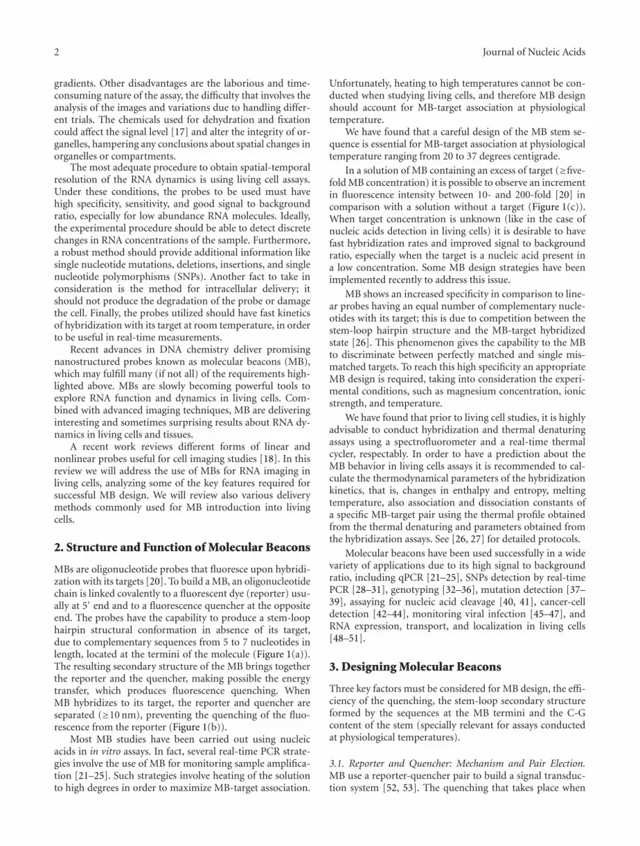
MBs are oligonucleotide probes that fluoresce upon hybridi-zation with its targets [20]. To build a MB, an oligonucleotidechain is linked covalently to a fluorescent dye (reporter) usu-ally at 5′ end and to a fluorescence quencher at the oppositeend. The probes have the capability to produce a stem-loophairpin structural conformation in absence of its target,due to complementary sequences from 5 to 7 nucleotides inlength, located at the termini of the molecule (Figure 1(a)).The resulting secondary structure of the MB brings togetherthe reporter and the quencher, making possible the energytransfer, which produces fluorescence quenching. WhenMB hybridizes to its target, the reporter and quencher areseparated (≥10 nm), preventing the quenching of the fluo-rescence from the reporter (Figure 1(b)).
Most MB studies have been carried out using nucleicacids in in vitro assays. In fact, several real-time PCR strate-gies involve the use of MB for monitoring sample amplifica-tion [21–25]. Such strategies involve heating of the solutionto high degrees in order to maximize MB-target association.
Unfortunately, heating to high temperatures cannot be con-ducted when studying living cells, and therefore MB designshould account for MB-target association at physiologicaltemperature.
We have found that a careful design of the MB stem se-quence is essential for MB-target association at physiologicaltemperature ranging from 20 to 37 degrees centigrade.
In a solution of MB containing an excess of target (≥five-fold MB concentration) it is possible to observe an incrementin fluorescence intensity between 10- and 200-fold [20] incomparison with a solution without a target (Figure 1(c)).When target concentration is unknown (like in the case ofnucleic acids detection in living cells) it is desirable to havefast hybridization rates and improved signal to backgroundratio, especially when the target is a nucleic acid present ina low concentration. Some MB design strategies have beenimplemented recently to address this issue.
MB shows an increased specificity in comparison to line-ar probes having an equal number of complementary nucle-otides with its target; this is due to competition between thestem-loop hairpin structure and the MB-target hybridizedstate [26]. This phenomenon gives the capability to the MBto discriminate between perfectly matched and single mis-matched targets. To reach this high specificity an appropriateMB design is required, taking into consideration the experi-mental conditions, such as magnesium concentration, ionicstrength, and temperature.
We have found that prior to living cell studies, it is highlyadvisable to conduct hybridization and thermal denaturingassays using a spectrofluorometer and a real-time thermalcycler, respectably. In order to have a prediction about theMB behavior in living cells assays it is recommended to cal-culate the thermodynamical parameters of the hybridizationkinetics, that is, changes in enthalpy and entropy, meltingtemperature, also association and dissociation constants ofa specific MB-target pair using the thermal profile obtainedfrom the thermal denaturing and parameters obtained fromthe hybridization assays. See [26, 27] for detailed protocols.
Molecular beacons have been used successfully in a widevariety of applications due to its high signal to backgroundratio, including qPCR [21–25], SNPs detection by real-timePCR [28–31], genotyping [32–36], mutation detection [37–39], assaying for nucleic acid cleavage [40, 41], cancer-celldetection [42–44], monitoring viral infection [45–47], andRNA expression, transport, and localization in living cells[48–51].
3. Designing Molecular Beacons
Three key factors must be considered for MB design, the effi-ciency of the quenching, the stem-loop secondary structureformed by the sequences at the MB termini and the C-Gcontent of the stem (specially relevant for assays conductedat physiological temperatures).
3.1. Reporter and Quencher: Mechanism and Pair Election.MB use a reporter-quencher pair to build a signal transduc-tion system [52, 53]. The quenching that takes place when

Journal of Nucleic Acids 3
(a)
(b)
(c)
Loop
Qu
ench
er
N
N
N
ONH
O
O
O
NH
N
NO
OO
O
O
OH
O−P
O
O
O
O−P
NH
OO
O−
OOO−
c
g
t
c
c g
a
g
c
g
A
T
A
C
A
G
C
TA G A
T
A
A
C
C
A
A
A
0
3
6
9
12
15
100 200 300 400
Time (s)
Flu
ores
cen
cein
ten
sity
(AU
)
MB without target
MB + target
Random-coilMB
Hyb
rid
MB
-tar
get
Target
Stem-loopMB
Reporter6-FAM
Stem
5lin
ker 3
linke
r
dabc
yl
0
Figure 1: Structure and function of MB. (a) Stem-loop hairpin structure of a MB showing its four structural components: loop, stem,quencher, and reporter. The chemical structure of the linkers is drawn according to the manufacturer (Integrated DNA Technologies, Iowa,USA). (b) Mechanism of MB function. A MB in a solution containing both MB and target could be in three states: free in a stem-loop hairpinconformation, hybridized with it target, or unbound in a random-coil conformation. The random-coil conformation of the MB contributesto the background. (c) Fluorescence intensity. The blue line shows the fluorescence intensity for a MB in solution (50 nM) during 400 seconds(background), and the red line corresponds to the addition (100 seconds) of a target oligonucleotide (500 nM) that hybridize the MB at theloop region. An increase in fluorescent intensity of approximately thirteenfold is observed. The reporter is represented with a red circle andthe quencher with a blue. UA means arbitrary units.
4 Journal of Nucleic Acids
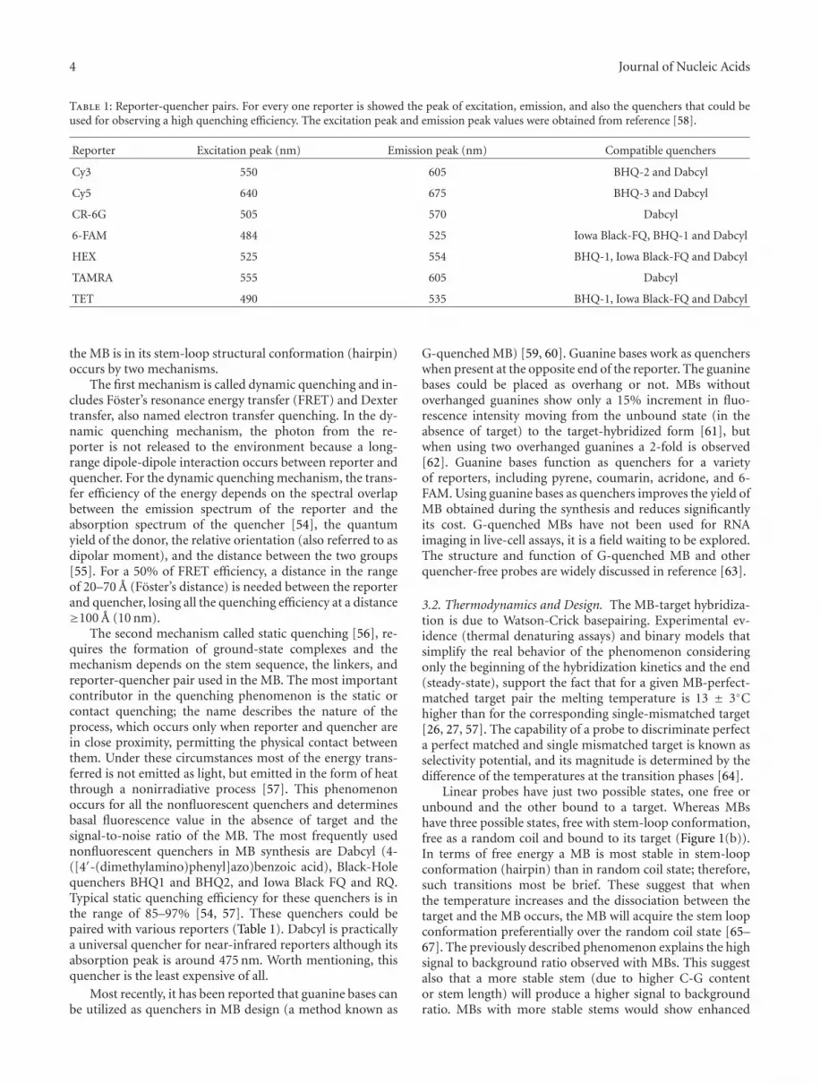
Table 1: Reporter-quencher pairs. For every one reporter is showed the peak of excitation, emission, and also the quenchers that could beused for observing a high quenching efficiency. The excitation peak and emission peak values were obtained from reference [58].
Reporter Excitation peak (nm) Emission peak (nm) Compatible quenchers
Cy3 550 605 BHQ-2 and Dabcyl
Cy5 640 675 BHQ-3 and Dabcyl
CR-6G 505 570 Dabcyl
6-FAM 484 525 Iowa Black-FQ, BHQ-1 and Dabcyl
HEX 525 554 BHQ-1, Iowa Black-FQ and Dabcyl
TAMRA 555 605 Dabcyl
TET 490 535 BHQ-1, Iowa Black-FQ and Dabcyl
the MB is in its stem-loop structural conformation (hairpin)occurs by two mechanisms.
The first mechanism is called dynamic quenching and in-cludes Foster’s resonance energy transfer (FRET) and Dextertransfer, also named electron transfer quenching. In the dy-namic quenching mechanism, the photon from the re-porter is not released to the environment because a long-range dipole-dipole interaction occurs between reporter andquencher. For the dynamic quenching mechanism, the trans-fer efficiency of the energy depends on the spectral overlapbetween the emission spectrum of the reporter and theabsorption spectrum of the quencher [54], the quantumyield of the donor, the relative orientation (also referred to asdipolar moment), and the distance between the two groups[55]. For a 50% of FRET efficiency, a distance in the rangeof 20–70 A (Foster’s distance) is needed between the reporterand quencher, losing all the quenching efficiency at a distance≥100 A (10 nm).
The second mechanism called static quenching [56], re-quires the formation of ground-state complexes and themechanism depends on the stem sequence, the linkers, andreporter-quencher pair used in the MB. The most importantcontributor in the quenching phenomenon is the static orcontact quenching; the name describes the nature of theprocess, which occurs only when reporter and quencher arein close proximity, permitting the physical contact betweenthem. Under these circumstances most of the energy trans-ferred is not emitted as light, but emitted in the form of heatthrough a nonirradiative process [57]. This phenomenonoccurs for all the nonfluorescent quenchers and determinesbasal fluorescence value in the absence of target and thesignal-to-noise ratio of the MB. The most frequently usednonfluorescent quenchers in MB synthesis are Dabcyl (4-([4′-(dimethylamino)phenyl]azo)benzoic acid), Black-Holequenchers BHQ1 and BHQ2, and Iowa Black FQ and RQ.Typical static quenching efficiency for these quenchers is inthe range of 85–97% [54, 57]. These quenchers could bepaired with various reporters (Table 1). Dabcyl is practicallya universal quencher for near-infrared reporters although itsabsorption peak is around 475 nm. Worth mentioning, thisquencher is the least expensive of all.
Most recently, it has been reported that guanine bases canbe utilized as quenchers in MB design (a method known as
G-quenched MB) [59, 60]. Guanine bases work as quencherswhen present at the opposite end of the reporter. The guaninebases could be placed as overhang or not. MBs withoutoverhanged guanines show only a 15% increment in fluo-rescence intensity moving from the unbound state (in theabsence of target) to the target-hybridized form [61], butwhen using two overhanged guanines a 2-fold is observed[62]. Guanine bases function as quenchers for a varietyof reporters, including pyrene, coumarin, acridone, and 6-FAM. Using guanine bases as quenchers improves the yield ofMB obtained during the synthesis and reduces significantlyits cost. G-quenched MBs have not been used for RNAimaging in live-cell assays, it is a field waiting to be explored.The structure and function of G-quenched MB and otherquencher-free probes are widely discussed in reference [63].
3.2. Thermodynamics and Design. The MB-target hybridiza-tion is due to Watson-Crick basepairing. Experimental ev-idence (thermal denaturing assays) and binary models thatsimplify the real behavior of the phenomenon consideringonly the beginning of the hybridization kinetics and the end(steady-state), support the fact that for a given MB-perfect-matched target pair the melting temperature is 13 ± 3◦Chigher than for the corresponding single-mismatched target[26, 27, 57]. The capability of a probe to discriminate perfecta perfect matched and single mismatched target is known asselectivity potential, and its magnitude is determined by thedifference of the temperatures at the transition phases [64].
Linear probes have just two possible states, one free orunbound and the other bound to a target. Whereas MBshave three possible states, free with stem-loop conformation,free as a random coil and bound to its target (Figure 1(b)).In terms of free energy a MB is most stable in stem-loopconformation (hairpin) than in random coil state; therefore,such transitions most be brief. These suggest that whenthe temperature increases and the dissociation between thetarget and the MB occurs, the MB will acquire the stem loopconformation preferentially over the random coil state [65–67]. The previously described phenomenon explains the highsignal to background ratio observed with MBs. This suggestalso that a more stable stem (due to higher C-G contentor stem length) will produce a higher signal to backgroundratio. MBs with more stable stems would show enhanced

Journal of Nucleic Acids 5
selectivity because the MB will hybridize only when theinteractions MB-targets are strong enough to overcome thestem stability. However, a word of caution, MBs with morestable stems may not hybridize to its target at a physiologicaltemperature, preventing its use for cellular studies. Thus adelicate balance between hairpin stability (stem design) andMB-target selectivity must be reached for successful use ofMBs in living cell assays.
3.3. Balance between Selectivity and Hybridization Rate.Clearly the greater advantage of MB over linear probes isits enhanced selectivity, which result from the hairpin stem-loop structure attained by the MB in the nonhybridized state(not associated to its target). The stability of the hairpin con-formation is ensured by building more stable stems (eitherincreasing its C-G content or the stem length). Unfortunate-ly, increasing stem strength decreases also the hybridizationrate. For a given MB the rates of hybridization decrease be-tween one and two orders of magnitude when the stemlength is increased from two to four nucleotides [27]. Shorterstems produce less stable hairpin structures, and in conse-quence reduce the signal background ratio and the selectivityof the MB-target interaction. For an optimal stem design, itis necessary to determine the balance between selectivity andhybridization rate for a given assay [26, 27, 68]. A typicalMB has a stem of 5–7 nucleotides and a loop of 15–25nucleotides in length (Figures 2(a) and 2(b)) and hybridizeits target using only the loop region (Figure 2(b)). In orderto accelerate the hybridization rate for MBs with more stablestems, one can increase the loop length, therefore augment-ing the region of the MB that hybridize to the target. Anotherpossibility, which prevents increasing the length of the loop,but ensures faster hybridization rates, is to include the stemsequence as part of the complementary region that willhybridize to the target. The MB could hybridize the targetusing one arm of the stem totally (Figure 2(c) for the 3′ armor Figure 2(d) for the 5′ arm) or partially, or using partiallyboth arms of the stem (Figure 2(e)). MBs using totally onearm of the stem to hybridize the target are known as sharedstem (Figures 2(c) or 2(d)) [57]. For a given target sequenceone shared-stem MB offers a high hybridization rate in com-parison with a MB with equal number of complementarynucleotides, using only the loop as hybridization region, thisis due to the complementary-nucleotides/MB-length ratio
Hybridization rate ∝ complementary nucleotidesMB-length
. (1)
For living cells assays the MB have to discriminate amongperfect matched and single mismatched targets at 37◦C, itmeans that the melting temperature of the MB-single-mis-matched target pair have to be less than 37◦C, while themelting temperature of the MB-perfect-matched target pairmust be above 37◦C [69]. This requirement could be coveredduring the design of the stem stability, loop length, and/orregions that will participate in the hybridization. If the MBis designed with too high MB-target melting temperatures, itwill be impossible to differentiate between the perfect com-plementary and the single mismatch targets. On the other
(a)
5′3′
(b)
5′3′
(c)
5′3′
(d)
5′3′
(e)
Figure 2: Different positions for MB-target hybridization. The loopregion is illustrated in green and the stem in purple for all panels,target is brown and the bonds MB target are black ((b), (c), (d)y (e)). (a) MB with a stem of five nucleotides in the stem-loophairpin conformation. (b) MB with a stem of five nucleotides andtwenty two nucleotides in the loop, target hybridization occursonly at loop region. (c) MB with a stem of five nucleotides and astem of seventeen nucleotides, using the loop and all the 3′ arm tohybridize the target (3′ shared stem MB). (d) MB with a stem offive nucleotides and a loop of seventeen nucleotides, using the loopand completely the 5′ arm to hybridize its target (5′ shared stemMB). (e) MB with a stem of five nucleotides and loop of eighteennucleotides using partially both arms to hybridize its target (twonucleotides of every arm). Notice that a target that makes possiblethe design of a MB that hybridizes it using completely both armscould have a strong secondary structure that makes impossible theMB-target hybridization.

6 Journal of Nucleic Acids
hand, if the MB-target melting temperatures were reducedtoo much, just a little fraction of the perfectly complemen-tary target would be bound to the MB at physiological tem-peratures.
4. Chemistry Approaches for High PerformanceMolecular Beacons
When a MB will be used for RNA imaging in living cells,it must have minimal requirements in terms of sensitivityand selectivity, to prevent false-positive signals derived fromnonspecific interactions, like protein binding and/or nucle-ase digestion. One could imagine that if the MB were digestedby a nuclease, then the separation of the quencher and thereporter would necessarily result in fluorescence signal,which would not reflect MB-target interactions. Similarly, ifprotein binding diminishes quencher efficiency, fluorescenceincrements will not reflect the expected MB-target associa-tion.
4.1. MB with Chemically Enhanced Sensitivity. Some RNAspecies have a very low number of copies in living cells (e.g.,microRNAs), which represent a big challenge for detectionusing MB. In such case, it is necessary to build MB with en-hanced sensitivity and increased signal to background ratio.Increasing the efficiency of the quenching or increasing thefluorescence intensity emitted by the reporter after targethybridization could accomplish this. Most of the recent strat-egies are focused on increasing the fluorescence intensity ofthe reporter.
A typical quenching efficiency by contact or staticquenching is in the range of 85–97% [70], depending onthe factors previously discussed. Increasing the quenchingefficiency would necessarily result in increased signal to back-ground ratios. The increment in the dynamic range increasesthe difference of the fluorescence emitted by a population ofMB-perfect-matched target hybrids in comparison with anequivalent population of MB-single-mismatched targets.
Recently developed MB synthesis strategies to increasethe quenching efficiency consist in adding several quenchermolecules in tandem (the so-called superquenching). RecentMBs have been synthesized using a FAM reporter and twoor three Dabcyl molecules as quenchers (Figure 3) at the 5′
end [19]. This strategy results in an increment in quenchingefficiency from 92.9% for one Dabcyl to 98.75% for twoDabcyl and 99.7% for three Dabcyl quenchers. Also the signalenhancement increases from 14 (one Dabcyl) to 80 (twoDabcyl) and 320 units (three Dabcyl molecules), increasingsignificantly the chances of detecting very low quantities oftarget [19]. Another interesting observation resulting fromthe use of three Dabcyl in tandem is that the increase in thehydrophobic interactions that occurs between the reporterand quenchers cause that the melting temperature of the MBstem increases in 4◦C in comparison with stems having a sin-gle Dabcyl quencher. In theory, the higher melting tempera-ture could enhance the capability of the MB to discriminatebetween perfect matched and single mismatched targets.
Since fluorescence quenching is based on static and dy-namic quenching mechanisms and the efficiency of both
depends on molecular distances and spatial orientation(dipole moment), having multiple quenchers in tandem mayfacilitate that one or more of them attain the correct spatialposition in respect to the reporter, thus explaining the highefficiency of the super-quenching phenomenon.
Some polymeric fluorescent dyes could be employed toincrease the fluorescence intensity and also the sensitivityof MB. Poly(phenylene ethynylene) (PPE) [71] is a watersoluble polyelectrolyte with a high quantum yield [72]. Usinga novel polymerization reaction it is possible to couple PPEdirectly to oligonucleotides. PPE have the highest fluorescentintensity compared to common fluorescent dyes used in MBsynthesis. PPE has a fluorescent intensity around 20-foldhigher than Cy3 and more than 6-fold in comparison withAlexa Fluor 488 (the fluorescent dye with highest fluores-cence intensity currently used for MB building). In additionthe fluorescence intensity of a single PPE chain is about 75%of the brightness intensity obtained with a quantum dot.The Dabcyl quencher shows good quenching performancewith PPE. Nevertheless, super-quenching with Dabcyl couldincrease the performance of a PPE-based MB and enhancethe signal to a level that may facilitate the identification oflow abundance RNA molecules in living cells.
4.2. Nuclease Resistant MB. Cells use nucleases for nucleicacid catabolism and reuse the nucleotides for nucleic acidsynthesis. Nucleases are used for cellular defense againstforeign nucleic acids and for degradation of damaged DNA.Unfortunately for the researcher, nucleases also depredateMB [73, 74], which constitutes a problem for RNA imagingin living cells using these probes. Recent experiments showthat living cells exhibit increments in fluorescence intensityafter 45 minutes of MB delivery, even in the absence of a tar-get [75]. Data indicate that this fluorescence increment is dueto MB nuclease-mediated degradation. To produce nucleaseresistant MB with improved stability at the cytoplasm, mod-ified nucleotides have been incorporated in MB synthesis.These modified nucleotides include 2′-O-methylated [76–78], phosphorothioate derivatives [79, 80], peptide nucleicacids (PNA) [81, 82], and locked nucleic acids (LNA) [83].2′-O-methylated MBs offer good nuclease resistance andalso resist RNase activity [76]; the main disadvantage for itsuse is the high background due to nonspecific interactionswith proteins [84–86], another common problem is probeaccumulation at the nucleus [87] and mitochondria [88].In order to avoid the nuclear accumulation a quantum dot(QD) has been recently linked to the 2′-O-methylated MB[89], the linker is a biotin-dT group at the 3′ stem. Theslow accumulation of the 2′-O-methylated MB at the mi-tochondria is reported only when using cyanine labeledMBs. PNA-based MBs have good affinity for both DNAand RNA targets in conjunction with high resistance tonuclease degradation. However, their reduced solubility andoccasional aggregation limits its use for in vivo localizationstudies. LNA have a bicyclic furanose unit locked in anRNA-mimicking sugar conformation. LNA-based MBs showhigher affinity for its target than DNA-based MBs; alsothe hybridization rate is slower in comparison. The use ofchimeric DNA-LNA MBs results in nuclease resistant probes,

Journal of Nucleic Acids 7
A
A
A
C
C
A
A
AT
G AT
C
G
A
C
A
T
A A
A
A
C
C
A
A
AT
G AT
C
G
A
C
A
T
A A
A
A
C
C
A
A
AT
G AT
C
G
A
C
A
T
A
HN
HN
HNNH
N
N
N
O
O
P O−OO
P
O
O O−
O−
NH
O O
O−
OOO−
O
P
O
O O− O
PO −
O
O−
NH
O O
O−
OOO−
O
O
P O−O
O
O
N
N
N
N
N
N
O
O
P O−O
O
OO
O O
O−
OO
O−
O−
O
P
O
O O−O
PO −
O
O
O
O
PO −
OO
O
PO −
O
O
O
PO −
O
HNO
N
N
N
HNO
N
N
N
HNO
N
N
N
g c
a t
c
g
g
g
c
c
g c
a t
c
g
g
g
c
c
g c
a t
c
g
g
g
c
c
MB with 1Q MB with 2Q MB with 3Q
Reporter6-FAM
Que
nch
erda
bcyl
O
Figure 3: Structures of the superquenched MB. MB with one (1Q), two (2Q), and three (3Q) Dabcyl quenchers. The structures arerepresented in their oxidized state. The linker structures, the reporter linked to the 3′ end and the quencher or quenchers attached at the 5′
terminus correspond to [19].
with very high selectivity. It is important to take in consid-eration the LNA/DNA ratio of the chimeric probes, and ifLNA bases will be part of the stem or not. By lowering theDNA/LNA ratio the hybridization rate is increased but alsothe nuclease degradation [83]. If the LNA bases are locatedat the stem, MB stability increases significantly, improvingthe selectivity. A shared-stem design with a four basepairstem and alternating DNA/LNA bases (ratio 1 : 1) has beenrecently suggested for living cell applications [90]. With thisdesign a reasonable hybridization rate is obtained in additionto lower unspecific protein interactions and high nucleaseresistance.
A novel strategy to avoid the possibility of the stem inter-actions with other nucleic acids or MB-MB between stickyends consists in the use of L-DNA (specular isomer for theD-DNA) in the stem region [91]. D-DNA cannot interactwith L-DNA and form left handed double helix. This designincreases stem stability and MB selectivity.
5. Methods for MB Delivery into Cells
The efficient delivery of the MB into living cells is not an easytask. The method used must be efficient in terms that suffi-cient probe must be introduced in order to detect even lowabundance RNA. Many delivery methods have been exploredfor MB introduction including microinjection, reversible cellmembrane permeabilization, electroporation, MB linked tocell-penetrating peptides, and bioballistics. Needless to saythat there is not a single method that can be used in allexperimental conditions, therefore method selection is alsobased on the sample utilized.
Many transfection agents produce punctuate fluorescentpatterns due to the passage of the MB by the endocytic path-way. Experimental evidence shows that linear fluorescentlylabeled probes enter in the endosomal/lysosomal pathwaywith the concomitant nuclease-mediated degradation. Re-duced amounts of the probe may escape from the endocytic

8 Journal of Nucleic Acids
pathway (0.01–10%) after several hours. For these reasonsa delivery method in which the MB could enter the cellcytoplasm evading the endosomal pathway is highly desirable(e.g., microinjection, permeabilization of cell membranes bytoxins, MB linked to cell penetration peptides, electropora-tion, or bioballistics).
5.1. Microinjection. The microinjection [42, 92] is in factthe most invasive method for MB delivery into living cells,which may result in cell damage and/or cell death. In thecase where cells do not die, the injection could interferewith normal cellular functions and also produce altered ornot reproducible results. An other disadvantage is the lownumber of cells that could be injected, given the fact that onemust inject cell by cell. The major advantage is that low MBvolumes are used reducing the cost of MB delivery. Whenusing this method it is important to include a nonfluorescentdye (ink) in the solution, to ensure that the small volumeenters the cell. Ink selection is also relevant to reduce sideeffects. Perhaps the main constrain for using this methodis that a specialized injection system must be used in orderto deliver a few nanoliters (nL) into the cell. Typicallymammalian cells cannot be injected with volumes larger than5 nL without compromising cell viability.
5.2. Toxin-Mediated Cell Membrane Permeabilization. Anonendocytic delivery method is the reversible membranepermeabilization mediated by toxins. Streptolysin O (SLO)is a bacterial toxin produced by streptococci hemolytic strain(Streptococcus pyogenes) having the ability to form pores onthe cell membrane [93–95]. SLO binds as a monomer tocholesterol located on the cell plasma membrane, later SLOoligomerizes into a ring structure, producing a transient porewith a diameter in the range of 25–30 nm. The pore allowsthe entrance of macromolecules like the MB. The SLO cellmembrane permeabilization method must be conducted inserum-free conditions; after membrane permeabilization theaddition of the culture media containing serum inactivatesthe toxin [93, 96]. Since cholesterol composition is not aconstant among cell types, the permeabilization process haveto be optimized for every cell type, testing the MB entranceefficiency under different conditions of temperature, SLOconcentration, cell number, and incubation time. We havefound the use of combined MB-target solutions extremelyuseful for such testing to visualize the increment of cytosolicfluorescence as the MB-target enter the cell via the SLO pore.
5.3. MB Linked to Cell-Penetrating Peptides. There are severalnaturally occurring peptides with the capacity to penetratecellular membranes (CPP: cell-penetrating peptides) [97–99]. One of the most widely used is, for instance, the transac-tivating transcriptional activator (Tat) from human immun-odeficiency virus (HIV-1). Another peptide is a fragmentof the Drosophila antennapedia homeodomain (RQIKI-WFQNRRMKWKK) and the peptide VP22 from the tegu-ment of the herpes simplex virus type 1 (HSV-1). In general,CPPs are short peptides rich in basic amino acids presentingan amphipathic arrange. CPPs penetrate across the cellular
membranes without toxic effects and with very high effi-ciency (near to 100% of cells exposed are affected). Themechanism underlying CPP internalization prevents mem-brane destabilization, loss of cellular integrity and appearsto be independent from the endocytic pathway (thereforereducing MB degradation by nuclease attack). Currently themost widely used CPP is the HIV-1 Tat peptide, and also thebest studied due to its small size and high delivery efficiency.
CPPs deliver a wide variety of cargo molecules in culturecells or tissues [100, 101]. A major advantage of CPPs is thatthey do not interfere with specificity or sensitivity of MBs.
Recently CCP and NLS peptides (peptides with nuclearlocalization sequences) were conjugated to MB by differentlinkers (being the most common the carbon saturated chains(Figures 5(b), 5(b′) and 5(c)). The maleimide-thiol system isa very useful tool to conjugate MB with peptides. Maleimidereacts with moieties having thiol groups (Figure 4(a)), likecysteine or free thiol groups. The first option is to have asulfhydryl in the terminus of the MB linker to react withmaleimide at the terminus of the peptide (Figure 4(b)). Thesecond option is the opposite, in that the MB linker has themaleimide and the peptide has the thiol group (Figure 4(b′)).A third option consists in having two sulfhydryl groups, oneat the terminus of the MB linker and another at the termi-nus of the peptide (Figure 4(c)) forming a disulphide bond.This last option is the most versatile, because it facilitatesthe separation of the MB and the peptide using a reducingenvironment. Using CCPs as tools made possible imagingmRNAs like GAPDH recently [102]. Another interestingstrategy involves the linking of the MB to peptides with nu-clear localization signals (NLSs), in conjunction with SLO fa-cilitates reaching nuclear targets like U1, U2, or U3 [103](Figure 4(d)).
5.4. Electroporation. Electroporation methods have beenused for delivering MBs into the cytoplasm of living cells,because electroporation avoid the endosomal pathway andin consequence reduces MB degradation. In the past, elec-troporation was associated with low cellular viability, butrecent advances in this technology have led to a reductionin the events associated with cell damage or stress, includingheat generation, metal ion dissolution, pH variations, orfree radicals generation. Recently a novel electroporationmethod called microporation shows that a short time afterthe electroporation process occurs, a uniform distributionof the probe is observed at the cytoplasm [104]. Using thisnovel method high delivery efficiencies superior to 90% andcell viability near to 86% are observed [104]. With microp-oration (or any electroporation method) cells must be insuspension during the delivery procedure, therefore a timeis required for cell to adhere before imaging studies can beconducted. This waiting period prevents the observation ofearly events of RNA mobilization or transport, a limitationto be taken into account during experiment planning.
5.5. Bioballistics. In the bioballistics technique, heavy metalparticles are coated with molecules (e.g., MB) and propelledinto the cells via a gas gun. The first application of this

Journal of Nucleic Acids 9
O
HS
R2
S
N
O
R1 R2+
O
O
N R1
(a)
Linker
CCP or NLS
Maleimide
SO
N
O
MB
(b)
O
N
O
S
CCP or NLS
MB
Maleimide
Linker
(b)
Cleavable
S S
CCP or NLS
MB
Linker
(c)
OS
N
O
O
S N
O
OS
N
O
(d)
SLO pore
NucleusNLS
CCPNLSNLS
CCP
OS
N
O
O
S N
O
Cytoplasm
Figure 4: MB linked to penetrating peptides. (a) Reaction of conjugation among the maleimide molecule with a carrier group (R1) andthe thiol group of another molecule. This reaction may possibly link MB-peptide and occurs at a pH between 6.5–7.5. (b) One possibilityusing the maleimide-thiol system is that the peptide (NLS or CCP) it is linked to the maleimide across the secondary amine and reactswith a sulfhydryl group at the terminus of the MB hydrocarbon linker. (b′) The other possibility for the maleimide-thiol system is that themaleimide is linked at the terminus of the MB hydrocarbon linker and reacts with a thiol group at the peptide (NLS or CCP). (c) The linkbetween the hydrocarbon linker of the MB and the peptide also could be across a disulphide bridge, in this case exists the possibility to cleavethe bond in a reducing environment. (d) Using SLO to make permeable the cytoplasmic membrane is possible to introduce a MB linked toan NLS peptide to the cytoplasm with the objective that it be transported into the nucleus by the cellular machinery. For a CCP-linked MBthe entrance to the nucleus is impossible.

10 Journal of Nucleic Acids
MB design MB synthesis
Nucleus
mRNA
ER
AAAAAA
CAPRibosomes
(a)
(b) (c)
3′ 5′
λ1 λ2λ1 > λ2
Nucleus
mRNA
AAAAAACAP
FRET
Donor dye
Cytoplasm Cytoplasm
Acceptor dye
Figure 5: Approaches for RNA visualization in living cells. (a) In order to reduce the possibility of false-positive signals and also increase thespecificity, one can design two shared stem MB having a pair reporters compatible with FRET, one to serve like a donor dye (red circle) andthe other like a acceptor dye (green circle). With this strategy the sequence to be recognized increases its length and thus the specificity of theassay (observe that one MB needs to have the reporter at the 3′ end and the quencher at the 5′ end, in a not conventional attachment). Afterthe MB design and synthesis the probe could be delivered using microinjection; in conjunction with other MB or fluorescent dyes couldbe used to measure RNA distribution at subcellular level. For example it is possible to use a specific fluorescent dye for the endoplasmicreticulum (ER) and also a MB designed to hybridize a given sequence of the ribosome. If a particular mRNA (based on the fluorescence ofthe MB) colocalizes with the ER dye and ribosome dye it means that this mRNA is translated in ribosomes at ER (b) or if the mRNA signalcolocalizes only with the ribosome dye means that the mRNA translation occurs at free ribosomes (c); also it is possible that the mRNAtranslation takes place at both free and ER associated ribosomes. These are only some examples of the power of using MB in combinationwith other compartment or structure-specific dyes.
technique was in 1987 on plant cells [105]. In 1990 thetechnique was used for the first time for nucleic acids deliveryinto mammalian cells [106]. The biobullets usually are madewith biocompatible metals, for example, gold or tungsten,having diameters from 1 to 5 micrometers [107]. Bulletsmust be coated with nucleic acids of interest prior to loadinginto the gun. The particles are propelled by the discharge ofa gas into the gun chamber at very high pressure, usuallyhelium [108] or water vapor [106]. The particles enter intothe cytoplasm independently of the endosomal pathway, thusavoiding degradation [109]. A disadvantage of this methodis that the cell membrane could present alterations that mayinduce cell stress. Bio-ballistic techniques are less invasivethan microinjection and membrane alterations associatedwith the shot are usually short lasting. Perhaps the majordisadvantage for bioballistics is that there is no control overthe amount of MB delivery into the cell. Also the amountof MB entering each cell is variable. This method is mostsuited for cell population studies, but may hamper theinterpretation of single cell imaging studies.
6. Real-Time Monitoring of RNA inLiving Cells Using MB
6.1. mRNA Imaging in Living Cells Using MB. MBs have beenonly recently used for monitoring mRNA in living cells. MBsare particularly suited for mRNA studies because these RNAmolecules are usually in high abundance, or at least higherthan noncoding RNAs, for instance. For a successful MBdesign one needs to take into consideration all the factorsinvolved in mRNA recognition by MB. Perhaps the mostcomplicated issue is the selection of an appropriate targetregion in the mRNA sequence. One must first analyze thepredicted secondary and tertiary structures in the mRNA tar-get in an attempt to identify those regions showing minimumstructure [68, 110]. In general, heavy secondary structurehampers MB binding to the target at room temperature, orresults in very slow association kinetics. One should alwaysconsider that the determination of the target secondarystructure is based on in silico predictions, which may or maynot reflect the real structure of that region selected in vivo.

Journal of Nucleic Acids 11
Another relevant consideration is to ensure that the regionselected is not present in other nucleic acid in the cell. Thiscan be partially accomplished using alignment tools like Blast[111], using the MB sequence as input. To ensure that theMB hybridizes with the expected target, a clever strategy hasbeen recently implemented. In this strategy, the use of twoshared stem MBs (one 3′ shared stem and the other 5′ sharedstem) in adjacent positions is required (Figure 5(a)), bothMBs are designed as FRET pair reporters to eliminate thepossibility that false positives detection occurs [112, 113].Shared stem design reduces the degree of freedom in themovements of both components of the FRET pair, enhancingFRET efficiency. Using two MBs reduces significantly thepossibility of false positives, since having adjacent sequencesin a given RNA that is recognized by two independent MBsis equivalent to recognizing a larger sequence (the sum of theMB-target complementary nucleotides in every MB).
Another promising application for MB research involvesits use in conjunction with fluorescent dyes for specific or-ganelles or subcellular structures. For example an assay de-signed to identify if a given mRNA is translated at ribosomesassociated with endoplasmic reticulum (ER) or in free ribos-omes could be developed, using a selective fluorescent dyefor the ER and a MB designed to hybridize with the riboso-mes and a second MB designed to detect the mRNA. Withthese elements it is possible to observe the colocalizationof the mRNA within the ER and ribosomes simultaneously(Figure 5(b)) or visualize the colocalization of the mRNAwith ribosomes only (no ER labeling) if the translation oc-curs at free ribosomes (Figure 5(c)).
6.2. Monitoring Gene Expression in Living Cells. The firstassays where MB were involved focused only in detectingsynthetic targets in in vitro studies, but in 2003 Bratu et al.demonstrated that a MB constitutes a powerful tool for RNAimaging and could be used for visualizing the distributionand transport of mRNA in living cells [48]. To reduce thebackground noise and ensure that the MB hybridizes its tar-get, two MBs that recognize adjacent positions at the targetwere designed. The binding of both MBs resulted in FRETsignal (energy transfer from one MB to the adjacent MB),indicating that both MBs were bound to adjacent regions inthe target [112, 113]. The robustness of this method allowsnot only the identification of the mRNA target localization inthe cell, but also opens the possibility to follow the transportof the mRNA through the cell in real time. Diffusion coeffi-cients of the mRNA can be accurately measured combiningfluorescence recovery after photobleaching (FRAP). In addi-tion Santangelo et al. were beyond and analyzed the distribu-tion and transport of the mRNA with respect in organellesfrom human fibroblasts. For this study MBs were used incombination with fluorescent stains of mitochondria, usingspectral resolution Santangelo et al. demonstrated that bothmRNA for glyceraldehydes 3-phospahate dehydrogenase andK-ras were specially enriched within the mitochondria [112].Recently Wang et al. described a method to determine theexpression levels of a specific RNA using MBs [114]. Theyuse a system with two MBs and a reference probe as an
internal standard for ratiometric analysis. The referenceprobe was used to avoid the variations due to different MBconcentrations in the cell. One MB was used to detect thedesired mRNA whereas the other MB was used to hybridize ahouse keeping mRNA, serving as reference. Three differentcell types were used to compare expression levels of themRNA of interest with excellent results.
Even though several methods for the study of uniquemRNA molecules have been implemented, monitoring mul-tiple RNA molecules in the same cell remains a technicalchallenge. The ratiometric analysis offers a good approachfor the quantification of gene expression [115], enabling amore accurate estimation of mRNA levels. Even though inthis particular study two different MBs were used, one canenvision an extension of this method to study several RNAmolecules simultaneously using spectrally different fluores-cent reporters for each MB.
6.3. Monitoring Viral Genomic RNA Replication in LivingCells. RNA viruses have a genome composed by RNA withstructural differences, which allow their classification intogroups. Three groups of RNA viruses have been described,group III with a segmented double-stranded RNA genome,group IV with positive-sense single-stranded RNA genome,and group V with a negative-sense single-stranded genome.Methods for live-cell imaging of viral genomic RNA repli-cation using MBs are currently available. Recently a methodwas implemented to monitor virus replication in living cellswith the B6 coxsackievirus [116]. Virus replication was mon-itored using 2′-O-methylated RNA-MB. MB was introducedin the cells using a thiol-maleimide bridge linked to the Tatpeptide. The MB was directed to a sequence of 18 basepairslocated in the 5′ noncoding region of the viral genome.Coxsackieviruses belong to a Picornaviridae family having apositive-sense single-stranded genome. In a different report,researchers monitored genomic RNA replication of thebovine respiratory syncytial virus (bRSV) [46] using a MBdirected to tandem-repeated consensus-sequences at inter-genic regions, in order to amplify the signal. Detection ofviral genomic RNA in living cells is a promising applicationbecause it facilitates the detection of viruses without theneed for RNA purification, cDNA synthesis steps and/oramplification procedures, and also open the possibility ofobtaining spatial-temporal information contributing to theunderstanding of viral replication, delivery, or propagationmechanisms. These contributions could facilitate the devel-opment of drugs for viral infection treatment or rapid di-agnostics techniques. Undoubtedly, MBs will impulse viralresearch by facilitating the study of genome replication andtransport in living cells, something difficult to envisionbefore the MB era.
7. Conclusion
We have discussed in this paper important advances in thedesign, synthesis, and use of MBs since its introductionby Tyagi and Kramer in 1996. We have highlighted rele-vant issues involved in sequence selection, loop, and stemconsiderations needed for a successful MB design. We
12 Journal of Nucleic Acids
have mentioned also new developments in MB synthesisusing modified nucleic acids highlighting its advantages andlimitations. Finally, we have gathered recent studies whereMBs have been used in rather clever experiments directed tothe study of RNA localization and dynamics in living cells.
The development of novel fluorescent indicators withgreater dynamic range in combination with robust quench-ers, in the next generation of MBs, will provide exceptionaltools for RNA studies in living cells and tissues. The com-bination of such tools with powerful imaging techniques,such as FRAP, FLIM, TIRFM, and multiphoton confocal mi-croscopy promises the possibility to conduct such studies inliving organisms.
Finally, a poorly explored area in MB research is the useof these tools in diagnostics. One can easily envision method-ologies capable of identifying replicating viruses in humansamples, using MB in combination with fluorescent assayswithout the need of time consuming more elaborated tech-niques such as qPCR.
Acknowledgments
This work was partially supported by a grant from the Insti-tuto de Ciencia y Tecnologıa del Distrito Federal (ICYTDF)and Direccion General de Asuntos del Personal Academico(DGAPA) to L. Vaca. R. Monroy-Contreras is a fellow fromthe Consejo Nacional de Ciencia y Tecnologia (CONACyT).The technical assistance from Alicia Sampieri is greatly ap-preciated.
References
[1] V. Glisin, R. Crkvenjakov, and C. Byus, “Ribonucleic acidisolated by cesium chloride centrifugation,” Biochemistry,vol. 13, no. 12, pp. 2633–2637, 1974.
[2] C. Auffray and F. Rougeon, “Purification of mouse immuno-globulin heavy-chain messenger RNAs from total myelomatumor RNA,” European Journal of Biochemistry, vol. 107,no. 2, pp. 303–314, 1980.
[3] P. Chomczynski and N. Sacchi, “Single-step method of RNAisolation by acid guanidinium thiocyanate-phenol-chloro-form extraction,” Analytical Biochemistry, vol. 162, no. 1,pp. 156–159, 1987.
[4] R. K. Saiki, S. Scharf, and F. Faloona, “Enzymatic ampli-fication of β-globin genomic sequences and restriction siteanalysis for diagnosis of sickle cell anemia,” Science, vol. 230,no. 4732, pp. 1350–1354, 1985.
[5] J. C. Alwine, D. J. Kemp, B. A. Parker et al., “Detection ofspecific RNAs or specific fragments of DNA by fractionationin gels and transfer to diazobenzyloxymethyl paper,” Methodsin Enzymology, vol. 68, pp. 220–242, 1979.
[6] V. E. Velculescu, L. Zhang, B. Vogelstein, and K. W.Kinzler, “Serial analysis of gene expression,” Science, vol. 270,no. 5235, pp. 484–487, 1995.
[7] L. Diatchenko, Y. F. C. Lau, A. P. Campbell et al., “Sup-pression subtractive hybridization: a method for generatingdifferentially regulated or tissue-specific cDNA probes andlibraries,” Proceedings of the National Academy of Sciences ofthe United States of America, vol. 93, no. 12, pp. 6025–6030,1996.
[8] P. Liang and A. B. Pardee, “Differential display of eukaryoticmessenger RNA by means of the polymerase chain reaction,”Science, vol. 257, no. 5072, pp. 967–971, 1992.
[9] N. Lisitsyn and M. Wigler, “Cloning the differences betweentwo complex genomes,” Science, vol. 259, no. 5097, pp. 946–951, 1993.
[10] M. D. Adams, M. Dubnick, A. R. Kerlavage et al., “Sequenceidentification of 2,375 human brain genes,” Nature, vol. 355,no. 6361, pp. 632–634, 1992.
[11] M. Schena, D. Shalon, R. W. Davis, and P. O. Brown,“Quantitative monitoring of gene expression patterns with acomplementary DNA microarray,” Science, vol. 270, no. 5235,pp. 467–470, 1995.
[12] G. J. Bassell, C. M. Powers, K. L. Taneja, and R. H. Singer,“Single mRNAs visualized by ultrastructural in situ hybridi-zation are principally localized at actin filament intersectionsin fibroblasts,” Journal of Cell Biology, vol. 126, no. 4, pp. 863–876, 1994.
[13] M. C. Vaccaro, S. Gigliotti, F. Graziani et al., “A transientasymmetric distribution of XNOA 36 mRNA and the associ-ated spectrin network bisects Xenopus laevis stage I oocytesalong the future A/V axis,” European Journal of Cell Biology,vol. 89, no. 7, pp. 525–536, 2010.
[14] Y. Shinmyo, T. Mito, T. Matsushita et al., “Caudal is requiredfor gnathal and thoracic patterning and for posterior elonga-tion in the intermediate-germband cricket Gryllus bimacu-latus,” Mechanisms of Development, vol. 122, no. 2, pp. 231–239, 2005.
[15] R. Krause, M. Hemberger, M. Messerschmid et al., “Molec-ular cloning and characterization of murine Mpgc60, a genepredominantly expressed in the intestinal tract,” Differentia-tion, vol. 63, no. 5, pp. 285–294, 1998.
[16] M. Mione, Z. Lele, C. T. Kwong, M. L. Concha, and J. D.Clarke, “Expression of pcp4a in subpopulations of CNS neu-rons in zebrafish,” Journal of Comparative Neurology, vol. 495,no. 6, pp. 769–787, 2006.
[17] S. Behrens, B. M. Fuchs, F. Mueller, and R. Amann, “Isthe in situ accessibility of the 16S rRNA of Escherichia colifor Cy3-labeled oligonucleotide probes predicted by a three-dimensional structure model of the 30S ribosomal subunit?”Applied and Environmental Microbiology, vol. 69, no. 8,pp. 4935–4941, 2003.
[18] G. Bao, J. R. Won, and A. Tsourkas, “Fluorescent probes forlive-cell RNA detection,” Annual Review of Biomedical Engi-neering, vol. 11, pp. 25–47, 2009.
[19] C. J. Yang, H. Lin, and W. Tan, “Molecular assembly of su-perquenchers in signaling molecular interactions,” Journal ofthe American Chemical Society, vol. 127, no. 37, pp. 12772–12773, 2005.
[20] S. Tyagi and F. R. Kramer, “Molecular beacons: probes thatfluoresce upon hybridization,” Nature Biotechnology, vol. 14,no. 3, pp. 303–308, 1996.
[21] S. D. Belanger, M. Boissinot, C. Menard, F. J. Picard, andM. G. Bergeron, “Rapid detection of Shiga toxin-producingbacteria in feces by multiplex PCR with molecular beaconson the Smart Cycler,” Journal of Clinical Microbiology, vol. 40,no. 4, pp. 1436–1440, 2002.
[22] S. D. Belanger, M. Boissinot, N. Clairoux, F. J. Picard, andM. G. Bergeron, “Rapid detection of Clostridium difficilein feces by real-time PCR,” Journal of Clinical Microbiology,vol. 41, no. 2, pp. 730–734, 2003.
[23] A. Beloukas, D. Paraskevis, M. Psichogiou, and A. Hatzakis,“The role of HIV-1 DNA as an additional marker of HIV-1

Journal of Nucleic Acids 13
infection,” Current HIV Research, vol. 7, no. 3, pp. 255–265,2009.
[24] M. G. Bergeron and D. Ke, “New DNA-based PCR approach-es for rapid real-time detection and prevention of group Bstreptococcal infections in newborns and pregnant women,”Expert Reviews in Molecular Medicine, vol. 3, no. 27, pp. 1–14,2001.
[25] W. Chen, G. Martinez, and A. Mulchandani, “Molecularbeacons: a real-time polymerase chain reaction assay for de-tecting Salmonella,” Analytical Biochemistry, vol. 280, no. 1,pp. 166–172, 2000.
[26] G. Bonnet, S. Tyagi, A. Libchaber, and F. R. Kramer, “Ther-modynamic basis of the enhanced specificity of structuredDNA probes,” Proceedings of the National Academy of Sciencesof the United States of America, vol. 96, no. 11, pp. 6171–6176,1999.
[27] A. Tsourkas, M. A. Behlke, S. D. Rose, and G. Bao, “Hybrid-ization kinetics and thermodynamics of molecular beacons,”Nucleic Acids Research, vol. 31, no. 4, pp. 1319–1330, 2003.
[28] I. Tapp, L. Malmberg, E. Rennel, M. Wik, and A. C. Syvanen,“Homogeneous scoring of single-nucleotide polymorphisms:comparison of the 5’-nuclease TaqMan� assay and molec-ular beacon probes (BioTechniques (2000) 28 (732-738)),”BioTechniques, vol. 28, no. 4, pp. 732–738, 2000.
[29] M. M. Mhlanga and L. Malmberg, “Using molecular beaconsto detect single-nucleotide polymorphisms with real-timePCR,” Methods, vol. 25, no. 4, pp. 463–471, 2001.
[30] S. A. Marras, F. R. Kramer, and S. Tyagi, “GenotypingSNPs with molecular beacons,” Methods in Molecular Biology,vol. 212, pp. 111–128, 2003.
[31] L. B. Barreiro, R. Henriques, and M. M. Mhlanga, “High-throughput SNP genotyping: combining tag SNPs and mo-lecular beacons,” Methods in Molecular Biology, vol. 578,pp. 255–276, 2009.
[32] L. G. Kostrikis, S. Tyagi, M. M. Mhlanga, D. D. Ho, and F.R. Kramer, “Spectral genotyping of human alleles,” Science,vol. 279, no. 5354, pp. 1228–1229, 1998.
[33] L. G. Kostrikis, S. Shin, and D. D. Ho, “Genotyping HIV-1and HCV strains by a combinatorial DNA melting assay(COMA),” Molecular Medicine, vol. 4, no. 7, pp. 443–453,1998.
[34] G. Orru, G. Faa, S. Pillai et al., “Rapid PCR real-time genoty-ping of M-Malton α1-antitrypsin deficiency alleles by molec-ular beacons,” Diagnostic Molecular Pathology, vol. 14, no. 4,pp. 237–242, 2005.
[35] G. Orru, F. Coghe, G. Faa et al., “Rapid multiplex real-timePCR by molecular beacons for different BRAF allele detectionin papillary thyroid carcinoma,” Diagnostic Molecular Pathol-ogy, vol. 19, no. 1, pp. 1–8, 2010.
[36] K. Szuhai, E. Sandhaus, S. M. Kolkman-Uljee et al., “A novelstrategy for human papillomavirus detection and genotypingwith sybrgreen and molecular beacon polymerase chain re-action,” American Journal of Pathology, vol. 159, no. 5,pp. 1651–1660, 2001.
[37] B. A. J. Giesendorf, J. A. M. Vet, S. Tyagi, E. J. M. G. Mensink,F. J. M. Trijbels, and H. J. Blom, “Molecular beacons: a newapproach for semiautomated mutation analysis,” ClinicalChemistry, vol. 44, no. 3, pp. 482–486, 1998.
[38] K. Frei, K. Szuhai, T. Lucas et al., “Connexin 26 mutations incases of sensorineural deafness in eastern Austria,” EuropeanJournal of Human Genetics, vol. 10, no. 7, pp. 427–432,2002.
[39] D. R. Hodgson, C. A. Foy, M. Partridge, S. Pateromichelakis,and N. J. Gibson, “Development of a facile fluorescent assayfor the detection of 80 mutations within the p53 gene,” Mo-lecular Medicine, vol. 8, no. 5, pp. 227–237, 2002.
[40] J. B. Biggins, J. R. Prudent, D. J. Marshall, M. Ruppen, andJ. S. Thorson, “A continuous assay for DNA cleavage: the ap-plication of ’break lights’ to enediynes, iron-dependent andnucleases,” Proceedings of the National Academy of Sciences ofthe United States of America, vol. 97, no. 25, pp. 13537–13542,2000.
[41] J. J. Li, R. Geyer, and W. Tan, “Using molecular beacons as asensitive fluorescence assay for enzymatic cleavage of single-stranded DNA,” Nucleic acids research, vol. 28, no. 11, articlee52, 2000.
[42] C. D. Medley, T. J. Drake, J. M. Tomasini, R. J. Rogers, and W.Tan, “Simultaneous monitoring of the expression of multiplegenes inside of single breast carcinoma cells,” AnalyticalChemistry, vol. 77, no. 15, pp. 4713–4718, 2005.
[43] X. H. Peng, Z. H. Cao, J. T. Xia et al., “Real-time detectionof gene expression in cancer cells using molecular beaconimaging: new strategies for cancer research,” Cancer Research,vol. 65, no. 5, pp. 1909–1917, 2005.
[44] L. Yang, Z. Cao, Y. Lin, W. C. Wood, and C. A. Staley,“Molecular beacon imaging of tumor marker gene expressionin pancreatic cancer cells,” Cancer Biology and Therapy, vol. 4,no. 5, pp. 561–570, 2005.
[45] A. Wang, A. M. Salazar, M. V. Yates, A. Mulchandani, and W.Chen, “Visualization and detection of infectious coxsackievi-rus replication using a combined cell culture-molecular bea-con assay,” Applied and Environmental Microbiology, vol. 71,no. 12, pp. 8397–8401, 2005.
[46] P. Santangelo, N. Nitin, L. LaConte, A. Woolums, and G. Bao,“Live-cell characterization and analysis of a clinical isolate ofbovine respiratory syncytial virus, using molecular beacons,”Journal of Virology, vol. 80, no. 2, pp. 682–688, 2006.
[47] H. Y. Yeh, M. V. Yates, A. Mulchandani, and W. Chen, “Vis-ualizing the dynamics of viral replication in living cells viaTat peptide delivery of nuclease-resistant molecular beacons,”Proceedings of the National Academy of Sciences of the UnitedStates of America, vol. 105, no. 45, pp. 17522–17525, 2008.
[48] D. P. Bratu, B. J. Cha, M. M. Mhlanga, F. R. Kramer, and S.Tyagi, “Visualizing the distribution and transport of mRNAsin living cells,” Proceedings of the National Academy of Sciencesof the United States of America, vol. 100, no. 23, pp. 13308–13313, 2003.
[49] D. P. Bratu, “Molecular beacons light the way: imaging nativemRNAs in living cells,” Discovery Medicine, vol. 3, no. 19,pp. 44–47, 2003.
[50] S. Tyagi and O. Alsmadi, “Imaging native β-actin mRNAin motile fibroblasts,” Biophysical Journal, vol. 87, no. 6,pp. 4153–4162, 2004.
[51] B. Simon, M. Sandhu, and K. L. Myhr, “Live FISH: imagingmRNA in living neurons,” Journal of Neuroscience Research,vol. 88, no. 1, pp. 55–63, 2010.
[52] X. Fang, J. J. Li, J. Perlette, W. Tan, and K. Wang, “Molecularbeacons: novel fluorescent probes,” Analytical Chemistry,vol. 72, no. 23, pp. 747A–753A, 2000.
[53] W. Tan, X. Fang, J. Li, and X. Liu, “Molecular beacons: a novelDNA probe for nucleic acid and protein studies,” Chemistry,vol. 6, no. 7, pp. 1107–1111, 2000.
[54] S. A. Marras, F. R. Kramer, and S. Tyagi, “Efficiencies of flu-orescence resonance energy transfer and contact-mediatedquenching in oligonucleotide probes,” Nucleic Acids Research,vol. 30, no. 21, article e122, 2002.

14 Journal of Nucleic Acids
[55] K. M. Parkhurst and L. J. Parkhurst, “Donor- acceptor dis-tance distributions in a double-labeled fluorescent oligonu-cleotide both as a single strand and in duplexes,” Biochemis-try�, vol. 34, no. 1, pp. 293–300, 1995.
[56] S. Bernacchi and Y. Mely, “Exciton interaction in molecularbeacons: a sensitive sensor for short range modifications ofthe nucleic acid structure,” Nucleic Acids Research, vol. 29,no. 13, article e62, 2001.
[57] A. Tsourkas, M. A. Behlke, and G. Bao, “Structure-functionrelationships of shared-stem and conventional molecularbeacons,” Nucleic Acids Research, vol. 30, no. 19, pp. 4208–4215, 2002.
[58] E. McGown, M. Su, and R. Dennis, “Measurement of molec-ular beacons in the SPECTRAmax� GEMINI spectrofluo-rometer,” Journal of Chemical Technology and Biotechnology,vol. 75, no. 10, pp. 942–944, 2000.
[59] C. A. M. Seidel, A. Schulz, and M. H. M. Sauer, “Nucleobase-specific quenching of fluorescent dyes. 1. Nucleobase one-electron redox potentials and their Correlation with staticand dynamic quenching efficiencies,” Journal of PhysicalChemistry, vol. 100, no. 13, pp. 5541–5553, 1996.
[60] T. Heinlein, J. P. Knemeyer, O. Piestert, J. Wolfrum, and M.Sauer, “Nucleobase-specific quenching of fluorescent dyes inDNA-hairpins,” The Journal of Physical Chemistry B, vol. 107,no. 31, pp. 7957–7964, 2003.
[61] G. Luo, L. Zheng, X. Zhang, J. Zhang, P. Nilsson-Ehle, and N.Xu, “Genotyping of single nucleotide polymorphisms usingbase-quenched probe: a method does not invariably dependon the deoxyguanosine nucleotide,” Analytical Biochemistry,vol. 386, no. 2, pp. 161–166, 2009.
[62] S. A. Oladepo and G. R. Loppnow, “Self-quenching smartprobes as a platform for the detection of sequence-specificUV-induced DNA photodamage,” Analytical and Bioanalyti-cal Chemistry, vol. 397, no. 7, pp. 2949–2957, 2010.
[63] N. Venkatesan, Y. J. Seo, and B. H. Kim, “Quencher-freemolecular beacons: a new strategy in fluorescence based nu-cleic acid analysis,” Chemical Society Reviews, vol. 37, no. 4,pp. 648–663, 2008.
[64] P. M. Holland, R. D. Abramson, R. Watson, and D. H.Gelfand, “Detection of specific polymerase chain reactionproduct by utilizing the 5’ → 3’ exonuclease activity of Ther-mus aquaticus DNA polymerase,” Proceedings of the NationalAcademy of Sciences of the United States of America, vol. 88,no. 16, pp. 7276–7280, 1991.
[65] S. Tyagi, S. A. E. Marras, and F. R. Kramer, “Wavelength-shift-ing molecular beacons,” Nature Biotechnology, vol. 18, no. 11,pp. 1191–1196, 2000.
[66] S. Tyagi, D. P. Bratu, and F. R. Kramer, “Multicolor molecularbeacons for allele discrimination,” Nature Biotechnology,vol. 16, no. 1, pp. 49–53, 1998.
[67] L. Tan, Y. Li, T. J. Drake et al., “Molecular beacons for bioan-alytical applications,” Analyst, vol. 130, no. 7, pp. 1002–1005,2005.
[68] M. Zuker, “Mfold web server for nucleic acid folding andhybridization prediction,” Nucleic Acids Research, vol. 31,no. 13, pp. 3406–3415, 2003.
[69] J. Perlette and W. Tan, “Real-time monitoring of intracellularmRNA hybridization inside single living cells,” AnalyticalChemistry, vol. 73, no. 22, pp. 5544–5550, 2001.
[70] J. R. Lakowicz, Principles of Fluorescence Spectroscopy,Springer, New York, NY, USA, 3rd edition, 2006.
[71] C. J. Yang, M. Pinto, K. Schanze, and W. Tan, “Direct synthe-sis of an oligonucleotide-poly-(phenylene ethynylene) conju-
gate with a precise one-to-one molecular ratio,” AngewandteChemie, vol. 44, no. 17, pp. 2572–2576, 2005.
[72] H. Huang, K. Wang, W. Tan et al., “Design of a modular-based fluorescent conjugated polymer for selective sensing,”Angewandte Chemie, vol. 43, no. 42, pp. 5635–5638, 2004.
[73] J. J. Li, R. Geyer, and W. Tan, “Using molecular beacons as asensitive fluorescence assay for enzymatic cleavage of single-stranded DNA,” Nucleic Acids Research, vol. 28, no. 11, articlee52, 2000.
[74] C. J. Yang, J. J. Li, and W. Tan, “Using molecular beaconsfor sensitive fluorescence assays of the enzymatic cleavage ofnucleic acids,” Methods in Molecular Biology, vol. 335, pp. 71–81, 2006.
[75] C. J. Yang, C. D. Medley, and W. Tan, “Monitoring nucleicacids using molecular beacons,” Current Pharmaceutical Bio-technology, vol. 6, no. 6, pp. 445–452, 2005.
[76] C. Molenaar, S. A. Marras, J. C. Slats et al., “Linear 2’ O-Methyl RNA probes for the visualization of RNA in livingcells,” Nucleic Acids Research, vol. 29, no. 17, article e89, 2001.
[77] A. Tsourkas, M. A. Behlke, and G. Bao, “Hybridization of 2′-O-methyl and 2-deoxy molecular beacons to RNA and DNAtargets,” Nucleic Acids Research, vol. 30, no. 23, pp. 5168–5174, 2002.
[78] R. H. Kehlenbach, “In vitro analysis of nuclear mRNA exportusing molecular beacons for target detection,” Nucleic AcidsResearch, vol. 31, no. 11, article e64, 2003.
[79] R. Shah and W. S. El-Deiry, “p53-dependent activation of amolecular beacon in tumor cells following exposure to dox-orubicin chemotherapy,” Cancer Biology and Therapy, vol. 3,no. 9, pp. 871–875, 2004.
[80] V. Vijayanathan, T. Thomas, L. H. Sigal, and T. J.Thomas, “Direct measurement of the association constant ofHER2/neu antisense oligonucleotide to its target RNA se-quence using a molecular beacon,” Antisense and Nucleic AcidDrug Development, vol. 12, no. 4, pp. 225–233, 2002.
[81] K. Petersen, U. Vogel, E. Rockenbauer et al., “Short PNAmolecular beacons for real-time PCR allelic discriminationof single nucleotide polymorphisms,” Molecular and CellularProbes, vol. 18, no. 2, pp. 117–122, 2004.
[82] H. Kuhn, V. V. Demidov, B. D. Gildea, M. J. Fiandaca, J.C. Coull, and M. D. Frank-Kamenetskii, “PNA beacons forduplex DNA,” Antisense and Nucleic Acid Drug Development,vol. 11, no. 4, pp. 265–270, 2001.
[83] C. J. Yang, L. Wang, Y. Wu et al., “Synthesis and investiga-tion of deoxyribonucleic acid/locked nucleic acid chimericmolecular beacons,” Nucleic Acids Research, vol. 35, no. 12,pp. 4030–4041, 2007.
[84] X. Fang, J. J. Li, and W. Tan, “Using molecular beacons toprobe molecular interactions between lactate dehydrogenaseand single-stranded DNA,” Analytical Chemistry, vol. 72,no. 14, pp. 3280–3285, 2000.
[85] R. Yang, J. Jin, Y. Chen et al., “Carbon nanotube-quenchedfluorescent oligonucleotides: probes that fluoresce uponhybridization,” Journal of the American Chemical Society,vol. 130, no. 26, pp. 8351–8358, 2008.
[86] N. Graf, M. Goritz, and R. Kramer, “A metal-ion-releasingprobe for DNA detection by catalytic signal amplification,”Angewandte Chemie, vol. 45, no. 24, pp. 4013–4015, 2006.
[87] A. K. Chen, M. A. Behlke, and A. Tsourkas, “Sub-cellulartrafficking and functionality of 2′-O-methyl and 2′-O-methyl-phosphorothioate molecular beacons,” Nucleic AcidsResearch, vol. 37, no. 22, article e149, 2009.

Journal of Nucleic Acids 15
[88] W. J. Rhee and G. Bao, “Slow non-specific accumulation of2’-deoxy and 2’-O-methyl oligonucleotide probes at mito-chondria in live cells,” Nucleic Acids Research, vol. 38, no. 9,article e109, 2010.
[89] A. K. Chen, M. A. Behlke, and A. Tsourkas, “Avoiding false-positive signals with nuclease-vulnerable molecular beaconsin single living cells,” Nucleic Acids Research, vol. 35, no. 16,article e105, 2007.
[90] Y. Wu, C. J. Yang, L. L. Moroz, and W. Tan, “Nucleic acidbeacons for long-term real-time intracellular monitoring,”Analytical Chemistry, vol. 80, no. 8, pp. 3025–3028, 2008.
[91] Y. Kim, C. J. Yang, and W. Tan, “Superior structure stabilityand selectivity of hairpin nucleic acid probes with an L-DNAstem,” Nucleic Acids Research, vol. 35, no. 21, pp. 7279–7287,2007.
[92] R. W. Dirks, C. Molenaar, and H. J. Tanke, “Visualizing RNAmolecules inside the nucleus of living cells,” Methods, vol. 29,no. 1, pp. 51–57, 2003.
[93] M. A. Barry and A. Eastman, “Identification of deoxyribonu-clease II as an endonuclease involved in apoptosis,” Archivesof Biochemistry and Biophysics, vol. 300, no. 1, pp. 440–450,1993.
[94] R. V. Giles, C. J. Ruddell, D. G. Spiller, J. A. Green, and D.M. Tidd, “Single base discrimination for ribonuclease H-dependent antisense effects within intact human leukaemiacells,” Nucleic Acids Research, vol. 23, no. 6, pp. 954–961,1995.
[95] R. V. Giles, D. G. Spiller, J. Grzybowski, R. E. Clark, P. Nicklin,and D. M. Tidd, “Selecting optimal oligonucleotide compo-sition for maximal antisense effect following streptolysin O-mediated delivery into human leukaemia cells,” Nucleic AcidsResearch, vol. 26, no. 7, pp. 1567–1575, 1998.
[96] I. Walev, S. C. Bhakdi, F. Hofmann et al., “Delivery of proteinsinto living cells by reversible membrane permeabilizationwith streptolysin-O,” Proceedings of the National Academy ofSciences of the United States of America, vol. 98, no. 6,pp. 3185–3190, 2001.
[97] M. Becker-Hapak, S. S. McAllister, and S. F. Dowdy, “TAT-mediated protein transduction into mammalian cells,” Meth-ods, vol. 24, no. 3, pp. 247–256, 2001.
[98] E. L. Snyder and S. F. Dowdy, “Protein/peptide transductiondomains: potential to deliver large DNA molecules into cells,”Current Opinion in Molecular Therapeutics, vol. 3, no. 2,pp. 147–152, 2001.
[99] J. S. Wadia and S. F. Dowdy, “Protein transduction technol-ogy,” Current Opinion in Biotechnology, vol. 13, no. 1, pp. 52–56, 2002.
[100] H. Brooks, B. Lebleu, and E. Vives, “Tat peptide-mediatedcellular delivery: back to basics,” Advanced Drug Delivery Re-views, vol. 57, no. 4, pp. 559–577, 2005.
[101] J. S. Wadia and S. F. Dowdy, “Transmembrane delivery ofprotein and peptide drugs by TAT-mediated transduction inthe treatment of cancer,” Advanced Drug Delivery Reviews,vol. 57, no. 4, pp. 579–596, 2005.
[102] N. Nitin, P. J. Santangelo, G. Kim, S. Nie, and G. Bao, “Pep-tide-linked molecular beacons for efficient delivery and rapidmRNA detection in living cells,” Nucleic Acids Research,vol. 32, no. 6, article e58, 2004.
[103] N. Nitin and G. Bao, “NLS peptide conjugated molecularbeacons for visualizing nuclear RNA in living cells,” Biocon-jugate Chemistry, vol. 19, no. 11, pp. 2205–2211, 2008.
[104] A. K. Chen, M. A. Behlke, and A. Tsourkas, “Efficient cy-tosolic delivery of molecular beacon conjugates and flow
cytometric analysis of target RNA,” Nucleic Acids Research,vol. 36, no. 12, article e69, 2008.
[105] T. M. Klein, E. D. Wolf, R. Wu, and J. C. Sanford, “High-ve-locity microprojectiles for delivering nucleic acids into livingcells,” Nature, vol. 327, no. 6117, pp. 70–73, 1987.
[106] N. S. Yang, J. Burkholder, B. Roberts, B. Martinell, and D.McCabe, “In vivo and in vitro gene transfer to mammaliansomatic cells by particle bombardment,” Proceedings of theNational Academy of Sciences of the United States of America,vol. 87, no. 24, pp. 9568–9572, 1990.
[107] E. F. Fynan, R. G. Webster, D. H. Fuller, J. R. Haynes, J.C. Santoro, and H. L. Robinson, “DNA vaccines: protectiveimmunizations by parenteral, mucosal, and gene- gun inoc-ulations,” Proceedings of the National Academy of Sciences ofthe United States of America, vol. 90, no. 24, pp. 11478–11482,1993.
[108] J. A. Wolff, P. Williams, G. Acsadi, S. Jiao, A. Jani, and W.Chong, “Conditions affecting direct gene transfer into rodentmuscle in vivo,” BioTechniques, vol. 11, no. 4, pp. 474–485,1991.
[109] C. Condon, S. C. Watkins, C. M. Celluzzi, K. Thompson, andL. D. Falo, “DNA-based immunization by in vivo transfectionof dendritic cells,” Nature Medicine, vol. 2, no. 10, pp. 1122–1128, 1996.
[110] D. H. Mathews, J. Sabina, M. Zuker, and D. H. Turner,“Expanded sequence dependence of thermodynamic param-eters improves prediction of RNA secondary structure,” Jour-nal of Molecular Biology, vol. 288, no. 5, pp. 911–940, 1999.
[111] S. F. Altschul, W. Gish, W. Miller, E. W. Myers, and D. J. Lip-man, “Basic local alignment search tool,” Journal of MolecularBiology, vol. 215, no. 3, pp. 403–410, 1990.
[112] P. J. Santangelo, B. Nix, A. Tsourkas, and G. Bao, “Dual FRETmolecular beacons for mRNA detection in living cells,” Nu-cleic Acids Research, vol. 32, no. 6, article e57, 2004.
[113] A. Tsourkas, M. A. Behlke, Y. Xu, and G. Bao, “Spectroscopicfeatures of dual fluorescence/luminescence resonance ener-gy-transfer molecular beacons,” Analytical Chemistry, vol. 75,no. 15, pp. 3697–3703, 2003.
[114] K. Wang, Z. Tang, C. J. Yang et al., “Molecular engineering ofDNA: molecular beacons,” Angewandte Chemie, vol. 48, no. 5,pp. 856–870, 2009.
[115] T. J. Drake, C. D. Medley, A. Sen, R. J. Rogers, and W. Tan,“Stochasticity of manganese superoxide dismutase mRNAexpression in breast carcinoma cells by molecular beaconimaging,” ChemBioChem, vol. 6, no. 11, pp. 2041–2047, 2005.
[116] H. Y. Yeh, M. V. Yates, A. Mulchandani, and W. Chen, “Vis-ualizing the dynamics of viral replication in living cells viaTat peptide delivery of nuclease-resistant molecular beacons,”Proceedings of the National Academy of Sciences of the UnitedStates of America, vol. 105, no. 45, pp. 17522–17525, 2008.
Recommended


