Molecular Diagnostics and Personalized Medicine inOncology: Challenges and Opportunities
Nicola Normanno,1* Anna Maria Rachiglio,2 Cristin Roma,2 Francesca Fenizia,2
Claudia Esposito,2 Raffaella Pasquale,2 Maria Libera La Porta,2 Alessia Iannaccone,2
Filippo Micheli,3 Michele Santangelo,4 Francesca Bergantino,2 Susan Costantini,5 andAntonella De Luca1
1Cell Biology and Biotherapy Unit, INT-Fondazione Pascale, Naples, Italy2Pharmacogenomic Laboratory, INT-Fondazione Pascale-Centro di Ricerche Oncologiche di Mercogliano (CROM),Mercogliano (AV), Italy
3Anatomia Patologica AORN ‘‘Ospedali dei Colli’’ Monaldi-Cotugno-CTO, Via Leonardo Bianchi 80131 Napoli, Italy4Dip.Universitario di Scienze Chirurgiche, Anestesiologiche, Rianimatorie e dell’Emergenza,Facolta di Medicina e Chirurgia, Universita degli Studi di Napoli Federico II, Napoli, Italy
5Drug Design and Systems Biology Laboratory, INT-Fondazione Pascale- Centro di Ricerche Oncologiche diMercogliano (CROM), Mercogliano (AV), Italy
ABSTRACTIncreasing evidence demonstrates that target-based agents are active only in molecularly selected populations of patients. Therefore, the
identification of predictive biomarkers has become mandatory to improve the clinical development of these novel drugs. Mutations of the
epidermal growth factor receptor (EGFR) or rearrangements of the ALK gene in non-small-cell lung cancer, and BRAFmutations in melanoma
are clear examples of driver mutations and predictive biomarkers of response to treatment with specific inhibitors. Predictive biomarkers
might also identify subgroups of patients that are not likely to respond to specific drugs, as shown for KRAS mutations and anti-EGFR
monoclonal antibodies in colorectal carcinoma. The discovery of novel driver molecular alterations and the availability of drugs capable to
selectively block such oncogenic mechanisms are leading to a rapid increase in the number of putative biomarkers that need to be assessed in
each single patient. In this respect, two different approaches are being developed to introduce a comprehensive molecular characterization in
clinical practice: high throughput genotyping platforms, which allow the detection of recognized genetic aberrations in clinical samples, and
next generation sequencing that can provide information on all the different types of cancer-causing alterations. The introduction of these
techniques in clinical practice will increase the possibility to identify molecular targets in each individual patient, and will also allow to follow
the molecular evolution of the disease during the treatment. By using these approaches, the development of personalized medicine for patients
with cancer will finally become possible. J. Cell. Biochem. 114: 514–524, 2013. � 2012 Wiley Periodicals, Inc.
KEY WORDS: MOLECULAR DIAGNOSTICS; PERSONALIZED MEDICINE; CANCER; NEXT GENERATION SEQUENCING
T he discovery of the molecular mechanisms involved in the
proliferation, survival, metastatization, and differentiation
of cancer cells has provided the knowledge to generate a novel
therapeutic approach based on drugs directed against specific
molecular targets. A number of target-based agents have been
developed in the past few years on the basis of a strong rationale
provided by preclinical studies. Nevertheless, the majority of
these drugs failed to demonstrate activity when administered to
unselected patient populations. These failures have underscored
the importance to employ target-based agents in molecularly
selected populations of patients and, therefore, to associate their
clinical development with the identification of predictive
biomarkers (i.e. markers that assess the effectiveness of a specific
treatment).
The identification of targets for therapeutic intervention and, at
the same time, of predictive biomarkers has been facilitated by the
Journal of CellularBiochemistry
PROSPECTSJournal of Cellular Biochemistry 114:514–524 (2013)
514
Grant sponsor: Associazione Italiana per la Ricerca sul Cancro (AIRC); Grant number: IG12118.
*Correspondence to: Dr. Nicola Normanno, Cell Biology and Biotherapy Unit, INT-Fondazione Pascale, 80131 Naples,Italy. E-mail: [email protected]
Manuscript Received: 3 September 2012; Manuscript Accepted: 7 September 2012
Accepted manuscript online in Wiley Online Library (wileyonlinelibrary.com): 18 September 2012
DOI 10.1002/jcb.24401 � � 2012 Wiley Periodicals, Inc.
discovery that somatic mutations and other genetic aberrations
drive human malignancies. Human cancer is characterized by a
number of genetic alterations [MacConaill and Garraway, 2010].
However, a peculiar phenomenon that has been defined ‘‘oncogene
addiction’’ has been demonstrated to occur in selected cancer types.
In fact, some cancers that contain multiple genetic, epigenetic, and
chromosomal abnormalities are dependent on or ‘‘addicted’’ to one
or a few genes for both maintenance of the malignant phenotype
and cell survival [Weinstein, 2002]. Identification of these genes has
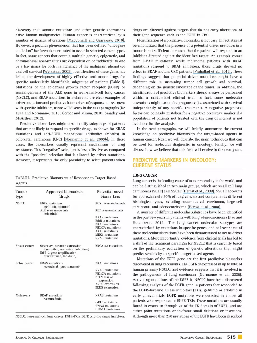
led to the development of highly effective anti-tumor drugs for
specific molecularly identifiable subgroups of patients (Table I).
Mutations of the epidermal growth factor receptor (EGFR) or
rearrangements of the ALK gene in non-small-cell lung cancer
(NSCLC), and BRAF mutations in melanoma are clear examples of
driver mutations and predictive biomarkers of response to treatment
with specific inhibitors, as we will discuss in the next paragraphs [De
Luca and Normanno, 2010; Gerber and Minna, 2010; Smalley and
McArthur, 2012].
Predictive biomarkers might also identify subgroups of patients
that are not likely to respond to specific drugs, as shown for KRAS
mutations and anti-EGFR monoclonal antibodies (MoAbs) in
colorectal carcinoma (CRC) [Normanno et al., 2009b]. In these
cases, the biomarkers usually represent mechanisms of drug
resistance. This ‘‘negative’’ selection is less effective as compared
with the ‘‘positive’’ selection that is allowed by driver mutations.
However, it represents the only possibility to select patients when
drugs are directed against targets that do not carry alterations of
their gene sequence such as the EGFR in CRC.
Identification of a predictive biomarker is not easy. In fact, it must
be emphasized that the presence of a potential driver mutation in a
tumor is not sufficient to ensure that the patient will respond to an
inhibitor directed against the identified target. An example comes
from BRAF mutations: while melanoma patients with BRAF
mutations respond to BRAF inhibitors, these drugs showed no
effect in BRAF mutant CRC patients [Prahallad et al., 2012]. These
findings suggest that potential driver mutations might have a
different role in sustaining tumor cell growth and survival,
depending on the genetic landscape of the tumor. In addition, the
identification of predictive biomarkers should always be performed
within a randomized clinical trial. In fact, some molecular
alterations might turn to be prognostic (i.e. associated with survival
independently of any specific treatment). A negative prognostic
factor can be easily mistaken for a negative predictive marker if a
population of patients not treated with the drug of interest is not
available for the analysis.
In the next paragraphs, we will briefly summarize the current
knowledge on predictive biomarkers for target-based agents in
human cancer. Next, we will describe the main techniques that can
be used for molecular diagnostic in oncology. Finally, we will
discuss how we believe that this field will evolve in the next years.
PREDICTIVE MARKERS IN ONCOLOGY:CURRENT STATUS
LUNG CANCER
Lung cancer is the leading cause of tumor mortality in the world, and
can be distinguished in two main groups, which are small cell lung
carcinomas (SCLC) and NSCLC [Herbst et al., 2008]. NSCLC accounts
for approximately 80% of lung cancers and comprehends different
histological types, including squamous cell carcinoma, large cell
carcinoma, and adenocarcinoma [Herbst et al., 2008].
A number of different molecular subgroups have been identified
in the past few years in patients with lung adenocarcinoma [Pao and
Hutchinson, 2012]. The lung cancer molecular subtypes are
characterized by mutations in specific genes, and at least some of
these molecular alterations have been demonstrated to act as driver
mutations. More importantly, evidence from clinical trials has led to
a shift of the treatment paradigm for NSCLC that is currently based
on the preliminary evaluation of genetic alterations that might
predict sensitivity to specific target-based agents.
Mutations of the EGFR gene are the first predictive biomarker
discovered in lung carcinoma. The EGFR is expressed in up to 80% of
human primary NSCLC, and evidence suggests that it is involved in
the pathogenesis of lung carcinoma [Normanno et al., 2006].
Activating mutations of the EGFR in NSCLC have been discovered
following analysis of the EGFR gene in patients that responded to
the EGFR-tyrosine kinase inhibitors (TKIs) gefitinib or erlotinib in
early clinical trials. EGFR mutations were detected in almost all
patients who responded to EGFR-TKIs. These mutations are usually
found in exons 18 through 21 of the TK domain of EGFR, and are
either point mutations or in-frame small deletions or insertions.
Althoughmore than 250mutations of the EGFR have been described
TABLE I. Predictive Biomarkers of Response to Target-Based
Agents
Tumortype
Approved biomarkers(drugs)
Potential novelbiomarkers
NSCLC EGFR mutations(gefitinib, erlotinib)
ROS1 rearrangements
ALK rearrangements(crizotinib)
RET rearrangements
KRAS mutationsErbB-2 mutationsBRAF mutationsPIK3CA mutationsAKT1 mutationsMEK1 mutationsNRAS mutations
Breast cancer Oestrogen receptor expression(tamoxifen, aromatase inhibitors)
BRCA1/2 mutations
ErbB-2 gene amplification(trastuzumab, lapatinib)
Colon cancer KRAS mutations(cetuximab, panitumumab)
BRAF mutations
NRAS mutationsPIK3CA mutationsPTEN loss of
expressionAREG expressionEREG expression
Melanoma BRAF mutations(vemurafenib)
NRAS mutations
c-KIT mutationsGNAQ mutationsGNA11 mutations
NSCLC, non-small-cell lung cancer; EGFR-TKIs, EGFR tyrosine kinase inhibitors.
JOURNAL OF CELLULAR BIOCHEMISTRY PREDICTIVE CANCER BIOMARKERS 515
up to now, two mutations, a single point mutation in exon 21, the
L858R, and a series of small in-frame deletions in exon 19, account
for approximately 90% of all EGFR mutations [De Luca and
Normanno, 2010]. EGFR mutations are not frequent in unselected
Caucasian NSCLC patients. However, a peculiar feature of these
mutations is that they are strongly associated with defined clinical
and pathological characteristics. In particular, EGFR mutations are
far more frequent in female patients as compared with male (38.7%
versus 10%); in adenocarcinoma as compared with other histologi-
cal types (29.4% versus 1.8%); in non-smokers as compared with
current smokers or former smokers (45.8% versus 7.1%); and in
East-Asian NSCLC patients (33.4%) as compared with Non-East-
Asian patients (5.5%) [Normanno et al., 2006].
Although the correlation between EGFR mutations and response
to EGFR-TKIs was evident since the early clinical trials with these
drugs, several other biomarkers have been hypothesized to be
associated with sensitivity (EGFR gene copy number variation,
EGFR protein expression, and AKT levels of activation) or resistance
(KRAS mutations) to EGFR-TKIs [De Luca and Normanno, 2010].
However, results of randomized phase III clinical trials have clearly
demonstrated that EGFR mutations are the only reliable marker that
predicts sensitivity to EGFR-TKIs [Fukuoka et al., 2011]. Following
the observation that administration of an EGFR-TKI as first line
treatment results in a prolonged progression free survival (PFS) as
compared with chemotherapy in patients carrying EGFR mutations,
treatment with an EGFR-TKI has became the recommended first line
therapy for EGFR mutant patients [Mok et al., 2009]. The EGFR-TKI
gefitinib has been approved in Europe for treatment of EGFR mutant
NSCLC in 2009. As a consequence, assessment of the mutational
status of the EGFR has become mandatory in order to choose the
most appropriate first-line treatment for NSCLC patients.
The fusion between anaplastic lymphoma kinase (ALK) and
echinoderm microtubule-associated protein-like 4 (EML4) genes is a
chromosomal translocation recently discovered in NSCLC [Gerber and
Minna, 2010; Shaw and Solomon, 2011]. ALK and EML4 are both
located in the short arm of chromosome 2. Several ALK fusions have
been characterized involving theC-terminal kinasedomainofALKand
N-terminal portions of the gene EML4; however, other genes such as
KIF5B, TGF, and ROS1 have been also described to form fusion
products with ALK [Sasaki et al., 2010]. EML4-ALK fusion oncogene
leads to the aberrant activation of ALK tyrosine kinase and to the
constitutive activation of downstream signaling pathways. This fusion
has been detected in �5% of NSCLC. Patients with NSCLC harboring
ALK rearrangements tend to be younger and have little or no smoking
history [Gerber and Minna, 2010; Sasaki et al., 2010]. ALK
rearrangements have been found exclusively in adenocarcinomas
and are associated with a signet-ring cell histology or with abundant
intracellular mucin. EML4-ALK alterations are mutually exclusive of
EGFR and KRAS mutations. Although some studies have suggested a
correlation with the male sex, this mutation has no clear association
with patient ethnicity and gender. Phase I and II trials of the ALK
inhibitor crizotinib in patients with ALK-positive advanced NSCLC
showedhigh response rates, and low incidenceof side effects [Scagliotti
et al., 2012]. Based on the phase I and II clinical trial data, the US Food
and Drug Administration granted approval of crizotinib for treatment
of NSCLC patients carrying ALK rearrangements in August 2011.
More recently, rearrangements of the ROS1 gene with different
partners have been reported to occur in approximately 1% of NSCLC
[Bergethon et al., 2012; Takeuchi et al., 2012]. ROS1 is a receptor
tyrosine kinase of the insulin receptor family, and the gene coding
for ROS1 is on chromosome 6q22. ROS1 fusion has been
predominantly identified in young and never-smokers individuals
with adenocarcinoma. Importantly, preclinical data suggest that the
proliferation of ROS1-positive tumors can be inhibited by crizotinib
and a significant response to crizotinib has been observed in an
NSCLC patient carrying a ROS1 rearrangement [Bergethon et al.,
2012].
Four different studies have described the occurrence of KIF5B-
RET fusions in approximately 1% of NSCLC adenocarcinoma [Pao
and Hutchinson, 2012]. Intriguingly, a RET TKI, vandetanib, has
been recently approved for treatment of patients with medullary
thyroid cancer.
A numberof other drivermutations have been discovered inNSCLC.
KRASmutations are found in approximately 30%of adenocarcinomas
andaremore frequent in smokerpatients [SubramanianandGovindan,
2008]. Although drugs targeting RAS proteins are not currently
available, assessment of KRAS mutations has been suggested to
identify patients that are resistant to EGFR-TKIs. Since KRAS, EGFR,
and ALK mutations are mutually exclusive, the presence of a KRAS
mutation is likely to predict resistance to EGFR-TKIs and crizotinib
since it excludes the possibility that the targets of these drugs are
present.
Subgroups of NSCLC adenocarcinoma carrying mutations of the
genes encoding for ErbB-2, BRAF, PI3K, AKT1, MEK1, and NRAS
have been also identified [Pao and Hutchinson, 2012]. However,
consistent clinical data demonstrating that the presence of these
mutations is correlated with response to specific inhibitors are not
available yet.
BREAST CANCER
Breast cancer (BC) is the most common malignancy in women, with
more than one million new cases diagnosed worldwide every year.
BC is the first solid tumor for which predictive biomarkers have been
made available. In fact, the leading parameters that define treatment
recommendations for early and advanced BC are oestrogen-receptor
(ER), progesterone-receptor (PgR), and ErbB-2 (HER2) status
[Normanno et al., 2009a]. Hormone receptor status dictates the use
of endocrine therapy for BC patients, and different types of endocrine
agents are now available for treatment of hormone receptor-positive
patients. Expression of ErbB-2 is associated with sensitivity to the
monoclonal antibody trastuzumab and to the TKI lapatinib. These
markers are evaluated by using immunohistochemistry and fluores-
cent in situ hybridization (FISH), and since they have long been
established we will not further discuss their role in BC.
The advent of gene-expression profiling techniques allowed the
classification of BC into five main subgroups according to their gene
expression pattern [Sotiriou and Pusztai, 2009]. Recently a sixth
group, named ‘‘claudin-negative’’, has been identified [Hennessy
et al., 2009]. More importantly, these techniques allowed the
identification of gene signatures that are associated with notably
different clinical outcomes in BC patients [Sotiriou and Pusztai,
516 PREDICTIVE CANCER BIOMARKERS JOURNAL OF CELLULAR BIOCHEMISTRY

2009]. However, these signatures have not been shown up to now to
predict response/resistance to specific agents in BC.
BRCA1 and BRCA2 (BC susceptibility gene 1 and 2) are tumor
suppressor genes involved in maintenance of genomic integrity
[O’Donovan and Livingston, 2010]. Germ-line mutations in these
genes identify individuals with an increased risk of developing
breast and ovarian cancer. BRCA1 is a gatekeeper of genomic
integrity with multiple roles including cell cycle checkpoint control
and DNA repair, while BRCA2 is likely to have a crucial role in
homologous recombination (HR). BRCA1-associated tumors com-
monly display a triple negative phenotype and a basal-like
morphology, while BRCA2-related BCs are a more heterogeneous
group [Carotenuto et al., 2010a].
Novel therapeutic approaches in BRCA-deficient tumors are
based on the inhibition of poly(ADP-ribose) polymerase (PARP), an
enzyme involved in the repair of single strand breaks. BRCA-related
tumors have a defective HR and are unable to repair double strand
breaks. The inhibition of alternative mechanisms of DNA repair
leads to the accumulation of multiple DNA breaks, so tumor cells
lacking homologous repair mechanisms invariably undergo
apoptosis whereas normal tissue is unaffected [Farmer et al.,
2005]. The introduction of PARP inhibitors in patients with BRCA-
related tumors represents the first treatment regimen based on a
synthetic lethal approach; this strategy has shown interesting
preliminary results [Fong et al., 2009]. There are currently at least
five PARP inhibitors in clinical trial development in BC. However,
these drugs have not been approved yet for treatment of BRCA-
deficient tumors.
Several other signal transduction inhibitors are in clinical
development in BC patients, and the number of biomarkers needed
to choose the most adequate therapy for BC patients is likely to
increase in the next future.
COLORECTAL CANCER
CRC is the third most common cancer worldwide and represents
approximately 10% of all cancer deaths. In the past decade, the
clinical development of targeted agents, such as anti-EGFR and anti-
angiogenic drugs, has significantly improved the survival of CRC
patients. To date, the most relevant data on predictive markers of
response concern the anti-EGFR agents.
Preclinical data strongly support the involvement of the EGFR
pathway in the pathogenesis and progression of CRC [Normanno
et al., 2006]. Cetuximab and panitumumab are two MoAbs that bind
and inactivate the extracellular domain of the EGFR, thus leading to
inhibition of its downstream signaling [Normanno et al., 2003].
These agents have been approved for the treatment of advanced CRC
when used as single agents (panitumumab and cetuximab) or
combined with standard chemotherapy (cetuximab) [Normanno
et al., 2009b].
Despite the EGFR protein is expressed in approximately 85% of
colorectal tumors, only a subgroup of patients with advanced CRC
benefits from treatment with anti-EGFR MoAbs [Normanno et al.,
2009b]. This observation highlighted the necessity to identify
markers of response or resistance to these drugs. In this regard, the
role of KRAS mutations in the resistance to anti-EGFR monoclonal
antibodies has been investigated in several different studies. KRAS
mutations are found in approximately 40% of CRC and are mainly
located in the codons 12 and 13 (85–90% of all mutations), although
other rare mutations have been described in exons 3 and 4 (codons
61 and 146) [Normanno et al., 2009b]. The presence of these
mutations leads a ligand-independent activation of intracellular
signaling pathways downstream the EGFR that sustain the
proliferation and survival of tumor cells.
A number of retrospective studies have clearly demonstrated that
the presence of KRAS mutations is associated with a loss of response
to cetuximab and panitumumab or, more generally to a lack of
benefit in metastatic CRC patients [Normanno et al., 2009b]. In
particular, the negative predictive role of KRAS mutations was
confirmed by subgroup analysis of patients enrolled in randomized
clinical trials in which CRC patients were treated with anti-EGFR
agents alone or in combination with chemotherapy. Based on these
results, the American and European health authorities restricted the
use of anti-EGFR MoAbs alone or in combination with chemothera-
py, only to patients with KRAS wild-type tumors. Intriguingly,
recent studies suggest that patients with the G13D KRAS mutation
might indeed respond to anti-EGFR MoAbs [De Roock et al., 2010c].
However, contrasting results have been reported up to now, and
patients with G13D mutations are currently excluded from
treatment with these agents.
Mutations in codons 61 and 146 also lead to activation of KRAS.
A large retrospective study of an European Consortium showed that
mutations in codon 61 had an adverse effect similar to codon 12
mutations, whereas codon 146 mutations did not affect cetuximab
efficacy [De Roock et al., 2010a]. However, these findings have not
been confirmed in randomized clinical trials.
A putative role in the resistance to anti-EGFR agents has been
hypothesized for other molecular alterations detected in CRC.
Retrospective subgroup analysis of patients treated with cetuximanb
or panitumumab suggest that patients carrying mutations of NRAS,
BRAF, PIK3CA or showing loss of expression of PTEN might indeed
be resistant to anti-EGFR agents [De Roock et al., 2010ab]. However,
these findings have been obtained by analyzing the outcome of
patients that were treated in clinical trials or in clinical practice with
EGFR MoAbs as single agent or in combination with irinotecan with
the aim to revert the resistance to this drug. In addition, these results
have not been confirmed yet in randomized clinical trials. By
instance, data from the CRYSTAL study of cetuximab plus FOLFIRI
versus FOLFIRI as first line therapy for metastatic CRC patients
suggest that BRAF mutant patients might benefit of cetuximab
treatment [Van Cutsem et al., 2011]. Therefore, there are no
consistent and sufficient data to preclude the use of anti-EGFR
agents in these subgroups of patients.
Some somatic mutations might provide important prognostic
information. For example, BRAF mutations are strongly associated
with a worse outcome in CRC patients [Van Cutsem et al., 2011]. The
prognostic role of KRAS mutations is more debated, since they have
been found to be associated with a worse prognosis in some studies,
but contrasting results have been reported [Normanno et al., 2009b].
Finally, recent studies have shown that high levels of expression
of the EGFR ligands amphiregulin (AREG) and epiregulin (EREG) are
associated with response to anti-EGFR MoAbs in CRC patients
[Khambata-Ford et al., 2007; Jacobs et al., 2009]. These data suggest
JOURNAL OF CELLULAR BIOCHEMISTRY PREDICTIVE CANCER BIOMARKERS 517

that in CRC the regulation of downstream EGFR signaling pathways
depends mainly on AREG and EREG binding to ErbB receptors in
KRAS wild-type patients. Consequently, higher expression of these
ligands results in increased activation of the EGFR pathway and in
sensitivity to anti-EGFR MoAbs.
MELANOMA
Metastatic melanoma is refractory to current therapies and has a
very poor prognosis, with a 5 years survival rate of 5–14% [Miller
and Mihm, 2006]. Several genetic aberrations are involved in
the pathogenesis and development of melanoma. In particular,
hyperactivation of the RAS/MEK/MAPK pathway occurs with high
frequency in melanoma (up to 90%), and it results from somatic
mutations of key genes of this pathway, including BRAF and NRAS
mutations [Miller and Mihm, 2006].
Approximately 50% of melanoma patients harbor BRAF
mutations, and the BRAF V600E mutation accounts for >90% of
BRAFmutations found in melanoma [Ribas and Flaherty, 2011]. The
presence of the V600E mutation identifies a subgroup of patients
that benefit of treatment with mutant BRAF inhibitors. Vemurafenib
(PLX4032) and dabrafenib (GSK2118436) have shown a significant
clinical response in melanoma patients carrying BRAF mutations
[Chapman et al., 2011; Hauschild et al., 2012], and vemurafenib has
been approved for treatment of melanoma patients carrying such
mutations. Actually, FDA approved vemurafenib for patients
carrying the V600E mutation, whereas EMA approved the drug
for patients with BRAF V600 mutations, since in the trial that led to
the approval of vemurafenib, patients with V600K and V600D
mutations also showed to respond to the BRAF inhibitor [Chapman
et al., 2011].
Given the important role of the MEK/ERK pathway in BRAF
mutant melanomas, the role of BRAFmutations as predictive marker
of response is also under investigation in clinical trials with MEK
inhibitors. Recently, results of a Phase III clinical study in metastatic
melanoma patients with a V600E or V600K BRAFmutation revealed
that the MEK1/2 inhibitor trametinib (GSK1120212) improved
progression-free survival (PFS) and overall survival (OS) in this
setting of patients, as compared with chemotherapy [Flaherty
et al., 2012].
NRASmutations are observed in 15–20%of cutaneousmelanomas
and cause RAS-independent activation of the MEK/ERK pathway
[Miller and Mihm, 2006]. Although therapeutic strategies to target
NRAS have not been developed yet, clinical studies are exploring
whether patients carryingNRASmutationsmight be sensitive toMEK
inhibitors.
Mutations of the type III transmembrane receptor tyrosine kinase
c-KIT have been frequently shown (20–25%) in acral and mucosal
melanomas [Smalley and McArthur, 2012]. These mutations are
expected to promote c-KIT dimerization in the absence of scatter
factor (SCF) resulting in its constitutive activation, or to prevent c-
KIT from maintaining its auto-inhibited conformation. Preliminary
observations suggest that inhibition of c-KIT in melanoma patients
harboring mutations of this receptor might result in objective
regression and disease control following treatment with imatinib, an
inhibitor of BCR-ABL kinase that also blocks the c-KIT receptor
tyrosine kinase. In particular, response rate to imatinib was better in
cases with mutations affecting recurrent hotspots or with a mutant
to wild-type allelic ratio of more than 1 (40% vs. 0%, P¼ .05),
indicating positive selection for the mutated allele [Carvajal et al.,
2011]. Additional multicenter trials are currently underway to
evaluate in melanoma patients’ agents that target c-KIT, including
imatinib and nilotinib (Phase III first-line NCT01028222 and Phase
II second-line NCT01099514). Interestingly, a significant response
to dasatinib, a TKI that inhibits c-Src, ABL and c-KIT, has also been
reported in two metastatic melanoma patients with the c-KITL756P
mutation. Clinical trials of dasatinib in melanoma are ongoing.
Recently, activating mutations in two highly related G protein-
coupled receptor alpha-subunit signaling molecules, guanine
nucleotide-binding protein Q (GNAQ) and GNA11, have been
described in approximately 70–80% of cases of uveal melanoma
[Smalley and McArthur, 2012]. A Phase II trial of the MEK inhibitor
AZD6244 as single-agent in patients with metastatic uveal mela-
noma, to correlate PFS, OS, and overall response rate (RR) with
GNAQ and GNA11 mutational status is ongoing (NCT01143402).
METHODS FOR ASSESSMENT OF BIOMARKERS
The methods used to detect predictive biomarkers depend on the
type of molecular alteration that underlies the activation of the
specific oncogenic pathway. Expression of hormone or growth
factor receptors can be assessed by using immunohistochemistry.
FISH analysis is used to evaluate gene amplification and
traslocations leading to oncogene activation. We will focus our
discussion on the methods that are used to detect point mutations
and small deletions or insertions.
Mutational analysis is performed with a variety of techniques that
can be subdivided into two main categories (Table II): (1) screening
techniques that can identify all mutations present within the
amplified DNA fragment, including new mutations and (2) targeted
methods that can specifically identify known and pre-defined
mutations.
Screening techniques are usually based on sequencing, whereas
targeted methods employ genotyping approaches. The different
methods employed for mutational analysis present advantages and
TABLE II. Current Methods for Mutational Analysis
Methods
Limit of detection(i.e., minimum % ofmutant alleles in a
wild type backgroundrequired for reliablemutation detection)
Range ofmutationsdetected
Screening methodsPCR/sequencing 20–30 ComprehensivePyrosequencing 1–10 ComprehensiveHigh-resolutionmelting (HRM)
10–20 Near comprehensive
Targeted methodsPNA-LNA clamp Up to 0.1 LimitedARMS (Therascreen) Up to 1 LimitedFragment analysis 5 LimitedReal time PCR (allelicdiscrimination)
10 Limited
PNA-LNA, peptide nucleic acid-locked nucleic acid; ARMS, allele refractorymutation system.
518 PREDICTIVE CANCER BIOMARKERS JOURNAL OF CELLULAR BIOCHEMISTRY

disadvantages, and the choice of the method depends on several
variables (Table II). Conventional direct Sanger sequencing of the
PCR product (PCR/sequencing) is the most widely used method for
mutational analysis. Pyrosequencing is a method of sequencing via
synthesis that has several advantages with respect to traditional
methods, including a greater sensitivity and the possibility of
analyzing short fragments, overcoming possible problems related to
DNA fragmentation. Sequencing-based techniques can detect a
larger number of mutations as compared with Real Time PCR-based
assays. However, these latter methods have usually an higher
sensitivity and can detect somatic mutations even when few cancer
cells are diluted in normal cells, a phenomenon that often occurs in
lung carcinoma specimens. For example, the Real Time PCR-based
Therascreen kits for EGFR and KRAS mutations employ ARMS
primers that selectively amplify mutant DNA, allowing detection of
as low as 1% mutant DNA diluted in wild-type DNA. Methods based
on peptide nucleic acid (PNA) clamp, which uses a PNA probe to
block the amplification of the wild-type strand during PCR
amplification, may reach a sensitivity of 0.1%. However, highly
sensitive methods should be cautiously used in clinical practice
since they have not been validated yet.
Several scientific societies have released recommendations on the
use of the different techniques for biomarkers’ testing [van Krieken
et al., 2008; Normanno et al., 2011]. PCR/sequencing techniques
should not be used if the specimen contains a percentage of tumor
cells <50%. In this regard, we have demonstrated that in CRC
samples with a tumor content <30%, PCR/sequencing has a
significant rate of false negative results [Carotenuto et al., 2010b].
The outcome of a molecular analysis is also significantly affected by
the quality and the quantity of the DNA, in particular when the
source of material is represented by formalin-fixed paraffin
embedded (FFPE) tissue.
CHALLENGES FOR BIOMARKERS’ ASSESSMENT INONCOLOGY
MOVING FROM A SINGLE BIOMARKER TO A COMPREHENSIVE
MOLECULAR CHARACTERIZATION
The field of predictive biomarkers is rapidly expanding. The
majority of clinical trials with target-based agents include
biomarker analysis. Pharmaceutical companies have indeed realized
the necessity to identify the populations of patients that might
benefit of treatment with specific drugs, and the experimentation of
new drugs is frequently associated with the development of
companion diagnostics. In this scenario, we expect that dramatic
changes will occur in the coming years in the field of biomarkers as
we discuss in the next paragraphs.
Although a restricted number of predictive biomarkers for solid
tumors is currently assessed in clinical practice, the need to identify
different biomarkers in the same patients’ population is rapidly
increasing (Table I). As above summarized, different subgroups of
NSCLC patients with defined molecular alterations have been
identified. Target-based agents are already available for some of
these groups (EGFR-TKIs for EGFR mutant patients, crizotinib for
patients with ALK rearrangement). Other drugs, such as BRAF or RET
inhibitors, have been already approved for treatment of other
diseases, or are in advanced phase of clinical development. In
melanoma, NRAS and c-KIT mutations may identify patients that
are sensitive to specific inhibitors, and these mutations are usually
mutually exclusive with BRAF mutations. The only predictive
biomarker of resistance to anti-EGFR agents in CRC patients is
represented by KRAS mutations. However, a number of other
mutations might offer the possibility of therapeutic intervention
with targeted agents.
Assessment of different molecular alterations might also provide
important prognostic information that might turn useful in the
clinical management of patients. For example, BRAF mutations
predict a worse prognosis in patients with CRC [Van Cutsem et al.,
2011]. In lung cancer, an association between worse prognosis and
BRAF V600E mutations has been also demonstrated [Marchetti
et al., 2011]. KRAS mutations have been hypothesized to represent
negative prognostic factors for colon and lung carcinoma patients
[Normanno et al., 2009b; De Luca and Normanno, 2010]. Data from
clinical trials also suggest that NSCLC patients with EGFR mutations
and metastatic disease have a better prognosis as compared with
patients with wild-type EGFR [De Luca and Normanno, 2010].
Finally, almost all patients treated with target-based agents
eventually become resistant to these treatment. In this regards,
evidence suggests that the mutational profile of the tumors
significantly changes during treatment with these drugs, since
clones of tumor cells carrying molecular alterations that produce
drug-resistance expand and are responsible of the recurrence of the
disease. In particular, several different mechanisms of resistance to
EGFR-TKI and crizotinib in lung carcinoma; to anti-EGFR MoAb in
colon carcinoma; and more recently to BRAF inhibitors in
melanoma have been described [De Luca and Normanno, 2010;
De Roock et al., 2010b; Ribas and Flaherty, 2011; Sequist et al.,
2011; Shaw and Solomon, 2011; Katayama et al., 2012]. Resistance
mechanisms are usually represented by mutations that reduce the
ability of the drug to bind to and inhibit the function of the target, or
by activation of alternative signaling pathways that are able to
sustain the proliferation and the survival of tumor cells. Assessment
of these resistance mutations is becoming extremely important for
the patients, since a numbers of signaling inhibitors that are poten-
tially able to block such mechanisms are in advanced clinical
development. Therefore, identification of resistance mechanismsmight
offer the possibility of different lines of treatment with targeted
agents with significant effects on both quality of life and survival.
Taken together, these findings clearly indicate that in several
different carcinoma types in the next future it will be necessary to
assess the status of a number of different biomarkers.
NEW TECHNOLOGIES FOR BIOMARKERS’ ANALYSIS
Assessment of different biomarkers in each individual patient is
difficult to accomplish with the currently available techniques. In
fact, the cost of a comprehensive molecular characterization with
the current methods would be too high to make possible to use this
approach in clinical practice. Since most of the driver mutations are
mutually exclusive, it is possible to reduce the cost of the screening
by performing consecutive analysis for the different biomarkers,
starting with the most frequent. For example, an NSCLC patient
might be screened first for EGFR mutations, then for ALK and so on.
JOURNAL OF CELLULAR BIOCHEMISTRY PREDICTIVE CANCER BIOMARKERS 519

However, each method requires at least 2–3 days to obtain the result
of the analysis. Therefore, if these analyses are sequentially
performed, several weeks might be necessary to analyze the
different biomarkers, and this time is not acceptable for patients
with advanced disease. An additional limiting factor is represented
by the availability of tissue for biomarker analysis. For a fraction of
patients with advanced disease small biopsies are only available, and
this frequently occurs for NSCLC patients. Analysis of different
categories of tumor genomic alterations (translocations, copy
number alterations, and point mutations) currently requires
techniques for which different samples are necessary in order to
allow analysis of protein expression (IHC), gene copy number (FISH),
mutations (DNA mutational analysis) or even gene expression.
Assessment of a wide range of biomarkers might result feasible only
in those patients that have a sufficient amount of available tissue.
Therefore, the possibility to introduce a comprehensive molecular
characterization in clinical practice relies on the development of
high throughput technologies that allow to detect the different
molecular alterations in a cost-effective and timely manner. In this
respect, two different approaches are being developed (Table III):
high throughput genotyping platforms, which allow the detection of
recognized genetic aberrations in clinical samples, and next
generation sequencing (NGS) that can provide information on all
the different types of cancer-causing alterations.
High-throughput genotyping platforms are usually based on
multiplexed assays and microarrays, as detailed in Table III. These
platforms can analyze hundreds to millions of selected germline
and/or somatic variants simultaneously. In addition, array-based
comparative genomic hybridization (aCGH) can detect gene copy
numbers with high resolution and high throughput, thus providing
information on gene copy number changes including deletions,
gains, and amplifications. The combined use of these platforms
might allow to detect all the most frequent molecular alterations that
have the potential to be predictive biomarkers. Indeed, these
platforms have been successfully used for genotyping clinical
samples and are currently the most used technologies for screening
of molecular alterations in patients with cancer.
The limit of genotyping platforms is that they can only provide
information on already known genetic alterations. Within the last
years, there has been an increasing interest in mapping human
genome to determine mutations and genetic events associated with
clonal evolution of cancer. A deeper understanding of these
alterations will lead to a further comprehension of tumor evolution
and to the identification of new potential targets for therapeutic
intervention as well. In this respect, NGS approach presents several
potential advantages over traditional methods, including the
opportunity of fully sequencing a large number of genes in a
single test and detecting at the same time deletions, insertions,
exome-wide single nucleotide polymorphisms (SNPs), transloca-
tions in several cancer-related genes [Metzker, 2009; Tran et al.,
2012]. NGS platforms, also known as second- and third-generation
sequencers, can generate millions of reads, each of limited length, by
TABLE III. High Throughput Platforms
Platform Method Application/notes
GenotypingTaqman OpenArray Genotyping System(Life Technologies, Carlsbad, CA)
Uses fluorescence-based PCR reagents to providequalitative detection of targets by post-PCR(endpoint) analysis.
Somatic mutations; SNPs.
MassARRAY (Sequenom, San Diego,CA)
Combines allele-specific PCR with MALDI-TOFmass spectrometry.
Somatic mutations; SNPs; methylation; gene expression.
SNaPshot Multiplex Kit (LifeTechnologies)
Consists of a multiplexed PCR step with labelednucleotides followed by single-base extensionreaction combined with capillary electrophoresis.
Somatic mutations; SNPs; methylation; gene expression.
Infinium (Illumina, San Diego, CA) 50-mer probes hybridize to loci of interest; enzymaticsingle-base extension incorporates a labelednucleotide; detection by iScan imaging system.
Somatic mutations; SNPs; methylation; gene expression.
aCGH (Agilent, Santa Clara, CA) Uses 60-mer oligonucleotide microarrays foraCGH analysis.
Gene copy number variation and rearrangements.
Next-generation sequencing (NGS)HeliScope (Helicos Biosciences,Cambridge, MA, USA)
No clonal amplification. Single molecules of DNAor RNA are sequenced by synthesis. Optical detection.
Lower biased sequence reads; high error rates comparedwith other NGS technologies.
454 (Roche, Basel, Switzerland) Clonally amplified beads generated by emulsion PCRserve as sequencing features. Sequencing isperformed by pyrosequencing methods. CCD-basedsignal detection.
Longer read-length than other platforms; high error ratesin homo-polymer repeats. Fast run times.
HiSeq (Illumina) Libraries are prepared by bridge PCR and sequenced bycyclic reversible termination. Optical detection.
Low multiplexing capability.
Discrete percentage of aberrant nucleotide incorporation.SOLiD (Life Technologies) Libraries are amplified by emulsion PCR and sequencing
by synthesis is driven by a DNA ligase. Opticaldetection.
The use of the two-bases encoding system enables a moreaccurate alignment of short reads and a considerableerror-rate reduction; run times are long and dataanalysis complex.
PacBio RS (Pacific Biosciences, MenloPark, CA)
Single-molecule real-time sequencing: detection of thetemporal order of enzymatic incorporation offluorescently labeled nucleotide into a growing DNAstrand. Optical detection.
Long read length; higher error rates than other NGSapparatus.
Ion Torrent Personal Genome Machine(Life Technologies)
Non-optical detection. Addition of a nucleotide isdetected as change in voltage due to release ofhydrogen ions.
Short read length. Short run time.
PCR, polymerase chain reaction; MALDI-TOF, matrix-assisted laser desorption/ionization-time-of-flight; aCGH, array-based comparative genomic hybridization; CCD,charge-coupled device; SOLiD, support oligonucleotide ligation and detection.
520 PREDICTIVE CANCER BIOMARKERS JOURNAL OF CELLULAR BIOCHEMISTRY

immobilizing amplified DNA fragments onto solid surfaces and
performing the sequencing reaction. For this reason, they result
more economical than Sanger sequencing and have higher
throughput. Since the launch of first NGS platforms, different
improvements have been made, such as improved sequencing
chemistry and new signal detection methodologies, although there
are still several challenges to face. Disadvantages still include short
read length, long run time, complex sample preparation and
amplification, and sophisticated data analysis. In fact, the enormous
amount of data produced in each experiment makes it essential the
development of appropriate bioinformatics approaches to analyze
the obtained data.
The characteristics of the main NGS platforms are summarized in
Table III. The HiSeq (Illumina, San Diego, CA) is the most widely
used NGS platform in the field, but its technology still exhibits
different issues such as low multiplexing capability and a discrete
percentage of aberrant nucleotide incorporation, due to polymerase
errors. The 454 system (Roche, Basel, Switzerland) is based on
emulsion PCR and pyrosequencing, has fast run times and provides
longer read lengths than other NGS technologies. It is also highly
sensitive compared with traditional sequencing but presents
contamination risks due to emulsion PCR steps, a poor performance
with homopolymer repeated regions and a relatively low through-
put. The HeliScope platform (Helicos Biosciences, Cambridge, MA,
USA) provides sequencing by synthesis of single molecules of DNA
or RNA without a preliminary PCR. The Helicos has been shown to
provide the lower biased sequence reads, although it displays a
relatively high error rate compared with other NGS technologies.
Supported Oligonucletide Ligation and Detection (SOLiD) (Life
Technologies, Carlsbad, CA) sequencing is carried out through
different round of ligase-mediate oligonucleotide ligation after an
emulsion PCR step. The sequence is determinated through a two-
base encoding system with color space that enables a more accurate
alignment of short reads and a considerable error-rate reduction.
Despite this, the SOLiD system presents relatively long run times and
complex analysis are required. The PacBio RS (Pacific Biosciences,
Menlo Park, CA) uses a process called single-molecule, real-time
detection of biologic processes that does not require DNA
amplification and results in longer read lengths. Finally, the Ion
Torrent, also called Personal Genome Machine (PGM) (Life
Technologies), detects nucleotide incorporation through an ion
sensor pH changes resulting from the release of hydrogen ions
during the nucleotide addition. Although its accuracy is good and
run time is very short, the read length is currently quite short.
The majority of these platforms are capable of performing whole
genome sequencing (WGS) in few days with costs that are
significantly cheaper as compared with Sanger sequencing.
However, the complexity and the cost of WGS is still too high to
make it feasible in clinical routine diagnostics. Information related
to few dozens of genes are sufficient to drive the choice of the most
appropriate treatment, at least according to our current knowledge.
Therefore, targeted sequencing, a strategy that enriches the input for
DNA regions of interest, is likely to represent the main approach
through which NGS technology will be applied to cancer diagnostics
in the next future. In this latter approach, the target regions are
enriched through PCR amplification or hybridization to oligonu-
cleotide arrays that are specially designed. Unfortunately, there are
few preliminary data available on the use of NGS apparatus in
molecular diagnostics, although this field is rapidly expanding.
CONCLUSIONS AND FUTURE DIRECTIONS
The identification of driver molecular alterations promoting tumor
growth and the development of drugs capable to block such
mechanisms will lead in the next few years to a significant
improvement of personalized medicine in oncology. However, the
progress in this field is limited by the availability of methods to
detect such target molecular alterations in the tumor tissue. In this
respect, determination of DNA sequence by using the Sanger method
has been the only sequencing method used for almost 30 years.
However, Sanger methods are limited to single-gene or hot spot
mutation analysis, due to the limited sequencing capacity and the
difficulties and high costs of multiplexing protocols.
Different methods that can detect somatic mutations with high
sensitivity and specificity have been more recently developed.
However, cancer somatic variations are not limited to single-
nucleotide mutations, but often consist of large deletions, insertions,
copy number variations, and rearrangements. In addition, increas-
ing evidence suggests that SNPs of different genes might affect the
activity of anti-cancer drugs. In this respect, NGS devices have made
the detection of these variations feasible and have surpassed
traditional Sanger sequencing in sensitivity, efficacy and time [Tran
et al., 2012]. With respect to genotyping approaches, NGS can detect
novel sequence variations that cannot be obtained with genotyping.
Importantly, NGS applications can also provide information on gene
expression and epigenetic regulation of gene expression such as
DNA methylation. Therefore, it is conceivable that the introduction
of NGS techniques in molecular diagnostics will allow a significant
progress in this field.
There are several aspects of molecular diagnostics that will be
possible to improve in the next future, thank to this technology
advancement. Evidence suggests that some mutations are specifi-
cally selected following treatment with anti-cancer agents. For
example, the EGFR T790M mutation that produces resistance to
EGFR-TKIs in NSCLC can be detected by using highly sensitive
techniques in approximately 30% of patients carrying activating
mutations of the EGFR [Maheswaran et al., 2008; Su et al., 2012].
Tumor cells carrying the T790M mutations are selected during the
treatment with EGFR-TKIs and are responsible of the recurrence of
the disease. Identification of this molecular alteration before
treatment with EGFR-TKIs might provide information on the
duration of the response. In addition, since agents capable to inhibit
the T790M mutant EGFR are in advanced clinical development, this
information might also allow to treat the patient with additional
target-based agents. Similar findings have been recently reported in
CRC, where minor clones of KRAS mutant cells seem to be involved
in the acquired resistance to anti-EGFR agents [Diaz et al., 2012].
Therefore, it is likely that analysis of genetic variants represented in
minor clones of tumor cells might allow to identify before the start
of the treatment mechanisms that might lead to drug resistance.
Since this phenomenon is mediated by several different molecular
JOURNAL OF CELLULAR BIOCHEMISTRY PREDICTIVE CANCER BIOMARKERS 521

alterations, diagnostic methods that allow to assess the mutational
statusof differentgenes ina singleanalysiswill benecessary to identify
such genetic variants. In addition, thesemethods need to have an high
sensitivity since the resistance-associated mutations are usually
represented in a small fraction of tumor cells and are not detectable
by using routine molecular diagnostic approaches. NGS technologies
have the features to overcome these methodological problems.
Another important goal of molecular diagnostic in the future will
be to move from qualitative to quantitative assessments. Many
tumors show an heterogeneous mutational pattern, and driver
mutations are not present in all tumor cells. Since qualitative
methods have been used in clinical trials to assess predictive
biomarkers, we do not know which is the lowest level of mutations
that is associated with sensitivity or resistance to specific drugs.
Quantitative assessment is hampered by the fact that tumor mass
also contain non-neoplastic cells. Nevertheless, quantitative
analysis might provide information on the effect of different levels
of mutant DNA on the activity of target-based agents. In this regard,
NGS systems are single-molecule counting instruments enabled to
measure the frequency of mutations in any tissue.
Mutational analysis might not be feasible for patients in
advanced stage of disease for which tissue is not available and a
new biopsy is not possible, as frequently occurs in lung carcinoma.
In addition, as above discussed, the mutational profile of the disease
might change during treatment, due to the selection of resistant
clones. Since specific inhibitors of intracellular signaling pathways
are in advanced phase of clinical development, identification of such
resistance mechanisms might allow to assign a personalized
treatment to each individual patient with cancer. However, in the
majority of the patients, it is not possible to perform repeated
biopsies to follow the mutational evolution of the disease. Recently,
it has been suggested that somatic mutations can be detected in
circulating tumor cells (CTC) or in the circulating free tumor DNA
(cftDNA) of patients carrying solid tumors [Maheswaran et al.,
2008]. Interestingly, it has been demonstrated that detection of
KRAS mutant DNA in the serum of KRAS wild-type CRC patients
treated with anti-EGFR MoAbs correlates with the development of
resistance to such agents [Diaz et al., 2012; Misale et al., 2012]. In
this respect, NGS-based techniques might be able to detect a wide
array of mutations in CTCs or serum from patients with advanced
solid tumors.
In conclusion, the significant advance in technologies to detect a
wide array of genetic alterations with an extremely high sensitivity
and specificity is allowing a significant improvement in molecular
diagnostic. By using these technologies, the application of a
personalized medicine approach to patients with cancer is finally
becoming possible (Fig. 1).
REFERENCES
Bergethon K, Shaw AT, Ou SH, Katayama R, Lovly CM, McDonald NT,Massion PP, Siwak-Tapp C, Gonzalez A, Fang R, Mark EJ, Batten JM,Chen H, Wilner KD, Kwak EL, Clark JW, Carbone DP, Ji H, Engelman JA,Mino-Kenudson M, Pao W, Iafrate AJ. 2012. ROS1 rearrangements define aunique molecular class of lung cancers. J Clin Oncol 30:863–870.
Fig. 1. Tumor DNA can be harvested from tumor specimens obtained by biopsy or surgery. Additional sources of tumor DNA are circulating tumor cells (CTC) (green arrows) or
circulating free tumor DNA (cftDNA) (blue arrows) that can be isolated from blood samples. A complete molecular profiling of tumor DNA can be performed with high
throughput genotyping and/or next generation sequencing platforms. The identification of predictive biomarkers will allow an individualized treatment with specific target-
based (kinase inhibitors and MoAbs, monoclonal antibodies) and conventional agents.
522 PREDICTIVE CANCER BIOMARKERS JOURNAL OF CELLULAR BIOCHEMISTRY

Carotenuto P, Roma C, Rachiglio AM, Botti G, D’Alessio A, Normanno N.2010a. Triple negative breast cancer: from molecular portrait to therapeuticintervention. Crit Rev Eukaryot Gene Expr 20:17–34.
Carotenuto P, Roma C, Rachiglio AM, Tatangelo F, Pinto C, Ciardiello F,Nappi O, Iaffaioli RV, Botti G, Normanno N. 2010b. Detection of KRASmutations in colorectal carcinoma patients with an integrated PCR/sequenc-ing and real-time PCR approach. Pharmacogenomics 11:1169–1179.
Carvajal RD, Antonescu CR, Wolchok JD, Chapman PB, Roman RA, TeitcherJ, Panageas KS, Busam KJ, Chmielowski B, Lutzky J, Pavlick AC, Fusco A,Cane L, Takebe N, Vemula S, Bouvier N, Bastian BC, Schwartz GK. 2011. KITas a therapeutic target in metastatic melanoma. JAMA 305:2327–2334.
Chapman PB, Hauschild A, Robert C, Haanen JB, Ascierto P, Larkin J,Dummer R, Garbe C, Testori A, Maio M, Hogg D, Lorigan P, Lebbe C, JouaryT, Schadendorf D, Ribas A, O’Day SJ, Sosman JA, Kirkwood JM, EggermontAMM, Dreno B, Nolop K, Li J, Nelson B, Hou J, Lee RJ, Flaherty KT, McArthurGA. 2011. Improved survival with vemurafenib in melanoma with BRAFV600E mutation. N Eng J Med 364:2507–2516.
De Luca A, Normanno N. 2010. Predictive biomarkers to tyrosine kinaseinhibitors for the epidermal growth factor receptor in non-small-cell lungcancer. Curr Drug Targets 11:851–864.
De Roock W, Claes B, Bernasconi D, De Schutter J, Biesmans B, Fountzilas G,Kalogeras KT, Kotoula V, Papamichael D, Laurent-Puig P, Penault-Llorca F,Rougier P, Vincenzi B, Santini D, Tonini G, Cappuzzo F, Frattini M, MolinariF, Saletti P, De Dosso S, Martini M, Bardelli A, Siena S, Sartore-Bianchi A,Tabernero J, Macarulla T, Di Fiore F, Gangloff AO, Ciardiello F, Pfeiffer P,Qvortrup C, Hansen TP, Van Cutsem E, Piessevaux H, Lambrechts D,Delorenzi M, Tejpar S. 2010a. Effects of KRAS, BRAF, NRAS, and PIK3CAmutations on the efficacy of cetuximab plus chemotherapy in chemothera-py-refractory metastatic colorectal cancer: a retrospective consortium anal-ysis. Lancet Oncol 11:753–762.
De RoockW, De Vriendt V, Normanno N, Ciardiello F, Tejpar S. 2010b. KRAS,BRAF, PIK3CA, and PTEN mutations: implications for targeted therapies inmetastatic colorectal cancer. Lancet Oncol 12:594–603.
De Roock W, Jonker DJ, Di Nicolantonio F, Sartore-Bianchi A, Tu D, Siena S,Lamba S, Arena S, Frattini M, Piessevaux H, Van Cutsem E, O’Callaghan CJ,Khambata-Ford S, Zalcberg JR, Simes J, Karapetis CS, Bardelli A, Tejpar S.2010c. Association of KRAS p.G13D mutation with outcome in patients withchemotherapy-refractory metastatic colorectal cancer treated with cetux-imab. JAMA 304:1812–1820.
Diaz LA Jr, Williams RT, Wu J, Kinde I, Hecht JR, Berlin J, Allen B, Bozic I,Reiter JG, Nowak MA, Kinzler KW, Oliner KS, Vogelstein B. 2012. Themolecular evolution of acquired resistance to targeted EGFR blockade incolorectal cancers. Nature 486:537–540.
Farmer H, McCabe N, Lord CJ, Tutt AN, Johnson DA, Richardson TB,Santarosa M, Dillon KJ, Hickson I, Knights C, Martin NM, Jackson SP, SmithGC, Ashworth A. 2005. Targeting the DNA repair defect in BRCAmutant cellsas a therapeutic strategy. Nature 434:917–921.
Flaherty KT, Robert C, Hersey P, Nathan P, Garbe C, Milhem M, Demidov LV,Hassel JC, Rutkowski P, Mohr P, Dummer R, Trefzer U, Larkin JM, Utikal J,Dreno B, Nyakas M, Middleton MR, Becker JC, Casey M, Sherman LJ, Wu FS,Ouellet D, Martin AM, Patel K, Schadendorf D. 2012. Improved survival withMEK inhibition in BRAF-mutated melanoma. N Engl J Med 367:107–114.
Fong PC, Boss DS, Yap TA, Tutt A, Wu P, Mergui-Roelvink M, Mortimer P,Swaisland H, Lau A, O’Connor MJ, Ashworth A, Carmichael J, Kaye SB,Schellens JH, de Bono JS. 2009. Inhibition of poly(ADP-ribose) polymerase intumors from BRCA mutation carriers. N Engl J Med 361:123–134.
Fukuoka M, Wu YL, Thongprasert S, Sunpaweravong P, Leong SS, Sriur-anpong V, Chao TY, Nakagawa K, Chu DT, Saijo N, Duffield EL, RukazenkovY, Speake G, Jiang H, Armour AA, To KF, Yang JC, Mok TS. 2011. Biomarkeranalyses and final overall survival results from a phase III, randomized,open-label, first-line study of gefitinib versus carboplatin/paclitaxel inclinically selected patients with advanced non-small-cell lung cancer inAsia (IPASS). J Clin Oncol 29:2866–2874.
Gerber DE, Minna JD. 2010. ALK inhibition for non-small cell lung cancer:from discovery to therapy in record time. Cancer Cell 18:548–551.
Hauschild A, Grob J-J, Demidov LV, Jouary T, Gutzmer R, Millward M,Rutkowski P, Blank CU, Miller WH Jr, Kaempgen E, MartAn-Algarra S,Karaszewska B, Mauch C, Chiarion-Sileni V, Martin A-M, Swann S, Haney P,Mirakhur B, Guckert ME, Goodman V, Chapman PB. 2012. Dabrafenib inBRAF-mutated metastatic melanoma: a multicentre, open-label, phase 3randomised controlled trial. Lancet 380:358–365.
Hennessy BT, Gonzalez-Angulo AM, Stemke-Hale K, Gilcrease MZ, Krish-namurthy S, Lee JS, Fridlyand J, Sahin A, Agarwal R, Joy C, Liu W, Stivers D,Baggerly K, Carey M, Lluch A, Monteagudo C, He X, Weigman V, Fan C,Palazzo J, Hortobagyi GN, Nolden LK, Wang NJ, Valero V, Gray JW, PerouCM, Mills GB. 2009. Characterization of a naturally occurring breast cancersubset enriched in epithelial-to-mesenchymal transition and stem cell char-acteristics. Cancer Res 69:4116–4124.
Herbst RS, Heymach JV, Lippman SM. 2008. Lung cancer. N Engl J Med 359:1367–1380.
Jacobs B, De RoockW, Piessevaux H, Van Oirbeek R, Biesmans B, De SchutterJ, Fieuws S, Vandesompele J, Peeters M, Van Laethem JL, Humblet Y,Penault-Llorca F, De Hertogh G, Laurent-Puig P, Van Cutsem E, Tejpar S.2009. Amphiregulin and epiregulin mRNA expression in primary tumorspredicts outcome in metastatic colorectal cancer treated with cetuximab.J Clin Oncol 27:5068–5074.
Katayama R, Shaw AT, Khan TM, Mino-Kenudson M, Solomon BJ, Halmos B,Jessop NA, Wain JC, Yeo AT, Benes C, Drew L, Saeh JC, Crosby K, Sequist LV,Iafrate AJ, Engelman JA. 2012. Mechanisms of acquired crizotinib resistancein ALK-rearranged lung cancers. Sci Transl Med 4:120ra17.
Khambata-Ford S, Garrett CR, Meropol NJ, Basik M, Harbison CT, Wu S,Wong TW, Huang X, Takimoto CH, Godwin AK, Tan BR, Krishnamurthi SS,Burris HA III, Poplin EA, Hidalgo M, Baselga J, Clark EA, Mauro DJ. 2007.Expression of epiregulin and amphiregulin and K-ras mutation status predictdisease control in metastatic colorectal cancer patients treated with cetux-imab. J Clin Oncol 25:3230–3237.
MacConaill LE, Garraway LA. 2010. Clinical implications of the cancergenome. J Clin Oncol 28:5219–5228.
Maheswaran S, Sequist LV, Nagrath S, Ulkus L, Brannigan B, Collura CV,Inserra E, Diederichs S, Iafrate AJ, Bell DW, Digumarthy S, Muzikansky A,Irimia D, Settleman J, Tompkins RG, Lynch TJ, Toner M, Haber DA. 2008.Detection of mutations in EGFR in circulating lung-cancer cells. N Engl JMed 359:366–377.
Marchetti A, Felicioni L, Malatesta S, Grazia Sciarrotta M, Guetti L, Chella A,Viola P, Pullara C, Mucilli F, Buttitta F. 2011. Clinical features and outcomeof patients with non-small-cell lung cancer harboring BRAF mutations.J Clin Oncol 29:3574–3579.
Metzker ML. 2009. Sequencing technologies—the next generation. Nat RevGenet 11:31–46.
Miller AJ, Mihm MC Jr. 2006. Melanoma. N Engl J Med 355:51–65.
Misale S, Yaeger R, Hobor S, Scala E, Janakiraman M, Liska D, Valtorta E,Schiavo R, Buscarino M, Siravegna G, Bencardino K, Cercek A, Chen CT,Veronese S, Zanon C, Sartore-Bianchi A, Gambacorta M, Gallicchio M,Vakiani E, Boscaro V, Medico E, Weiser M, Siena S, Di Nicolantonio F,Solit D, Bardelli A. 2012. Emergence of KRAS mutations and acquiredresistance to anti-EGFR therapy in colorectal cancer. Nature 486:532–536.
Mok TS, Wu YL, Thongprasert S, Yang CH, Chu DT, Saijo N, SunpaweravongP, Han B, Margono B, Ichinose Y, Nishiwaki Y, Ohe Y, Yang JJ, Chewasku-lyong B, Jiang H, Duffield EL, Watkins CL, Armour AA, Fukuoka M. 2009.Gefitinib or carboplatin-paclitaxel in pulmonary adenocarcinoma. N Engl JMed 361:947–957.
Normanno N, Bianco C, De Luca A, Maiello MR, Salomon DS. 2003. Target-based agents against ErbB receptors and their ligands: a novel approach tocancer treatment. Endocr Relat Cancer 10:1–21.
JOURNAL OF CELLULAR BIOCHEMISTRY PREDICTIVE CANCER BIOMARKERS 523

Normanno N, De Luca A, Bianco C, Strizzi L, Mancino M, Maiello MR,Carotenuto A, De Feo G, Caponigro F, Salomon DS. 2006. Epidermal growthfactor receptor (EGFR) signaling in cancer. Gene 366:2–16.
Normanno N, Morabito A, De Luca A, Piccirillo MC, Gallo M, Maiello MR,Perrone F. 2009a. Target-based therapies in breast cancer: current status andfuture perspectives. Endocr Relat Cancer 16:675–702.
Normanno N, Tejpar S, Morgillo F, De Luca A, Van Cutsem E, Ciardiello F.2009b. Implications for KRAS status and EGFR-targeted therapies in meta-static CRC. Nat Rev Clin Oncol 6:519–527.
Normanno N, Pinto C, Castiglione F, Bardelli A, Gambacorta M, Botti G,Nappi O, Siena S, Ciardiello F, Taddei G, Marchetti A. 2011. KRAS mutationstesting in colorectal carcinoma patients in Italy: from guidelines to externalquality assessment. PLoS ONE 6:e29146.
O’Donovan PJ, Livingston DM. 2010. BRCA1 and BRCA2: breast/ovariancancer susceptibility gene products and participants in DNA double-strandbreak repair. Carcinogenesis 31:961–967.
Pao W, Hutchinson KE. 2012. Chipping away at the lung cancer genome. NatMed 18:349–351.
Prahallad A, Sun C, Huang S, Di Nicolantonio F, Salazar R, Zecchin D,Beijersbergen RL, Bardelli A, Bernards R. 2012. Unresponsiveness of coloncancer to BRAF(V600E) inhibition through feedback activation of EGFR.Nature 483:100–103.
Ribas A, Flaherty KT. 2011. BRAF targeted therapy changes the treatmentparadigm in melanoma. Nat Rev Clin Oncol 8:426–433.
Sasaki T, Rodig SJ, Chirieac LR, Janne PA. 2010. The biology and treatment ofEML4-ALK non-small cell lung cancer. Eur J Cancer 46:1773–1780.
Scagliotti G, Stahel RA, Rosell R, Thatcher N, Soria JC. 2012. ALK translo-cation and crizotinib in non-small cell lung cancer: an evolving paradigm inoncology drug development. Eur J Cancer 48:961–973.
Sequist LV, Waltman BA, Dias-Santagata D, Digumarthy S, Turke AB, FidiasP, Bergethon K, Shaw AT, Gettinger S, Cosper AK, Akhavanfard S, Heist RS,Temel J, Christensen JG, Wain JC, Lynch TJ, Vernovsky K, Mark EJ, Lanuti M,Iafrate AJ, Mino-Kenudson M, Engelman JA. 2011. Genotypic and histolog-ical evolution of lung cancers acquiring resistance to EGFR inhibitors. SciTransl Med 3:75ra26.
Shaw AT, Solomon B. 2011. Targeting anaplastic lymphoma kinase in lungcancer. Clin Cancer Res 17:2081–2086.
Smalley KS, McArthur GA. 2012. The current state of targeted therapy inmelanoma: this time it’s personal. Semin Oncol 39:204–214.
Sotiriou C, Pusztai L. 2009. Gene-expression signatures in breast cancer.N Engl J Med 360:790–800.
Su KY, Chen HY, Li KC, Kuo ML, Yang JC, Chan WK, Ho BC, Chang GC, ShihJY, Yu SL, Yang PC. 2012. Pretreatment epidermal growth factor receptor(EGFR) T790M mutation predicts shorter EGFR tyrosine kinase inhibitorresponse duration in patients with non-small-cell lung cancer. J Clin Oncol30:433–440.
Subramanian J, Govindan R. 2008. Molecular genetics of lung cancer inpeople who have never smoked. Lancet Oncol 9:676–682.
Takeuchi K, Soda M, Togashi Y, Suzuki R, Sakata S, Hatano S, Asaka R,Hamanaka W, Ninomiya H, Uehara H, Lim Choi Y, Satoh Y, Okumura S,Nakagawa K, Mano H, Ishikawa Y. 2012. RET, ROS1 and ALK fusions in lungcancer. Nat Med 18:378–381.
Tran B, Dancey JE, Kamel-Reid S, McPherson JD, Bedard PL, Brown AM,Zhang T, Shaw P, Onetto N, Stein L, Hudson TJ, Neel BG, Siu LL. 2012.Cancer genomics: technology, discovery, and translation. J Clin Oncol 30:647–660.
Van Cutsem E, Kohne CH, Lang I, Folprecht G, Nowacki MP, Cascinu S,Shchepotin I, Maurel J, Cunningham D, Tejpar S, Schlichting M, Zubel A,Celik I, Rougier P, Ciardiello F. 2011. Cetuximab plus irinotecan, fluorouracil,and leucovorin as first-line treatment for metastatic colorectal cancer:updated analysis of overall survival according to tumor KRAS and BRAFmutation status. J Clin Oncol 29:2011–2019.
van Krieken JH, Jung A, Kirchner T, Carneiro F, Seruca R, Bosman FT, QuirkeP, Flejou JF, Plato Hansen T, de Hertogh G, Jares P, Langner C, Hoefler G,Ligtenberg M, Tiniakos D, Tejpar S, Bevilacqua G, Ensari A. 2008. KRASmutation testing for predicting response to anti-EGFR therapy for colorectalcarcinoma: proposal for an European quality assurance program. VirchowsArch 453:417–431.
Weinstein IB. 2002. Cancer. Addiction to oncogenes—the Achilles heal ofcancer. Science 297:63–64.
524 PREDICTIVE CANCER BIOMARKERS JOURNAL OF CELLULAR BIOCHEMISTRY
Recommended


