
Research
Nicotinic Cholinergic Synaptic Mechanismsin the Ventral Tegmental Area Contributeto Nicotine AddictionVolodymyr I. Pidoplichko, Jun Noguchi, Oluwasanmi O. Areola, Yong Liang,Jayms Peterson, Tianxiang Zhang, and John A. Dani1
Division of Neuroscience, Baylor College of Medicine, Houston, Texas 77030, USA
Tobacco use is a major health problem that is estimated to cause 4 million deaths a year worldwide. Nicotine is themain addictive component of tobacco. It acts as an agonist to activate and desensitize nicotinic acetylcholinereceptors (nAChRs). A component of nicotine’s addictive power is attributable to actions on the mesolimbicdopaminergic system, which serves a fundamental role in the acquisition of behaviors that are inappropriatelyreinforced by addictive drugs. Here we show that nicotine, in the same concentration and time ranges as obtainedfrom tobacco, has three main actions that regulate the activity of midbrain dopamine (DA) neurons. Nicotine firstactivates and then desensitizes nAChRs on the DA neurons. This process directly excites the DA neurons for a shortperiod of time before the nAChRs desensitize. Nicotine also enhances glutamatergic excitation and decreasesGABAergic inhibition onto DA neurons. These events increase the probability for synaptic plasticity, such aslong-term potentiation. The short-lived direct excitation of the DA neurons coupled with the enhanced glutamatergicafferent activity provides the presynaptic and postsynaptic coincidence necessary to initiate synaptic potentiation. Intotal, these synaptic events lead to a relatively long-lasting heightened activity of midbrain DA neurons. Consistentwith other summarized studies, this work indicates that the synaptic changes normally associated with learning andmemory can be influenced and commandeered during the nicotine addiction process.
About one-third of the adults in the world smoke tobacco. Mostof them start as adolescents, and half of those that continuesmoking die from smoking-related diseases (WHO 1997). Becausetobacco usage is increasing in less developed countries, it is oneof the few causes of death that is on the rise (Peto et al. 1996). Indeveloped countries, smoking is estimated to be the largest singlecause of premature death (Peto et al. 1992). From 1995 to 1999 inthe United States alone, smoking annually caused ∼440,000deaths and $157 billion in health-related economic losses (CDC2002).
Nicotine is the main addictive component of tobacco (Karanet al. 2003). When studied under laboratory conditions, nicotineelicits drug-seeking behavior in animals (Corrigall and Coen1989; Stolerman and Shoaib 1991; Corrigall 1999; Di Chiara2000). It also produces additional effects commonly seen withaddictive drugs such as amphetamines and cocaine. Nicotine re-inforces self-administration, increases locomotor activity, en-hances reward from brain stimulation, and reinforces place pref-erence (Clarke 1990, 1991; Stolerman and Shoaib 1991; Stoler-man and Jarvis 1995; Dani and Heinemann 1996; Corrigall 1999;Di Chiara 2000; Dani and De Biasi 2001; Mansvelder and McGe-hee 2002). Furthermore, nicotine cessation produces a with-drawal syndrome, and those symptoms can be relieved by nico-tine replacement (Stolerman and Jarvis 1995).
Midbrain dopaminergic systems serve an important role innicotine addiction because they reinforce acquisition of behav-iors that are driven by salient environmental cues or by the in-appropriate stimuli of addictive drugs (Wonnacott et al. 1990;
Corrigall et al. 1992; Nestler 1992, 1993; Nisell et al. 1994; Pon-tieri et al. 1996; Spanagel and Weiss 1999; Balfour et al. 2000; DiChiara 2000; Dani and De Biasi 2001; Dani et al. 2001; Karan etal. 2003). The role of the midbrain DA systems in nicotine ad-diction is supported by the findings that DA antagonists or le-sions of DA neurons or of the nucleus accumbens (NAc) reduceself-administration (Corrigall and Coen 1989; Corrigall et al.1992, 1994; Corrigall 1999; Di Chiara 2000). By acting at nico-tinic acetylcholine receptors (nAChRs), nicotine can activateneurons of the ventral tegmental area (VTA) and the substantianigra compacta (SNc; Clarke et al. 1985; Grenhoff and Johnson1996; Calabresi et al. 1989; Pidoplichko et al. 1997; Picciotto etal. 1998; Dani et al. 2001; Mansvelder and McGehee 2002) andcause release of DA in the NAc of rats (Clarke 1991; Nisell et al.1994, 1995; Pontieri et al. 1996). Furthermore, presynapticallylocated nAChRs can potently regulate DA release in the striatum,including the NAc (Wonnacott et al. 2000; Jones et al. 2001;Zhou et al. 2001).
Neuronal nAChRs are formed from combinations of fivesubunits arising from �2–�10 and �2–�4. Therefore, many com-positionally and functionally different nAChR subtypes are pos-sible (McGehee and Role 1995; Role and Berg 1996; Wonnacott1997; Jones et al. 1999; Wooltorton et al. 2003). Five � subunits(�2–�6) and three � subunits (�2–�4) assemble in various com-binations into the vast majority of neuronal hetero-oligomericnAChRs. These various hetero-oligomeric receptors commonlyshare some functional and pharmacological properties. The othercommon neuronal subtype arises from nAChRs containing the�7 (�7*) subunit. The neuronal �7* nAChRs are functionallysimilar to homo-oligomeric �7 receptors studied in exogenousexpression systems, but �7 also may more rarely form hetero-oligomeric nAChRs (Yu and Role 1998). The �7* nAChRs haverapid activation and desensitization kinetics, and are specificallyinhibited by �-bungarotoxin (�-BTX) and by low concentrations
1Corresponding author.E-MAIL [email protected]; FAX (713) 798-3946.Article and publication are at http://www.learnmem.org/cgi/doi/10.1101/lm.70004.
60 Learning & Memory 11:60–69 ©2004 by Cold Spring Harbor Laboratory Press ISSN 1072-0502/04; www.learnmem.orgwww.learnmem.org
of methyllycaconitine (MLA; Alkondon et al. 1992; Castro andAlbuquerque 1995; Gray et al. 1996). Although �7* nAChRs arequickly desensitized by very high agonist concentrations (e.g.,500 µM ACh or nicotine), they have a lower affinity for nicotine.Consequently, the �7* nAChRs are not significantly desensitizedby the low concentrations of nicotine obtained from tobacco (seeQuick and Lester 2002; Wooltorton et al. 2003).
The predominant nAChR-mediated currents from VTA andSNc neurons have relatively slow kinetics and are inhibited bythe �2-selective inhibitor, dihydro-�-erythroidine (DH�E; Pido-plichko et al. 1997; Picciotto et al. 1998; Klink et al. 2001; Wool-torton et al. 2003). Those characteristics indicate that the vastmajority of the nicotinic currents on midbrain neurons are me-diated by �2-containing (�2*) nAChRs. The �2 subunit is in com-bination with other nicotinic subunits that are expressed in theseareas, particularly �4, �6, and �3 (Wada et al. 1989, 1990; LeNovere et al. 1996; Goldner et al. 1997; Charpantier et al. 1998;Klink et al. 2001). That conclusion was verified using �2-nullmice in which nicotinic currents were dramatically decreased inthe midbrain neurons (Picciotto et al. 1998; Wooltorton et al.2003). In midbrain slices from the �2-null mice, the only consis-tent nicotinic current remaining was a small minority currentmediated by �7* nAChRs. Therefore, the �7* nAChRs are presenton VTA/SNc neurons, but at a much lower density than �2*nAChRs (Wooltorton et al. 2003).
The purpose of this report is to review and further examinenicotinic synaptic mechanisms in midbrain dopaminergic areas.New data are presented along with review material to show thatthe majority subtypes of nAChRs on DA neurons are directlyactivated by nicotine, but after a short time, they desensitize.Glutamatergic afferents into this region supply convergent exci-tation from a number of brain areas. The predominant subtype ofnAChR located on the glutamatergic presynaptic terminals is notsignificantly desensitized by the concentration of nicotine ob-tained from smoking. Thus, nicotine causes a persistent enhance-ment of the afferent glutamatergic excitation onto DA neurons.The GABAergic inhibition in this midbrain region arises mainlyfrom the NAc, the ventral pallidum, and local midbrain inter-neurons (Kalivas et al. 1993; Steffensen et al. 1998). The predomi-nant nAChR subtypes on the GABAergic neurons desensitize af-ter some exposure to nicotine, thereby decreasing the inherentinhibition onto DA neurons. The consequence of these synapticevents is a prolonged firing of DA neurons in response to nico-tine. In summary, nicotine as obtained from tobacco interactswith multiple nAChR subtypes on DA neurons and on afferentneurons, fibers, and presynaptic terminals to produce synapticevents much like those that underlie the synaptic changes asso-ciated with learning and memory.
RESULTS
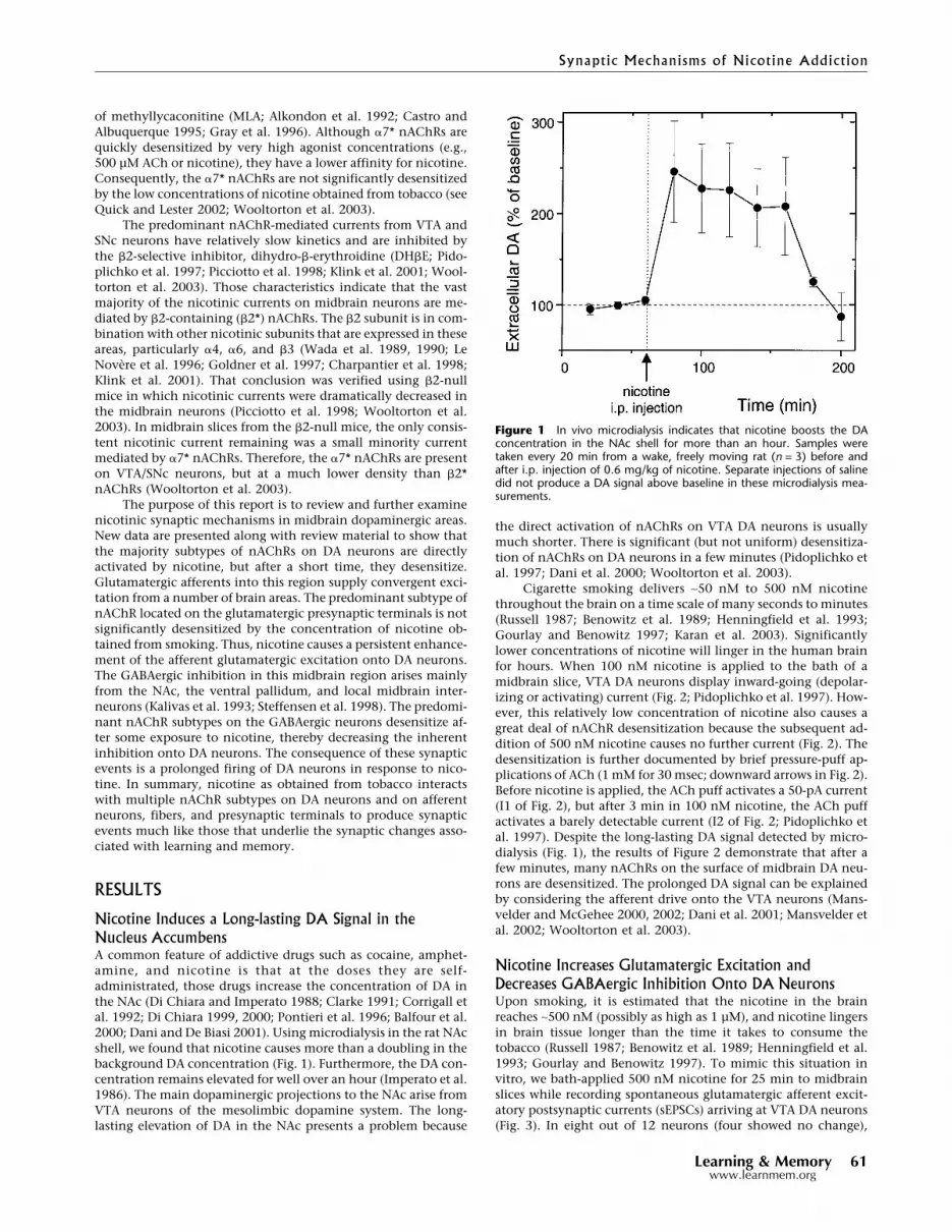
Nicotine Induces a Long-lasting DA Signal in theNucleus AccumbensA common feature of addictive drugs such as cocaine, amphet-amine, and nicotine is that at the doses they are self-administrated, those drugs increase the concentration of DA inthe NAc (Di Chiara and Imperato 1988; Clarke 1991; Corrigall etal. 1992; Di Chiara 1999, 2000; Pontieri et al. 1996; Balfour et al.2000; Dani and De Biasi 2001). Using microdialysis in the rat NAcshell, we found that nicotine causes more than a doubling in thebackground DA concentration (Fig. 1). Furthermore, the DA con-centration remains elevated for well over an hour (Imperato et al.1986). The main dopaminergic projections to the NAc arise fromVTA neurons of the mesolimbic dopamine system. The long-lasting elevation of DA in the NAc presents a problem because
the direct activation of nAChRs on VTA DA neurons is usuallymuch shorter. There is significant (but not uniform) desensitiza-tion of nAChRs on DA neurons in a few minutes (Pidoplichko etal. 1997; Dani et al. 2000; Wooltorton et al. 2003).
Cigarette smoking delivers ∼50 nM to 500 nM nicotinethroughout the brain on a time scale of many seconds to minutes(Russell 1987; Benowitz et al. 1989; Henningfield et al. 1993;Gourlay and Benowitz 1997; Karan et al. 2003). Significantlylower concentrations of nicotine will linger in the human brainfor hours. When 100 nM nicotine is applied to the bath of amidbrain slice, VTA DA neurons display inward-going (depolar-izing or activating) current (Fig. 2; Pidoplichko et al. 1997). How-ever, this relatively low concentration of nicotine also causes agreat deal of nAChR desensitization because the subsequent ad-dition of 500 nM nicotine causes no further current (Fig. 2). Thedesensitization is further documented by brief pressure-puff ap-plications of ACh (1 mM for 30 msec; downward arrows in Fig. 2).Before nicotine is applied, the ACh puff activates a 50-pA current(I1 of Fig. 2), but after 3 min in 100 nM nicotine, the ACh puffactivates a barely detectable current (I2 of Fig. 2; Pidoplichko etal. 1997). Despite the long-lasting DA signal detected by micro-dialysis (Fig. 1), the results of Figure 2 demonstrate that after afew minutes, many nAChRs on the surface of midbrain DA neu-rons are desensitized. The prolonged DA signal can be explainedby considering the afferent drive onto the VTA neurons (Mans-velder and McGehee 2000, 2002; Dani et al. 2001; Mansvelder etal. 2002; Wooltorton et al. 2003).
Nicotine Increases Glutamatergic Excitation andDecreases GABAergic Inhibition Onto DA NeuronsUpon smoking, it is estimated that the nicotine in the brainreaches ∼500 nM (possibly as high as 1 µM), and nicotine lingersin brain tissue longer than the time it takes to consume thetobacco (Russell 1987; Benowitz et al. 1989; Henningfield et al.1993; Gourlay and Benowitz 1997). To mimic this situation invitro, we bath-applied 500 nM nicotine for 25 min to midbrainslices while recording spontaneous glutamatergic afferent excit-atory postsynaptic currents (sEPSCs) arriving at VTA DA neurons(Fig. 3). In eight out of 12 neurons (four showed no change),
Figure 1 In vivo microdialysis indicates that nicotine boosts the DAconcentration in the NAc shell for more than an hour. Samples weretaken every 20 min from a wake, freely moving rat (n = 3) before andafter i.p. injection of 0.6 mg/kg of nicotine. Separate injections of salinedid not produce a DA signal above baseline in these microdialysis mea-surements.
Synaptic Mechanisms of Nicotine Addiction
Learning & Memory 61www.learnmem.org

nicotine caused an increase in the frequency (but not the ampli-tude) of sEPSCs (Fig. 3; Mansvelder and McGehee 2000, 2002;Dani et al. 2001; Mansvelder et al. 2002). The frequency of thesEPSCs remained elevated throughout the 25-min period, indi-cating that desensitization did not terminate the nicotinic effect.
Nicotine also increased the amplitude of electrically evokedEPSCs on DA neurons (Fig. 4). The enhancement was favored bycutting sagittal slices and stimulating with a low stimulusstrength (∼10% of the maximal response). In five out of 11 neu-rons (six showed no change), the amplitude of the eEPSCs in-creased (Mansvelder and McGehee 2000, 2002; Dani et al. 2001;Mansvelder et al. 2002). It is interesting to note that both thespontaneous and evoked EPSCs remain elevated after the nico-tine was washed away, consistent with the induction of long-term potentiation of glutamatergic afferents (Mansvelder andMcGehee 2000, 2002; Dani et al. 2001; Ji et al. 2001; Mansvelderet al. 2002). This result is consistent with presynaptic nAChRsthat boost EPSC frequency without changing the amplitude, ashas been demonstrated in the hippocampus and elsewhere (Mc-Gehee and Role 1995; McGehee et al. 1995; Gray et al 1996; Roleand Berg 1996; Albuquerque et al 1997; Wonnacott 1997; Guo etal 1998; Li et al 1998; Radcliffe and Dani 1998; Jones et al 1999;Radcliffe et al 1999; Mansvelder and McGehee 2000, 2002; Daniet al. 2001; Mansvelder et al. 2002). The nicotine-induced long-lasting potentiation of glutamatergic afferent excitation onto DAneurons is similar to the synaptic plasticity that is normallythought to underlie learning and memory (Martin et al. 2000).
The response to nicotine by the spontaneous GABAergic af-ferent inhibitory postsynaptic currents (sIPSCs) was markedlydifferent from the long-lasting boost of glutamatergic spontane-ous or evoked EPSCs. Bath-applied 500 nM nicotine for 25 minbriefly boosted sIPSCs, but that was followed by a strong long-lasting inhibition (n = 13 of 18; five showed no change; Fig. 5).The amplitudes of the sIPSCs also responded. During the increasein sIPSC frequency, there were larger sIPSCs and the average in-crease in amplitude was 16% � 6% (Mansvelder et al. 2002). Thisamplitude increase is consistent with the interpretation that thenAChRs are located on preterminal and somal locations wherenicotine can boost the fraction of action potential-dependent
IPSCs (Lena et al 1993; McMahon et al 1994; Alkondon et al.1997; Ji and Dani 2000; Mansvelder et al. 2002). Activation ofnAChRs located on the soma or preterminally has been found todepolarize the membrane locally, leading to activation of volt-age-dependent channels that directly mediate action potentials.Thus, adding nicotine to the bath briefly boosts the action po-tential firing of GABAergic neurons, decreasing the relative con-tribution from the smaller-amplitude miniature IPSCs, whicharise from the stochastic release of a single quantum of neuro-transmitter. After a short time, the nAChRs desensitize, removingthe nicotine-derived excitation as well as removing any endog-enous nicotinic cholinergic drive onto the GABAergic neurons.
Endogenous Cholinergic Activity Influences GABAergicand Glutamatergic AfferentsNicotine activates and desensitizes nAChRs, and in that way di-rectly influences afferent activity into the midbrain and the fir-ing of the DA neurons (Pidoplichko et al. 1997; Picciotto et al.1998; Mansvelder and McGehee 2000, 2002; Dani et al. 2001;Mansvelder et al. 2002). Desensitization also can have a long-lasting effect on the normal nicotinic mechanisms driven by en-dogenous cholinergic activity. For example, the long-lasting de-crease of sIPSC frequency (Fig. 5) is consistent with nicotine-desensitizing cholinergic afferents that partially drive theGABAergic activity, as has been seen in the hippocampus (Alkon-
Figure 2 Nicotine, at the concentration experienced by smokers, acti-vates and desensitizes nAChRs. (Left) Bath application of 0.1 µM nicotineactivated a 17-pA current, and after 3 min application of 0.5 µM nicotineactivated very little additional current. If there had been no desensitiza-tion, the solid square marks the average size of the current that wouldhave been activated by 0.5 µM nicotine. ACh pressure injections (1 mM,30 msec, downward arrows) were applied before (I1) and near the end(I2) of the 0.1 µM nicotine. Those ACh-induced currents are shown on anexpanded time scale (right) to illustrate the extent of desensitization.(These data were adapted from Pidoplichko et al. 1997.)
Figure 3 Nicotine increases the frequency not the amplitude of spon-taneous EPSCs recorded from VTA DA neurons. Example traces (upper)show that bath-applied nicotine (500 nM) increases the frequency ofsEPSCs, and the average (eight of 12; four showed no change) shows thelong-lasting effect of the frequency increase (lower). The recordings wereat room temperature, at a holding potential of �60 mV. Scale barsrepresent 10 pA and 500 msec.
Pidoplichko et al.
62 Learning & Memorywww.learnmem.org

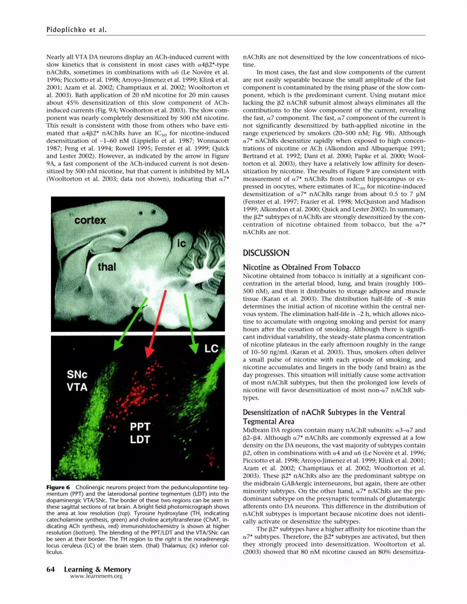
don et al. 1998; Frazier et al. 1998; Hefft et al. 1999). Cholinergicinnervation into the midbrain DA areas arises from neurons inthe pedunculopontine tegmentum (PPT) and the laterodorsalpontine tegmentum (LDT), which provide widespread innerva-tion mainly to the thalamus and midbrain areas and descendinginnervation that reaches to the brain stem.
When we cut horizontal slices and electrically stimulatedrostral inputs to the VTA, we found that inhibiting nAChRs hadlittle or no effect on the amplitude of GABAergic-evoked IPSCs(Mansvelder et al. 2002; data not shown). Neither 1 µM DH�E(which is selective for non-�7, mainly �2*, nAChRs) nor 5 nMMLA (which is selective for �7* nAChRs) influenced the eIPSCs.Therefore, to better preserve the cholinergic inputs into the VTAfrom the PPT/LDT, we cut parasagittal slices and stimulated cau-dally to the VTA. Figure 6 shows that the cholinergic neurons ofthe PPT/LDT (labeled red for ChAT activity) are near to the DAneurons of the VTA/SNc (labeled green for TH activity). For ourelectrophysiological studies, we tried to select DA neurons nearthe interface with the cholinergic neurons, and stimulated nearthe border between the PPT/LDT and the VTA. Under those con-ditions, we found that the �2-selective inhibitor, 1 µM DH�E,inhibited the amplitude of the GABAergic eIPSCs (n = 4 out of 10neurons; six showed no effect; Fig. 7). MLA (5 nM), which inhib-its �7* nAChRs, did not influence the eIPSCs when it was addedseparately (data not shown) or when it was added prior to theDH�E (Fig. 7). The results indicate that non-�7, mainly �2*
nAChRs, are activated by endogenous cholinergic activity thathelps to drive GABAergic IPSCs onto DA neurons. Thus, ongoingendogenous nicotinic cholinergic activity contributes to thebackground GABAergic inhibition onto DA neurons.
When we inhibited nAChRs while recording glutamatergiceEPSCs onto VTA DA neurons, we found that �7* nAChRs wereimportant. Inhibition with DH�E had no effect on the eEPSCamplitude (data not shown), but inhibition of �7* nAChRs with5 or 10 nM MLA decreased the eEPSC amplitude (n = 3 out of 10;seven showed no effect; Fig. 8). The results are consistent withsparse endogenous cholinergic innervation stimulating presyn-aptic �7* nAChRs on some glutamatergic terminals, and in thatway boosting excitatory eEPSCs onto DA neurons.
Nicotine Differentially Desensitizes the nAChR SubtypesMidbrain DA neurons express different nAChR subunits, but thepharmacological and physiological characteristics of nAChRscurrents indicate that �2* nAChRs are by far the predominantsubtypes (Pidoplichko et al. 1997; Picciotto et al. 1998; Klink etal. 2001; Wooltorton et al. 2003). Nicotine at the concentrationachieved by smokers desensitizes nAChR currents from VTA DAneurons (Pidoplichko et al. 1997; Dani et al. 2000; Wooltorton etal. 2003). An important characteristic, however, is the differencein desensitization of nAChR subtypes (Wooltorton et al. 2003).
Figure 5 Nicotine increases then decreases the frequency of sponta-neous IPSCs recorded from VTA DA neurons. Example traces (upper) showthat bath-applied nicotine (0.5 µM) first increases the frequency (andamplitude) of sIPSCs, but the average (13 of 18; five showed no change)shows the more potent, long-lasting effect is an inhibition of sIPSC fre-quency (lower). The recordings were at room temperature at a holdingpotential of �20 mV. The scale bars represent 20 pA and 0.5 sec.
Figure 4 Nicotine increases the amplitude of evoked EPSCs recordedfrom VTA DA neurons. Example traces (upper) show that bath-appliednicotine (1 µM) increases the amplitude of eEPSCs, and the average (fiveof 11; six showed no change) shows the long-lasting effect of the ampli-tude increase (lower). A low concentration of EGTA (0.4 µM) was used inthe patch pipette to enhance the long-lasting increase in amplitude. Therecordings were at room temperature at a holding potential of �65 mV,and a weak simulation strength was used. The scale bars represent 100pA and 10 msec.
Synaptic Mechanisms of Nicotine Addiction
Learning & Memory 63www.learnmem.org
Nearly all VTA DA neurons display an ACh-induced current withslow kinetics that is consistent in most cases with �4�2*-typenAChRs, sometimes in combinations with �6 (Le Novere et al.1996; Picciotto et al. 1998; Arroyo-Jimenez et al. 1999; Klink et al.2001; Azam et al. 2002; Champtiaux et al. 2002; Wooltorton etal. 2003). Bath application of 20 nM nicotine for 20 min causesabout 45% desensitization of this slow component of ACh-induced currents (Fig. 9A; Wooltorton et al. 2003). The slow com-ponent was nearly completely desensitized by 500 nM nicotine.This result is consistent with those from others who have esti-mated that �4�2* nAChRs have an IC50 for nicotine-induceddesensitization of ∼1–60 nM (Lippiello et al. 1987; Wonnacott1987; Peng et al. 1994; Rowell 1995; Fenster et al. 1999; Quickand Lester 2002). However, as indicated by the arrow in Figure9A, a fast component of the ACh-induced current is not desen-sitized by 500 nM nicotine, but that current is inhibited by MLA(Wooltorton et al. 2003; data not shown), indicating that �7*
nAChRs are not desensitized by the low concentrations of nico-tine.
In most cases, the fast and slow components of the currentare not easily separable because the small amplitude of the fastcomponent is contaminated by the rising phase of the slow com-ponent, which is the predominant current. Using mutant micelacking the �2 nAChR subunit almost always eliminates all thecontributions to the slow component of the current, revealingthe fast, �7 component. The fast, �7 component of the current isnot significantly desensitized by bath-applied nicotine in therange experienced by smokers (20–500 nM; Fig. 9B). Although�7* nAChRs desensitize rapidly when exposed to high concen-trations of nicotine or ACh (Alkondon and Albuquerque 1991;Bertrand et al. 1992; Dani et al. 2000; Papke et al. 2000; Wool-torton et al. 2003), they have a relatively low affinity for desen-sitization by nicotine. The results of Figure 9 are consistent withmeasurement of �7* nAChRs from rodent hippocampus or ex-pressed in oocytes, where estimates of IC50 for nicotine-induceddesensitization of �7* nAChRs range from about 0.5 to 7 µM(Fenster et al. 1997; Frazier et al. 1998; McQuiston and Madison1999; Alkondon et al. 2000; Quick and Lester 2002). In summary,the �2* subtypes of nAChRs are strongly desensitized by the con-centration of nicotine obtained from tobacco, but the �7*nAChRs are not.
DISCUSSION
Nicotine as Obtained From TobaccoNicotine obtained from tobacco is initially at a significant con-centration in the arterial blood, lung, and brain (roughly 100–500 nM), and then it distributes to storage adipose and muscletissue (Karan et al. 2003). The distribution half-life of ∼8 mindetermines the initial action of nicotine within the central ner-vous system. The elimination half-life is ∼2 h, which allows nico-tine to accumulate with ongoing smoking and persist for manyhours after the cessation of smoking. Although there is signifi-cant individual variability, the steady-state plasma concentrationof nicotine plateaus in the early afternoon roughly in the rangeof 10–50 ng/mL (Karan et al. 2003). Thus, smokers often delivera small pulse of nicotine with each episode of smoking, andnicotine accumulates and lingers in the body (and brain) as theday progresses. This situation will initially cause some activationof most nAChR subtypes, but then the prolonged low levels ofnicotine will favor desensitization of most non-�7 nAChR sub-types.
Desensitization of nAChR Subtypes in the VentralTegmental AreaMidbrain DA regions contain many nAChR subunits: �3–�7 and�2–�4. Although �7* nAChRs are commonly expressed at a lowdensity on the DA neurons, the vast majority of subtypes contain�2, often in combinations with �4 and �6 (Le Novere et al. 1996;Picciotto et al. 1998; Arroyo-Jimenez et al. 1999; Klink et al. 2001;Azam et al. 2002; Champtiaux et al. 2002; Wooltorton et al.2003). These �2* nAChRs also are the predominant subtype onthe midbrain GABAergic interneurons, but again, there are otherminority subtypes. On the other hand, �7* nAChRs are the pre-dominant subtype on the presynaptic terminals of glutamatergicafferents onto DA neurons. This difference in the distribution ofnAChR subtypes is important because nicotine does not identi-cally activate or desensitize the subtypes.
The �2* subtypes have a higher affinity for nicotine than the�7* subtypes. Therefore, the �2* subtypes are activated, but thenthey strongly proceed into desensitization. Wooltorton et al.(2003) showed that 80 nM nicotine caused an 80% desensitiza-
Figure 6 Cholinergic neurons project from the pedunculopontine teg-mentum (PPT) and the laterodorsal pontine tegmentum (LDT) into thedopaminergic VTA/SNc. The border of these two regions can be seen inthese sagittal sections of rat brain. A bright field photomicrograph showsthe area at low resolution (top). Tyrosine hydroxylase (TH, indicatingcatecholamine synthesis, green) and choline acetyltransferase (ChAT, in-dicating ACh synthesis, red) immunohistochemistry is shown at higherresolution (bottom). The blending of the PPT/LDT and the VTA/SNc canbe seen at their border. The TH region to the right is the noradrenergiclocus ceruleus (LC) of the brain stem. (thal) Thalamus; (ic) inferior col-liculus.
Pidoplichko et al.
64 Learning & Memorywww.learnmem.org

tion of the �2* nAChRs in the midbrain. That result is consistentwith previous estimates that �2* nAChRs have an IC50 for nico-tine-induced desensitization of ∼1–60 nM (Lippiello et al. 1987;Wonnacott 1987; Peng et al. 1994; Rowell 1995; Fenster et al.1999; Quick and Lester 2002). Thus, nicotine strongly desensi-tizes the majority subtypes of nAChRs (i.e., �2*) on midbrain DAand GABA neurons. On the other hand, �7* nAChRs are notstrongly desensitized by the low concentrations of nicotine ob-tained from tobacco. Wooltorton et al. (2003) showed that up to500 nM nicotine caused very little desensitization of VTA �7*nAChRs. That result is consistent with previous estimates that�7* nAChRs have an IC50 for nicotine-induced desensitization ofup to 7 µM (Fenster et al. 1997; Frazier et al. 1998; McQuistonand Madison 1999; Alkondon et al. 2000; Quick and Lester 2002).This result may be surprising to some because high concentra-tions of agonist desensitize �7* nAChRs more rapidly than othernAChR types (Alkondon and Albuquerque 1991; Bertrand et al.1992; Dani et al. 2000; Papke et al. 2000). Despite the rapid ki-netics, the lower affinity of �7* nAChRs for nicotine underliesthe lack of desensitization. At low nicotine concentrations, onlya small proportion of the �7* nAChR population is occupied bynicotine, and that proportion proceeds toward desensitization.
Because of the rapid kinetics, those �7* nAChRs rapidly recoverfrom desensitization when nicotine unbinds. Consequently,only a very small portion of the overall �7 population is desen-sitized at any moment, leaving the remaining receptors availableto activate.
Model of the Synaptic Action of Nicotinein the Ventral Tegmental AreaAlthough other minority subtypes are present, �2* nAChRs makeup the vast majority of subtypes on VTA DA neurons andGABAergic interneurons. Those receptors underlie the initial di-rect activation of DA neurons by nicotine (see Fig. 10; Mans-velder and McGehee 2000, 2002; Dani et al. 2001; Mansvelder etal. 2002; Wooltorton et al. 2003). When nicotine first arrives,these �2* nAChRs are activated, causing direct excitation of theDA neurons and the GABAergic interneurons (indicated by Figs.2 and 5; Calabresi et al. 1989; Pidoplichko et al. 1997; Picciotto etal. 1998; Dani et al. 2000, 2001; Mansvelder and McGehee 2002).In minutes, significant desensitization affects these (predomi-nantly) �2* nAChRs (see Figs. 2, 5, and 9). The GABAergic activitydeclines more rapidly because desensitization removes the directexcitation caused by nicotine and decreases the endogenous cho-linergic drive onto the GABAergic interneurons (Figs. 5 and 7).Whereas a small subset of DA neurons receives cholinergic in-
Figure 7 The �2* antagonist, DH�E, decreased the amplitude ofGABAa-evoked IPSCs onto DA neurons. Example traces (upper) show thatbath-applied DH�E (1 µM) decreases the amplitude of eIPSCs, and theaverage (four out of 10 neurons; six showed no effect) shows the ampli-tude decrease (lower). The �7* nAChR antagonist, MLA, had no effect inseparate experiments (data not shown) or when applied before DH�E.The recordings were at room temperature at a holding potential of �60mV, and weak simulation strength was used. The scale bars represent 50pA and 20 msec.
Figure 8 The �7* antagonist, MLA, decreased the amplitude of Glu-evoked EPSCs. Example traces (upper) show that bath-applied MLA (10nM) decreases the amplitude of eEPSCs, and the average (three out of 10;seven showed no effect) shows the amplitude decrease (lower). The re-cordings were at room temperature at a holding potential of �65 mV,and a weak simulation strength was used. The scale bars represent 100pA and 10 msec.
Synaptic Mechanisms of Nicotine Addiction
Learning & Memory 65www.learnmem.org

puts, there is greater innervation of GABAergic neurons in theVTA (Garzon et al. 1999).
As the excitation of DA neurons by nicotine decreases owingto desensitization of the predominant nAChR subtypes, othersynaptic factors have already provided further excitation to pro-duce the prolonged DA signal observed in the NAc (Fig. 1; Im-perato et al. 1986; Di Chiara and Imperato 1988; Clarke 1991;Corrigall et al. 1992; Pontieri et al. 1996; Di Chiara 1999, 2000;Dani and De Biasi 2001). The initial “pulse” of nicotine providedby smoking activates presynaptic �7* nAChRs located on gluta-matergic terminals that synapse onto DA neurons (see Figs. 3, 4,and 8), and the �7* subtype does not desensitize even at thehigher concentrations of nicotine obtained from smoking (Fig.9). Because the �7* subtype is the most highly permeable nAChRto calcium, it often mediates a direct Ca2+ increase as well asinitiating indirect Ca2+ influx caused by the local depolarizationand via intracellular Ca2+ stores (Séguéla et al. 1993; McGeheeand Role 1995; McGehee et al. 1995; Gray et al. 1996; Rathouz etal. 1996; Radcliffe and Dani 1998; Mansvelder and McGehee2000, 2002; Dani et al. 2001; Ji et al. 2001; Mansvelder et al.2002). Consequently, the activation of presynaptic �7* nAChRsinitiates a calcium increase in the glutamatergic presynaptic ter-minals that increases glutamate release and excitation of DA neu-rons, even while the �2* nAChRs on the DA neurons are desen-sitizing (see Fig. 10).
The situation favors longer-term synaptic plasticity that po-tentiates the glutamatergic drive. Long-term potentiation (LTP)arises when presynaptic glutamatergic excitation is coincident
with a large enough postsynaptic responseto induce a Ca2+ signal through NMDA-typeglutamate receptors (NMDARs). Whennicotine initially arrives, it excites the DAneurons to increase their action potentialfiring rate. That postsynaptic DA neuron ac-tivity is coupled with a nicotine-induced in-crease in presynaptic glutamatergic afferentexcitation (see Fig. 10). That combinationproduces the presynaptic and postsynapticcoincidence that boosts the production ofLTP (see Mansvelder and McGehee 2000,2002; Dani et al. 2001; Ji et al. 2001; Mans-velder et al. 2002). Subsequently, non-�7subtypes desensitize, thereby, decreasingthe inhibition onto DA neurons by GABAer-gic neurons. In addition, the �7* nAChRson presynaptic glutamate terminals do notdesensitize. Thus, they continue to enhanceglutamatergic excitation as long as the nico-tine signal is present. This seemingly cho-reographed complex of nicotinic synapticmechanisms contributes to the prolongedDA signal in the NAc and elsewhere that isthought to be a critical component in theaddiction process.
The synaptic changes that are inducedby nicotine are much like the normal syn-aptic plasticity that underlies learning andmemory: Presynaptic calcium signals en-hance excitatory transmission coupled to astrong postsynaptic response, leading toshort-term and long-term potentiation.Nicotine tips the normal balance, inappro-priately favoring potentiation of synapsesand, ultimately, favoring inappropriate be-haviors. In this way, the addictive drug,nicotine, commandeers fundamental syn-
aptic mechanisms that normally subserve learning and memory.
MATERIALS AND METHODS
Brain Slice Preparation and ElectrophysiologyMidbrain horizontal or sagittal slices containing the VTA andSNc were prepared from 14- to 25-day-old Sprague Dawley ratsthat were anesthetized before decapitation (see Wooltorton et al.2003). Slices (300–350 µm thick) were cut in ice-cold cuttingsolution, and all the solutions were saturated with 95% O2, 5%CO2 to achieve a pH near 7.4 during the experiments. The cut-ting solution was either of the following or a 50%/50% mixtureof the two solutions. The sucrose cutting solution was 230 mMsucrose, 1 mM KCl, 1.25 mM NaH2PO4, 30 mM NaHCO3, 1 mMCaCl2, 7 mM MgCl2, and 25 mM D-glucose. The N-methyl-D-glucamine (NMDG) cutting solution was 144 mM NMDG, 1.5mM KCl, 1.25 mM NaH2PO4, 30 mM NaHCO3, 2 mM CaCl2, 2mM MgCl2, and 25 mM D-glucose, and the pH was adjusted to7.4 with 50% D-gluconic acid and sodium bicarbonate. The sliceswere then transferred to a holding chamber containing the bathsolution: 125 mM NaCl, 2.5 mM KCl, 1.25 mM NaH2PO4 (or 1.24mM KH2PO4), 21 mM NaHCO3, 2.5 mM CaCl2 (or 2.1 mMCaCl2), 1 mM MgCl2 (or 0.3 mM MgCl2), and 25 mM D-glucose25. Slices were held either for 1 h at room temperature or for 20min at 34°C and, then, for a minimum of 20 min at room tem-perature. The experimental chamber (0.8 mL capacity) had con-tinuously flowing bath solution (∼5 mL/min) at room tempera-ture near 23°C. The osmolality of the external solution was ad-justed to 320 mOsm with D-glucose. All the experimentsstudying evoked responses had 0.5 µM atropine added to the
Figure 9 Exposure to low concentrations of nicotine differentially desensitizes the fast and slowcomponents of nicotinic currents from VTA DA neurons. (A) ACh-induced currents (1 mM ACh, 200msec puff, horizontal line) in the absence (Control) and presence of bath-applied nicotine (20 nMor 500 nM for 20 min). Although the slow component of the current (mainly �4�2* nAChRs) wasdesensitized, the fast component (indicated by the notch, arrow) was not desensitized. The scalebars represent 100 pA and 0.5 sec. (B) Exposure to low concentrations of nicotine does notdesensitize the fast, MLA-sensitive currents from VTA DA neurons of �2-null mice. ACh-inducedcurrents (1 mM ACh, 200 msec puff, horizontal line) are shown from the same neuron. In theabsence (Control) or presence of nicotine (500 nM for 20 min) the currents are the same. TheACh-induced currents were inhibited by 5 nMMLA, confirming that these are �7* nAChR currents.The ACh-induced currents were not inhibited by 1 µM DH�E (data not shown). The scale barsrepresent 50 pA and 0.5 sec. (These data are taken and adapted from Wooltorton et al. 2003.)
Pidoplichko et al.
66 Learning & Memorywww.learnmem.org

external bath solutions to block muscarinic AChRs. During themeasurement of glutamatergic evoked EPSCs, the bath solutioncontained 20 µM bicuculline to block GABAergic transmission.During the measurement of evoked GABAergic IPSCs, the bathsolution contained 20 µM CNQX to block non-NMDA Glu re-ceptors. When used, nicotine and nAChR antagonists were ap-plied via the continuously flowing bath solution.
Neurons were visualized under infrared light using Nomar-ski optics of an upright microscope (Zeiss Axioscope) equippedwith a CCD camera (Hamamatsu Photonics K.K.). The patch elec-trodes had resistances of 3.5–4.5 M� for evoked responses and upto 7 M� for spontaneous events when filled with the internalsolution: 60 mM CsCH3SO3, 60 mM KCH3SO3, 1 or 10 mM KCl,0.4 mM EGTA, 10 mM HEPES, 5 mM Mg-ATP, 0.3 mM Na3GTP(pH 7.2), 290 mOsm osmolality; or 140 mM K-gluconate; 1 mMMgCl2; 1 mM CaCl2; 10 mM HEPES; 10 mM EGTA; 5 mM ATP-Mg; and 0.3 mM GTP-Na (pH 7.3 adjusted with Tris-base). Theholding potential for voltage-clamp recordings was the follow-ing: for eEPSCs, �65 mV; for sEPSCs and eIPSCs, �60 mV; forsIPSCs, �20 mV. The spontaneous or evoked glutamatergicevents were completely inhibited by CNQX (20 µM) and AP-5 (50µM), and the spontaneous or evoked GABAergic events werecompletely inhibited by bicuculline (20 µM).
The stimulation arrangement we used attempted to opti-mize the stimulation of the sparse cholinergic fibers that rangethrough the thickness of the slice. Nine Teflon-coated wires, withthe isolation stripped at the very end of each wire, formed aV-shaped sink electrode array beneath the slice. The VTA-containing mesolimbic system was positioned inside the V-shaped area formed by the tips of nine wires. A monopolar tung-sten stimulation electrode could be positioned near the top of theslice either on the rostral or caudal side close to the VTA. Switch-ing between sink electrodes allowed choosing the best input. Inother cases to favor rostral or caudal stimulation, two of the nineV-shaped electrodes serve as source and sink. EPSCs or IPSCs were
evoked via a computer-controlled stimulusisolator model A-360 (WPI) applying mini-mal stimulation intensity. The intensity ofthe stimulus was adjusted to elicit ampli-tudes at 10%–20% of the maximum betterto see the effect of nicotine (see Mansvelderand McGehee 2000). Stimulation currentsranged from 150 to 450 µA and usuallylasted 0.7 msec. Currents were amplifiedand filtered (1 kHz) using Axopatch 200B or1C amplifiers (Axon Instruments) with afour-pole low-pass Bessel filter and weredigitally sampled (up to 5 kHz). Currentswere recorded using pClamp software(Axon), and further analyzed using Origin(MicroCal Software). Additional off-line fil-tering and signal averaging were sometimesused in the figures. Neurons that werepatch-clamped were identified as VTA DAbased on large Ih currents (Pidoplichko et al.1997; Bonci and Malenka 1999).
ImmunohistochemistryTyrosine hydroxylase (TH, indicating cat-echolamine synthesis) and choline acetyl-transferase (ChAT, indicating ACh synthe-sis) immunohistochemistry were adaptedfrom published methods (Zhou et al. 2001).For ChAT and TH double immunofluores-cence, brains were fixed in 0.1 M phosphatebuffer containing 4% (w/v) paraformalde-hyde and 14% (v/v) picric acid. Sectionswere cut on a cryostat, and were incubatedwith goat anti-ChAT and rabbit anti-TH an-tibodies. The secondary antibody mixturecontaining Cy2-conjugated donkey anti-rabbit and rhodamine-conjugated donkeyanti-goat IgG antisera was used. All images
were captured with a digital camera and processed in Adobe Pho-toShop.
MicrodialysisLong-Evans male rats (Harlan) were housed together on a 12-hlight/dark cycle. Their body weight was from 280–300 g. The ratswere anesthetized using a ketamine–xylazine combo injected in-traperitoneally (1.8 µL per gram body weight) and subsequentlymaintained on an isoflurane-gas mixture. The microdialysisCMA/12 (CMA/Microdialysis) guide cannula was aimed at theNAc shell (1.7 mm AP; 0.8 mm L; 6.5 mm DV, with the probe at7.5 mm) and was secured with bone wax reinforced with acryliccement and three screws into the skull. The rat was allowed torecover fully for a minimum of 48 h.
CMA/12 probes (diameter, 0.5 mm; length, 1 mm; mem-brane, polycarbonate; cutoff, 20,000 D) were prepared, and fil-tered degassed artificial cerebrospinal fluid (aCSF; from ESA) wasperfused through the probe at a flow rate of 1 µL/min using aCMA/100 pump. Following a 2-h recovery period, three 20-minfractions were collected to assess the basal output of dopamine inthe dialysate. Subsequently, saline and nicotine (0.6 mg/kg i.p.)were injected, and samples were collected every 20 min for 3 h.After these experiments, rats were killed with an overdose ofanesthetics and trans-cardially perfused with PBS and then 10%formalin. The brain was removed and fixed in 10% formalin. Theaccuracy of probe placement was later confirmed by histologicalsectioning.
Dopamine contents of microdialysates were determined us-ing a high-performance liquid chromatography (HPLC) system(model 580 pump, Coulochem II electrochemical detector,model 5014B analytical cell; ESA, Inc.). Separation of dopaminewas achieved on a 150 � 3 mm column with 3 µm particle size(ESA, Inc.; MD-150). An isocratic mobile phase (pH 4.0) contain-ing 75 mM NaH2PO4, 2 mM 1-octane sulphonic acid-sodium salt,
Figure 10 The major sites of influence by nAChR subtypes at glutamatergic and GABAergicsynapses onto VTA DA neurons are shown. Activation of nAChRs will induce a local depolarization(lightning bolt) than can activate voltage-dependent channels and in some cases an action po-tential. The activity also will initiate a direct and indirect Ca2+ signal. The Ca2+ signal can influencesubsequent Ca2+ release from intracellular stores and initiate intracellular cascades. The size of thesignals will depend on many factors, including the subtypes of nAChRs that are present andactivation versus desensitization by agonists and modulators. The diagram is simplified to the majorsubtypes at each location, but other minority subtypes also can be present. Nicotine, as obtainedfrom tobacco, will briefly activate then begin to significantly desensitize the non-�7 (usually �2*)nAChRs located on DA and GABA neurons. The �7* nAChRs will be activated somewhat, but willnot be strongly desensitized by those levels of nicotine. Thus, the increased excitatory drive viapresynaptic �7* nAChR on glutamatergic (Glu) terminals coupled with the short-lasting increase inDA neuron firing caused by nicotine’s direct action creates the coincidence of presynaptic andpostsynaptic activity that favors the initiation of synaptic plasticity, such as STP and LTP. Thus,multiple synaptic events contribute to the prolonged increased firing by DA neurons.
Synaptic Mechanisms of Nicotine Addiction
Learning & Memory 67www.learnmem.org

20 mM EDTA, 100 µL/L triethylamine, and 18% methanol (fromESA) was used at a flow rate of 0.6 mL/min. This mobile phaseand the protocols used produced clearly separable dopaminepeaks with a retention time of ∼5.6 min. Chromatograms wereanalyzed with the ESA software. Freshly prepared standards rang-ing from 0–6 nM DA were used to calibrate the readings.
ACKNOWLEDGMENTSThis work was supported by the National Institute of Neurologi-cal Disorders and Stroke (NS21229) and the National Institute onDrug Abuse (DA09411 and DA12661).
The publication costs of this article were defrayed in part bypayment of page charges. This article must therefore be herebymarked “advertisement” in accordance with 18 USC section 1734solely to indicate this fact.
REFERENCESAlbuquerque, E.X., Alkondon, M., Pereira, E.F., Castro, N.G.,
Schrattenholz, A., Barbosa, C.T., Bonfante-Cabarcas, R., Aracava, Y.,Eisenberg, H.M., and Maelicke, A. 1997. Properties of neuronalnicotinic acetylcholine receptors: Pharmacological characterizationand modulation of synaptic function. J. Pharmacol. Exp. Ther.280: 1117–1136.
Alkondon, M. and Albuquerque, E.X. 1991. Initial characterization ofthe nicotinic acetylcholine receptors in rat hippocampal neurons. J.Recept. Res. 11: 1001–1021.
Alkondon, M., Pereira, E.F., Wonnacott, S., and Albuquerque, E.X. 1992.Blockade of nicotinic currents in hippocampal neurons definesmethyllycaconitine as a potent and specific receptor antagonist. Mol.Pharmacol. 41: 802–808.
Alkondon, M., Pereira, E.F., Barbosa, C.T., and Albuquerque, E.X. 1997.Neuronal nicotinic acetylcholine receptor activation modulates�-aminobutyric acid release from CA1 neurons of rat hippocampalslices. J. Pharmacol. Exp. Ther. 283: 1396–1411.
Alkondon, M., Pereira, E.F., and Albuquerque, E.X. 1998.�-Bungarotoxin- and methyllycaconitine-sensitive nicotinicreceptors mediate fast synaptic transmission in interneurons of rathippocampal slices. Brain Res. 810: 257–263.
Alkondon, M., Pereira, E.F., Almeida, L.E., Randall, W.R., andAlbuquerque, E.X. 2000. Nicotine at concentrations found incigarette smokers activates and desensitizes nicotinic acetylcholinereceptors in CA1 interneurons of rat hippocampus.Neuropharmacology 39: 2726–2739.
Arroyo-Jimenez, M.M., Bourgeois, J.P., Marubio, L.M., Le Sourd, A.M.,Ottersen, O.P., Rinvik, E., Fairen, A., and Changeux, J.P. 1999.Ultrastructural localization of the �4-subunit of the neuronalacetylcholine nicotinic receptor in the rat substantia nigra. J.Neurosci. 19: 6475–6487.
Azam, L., Winzer-Serhan, U.H., Chen, Y., and Leslie, F.M. 2002.Expression of neuronal nicotinic acetylcholine receptor subunitmRNAs within midbrain dopamine neurons. J. Comp. Neurol.444: 260–274.
Balfour, D.J., Wright, A.E., Benwell, M.E., and Birrell, C.E. 2000. Theputative role of extra-synaptic mesolimbic dopamine in theneurobiology of nicotine dependence. Behav. Brain Res. 113: 73–83.
Benowitz, N.L., Porchet, H., and Jacob III, P. 1989. Nicotine dependenceand tolerance in man: Pharmacokinetic and pharmacodynamicinvestigations. Prog. Brain Res. 79: 279–287.
Bertrand, D., Devillers-Thiery, A., Revah, F., Galzi, J.L., Hussy, N., Mulle,C., Bertrand, S., Ballivet, M., and Changeux, J.P. 1992.Unconventional pharmacology of a neuronal nicotinic receptormutated in the channel domain. Proc. Natl. Acad. Sci.89: 1261–1265.
Bonci, A. and Malenka, R.C. 1999. Properties and plasticity of excitatorysynapses on dopaminergic and GABAergic cells in the ventraltegmental area. J. Neurosci. 19: 3723–3730.
Calabresi, P., Lacey, M.G., and North, R.A. 1989. Nicotinic excitation ofrat ventral tegmental neurones in vitro studied by intracellularrecording. Br. J. Pharmacol. 98: 135–140.
Castro, N.G. and Albuquerque, E.X. 1995. �-Bungarotoxin-sensitivehippocampal nicotinic receptor channel has a high calciumpermeability. Biophys. J. 68: 516–524.
CDC. 2002. Annual smoking-attributable mortality, years of potentiallife lost, and economic costs; United States, 1995–1999. Morbidityand Mortality Weekly Report, 51: 300–303. CDC.
Champtiaux, N., Han, Z.Y., Bessis, A., Rossi, F.M., Zoli, M., Marubio, L.,McIntosh, J.M., and Changeux, J.P. 2002. Distribution andpharmacology of � 6-containing nicotinic acetylcholine receptorsanalyzed with mutant mice. J. Neurosci. 22: 1208–1217.
Charpantier, E., Barneoud, P., Moser, P., Besnard, F., and Sgard, F. 1998.Nicotinic acetylcholine subunit mRNA expression in dopaminergicneurons of the rat substantia nigra and ventral tegmental area.Neuroreport 9: 3097–3101.
Clarke, P.B. 1990. Dopaminergic mechanisms in the locomotorstimulant effects of nicotine. Biochem. Pharmacol. 40: 1427–1432.
. 1991. The mesolimbic dopamine system as a target for nicotine.In Effects of nicotine on biological systems (eds. F. Adlkofer and K.Thurau), pp. 285–294. Birkhäuser Verlag, Basel.
Clarke, P.B., Schwartz, R.D., Paul, S.M., Pert, C.B., and Pert, A. 1985.Nicotinic binding in rat brain: Autoradiographic comparison of[3H]acetylcholine, [3H]nicotine, and [125I]-�-bungarotoxin. J.Neurosci. 5: 1307–1315.
Corrigall, W.A. 1999. Nicotine self-administration in animals as adependence model. Nicotine Tob. Res. 1: 11–20.
Corrigall, W.A. and Coen, K.M. 1989. Nicotine maintains robustself-administration in rats on a limited-access schedule.Psychopharmacology 99: 473–478.
Corrigall, W.A., Franklin, K.B., Coen, K.M., and Clarke, P.B. 1992. Themesolimbic dopaminergic system is implicated in the reinforcingeffects of nicotine. Psychopharmacology 107: 285–289.
Corrigall, W.A., Coen, K.M., and Adamson, K.L. 1994. Self-administerednicotine activates the mesolimbic dopamine system through theventral tegmental area. Brain Res. 653: 278–284.
Dani, J.A. and De Biasi, M. 2001. Cellular mechanisms of nicotineaddiction. Pharmacol. Biochem. Behav. 70: 439–446.
Dani, J.A. and Heinemann, S. 1996. Molecular and cellular aspects ofnicotine abuse. Neuron 16: 905–908.
Dani, J.A., Radcliffe, K.A., and Pidoplichko, V.I. 2000. Variations indesensitization of nicotinic acetylcholine receptors fromhippocampus and midbrain dopamine areas. Eur. J. Pharmacol.393: 31–38.
Dani, J.A., Ji, D., and Zhou, F.M. 2001. Synaptic plasticity and nicotineaddiction. Neuron 31: 349–352.
Di Chiara, G. 1999. Drug addiction as dopamine-dependent associativelearning disorder. Eur. J. Pharmacol. 375: 13–30.
. 2000. Role of dopamine in the behavioural actions of nicotinerelated to addiction. Eur. J. Pharmacol. 393: 295–314.
Di Chiara, G. and Imperato, A. 1988. Drugs abused by humanspreferentially increase synaptic dopamine concentrations in themesolimbic system of freely moving rats. Proc. Natl. Acad. Sci.85: 5274–5278.
Fenster, C.P., Rains, M.F., Noerager, B., Quick, M.W., and Lester, R.A.1997. Influence of subunit composition on desensitization ofneuronal acetylcholine receptors at low concentrations of nicotine.J. Neurosci. 17: 5747–5759.
Fenster, C.P., Whitworth, T.L., Sheffield, E.B., Quick, M.W., and Lester,R.A. 1999. Upregulation of surface �4�2 nicotinic receptors isinitiated by receptor desensitization after chronic exposure tonicotine. J. Neurosci. 19: 4804–4814.
Frazier, C.J., Buhler, A.V., Weiner, J.L., and Dunwiddie, T.V. 1998.Synaptic potentials mediated via �-bungarotoxin-sensitive nicotinicacetylcholine receptors in rat hippocampal interneurons. J. Neurosci.18: 8228–8235.
Garzon, M., Vaughan, R.A., Uhl, G.R., Kuhar, M.J., and Pickel, V.M.1999. Cholinergic axon terminals in the ventral tegmental areatarget a subpopulation of neurons expressing low levels of thedopamine transporter. J. Comp. Neurol. 410: 197–210.
Goldner, F.M., Dineley, K.T., and Patrick, J.W. 1997.Immunohistochemical localization of the nicotinic acetylcholinereceptor subunit �6 to dopaminergic neurons in the substantia nigraand ventral tegmental area. Neuroreport 8: 2739–2742.
Gourlay, S.G. and Benowitz, N.L. 1997. Arteriovenous differences inplasma concentration of nicotine and catecholamines and relatedcardiovascular effects after smoking, nicotine nasal spray, andintravenous nicotine. Clin. Pharmacol. Ther. 62: 453–463.
Gray, R., Rajan, A.S., Radcliffe, K.A., Yakehiro, M., and Dani, J.A. 1996.Hippocampal synaptic transmission enhanced by low concentrationsof nicotine. Nature 383: 713–716.
Grenhoff, J. and Johnson, S.W. 1996. Sulfonylureas enhance GABAAsynaptic potentials in rat midbrain dopamine neurones. Acta Physiol.Scand. 156: 147–148.
Guo, J.Z., Tredway, T.L., and Chiappinelli, V.A. 1998. Glutamate andGABA release are enhanced by different subtypes of presynapticnicotinic receptors in the lateral geniculate nucleus. J. Neurosci.18: 1963–1969.
Hefft, S., Hulo, S., Bertrand, D., and Muller, D. 1999. Synaptictransmission at nicotinic acetylcholine receptors in rat hippocampalorganotypic cultures and slices. J. Physiol. 515: 769–776.
Henningfield, J.E., Stapleton, J.M., Benowitz, N.L., Grayson, R.F., andLondon, E.D. 1993. Higher levels of nicotine in arterial than in venousblood after cigarette smoking. Drug Alcohol Depend. 33: 23–29.
Pidoplichko et al.
68 Learning & Memorywww.learnmem.org

Imperato, A., Mulas, A., and Di Chiara, G. 1986. Nicotine preferentiallystimulates dopamine release in the limbic system of freely movingrats. Eur. J. Pharmacol. 132: 337–338.
Ji, D. and Dani, J.A. 2000. Inhibition and disinhibition of pyramidalneurons by activation of nicotinic receptors on hippocampalinterneurons. J. Neurophysiol. 83: 2682–2690.
Ji, D., Lape, R., and Dani, J.A. 2001. Timing and location of nicotinicactivity enhances or depresses hippocampal synaptic plasticity.Neuron 31: 131–141.
Jones, S., Sudweeks, S., and Yakel, J.L. 1999. Nicotinic receptors in thebrain: Correlating physiology with function. Trends. Neurosci.22: 555–561.
Jones, I.W., Bolam, J.P., and Wonnacott, S. 2001. Presynapticlocalization of the nicotinic acetylcholine receptor �2 subunitimmunoreactivity in rat nigrostriatal dopaminergic neurones. J.Comp. Neurol. 439: 235–247.
Kalivas, P.W., Churchill, L., and Klitenick, M.A. 1993. GABA andenkephalin projection from the nucleus accumbens and ventralpallidum to the ventral tegmental area. Neuroscience 57: 1047–1060.
Karan, L.D., Dani, J.A., and Benowitz, N.L. 2003. The pharmacology ofnicotine dependence. In Principles of addiction medicine, 3rd ed. (eds.A. Graham et al.), pp. 225–248. American Society of AddictionMedicine, Inc.
Klink, R., de Kerchove d’Exaerde, A., Zoli, M., and Changeux, J.P. 2001.Molecular and physiological diversity of nicotinic acetylcholinereceptors in the midbrain dopaminergic nuclei. J. Neurosci.21: 1452–1463.
Lena, C., Changeux, J.P., and Mulle, C. 1993. Evidence for“preterminal” nicotinic receptors on GABAergic axons in the ratinterpeduncular nucleus. J. Neurosci. 13: 2680–2688.
Le Novere, N., Zoli, M., and Changeux, J.P. 1996. Neuronal nicotinicreceptor � 6 subunit mRNA is selectively concentrated incatecholaminergic nuclei of the rat brain. Eur. J. Neurosci.8: 2428–2439.
Li, X., Rainnie, D.G., McCarley, R.W., and Greene, R.W. 1998.Presynaptic nicotinic receptors facilitate monoaminergictransmission. J. Neurosci. 18: 1904–1912.
Lippiello, P.M., Sears, S.B., and Fernandes, K.G. 1987. Kinetics andmechanism of L-[3H]nicotine binding to putative high affinityreceptor sites in rat brain. Mol. Pharmacol. 31: 392–400.
Mansvelder, H.D. and McGehee, D.S. 2000. Long-term potentiation ofexcitatory inputs to brain reward areas by nicotine. Neuron27: 349–357.
. 2002. Cellular and synaptic mechanisms of nicotine addiction. J.Neurobiol. 53: 606–617.
Mansvelder, H.D., Keath, J.R., and McGehee, D.S. 2002. Synapticmechanisms underlie nicotine-induced excitability of brain rewardareas. Neuron 33: 905–919.
Martin, S.J., Grimwood, P.D., and Morris, R.G. 2000. Synaptic plasticityand memory: An evaluation of the hypothesis. Annu. Rev. Neurosci.23: 649–711.
McGehee, D.S. and Role, L.W. 1995. Physiological diversity of nicotinicacetylcholine receptors expressed by vertebrate neurons. Annu. Rev.Physiol. 57: 521–546.
McGehee, D.S., Heath, M.J., Gelber, S., Devay, P., and Role, L.W. 1995.Nicotine enhancement of fast excitatory synaptic transmission inCNS by presynaptic receptors. Science 269: 1692–1696.
McMahon, L.L., Yoon, K.W., and Chiappinelli, V.A. 1994. Nicotinicreceptor activation facilitates GABAergic neurotransmission in theavian lateral spiriform nucleus. Neuroscience 59: 689–698.
McQuiston, A.R. and Madison, D.V. 1999. Nicotinic receptor activationexcites distinct subtypes of interneurons in the rat hippocampus. J.Neurosci. 19: 2887–2896.
Nestler, E.J. 1992. Molecular mechanisms of drug addiction. J. Neurosci.12: 2439–2450.
. 1993. Cellular responses to chronic treatment with drugs ofabuse. Crit. Rev. Neurobiol. 7: 23–39.
Nisell, M., Nomikos, G.G., and Svensson, T.H. 1994. Systemicnicotine-induced dopamine release in the rat nucleus accumbens isregulated by nicotinic receptors in the ventral tegmental area.Synapse 16: 36–44.
. 1995. Nicotine dependence, midbrain dopamine systems andpsychiatric disorders. Pharmacol. Toxicol. 76: 157–162.
Papke, R.L., Meyer, E., Nutter, T., and Uteshev, V.V. 2000. �7receptor-selective agonists and modes of �7 receptor activation. Eur.J. Pharmacol. 393: 179–195.
Peng, X., Gerzanich, V., Anand, R., Whiting, P.J., and Lindstrom, J.1994. Nicotine-induced increase in neuronal nicotinic receptorsresults from a decrease in the rate of receptor turnover. Mol.Pharmacol. 46: 523–530.
Peto, R., Lopez, A.D., Boreham, J., Thun, M., and Heath Jr., C. 1992.
Mortality from tobacco in developed countries: Indirect estimationfrom national vital statistics. Lancet 339: 1268–1278.
Peto, R., Lopez, A.D., Boreham, J., Thun, M., Heath Jr., C., and Doll, R.1996. Mortality from smoking worldwide. Br. Med. Bull. 52: 12–21.
Picciotto, M.R., Zoli, M., Rimondini, R., Lena, C., Marubio, L.M., Pich,E.M., Fuxe, K., and Changeux, J.P. 1998. Acetylcholine receptorscontaining the �2 subunit are involved in the reinforcing propertiesof nicotine. Nature 391: 173–177.
Pidoplichko, V.I., De Biasi, M., Williams, J.T., and Dani, J.A. 1997.Nicotine activates and desensitizes midbrain dopamine neurons.Nature 390: 401–404.
Pontieri, F.E., Tanda, G., Orzi, F., and Di Chiara, G. 1996. Effects ofnicotine on the nucleus accumbens and similarity to those ofaddictive drugs. Nature 382: 255–257.
Quick, M.W. and Lester, R.A. 2002. Desensitization of neuronalnicotinic receptors. J. Neurobiol. 53: 457–478.
Radcliffe, K.A. and Dani, J.A. 1998. Nicotinic stimulation producesmultiple forms of increased glutamatergic synaptic transmission. J.Neurosci. 18: 7075–7083.
Radcliffe, K.A., Fisher, J.L., Gray, R., and Dani, J.A. 1999. Nicotinicmodulation of glutamate and GABA synaptic transmission ofhippocampal neurons. Ann. NY Acad. Sci. 868: 591–610.
Rathouz, M.M., Vijayaraghavan, S., and Berg, D.K. 1996. Elevation ofintracellular calcium levels in neurons by nicotinic acetylcholinereceptors. Mol. Neurobiol. 12: 117–131.
Role, L.W. and Berg, D.K. 1996. Nicotinic receptors in the developmentand modulation of CNS synapses. Neuron 16: 1077–1085.
Rowell, P.P. 1995. Nanomolar concentrations of nicotine increase therelease of [3H]dopamine from rat striatal synaptosomes. Neurosci.Lett. 189: 171–175.
Russell, M.A. 1987. Estimation of smoke dosage and mortality ofnon-smokers from environmental tobacco smoke. Toxicol. Lett.35: 9–18.
Séguéla, P., Wadiche, J., Dineley-Miller, K., Dani, J.A., and Patrick, J.W.1993. Molecular cloning, functional properties, and distribution ofrat brain � 7: A nicotinic cation channel highly permeable tocalcium. J. Neurosci. 13: 596–604.
Spanagel, R. and Weiss, F. 1999. The dopamine hypothesis of reward:Past and current status. Trends Neurosci. 22: 521–527.
Steffensen, S.C., Svingos, A.L., Pickel, V.M., and Henriksen, S.J. 1998.Electrophysiological characterization of GABAergic neurons in theventral tegmental area. J. Neurosci. 18: 8003–8015.
Stolerman, I.P. and Jarvis, M.J. 1995. The scientific case that nicotine isaddictive. Psychopharmacology (Berl) 117: 2–20.
Stolerman, I.P. and Shoaib, M. 1991. The neurobiology of tobaccoaddiction. Trends Pharmacol. Sci. 12: 467–473.
Wada, E., Wada, K., Boulter, J., Deneris, E., Heinemann, S., Patrick, J.,and Swanson, L.W. 1989. Distribution of � 2, � 3, � 4, and � 2neuronal nicotinic receptor subunit mRNAs in the central nervoussystem: A hybridization histochemical study in the rat. J. Comp.Neurol. 284: 314–335.
Wada, E., McKinnon, D., Heinemann, S., Patrick, J., and Swanson, L.W.1990. The distribution of mRNA encoded by a new member of theneuronal nicotinic acetylcholine receptor gene family (� 5) in the ratcentral nervous system. Brain Res. 526: 45–53.
WHO. 1997. Tobacco or health, a global status report, p. 495. WorldHealth Organization Publications, Geneva.
Wonnacott, S. 1987. Brain nicotine binding sites. Hum. Toxicol.6: 343–353.
. 1997. Presynaptic nicotinic ACh receptors. Trends Neurosci.20: 92–98.
Wonnacott, S., Drasdo, A., Sanderson, E., and Rowell, P. 1990.Presynaptic nicotinic receptors and the modulation of transmitterrelease. Ciba Found. Symp. 152: 87–101.
Wonnacott, S., Kaiser, S., Mogg, A., Soliakov, L., and Jones, I.W. 2000.Presynaptic nicotinic receptors modulating dopamine release in therat striatum. Eur. J. Pharmacol. 393: 51–58.
Wooltorton, J.R., Pidoplichko, V.I., Broide, R.S., and Dani, J.A. 2003.Differential desensitization and distribution of nicotinicacetylcholine receptor subtypes in midbrain dopamine areas. J.Neurosci. 23: 3176–3185.
Yu, C.R. and Role, L.W. 1998. Functional contribution of the �7 subunitto multiple subtypes of nicotinic receptors in embryonic chicksympathetic neurones. J. Physiol. 509: 651–665.
Zhou, F.M., Liang, Y., and Dani, J.A. 2001. Endogenous nicotiniccholinergic activity regulates dopamine release in the striatum. Nat.Neurosci. 4: 1224–1229.
Received September 5, 2003; accepted in revised form November 21, 2003.
Synaptic Mechanisms of Nicotine Addiction
Learning & Memory 69www.learnmem.org
Recommended


![PET imaging with [18F]fluoroethoxybenzovesamicol ([18F]FEOBV) following selective lesion of cholinergic pedunculopontine tegmental neurons in rat](https://img.pdfslide.net/doc/110x75/634aa5b56bb2dc8f25054806/pet-imaging-with-18ffluoroethoxybenzovesamicol-18ffeobv-following-selective.jpg)


