© 2006 EAAM
Organ Weights and Growth Profiles in Bottlenose Dolphins (Tursiops truncatus) from the Northwestern Gulf of Mexico
Jason P. Turner,1 Lance S. Clark,2 Elsa M. Haubold,2, 3 Graham A. J. Worthy,1, 2 and Daniel F. Cowan2, 3
1Physiological Ecology and Bioenergetics Lab, Texas A&M University–Galveston, Galveston, TX 77551, USA 2Texas Marine Mammal Stranding Network, Galveston, TX 77551-5926, USA
3Department of Pathology, and the Marine Biomedical Institute, University of Texas Medical Branch, Galveston, TX 77555-0555, USA
Current addresses: Department of Marine Science, University of Hawai‘i at Hilo, Hilo, HI 96720 (JPT) Department of Biological Sciences, University of Alaska–Anchorage, Anchorage, AK 99508 (LSC)
Florida Fish and Wildlife Conservation Commission, St. Petersburg, FL 33701-5095 (EMH) Department of Biology, University of Central Florida, Orlando, FL 32816-2368 (GAJW)
Abstract
Systematic necropsies were performed on 63 bottlenose dolphins (Tursiops truncatus), and data on organ mass, standard body length (SBL), body mass (BM), gender, sexual maturity, and age were measured and/or estimated. Animals were extremely fresh and recovered from along the Texas and Louisiana coastline in the north-western Gulf of Mexico. Organ reference tables were established for this species to facilitate comparisons with other bottlenose dolphins and to provide a baseline for other cetacean spe-cies. Organs examined included lungs, adre-nal glands, kidneys, testes, ovaries, heart, liver, pancreas, brain, pituitary, thyroid, thymus, and spleen. Individuals were separated into three size classes—< 175 cm, 175-225 cm, and > 225 cm—based upon SBL to further facilitate comparisons. Growth rates of length and mass were described using Gompertz nonlinear models as a function of gender. No sexual dimorphism was identified in BM or organ weights, and SBL was only sig-nificantly larger for older mature males. SBL and BM were strongly correlated with age when all animals were included in analyses, although this is not an accurate predictor of age, especially in older individuals. Organ weights were signifi-cantly correlated with both SBL (except thymus and spleen) and BM (except left ovary, spleen, and thymus). Age was significantly correlated with all organ weights (except thymus, thyroid, and ova-ries). There were no significant differences in the weight of any paired organs (adrenal glands, kid-neys, lungs, ovaries, testes), and all were signifi-cantly correlated with BM. These data on organ weights of bottlenose dolphins, when interpreted
with SBL, BM, and age, are significant tools for pathologists and veterinarians interpreting animal health status.
Key Words: organ weights, bottlenose dol-phin, Tursiops truncatus, growth profile, marine mammal, organ weights, stranding, Gulf of Mexico
Introduction
Cetacean (dolphins and whales) strandings along the Texas coast are relatively common with an average stranding rate of 138 animals per year (Worthy, 1998). Stranded animals, whether dead or alive, are of great biological importance to the scientific community. These specimens provide invaluable information pertaining to identifica-tion of new species, their natural history and ecol-ogy, diseases, and the potentially adverse effects of environmental conditions. By analyzing and interpreting tissues from these stranded animals, scientists will be better able to diagnose and treat illnesses in live dolphins and further understand the potentially problematic effects of oceanic pol-lution or contamination. To better interpret patho-logical changes in these animals, it is important to first understand the normal variability in organ size as a function of age and body mass (BM). Therefore, it has become necessary to supplement our knowledge of cetaceans inhabiting coastal oceans via comprehensive assessments of the var-ious organ systems within these animals.
From 1991 to 2003, the weight of nearly every organ, as well as standard body length (SBL), BM, gender, sexual maturity status, and animal age, was determined from beach stranded Atlantic
Aquatic Mammals 2006, 32(1), 46-57, DOI 10.1578/AM.32.1.2006.46

bottlenose dolphins (Tursiops truncatus) along the Texas and Louisiana coasts in northwestern Gulf of Mexico (GOM). Although bottlenose dolphins are cosmopolitan in their distribution (except for the high latitudes) and are one of the dominant cetacean species inhabiting many coastal seas, information regarding specific organ weights and their correlation with measures of SBL, BM, and age are limited at best.
The purpose of the present study was to estab-lish baseline data for organ weights in bottlenose dolphins (T. truncatus) and to generate tables of reference for use in identifying abnormally sized organs (e.g., enlarged or stunted) that often cor-respond with disease or exposure to toxic sub-stances. Furthermore, we sought to determine whether organ weights correlate with SBL, BM, or age in bottlenose dolphins throughout their life history. Finally, we tested for sexual dimor-phic characters of SBL, BM, and individual organ weights in these animals and compared them to published values for similar delphinids.
These data from the present study will provide standardized organ weight reference measure-ments for comparison with other bottlenose dol-phins or other cetacean species. To our knowl-edge, organ weights and growth profiles have not been established for this species within the GOM. Fernandez & Hohn (1998) recorded age and growth profiles in the same population from 1981 to 1990, the period immediately preceding our period of observation, but did not include organ weights in their study. Thus, the focus of the pres-ent study was to provide established organ weights and growth data on bottlenose dolphins from the northwestern GOM for reference and comparison with other specimens examined elsewhere.
Materials and Methods
Number and Source of AnimalsBottlenose dolphins included in this study (n = 63) were collected by the Texas Marine Mammal Stranding Network (TMMSN), under the auspices of the National Marine Fisheries Service. The col-lection area ranged along the entire Texas Gulf coast and part of western Louisiana (Brownsville, Texas at the Texas/Mexican border to Cameron Parish in Louisiana; Figure 1). Animals examined had either stranded alive and died shortly after-wards (n = 21), stranded already deceased (n = 38), or were accidentally net-captured/killed (n = 4).
Age and Sexual Maturity DeterminationAge was estimated in a subsample of bottlenose dolphins (n = 36) utilized in the present study for animals where teeth samples were collected; no estimation or approximation of age was
determined for animals where teeth were not available. Estimation of age was conducted by counting the growth layer groups (GLGs) of den-tinal lamination of the teeth following methods described by Hohn et al. (1989) and Turner (1998). This method of age estimation is widely accepted as an accurate determination of age in dolphins, porpoises, and toothed whales (e.g., Hohn, 1980; Perrin & Myrick, 1980). Sexual maturity in males was determined by gross and histological identi-fication of spermatozoa in the testes and epididy-mis, and in females by the presence or absence of follicular development or corpora lutea.
Selection and SamplingAll animals were brought to the TMMSN’s state laboratory at Texas A&M University at Galveston for necropsy, which included gross examination and systematic histological sampling of lungs, adrenal glands, kidneys, testes, ovaries, heart, liver, pancreas, brain, pituitary, thyroid, thymus, and spleen. All methods for necropsy, gross exam-ination, and histological sampling were standard-ized by one of us (DFC) for the duration of the study. Only freshly dead animals (< 24 h), suit-able for histological examination, were included in this study. During the course of necropsy, all viscera were removed, as well as all soft tissues from the skeleton. All organs were weighed (g) on a Sartorius model 4800P electronic platform scale from 1991 to 2001, and on a Mettler/Toledo AB54-S thereafter, and later dissected, includ-ing gross and microscopic tissue examination. Individual dolphins were initially separated into two groups—above or below 225 cm SBL—based upon a separation of organ weights that corre-sponds to the onset of sexual maturity and a SBL of 225 cm; however, since animals < 225 cm tend to be immature, but undergo very different life his-tories, dolphins were further separated into three groups (Groups A, B, and C) for further analysis based on SBL. Group A consisted of animals less than 175 cm (n = 7; 5 females and 2 males), Group B were animals between 175 and 225 cm (n = 20; 9 females and 11 males), and animals in Group C were greater than 225 cm in length (n = 36; 17 females and 19 males) (Figures 1 & 2).
During the necropsy, all tissue samples col-lected were placed in 10% neutral buffered for-malin for a period of 7 to 14 d for fixation. After fixation, these tissues were processed for routine histological examination to determine whether these organs were normal (without pathologies). Briefly, tissue samples were embedded in par-affin wax, sectioned at 5 to 7 µm, and stained with either Hematoxylin and Eosin (H&E) or Hematoxylin, Phloxine, and Saffron (HPS) (a tri-chrome stain used to differentiate collagen from
Organ Weights in Bottlenose Dolphins 47

muscle). Histological examination was used to support evaluation of organs as being normal or abnormal. All histological sections were exam-ined using a Nikon Optiphot-2 microscope with a top-mounted, 35-mm Nikon camera. Any samples deemed abnormal (having pathologies) during histological examination were removed from the present study.
Data AnalysisGompertz growth curves were fitted to size-at-
age data using nonlinear, least squares techniques. In the present study,
(1) S = A(exp(-b exp(-kt)))where, S is a measure of size, A is the symp-
totic value, b is the constant of integration, k is the growth rate constant, and t is age (Fitzhugh, 1975; Read et al., 1993). Separate equations were
derived for the two measures of size (SBL and BM) and for each sex.
Pearson’s correlation coefficients were calcu-lated among SBL, age, and various organ weights, including lungs, adrenal glands, kidneys, testes, ovaries, as well as the heart, liver, pancreas, brain, pituitary, thyroid, thymus, and spleen. Correlation coefficients between paired organs (left and right) also were calculated for lungs, adrenal glands, kidneys, testes, and ovaries. Analysis of vari-ance (ANOVA) tests were conducted to deter-mine whether animals exhibited significant sexual dimorphism in SBL, BM, or individual organ weights and functional symmetry in pared organs. A significance value of 0.05 was utilized for both Pearson’s correlation and ANOVA tests. All sta-tistical analyses were performed using SPSS for Windows, Version 11.5.0.
Age ( glgs )
Male Group AMale Group BMale Group CFemale Group AFemale Group BFemale Group C
)
50403020100
300
200
100
Age ( glgs )
Male Group AMale Group BMale Group CFemale Group AFemale Group BFemale Group C
)
50403020100
300
200
100
Age ( glgs )
Male Group AMale Group BMale Group CFemale Group AFemale Group BFemale Group C
)
50403020100
300
200
100
Age ( glgs )
Male Group AMale Group BMale Group CFemale Group AFemale Group BFemale Group C
)
50403020100
300
200
100
Stan
dar
d b
od
y le
ng
th (c
m)
Figure 1. Scatterplot (with Gompertz curve) of age (growth layer groups) versus standard body length (cm) of male and female bottlenose dolphins from the present study
Age ( glgs )
50403020100
300
200
100
0
Male Group AMale Group BMale Group CFemale Group AFemale Group BFemale Group C
Age ( glgs )
50403020100
300
200
100
0
Male Group AMale Group BMale Group CFemale Group AFemale Group BFemale Group C
Age ( glgs )
50403020100
300
200
100
0
Male Group AMale Group BMale Group CFemale Group AFemale Group BFemale Group C
Age ( glgs )
50403020100
300
200
100
0
Male Group AMale Group BMale Group CFemale Group AFemale Group BFemale Group C
Bo
dy
mas
s (k
g)
Figure 2. Scatterplot (with Gompertz curve) of age (growth layer groups) versus body weight (kg) of Age Groups A, B, and C for male and female bottlenose dolphins from the present study
48 Turner et al.
Similar statistical analyses were conducted on published data of organ weights from two species of delphinid: Stenella graffmani (now S. attenu-atta) and S. longirostris from Perrin & Roberts (1972).
Results
Animals that died from unknown causes and animals that died from human interactions did not have significantly different organ weights (ANOVA, p = 0.791). Therefore, it appears that the organ weights in the present study were rep-resentative of a normal population of bottlenose dolphins regardless of the cause of death.
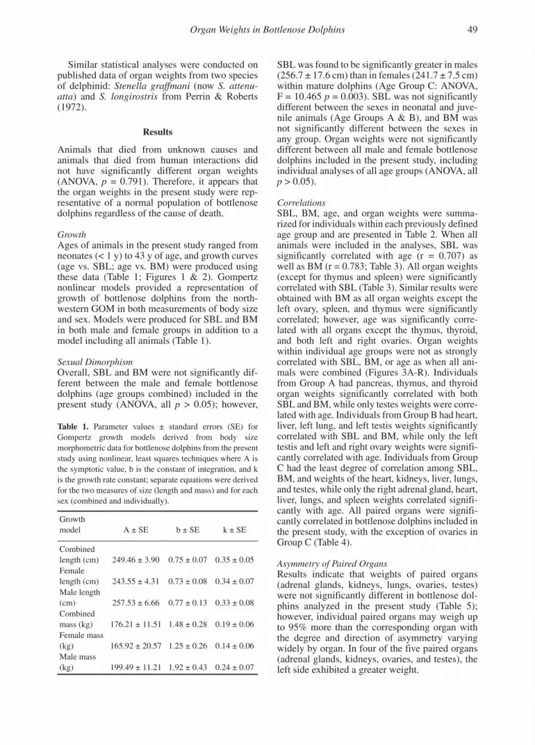
GrowthAges of animals in the present study ranged from neonates (< 1 y) to 43 y of age, and growth curves (age vs. SBL; age vs. BM) were produced using these data (Table 1; Figures 1 & 2). Gompertz nonlinear models provided a representation of growth of bottlenose dolphins from the north-western GOM in both measurements of body size and sex. Models were produced for SBL and BM in both male and female groups in addition to a model including all animals (Table 1).
Sexual DimorphismOverall, SBL and BM were not significantly dif-ferent between the male and female bottlenose dolphins (age groups combined) included in the present study (ANOVA, all p > 0.05); however,
SBL was found to be significantly greater in males (256.7 ± 17.6 cm) than in females (241.7 ± 7.5 cm) within mature dolphins (Age Group C: ANOVA, F = 10.465 p = 0.003). SBL was not significantly different between the sexes in neonatal and juve-nile animals (Age Groups A & B), and BM was not significantly different between the sexes in any group. Organ weights were not significantly different between all male and female bottlenose dolphins included in the present study, including individual analyses of all age groups (ANOVA, all p > 0.05).
CorrelationsSBL, BM, age, and organ weights were summa-rized for individuals within each previously defined age group and are presented in Table 2. When all animals were included in the analyses, SBL was significantly correlated with age (r = 0.707) as well as BM (r = 0.783; Table 3). All organ weights (except for thymus and spleen) were significantly correlated with SBL (Table 3). Similar results were obtained with BM as all organ weights except the left ovary, spleen, and thymus were significantly correlated; however, age was significantly corre-lated with all organs except the thymus, thyroid, and both left and right ovaries. Organ weights within individual age groups were not as strongly correlated with SBL, BM, or age as when all ani-mals were combined (Figures 3A-R). Individuals from Group A had pancreas, thymus, and thyroid organ weights significantly correlated with both SBL and BM, while only testes weights were corre-lated with age. Individuals from Group B had heart, liver, left lung, and left testis weights significantly correlated with SBL and BM, while only the left testis and left and right ovary weights were signifi-cantly correlated with age. Individuals from Group C had the least degree of correlation among SBL, BM, and weights of the heart, kidneys, liver, lungs, and testes, while only the right adrenal gland, heart, liver, lungs, and spleen weights correlated signifi-cantly with age. All paired organs were signifi-cantly correlated in bottlenose dolphins included in the present study, with the exception of ovaries in Group C (Table 4).
Asymmetry of Paired OrgansResults indicate that weights of paired organs (adrenal glands, kidneys, lungs, ovaries, testes) were not significantly different in bottlenose dol-phins analyzed in the present study (Table 5); however, individual paired organs may weigh up to 95% more than the corresponding organ with the degree and direction of asymmetry varying widely by organ. In four of the five paired organs (adrenal glands, kidneys, ovaries, and testes), the left side exhibited a greater weight.
Organ Weights in Bottlenose Dolphins 49
Table 1. Parameter values ± standard errors (SE) for Gompertz growth models derived from body size morphometric data for bottlenose dolphins from the present study using nonlinear, least squares techniques where A is the symptotic value, b is the constant of integration, and k is the growth rate constant; separate equations were derived for the two measures of size (length and mass) and for each sex (combined and individually).
Growth model A ± SE b ± SE k ± SE
Combined length (cm) 249.46 ± 3.90 0.75 ± 0.07 0.35 ± 0.05Female length (cm) 243.55 ± 4.31 0.73 ± 0.08 0.34 ± 0.07Male length (cm) 257.53 ± 6.66 0.77 ± 0.13 0.33 ± 0.08Combined mass (kg) 176.21 ± 11.51 1.48 ± 0.28 0.19 ± 0.06Female mass (kg) 165.92 ± 20.57 1.25 ± 0.26 0.14 ± 0.06Male mass (kg) 199.49 ± 11.21 1.92 ± 0.43 0.24 ± 0.07

Discussion
Age classes utilized in the present study were based upon the developmental growth curves of these ani-mals and were similar to those defined in previous studies of Tursiops truncatus (Read et al., 1993; Fernandez & Hohn, 1998; Turner, 1998; Turner & Worthy, 2003), other delphinids (Perrin et al., 1976, 1977), and odontocetes (Doidge, 1990; Read & Gaskin, 1990). Dolphins in the present study exhib-ited asymptotic values of length which were compa-rable to those calculated from a previous study of T. truncatus from a coastal Texas population (Fernandez & Hohn, 1998); however, both female and male dol-phins in the present study had smaller asymptotic values of SBL and BM than animals from the Gulf coast of Florida (Read et al., 1993), indicating that the individuals in the present study were inherently shorter and had smaller BM. SBL of dolphins in the present study were also compared with animals included in a study examining the morphometric variability in T. truncatus from both Texas and Florida GOM populations. Results indicate that the dolphins from the present study are morphometri-cally similar to those from coastal Texas populations and are smaller than those presumed to belong to the larger “offshore” ecotype (Turner, 1998).
Bottlenose dolphins in the present study did not exhibit sexual dimorphism in measures of SBL
and BM when all groups were combined, although male dolphins from Age Group C (i.e., mature males) were significantly larger. This is similar to results obtained from previous studies, indicat-ing that sexual dimorphism in SBL of bottlenose dolphins does not occur until physical matura-tion (Sergeant et al., 1973; Hohn, 1980; Kasuya et al., 1986; Cockroft & Ross, 1990; Read et al., 1993); however, BM of T. truncatus included in the present study were not sexually dimorphic, regardless of age, which is similar to results from the Atlantic Coast of the United States (Mead & Potter, 1990), but differs from the results of previ-ous studies from coastal South Africa (Cockroft & Ross, 1990) and Florida (Read et al., 1993), where males have considerably greater BM than females (26% and 11%, respectively).
Sexual dimorphism was not identified in weights of any of the organs assessed in the pres-ent study. Information on sexual dimorphism in individual organ weights of T. truncatus, as well as other delphinids, is limited at best. ANOVA tests were run using published data from a previ-ous study of the organ weights of two species of Stenella (Perrin & Roberts, 1972), indicating that sexual dimorphism was not present in the seven organs they examined (heart, lungs, liver, kidneys, and spleen), including all age groups (ANOVA, all p > 0.05). Therefore, from the limited amount of
Table 2. Summary statistics of standard body length (SBL), body mass (BM), age, and organ weights of bottlenose dolphins from the present study grouped by body size parameters A, B, and C as previously defined
Group A (£ 175 cm) Group B (175-225 cm) Group C (> 225 cm)
Organ Mean SD n Mean SD n Mean SD n
SBL (cm) 143.6 25.7 7 201.7 12.7 20 249.6 15.6 36BM (kg) 44.5 16.9 6 91.5 19.1 17 166.3 42.0 30Age (glg) 1.0 0.6 6 3.9 1.7 9 18.1 8.0 20L. adrenal (g) 2.6 1.2 7 6.3 1.9 19 11.3 3.3 36R. adrenal (g) 2.5 1.2 7 6.0 1.5 17 11.5 3.8 36Brain (g) 909.1 151.9 6 1,332.2 105.3 13 1,471.7 115.3 27Heart (g) 262.8 78.3 6 452.1 97.7 18 940.3 223.9 36L. kidney (g) 137.9 66.2 7 309.7 108.2 17 611.8 134.4 35R. kidney (g) 158.1 80.6 6 306.0 112.3 15 597.5 137.8 34Liver (g) 978.3 535.8 7 2,546.9 1,141.4 18 4,304.5 1,384.1 33L. lung (g) 623.8 279.1 7 1,204.3 519.6 18 2,872.9 900.9 34R. lung (g) 722.6 394.6 7 1,338.1 553.5 18 3,235.8 970.7 33L. ovary (g) 1.5 -- 1 2.7 1.0 5 12.3 9.7 10R. ovary (g) 1.7 -- 1 2.3 0.8 5 6.3 2.5 10Pancreas (g) 134.1 120.9 4 209.9 74.7 13 364.2 138.9 21Pituitary (g) 0.8 0.2 4 1.1 0.4 11 2.1 0.5 18Spleen (g) 60.8 42.7 5 73.4 31.5 17 92.9 44.4 34L. testis (g) 3.1 1.1 2 16.0 5.5 11 439.2 310.6 15R. testis (g) 2.9 0.9 2 15.7 5.8 11 443.2 308.9 15Thymus (g) 71.1 55.2 5 39.0 19.7 4 41.5 12.5 5Thyroid (g) 20.3 7.0 4 26.6 5.6 10 34.1 11.8 24
50 Turner et al.

data available, it appears that sexual dimorphism is not present in the organ weights of T. truncatus and at least two other similar delphinids, regard-less of age group.
Allometric relationships between organ weight and BM were similar to those described for other mammalian species (Schmidt-Nielsen, 1984). Weak correlations between age and most organ weights observed in the present study is con-sistent with previous work reported by Cowan (1966) on pilot whales (Globicephala melaena). Additionally, Cowan reported that all non-endo-crine organs were correlated with SBL (and BM through indirect calculations). During the expo-nential phase of the growth curve, it appears that SBL can be used as a relatively accurate mea-surement of age, although the phase of negative growth acceleration does not yield an accurate relationship. Therefore, data from the present study reveals that SBL is not a reliable indica-tor of age estimation in sexually mature animals. Results indicate that paired organs in bottlenose
dolphins are significantly correlated except for the ovaries in mature females. Data from T. truncatus and other related species were not available for comparison, making it difficult to determine if this feature is common in other delphinids.
Data from the present study indicate that weights of paired organs in T. truncatus were not significantly different, including adrenal glands, kidneys, lungs, ovaries, and testes. Similar results were identified in the kidneys of S. attenuatta and S. longirostris as no significant differences were identified; however, in both T. truncatus from the present study and Stenella spp. from a previous study (Perrin & Roberts, 1972), it appears that a large degree of variability exists between paired organs within individuals. For example, in both S. attenuatta and S. longirostris, the right kidney was often heavier, typically by 2 to 4%. Similar results were identified in kidneys of T. truncatus, although all other paired organs (adrenal glands, lungs, testes, and ovaries) exhibited a much larger degree of asymmetry (12.8%, 15.3%, 21.0%, and 95.9%, respectively). Furthermore, contrary to
Organ Weights in Bottlenose Dolphins 51
Table 3. Correlation coefficients of organ weights with standard body length (SBL), body mass (BM), and age for bottlenose dolphins from the present study; bolded values are p < 0.05.
Variable Group AgeL.
adrenalR.
adrenal Brain HeartL.
kidneyR.
kidney Liver L. lung R. lung
SBL All 0.707 0.758 0.699 0.824 0.848 0.865 0.859 0.761 0.800 0.812A 0.428 0.855 0.876 0.875 0.823 0.885 0.850 0.870 0.978 0.988B 0.754 0.598 0.589 0.308 0.634 0.447 0.513 0.528 0.644 0.576C 0.171 0.147 -0.002 0.218 0.488 0.537 0.580 0.400 0.427 0.441
BM All 0.783 0.715 0.674 0.736 0.923 0.859 0.861 0.778 0.840 0.869A 0.534 0.883 0.846 0.658 0.965 0.954 0.921 0.963 0.836 0.894
B 0.170 0.051 0.202 0.474 0.568 0.636 0.687 0.532 0.586 0.515C 0.439 0.252 0.153 0.092 0.808 0.567 0.575 0.533 0.578 0.650
Age All -- 0.725 0.790 0.490 0.857 0.805 0.837 0.828 0.837 0.834A -- 0.681 0.776 0.134 0.540 0.627 0.764 0.596 0.512 0.575B -- 0.669 0.714 0.503 0.443 0.089 -0.058 0.110 0.295 0.419C -- 0.283 0.489 -0.289 0.629 0.267 0.395 0.631 0.598 0.556
Variable Group L. ovary R. ovary Pancreas Pituitary L. testis R. testis Spleen Thymus Thyroid
SBL All 0.537 0.562 0.704 0.699 0.726 0.710 0.250 -0.294 0.493A -- -- 0.909 0.202 1.000 1.000 0.652 0.013 0.710B 0.352 0.231 0.530 0.277 0.770 0.633 0.096 0.942 0.670C 0.268 -0.414 0.421 0.017 0.491 0.607 0.149 -0.273 0.148
BM All 0.465 0.721 0.699 0.619 0.886 0.880 0.274 -0.266 0.526A -- -- 0.794 0.420 1.000 1.000 0.884 0.102 0.895B 0.986 0.995 0.574 0.529 0.695 0.525 0.772 0.445 0.455C 0.161 0.447 0.410 -0.379 0.795 0.923 -0.080 -0.556 0.084
Age All 0.126 0.349 0.711 0.489 0.778 0.764 0.507 -0.396 0.315A -- -- 0.559 -0.574 1.000 1.000 0.820 -0.286 -0.200B 1.000 1.000 0.006 -0.414 0.853 0.343 0.165 -- 0.800C -0.271 -0.250 0.206 -0.204 0.492 0.468 0.459 -0.392 -0.102

results from the study examining Stenella spp.’s kid-neys, the left side of most paired organs in the pres-ent study were heavier.
Correlation coefficients and the percent of organ weight of BM for T. truncatus from the
GOM were compared to published values for S. attenuatta and S. longirostris in the Pacific (Table 6). Similar correlation patterns were iden-tified across all three species, indicating that the overall relationships among SBL, BM, age, and
Figures 3A-F. Scatterplots of organ weight (g) versus standard body length (cm) of bottlenose dolphins from the present study, including A. left adrenal gland, B. right adrenal gland, C. brain, D. heart, E. left kidney, and F. right kidney; age classes (A, B, and C) are those defined in text.
52 Turner et al.

organ weights may be inherent among these three delphinids; however, significant differences were identified among the three species in regard to the percent of organ weight of BM (MANOVA, F = 8.436, p < 0.001). Univariate tests further revealed that of the six organs tested, there was no
significant difference in the proportion of heart and lung weight to body weight among the three species, while the percent weight of liver, left kidney, right kidney, and spleen (ANOVA, all p < 0.05) Tukey’s HSD tests indicated that propor-tional organ weights of the kidneys and spleen
Organ Weights in Bottlenose Dolphins 53
Figures 3G-L. Scatter plots of organ weight (g) versus standard body length (cm) of bottlenose dolphins from the present study, including G. liver, H. pancreas, I. left lung, J. right lung, K. left ovary, and L. right ovary; age classes (A, B, and C) are those defined in text.

were more similar between T. truncatus and S. longirostris than between S. longirostris and S. attenuatta. It is unknown whether these differ-ences relate to similarities in life history patterns or physiological parameters between T. trunca-tus and S. longirostris or whether the differences
between S. longirostris and S. attenuatta are a function of a large degree of variability within the Stenella genus.
The causes of stranding and/or death of animals in the present study varied and usually involved several factors. Thirteen of the 63 (20.6%) animals
Figures 3M-R. Scatter plots of organ weight (g) versus standard body length (cm) of bottlenose dolphins from the present study, including M. pituitary gland, N. spleen, O. left testis, P. right testis, Q. thymus, and R. thyroid; age classes (A, B, and C) are those defined in text.
54 Turner et al.

had evidence of severe trauma; nine of these 13 animals stranded/died due to human interaction (net entanglement, boat collision, or entanglement in fishing gear), two of the animals stranded/died due to intraspecific aggression, and two animals stranded/died from nontrauma-related causes. The remaining 50 bottlenose dolphins stranded/died from apparently natural causes or diseases. Since a stranded animal may not show signs of chronic disease or illness, we compared organ weights from human interaction vs natural causes or diseases and found that, with the exception of the lungs, diseases or pathologies observed in the present study did not significantly alter the weight of the organs.
Data on organ weights of bottlenose dolphins in the GOM, when correlated with SBL and age,
offer an important tool for pathologists and vet-erinarians for aiding in interpretation of health status. In general, it appears that dolphins that died from unknown causes and animals that died from human interactions did not have significantly different organ weights and, therefore, the organ weights presented here represent a normal popula-tion of bottlenose dolphins. Additionally, a special note should be made for two specific organs in bottlenose dolphins from the western GOM: lungs and thymus. First, we have described a previously unrecognized lung disease, angiomatosis, in T. truncatus within the GOM coastal area, which increased in both incidence and severity over the course of the study period (Turnbull & Cowan, 1999). In its less advanced stage, lungs with this disease are indistinguishable from lungs without the disease on the basis of weight, while the heavi-est lungs all have the advanced disease. Second, because of the location of the thymus (intimately related to the vessels of the aortic arch) and its involution with age, thymus weights are less accurate in older animals (Clark et al., 2005). In calves, it is easy to identify and collect the entire gland; however, in adults, the thymus softens and becomes progressively more indistinct, making complete removal problematic. Furthermore, in many animals, the thymus often develops large cysts, making weight disproportionately large (Cowan, 1994). Though no significant differences were detected in lung or thymus weights between animals that died from unknown causes and ani-mals that died from human interactions, caution should be used in interpreting lung and thymus weights included in the present study for reasons previously stated.
In summary, dolphins included in the present study were thought to belong to the coastal Texas population and were smaller and had less BM than similarly aged animals from Florida. Sexual dimor-phism was not present in T. truncatus from the pres-ent study when all groups were combined, although male dolphins from Group C were significantly larger. BM of T. truncatus included in the present study were not sexually dimorphic, regardless of age, which differs from the results of some previ-ous studies. Additionally, sexual dimorphism was not identified in weight of any of the organs assessed in the present study, which is comparable to results found in similar delphinids. Allometric relationships between organ weight and BM were similar to those described for other mammalian species, and data from the present study reveal that SBL is not a reli-able indicator of age estimation in sexually mature animals. Finally, results indicate that paired organs in bottlenose dolphins are significantly correlated, except for ovaries in mature females, and that no paired organs had significantly different weights.
Organ Weights in Bottlenose Dolphins 55
Table 4. Correlation coefficients of paired organ weights for bottlenose dolphins from the present study grouped by body size parameters A, B, and C as previously defined; all p < 0.05.
Organ Group r
Adrenals All 0.943Group A 0.974Group B 0.875Group C 0.878
Kidneys All 0.940Group A 0.992Group B 0.976Group C 0.772
Lungs All 0.972Group A 0.979Group B 0.927Group C 0.927
Ovaries All 0.583Group A --Group B 0.929Group C 0.320
Testes All 0.982Group A 1.000Group B 0.827Group C 0.785
Table 5. Asymmetry of paired organ weights within bottlenose dolphins from the present study
Paired organ
% difference in weight (g)
Larger side
% frequency n
Adrenals 12.8 ± 11.8 Left 61.6 60Kidneys 8.2 ± 15.4 Left 52.8 55Lungs 15.3 ± 14.7 Right 84.7 58Ovaries 95.9 ± 106.9 Left 80.0 16Testes 21.0 ± 37.4 Left 55.1 29

Tabl
e 6.
Cor
rela
tion
coef
fici
ents
and
per
cent
org
an w
eigh
t of
body
mas
s (B
M)
for
bottl
enos
e do
lphi
ns f
rom
the
pres
ent s
tudy
and
dat
a fr
om P
erri
n &
Rob
erts
(19
72)
on S
tene
lla
spp.
; G
roup
s of
wer
e th
ose
prev
ious
ly d
efin
ed (
T. tr
unca
tus)
or
usin
g si
mila
r cr
iteri
a (S
tene
lla
spp.
). B
olde
d va
lues
are
p <
0.0
5.
T. tr
unca
tus
S. a
tten
uatt
aS.
long
iros
tris
Org
anG
roup
r SB
Lr
BM
Ave
% o
f B
M ±
sd
r SB
Lr
BM
Ave
% o
f B
M ±
sd
r SB
Lr
BM
Ave
% o
f B
M ±
sd
Hea
rtA
ll0.
850.
920.
55±
0.07
0.97
0.97
0.51
±0.
120.
930.
940.
51±
0.06
Gro
up A
0.82
0.97
0.58
±0.
050.
900.
920.
75±
0.06
0.84
0.93
0.56
±0.
03G
roup
B0.
630.
570.
51±
0.08
0.81
0.92
0.48
±0.
040.
970.
970.
58±
0.04
Gro
up C
0.49
0.81
0.56
±0.
070.
550.
650.
42±
0.05
0.12
0.54
0.46
±0.
05L
. kid
ney
All
0.87
0.86
0.36
±0.
070.
880.
920.
45±
0.09
0.79
0.74
0.34
±0.
10G
roup
A0.
890.
950.
34±
0.04
0.93
0.90
0.53
±0.
050.
900.
940.
32±
0.02
Gro
up B
0.45
0.64
0.33
±0.
070.
790.
820.
44±
0.10
0.96
0.97
0.31
±0.
02G
roup
C0.
540.
570.
38±
0.07
0.78
0.71
0.46
±0.
07-0
.05
-0.3
00.
36±
0.15
R. k
idne
yA
ll0.
860.
860.
36±
0.07
0.88
0.92
0.48
±0.
100.
790.
740.
36±
0.10
Gro
up A
0.85
0.92
0.35
±0.
060.
520.
500.
56±
0.11
0.95
0.85
0.36
±0.
02G
roup
B0.
510.
690.
33±
0.07
0.77
0.82
0.46
±0.
090.
790.
810.
36±
0.03
Gro
up C
0.58
0.58
0.38
±0.
070.
760.
720.
48±
0.08
-0.1
1-0
.31
0.37
±0.
14L
iver
All
0.76
0.78
2.61
±0.
750.
920.
952.
28±
0.36
0.95
0.93
1.97
±0.
25G
roup
A0.
870.
962.
39±
0.26
0.67
0.75
1.84
±0.
270.
980.
881.
98±
0.14
Gro
up B
0.53
0.53
2.73
±0.
960.
820.
932.
34±
0.30
0.85
0.86
2.17
±0.
25G
roup
C0.
400.
532.
60±
0.69
0.56
0.50
2.37
±0.
37-0
.22
0.26
1.84
±0.
22L
ungs
All
0.73
0.81
3.26
±1.
000.
970.
993.
57±
0.55
0.95
0.99
3.27
±0.
13G
roup
A0.
990.
883.
24±
0.66
0.67
0.76
4.92
±0.
651.
001.
003.
35±
0.05
Gro
up B
0.29
0.43
2.57
±1.
050.
880.
953.
41±
0.26
1.00
1.00
3.26
±0.
15G
roup
C0.
490.
633.
66±
0.82
0.68
0.71
3.19
±0.
31--
----
Sple
enA
ll0.
250.
270.
07±
0.03
0.58
0.51
0.09
±0.
040.
140.
190.
07±
0.04
Gro
up A
0.65
0.88
0.13
±0.
040.
580.
600.
07±
0.02
0.46
0.63
0.11
±0.
04G
roup
B0.
100.
770.
08±
0.02
0.35
0.48
0.11
±0.
030.
250.
270.
08±
0.03
Gro
up C
0.15
-0.0
80.
06±
0.03
0.33
0.31
0.06
±0.
010.
170.
740.
04±
0.01
56 Turner et al.

Acknowledgments
The authors thank the Histopathology Department at the University of Texas Medical Branch for tissue processing. Similarly, thanks to the Texas Marine Mammal Stranding Network and all of the volunteers that made this research possible. Also, the authors would like to thank all reviewers of this manuscript.
Literature Cited
Clark, L. S., Turner, J. P., & Cowan, D. F. (2005). Involution of lymphoid organs in bottlenose dolphins (Tursiops trun-catus) from the western Gulf of Mexico: Implications for life in an aquatic environment. Anatomical Record, 282A(1), 67-73.
Cockroft, V. G., & Ross, G. J. B. (1990). Age, growth, and reproduction of bottlenose dolphins, Tursiops truncatus, from the east coast of southern Africa. Fishery Bulletin, 88(2), 289-302.
Cowan, D. F. (1966). Observations on the pilot whale Globicephala melaena: Organ weight and growth. Anatomical Record, 155, 623-628.
Cowan, D. F. (1994). Involution and cystic transformation of the thymus in the bottlenose dolphin, Tursiops trun-catus. Veterinary Pathology, 31, 648-653.
Doidge, D. W. (1990). Age-length and length-weight com-parisons in the beluga, Delphinapterus leucas. Canadian Bulletin of Fisheries and Aquatic Sciences, 244, 59-68.
Fernandez, S., & Hohn, A. A. (1998). Age, growth, and calving season of bottlenose dolphins, Tursiops trunca-tus, off coastal Texas. Fishery Bulletin, 96, 357-365.
Fitzhugh, H. A., Jr. (1975). Analysis of growth curves and strategies for altering their shape. Journal of Animal Science, 42, 1036-1051.
Hohn, A. A. (1980). Age determination and age related factors in the teeth of western north Atlantic bottlenose dolphins. Scientific Reports of the Whales Research Institute, 32, 39-66.
Hohn, A. A., Scott, M. S., Wells, R. S., Sweeney, J. C., & Irvine, A. B. (1989). Growth layers in teeth from known-age free-ranging bottlenose dolphins. Marine Mammal Science, 5, 315-342.
Kasuya, T., Tobayama, T., Siaga, T., & Kataoka, T. (1986). Perinatal growth of delphinids: Information from aquar-ium reared bottlenose dolphins and finless porpoises. Scientific Reports of the Whales Research Institute, 37, 85-97.
Mead, J. G., & Potter, C. W. (1990). Natural history of bot-tlenose dolphins along the central Atlantic Coast of the United States. In S. Leatherwood & R. R. Reeves (Eds.), The bottlenose dolphin (pp. 165-195). San Diago: Academic Press.
Perrin, W. J., & Myrick, A. C. (1980). Age determination of toothed whales and sirenians. Report of the International Whaling Commission, 3(Special Issue). 229 pp.
Perrin, W. J., & Roberts, E. L. (1972). Organ weights of non-captive porpoise (Stenella spp.). Bulletin of the Southern California Academy of Sciences, 71, 19-32.
Perrin, W. J., Coe, J. M., & Zwiefel, J. R. (1976). Growth and reproduction of the spotted porpoise, Stenella atten-uata, in the offshore eastern tropical Pacific. Fishery Bulletin, 74, 229-269.
Perrin, W. J., Holts, D. B., & Miller, R. B. (1977). Growth and reproduction in the eastern spinner dolphin, a geo-graphical form of Stenella longirostris in the eastern tropical Pacific. Fishery Bulletin, 75, 725-750.
Read, A. J., & Gaskin, D. E. (1990). Changes in growth and reproduction of harbour porpoises, Phocoena phocoena, from the Bay of Fundy. Canadian Journal of Fisheries and Aquatic Sciences, 47, 2158-2163.
Read, A. J., Wells, R. S., Hohn, A. A., & Scott, M. D. (1993). Patterns of growth in wild bottlenose dolphins, Tursiops truncatus. Journal of Zoology, 231, 107-123.
Schmidt-Nielsen, K. (1984). Scaling: Why is animal size so important? New York: Cambridge University Press. 241 pp.
Sergeant, D. E., Caldwell, D. K., & Caldwell, M. C. (1973). Age, growth, and maturity of bottlenose dolphins (Tursiops truncatus) from northeast Florida. Journal of Fisheries Research Board of Canada, 30, 1009-1011.
Turnbull, B. S., & Cowan, D. F. (1999). Angiomatosis: A newly recognized disease in Atlantic bottlenose dol-phins (Tursiops truncatus) from the Gulf of Mexico. Veterinary Pathology, 36, 28-34.
Turner, J. P. (1998). A comparison of the cranial morphol-ogy of bottlenose dolphins (Tursiops truncatus) in the Gulf of Mexico. Master’s of Science thesis, Texas A&M University, College Station, TX.
Turner, J. P., & Worthy, G. A. J. (2003). Skull morphometry of bottlenose dolphins (Tursiops truncatus) from the Gulf of Mexico. Journal of Mammalogy, 84, 665-672.
Worthy, G. A. J. (1998). Patterns of bottlenose dol-phin, Tursiops truncatus, stranding in Texas. In R. Zimmerman (Ed.), Characteristics and causes of Texas marine strandings (NMFS/NOAA Technical Report 143) (pp. 47-55).
Organ Weights in Bottlenose Dolphins 57
Recommended


