
Possible Interactions between Gonadotrophs andSomatotrophs in the Pituitary of Tilapia: ApparentRoles for Insulin-Like Growth Factor I and Estradiol*
PHILIPPA MELAMED†, GAL GUR, HANNAH ROSENFELD, ABIGAIL ELIZUR,AND ZVI YARON‡
Department of Zoology, Tel-Aviv University (P.M., G.G., H.R., Z.Y.), Ramat Aviv 69978, Israel; and theNational Center for Mariculture, Israel Oceanographic and Limnological Research (H.R., A.E.),Eilat, Israel
ABSTRACTThe unique organization of the teleost pituitary, in which cells are
grouped according to their characteristic hormone, makes this a suit-able model for studying pituitary paracrine interactions. In a numberof fish, including tilapia, there are variations in the circulating levelsof the gonadotropins and GH, which are elevated during the repro-ductive season, suggesting interactions between the reproductive andgrowth axes. The aim of this study was to investigate paracrineinteractions between the gonadotrophs and somatotrophs in the ti-lapia pituitary. Initially, dispersed pituitary cells were separated ona density gradient in which the gonadotrophs were found in the leastdense fractions, and the somatotrophs were concentrated in the dens-est fraction. After 4 days in culture, cells in the least dense fractionsshowed characteristic cytoplasmic extensions not seen in the soma-totrophs, which appeared small and failed to form aggregates; soma-totrophs were found, however, attached to other non-GH cells. Stain-ing of the nuclei with 4,6-diaminidino-2-phenyl-dihydrochloride
revealed that the isolated somatotrophs had undergone nuclear con-densation and fragmentation typical of apoptosis. Addition of eitherestradiol or human recombinant insulin-like growth factor I (IGF-I;10 nM) to the somatotroph cultures increased the number of cellaggregates and reduced the number of condensed or fragmented nu-clei. Immunocytochemical studies on pituitary sections revealedIGF-I immunoreactivity in regions of the proximal pars distalis thatstain with gonadotropin IIb antisera and also in regions of the rostralpars distalis characteristic of corticotrophs; immunoreactive IGF-Iwas never seen in the region of the somatotrophs. Incubation of cellsfrom the different fractions with testosterone (10 nM; 24 h) revealedthat cells of the least dense fractions, which were rich in gonado-trophs, possessed aromatizing ability, which was absent in the so-matotroph-enriched fraction. These results suggest that estradiol andIGF-I, both generated from cells other than the somatotrophs, mayexert antiapoptotic effects and thus possibly control the size of thispopulation of cells. (Endocrinology 140: 1183–1191, 1999)
THE CELLS of the teleost pituitary pars distalis, unlikethose of mammals, are segregated into distinct regions
according to the characteristic hormone that they secrete. Intilapia, the somatotrophs are located in the proximal parsdistalis (PPD) forming a palisade around the nerve ramifi-cations; the gonadotropin (GtH) I (FSH-like) gonadotrophsare adjacent but slightly peripheral to them, whereas the GtHII (LH-like) gonadotrophs outlay these cells, and the lac-totrophs are found in a separate location in the rostral parsdistalis (RPD) (1–3). The hypothalamic regulatory factors,which in the absence of a portal system reach their target cellsvia nerve fibers, appear to show somewhat less specificitythan in mammals. For example, GnRH stimulates the releaseof both GtH and GH in a number of teleosts, and in vitrostudies in tilapia have suggested that PRL is similarly af-fected (2, 4, 5). In contrast, dopamine inhibits both GtH andPRL release while stimulating that of GH (2, 6, 7).
Studies of the circulating levels of these hormones alsosuggest some correlation in their regulation; GH levels ap-pear to increase with GtH II at the time of ovulation andspawning, whereas levels of GtH I, which is predominantduring vitellogenesis, decrease at this time (8–12). Apartfrom these changes in circulating hormone levels, changes inthe sizes of these cell populations have been noted in rainbowtrout in which the GtH I gonadotrophs are found in greaternumber in immature fish, whereas in mature fish the GtH IIgonadotrophs predominate (13, 14). At the time of spermi-ation, the somatotroph population is also notably larger inthese fish (15).
Studies in tilapia suggest that the direct effects of steroids onthe expression of the GtH b-subunits may explain some of thechanging patterns in their circulatory levels. No direct effects ofthe gonadal steroids were seen on expression of the tilapia GHgene, although they did appear to increase the sensitivity of thesomatotrophs to some of the hypothalamic GH-releasing hor-mones. The effects of testosterone could be mimicked by es-tradiol (E2), but were not mimicked by the nonaromatizable11-ketotesoterone, suggesting that the testosterone is aroma-tized before eliciting these effects (3, 16, 17).
Despite the apparent overlap in the regulation and levelsof activity of gonadotrophs and somatotrophs, little attentionhas been paid to the possible interactions between them. Inmammals, locally produced paracrine factors have been re-
Received June 23, 1998.Address all correspondence and requests for reprints to: Dr. Zvi
Yaron, Department of Zoology, Tel Aviv University, Ramat Aviv 69978,Israel. E-mail: [email protected].
* This work was supported by the Israel Science Foundation foundedby the Israel Academy of Sciences and Humanities.
† Current address: Department of Laboratory Medicine and Patho-biology, University of Toronto, 100 College Street, Room 351, Toronto,Ontario, Canada M5G 1L5.
‡ The Norman and Rose Lederer Chair of Experimental Biology.
0013-7227/99/$03.00/0 Vol. 140, No. 3Endocrinology Printed in U.S.A.Copyright © 1999 by The Endocrine Society
1183

ported to regulate the synthesis and release of gonadotro-pins, GH and PRL, and also the differentiation of the cells.These paracrine factors include locally produced cytokinesand growth factors, neurohormones produced in pituitarycells such as GnRH, and also the pituitary hormones them-selves or parts thereof, such as the glycoprotein a-subunit(18–22). In the goldfish pituitary, activin and inhibin sub-units have also been implicated, as these are found in thesomatotrophs and stimulate the release of both GtH and GH(23, 24). Little information is available regarding the locationor actions of other cytokines and growth factors in the teleostpituitary. However, after the finding that insulin-like growthfactor I (IGF-I) is produced locally in multiple organs intilapia, it has been hypothesized that it may be involved inautocrine or paracrine actions in organ-specific functions(25). The effects of IGF-I in the teleost pituitary, depressingGH release and messenger RNA (mRNA) levels, have beenshown (3, 26).
The aim of this study was to examine possible paracrineinteractions operating between the gonadotrophs and soma-totrophs in the pituitary of tilapia and to investigate the rolesof E2 and IGF-I. This required the initial establishment of atechnique for separation of the pituitary cell populations.
Materials and MethodsFish
The fish used in the study were tilapia hybrids (Oreochromis aureus 3O. niloticus) collected from the ponds of local fish farms. At watertemperatures above 22 C, these fish will start gonadal development ata body weight of 30 g. As temperatures fall below this, fish that havealready reached sexual maturity will undergo gonadal regression. Cellsfrom fish at various reproductive stages were used for validation of thecell separation technique, whereas for other experiments cells from fishat specific reproductive stages were employed, as stated in Results. Thereproductive stage of the fish was assessed by measurement of theirgonadosomatic index (percent gonadal weight/body weight) taken to-gether with their absolute weight and the season when they died.
Separation of pituitary cells
Pituitaries were collected aseptically, and the cells were dispersed bytrypsinization as described previously (27). After the addition of FCS(final concentration, 20%) to stop the reaction, the cells were counted,and the cell suspension (5 ml) was loaded onto the density gradient (10ml). The gradient was made up of four concentrations (35%, 45%, 55%,and 65%) of Universal Separation Media (Sigma Chemical Co., St. Louis,MO) with densities of 1.0544–1.0985 g/ml. The cells were centrifugedthrough the gradient at 1200 3 g for 15 min at 18–20 C. The majority ofcells were found to segregate into three clear fractions (fractions 1–3),whereas some cells failed to penetrate the gradient, and a few (mostlyred blood cells) were found below the densest part of the gradient. Eachfraction was collected by gentle aspiration and rinsed in 10 ml Hanks’Balanced Salt Solution (5 min, 800 3 g, 18–20 C). The cells of each fractionwere then resuspended in 1 ml Hanks’ Balanced Salt Solution andcounted. The viability of the cells in each of three major fractions wasgreater than 95%. Repeated experiments showed that an average of59.14 6 6.57% of the initial number of cells were recovered (n 5 8), with49.47 6 2.47% of these found in the least dense fraction 1, 29.24 6 2.31%in fraction 2, and 20.3 6 0.97% in the densest fraction 3 (n 5 9). Thevalidation of this technique was performed on numerous occasions,using fish at different reproductive stages.
Culture of separated cells
After separation, cells were plated in Corning 24- or 96-well tissueculture plates (Corning, Corning, NY), at a density of 2.5 3 105 cells/wellin 1 ml or a density of 6.25 3 104 cells/well in 200 ml medium [medium
199, 10% FCS, 10 mm HEPES, and 1% antibiotic suspension (Pen-strep-nystatin suspension, Biological Industries, Bet HaEmek, Israel)]. Previ-ous experiments on GH and GtH release have shown these to be optimaldensities for hormone release. Additional preliminary experiments werecarried out in the present study in which the cells from fraction 3 wereplated at as much as twice this density. On finding that the increaseddensity had no effect on cell morphology, subsequent experiments em-ployed the cell densities stated above. The cells were incubated for 4 daysat 28 C under 5% CO2.
On the fourth day, cultured cells were photographed using an in-verted microscope (Olympus Corp., Melville, NY) and phase contrastoptics. In various experiments, the cells were exposed to recombinanthuman IGF-I (rhIGF-I; dissolved in 0.1 m acetic acid; Life Technologies,Grand Island, NY), recombinant salmon IGF-I (provided by GroPep,Adelaide, Australia; dissolved in 10 nm HCl), E2, or testosterone (SigmaChemical Co., dissolved in ethanol). The final concentration of the sol-vents comprised less than 0.1% of the culture medium. The undilutedFCS contained 7.2 nm IGF-I determined as human IGF-I (Silbergeld, A.,personal communication).
Immunocytochemistry (ICC)
ICC was carried out on paraffin-embedded pituitary sections, asdescribed previously (2), after initial blocking with 10% normal goatserum. The primary antisera used were anti salmon IGF-I (1:2500; a giftfrom GroPep, Adelaide, Australia), antirecombinant tilapia GH (1:8000)(2), and antiserum produced against the recombinant b-subunit of ti-lapia GtH II (1:4000; Zmora, N., and A. Elizur, unpublished). The secondantibody (goat antirabbit) was used at a dilution of 1:600, and peroxi-dase-antiperoxidase (Sigma Chemical Co.) was used at a dilution of1:200. The reaction was detected using diaminobenzidine (DAB) andperoxidase.
Cells were fixed in the culture wells (after plating as described above)overnight by the addition of formaldehyde to the incubation medium toa final concentration of 4%. The ICC reactions employed the GH and GtHIIb antisera as described above, and the technique was essentially thesame.
4,6-Diaminidino-2-phenyl-dihydrochloride (DAPI) stainingof fixed cells
The cells were fixed as described above and rinsed in phosphatebuffer solution (1 m PBS, pH 7.4) before the addition of DAPI (1 mg/mlPBS; Sigma Chemical Co.) for 5 min. After the removal of DAPI, the cellswere rinsed again in PBS before being examined under an invertedmicroscope fitted with a U-MNU filter (Olympus Corp.) and photo-graphed using Fuji Photo Film Co. Ltd. TMX 400 film (Tokyo, Japan).
In the case of double staining, nuclei were stained first with DAPI, andconsequently, ICC reactions were performed as described above. Thecells were photographed with and without the U-MNU filter, using FujiPhoto Film Co. Ltd. TMX 400 film.
RIAs and measurement of aromatase activity
The cellular hormone content of the different fractions was measuredusing RIAs specific for tilapia GtH or GH. The RIA for GH, using therecombinant hormone, has been described previously (2). The RIA forGtH was based on native GtH that was purified from tilapia pituitariesharvested during the spawning season (28).
For the measurement of aromatase activity in cells of the variousfractions, the level of E2 secreted into the incubation medium was mea-sured after 24-h exposure to graded concentrations of testosterone (Sig-ma Chemical Co.). This RIA employed a previously validated antiserumand an iodinated standard (Diagnostics Products Corp., Los Angeles,CA). The cross-reaction of the assay was 0.79% with estrone, 0.34% withestriol, and less than 0.0001% with testosterone, 17a,20b-dihydroxypro-gesterone and cortisol (29). The sensitivity of this assay was 0.66pg/tube.
Measurement of mRNA levels
Comparative levels of GH, GtH Ib, and GtH IIb mRNA were mea-sured in 1 3 106 cells from each of the fractions. Total RNA was extracted
1184 PARACRINE INTERACTIONS IN THE TILAPIA PITUITARY Endo • 1999Vol 140 • No 3
from the cells using a modification of the guanidinium-phenol-chloro-form method, as previously described (27). The samples were run on a1.2% agarose gel and transferred to nylon membranes (GeneScreen Plus,New England Nuclear Research Products, Boston, MA) by Northernblotting. RNA on the membranes was hybridized with complementaryDNA probes for GH and GtH IIb and a DNA probe for GtH Ib, asdescribed previously (17). After rinsing, the membranes were exposedto the image plate of a phosphorimager (BAS 1000, Fuji Photo Film Co.,Ltd.) for 1 h.
Statistical analysis
Quantitative analysis of apoptotic cells was performed by countingthe number of normal nuclei in a field (6.4 3 0.95 mm) that spanned thediameter of the well; this was then compared with the situation in control(untreated) cells. In addition, the degree of aggregation of the cells wasassessed by counting the number of aggregates with a diameter greaterthan 0.15 mm in a similar field. These measurements were repeated inthree or four wells in three separate experiments.
Statistical analysis employed ANOVA followed by the least signifi-cant difference test. All experiments were carried out numerous times.
ResultsValidation of cell separation technique
Contents of GtH and GH in cells of the different fractions. Pitu-itary cells were dispersed and separated as described above,and 250,000 cells from each fraction were homogenized inTriton X-100 (0.1%). The GtH and GH contents were mea-sured in an aliquot from each fraction of cells. In fraction 1,there was 8.4 times more GtH than GH, whereas in fraction2 the GH was 2.5 times the quantity of GtH, and in fraction3 GH was found at over 20 times the amount of GtH. Thisdistribution of hormone over the gradient was characteristicregardless of the reproductive stage of the fish.
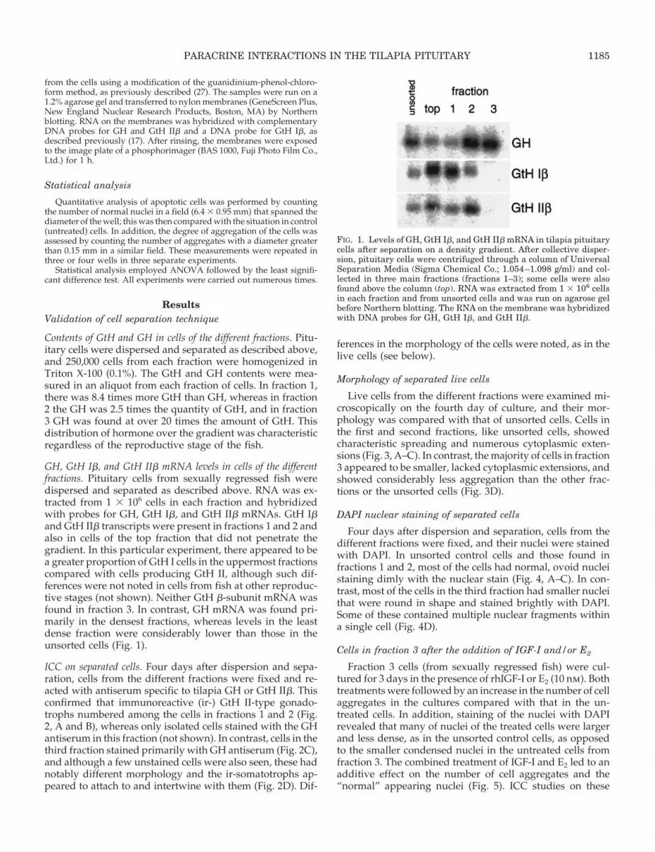
GH, GtH Ib, and GtH IIb mRNA levels in cells of the differentfractions. Pituitary cells from sexually regressed fish weredispersed and separated as described above. RNA was ex-tracted from 1 3 106 cells in each fraction and hybridizedwith probes for GH, GtH Ib, and GtH IIb mRNAs. GtH Iband GtH IIb transcripts were present in fractions 1 and 2 andalso in cells of the top fraction that did not penetrate thegradient. In this particular experiment, there appeared to bea greater proportion of GtH I cells in the uppermost fractionscompared with cells producing GtH II, although such dif-ferences were not noted in cells from fish at other reproduc-tive stages (not shown). Neither GtH b-subunit mRNA wasfound in fraction 3. In contrast, GH mRNA was found pri-marily in the densest fractions, whereas levels in the leastdense fraction were considerably lower than those in theunsorted cells (Fig. 1).
ICC on separated cells. Four days after dispersion and sepa-ration, cells from the different fractions were fixed and re-acted with antiserum specific to tilapia GH or GtH IIb. Thisconfirmed that immunoreactive (ir-) GtH II-type gonado-trophs numbered among the cells in fractions 1 and 2 (Fig.2, A and B), whereas only isolated cells stained with the GHantiserum in this fraction (not shown). In contrast, cells in thethird fraction stained primarily with GH antiserum (Fig. 2C),and although a few unstained cells were also seen, these hadnotably different morphology and the ir-somatotrophs ap-peared to attach to and intertwine with them (Fig. 2D). Dif-
ferences in the morphology of the cells were noted, as in thelive cells (see below).
Morphology of separated live cells
Live cells from the different fractions were examined mi-croscopically on the fourth day of culture, and their mor-phology was compared with that of unsorted cells. Cells inthe first and second fractions, like unsorted cells, showedcharacteristic spreading and numerous cytoplasmic exten-sions (Fig. 3, A–C). In contrast, the majority of cells in fraction3 appeared to be smaller, lacked cytoplasmic extensions, andshowed considerably less aggregation than the other frac-tions or the unsorted cells (Fig. 3D).
DAPI nuclear staining of separated cells
Four days after dispersion and separation, cells from thedifferent fractions were fixed, and their nuclei were stainedwith DAPI. In unsorted control cells and those found infractions 1 and 2, most of the cells had normal, ovoid nucleistaining dimly with the nuclear stain (Fig. 4, A–C). In con-trast, most of the cells in the third fraction had smaller nucleithat were round in shape and stained brightly with DAPI.Some of these contained multiple nuclear fragments withina single cell (Fig. 4D).
Cells in fraction 3 after the addition of IGF-I and/or E2
Fraction 3 cells (from sexually regressed fish) were cul-tured for 3 days in the presence of rhIGF-I or E2 (10 nm). Bothtreatments were followed by an increase in the number of cellaggregates in the cultures compared with that in the un-treated cells. In addition, staining of the nuclei with DAPIrevealed that many of nuclei of the treated cells were largerand less dense, as in the unsorted control cells, as opposedto the smaller condensed nuclei in the untreated cells fromfraction 3. The combined treatment of IGF-I and E2 led to anadditive effect on the number of cell aggregates and the“normal” appearing nuclei (Fig. 5). ICC studies on these
FIG. 1. Levels of GH, GtH Ib, and GtH IIb mRNA in tilapia pituitarycells after separation on a density gradient. After collective disper-sion, pituitary cells were centrifuged through a column of UniversalSeparation Media (Sigma Chemical Co.; 1.054–1.098 g/ml) and col-lected in three main fractions (fractions 1–3); some cells were alsofound above the column (top). RNA was extracted from 1 3 106 cellsin each fraction and from unsorted cells and was run on agarose gelbefore Northern blotting. The RNA on the membrane was hybridizedwith DNA probes for GH, GtH Ib, and GtH IIb.
PARACRINE INTERACTIONS IN THE TILAPIA PITUITARY 1185

same treated cells confirmed that they were indeed soma-totrophs (Fig. 6, A and B). Similar results were obtained whensalmon IGF-I was used in place of the recombinant humanpeptide (not shown).
Cells from fraction 3 after addition of conditioned mediumfrom fraction 1 or 2
Conditioned medium was collected from fraction 1 and 2cells (from sexually regressed fish) after 4 days in culture.This medium was added, either undiluted or after dilution(1:1) with freshly prepared medium, to cells from fraction 3immediately after their separation. After 2–4 days the cellswere fixed, and their nuclei were stained with DAPI. Nochange was noted in either the morphology of the live cellsor in the size or shape of the nuclei, and the cells remainedsimilar in appearance to untreated cells from this fraction(not shown).
ICC localization of IGF-I in the tilapia pituitary
Numerous ICC reactions on sections of tilapia pituitariesfailed to show ir-IGF-I in the region of the somatotrophs (e.g.Figs. 7 and 8). In some, but not all, pituitaries from maturefish, IGF-I immunoreactivity was seen in areas of the prox-imal pars distalis corresponding to those also reacting withantiserum to GtH IIb (Fig. 7, A and C). In another pituitary,
ir-IGF-I was also apparent in the rostral pars distalis in a thinlayer of epithelial cells lining the junction of the RPD and theneurohypophysis (Fig. 8A).
Aromatase activity in separated pituitary cells
Pituitaries were collected from sexually mature male andfemale tilapia at the height of the spawning season (gona-dosomatic index of 0.4 6 0.09 for males or 1.91 6 0.35 forfemales; n 5 13); the cells were dispersed, separated, andcultured at 6.25 3 104 cells/well as described above. On thethird day of culture, testosterone (1–100 nm) was added for24 h, after which the medium was collected for the mea-surement of E2. In cultures of both male (Fig. 9A) and female(Fig. 9B) pituitary cells, the highest levels of E2 were foundin cells of fraction 1. Cultures of fraction 2 cells containedlower levels of E2, similar to the levels in unsorted cells. Incells not exposed to testosterone and those from fraction 3after all levels of testosterone exposure, E2 was undetectable(Fig. 9, A and B).
Discussion
This study has demonstrated that tilapia somatotrophsand gonadotrophs can be separated on a density gradientand remain viable. Consistently, regardless of the reproduc-tive state of the fish, the gonadotrophs were found in the least
FIG. 2. ICC of tilapia GH and GtH on cells from fractions 1, 2, and 3. Pituitary cells were dispersed and separated as described in Fig. 1 andwere cultured for 4 days before fixation. ICC studies employed antiserum specific to tilapia GtH IIb (1:4000) or GH (1:8000), with goat antirabbitantiserum (1:600) and peroxidase-antiperoxidase complex (1:200); detection was performed using DAB. Fraction 1 (A) and fraction 2 (B) cellsafter reaction with GtH IIb antiserum. C and D, Cells from fraction 3 after reaction with GH antiserum. Arrows indicate immunoreactive cells.Scale bar, 50 mm.
1186 PARACRINE INTERACTIONS IN THE TILAPIA PITUITARY Endo • 1999Vol 140 • No 3

dense fractions, although in sexually regressed fish, slightdifferences were noted between the two gonadotroph pop-ulations; the somatotrophs were invariably found in thedensest fractions. The densities of these pituitary cells appearto differ in various teleosts; for example, in the African cat-fish, gonadotrophs separated on a Percoll gradient werefound below the densest fraction (1.095 g/ml), whereas theacidophils were mostly in the least dense (1.049–1.083 g/ml)fractions (30). Goldfish gonadotrophs were found in Percolldensity fractions similar to those in the catfish (1.095 g/ml),but the somatotrophs were largely in the 1.083 g/ml fraction(31). In contrast, but similar to the current findings in tilapia,rainbow trout somatotrophs were found in the densest Per-coll fractions (1.066–1.102 g/ml), although the GtH II gona-dotrophs were almost equally distributed among the remain-ing fractions (1.027–1.096) (15).
In the separated tilapia pituitary cell cultures, markeddifferences in morphology were noted in cells of the variousfractions. Notably, cells in the somatotroph-enriched fractionhad a peculiar appearance and failed to aggregate. Unsortedcells or those from the least dense fractions examined after4 days in culture show numerous cytoplasmic extensions.ICC studies revealed that the extensions were present ingonadotrophs as well as in other unidentified cells in thesefractions. However, the ir-somatotrophs in fraction 3 weredevoid of such extensions. This contrasts with the situationin goldfish, where small cytoplasmic extensions were seen on
pituitary cells identified as somatotrophs (31). In fact, whencultured with other cell types, the tilapia somatotrophs ap-peared to be attached to the spreading cells.
After staining the nuclei with DAPI, a reason for the pe-culiar morphology of the separated somatotrophs becameevident. The nuclei of isolated somatotrophs had undergonecondensation and fragmentation typical of apoptosis. Alsotypical of this kind of cell death was a decrease in the cy-toplasmic volume of the cells leading to a shrunken appear-ance. In contrast, tilapia somatotrophs in mixed culture, asshown in previous studies, retained their viability withoutshowing signs of nuclear damage or cell death (17, 27) (ourunpublished observations).
To test the hypothesis that substances secreted from othercell types prevent the peculiar appearance of the isolatedsomatotrophs, the somatotroph-enriched culture was incu-bated with conditioned medium from the uppermost frac-tion, but the appearance of the cells was not altered. How-ever, the addition of either rhIGF-I or E2 to the somatotroph-enriched cultures increased the numbers of normalappearing nuclei and increased the degree of cell aggrega-tion. Previous studies on tilapia pituitary cells have shownthat somatotrophs respond to treatment with rhIGF-I byreducing the GH mRNA levels (3), whereas studies in mam-mals have demonstrated the presence of IGF-I receptors onthese cells (32). The present study has demonstrated that inthe tilapia pituitary, ir-IGF-I is not found in the somatotrophs
FIG. 3. Live pituitary cells 4 days after separation. Pituitary cells were dispersed and separated as described in Fig. 1 and cultured for 4 days.A, Unsorted cells; B, fraction 1 cells; C, fraction 2 cells; D, fraction 3 cells. Phase contrast. Scale bar, 50 mm.
PARACRINE INTERACTIONS IN THE TILAPIA PITUITARY 1187

but was seen in the same region of the PPD reacting withantisera to GtH IIb in some, but not all, pituitaries. The reasonfor these variations is not yet apparent. In addition, cells inthe RPD, in a location typical of the corticotrophs (33), alsoreacted with the IGF-I antiserum. Although the type of pi-tuitary cells producing IGF-I in mammals remains in dispute,most evidence points to the folliculostellate cells, with othertypes, including the somatotrophs, possibly also being in-volved (20, 21, 32).
Aromatase activity was found only in the least dense frac-tions of separated cells where the majority of gonadotrophsare located. It is suggested, therefore, that the source ofpituitary E2 in tilapia is the gonadotrophs, although the pos-sibility that it originates from other unidentified cells in thisfraction cannot be excluded. This is opposed to previousstudies of the Oreochromis mossambicus, in which it was sug-gested that aromatase activity, predominant in the PPD, orig-inates from the somatotrophs (34). Also in another teleost, thelonghorn sculpin, and in mammals, somatotrophs have beenimplicated as the location of this enzyme (35, 36). Aromataseactivity was seen in separated gonadotrophs of the Africancatfish, although it was also present in other cell types (37).It appears that in teleosts, brain and pituitary aromataselevels far exceed those in mammals and also exceed thosefound in the gonads, suggesting that in situ synthesis may bethe major source of estrogen within these tissues (38). In the
FIG. 5. Addition of IGF-I or E2 to cells from fraction 3. After disper-sion and separation (as described in Fig. 1), pituitary cells werecultured for 3 days in the presence of IGF-I and/or E2 (10 nM) beforefixation and staining of the nuclei with DAPI. The number of cellaggregates with a diameter of at least 0.15 mm was counted in a fieldspanning the diameter of the well (6.4 3 0.95 mm) and was comparedwith that for untreated fraction 3 cells. The same procedure was usedto evaluate the number of normal appearing nuclei. Means of alltreatment groups differed from those of controls (P , 0.05). Values arethe mean 6 SEM (n 5 3).
FIG. 4. DAPI staining of nuclei in cells from fractions 1–3. Cells were dispersed and separated as described in Fig. 1 and were cultured for 4days before fixation and staining of the nuclei with DAPI. Control cells (A) and those in fraction 1 (B) and fraction 2 (C) have mostly large ovoidnuclei that stain dimly with the nuclear stain. D, Cells in fraction 3 have smaller, dense nuclei that stain brightly with DAPI, and some cellsshow multiple nuclear fragments. Scale bar, 100 mm.
1188 PARACRINE INTERACTIONS IN THE TILAPIA PITUITARY Endo • 1999Vol 140 • No 3

present study, the location of the aromatizing ability in ti-lapia pituitary cells was similar in cells from sexually maturemales and females, although it was more potent in cells fromthe male fish. This difference presumably arose because ofslight differences in the reproductive state of the fish.
The additive effects of IGF-I and E2 treatment suggest thatthese factors activate different mechanisms to preserve thesomatotroph population. In mammals, endogenously pro-duced IGF-I prevents apoptosis of the ovarian follicles andalso slightly increased the production of E2 by cultured pre-ovulatory follicles (39, 40). In granulosa cells of early or
FIG. 6. After combined IGF-I and E2 treatment, cells from fraction 3were stained with DAPI and then reacted with antiserum to GH asdescribed in Fig. 2. A, Cluster of cells showing immunoreactivity toGH; B, the same cells after staining with DAPI. Scale bar, 10 mm.
FIG. 7. IGF-I immunoreactivity in the proximal pars distalis of thetilapia pituitary. Paraffin sections were reacted with antisera tosalmon IGF-I (A; 1:2500), tilapia GH (B; 1:8000), or tilapia GtH IIb(C; 1:4000), which were detected using goat antirabbit antiserum(1:600) and peroxidase-antiperoxidase complex (1:200). PPD, Proxi-mal pars distalis. The DAB reaction product is marked by arrows.Scale bar, 40 mm.
PARACRINE INTERACTIONS IN THE TILAPIA PITUITARY 1189

preantral follicles, apoptosis increased after estrogen with-drawal, and this was completely prevented by replacementwith diethylstilbesterol or estradiol benzoate, whereas an-drogens had the opposite effect (41, 42).
The reason for the failure of conditioned medium fromcells of the upper fractions to prevent the morphologicalchanges in the isolated somatotrophs is not entirely clear. Ithas been suggested, however, that the control of IGF-I in thepituitary is mediated by GH (21). If a similar situation pre-vails in tilapia, then a reduction in the number of soma-totrophs in the upper fractions could have abated the releaseof IGF-I. It should be noted that the basal E2 levels in allfractions were below the detection limit (i.e. ,6.6 pg/ml wassecreted during 24 h) even though these cells were fromsexually mature fish. It is probable, therefore, that the con-ditioned medium (taken from cells of sexually regressed fish)simply did not contain enough E2 or IGF-I to elicit an effect.
There is direct evidence in certain teleosts that the numbersof somatotrophs and type II gonadotrophs increase duringthe breeding season and are lower in sexually regressed fish(13–15). In tilapia, cyclical circulating GH levels and their
response to GH-releasing hormones are greatest at the heightof the reproductive season, whereas GtH IIb mRNA levelspeak with maximal gonadal development (16, 17). Thesechanges could arise from either an increase in the size ofspecific cell populations or simply from increased activity ofthe same cells. Results from the present study suggest that adecrease in the number of cells producing GtH II, for what-ever reason, could lead to a reduction in both the aromatizingability of the pituitary and the output of IGF-I. This coupledwith a decrease in circulating steroid levels (occurring in thesexually regressing fish) could also lead to a decrease in thenumber of somatotrophs by allowing programmed celldeath.
Thus, IGF-I appears to have paradoxical effects on thesomatotrophs in tilapia: on the one hand, reducing GH re-lease and synthesis (3) and, on the other, preventing a re-duction in the somatotroph population. Such actions mean
FIG. 8. A, IGF-I immunoreactivity in the RPD of the tilapia pituitary.The ir-IGF-I is seen in a thin layer of epithelial cells lining the junctionof the RPD and the pars neurointermedia (N). B, GH immunoreac-tivity is seen in a different region of the pituitary, in the proximal parsdistalis. Reaction details are given in Fig. 7. RD, RPD; PD, proximalpars distalis; N, pars neurointermedia. An asterisk marks a nervetract in the RPD; arrows mark the DAB reaction product. Scalebar, 100 mm.
FIG. 9. Aromatase activity in separated pituitary cells. Cells werecollected from sexually mature male (A) or female (B) fish, dispersed,and separated as described in Fig. 1. The separated cells were cul-tured, and on the third day, testosterone (1–100 nM) was added for24 h. The level of E2 accumulated in the medium was measured.Values are the mean 6 SEM (n 5 4–5). Mean values not visible on thegraph were undetectable (i.e. ,0.66 pg E2/tube).
1190 PARACRINE INTERACTIONS IN THE TILAPIA PITUITARY Endo • 1999Vol 140 • No 3

that although negative feedback operates to control the ef-fects of GH on IGF-I, the positive effects on the viability ofthe cells ensure the capacity of the GH-IGF-I axis to operateoptimally. In contrast, the ability of E2 to have similar pos-itive effects on the condition of the somatotrophs suggeststhat gonadal steroids may act indirectly to increase this pop-ulation of cells in reproductively active fish. There is evi-dence to suggest that both of these actions are of a paracrinenature.
Acknowledgments
We thank GroPep Pty. Ltd. (Adelaide, Australia) for the gift of salmonIGF-I recombinant peptide and antisera, and Dr. Rentier-Delrue for thegift of tilapia GH complementary DNA. Thanks also to Mr. AhikamGissis and the team of HaMa’apil fish farm for supplying the fish, andto Dr. A. Silbergeld, Felsenstein Medical Research Center (Petah-Tiqwa,Israel), for the hIGF-I determinations.
References
1. Specker JL, Kishida M, Huang L, King DS, Nagahama Y, Ueda H, AndersonTR 1993 Immunocytochemical and immunogold localization of two prolactinisoforms in the same pituitary cells and in the same granules in the tilapia(Oreochromis mossambicus). Gen Comp Endocrinol 89:28–38
2. Melamed P, Eliahu N, Levavi-Sivan B, Ofir M, Farchi-Pisanty O, Rentier-Delrue F, Smal J, Yaron Z, Naor Z 1995 Hypothalamic and thyroidal regulationof growth hormone in tilapia. Gen Comp Endocrinol 97:13–30
3. Melamed P, Rosenfeld H, Elizur A, Yaron Z 1998 Endocrine regulation ofgonadotropin and growth hormone gene transcription in fish. Comp BiochemPhysiol (C), 119:325–338
4. Marchant TA, Chang JP, Nahorniak CS, Peter RE 1989 Evidence that gona-dotropin-releasing hormone also functions as a growth hormone-releasingfactor in the goldfish. Endocrinology 124:2509–2518
5. Weber GM, Powell JFF, Park M, Fischer WH, Craig AG, Rivier RE, NanakornU, Parhar IS, Ngamvongchon S, Grau EG, Sherwood NM 1997 Evidence thatgonadotropin-releasing hormone (GnRH) functions as a prolactin-releasingfactor in a teleost fish (Oreochromis mossambicus) and primary structures forthree native GnRH molecules. J Endocrinol 155:121–132
6. Wigham T, Ball JN, Ingleton PM 1975 Secretion of prolactin and growthhormone by teleost pituitaries in vitro. III. Effect of dopamine on hormonerelease in Poecilia latipinna. J Comp Physiol 104:87–96
7. Chang JP, Yu K, Wong AOL, Peter RE 1990 Differential actions of dopaminereceptor subtypes on gonadotropin and growth hormone release in vitro ingoldfish. Neuroendocrinology 51:664–674
8. Stacey NE, MacKenzie DS, Marchant TA, Kyle AL, Peter RE 1984 Endocrinechanges during natural spawning in the white sucker, Catostomus commersoni.I. Gonadotropin, growth hormone and thyroid hormones. Gen Comp Endo-crinol 56:333–348
9. Yu KL, Peng C, Peter RE 1991 Changes in gonadotropin-releasing hormoneand serum levels of gonadotropin and growth hormone in goldfish duringspawning. Can J Zool 69:182–188
10. Swanson P 1991 Salmon gonadotropins: reconciling old and new ideas. In:Scott AP, Sumpter JP, Kime DE, Rolfe MS (eds), Proceedings of the FourthInternational Symposium on the Reproductive Physiology of Fish, Fish-Symp91, Sheffield. pp 2–7
11. Slater CH, Schreck CB, Swanson P 1994 Plasma profiles of the sex steroids andgonadotropins in maturing female spring chinook salmon (Oncorhynchustshawytscha). Comp Biochem Physiol 109A:165–175
12. Prat F, Sumpter JP, Tyler C 1996 Validation of radioimmunoassays for twosalmon gonadotropins (GTH I and GTH II) and their plasma concentrationsthroughout the reproductive cycle in male and female rainbow trout (On-corhynchus mykiss). Biol Reprod 54:1375–1382
13. Nozaki M, Naito N, Swanson P, Dickhoff WW, Nakai Y, Suzuki K, Kawau-chi H 1990 Salmonid pituitary gonadotrophs. II. Ontogeny of GTH I and GTHII cells in the rainbow trout (Salmo gairdneri irideus). Gen Comp Endocrinol77:358–367
14. Naito N, Hyodo S, Okumoto N, Urano A, Nakai Y 1991 Differential produc-tion and regulation of gonadotropins (GTH I and GTH II) in the pituitary glandof rainbow trout, Oncorhynchus mykiss, during ovarian development. CellTissue Res 266:457–467
15. Weil C, Sambroni E, Bougoussa M, Dacheux F, Le Bail P-Y, Loir M 1994Isolation and culture of somatotrophs from the pituitary of the rainbow trout:immunological and physiological characterization. In Vitro Cell Dev Biol30A:162–167
16. Melamed P, Eliahu N, Ofir M, Levavi-Sivan B, Smal J, Rentier-Delrue F,
Yaron Z 1995 The effects of gonadal development and sex steroids on GHsecretion in the male tilapia hybrid. Fish Physiol Biochem 14:267–277
17. Melamed P, Rosenfeld H, Elizur A, Yaron Z 1997 The mRNA levels of GtHIb, GtH IIb and GH in relation to testicular development and testosteronetreatment in pituitary cells of male tilapia. Fish Physiol Biochem 17:93–95
18. Schwartz J, Cherny R 1992 Intercellular communication within the anteriorpituitary influencing the secretion of hypophysial hormones. Endocr Rev13:453–475
19. Denef C 1994 Paracrine mechanisms in the pituitary. In: Imura H (ed) ThePituitary Gland, ed 2. Raven Press, New York, pp 351–378
20. Renner U, Pagotto U, Arzt E, Stalla GK 1996 Autocrine and paracrine rolesof polypeptide growth factors, cytokines and vasogenic substances in normaland tumorous pituitary function and growth: a review. Eur J Endocrinol135:515–532
21. Ray D, Melmed S 1997 Pituitary cytokine and growth factor expression andaction. Endocr Rev 18:206–228
22. Oguchi A, Tanaka S, Yamamoto K, Kikuyama S 1996 Release of a subunit ofglycoproteins from the bullfrog pituitary: possible effect on prolactin cellfunction. Gen Comp Endocrinol 102:141–146
23. Ge W, Chang JP, Peter RE, Vaughan J, Rivier J, Vale W 1992 Effects of porcinefollicular fluid, inhibin-A, and activin-A on goldfish gonadotropin release invitro. Endocrinology 131:1922–1929
24. Ge W, Peter RE 1994 Activin-like peptides in somatotrophs and activin stim-ulation of growth hormone release in goldfish. Gen Comp Endocrinol95:213–222
25. Reinecke M, Schmid A, Ermatinger R, Loffing-Cueni D 1997 Insulin-likegrowth factor I in the teleost Oreochromis mossambicus, the tilapia: genesequence, tissue expression, and cellular localization. Endocrinology 138:3613–3619
26. Blaise O, Weil C, Le Bail P-Y 1995 Role of IGF-I in the control of GH secretionin rainbow trout (Oncorhynchus mykiss). Growth Regul 5:142–150
27. Melamed P, Gur G, Elizur A, Rosenfeld H, Sivan B, Rentier-Delrue F, YaronZ 1996 Differential effects of gonadotropin-releasing hormone, dopamine andsomatostatin and their second messengers on the mRNA levels of gonado-tropin IIb subunit and growth hormone in the teleost fish, tilapia. Neuroen-docrinology 64:320–328
28. Bogomolnaya A, Yaron Z, Hilge V, Graesslin D, Lichtenberg V, Abraham M1989 Isolation and radioimmunoassay of a steroidogenic gonadotropin oftilapia. Isr J Aquacult-Bamidgeh 41:123–136
29. Levavi-Zermonsky B, Yaron Z 1986 Changes in gonadotropin and ovariansteroids associated with oocyte maturation during spawning induction in thecarp. Gen Comp Endocrinol 62:89–98
30. de Leeuw R, Goos HJTh, Peute J, van Pelt AMM, Burzawa-Gerard E, vanOordt PGWJ 1984 Isolation of gonadotrops from the pituitary of the Africancatfish, Clarias lazera. Cell Tissue Res 236:669–675
31. Van Goor F, Goldberg JI, Wong AOL, Jobin RM, Chang JP 1994 Morpho-logical identification of live gonadotropin, growth-hormone, and prolactincells in goldfish (Carassius auratus) pituitary-cell cultures. Cell Tissue Res276:253–261
32. Bach MA, Bondy CA 1992 Anatomy of the pituitary insulin-like growth factorsystem. Endocrinology 131:2588–2594
33. Leatherland JF, Ball JN, Hyder M 1974 Structure and fine structure of thehypophyseal pars distalis in endigenous African species of genus Tilapia. CellTissue Res 149:245–266
34. Callard GV, Specker JL, Knapp J, Nishioka RS, Bern HA 1988 Aromatase isconcentrated in the proximal pars distalis of tilapia pituitary. Gen CompEndocrinol 71:70–79
35. Callard GV, Petro Z, Tashjian Jr AH 1983 Identification of aromatase activityin rodent pituitary cell strains. Endocrinology 113:152–158
36. Olivereau M, Callard GV 1985 Distribution of cell types and aromatase ac-tivity in the sculpin (Myoxocephalus) pituitary. Gen Comp Endocrinol58:280–290
37. de Leeuw R, Smit-van Dijk W, Zigterman JWJ, van der Loo JCM, LambertJGD, Goos HJTh 1985 Aromatase, estrogen 2-hydroxylase, and catechol-O-methyltransferase activity in isolated, cultured gonadotropic cells of matureAfrican catfish, Clarias gariepinus (Burchell). Gen Comp Endocrinol 60:171–177
38. Pasmanik M, Callard GV 1985 Aromatase and 5a-reductase in the teleostbrain, spinal cord, and pituitary gland. Gen Comp Endocrinol 60:244–251
39. Chun S-Y, Billig H, Tilly JL, Furuta I, Tsafriri A, Hsueh AJW 1994 Gonad-otropin suppression of apoptosis in cultured preovulatory follicles: mediatoryrole of endogenous insulin-like growth factor-I. Endocrinology 135:1845–1853
40. Eisenhauer KM, Chun S-Y, Billig H, Hsueh AJW 1995 Growth hormonesuppression of apoptosis in preovulatory rat follicles and partial neutralizationby insulin-like growth factor binding protein. Biol Reprod 53:13–20
41. Billig H, Furuta I, Hsueh AJW 1993 Estrogens inhibit and androgens enhanceovarian granulosa cell apoptosis. Endocrinology 133:2204–2212
42. Hsueh AJW, Billig H, Tsafriri A 1994 Ovarian follicle atresia: a hormonallycontrolled apoptotic process. Endocr Rev 15:707–724
PARACRINE INTERACTIONS IN THE TILAPIA PITUITARY 1191
Recommended


