1
Retrospective study of the impact of pharmacogenetic variants on
paclitaxel toxicity and survival in patients with ovarian cancer
TK Bergmann*(1) H Gréen*(2) C Brasch-Andersen(1,6) MR Mirza(3) J Herrstedt(3) B Hølund(4)
A du Bois (5) P Damkier(6) W Vach(7) K Brosen(1) C Peterson(2)
(1) – Clinical Pharmacology, Institute of Public Health, University of Southern Denmark,
Odense, Denmark
(2) – Clinical Pharmacology, Department of Medical and Health Sciences, Linköping University,
Linköping, Sweden
(3) – Department of Oncology, Odense University Hospital, Odense, Denmark
(4) – Department of Pathology, Odense University Hospital, Odense, Denmark
(5) – Gynecology & Gynecological Oncology, Dr.Horst Schmidt Clinic, Wiesbaden, Germany
(6) – Department of Clinical Chemistry & Pharmacology, Odense University Hospital, Odense,
Denmark
(7) – Clinical Epidemiology, Institute of Medical Biometry and Medical Informatics,
University Medical Center Freiburg, Germany
*H Gréen and TK Bergmann contributed equally to the work
Corresponding author:
TK Bergmann
Clinical Pharmacology, Inst of Public Health
University of Southern Denmark
J.B. Winsleows vej 19, 2nd
floor
5000 Odense C.
Denmark
Fax: +45 65 91 60 89
Phone +45 61 70 47 11
mail: [email protected]
Word count: 2589
Figures: 1
2
Abstract
Purpose
Paclitaxel has a broad spectrum of anti tumor activity and is useful in the treatment of ovarian, breast
and lung cancer. Paclitaxel is metabolized in the liver by CYP2C8 and CYP3A4 and transported by P-
glycoprotein. The dose limiting toxicity is neuropathy and neutropenia, but the inter-individual
variability in toxicity and also survival is large. The main purpose of this study was to investigate the
impact of genetic variants in CYP2C8 and ABCB1 on the toxicity and survival.
Methods
The 182 patients previously treated for ovarian cancer with carboplatin and paclitaxel in either the
AGO-OVAR-9 or the NSGO-OC9804 trial in Denmark or Sweden were eligible for this study.
Genotyping was carried out on formalin fixed tissue. The patients’ toxicity profiles and survival data
were derived from retrospective data. CYP2C8*3, ABCB1 C1236T, G2677T/A and C3435T were
chosen pre hoc for primary analysis; a host of other variants entered an exploratory analysis.
Results
Clinical data and tissue was available from a total of 119 patients. Twenty-two single nucleotide
polymorphism(SNPs) in 10 genes were determined. Toxicity registration was available from 710
treatment cycles. Primary analysis: no statistical significant correlation was found between
CYP2C8*3, ABCB1 C1236T, G2677T/A and C3435T and neutropenia, sensoric neuropathy and
overall survival.
Conclusion
CYP2C8*3 and the ABCB1 SNPs C1236T, G2677T/A and C3435T are not statistically significantly
correlated to overall survival, sensoric neuropathy and neutropenia in 119 patients treated for ovarian
cancer with paclitaxel/carboplatin.
Keywords: paclitaxel, CYP2C8, ABCB1, ovarian cancer, neutropenia, neuropathy
3
Introduction
Paclitaxel is a highly active anticancer drug useful in the treatment of patients with e.g. breast, lung or
ovarian cancer. It was first isolated in the seventies from the bark of the western yew, Taxus
brevifolia[1]. In advanced ovarian cancer the initial response rate is almost 80 % but unfortunately
most patients eventually relapse[2]. Patients are allocated to treatment according to the FIGO stage
(International Federation of Gynecology and Obstetrics) with post-surgery chemotherapy with a
taxane and a platin being the widely accepted treatment for stage II-IV. Paclitaxel is dosed normalized
to body surface area (BSA) and in most regimens infused over 1, 3 or 24 hours. Dose limiting
toxicities are neutropenia and neuropathy; other toxicities include arthralgia, myalgia and alopecia
whereas nausea and vomiting are experienced by less than 30 % of the patients. Toxicity and clinical
effects of paclitaxel vary greatly among patients and remain a clinically relevant problem with
implications on survival and quality of life. Toxicity can result in dose delay, dose reduction or even
early cessation of the treatment. It has been proposed and investigated that single nucleotide
polymorphism (SNPs) could explain the variability in toxicity and effect. Sequence variants in the
genome might have an impact on clinical outcome either indirectly by altering the elimination and/or
disposition or directly by changes in the drug concentration in the micro environment of the cells in
the different tissues.
Paclitaxel is metabolized to inactive compounds by CYP2C8 and CYP3A4 in the liver[3-5], and is
also a substrate of the ATP driven efflux pump P-glycoprotein encoded by the ABCB1 gene[6]. This
study was initiated based on reports on CYP2C8*3 as having decreased paclitaxel metabolism[7, 8]
and reports of a functional significance of ABCB1 SNPs with particular interest in C1236T,
G2677T/A and C3435T[9-11]. In the study design these four ‘highly implicated’ and relatively
frequent SNPs were selected (pre hoc) for the primary analysis; in addition other candidate genes
linked to the pharmacodynamics and pharmacokinetics of paclitaxel were chosen for an exploratory
analysis namely CYP3A5, ABCC1, ABCC2, ABCG2, ABCC10, CYP1B1 and SLCO1B3[12-18]. These
are all drug transporters except CYP3A5 which has a substrate range similar to CYP3A4, and CYP1B1
which has been implied to play a role in taxane metabolism and therapeutic effects[12].
The aim of the study was primarily to evaluate the impact of the CYP2C8*3, ABCB1 C1236T,
G2677T/A and C3435T variants on paclitaxel toxicity and survival and secondarily to explore the role
of the other variants in CYP2C8 and ABCB1 as well as the variants in CYP1B1, CYP3A4/5,
SLCO1B3, ABCC1, ABCC2, ABCG2 and ABCC10. We present a retrospective pharmacogenetic study
of 119 Scandinavian Caucasian patients who were treated with paclitaxel and carboplatin in either the
AGO-OVAR-9 trial (ClinicalTrials.gov identifier: NCT00052468) or the NSGO-OC9804 trial
(NCT00004934) (phase III trials of first-line treatment of ovarian cancer).
4
Patients and methods
Patients
In this study clinical data and tissue was collected from 119 patients (Table 1). The patients were all
previously treated with paclitaxel (175mg/m2) and carboplatin (AUC 5-6, using Calvert´s formula) in
either the AGO-OVAR-9 trial (OVAR-9) or the NSGO-OC9804 trial (TEC). Both trials compared a
three compound regimen to the standard treatment of paclitaxel/carboplatin in patients with primary
epithelial ovarian cancer. In the OVAR-9 trial the primary and secondary endpoints were overall
survival and progression free survival, response rate, duration of response, toxicity and quality of life.
The study closed for inclusion in April 2004. A total of 83 patients in Denmark and Sweden were
randomized to paclitaxel and carboplatin. In the TEC trial the primary and secondary endpoints were
progression free survival and overall survival, toxicity and quality of life. The study closed for
inclusion October 2001. A total of 99 patients in Denmark and Sweden were randomized to paclitaxel
and carboplatin. Treatment records and follow-up data for OVAR-9 and TEC are kept by the AGO
Ovarian Cancer Study Group in Wiesbaden, Germany and by the NSGO in Odense, Denmark.
Formalin fixed paraffin embedded tissue samples from the patients were collected from departments
of pathology at the participating centers in Denmark and Sweden. Eligibility criteria for this study
were: 1) participation in either the OVAR-9 or the TEC study in either Denmark or Sweden, 2)
randomization to paclitaxel and carboplatin treatment, 3) availability of formalin fixed paraffin
embedded tissue (any type available) and 4) availability of treatment records and follow-up data. No
patients were contacted. The study was approved by the regional ethics committee of Southern
Denmark and the regional ethics committee in Linkoping, Sweden.
Toxicity
Patients were originally evaluated for toxicity using Common Toxicity Criteria (CTC-NCIC). The two
studies used revisions from March 1998 and December 1994 which have only insignificant
differences regarding the chosen toxicities. Generally, grade 0 was used for absence of the toxicity, 1,
2, 3 and 4 were assigned for mild, moderate, severe toxicities and very severe toxicity. The principal
characteristic of grade 3 and 4 is interference with normal daily activity as opposed to grade 1 and 2.
Summary statistics of toxicities were evaluated pre hoc with regard to frequency distribution and
cross-wise correlation. Overall survival and two toxicities with no indication of being strongly
correlated (Spearman correlation coefficient = 0.07) were picked for primary analysis: 1) Sensoric
neuropathy: grades seemed to be increasing with increasing number of cycles, and so time-to-event
analysis was used, with time being the cycle number (0 to 9) and event time being the first cycle with
reported increase in sensoric neuropathy score (CTC) of two from baseline, 2) Neutrophil depression
calculated as difference between baseline and nadir divided by the baseline value in %. Nadir was
defined as the lowest observed value between two cycles regardless of the actual sampling time.
5
Seven other toxicities of which none seemed to be strongly cross-wise correlated (Spearman
coefficient ≤ 0.40) were left for explorative analysis: myalgia, arthralgia, mucositis, hearing loss,
vomiting/nausea, motor neuropathy and compliance. For all toxicities the highest grade observed for
any cycle was used. Nausea and vomiting seemed to be correlated (Spearman coefficient of 0.66) and
the highest grade for either of the two was used. The grading for compliance was constructed using
the actual dose of paclitaxel administered. Grade 1 was defined as at least one episode of reduced
paclitaxel dose (range of 50 % - 90 % of initial dose) and grade 2 as occurrence of at least one cycle
where paclitaxel was not given. In two patients who experienced an allergic reaction during paclitaxel
infusion only the subsequent cycles were evaluated for compliance. Evaluation of all toxicity data
were completed and inserted in the database, which was locked, before the analyses of genetic
variants were disclosed. Summary of the toxicity is presented in Table 2.
Genotyping procedures
Genomic DNA was extracted from formalin fixed paraffin embedded tissue using QIAamp DNA
Mini Kit, tissue protocol (VWR International A/S, Denmark) according the manufacturers protocol.
SNPs in CYP2C8, CYP3A4, SLCO1B3 (except G767C) and ABCB1(except A-1G) were determined
using Pyrosequencing as previously described[19-21]. The variants in CYP1B1, CYP3A5, ABCC1,
ABCC10 and G767C in SLCO1B3, A-1G in ABCB1 and rs2273697 in ABCC2 were genotyped using
TaqMan® pre-designed SNP genotyping assays by Applied Biosystems (Foster city, CA). SNPs in
ABCG2 and rs17222723 and rs8187710 in ABCC2 were genotyped using SYBRgreen real time PCR
assays as previously described[22].
Statistics
In order to overcome the problem of multiple testing we split the analysis into a primary and an
exploratory analysis. For the primary analysis two sided P-values with a significance level of 0.05 was
used. P-values were calculated for the exploratory analysis in similar fashion and reported without any
correction for multiple testing - the results should therefore be interpreted with caution. Briefly, we
assumed that heterozygocity is an intermediate between the homozygotes and that CTC scores are on
an ordinal scale we thus used a non parametric test for trend (STATA10 command: nptrend, 2009,
StataCorp, Texas, USA) for SNPs with three genotypes and similarly the Wilcoxon rank sum test for
those with two genotypes. For the tri-allelic SNP G2677T/A in ABCB1 patients carrying an A allele
was omitted from the analysis. The Kaplan-Meier method with Log-rank test was used to test the risk
of developing sensoric neuropathy (STATA10 command: sts test, with trend option). A Spearman
correlation coefficient > 0.5 was arbitrarily set to determine if different toxicities were correlated.
Overall survival analysis was carried out fitting a Cox model under the assumption of proportional
hazard. This assumption was tested visually and by Schoenfeld residuals (data not shown). Classical
prognostic variables[2] were tested one at a time in a univariate analysis (Table 3). Age ( 65 or >65
years), CA125 levels at baseline ( 35 or >35 U/ml) and FIGO stage (trend from stage II to IV) were
6
picked for the final model in which the SNPs were tested for trend. All calculations were carried out
in STATA 10 (2009, StataCorp, Texas, USA).
Results
Patients
One hundred nineteen of 182 patients were eligible for analysis; 56 patients from the OVAR-9 trial
and 63 from the TEC trial. Twenty and 31 patients were ineligible from the OVAR-9 and TEC trial
respectively, because of oncology departments not participating. Tissue was unavailable from a total
of 12 patients from the participating departments. Characteristics are summarized in Table 1.
Results of genotyping
Twenty-two single nucleotide polymorphisms (SNPs) were analyzed in 10 genes. For 57 patients both
tumor and non cancerous tissue was available. The total call rate (across all SNPs) was 94.7 % evenly
distributed (data not shown). Mismatching genotypes were found evenly distributed in 2.3 % of the
cases where both tumor and non cancerous tissue was available. In these cases no final genotype was
called. In one patient all calls from the tumor sample were discarded because the DNA quality was
obviously very poor with a call rate of 41 % across the 22 SNPs. Allele frequencies were all similar to
frequencies reported for Caucasians on the NCBI SNP database (dbSNP). The genotype distributions
were all in Hardy-Weinberg equilibrium except CYP2C8*4, *1B and *1C, and ABCB1 A61G.
Toxicity
Toxicity registration was available from 710 treatment cycles (Table 2). Twenty-three patients
received treatment beyond the 6th cycle. One patient did not receive chemotherapy (no cause stated).
Toxicity registration for this patient was not done, but survival data was included in the final analysis
(intention to treat).
Primary analysis
No statistical significant correlations was found between the SNPs: CYP2C8*3, ABCB1 C1236T,
G2677T/A and C3435T and neutrophil depression and sensoric neuropathy (Table 4).
For overall survival the calculated hazard ratio for the individual genotype as a covariate in the Cox
model is reported along with the 95% confidence interval (Table 4). None of the SNPs selected for
primary analysis were statistically significantly associated with overall survival of the patients.
ABCB1 G2677T/A which is a tri-allelic SNP was tested excluding patients carrying an A allele. For
completeness analyses treating the A allele as a T allele and using the five genotypes as a categorical
variable were also carried out.
Exploratory analysis
Among the 190 tests of association between particular toxicities and SNPs, seven were found to have
7
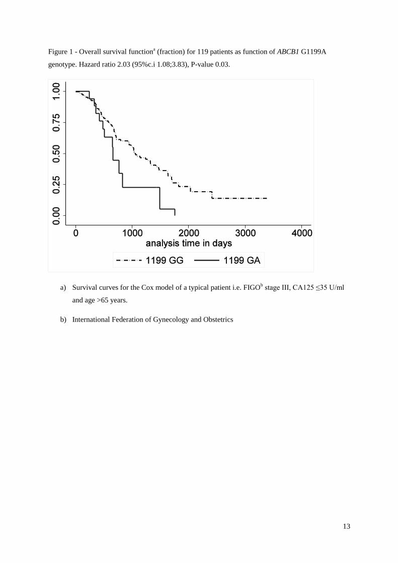
a P-value ≤ 0.05 (data not shown). ABCB1 G1199A was associated with increased risk of death with a
hazard ratio of 2.03 (95% ci: 1.08-3.83), P-value = 0.03 (Figure 1).
Discussion
Definite results linking frequent ABCB1 variants to taxane toxicity and effects in vivo have been
elusive. This study retrospectively investigates possible correlations between a wide selection of
candidate genes all with a reputed role in either the transport or the metabolism of paclitaxel and the
toxicity and survival of patients treated with paclitaxel. Clinical data and formalin fixed paraffin
embedded tissue was gathered from 119 patients with ovarian cancer who were treated with paclitaxel
and carboplatin in either the OVAR-9 or the TEC trial. The genetic variant CYP2C8*3 (a paclitaxel
metabolizing enzyme) and three variants in the drug transporter ABCB1: C1236T, G2677T/A and
C3435T were picked pre hoc for primary analysis; no statistical significant correlation for sensoric
neuropathy, neutrophil depression and overall survival was found. This finding contrasts the
correlation reported by Nakajima et al[23] for leukocytopenia and C3435T in 23 Japanese women
with ovarian cancer and the report of Sissung et al[24] who found a trend between neutrophil decrease
and the ABCB1 C3435T/G2677T/A compound genotype in 26 patients with advanced solid tumors.
Interestingly Green et al[25] recently reported a correlation between CYP2C8*3 and hematological
toxicity.
We also conducted a wide exploratory analysis of 19 SNPs in a total of 10 genes and both sensoric
neuropathy, neutrophil depression and overall survival and other toxicities that are related to
paclitaxel treatment i.e. myalgia, arthralgia, mucositis, hearing loss, motor neuropathy,
vomiting/nausea and compliance. This analysis encompasses some 190 tests (data not shown) of
which seven had a P-value ≤ 0.05. This finding is likely due to chance alone, given the multiple
testing. This multitude of statistical testing can serve a purpose for future investigations but have no
value without being confirmed in other studies. None of the seven possible associations have to our
knowledge been reported in the literature before. In the explorative analysis of overall survival, an
association with the non synonymous G1199A SNP in the ABCB1 transporter was found with a P-
value of 0.03 (uncorrected) and a hazard ratio of 2.03 [95 % c.i. 1.08-3.83] favoring patients with
1199GG genotype (Figure 1). This is in accordance with the shorter progression free survival for
patients carrying the 1199GA genotype found by Green et al[26]. Similarly Johnatty et al[27] found a
better progression-free survival for patients carrying the 2677TA alleles and Gréen et al[28] found an
association for the same variant and good response to paclitaxel treatment. The finding should be
confirmed in other studies before any conclusions can be made. CYP1B1*3 has previously been
associated with survival after paclitaxel treatment in breast cancer [29, 30]; but this finding was not
reproduced in our material of ovarian cancer.
In 2007 Marsh et al[31] reported a very comprehensive retrospective pharmacogenetic study of
toxicity and response to treatment with a taxane in 914 ovarian cancer patients. No significant
8
association was found for a selection of variants in the candidate genes: ABCB1, ABCC1, ABCG2,
CYP1B1, CYP2C8 and CYP3A4/5 and hematologic toxicity, neurotoxicity and gastrointestinal
toxicity. While this study included far more patients than our study and other previous studies it also
raised a debate concerning power and the interpretation of P-values[32-34]. Our results however, do
agree with the conclusions made by Marsh et al; but are again contrasted by a very recent paper by
Leskelä et al[35] who demonstrates an association between CYP2C8*3 and CYP3A5 in a mixed
retrospective/prospective study.
In this study we investigate candidate genes with putative impact on the pharmacokinetics of
paclitaxel under the assumption that these might influence on toxicity or survival either directly
through impact on clearance or the distribution of paclitaxel or indirectly through the paclitaxel
concentration in the micro environment in the tumor and susceptible tissues (bone marrow, peripheral
nerves etc.). We have thus not addressed the possibility that the susceptibility of the mentioned targets
by themselves is determined by genetic variation. Ideally, future pharmacogenetic studies of
chemotherapy should try to combine the pharmacokinetic and the pharmacodynamic components.
In conclusion this study primarily investigated the notion that the genetic variants CYP2C8*3 and
ABCB1 C1236T, G2677T/A and C3435T explain some of the observed variability in toxicity and
survival of patients treated with paclitaxel and carboplatin in ovarian cancer. No statistical significant
associations were found in the primary analysis. The study also involved an explorative analysis of
other candidate genes which might serve a purpose for studies in the future.
9
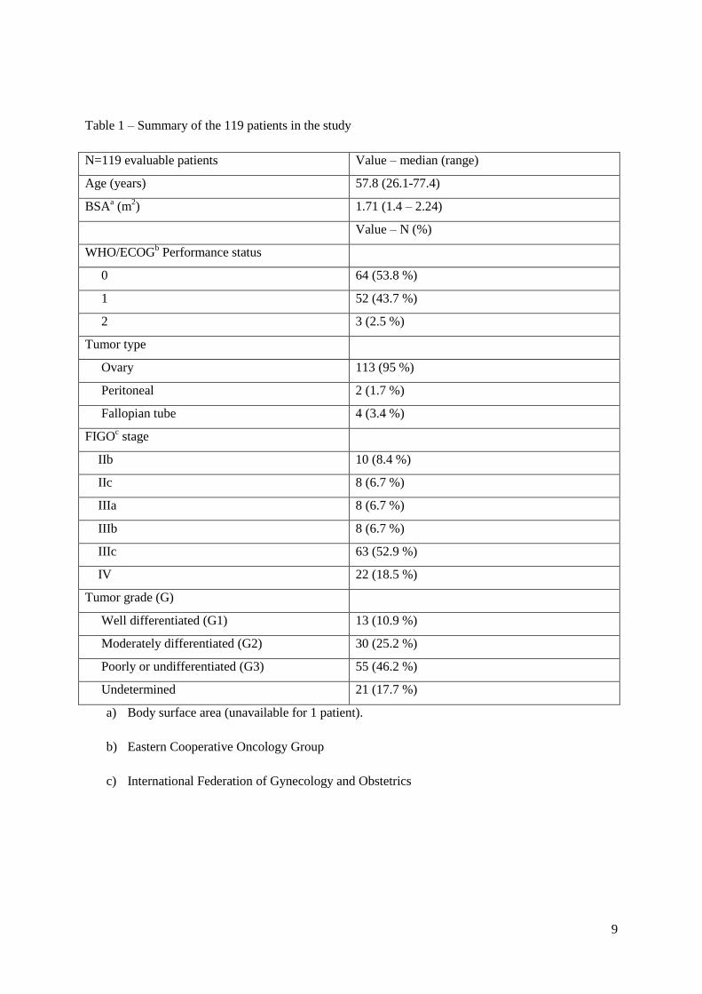
Table 1 – Summary of the 119 patients in the study
N=119 evaluable patients Value – median (range)
Age (years) 57.8 (26.1-77.4)
BSAa (m
2) 1.71 (1.4 – 2.24)
Value – N (%)
WHO/ECOGb Performance status
0 64 (53.8 %)
1 52 (43.7 %)
2 3 (2.5 %)
Tumor type
Ovary 113 (95 %)
Peritoneal 2 (1.7 %)
Fallopian tube 4 (3.4 %)
FIGOc stage
IIb 10 (8.4 %)
IIc 8 (6.7 %)
IIIa 8 (6.7 %)
IIIb 8 (6.7 %)
IIIc 63 (52.9 %)
IV 22 (18.5 %)
Tumor grade (G)
Well differentiated (G1) 13 (10.9 %)
Moderately differentiated (G2) 30 (25.2 %)
Poorly or undifferentiated (G3) 55 (46.2 %)
Undetermined 21 (17.7 %)
a) Body surface area (unavailable for 1 patient).
b) Eastern Cooperative Oncology Group
c) International Federation of Gynecology and Obstetrics
10
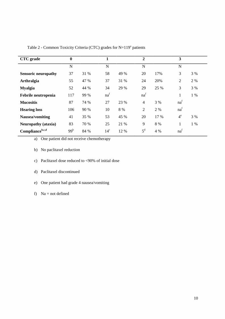
Table 2 - Common Toxicity Criteria (CTC) grades for N=119a patients
CTC grade 0 1 2 3
N N N N
Sensoric neuropathy 37 31 % 58 49 % 20 17% 3 3 %
Arthralgia 55 47 % 37 31 % 24 20% 2 2 %
Myalgia 52 44 % 34 29 % 29 25 % 3 3 %
Febrile neutropenia 117 99 % naf
naf
1 1 %
Mucositis 87 74 % 27 23 % 4 3 % naf
Hearing loss 106 90 % 10 8 % 2 2 % naf
Nausea/vomiting 41 35 % 53 45 % 20 17 % 4e 3 %
Neuropathy (ataxia) 83 70 % 25 21 % 9 8 % 1 1 %
Complianceb,c,d 99
b 84 % 14
c 12 % 5
d 4 % na
f
a) One patient did not receive chemotherapy
b) No paclitaxel reduction
c) Paclitaxel dose reduced to <90% of initial dose
d) Paclitaxel discontinued
e) One patient had grade 4 nausea/vomiting
f) Na = not defined
11
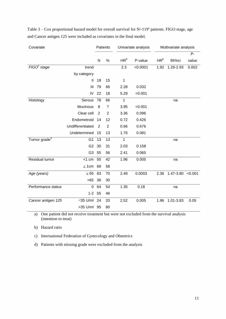
Table 3 – Cox proportional hazard model for overall survival for N=119a patients. FIGO stage, age
and Cancer antigen 125 were included as covariates in the final model.
Covariate
Patients
Univariate analysis
Multivariate analysis
N % HRb
P-value HRb
95%ci
P-
value
FIGOc stage trend
2.3 <0.0001
1.92 1.26-2.93 0.002
by category
II 18 15
1
III 79 66
2.28 0.032
IV 22 18 5.29 <0.001
Histology Serous 78 66
1
na
Mucinous 8 7
3.95 <0.001
Clear cell 2 2
3.36 0.096
Endometroid 14 12
0.72 0.426
Undifferentiated 2 2
0.66 0.676
Undetermined 15 13 1.76 0.081
Tumor graded G1 13 13
1
na
G2 30 31
2.03 0.158
G3 55 56 2.41 0.065
Residual tumor <1 cm 50 42
1.96 0.005
na
1cm 69 58
Age (years) 65 83 70
2.49 0.0003
2.36 1.47-3.80 <0.001
>65 36 30
Performance status 0 64 54
1.35 0.18
na
1-2 55 46
Cancer antigen 125 35 U/ml 24 20
2.52 0.005
1.96 1.01-3.83 0.05
>35 U/ml 95 80
a) One patient did not receive treatment but were not excluded from the survival analysis
(intention to treat)
b) Hazard ratio
c) International Federation of Gynecology and Obstetrics
d) Patients with missing grade were excluded from the analysis
12
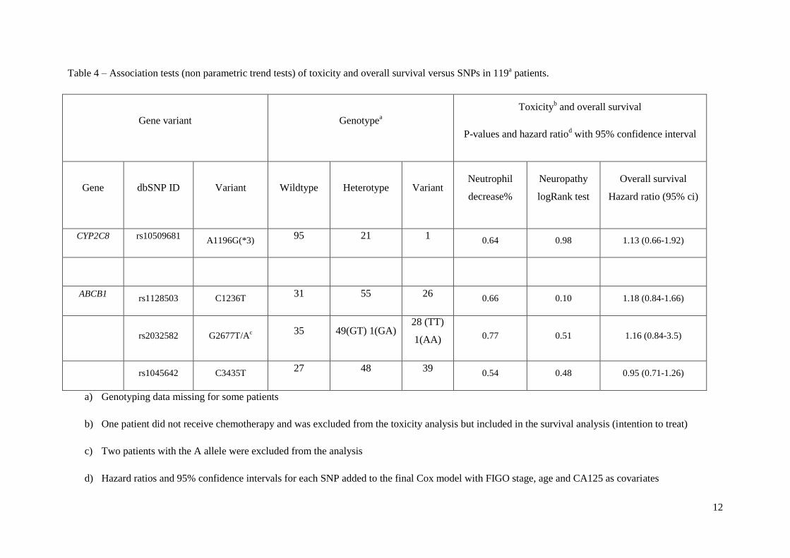
Table 4 – Association tests (non parametric trend tests) of toxicity and overall survival versus SNPs in 119a patients.
Gene variant Genotypea
Toxicityb and overall survival
P-values and hazard ratiod with 95% confidence interval
Gene dbSNP ID Variant Wildtype Heterotype Variant Neutrophil
decrease%
Neuropathy
logRank test
Overall survival
Hazard ratio (95% ci)
CYP2C8 rs10509681 A1196G(*3)
95 21 1 0.64 0.98 1.13 (0.66-1.92)
ABCB1 rs1128503 C1236T
31 55 26 0.66 0.10 1.18 (0.84-1.66)
rs2032582 G2677T/A
c 35 49(GT) 1(GA)
28 (TT)
1(AA) 0.77 0.51 1.16 (0.84-3.5)
rs1045642 C3435T
27 48 39 0.54 0.48 0.95 (0.71-1.26)
a) Genotyping data missing for some patients
b) One patient did not receive chemotherapy and was excluded from the toxicity analysis but included in the survival analysis (intention to treat)
c) Two patients with the A allele were excluded from the analysis
d) Hazard ratios and 95% confidence intervals for each SNP added to the final Cox model with FIGO stage, age and CA125 as covariates
13
Figure 1 - Overall survival functiona (fraction) for 119 patients as function of ABCB1 G1199A
genotype. Hazard ratio 2.03 (95%c.i 1.08;3.83), P-value 0.03.
a) Survival curves for the Cox model of a typical patient i.e. FIGOb stage III, CA125 ≤35 U/ml
and age >65 years.
b) International Federation of Gynecology and Obstetrics
14
Acknowledgements
We wish to acknowledge the AGO Ovarian Cancer study group in Wiesbaden, Germany and The
Nordic Society of Gynecologic Oncology, Clinical Trial Unit, Odense, Denmark for making available
the clinical data from the AGO-OVAR-9 study and the NSGO-OC9804 (TEC) study.
We wish to acknowledge the work and invaluable help of lab technicians Ingrid Jakobsen Falk and
Karin Skoglund in Linköping and Pernille Jordan in Odense.
The work was financially supported by grants from European Commission (CHEMORES
LSHC-CT-2007-037665), the Swedish Cancer Society, the Swedish Medical Society- Linköping
branch, the County Council in Östergötland, the Danish Ministry of Interior Affairs and Health (2001-
2007)(J.nr 2006-12103-276), the Danish Research Agency (J.nr 271-05-0266), Borgholm Rotary club,
Sweden and Roche Denmark, Hvidovre, Denmark.
Conflicts of Interest The authors declare that they have no conflict of interest.
15
Reference List
[1] Wani MC, Taylor HL, Wall ME, Coggon P, McPhail AT. Plant antitumor agents. VI. The
isolation and structure of taxol, a novel antileukemic and antitumor agent from Taxus
brevifolia. J Am Chem Soc 1971; 93:2325-2327.
[2] Karlan,BY, Markman,MA, Eifel,PJ. Gynecologic Cancers. In: DeVita,VT, Hellman,S,
Rosenberg,SA, editors. Cancer. Principles & Practice of Oncology. Lippincott
Williams & Wilkins; 2004.
[3] Harris JW, Rahman A, Kim BR, Guengerich FP, Collins JM. Metabolism of taxol by human
hepatic microsomes and liver slices: participation of cytochrome P450 3A4 and an
unknown P450 enzyme. Cancer Res 1994; 54:4026-4035.
[4] Rahman A, Korzekwa KR, Grogan J, Gonzalez FJ, Harris JW. Selective biotransformation of
taxol to 6 alpha-hydroxytaxol by human cytochrome P450 2C8. Cancer Res 1994;
54:5543-5546.
[5] Kumar G, Ray S, Walle T, Huang Y, Willingham M, Self S et al. Comparative in vitro
cytotoxic effects of taxol and its major human metabolite 6 alpha-hydroxytaxol.
Cancer Chemother Pharmacol 1995; 36:129-135.
[6] Sparreboom A, van Asperen J, Mayer U, Schinkel AH, Smit JW, Meijer DK et al. Limited
oral bioavailability and active epithelial excretion of paclitaxel (Taxol) caused by P-
glycoprotein in the intestine. PNAS 1997; 94:2031-2035.
[7] Dai D, Zeldin DC, Blaisdell JA, Chanas B, Coulter SJ, Ghanayem BI et al. Polymorphisms in
human CYP2C8 decrease metabolism of the anticancer drug paclitaxel and
arachidonic acid. Pharmacogenetics 2001; 11:597-607.
[8] Soyama A, Saito Y, Hanioka N, Murayama N, Nakajima O, Katori N et al. Non-synonymous
single nucleotide alterations found in the CYP2C8 gene result in reduced in vitro
paclitaxel metabolism. Biol Pharm Bull 2001; 24:1427-1430.
[9] Tanabe M, Ieiri I, Nagata N, Inoue K, Ito S, Kanamori Y et al. Expression of P-glycoprotein
in human placenta: relation to genetic polymorphism of the multidrug resistance
(MDR)-1 gene. J Pharmacol Exp Ther 2001; 297:1137-1143.
[10] Kim RB, Leake BF, Choo EF, Dresser GK, Kubba SV, Schwarz UI et al. Identification of
functionally variant MDR1 alleles among European Americans and African
Americans. Clin Pharmacol Ther 2001; 70:189-199.
[11] Hoffmeyer S, Burk O, von Richter O, Arnold HP, Brockmoller J, Johne A et al. Functional
polymorphisms of the human multidrug-resistance gene: multiple sequence variations
and correlation of one allele with P-glycoprotein expression and activity in vivo. Proc
Natl Acad Sci U S A 2000; 97:3473-3478.
[12] Marsh S. Taxane pharmacogenetics. Personalized Medicine 2006; 3:33-43.
16
[13] Huisman MT, Chhatta AA, van Tellingen O, Beijnen JH, Schinkel AH. MRP2 (ABCC2)
transports taxanes and confers paclitaxel resistance and both processes are stimulated
by probenecid. Int J Cancer 2005; 116:824-829.
[14] McFadyen MC, Cruickshank ME, Miller ID, McLeod HL, Melvin WT, Haites NE et al.
Cytochrome P450 CYP1B1 over-expression in primary and metastatic ovarian
cancer. Br J Cancer 2001; 85:242-246.
[15] Marsh S, Paul J, King CR, Gifford G, McLeod HL, Brown R. Pharmacogenetic Assessment
of Toxicity and Outcome After Platinum Plus Taxane Chemotherapy in Ovarian
Cancer: The Scottish Randomised Trial in Ovarian Cancer. J Clin Oncol 2007;
25:4528-4535.
[16] Smith NF, Marsh S, Scott-Horton TJ, Hamada A, Mielke S, Mross K et al. Variants in the
SLCO1B3 gene: interethnic distribution and association with paclitaxel
pharmacokinetics. Clin Pharmacol Ther 2007; 81:76-82.
[17] Hopper-Borge E, Chen ZS, Shchaveleva I, Belinsky MG, Kruh GD. Analysis of the drug
resistance profile of multidrug resistance protein 7 (ABCC10): resistance to
docetaxel. Cancer Res 2004; 64:4927-4930.
[18] McFadyen MCE, McLeod HL, Jackson FC, Melvin WT, Doehmer J, Murray GI. Cytochrome
P450 CYP1B1 protein expression: A novel mechanism of anticancer drug resistance.
Biochemical Pharmacology 2001; 62:207-212.
[19] Green H, Soderkvist P, Rosenberg P, Horvath G, Peterson C. mdr-1 single nucleotide
polymorphisms in ovarian cancer tissue: G2677T/A correlates with response to
paclitaxel chemotherapy. Clin Cancer Res 2006; 12:854-859.
[20] Green H, Soderkvist P, Rosenberg P, Mirghani RA, Rymark P, Lundqvist EA et al.
Pharmacogenetic studies of Paclitaxel in the treatment of ovarian cancer. Basic Clin
Pharmacol Toxicol 2009; 104:130-137.
[21] Green H, Soderkvist P, Rosenberg P, Horvath G, Peterson C. ABCB1 G1199A polymorphism
and ovarian cancer response to paclitaxel. J Pharm Sci 2008; 97:2045-2048.
[22] Bergmann TK, Brasch-Andersen C, Green H, Mirza M, Pedersen RS, Nielsen F et al. Impact
of CYP2C8*3 on paclitaxel clearance: a population pharmacokinetic and
pharmacogenomic study in 93 patients with ovarian cancer. Pharmacogenomics J
2010; advance online publication:6 April 2010.
[23] Nakajima M, Fujiki Y, Kyo S, Kanaya T, Nakamura M, Maida Y et al. Pharmacokinetics of
paclitaxel in ovarian cancer patients and genetic polymorphisms of CYP2C8,
CYP3A4, and MDR1. J Clin Pharmacol 2005; 45:674-682.
[24] Sissung TM, Mross K, Steinberg SM, Behringer D, Figg WD, Sparreboom A et al.
Association of ABCB1 genotypes with paclitaxel-mediated peripheral neuropathy
and neutropenia. European Journal of Cancer 2006; 42:2893-2896.
[25] Green H, Soderkvist P, Rosenberg P, Mirghani RA, Rymark P, Lundqvist EA et al.
Pharmacogenetic studies of Paclitaxel in the treatment of ovarian cancer. Basic Clin
Pharmacol Toxicol 2009; 104:130-137.
[26] Green H, Soderkvist P, Rosenberg P, Horvath G, Peterson C. ABCB1 G1199A polymorphism
and ovarian cancer response to paclitaxel. J Pharm Sci 2008; 97:2045-2048.
17
[27] Johnatty SE, Beesley J, Paul J, Fereday S, Spurdle AB, Webb PM et al. ABCB1 (MDR 1)
Polymorphisms and Progression-Free Survival among Women with Ovarian Cancer
following Paclitaxel/Carboplatin Chemotherapy. Clin Cancer Res 2008; 14:5594-
5601.
[28] Green H, Soderkvist P, Rosenberg P, Horvath G, Peterson C. mdr-1 single nucleotide
polymorphisms in ovarian cancer tissue: G2677T/A correlates with response to
paclitaxel chemotherapy. Clin Cancer Res 2006; 12:854-859.
[29] Gehrmann M, Schmidt M, Brase JC, Roos P, Hengstler JG. Prediction of paclitaxel resistance
in breast cancer: is CYP1B1*3 a new factor of influence? Pharmacogenomics 2008;
9:969-974.
[30] Marsh S, Somlo G, McLeod HL, Li X, Frankel P, King CR et al. Pharmacogenetic analysis of
paclitaxel in breast cancer. J Clin Oncol (Meeting Abstracts) 2005; 23:3058.
[31] Marsh S, Paul J, King CR, Gifford G, McLeod HL, Brown R. Pharmacogenetic Assessment
of Toxicity and Outcome After Platinum Plus Taxane Chemotherapy in Ovarian
Cancer: The Scottish Randomised Trial in Ovarian Cancer. J Clin Oncol 2007;
25:4528-4535.
[32] Vach W, Bergmann TK, Brosen K. No evidence for taxane/platinum pharmacogenetic
markers: just lack of power? J Clin Oncol 2008; 26:1903-1904.
[33] Branford RA, Pantelidis P, Ross JR. Ethnic considerations in pharmacogenetic studies. J Clin
Oncol 2008; 26:1766-1767.
[34] Maitland ML, Ratain MJ, Cox NJ. Interpreting P values in pharmacogenetic studies: a call for
process and perspective. J Clin Oncol 2007; 25:4513-4515.
[35] Leskela S. Polymorphisms in cytochromes P450 2C8 and 3A5 are associated with paclitaxel
neurotoxicity. 2010.
Recommended


