METHODOLOGY ARTICLE Open Access
Sensitive detection of Ab protofibrils by proximityligation - relevance for Alzheimer’s diseaseMasood Kamali-Moghaddam1*, Frida Ekholm Pettersson2, Di Wu1, Hillevi Englund2, Spyros Darmanis1, Anna Lord2,Gholamreza Tavoosidana1, Dag Sehlin2, Sigrun Gustafsdottir1, Lars NG Nilsson2, Lars Lannfelt2, Ulf Landegren1
Abstract
Background: Protein aggregation plays important roles in several neurodegenerative disorders. For instance,insoluble aggregates of phosphorylated tau and of Ab peptides are cornerstones in the pathology of Alzheimer’sdisease. Soluble protein aggregates are therefore potential diagnostic and prognostic biomarkers for their cognatedisorders. Detection of the aggregated species requires sensitive tools that efficiently discriminate them frommonomers of the same proteins. Here we have established a proximity ligation assay (PLA) for specific andsensitive detection of Ab protofibrils via simultaneous recognition of three identical determinants present in theaggregates. PLA is a versatile technology in which the requirement for multiple target recognitions is combinedwith the ability to translate signals from detected target molecules to amplifiable DNA strands, providing very highspecificity and sensitivity.
Results: For specific detection of Ab protofibrils we have used a monoclonal antibody, mAb158, selective for Abprotofibrils in a modified PLA, where the same monoclonal antibody was used for the three classes of affinityreagents required in the assay. These reagents were used for detection of soluble Ab aggregates in solid-phasereactions, allowing detection of just 0.1 pg/ml Ab protofibrils, and with a dynamic range greater than six orders ofmagnitude. Compared to a sandwich ELISA setup of the same antibody the PLA increases the sensitivity of the Abprotofibril detection by up to 25-fold. The assay was used to measure soluble Ab aggregates in brain homogenatesfrom mice transgenic for a human allele predisposing to Ab aggregation.
Conclusions: The proximity ligation assay is a versatile analytical technology for proteins, which can provide highlysensitive and specific detection of Ab aggregates - and by implication other protein aggregates of relevance inAlzheimer’s disease and other neurodegenerative disorders.
BackgroundIn Alzheimer’s disease (AD), brain deposits of extracel-lular amyloid-b (Ab) and intracellular tau tangles arecharacteristic of the disease. Cerebrospinal fluid (CSF) isoften investigated for levels of Ab42, tau and phosho-tau in routine diagnostics of AD [1], where decreasedAb42 and increased tau and/or phospho-tau (Thr181P)in CSF are indicative of the disease. These measuresare reasonably good predictors of future conversionto AD among subjects with mild cognitive impairment,but they are not suitable to follow disease progressionor to monitor drug intervention. Novel biomarkers are
therefore needed, and evidence suggests that soluble,oligomeric aggregates of Ab could be such a marker.For instance, levels of soluble forms of Ab correlatemore closely with disease severity than do the amountsof insoluble Ab aggregates in the brain [2], and oligo-meric Ab has been shown to be neurotoxic, lead tosynaptic dysfunction and to inhibit maintenance of hip-pocampal long-term potentiation [3-7]. Moreover, theso-called Arctic mutation causing early onset AD islocated within the Ab domain as are other mutationssuch as the Flemish, the Dutch and the Italian muta-tions, and this particular mutation has been shown tospecifically enhance the formation of large soluble oligo-mers of Ab (i. e. protofibrils), suggesting the notion thatthis Ab species plays a central role in disease pathogen-esis [8,9]. We previously developed a sensitive sandwich
* Correspondence: [email protected] of Genetics and Pathology, Molecular Medicine, UppsalaUniversity, Uppsala, SwedenFull list of author information is available at the end of the article
Kamali-Moghaddam et al. BMC Neuroscience 2010, 11:124http://www.biomedcentral.com/1471-2202/11/124
© 2010 Kamali-Moghaddam et al; licensee BioMed Central Ltd. This is an Open Access article distributed under the terms of theCreative Commons Attribution License (http://creativecommons.org/licenses/by/2.0), which permits unrestricted use, distribution, andreproduction in any medium, provided the original work is properly cited.
ELISA where the protofibril-selective mAb158 was usedboth as capture and detecting antibody [10]. Using thisassay, the antibody used herein has been shown todetect Ab protofibrils also in other, well-known, tg-micesuch as PSAPP and tg2576 [11]. Here, we demonstratethat the proximity ligation assay (PLA) can provide evenmore sensitive detection of synthetic Ab protofibrils.PLA is an affinity-based technology enabling sensitive
and specific detection of proteins in which the detectionof proteins by sets of antibodies results in the formationof a specific DNA sequence by ligation of two parts.This sequence can then be amplified and quantified bymethods such as real-time, PCR [12,13]. The techniquemakes use of affinity probes, typically antibodies coupledto oligonucleotides. Upon recognition of a common tar-get molecule by a pair of such probes, the attachedDNA strands are brought in proximity, allowing theirfree ends to be hybridized to a connector oligonucleo-tide that directs their joining by ligation. The reporterDNA strand that forms upon ligation can be amplifiedand quantified by methods such as real-time PCR. Theassays can be performed in the homogenous phase withno need for washes or separations [12,13]. Alternatively,a solid support-bound affinity reagent can be used thatoffers the possibility to search for target molecules inlarger sample volumes and to remove excess probes andundesired sample components before the ligation andamplification steps. This approach also adds specificityby requiring simultaneous recognition of three epitopeson the targets [14,15]. By using a single monoclonalantibody as the affinity reagent in all three affinityreagents required for solid-phase PLA (SP-PLA) wehave achieved a highly sensitive assay that is specific forAb protofibrils, with excellent discrimination againstmonomers. We demonstrate that PLA is capable ofdetecting soluble Ab aggregates in brains from mice
transgenic for a pathogenic form of APP - the proteinfrom which Ab is derived.
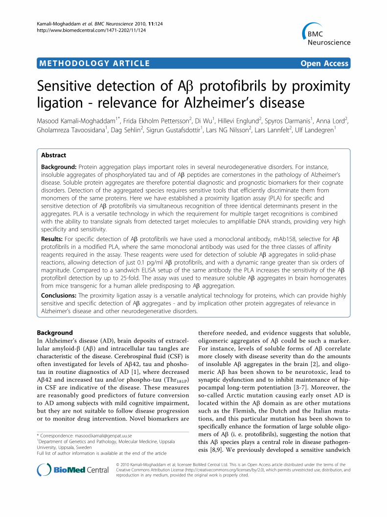
ResultsWe have developed a solid-phase form of PLA (Figure 1)using the mAb158 antibody for sensitive and specificdetection of Ab protofibrils, and we have compared thisassay to the previously established sandwich ELISA usingthe same antibody.The SP-PLA has been previously shown to provide a
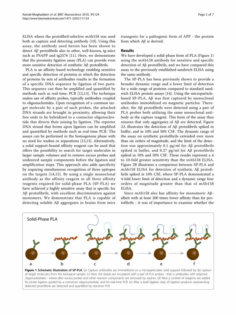
broader dynamic range and a lower limit of detectionfor a wide range of proteins compared to standard sand-wich ELISA protein assays [14]. Using the microparticle-based SP-PLA, Ab was first captured by monoclonalantibodies immobilized on magnetic particles. There-after, the Ab protofibrils were detected using a pair ofPLA probes both utilizing the same monoclonal anti-body as the capture reagent. This form of the assay thusensures that only aggregates of Ab are detected. Figure2A illustrates the detection of Ab protofibrils spiked inbuffer, and in 10% and 50% CSF. The dynamic range ofthe assay on synthetic protofibrils extended over morethan six orders of magnitude, and the limit of the detec-tion was approximately 0.1 pg/ml for Ab protofibrilsspiked in buffer, and 0.27 pg/ml for Ab protofibrilsspiked in 10% and 50% CSF. These results represent a 4to 10-fold greater sensitivity than the mAb158 ELISA.Figure 2B illustrates a comparison between SP-PLA andmAb158 ELISA for detection of synthetic Ab protofi-brils spiked in 10% CSF, where SP-PLA demonstrated a4-fold lower limit of detection and a dynamic range fourorders of magnitude greater than that of mAb158ELISA.Since mAb158 also has affinity for monomeric Ab-
albeit with at least 200 times lower affinity than for pro-tofibrils - it was of importance to examine whether the
Solid-Phase PLA
eda fcbFigure 1 Schematic illustration of SP-PLA. (a) Capture antibodies are immobilized on a microparticulate solid support followed by (b) captureof target molecules from the biological sample. (c) Next, the beads are incubated with a pair of PLA probes - that is antibodies with attachedoligonucleotides - where after excess probes and other reaction components are removed by washes. (d) Next a cocktail of reagents are addedfor probe ligation guided by a connector oligonucleotide, and for real-time PCR. (e) After a brief ligation step, (f) ligation products representingdetected protofibrils are detected and quantified by real-time PCR.
Kamali-Moghaddam et al. BMC Neuroscience 2010, 11:124http://www.biomedcentral.com/1471-2202/11/124
Page 2 of 7
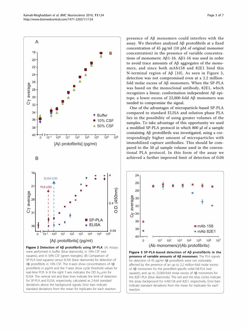
presence of Ab monomers could interfere with theassay. We therefore analyzed Ab protofibrils at a fixedconcentration of 45 pg/ml (10 pM of original monomerconcentration) in the presence of variable concentra-tions of monomeric Ab1-16. Ab1-16 was used in orderto avoid trace amounts of Ab aggregates of the mono-mers, and since both mAb158 and 82E1 bind thisN-terminal region of Ab [10]. As seen in Figure 3,detection was not compromised even at a 2.2 million-fold molar excess of Ab monomers. When the SP-PLAwas based on the monoclonal antibody, 82E1, whichrecognizes a linear, conformation independent Ab epi-tope, a lower excess of 22,000-fold Ab monomers wasneeded to compromise the signal.One of the advantages of microparticle-based SP-PLA
compared to standard ELISA and solution phase PLAlies in the possibility of using greater volumes of thesamples. To take advantage of this opportunity we useda modified SP-PLA protocol in which 800 μl of a samplecontaining Ab protofibrils was investigated, using a cor-respondingly higher amount of microparticles withimmobilized capture antibodies. This should be com-pared to the 50 μl sample volume used in the conven-tional PLA protocol. In this form of the assay weachieved a further improved limit of detection of 0.04
18
20
22
24
26
28
30
32
34
36
38
40
Buffer10% CSF 50% CSF
0 10-1 100 101 102 103 104 105 106
CT
aver
age
0 10-1 100 101 102 103 104 105 1060.04
0.4
4 16
21
26
31
36
PLA LOD
ELISA LOD
SP-PLAELISA
O.D
. (45
0nm
)
CT
aver
age
[Aβ protofibrils] (pg/ml)
[Aβ protofibrils] (pg/ml)
A
B
Figure 2 Detection of Ab protofibrils using SP-PLA. (A) Assayswere performed in buffer (blue diamonds), in 10% CSF (redsquares), and in 50% CSF (green triangles). (B) Comparison ofSP-PLA (red squares) versus ELISA (blue diamonds) for detection ofAb protofibrils in 10% CSF. The X-axes show concentrations of Abprotofibrils in pg/ml and the Y-axes show cycle threshold values forreal-time PCR. In B the right Y-axis indicates the OD A450nm forELISA. The vertical red and blue lines indicate the limit of detectionfor SP-PLA and ELISA, respectively, calculated as 2-fold standarddeviations above the background signals. Error bars indicatestandard deviations from the mean for triplicates for each reaction.
C
aver
age
T
24
26
28
30
32
34
36
38
400
mAb 158mAb 82E1
(Ab monomers)/(Ab protofibrils)101 102 103 104 105 106 107
Figure 3 SP-PLA-based detection of Ab protofibrils in thepresence of variable amounts of Ab monomers. The PLA signalsfor detection of 45 pg/ml Ab protofibrils were not noticeablyaffected by the presence of an up to 2.2 million-fold molar excessof Ab monomers for the protofibril-specific mAb158-PLA (redsquares), and up to 22,000-fold molar excess of Ab monomers forthe 82E1-PLA (blue diamonds). The red and the blue circles indicatethe assay background for mAb158 and 82E1, respectively. Error barsindicate standard deviations from the mean for triplicates for eachreaction.
Kamali-Moghaddam et al. BMC Neuroscience 2010, 11:124http://www.biomedcentral.com/1471-2202/11/124
Page 3 of 7
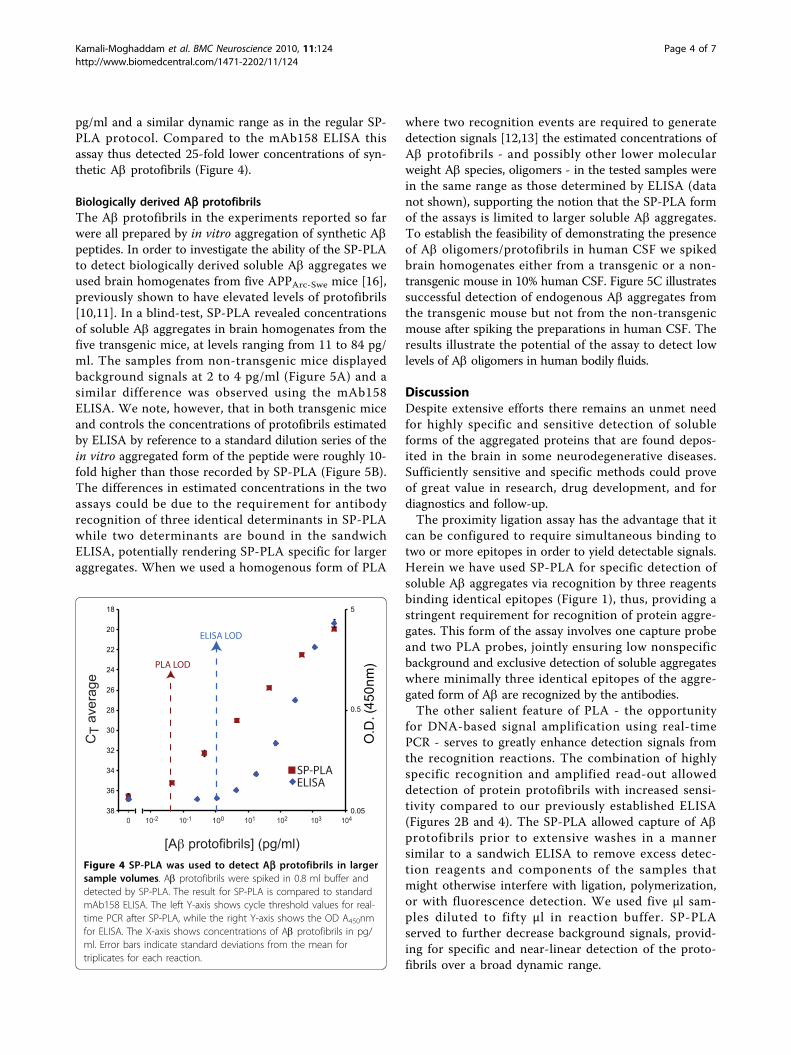
pg/ml and a similar dynamic range as in the regular SP-PLA protocol. Compared to the mAb158 ELISA thisassay thus detected 25-fold lower concentrations of syn-thetic Ab protofibrils (Figure 4).
Biologically derived Ab protofibrilsThe Ab protofibrils in the experiments reported so farwere all prepared by in vitro aggregation of synthetic Abpeptides. In order to investigate the ability of the SP-PLAto detect biologically derived soluble Ab aggregates weused brain homogenates from five APPArc-Swe mice [16],previously shown to have elevated levels of protofibrils[10,11]. In a blind-test, SP-PLA revealed concentrationsof soluble Ab aggregates in brain homogenates from thefive transgenic mice, at levels ranging from 11 to 84 pg/ml. The samples from non-transgenic mice displayedbackground signals at 2 to 4 pg/ml (Figure 5A) and asimilar difference was observed using the mAb158ELISA. We note, however, that in both transgenic miceand controls the concentrations of protofibrils estimatedby ELISA by reference to a standard dilution series of thein vitro aggregated form of the peptide were roughly 10-fold higher than those recorded by SP-PLA (Figure 5B).The differences in estimated concentrations in the twoassays could be due to the requirement for antibodyrecognition of three identical determinants in SP-PLAwhile two determinants are bound in the sandwichELISA, potentially rendering SP-PLA specific for largeraggregates. When we used a homogenous form of PLA
where two recognition events are required to generatedetection signals [12,13] the estimated concentrations ofAb protofibrils - and possibly other lower molecularweight Ab species, oligomers - in the tested samples werein the same range as those determined by ELISA (datanot shown), supporting the notion that the SP-PLA formof the assays is limited to larger soluble Ab aggregates.To establish the feasibility of demonstrating the presenceof Ab oligomers/protofibrils in human CSF we spikedbrain homogenates either from a transgenic or a non-transgenic mouse in 10% human CSF. Figure 5C illustratessuccessful detection of endogenous Ab aggregates fromthe transgenic mouse but not from the non-transgenicmouse after spiking the preparations in human CSF. Theresults illustrate the potential of the assay to detect lowlevels of Ab oligomers in human bodily fluids.
DiscussionDespite extensive efforts there remains an unmet needfor highly specific and sensitive detection of solubleforms of the aggregated proteins that are found depos-ited in the brain in some neurodegenerative diseases.Sufficiently sensitive and specific methods could proveof great value in research, drug development, and fordiagnostics and follow-up.The proximity ligation assay has the advantage that it
can be configured to require simultaneous binding totwo or more epitopes in order to yield detectable signals.Herein we have used SP-PLA for specific detection ofsoluble Ab aggregates via recognition by three reagentsbinding identical epitopes (Figure 1), thus, providing astringent requirement for recognition of protein aggre-gates. This form of the assay involves one capture probeand two PLA probes, jointly ensuring low nonspecificbackground and exclusive detection of soluble aggregateswhere minimally three identical epitopes of the aggre-gated form of Ab are recognized by the antibodies.The other salient feature of PLA - the opportunity
for DNA-based signal amplification using real-timePCR - serves to greatly enhance detection signals fromthe recognition reactions. The combination of highlyspecific recognition and amplified read-out alloweddetection of protein protofibrils with increased sensi-tivity compared to our previously established ELISA(Figures 2B and 4). The SP-PLA allowed capture of Abprotofibrils prior to extensive washes in a mannersimilar to a sandwich ELISA to remove excess detec-tion reagents and components of the samples thatmight otherwise interfere with ligation, polymerization,or with fluorescence detection. We used five μl sam-ples diluted to fifty μl in reaction buffer. SP-PLAserved to further decrease background signals, provid-ing for specific and near-linear detection of the proto-fibrils over a broad dynamic range.
O.D
. (45
0nm
)
CT
aver
age
10-1 100 101 102 103 10410-20
SP-PLAELISA
0.05
0.5
5 18
20
22
24
26
28
30
32
34
36
38
PLA LOD
ELISA LOD
[Aβ protofibrils] (pg/ml)Figure 4 SP-PLA was used to detect Ab protofibrils in largersample volumes. Ab protofibrils were spiked in 0.8 ml buffer anddetected by SP-PLA. The result for SP-PLA is compared to standardmAb158 ELISA. The left Y-axis shows cycle threshold values for real-time PCR after SP-PLA, while the right Y-axis shows the OD A450nmfor ELISA. The X-axis shows concentrations of Ab protofibrils in pg/ml. Error bars indicate standard deviations from the mean fortriplicates for each reaction.
Kamali-Moghaddam et al. BMC Neuroscience 2010, 11:124http://www.biomedcentral.com/1471-2202/11/124
Page 4 of 7
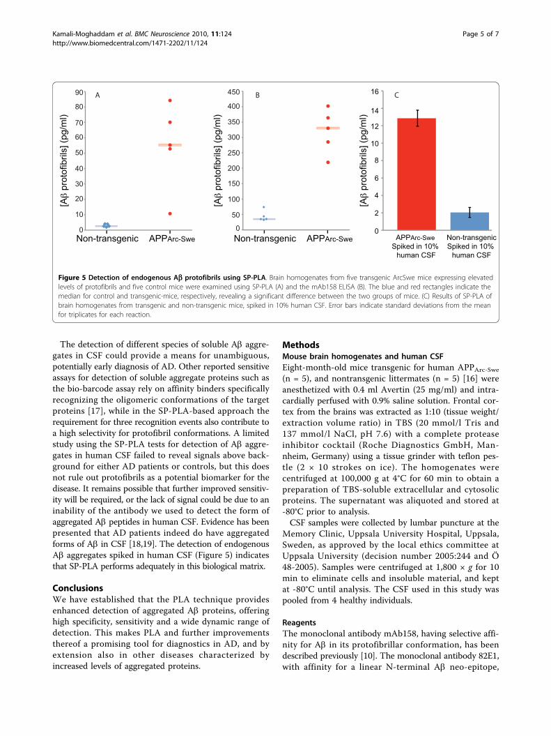
The detection of different species of soluble Ab aggre-gates in CSF could provide a means for unambiguous,potentially early diagnosis of AD. Other reported sensitiveassays for detection of soluble aggregate proteins such asthe bio-barcode assay rely on affinity binders specificallyrecognizing the oligomeric conformations of the targetproteins [17], while in the SP-PLA-based approach therequirement for three recognition events also contribute toa high selectivity for protofibril conformations. A limitedstudy using the SP-PLA tests for detection of Ab aggre-gates in human CSF failed to reveal signals above back-ground for either AD patients or controls, but this doesnot rule out protofibrils as a potential biomarker for thedisease. It remains possible that further improved sensitiv-ity will be required, or the lack of signal could be due to aninability of the antibody we used to detect the form ofaggregated Ab peptides in human CSF. Evidence has beenpresented that AD patients indeed do have aggregatedforms of Ab in CSF [18,19]. The detection of endogenousAb aggregates spiked in human CSF (Figure 5) indicatesthat SP-PLA performs adequately in this biological matrix.
ConclusionsWe have established that the PLA technique providesenhanced detection of aggregated Ab proteins, offeringhigh specificity, sensitivity and a wide dynamic range ofdetection. This makes PLA and further improvementsthereof a promising tool for diagnostics in AD, and byextension also in other diseases characterized byincreased levels of aggregated proteins.
MethodsMouse brain homogenates and human CSFEight-month-old mice transgenic for human APPArc-Swe(n = 5), and nontransgenic littermates (n = 5) [16] wereanesthetized with 0.4 ml Avertin (25 mg/ml) and intra-cardially perfused with 0.9% saline solution. Frontal cor-tex from the brains was extracted as 1:10 (tissue weight/extraction volume ratio) in TBS (20 mmol/l Tris and137 mmol/l NaCl, pH 7.6) with a complete proteaseinhibitor cocktail (Roche Diagnostics GmbH, Man-nheim, Germany) using a tissue grinder with teflon pes-tle (2 × 10 strokes on ice). The homogenates werecentrifuged at 100,000 g at 4°C for 60 min to obtain apreparation of TBS-soluble extracellular and cytosolicproteins. The supernatant was aliquoted and stored at-80°C prior to analysis.CSF samples were collected by lumbar puncture at the
Memory Clinic, Uppsala University Hospital, Uppsala,Sweden, as approved by the local ethics committee atUppsala University (decision number 2005:244 and Ö48-2005). Samples were centrifuged at 1,800 × g for 10min to eliminate cells and insoluble material, and keptat -80°C until analysis. The CSF used in this study waspooled from 4 healthy individuals.
ReagentsThe monoclonal antibody mAb158, having selective affi-nity for Ab in its protofibrillar conformation, has beendescribed previously [10]. The monoclonal antibody 82E1,with affinity for a linear N-terminal Ab neo-epitope,
Non-transgenic APPArc-Swe
[Aβ
prot
ofib
rils]
(pg/
ml)
[Aβ
prot
ofib
rils]
(pg/
ml)
450
400
350
300
250
200
150
100
50
0Non-transgenic APPArc-Swe
A B80
70
60
50
40
30
20
10
0
90
Non-transgenicSpiked in 10%
human CSF
APPArc-Swe Spiked in 10%
human CSF
16
14
12
10
8
6
4
2
0
[Aβ
prot
ofib
rils]
(pg/
ml)
C
Figure 5 Detection of endogenous Ab protofibrils using SP-PLA. Brain homogenates from five transgenic ArcSwe mice expressing elevatedlevels of protofibrils and five control mice were examined using SP-PLA (A) and the mAb158 ELISA (B). The blue and red rectangles indicate themedian for control and transgenic-mice, respectively, revealing a significant difference between the two groups of mice. (C) Results of SP-PLA ofbrain homogenates from transgenic and non-transgenic mice, spiked in 10% human CSF. Error bars indicate standard deviations from the meanfor triplicates for each reaction.
Kamali-Moghaddam et al. BMC Neuroscience 2010, 11:124http://www.biomedcentral.com/1471-2202/11/124
Page 5 of 7
was purchased from IBL International (Hamburg,Germany). Synthetic Ab42 was purchased from AmericanPeptide (Sunnyvale, Ca, USA), and the Ab protofibrilswere prepared as previously described [20]. Briefly, lyophi-lized synthetic Ab42wt was dissolved in 10 mM NaOH toa concentration of 100 μM, and then further diluted 1:1with 2 × PBS (50 mM phosphate buffer and 100 mMNaCl, pH 7.4), and incubated at 37°C over night (ON) inthe presence of 50 μM Docosahexaenoic acid (DHA) tostabilize the protofibrils. To remove fibrillar material thesample was centrifuged for 5 min at 17,900 × g before ana-lyses. As determined by density gradient ultracentrifuga-tion the mass of the Ab protofibrils in this preparation areapproximately 100-400 kDa (unpublished data). Lyophi-lized synthetic Ab1-16wt peptide (Bachem, Bubendorf,Switzerland) was dissolved in 10 mM NaOH, prior to use,and diluted in 2 × PBS to a final concentration of 50 μM.Oligonucleotide-streptavidin conjugates SLC1 (5’-
streptavidin CGCATCGCCCTTGGACTACGACTGAC-GAACCGCTTTGCCTGACTGATCGCTAAATCGTG-3’)and SLC2 (5’-TCGTGTCTAAAGTCCGTTACCTT-GATTCCCCTAACCCTCTTGAAAAATTCGGCATCGGTGA-streptavidin 3’) were purchased from Solulink(San Diego, CA, USA), and treated prior to use withfree streptavidin to reduce the numbers of oligonucleo-tides per streptavidin tetramer, as described [14].The same PCR forward primer, Biofwd, 5’-CATCGC-
CCTTGGACTACGA-3’, PCR reverse primer, Biorev,5’-GGGAATCAAGGTAACGGACTTTAG-3’, and con-nector oligonucleotide, 5’-TACTTAGACACGACACG-ATTTAGTTT-3’ were used in all PLA tests. Theseoligonucleotides were purchased from Biomers (Ger-many). A TaqMan probe (5’ FAM-TGACGAAC CGCT-TTGCCTGA-MGB 3’) was obtained from AppliedBiosystems.
Solid-phase proximity ligation assayFor all PLA reactions the oligonucleotide-streptavidinconjugates SLC1 and SLC2 were coupled to biotinylatedantibodies by incubating identical volumes of 100 nMantibodies with 100 nM streptavidin-oligonucleotideconjugates for 1 h at room temperature. The antibody-oligonucleotide conjugates (PLA probes) thus obtainedwere used without purification after being separatelydiluted in PLA buffer (1 mM D-Biotin (Invitrogen), 0.1%purified BSA (New England Biolabs), 0.05% Tween 20(Sigma-Aldrich), 100 nM goat serum IgG (Sigma-Aldrich), 0.1 μg/μl salmon sperm DNA (Invitrogen), 5mM EDTA, 1 × PBS), and incubated for 15 min atroom temperature prior to mixing the reagents to forma PLA probe mix.Microparticle-based SP-PLA was carried out as
described by Darmanis et al. [14], with some modifica-tions as follows. Briefly, capture antibodies were bound
to microparticles by using one mg of Dynabeads®MyOne™ Streptavidin T1 microparticles (Invitrogen)that had been washed twice with 500 μl washing buffer(1 × PBS, 0.05% Tween 20 (Sigma-Aldrich)), using a 96-well plate magnet (Perkin Elmer) for separation ofmicroparticles. The microparticles were mixed with 200μl of 50 nM (1.5 μg) of the same biotinylated monoclo-nal antibody that was used for PLA probe, and incu-bated for 1 h at RT under rotation, followed by washesas above. The antibody-coated microparticles were sus-pended in 200 μl of storage buffer (1xPBS, 0.1% purifiedBSA (New England Biolabs)), and stored at 4°C for upto 2 months.For each assay the storage buffer of one μl of anti-
body-coated microparticles (≈5 μg of microparticles and7.5 ng of antibody) was replaced by 5 μl of PLA buffer,and the microparticles were mixed with 45 μl samplesto be investigated for Ab protofibrils. The binding reac-tions were incubated ON at 4°C or for 1.5 h at RTunder rotation with similar efficiencies (data notshown). The microparticles were washed twice, and 50μl of PLA probe mix at a concentration of 30 pM foreach probe was added to each well, and incubated for1.5 h at RT with rotation, followed by washing. Finally,50 μl of ligation/PCR mix (1 × PCR buffer (Invitrogen),2.5 mM MgCl2 (Invitrogen), 0.2 μM of each primerBiofwd and Biorev, 0.4 μM TaqMan probe, 0.08 mMATP, 100 nM connector oligonucleotide, 0.2 mMdNTPs (containing dUTP) (Fermentas), 1.5 units Plati-num Taq polymerase (Invitrogen), 0.5 units T4 DNAligase (Fermentas), 0.1 units uracil-DNA glycosylase(Fermentas)) were added, followed by a 5 min incuba-tion at room temperature for the proximity ligationstep, before a real-time PCR was performed on an Mx-3000 real-time PCR instrument (Stratagene), with aninitial incubation for 2 min at 95°C, and then 45 cyclesof 15 s at 95°C and 1 min at 60°C.For higher volume samples, 10 μl of antibody-coated
microparticles were transferred to a 1.5 ml tube, andafter removing the storage buffer the particles weremixed with 0.8 ml samples to be investigated for thepresence of Ab protofibrils, and incubated ON at 4°Cwith end-over-end rotation. The microparticles werecollected by spinning at 15,000 rpm for 30 s, andwashed twice. 50 μl PLA probe mix was added followedby incubation for 1.5 h at RT. Next, the microparticleswere washed twice and transferred to optical PCR tubes,50 μl ligation/PCR mix was added, and the real-timePCR was performed as described above.
ELISAThe mAb158 sandwich ELISA was carried out as pre-viously described [10]. In short, 96-well plates werecoated with 200 ng/well of mAb158 at 4°C ON before
Kamali-Moghaddam et al. BMC Neuroscience 2010, 11:124http://www.biomedcentral.com/1471-2202/11/124
Page 6 of 7
being blocked with 1% BSA in PBS. 100 μl samples wereadded to the plate in triplicates and incubated for 2 h atRT. 0.5 μg/ml of biotinylated mAb158 was added andincubated for 1 h at RT, and then streptavidin-coupledhorse radish peroxidase (Mabtech, Sweden) was addedfor 1 h at RT. K-blue enhanced (ANL produkter,Sweden) was used as a peroxidase substrate and the reac-tions were stopped with 1 M H2SO4. Wells were washedthree times between each step after blocking the plates,and antibodies and samples were diluted in ELISA incu-bation buffer (PBS with 0.1% BSA, 0.05% Tween-20).
AbbreviationsAbβ: Amyloid-bβ; AD: Alzheimer’s disease; APP: Amyloid-bβ precursorprotein; CSF: Cerebrospinal fluid; ELISA: Enzyme-linked immunosorbent assay;PBS: Phosphate buffered saline; PLA: Proximity ligation assay; SP-PLA: Solid-phase PLA.
AcknowledgementsThis work was funded by the Knut and Alice Wallenberg Foundation,Uppsala Berzelii Centre for Neurodiagnostics, FORMAS (2006-2856, MKM),Alliance BioSecure, Åke Wiberg Foundation, European Science Foundation,the Swedish Alzheimer Foundation, the Swedish Brain Foundation, theSwedish Research Council for medicine (2004-2167, DS; 2009-4567, LL; 2009-4389, LN; 2007-2720, UL) and for natural sciences and technology (2006-5168, UL), and by the European Community’s 6th and 7th FrameworkPrograms.
Author details1Department of Genetics and Pathology, Molecular Medicine, UppsalaUniversity, Uppsala, Sweden. 2Department of Public Health and CaringSciences, Molecular Geriatrics, Uppsala University, Uppsala, Sweden.
Authors’ contributionsMKM designed the study, carried out PLA experiments, and wrote themanuscript; FEP participated in designing the study, carried out ELISAexperiments and wrote parts of the manuscript; DW carried out PLAexperiments and helped draft the manuscript; HE carried out initial PLA andELISA experiments and helped draft the manuscript; SD participated inmethods development of PLA and helped draft the manuscript; AL preparedand characterized mouse brain homogenates; GT contributed PLA reagents;DS carried out ELISA experiments, prepared and characterized mouse brainhomogenates; SG contributed PLA regents and methods development;LNGN provided samples from mice and helped draft the manuscript; LLprovided antibodies and human CSF samples and helped draft themanuscript; and UL participated in designing the study and writing themanuscript. All authors read and approved the final manuscript.
Competing interestsU. Landegren and L. Lannfelt are founders and stockholders of OlinkBioscience and BioArctic Neuroscience, respectively.
Received: 30 June 2010 Accepted: 5 October 2010Published: 5 October 2010
References1. Hansson O, Zetterberg H, Buchhave P, Londos E, Blennow K, Minthon L:
Association between CSF biomarkers and incipient Alzheimer’s diseasein patients with mild cognitive impairment: a follow-up study. LancetNeurol 2006, 5(3):228-234.
2. McLean CA, Cherny RA, Fraser FW, Fuller SJ, Smith MJ, Beyreuther K,Bush AI, Masters CL: Soluble pool of Abeta amyloid as a determinant ofseverity of neurodegeneration in Alzheimer’s disease. Ann Neurol 1999,46(6):860-866.
3. Hartley DM, Walsh DM, Ye CP, Diehl T, Vasquez S, Vassilev PM, Teplow DB,Selkoe DJ: Protofibrillar intermediates of amyloid beta-protein induce
acute electrophysiological changes and progressive neurotoxicity incortical neurons. J Neurosci 1999, 19(20):8876-8884.
4. Klyubin I, Walsh DM, Cullen WK, Fadeeva JV, Anwyl R, Selkoe DJ, Rowan MJ:Soluble Arctic amyloid beta protein inhibits hippocampal long-termpotentiation in vivo. Eur J Neurosci 2004, 19(10):2839-2846.
5. Lacor PN, Buniel MC, Furlow PW, Clemente AS, Velasco PT, Wood M,Viola KL, Klein WL: Abeta oligomer-induced aberrations in synapsecomposition, shape, and density provide a molecular basis for loss ofconnectivity in Alzheimer’s disease. J Neurosci 2007, 27(4):796-807.
6. Shankar GM, Bloodgood BL, Townsend M, Walsh DM, Selkoe DJ, Sabatini BL:Natural oligomers of the Alzheimer amyloid-beta protein inducereversible synapse loss by modulating an NMDA-type glutamatereceptor-dependent signaling pathway. J Neurosci 2007, 27(11):2866-2875.
7. Walsh DM, Klyubin I, Fadeeva JV, Cullen WK, Anwyl R, Wolfe MS, Rowan MJ,Selkoe DJ: Naturally secreted oligomers of amyloid beta protein potentlyinhibit hippocampal long-term potentiation in vivo. Nature 2002,416(6880):535-539.
8. Johansson AS, Berglind-Dehlin F, Karlsson G, Edwards K, Gellerfors P,Lannfelt L: Physiochemical characterization of the Alzheimer’s disease-related peptides A beta 1-42Arctic and A beta 1-42wt. FEBS J 2006,273(12):2618-2630.
9. Nilsberth C, Westlind-Danielsson A, Eckman CB, Condron MM, Axelman K,Forsell C, Stenh C, Luthman J, Teplow DB, Younkin SG, et al: The ‘Arctic’APP mutation (E693G) causes Alzheimer’s disease by enhanced Abetaprotofibril formation. Nat Neurosci 2001, 4(9):887-893.
10. Englund H, Sehlin D, Johansson AS, Nilsson LN, Gellerfors P, Paulie S,Lannfelt L, Pettersson FE: Sensitive ELISA detection of amyloid-betaprotofibrils in biological samples. J Neurochem 2007, 103(1):334-345.
11. Lord A, Englund H, Soderberg L, Tucker S, Clausen F, Hillered L, Gordon M,Morgan D, Lannfelt L, Pettersson FE, et al: Amyloid-beta protofibril levelscorrelate with spatial learning in Arctic Alzheimer’s disease transgenicmice. FEBS J 2009, 276(4):995-1006.
12. Fredriksson S, Gullberg M, Jarvius J, Olsson C, Pietras K, Gustafsdottir SM,Ostman A, Landegren U: Protein detection using proximity-dependentDNA ligation assays. Nat Biotechnol 2002, 20(5):473-477.
13. Gullberg M, Gustafsdottir SM, Schallmeiner E, Jarvius J, Bjarnegard M,Betsholtz C, Landegren U, Fredriksson S: Cytokine detection by antibody-based proximity ligation. Proc Natl Acad Sci USA 2004, 101(22):8420-8424.
14. Darmanis S, Nong RY, Hammond M, Gu J, Alderborn A, Vanelid J,Siegbahn A, Gustafsdottir S, Ericsson O, Landegren U, et al: Sensitiveplasma protein analysis by microparticle-based proximity ligation assays.Mol Cell Proteomics 2010, 9(2):327-335.
15. Gustafsdottir SM, Nordengrahn A, Fredriksson S, Wallgren P, Rivera E,Schallmeiner E, Merza M, Landegren U: Detection of individual microbialpathogens by proximity ligation. Clin Chem 2006, 52(6):1152-1160.
16. Lord A, Kalimo H, Eckman C, Zhang XQ, Lannfelt L, Nilsson LN: The ArcticAlzheimer mutation facilitates early intraneuronal Abeta aggregationand senile plaque formation in transgenic mice. Neurobiol Aging 2006,27(1):67-77.
17. Georganopoulou DG, Chang L, Nam JM, Thaxton CS, Mufson EJ, Klein WL,Mirkin CA: Nanoparticle-based detection in cerebral spinal fluid of asoluble pathogenic biomarker for Alzheimer’s disease. Proc Natl Acad SciUSA 2005, 102(7):2273-2276.
18. Englund H, Degerman Gunnarsson M, Brundin RM, Hedlund M, Kilander L,Lannfelt L, Pettersson FE: Oligomerization partially explains the loweringof Abeta42 in Alzheimer’s disease cerebrospinal fluid. Neurodegener Dis2009, 6(4):139-147.
19. Fukumoto H, Tokuda T, Kasai T, Ishigami N, Hidaka H, Kondo M, Allsop D,Nakagawa M: High-molecular-weight beta-amyloid oligomers areelevated in cerebrospinal fluid of Alzheimer patients. FASEB J24(8):2716-2726.
20. Johansson AS, Garlind A, Berglind-Dehlin F, Karlsson G, Edwards K,Gellerfors P, Ekholm-Pettersson F, Palmblad J, Lannfelt L: Docosahexaenoicacid stabilizes soluble amyloid-beta protofibrils and sustains amyloid-beta-induced neurotoxicity in vitro. FEBS J 2007, 274(4):990-1000.
doi:10.1186/1471-2202-11-124Cite this article as: Kamali-Moghaddam et al.: Sensitive detection of Abβprotofibrils by proximity ligation - relevance for Alzheimer’s disease.BMC Neuroscience 2010 11:124.
Kamali-Moghaddam et al. BMC Neuroscience 2010, 11:124http://www.biomedcentral.com/1471-2202/11/124
Page 7 of 7
Recommended


