Part OneTheory
Alternative pre-mRNA Splicing: Theory and Protocols, First Edition. Edited by Stefan Stamm, Chris Smith, and Reinhard Lührmann.! 2012 Wiley-VCH Verlag GmbH & Co. KGaA. Published 2012 by Wiley-VCH Verlag GmbH & Co. KGaA.
j 1

1Splicing in the RNA WorldEmanuele Buratti, Maurizio Romano, and Francisco E. Baralle
Key Concepts. Alternative splicing is a key element of eukaryotic gene expression.. Almost all polymerase II transcripts are alternatively spliced.. RNA is chemically and structurally more !exible than DNA, and can act as acatalyst.
. RNA is an active player in mediating genetic information, not just a staticmessenger.
. Almost the entire human genome is transcribed into RNA and new classes ofnoncoding RNA molecules are emerging.
. The number of diseases known to be associated with alternative splicing issteadily increasing.
1.1Introduction: The Fascination of Alternative Pre-mRNA Splicing
The genetic information is stored in DNA, which is transferred from one generationto the next. During the life of a cell, this DNA information is retrieved as RNA.Whereas DNA is chemically very stable and therefore well suited to archive thegenetic information, RNA is chemically more reactive, and thus unstable. Therefore,with the exception of RNA viruses, RNA does not store the genetic information butrather acts as an intermediate between DNA and proteins.However, RNA does not simply copy the genetic information, as the primary RNA
transcript generated from DNA undergoes processing. Most human polymerase IItranscripts contain exonic sequences that are "nally exported into the cytoplasm(exons, for exported sequence), whereas the intervening sequences (introns) remainin the nucleus. The removal of the introns and the joining of the exons is known aspre-mRNA splicing [1–3]. Almost all human protein-coding genes undergo alterna-tive splicing (AS; see Chapter 3 Hertel) [4]; this means that, depending on the cellularconditions, an alternative exon can be either included or removed from the "nalmessenger RNA (mRNA). For example, the protein kinase CbII gene contains analternative exon encoding a protein part that regulates the subcellular localization andsubstrate speci"city of the kinase. In skeletal muscle, the inclusion of this exon ispromoted by insulin, via a phosphatidylinositol 3-kinase-dependent pathway [5,6].This example shows how the readout of the genetic information is regulated by AS inresponse to a daily activity, such as the eating of ameal. The carbohydrates in the foodtrigger an insulin response; the insulin binds to receptors onmuscle cells that initiatea phosphorylation cascade which modulates the splicing machinery to use onlycertain parts of the genetic information, which in turn generates a regulatory proteinwith altered properties (see Chapter 48 Patel for signaling and splicing). This showsthat the type of information transferred from the genome to the cell depends oninputs that the cell receives, which implies that the output of a gene is only de"ned inthe context of the cellular state.
j 3
Alternative pre-mRNA Splicing: Theory and Protocols, First Edition. Edited by Stefan Stamm, Chris Smith, and Reinhard Lührmann.! 2012 Wiley-VCH Verlag GmbH & Co. KGaA. Published 2012 by Wiley-VCH Verlag GmbH & Co. KGaA.
RNA is therefore more than just a copy of the genetic information: RNA can!interpret" the genetic information depending on environmental cues that the cellreceives. Alternative splicing is a central mechanism in this interpretation process, asit allows the expression of selected parts of genetic information.Due to its role as a !exible !interpreter," AS strongly enhances the number
of proteins that can be encoded by the genome. For example, by combining oneexon out of four alternatively spliced regions that contain 12, 48, 33, and 2alternative exons each, the Drosophila Dscam gene can generate 38 016 proteinisoforms (12! 48! 33! 2) [7]. Deep sequencing results (see Chapters 50Guig!o, 51Zhang for this method) indicate that the !y actually generates this large number ofisoforms. Alternative splicing can thus generate from a single gene a numberof protein isoforms that is larger than the total number of protein coding genesin Drosophila.The ability to change the output of the genetic information depending on cellular
states, and the ability to expand the information content of the genome, makes ASa central element in gene expression. About 30 years after the discovery of splic-ing [1,2], we are now beginning to understand on a molecular level how AS canbe such a fascinating biological process (see Chapters 3 Hertel, 5 L"uhrmann, and8 Smith).
1.2RNA Can Adopt a Flexible Conformation
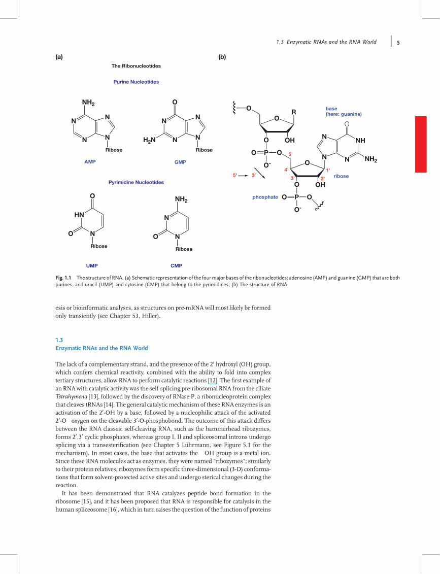
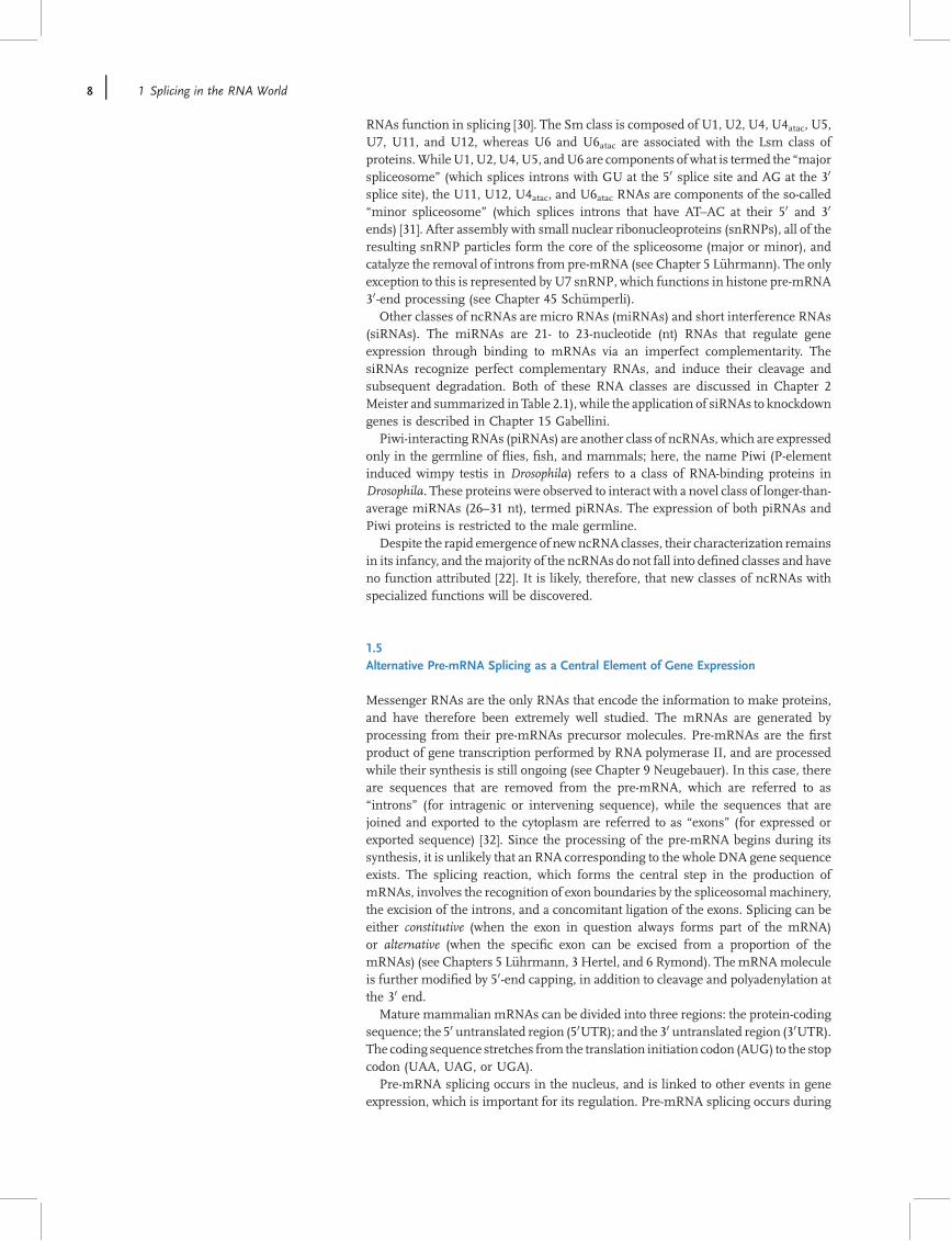
RNA molecules can be represented by a linear sequence of four classical bases:adenine and guanine (A/G, both purines); and cytosine and uracil (C/U, bothpyrimidines). These bases can be subjected to more than 100 post-transcriptionalmodi"cations that are currently listed in the RNA modi"cation database [8]http://library.med.utah.edu/RNAmods (see also Chapter 14 by H"obartner for syn-thetic available bases). In the RNA molecule, each of these bases (schematicallyrepresented in Figure 1.1a) is bound to the 10 position of a ribose sugar that, throughits 30 position, utilizes a phosphate group to linkwith the 50 position of the next ribose.The most important features that distinguish RNA (ribonucleic acid) from DNA(deoxyribonucleic acid) is the presence of a hydroxyl group ("OH) in the 20 positionof the ribose sugar (Figure 1.1b). The 20 hydroxyl group is chemically reactive,which not only makes RNA more vulnerable to degradation but also it to participatein chemical reactions.Although RNAmolecules are described as a single-stranded sequence, most RNA
molecules exhibit a high degree of double-helical character, as complementarysegments of the RNA fold back on each other. The base-pairing of RNA is more!exible than that of DNA. In addition to the canonical Watson–Crick base pairs(cytosine with guanine, adenine with uracil), there are !noncanonical" base pairs,such as G–U pairing, and numerous other base pairings are possible [9]. Since theareas of complementarity in an RNA molecule are short, RNA molecules show localregions of base-pairing, which is referred to as !secondary structure."RNA secondarystructures are locally con"ned, which is in contrast to the extended double-strandedDNA helix.As RNAs do not form a long-range double-stranded structure, the short RNA
helices themselves can interact with each other to formwhat is known as the !tertiarystructure" [10]. The rules that exactly de"ne the "nal outcome of these foldingprocesses, and the various factors that in!uence them, remain the subject of manyactive studies. In contrast to proteins, it is currently not possible to predict in vivoRNAtertiary structures accurately [10] (see Chapter 54 Bujnicki for structure prediction ofsplicing proteins). X-ray crystallography experiments have clearly shown de"nedtertiary structures for metabolically stable RNAs, such as transfer RNAs (tRNAs) orribosomal RNAs (rRNAs) [11]. In contrast, structures in pre-mRNAs that form thesubstrate of the spliceosome can currently be predicted only indirectly by mutagen-
4 j 1 Splicing in the RNA World
esis or bioinformatic analyses, as structures on pre-mRNAwill most likely be formedonly transiently (see Chapter 53, Hiller).
1.3Enzymatic RNAs and the RNA World
The lack of a complementary strand, and the presence of the 20 hydroxyl (OH) group,which confers chemical reactivity, combined with the ability to fold into complextertiary structures, allow RNA to perform catalytic reactions [12]. The "rst example ofanRNAwith catalytic activity was the self-splicing pre-ribosomal RNA from the ciliateTetrahymena [13], followed by the discovery of RNase P, a ribonucleoprotein complexthat cleaves tRNAs [14]. The general catalyticmechanism of these RNAenzymes is anactivation of the 20-OH by a base, followed by a nucleophilic attack of the activated20-O" oxygen on the cleavable 30-O-phosphobond. The outcome of this attack differsbetween the RNA classes: self-cleaving RNA, such as the hammerhead ribozymes,forms 20,30 cyclic phosphates, whereas group I, II and spliceosomal introns undergosplicing via a transesteri"cation (see Chapter 5 L"uhrmann, see Figure 5.1 for themechanism). In most cases, the base that activates the "OH group is a metal ion.Since these RNAmolecules act as enzymes, they were named !ribozymes"; similarlyto their protein relatives, ribozymes form speci"c three-dimensional (3-D) conforma-tions that form solvent-protected active sites and undergo sterical changes during thereaction.It has been demonstrated that RNA catalyzes peptide bond formation in the
ribosome [15], and it has been proposed that RNA is responsible for catalysis in thehuman spliceosome [16], which in turn raises the question of the function of proteins
(a) (b)The Ribonucleotides
Purine Nucleotides
Pyrimidine Nucleotides
UMP CMP
N
N N
N
NH2
Ribose
N
N N
N
O
Ribose
H2N
HN
N
Ribose
O
O
N
N
Ribose
NH2
O
AMP GMP
NH
N
N
O
NH2N
O
OHO
PO
O-
O
O
O
OHO
PO
O-
O
1'
2'3'
4'
5'
base(here: guanine)
ribose
phosphate
5' 3'
R
Fig. 1.1 The structure of RNA. (a) Schematic representation of the fourmajor bases of the ribonucleotides: adenosine (AMP) and guanine (GMP) that are bothpurines, and uracil (UMP) and cytosine (CMP) that belong to the pyrimidines; (b) The structure of RNA.
1.3 Enzymatic RNAs and the RNA World j 5
in ribonucleoproteins (RNPs). The study of RNase P showed the importance ofprotein components associating with ribozymes. RNase P is an RNA–proteincomplex that cleaves tRNA precursors. In bacteria, the catalytic activity resideswithin the RNA [14], but in human mitochondria RNase P catalyzes the reactionwithout RNA, demonstrating that proteins can substitute for RNA functions [17].The question then is, since similar biological functions can be performed by eitherRNA or protein complexes, why did evolution select RNPs such as ribosomes andspliceosomes for protein synthesis and pre-mRNA processing?One possibility is that proteins facilitate the conformational changes of RNA that
are necessary for catalysis. The spliceosome catalyzes the reaction between twostructurally different substrates, which necessitates large spatial rearrangementsduring the reaction (see Chapter 5, L"uhrmann), which could be stabilized byadditional RNA–protein interactions in the spliceosome. In fact, the spliceosomeis an excellent example of an RNP machine, where the degree of interdependencebetween RNA and protein for catalytic function is such that it is justi"ed to consider ita veritable RNP enzyme.The discovery of the enzymatic activity of RNAs led to the concept of a primitive
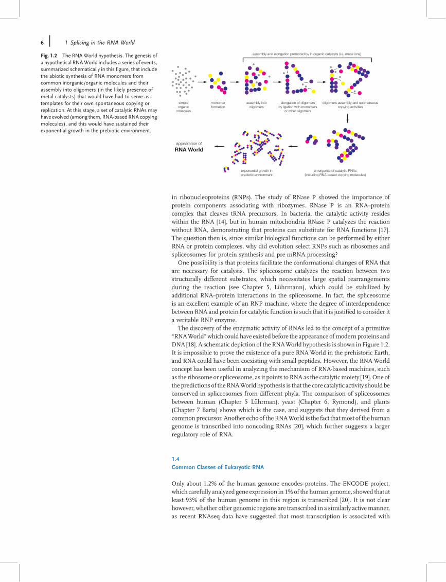
!RNAWorld"which could have existed before the appearance ofmodern proteins andDNA [18]. A schematic depiction of the RNAWorld hypothesis is shown in Figure 1.2.It is impossible to prove the existence of a pure RNAWorld in the prehistoric Earth,and RNA could have been coexisting with small peptides. However, the RNAWorldconcept has been useful in analyzing the mechanism of RNA-based machines, suchas the ribosome or spliceosome, as it points to RNA as the catalyticmoiety [19]. One ofthe predictions of theRNAWorldhypothesis is that the core catalytic activity should beconserved in spliceosomes from different phyla. The comparison of spliceosomesbetween human (Chapter 5 L"uhrman), yeast (Chapter 6, Rymond), and plants(Chapter 7 Barta) shows which is the case, and suggests that they derived from acommonprecursor. Another echo of theRNAWorld is the fact thatmost of the humangenome is transcribed into noncoding RNAs [20], which further suggests a largerregulatory role of RNA.
1.4Common Classes of Eukaryotic RNA
Only about 1.2% of the human genome encodes proteins. The ENCODE project,which carefully analyzed gene expression in 1%of thehumangenome, showed that atleast 93% of the human genome in this region is transcribed [20]. It is not clearhowever, whether other genomic regions are transcribed in a similarly activemanner,as recent RNAseq data have suggested that most transcription is associated with
assembly and elongation promoted by in organic catalysts (i.e. metal ions)
emergence of catalytic RNAs(including RNA-based copying molecules)
exponential growth inprebiotic environment
simpleorganic
molecules
monomerformation
assembly intooligomers
elongation of oligomersby ligation with monomers
or other oligomers
oligomers assembly and spontaneouscopying activities
appearance of
RNA World
Fig. 1.2 The RNA World hypothesis. The genesis ofa hypothetical RNAWorld includes a series of events,summarized schematically in this figure, that includethe abiotic synthesis of RNA monomers fromcommon inorganic/organic molecules and theirassembly into oligomers (in the likely presence ofmetal catalysts) that would have had to serve astemplates for their own spontaneous copying orreplication. At this stage, a set of catalytic RNAs mayhave evolved (among them, RNA-basedRNA copyingmolecules), and this would have sustained theirexponential growth in the prebiotic environment.
6 j 1 Splicing in the RNA World
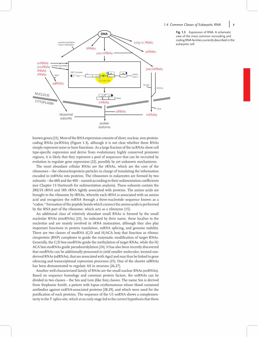
knowngenes [21].Most of theRNAexpression consists of short, nuclear, non-protein-coding RNAs (ncRNAs) (Figure 1.3), although it is not clear whether these RNAssimply represent noise or have functions. As a large fraction of the ncRNAs show celltype-speci"c expression and derive from evolutionary highly conserved promoterregions, it is likely that they represent a pool of sequences that can be recruited byevolution to regulate gene expression [22], possibly by yet unknown mechanisms.The most abundant cellular RNAs are the rRNAs, which are the core of the
ribosomes – the ribonucleoprotein particles in charge of translating the informationencoded in mRNAs into proteins. The ribosomes in eukaryotes are formed by twosubunits – the 60S and the 40S – named according to their sedimentation coef"cients(see Chapter 13 Hartmuth for sedimentation analysis). These subunits contain the28S/5S rRNA and 18S rRNA tightly associated with proteins. The amino acids arebrought to the ribosome by tRNAs, whereby each tRNA is associated with an aminoacid and recognizes the mRNA through a three-nucleotide sequence known as a!codon."Formation of the peptide bondswhich connect the amino acids is performedby the RNA part of the ribosome, which acts as a ribozyme [15].An additional class of relatively abundant small RNAs is formed by the small
nucleolar RNAs (snoRNAs) [23]. As indicated by their name, these localize to thenucleolus and are mainly involved in rRNA maturation, although they also playimportant functions in protein translation, mRNA splicing, and genome stability.There are two classes of snoRNA (C/D and H/ACA box) that function as ribonu-cleoprotein (RNP) complexes to guide the enzymatic modi"cation of target RNAs.Generally, the C/D box snoRNAs guide the methylation of target RNAs, while the H/ACA box snoRNAs guide pseudouridylation [24]. It has also been recently discoveredthat snoRNAs can be additionally processed to yield smaller molecules, termed sno-derivedRNAs (sdRNAs), that are associatedwithAgo2 andmay thus be linked to genesilencing and transcriptional repression processes [25]. One of the shorter sdRNAshas been demonstrated to regulate AS in neurons [26,27].Another well-characterized family of RNAs are the small nuclear RNAs (snRNAs).
Based on sequence homology and common protein factors, the snRNAs can bedivided in two classes – the Sm and Lsm (like Sm) classes. The name Sm is derivedfrom Stephanie Smith, a patient with lupus erythematosus whose blood containedantibodies against snRNA-associated proteins [28,29], and which were used for thepuri"cation of such proteins. The sequence of the U1 snRNA shows a complemen-tarity to the 50 splice site, which at an early stage led to the correct hypothesis that these
DNA
long nc RNAs
tiRNAspre-mRNAs piRNAs
pre-miRNAs
mirtrons
Dicer
miRNAssiRNAs
mRNAs
ribosomalsubunits
proteinisoforms
Drosha
snRNAssnoRNAstRNAsrRNAs
sdRNAs
Ago7
smRNPs/snoRNPs
- pseudourydinilation- ribose methylation
CYTOPLASM
NUCLEUSAAAA
AAAA...poly A
7mG -Cap
CBP Complex
spliceosome
Fig. 1.3 Expression of RNA. A schematicview of the most common noncoding andcoding RNA families currently described in theeukaryotic cell.
1.4 Common Classes of Eukaryotic RNA j 7
RNAs function in splicing [30]. The Sm class is composed of U1, U2, U4, U4atac, U5,U7, U11, and U12, whereas U6 and U6atac are associated with the Lsm class ofproteins.WhileU1,U2,U4,U5, andU6 are components ofwhat is termed the !majorspliceosome" (which splices introns with GU at the 50 splice site and AG at the 30
splice site), the U11, U12, U4atac, and U6atac RNAs are components of the so-called!minor spliceosome" (which splices introns that have AT–AC at their 50 and 30
ends) [31]. After assembly with small nuclear ribonucleoproteins (snRNPs), all of theresulting snRNP particles form the core of the spliceosome (major or minor), andcatalyze the removal of introns from pre-mRNA (see Chapter 5 L"uhrmann). The onlyexception to this is represented by U7 snRNP, which functions in histone pre-mRNA30-end processing (see Chapter 45 Sch"umperli).Other classes of ncRNAs are micro RNAs (miRNAs) and short interference RNAs
(siRNAs). The miRNAs are 21- to 23-nucleotide (nt) RNAs that regulate geneexpression through binding to mRNAs via an imperfect complementarity. ThesiRNAs recognize perfect complementary RNAs, and induce their cleavage andsubsequent degradation. Both of these RNA classes are discussed in Chapter 2Meister and summarized in Table 2.1), while the application of siRNAs to knockdowngenes is described in Chapter 15 Gabellini.Piwi-interacting RNAs (piRNAs) are another class of ncRNAs, which are expressed
only in the germline of !ies, "sh, and mammals; here, the name Piwi (P-elementinduced wimpy testis in Drosophila) refers to a class of RNA-binding proteins inDrosophila. These proteins were observed to interact with a novel class of longer-than-average miRNAs (26–31 nt), termed piRNAs. The expression of both piRNAs andPiwi proteins is restricted to the male germline.Despite the rapid emergence of newncRNAclasses, their characterization remains
in its infancy, and themajority of the ncRNAs do not fall into de"ned classes and haveno function attributed [22]. It is likely, therefore, that new classes of ncRNAs withspecialized functions will be discovered.
1.5Alternative Pre-mRNA Splicing as a Central Element of Gene Expression
Messenger RNAs are the only RNAs that encode the information to make proteins,and have therefore been extremely well studied. The mRNAs are generated byprocessing from their pre-mRNAs precursor molecules. Pre-mRNAs are the "rstproduct of gene transcription performed by RNA polymerase II, and are processedwhile their synthesis is still ongoing (see Chapter 9 Neugebauer). In this case, thereare sequences that are removed from the pre-mRNA, which are referred to as!introns" (for intragenic or intervening sequence), while the sequences that arejoined and exported to the cytoplasm are referred to as !exons" (for expressed orexported sequence) [32]. Since the processing of the pre-mRNA begins during itssynthesis, it is unlikely that an RNA corresponding to the whole DNA gene sequenceexists. The splicing reaction, which forms the central step in the production ofmRNAs, involves the recognition of exon boundaries by the spliceosomalmachinery,the excision of the introns, and a concomitant ligation of the exons. Splicing can beeither constitutive (when the exon in question always forms part of the mRNA)or alternative (when the speci"c exon can be excised from a proportion of themRNAs) (see Chapters 5 L"uhrmann, 3 Hertel, and 6 Rymond). ThemRNAmoleculeis further modi"ed by 50-end capping, in addition to cleavage and polyadenylation atthe 30 end.Mature mammalian mRNAs can be divided into three regions: the protein-coding
sequence; the 50 untranslated region (50UTR); and the 30 untranslated region (30UTR).The coding sequence stretches from the translation initiation codon (AUG) to the stopcodon (UAA, UAG, or UGA).Pre-mRNA splicing occurs in the nucleus, and is linked to other events in gene
expression, which is important for its regulation. Pre-mRNA splicing occurs during
8 j 1 Splicing in the RNA World
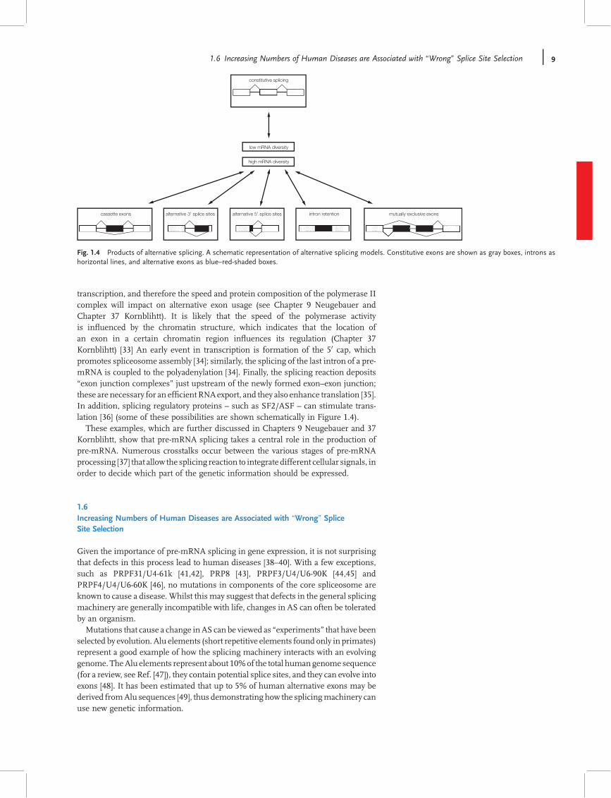
transcription, and therefore the speed and protein composition of the polymerase IIcomplex will impact on alternative exon usage (see Chapter 9 Neugebauer andChapter 37 Kornblihtt). It is likely that the speed of the polymerase activityis in!uenced by the chromatin structure, which indicates that the location ofan exon in a certain chromatin region in!uences its regulation (Chapter 37Kornblihtt) [33] An early event in transcription is formation of the 50 cap, whichpromotes spliceosome assembly [34]; similarly, the splicing of the last intron of a pre-mRNA is coupled to the polyadenylation [34]. Finally, the splicing reaction deposits!exon junction complexes" just upstream of the newly formed exon–exon junction;these are necessary for an ef"cientRNAexport, and they also enhance translation [35].In addition, splicing regulatory proteins – such as SF2/ASF – can stimulate trans-lation [36] (some of these possibilities are shown schematically in Figure 1.4).These examples, which are further discussed in Chapters 9 Neugebauer and 37
Kornblihtt, show that pre-mRNA splicing takes a central role in the production ofpre-mRNA. Numerous crosstalks occur between the various stages of pre-mRNAprocessing [37] that allow the splicing reaction to integrate different cellular signals, inorder to decide which part of the genetic information should be expressed.
1.6Increasing Numbers of Human Diseases are Associated with !Wrong" SpliceSite Selection
Given the importance of pre-mRNA splicing in gene expression, it is not surprisingthat defects in this process lead to human diseases [38–40]. With a few exceptions,such as PRPF31/U4-61k [41,42], PRP8 [43], PRPF3/U4/U6-90K [44,45] andPRPF4/U4/U6-60K [46], no mutations in components of the core spliceosome areknown to cause a disease. Whilst this may suggest that defects in the general splicingmachinery are generally incompatible with life, changes in AS can often be toleratedby an organism.Mutations that cause a change inAS can be viewed as !experiments" that have been
selected by evolution. Alu elements (short repetitive elements found only in primates)represent a good example of how the splicing machinery interacts with an evolvinggenome. TheAlu elements represent about 10%of the total humangenome sequence(for a review, see Ref. [47]), they contain potential splice sites, and they can evolve intoexons [48]. It has been estimated that up to 5% of human alternative exons may bederived fromAlu sequences [49], thus demonstrating how the splicingmachinery canuse new genetic information.
constitutive splicing
low mRNA diversity
high mRNA diversity
cassette exons alternative 5' splice sitesalternative 3' splice sites intron retention mutually exclusive exons
Fig. 1.4 Products of alternative splicing. A schematic representation of alternative splicing models. Constitutive exons are shown as gray boxes, introns ashorizontal lines, and alternative exons as blue–red-shaded boxes.
1.6 Increasing Numbers of Human Diseases are Associated with !Wrong" Splice Site Selection j 9
By far the largest number of currently known splicing diseases are caused bymutations in the pre-mRNA,which leads to aberrant exonusage (Chapter 10, Baralle).The analysis of these mutations has been highly informative for the mechanism ofsplice site regulation, and has provided insights into disease mechanisms thatallowed rational therapies "rst to be devised. For example, the sequencing ofdisease-associatedmRNAs led to the detection of numerous synonymousmutations.Initially, it was dif"cult to understand how these mutations could lead to a disease, assynonymousmutations did not alter the protein encoded by themRNA.However, thedisease mechanism became clear when it was realized that exons contain sequencesthat regulate their AS [50]. Synonymous mutations can, therefore, act by changingalternative exon usage. Based on this insight into the disease mechanism, it ispossible to test therapeutic approaches. Currently, themost common genetic cause ofdeath in children is spinal muscular atrophy (SMA). This deadly disease is caused bydeletion of the SMN1 gene which, unfortunately, cannot be substituted by the almostidentical SMN2 gene that is present in all patients. The difference between the twogenes is a synonymous mutation that causes exon skipping in SMN2. Yet, thedisease could be treated if the inclusion of this particular exon into the SMN2mRNAcould be promoted. Towards this aim, the regulation of the exonhas been investigatedin great detail (see Chapters 18 and 19 Singh), and this has led not only to thedevelopment of therapeutic approaches currently undergoing clinical trials [51], butalso to new experimental approaches to alter splice site selection (see Chapter 45Sch"umperli).The majority of currently known mutations that have a clear effect on alternative
exon usage have become apparent in only a few patients. One such example – theHutchinson–Gilford progeria syndrome (HGPS) –highlights the bene"ts of studyingthese rare diseases. HGPS is genetic disorder that is characterized phenotypically bymany features of premature aging, with patients typically dying at the age of 13 years.Mutations causing HGPS have been identi"ed in the nuclear lamin A/C (LMNA)gene, and three out of 14 mutations affecting lamin A/C have been reported tospeci"cally alter laminA splicing (see Chapter 36, Tazi). This particular splicing eventcauses an aberrant farnesylation of the resulting protein, which causes the disease. Ascreening effort has identi"ed a previously tested farnesyltransferase inhibitor thatcould be used to treat the disease [52]. HGPS, which has an incidence of 1 per 4–8million live births, may prove to be highly informative for the normal aging process,as the mutant splice variant accumulates in the skin of aging individuals [53]. Thisexample shows that screening for substances that change alternative splice siteselection (as shown inChapter 46 Stoilov)may, in time, have a huge impact onhumanhealth.The development of new experimental techniques was a strong driving force in
research on AS. The current experimental protocols collected in this book have beenbuilt onmore than 30 years of experimental experience. For example, the identi"cationof antisera against splicing components [28] allowed their puri"cation such that, today,is it possible to purify different stages of spliceosomes (see Chapters 13 and 31L"uhrmann) and to generate speci"c antisera (as shown in Chapter 43 Fishman). Theidenti"cation of alternative exons is based on comparing cDNA and genomicsequences. The completion of the human genome showed, for the "rst time, theunexpected high usage of AS on an organismal level [54]. The sequencing techniqueshave been improved (as shown in Chapters 25 Guigo and 51 Zhang), and it is nowpossible to rapidly sequence the genome of an individual [55]. The knowledge ofindividual genome sequences, and their analysis by genome-wide DNA arrays (seeChapter 24 de la Grange), marks the beginning of personalized medicine. This willallow the analysis of potential changes in the AS of individuals by PCR (Chapter 21Smith), cell-based (Chapters 35–37, Stamm, Tazi, Kornblihtt), and in vitro assays(Chapter 30 Krainer). Knowledge of disease-causing mechanisms may lead to a bettergenetic counseling (see Chapter 11 Baralle), and might also pave the way to thedevelopment of therapies (as discussed in Chapters 45–48 Sch"umperli, Stoilov,Annemieke, Patel).
10 j 1 Splicing in the RNA World
Acknowledgments
This work was supported by Telethon, and by the EC grant EURASNET.
References
1 Berget, S.M., Moore, C., and Sharp, P.A.(1977) Spliced segments at the 50 terminusof adenovirus 2 latemRNA. Proc. Natl Acad.Sci. USA, 74, 3171–3175.
2 Chow, L.T., Roberts, J.M., Lewis, J.B., andBroker, T.R. (1977) A map of cytoplasmicRNA transcripts from lytic adenovirus type2, determined by electron microscopy ofRNA:DNA hybrids. Cell, 11, 819–836.
3 Sharp, P.A. (1994) Split genes and RNAsplicing. Cell, 77, 805–815.
4 Pan, Q., Shai, O., Lee, L.J., Frey, B.J., andBlencowe, B.J. (2008) Deep surveying ofalternative splicing complexity in thehuman transcriptome by high-throughputsequencing. Nat. Genet., 40, 1413–1415.
5 Patel, N.A., Chalfant, C.E., Watson, J.E.,Wyatt, J.R., Dean, N.M., Eichler, D.C., andCooper, D.R. (2001) Insulin regulatesalternative splicing of protein kinase Cbeta II through a phosphatidylinositol3-kinase-dependent pathway involving thenuclear serine/arginine-rich splicing factor,SRp40, in skeletal muscle cells. J. Biol.Chem., 276, 22648–22654.
6 Weg-Remers, S., Ponta, H., Herrlich, P.,and Konig, H. (2001) Regulation ofalternative pre-mRNA splicing by the ERKMAP-kinase pathway. EMBO J., 20,4194–4203.
7 Celotto, A.M. and Graveley, B.R. (2001)Alternative splicing of the DrosophilaDscam pre-mRNA is both temporally andspatially regulated. Genetics, 159, 599–608.
8 Rozenski, J., Crain, P.F., and McCloskey,J.A. (1999) The RNA Modi"cationDatabase: 1999 update. Nucleic Acids Res.,27, 196–197.
9 Leontis, N.B. and Westhof, E. (2001)Geometric nomenclature and classi"cationof RNA base pairs. RNA, 7, 499–512.
10 Brion, P. and Westhof, E. (1997) Hierarchyand dynamics of RNA folding. Annu. Rev.Biophys. Biomol. Struct., 26, 113–137.
11 Noller, H.F. (2005) RNA structure: readingthe ribosome. Science, 309, 1508–1514.
12 Doudna, J.A. and Cech, T.R. (2002) Thechemical repertoire of natural ribozymes.Nature, 418, 222–228.
13 Kruger, K., Grabowski, P.J., Zaug, A.J.,Sands, J., Gottschling, D.E., and Cech, T.R.(1982) Self-splicing RNA: autoexcision andautocyclization of the ribosomal RNAintervening sequence of Tetrahymena. Cell,31, 147–157.
14 Guerrier-Takada, C., Gardiner, K.,Marsh, T.,Pace, N., and Altman, S. (1983) The RNAmoiety of ribonuclease P is the catalyticsubunit of the enzyme. Cell, 35, 849–857.
15 Steitz, T.A. (2008) A structuralunderstanding of the dynamic ribosomemachine. Nat. Rev. Mol. Cell Biol., 9,242–253.
16 Valadkhan, S., Mohammadi, A., Wachtel,C., and Manley, J.L. (2007) Protein-freespliceosomal snRNAs catalyze a reactionthat resembles the "rst step of splicing.RNA, 13, 2300–2311.
17 Holzmann, J., Frank, P., Lof!er, E., Bennett,K.L., Gerner, C., and Rossmanith, W. (2008)RNase P without RNA: identi"cation andfunctional reconstitution of the humanmitochondrial tRNA processing enzyme.Cell, 135, 462–474.
18 Orgel, L.E. (1998) The origin of life – areview of facts and speculations. TrendsBiochem. Sci., 23, 491–495.
19 Cech, T.R. (2009) Crawling out of the RNAworld. Cell, 136, 599–602.
20 Birney, E., Stamatoyannopoulos, J.A., Dutta,A., Guigo, R., Gingeras, T.R., Margulies,E.H., Weng, Z., Snyder, M., Dermitzakis,E.T., Thurman, R.E. et al. (2007)Identi"cation and analysis of functionalelements in 1% of the human genome bythe ENCODE pilot project. Nature, 447,799–816.
21 van Bakel, H., Nislow, C., Blencowe, B.J.,and Hughes, T.R. (2010) Most #dark matter$transcripts are associated with knowngenes. PLoS Biol., 8 (5), e1000371.
22 Amaral, P.P., Dinger, M.E., Mercer, T.R.,and Mattick, J.S. (2008) The eukaryoticgenome as an RNA machine. Science, 319,1787–1789.
23 Dieci, G., Preti, M., and Montanini, B.(2009) Eukaryotic snoRNAs: a paradigm forgene expression !exibility. Genomics, 94,83–88.
24 Matera, A.G., Terns, R.M., and Terns, M.P.(2007) Non-coding RNAs: lessons from thesmall nuclear and small nucleolar RNAs.Nat. Rev. Mol. Cell Biol., 8, 209–220.
25 Taft, R.J., Glazov, E.A., Lassmann, T.,Hayashizaki, Y., Carninci, P., and Mattick,J.S. (2009) Small RNAs derived fromsnoRNAs. RNA, 15, 1233–1240.
26 Kishore, S., Khanna, A., Zhang, Z., Hui, J.,Balwierz, P., Stefan,M., Beach, C., Nicholls,R.D., Zavolan, M., and Stamm, S. (2010)The snoRNA MBII-52 (SNORD 115) isprocessed into smaller RNAs and regulatesalternative splicing. Hum. Mol. Genet., 19,1153–1164.
27 Kishore, S. and Stamm, S. (2006) ThesnoRNA HBII-52 regulates alternativesplicing of the serotonin receptor 2C.Science, 311, 230–232.
28 Lerner, M.R. and Steitz, J.A. (1979)Antibodies to small nuclear RNAscomplexed with proteins are produced bypatients with systemic lupuserythematosus. Proc. Natl Acad. Sci. USA,76, 5495–5499.
29 Reeves, W.H., Narain, S., and Satoh, M.(2003) Henry Kunkel, Stephanie Smith,clinical immunology, and split genes. Lupus,12, 213–217.
30 Lerner, M.R., Boyle, J.A., Mount, S.M.,Wolin, S.L., and Steitz, J.A. (1980) AresnRNPs involved in splicing? Nature, 283,220–224.
31 Will, C.L. and L"uhrmann, R. (2005) Splicingof a rare class of introns by the U12-dependent spliceosome. Biol. Chem., 386,713–724.
32 Gilbert, W. (1978) Why genes in pieces?Nature, 271, 501.
33 Kornblihtt, A.R. (2007) Couplingtranscription and alternative splicing. Adv.Exp. Med. Biol., 623, 175–189.
34 Schwer, B. and Shuman, S. (1996)Conditional inactivation of mRNA cappingenzyme affects yeast pre-mRNA splicingin vivo. RNA, 2, 574–583.
35 Luo, M.L., Zhou, Z., Magni, K.,Christoforides, C., Rappsilber, J., Mann,M.,and Reed, R. (2001) Pre-mRNA splicing andmRNA export linked by direct interactionsbetween UAP56 and Aly. Nature, 413,644–647.
36 Sanford, J.R., Gray,N.K., Beckmann,K., andCaceres, J.F. (2004) A novel role forshuttling SR proteins in mRNA translation.Genes Dev., 18, 755–768.
37 Moore, M.J. (2005) From birth to death: thecomplex lives of eukaryoticmRNAs.Science,309, 1514–1518.
38 Cooper, T.A., Wan, L., and Dreyfuss, G.(2009) RNA and disease. Cell, 136, 777–793.
39 Jeanteur, P. (2006) Alternative Splicing andDisease, Springer.
40 Tazi, J., Bakkour, N., and Stamm, S. (2009)Alternative splicing and disease. Biochim.Biophys. Acta, 1792, 14–26.
41 Vithana, E.N., Abu-Sa"eh, L., Allen, M.J.,Carey, A., Papaioannou, M., Chakarova, C.,Al-Maghtheh, M., Ebenezer, N.D., Willis,C., Moore, A.T. et al. (2001) A humanhomolog of yeast pre-mRNA splicing gene,PRP31, underlies autosomal dominantretinitis pigmentosa on chromosome19q13.4 (RP11). Mol. Cell, 8, 375–381.
42 Wilkie, S.E., Vaclavik, V., Wu, H.,Bujakowska, K., Chakarova, C.F.,Bhattacharya, S.S., Warren, M.J., and Hunt,D.M. (2008) Disease mechanism for
References j 11
retinitis pigmentosa (RP11) caused bymissense mutations in the splicing factorgene PRPF31. Mol. Vis., 14, 683–690.
43 Boon, K.L., Grainger, R.J., Ehsani, P.,Barrass, J.D., Auchynnikava, T., Inglehearn,C.F., and Beggs, J.D. (2007) prp8 mutationsthat cause human retinitis pigmentosa leadto a U5 snRNP maturation defect in yeast.Nat. Struct. Mol. Biol., 14, 1077–1083.
44 Chakarova, C.F., Hims, M.M., Bolz, H.,Abu-Sa"eh, L., Patel, R.J., Papaioannou,M.G., Inglehearn,C.F., Keen, T.J.,Willis, C.,Moore, A.T. et al. (2002) Mutations inHPRP3, a third member of pre-mRNAsplicing factor genes, implicated inautosomal dominant retinitis pigmentosa.Hum. Mol. Genet., 11, 87–92.
45 Gonzalez-Santos, J.M., Cao, H., Duan, R.C.,and Hu, J. (2008) Mutation in the splicingfactorHprp3p linked to retinitis pigmentosaimpairs interactions within the U4/U6snRNP complex. Hum. Mol. Genet., 17,225–239.
46 Schmidt-Kastner, R., Yamamoto, H.,Hamasaki, D., Parel, J.M., Schmitz, C.,Dorey, C.K., Blanks, J.C., andPreising,M.N.
(2008) Hypoxia-regulated components oftheU4/U6.U5 tri-small nuclear riboproteincomplex: possible role in autosomaldominant retinitis pigmentosa. Mol. Vis.,14, 125–135.
47 Hasler, J., Samuelsson, T., and Strub, K.(2007) Useful #junk$: Alu RNAs in thehuman transcriptome.Cell Mol. Life Sci., 64,1793–1800.
48 Lev-Maor, G., Sorek, R., Shomron, N., andAst, G. (2003) The birth of an alternativelyspliced exon: 30 splice-site selection in Aluexons. Science, 300, 1288–1291.
49 Sorek, R., Ast, G., and Graur, D. (2002) Alu-containing exons are alternatively spliced.Genome Res., 12, 1060–1067.
50 Cooper, T.A. and Mattox, W. (1997) Theregulation of splice-site selection, and itsrole in human disease. Am. J. Hum. Genet.,61, 259–266.
51 Darras, B.T. and Kang, P.B. (2007) Clinicaltrials in spinalmuscular atrophy.Curr.Opin.Pediatr., 19, 675–679.
52 Capell, B.C., Olive, M., Erdos,M.R., Cao, K.,Faddah, D.A., Tavarez, U.L., Conneely, K.N.,Qu, X., San, H., Ganesh, S.K. et al. (2008) A
farnesyltransferase inhibitor prevents boththe onset and late progression ofcardiovascular disease in a progeria mousemodel. Proc. Natl Acad. Sci. USA, 105,15902–15907.
53 McClintock, D., Ratner, D., Lokuge, M.,Owens, D.M., Gordon, L.B., Collins, F.S.,and Djabali, K. (2007) The mutant form oflamin A that causes Hutchinson-Gilfordprogeria is a biomarker of cellular aging inhuman skin. PLoS One, 2, e1269.
54 Lander, E.S., Linton, L.M., Birren, B.,Nusbaum, C., Zody, M.C., Baldwin, J.,Devon, K., Dewar, K., Doyle, M., FitzHugh,W. et al. (2001) Initial sequencing andanalysis of the human genome.Nature, 409,860–921.
55 Wheeler, D.A., Srinivasan, M., Egholm, M.,Shen, Y., Chen, L., McGuire, A., He, W.,Chen, Y.J., Makhijani, V., Roth, G.T. et al.(2008) The complete genome of anindividual by massively parallel DNAsequencing. Nature, 452, 872–876.
12 j 1 Splicing in the RNA World
Recommended


