
FULL PAPER
DOI: 10.1002/ejoc.200901346
Supramolecular Assemblies Derived from Formyl-Substituted π-ConjugatedAcyclic Anion Receptors
Hiromitsu Maeda,*[a,b] Rika Fujii,[a] and Yohei Haketa[a]
Keywords: Anions / Boron / Formyl units / Pyrrole derivatives / Receptors / Supramolecular chemistry
We report on the synthesis and properties of formyl-substi-tuted dipyrrolyl diketone–BF2 complexes (anion receptors)and their extended derivatives. The formyl-substituted re-ceptors exhibited efficient anion-binding behavior and com-plicated solid-state hydrogen-bonding assembly patterns,due to the formyl group’s electron-withdrawing and hydro-gen-bond-accepting properties. The extended derivatives,
IntroductionSoft matter has been attracting increasing attention as
a “transformable” functional material due to its moderatemobility and flexibility, which readily enable it to change itsbulk shape and properties depending on the conditions.[1]
Supramolecular gels are dimensionally controlled assem-blies made up of low-molecular-weight (LMW) moleculesheld together by noncovalent interactions such as hydrogenbonding, metal coordination, van der Waals interaction,and π–π stacking. Gels derived from molecular assemblies,in which the components can be readily replaced with alter-natives, may provide promising material systems for drugdelivery and tissue engineering.[2,3] Supramolecular gelsmade up of molecules sensitive to external physical stimulican be modulated and controlled by the conditions.[4] Incontrast to physical stimuli, chemical stimuli such as theincorporation of a specific species can afford versatile sup-ramolecular gels that change their states as a result of theinteractions between the additives and the gelator molec-ules.[5] Of the various available chemical stimuli available,inorganic and biotic anions, such as halides, acetates, andphosphates, which are ubiquitous in biology, are essential,as can be seen in the activity of enzymes, transport of hor-mones, protein synthesis, and DNA regulation.[6,7] Recently,
the chemistry of anion-responsive supramolecular gels has
[a] College of Pharmaceutical Sciences, Institute of Science andEngineering, Ritsumeikan UniversityKusatsu 525-8577, JapanFax: +81-77-561-2659E-mail: [email protected]
[b] PRESTO, Japan Science and Technology Agency (JST)Kawaguchi 332-0012, JapanSupporting information for this article is available on theWWW under http://dx.doi.org/10.1002/ejoc.200901346.
Eur. J. Org. Chem. 2010, 1469–1482 © 2010 Wiley-VCH Verlag GmbH & Co. KGaA, Weinheim 1469
prepared by the formation of Schiff bases and their subse-quent reduction, behaved as building subunits to providegel-like materials in spite of the existence of β-ethyl substitu-ents. Fairly unspecific spherical particle morphologies wereobserved for these gel-like materials by optical microscopyand AFM. The anion-responsive behavior of the supramolec-ular assemblies (gelated materials) was also examined.
become all the more fascinating as a result of their potentialas stimuli-responsive functional soft materials.[8,9]
Supramolecular gels based on metal complexes also ex-hibit transformation by anions.[9a–9c] Lee et al., for example,prepared a dendrimer-like oxyethylene-substituted bis(pyr-idine) derivative that forms coordination polymers or oligo-mers, such as dispersed helical structures and discrete cyclicconformations, through Ag+ complexation; the mor-phologies of the organized structures depend on the counteranions present, such as NO3
–, BF4–, and CF3SO3
–, withoutspecific interactions with the gelators.[9a,9b] Aida et al. re-ported the synthesis of an alkyl-substituted pyrazole–Au+
trinuclear complex that forms a red-emissive supramolec-ular hexane gel with states and emission behavior controlledby the anion-driven “liberation” of metal ions.[9c] Further,some of the amide- and urea-based supramolecular gels areanion-responsive because of the affinities of the polarizedNH sites for anions, which disrupt the hydrogen-bond-as-sembled structures.[9d–9k] Under suitable conditions, thechemical stimuli not only act as inhibitors of the formationof supramolecular assemblies but also function as tuneablecomponents that are included in the organized structures.
As efficient receptors for anions, we have reported acyclicplanar π-conjugated systems in the form of BF2 complexesof 1,3-dipyrrolylpropane-1,3-diones (e.g., 1a–c, 2a–d, Fig-ure 1a),[10–12] which can bind anions through the action oftwo pyrrole NH moieties and the bridging CH system, withthe inversion of the pyrrole rings (Figure 1b). In fact, α-aryl-substituted receptors containing long aliphatic chains(e.g., 1b) form anion-responsive supramolecular organogelson gelation from octane solutions and exhibit transitions tothe solution state on addition of appropriate anions,[8,12a]
whereas amphiphilic receptors possessing hydrophilicchains (e.g., 1c) form solvent-assisted assemblies, such asvesicular structures, in aqueous solution.[12d]

H. Maeda, R. Fujii, Y. HaketaFULL PAPER
Figure 1. (a) BF2 complexes of dipyrrolyl diketones 1a–c, 2a–d, and formyl-substituted derivatives (3m1, 3m2, 3p1, and 3p2). (b) Anion-binding mode of 1a.
Unlike the β-unsubstituted compounds 1a–c, which areobtained from α-arylpyrroles as starting materials, the β-alkyl-substituted receptor 2a, as an essential key starting ma-terial,[11e] can be converted into various π-extended receptorssuch as α-phenyl- (2b, 2c) and α-pyrrolyl-substituted (2d) de-rivatives via α-iodinated intermediates.[12b,12c] Accordingly,the introduction of appropriate moieties that are readilytransformable into other units would afford various anion-receptor derivatives. Of the potentially transformable groups,we chose the formyl (CHO) group, which is reactive for re-duction to alcohols, oxidation to carboxylic acids, couplingto alkenes, condensation with amines to Schiff bases, etc.[13]
In this paper we discuss formyl-substituted derivatives of theacyclic anion receptors (3m1 and 3m2, or 3p1 and 3p2),which were converted into various derivatives that behavedas building blocks for supramolecular assemblies with/with-out anions in hydrocarbon solvents to afford gel-like materi-als. Effects of the sp3-linkages between the core π-plane andthe side aryl moieties are also discussed.
Results and Discussion
Synthesis and Characterization of BF2 Complexes ofFormyl-Substituted Dipyrrolyl Diketones
Mono- and diformyl substituents were introduced intoacyclic anion receptors to yield the meta-monoformyl com-pound 3m1, the meta-diformyl compound 3m2, the para-monoformyl compound 3p1, and the para-diformyl com-pound 3p2, in 40, 44, 29, and 15% yields, respectively,through cross-coupling reactions with mono- and bis(α-iodo)-substituted BF2 complexes[12b] and the corresponding
www.eurjoc.org © 2010 Wiley-VCH Verlag GmbH & Co. KGaA, Weinheim Eur. J. Org. Chem. 2010, 1469–14821470
(formylphenyl)boronic acid derivatives. The synthesis pro-cedure with iodo-substituted receptors afforded betteryields than those obtained with α-borylated dipyrrolyl dike-tones.[12d] The synthetic route starting from α-form-ylphenyl-substituted pyrrole and malonyl chloride, as inthe case of 1a–c,[12a] did not work well, possibly due to po-lymerization during the reactions to yield unidentified com-pounds. Chemical identification of 3m1, 3m2, 3p1, and 3p2was carried out by 1H NMR and MALDI-TOF MS. TheUV/Vis absorption bands of 3m1, 3m2, 3p1, and 3p2 inCH2Cl2 were observed at 474, 495, 482, and 510 nm, respec-tively, suggesting that the formyl substitution at the paraposition extended the π-conjugation, whereas that at themeta position disrupted the π-conjugation to some extent,comparable to the situation with α-unsubstituted 2a(451 nm),[11e] mono-α-phenylsubstituted 2b (476 nm), andbis(α-phenyl)-substituted 2c (499 nm).[12b] The order ofthese λmax values was correlated to the order of theHOMO–LUMO gaps (3m1: 3.300 eV; 3m2: 3.191 eV; 3p1:2.844 eV; 3p2: 3.163 eV) as estimated by DFT calculations(Figures S3 and S4 in the Supporting Information).[14] Fur-ther, fluorescence emissions (and quantum yields ΦF) ex-cited at each absorption maximum in CH2Cl2 were ob-served at 504 (0.92) (3m1), 532 (0.98) (3m2), 511 (0.95)(3p1), and 547 nm (0.97) (3p2), suggesting that the formyl-substituted derivatives, like 2b (ΦF = 0.70) and 2c (0.94),were also highly emissive fluorophores.
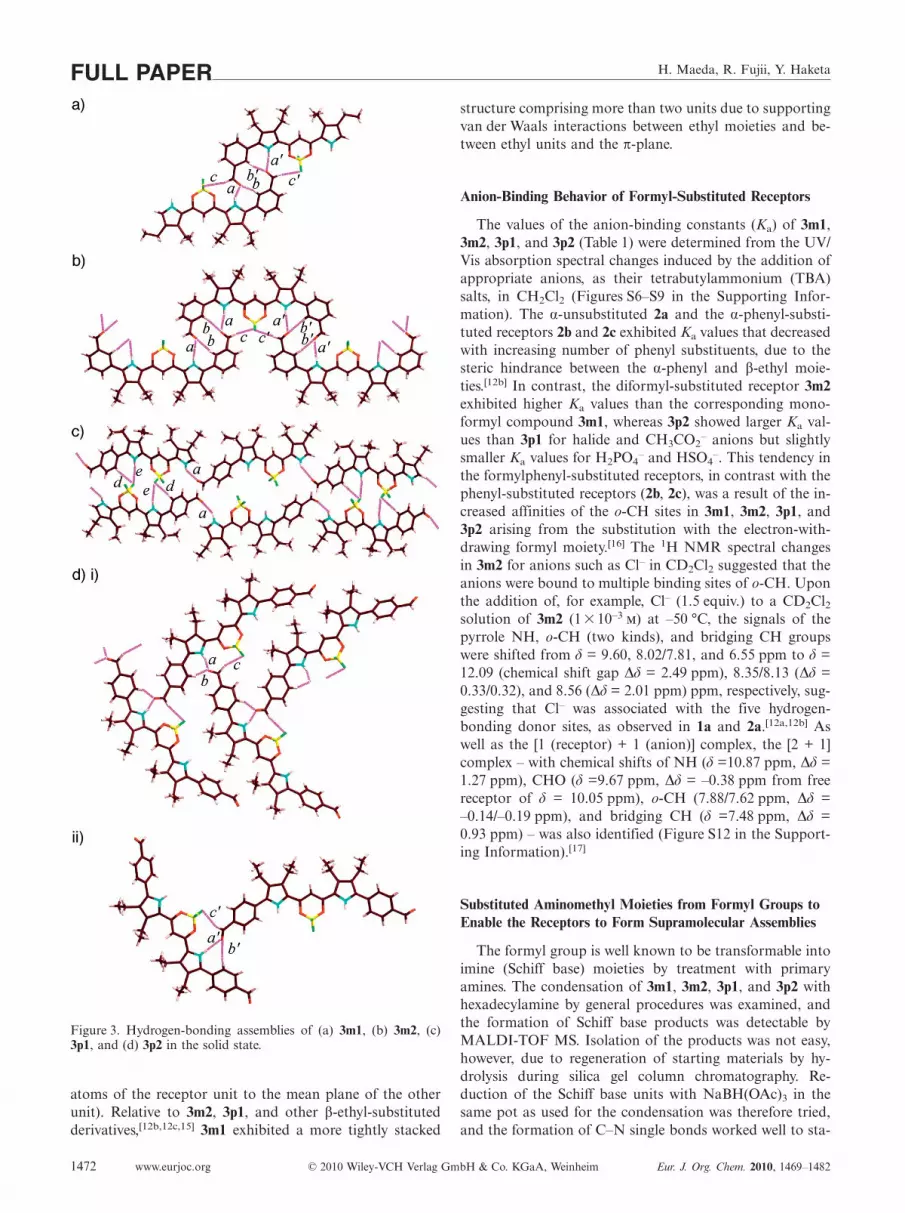
The solid-state structures of 3m1, 3m2, 3p1, and 3p2 weredetermined by single-crystal X-ray analyses (Figure 2). Allthe receptor molecules formed hydrogen-bonding assembl-ies through their CHO moieties, along with the pyrrole NHand BF units. Here we have labeled the distances (C=)O···

Supramolecular Assemblies from Formyl-Substituted Anion Receptors
(H–)N, (C=)O···(H–)o-C, F···(H–)C(=O), F···(H–)o-C, andF···(H–)N as a, b, c, d, and e, respectively. The monoformylcompound 3m1 formed hydrogen-bonding dimers with(C=)O···(H–)N and (C=)O···(H–)o-C distances of 2.963 (a)/3.418 (a�) and 3.080 (b)/3.331 (b�) Å, respectively. The B–F···H–C(=O) interaction [F···(H–)C(=O): 3.486 (c)/3.301(c�) Å] also supported the assembled dimer structure (Fig-ure 3a). On the other hand, the receptors 3m2, 3p1, and3p2 exhibited the formation of 1-D hydrogen-bonding chainstructures.
In the case of 3m2, the (C=)O···(H–)N, (C=)O···(H–)o-C, and F···(H–)C(=O) distances in the chain structures were3.075 (a)/3.029 (a�), 3.121 (b)/3.163 (b�), and 3.400 (c)/3.436(c�) Å, respectively. The two formyl units in the receptordisplayed inequivalent interaction, as observed in the hy-drogen bond lengths (Figure 3b). The derivative 3m1formed fairly planar supramolecular assemblies, which wereefficient stacking structures. Interestingly, the monoformylcompound 3p1 produced 1-D chains through C=O···H–Ninteractions, which further constructed “knit”-like assem-blies through B–F···H–N and B–F···H–o-C hydrogen bond-ing. The (C=)O···(H–)N, F···(H–)o-C, and F···(H–)N dis-tances were 2.888 (a), 3.300 (d), and 3.235 (e) Å, respec-tively. A dihedral angle of 117.9° was observed between thetwo neighboring units (core π-planes) in the 1-D chainsformed by C=O···H–N interactions (Figure 3c). The “cen-ter” molecules in this figure formed hydrogen-bonding as-semblies through F···(H–)o-C and F···(H–)N with anotherC=O···H–N-based 1-D chain. Furthermore, the (C=)O···(H–)N, (C=)O···(H–)o-C, and F···(H–)C(=O) distances in3p2 were 2.826 (a)/2.856 (a�), 3.248 (b)/3.113 (b�), and 3.426(c)/3.301 (c�) Å, respectively. “Disordered” moleculesformed 1-D chain structures through CHO units [Fig-ure 3d(i)], whereas one of the CHO units of a moleculewithout a disordered structure [right in Figure 3d(ii)] servedto associate with the hydrogen-bonding sites of the disor-dered molecule [left in Figure 3d(ii)]. The remaining hydro-gen-bonding sites were partially associated with water mo-lecules.
These formyl-substituted receptors had slightly distortedside aryl rings: the dihedral angles (minimum and maxi-mum values) between the aryl rings and the dipyrrolyl coreplane comprising 16 atoms were 27.5° and 30.7° for 3m1(two independent structures), 29.2°/31.5° for 3m2, 29.2° for3p1, and 33.1°/44.0° and 32.4°/44.1° for 3p2 (two indepen-dent structures). These values were comparable to orslightly larger than those in 2c (24.3°/31.9°). The larger val-ues were due to the intermolecular hydrogen bonding. Thedeviations in the mean plane consisting of 16 atoms were0.26/0.35 Å (3m1), 0.20 Å (3m2), 0.37 Å (3p1), and 0.12/0.21 Å (3p2); these values are comparable to the value in 2c(0.30 Å). Furthermore, as in other β-ethyl-substituted re-ceptors,[11h,12b,12c] π–π stacking dimer structures were ob-served in the cases of 3m2 and 3p1 (Figure S2 in the Sup-porting Information), which had short distances of 3.774(3m2) and 3.695 (3p1) Å. The longer distances of 4.085(3m2) and 4.389 (3p1) Å between the stacking dimers sug-gested the influence of van der Waals interactions in stack-
Eur. J. Org. Chem. 2010, 1469–1482 © 2010 Wiley-VCH Verlag GmbH & Co. KGaA, Weinheim www.eurjoc.org 1471
Figure 2. Single-crystal X-ray structures (top and side view) of: (a)3m1 (one of the two independent structures), (b) 3m2, (c) 3p1, and(d) 3p2 (one of the two independent structures including a disor-dered pair). Atom color code: brown, pink, yellow, green, blue, andred refer to carbon, hydrogen, boron, fluorine, nitrogen, and oxy-gen, respectively. See also Figure S1 in the Supporting Information.
ing up the receptors. On the other hand, 3m1 gave infinitestacking assemblies, in which the parallel stacking distanceswere 3.600 and 3.994 Å and the nonparallel one was ca.3.40 Å (estimated from the averaged distances from the 16
H. Maeda, R. Fujii, Y. HaketaFULL PAPER
Figure 3. Hydrogen-bonding assemblies of (a) 3m1, (b) 3m2, (c)3p1, and (d) 3p2 in the solid state.
atoms of the receptor unit to the mean plane of the otherunit). Relative to 3m2, 3p1, and other β-ethyl-substitutedderivatives,[12b,12c,15] 3m1 exhibited a more tightly stacked
www.eurjoc.org © 2010 Wiley-VCH Verlag GmbH & Co. KGaA, Weinheim Eur. J. Org. Chem. 2010, 1469–14821472
structure comprising more than two units due to supportingvan der Waals interactions between ethyl moieties and be-tween ethyl units and the π-plane.
Anion-Binding Behavior of Formyl-Substituted Receptors
The values of the anion-binding constants (Ka) of 3m1,3m2, 3p1, and 3p2 (Table 1) were determined from the UV/Vis absorption spectral changes induced by the addition ofappropriate anions, as their tetrabutylammonium (TBA)salts, in CH2Cl2 (Figures S6–S9 in the Supporting Infor-mation). The α-unsubstituted 2a and the α-phenyl-substi-tuted receptors 2b and 2c exhibited Ka values that decreasedwith increasing number of phenyl substituents, due to thesteric hindrance between the α-phenyl and β-ethyl moie-ties.[12b] In contrast, the diformyl-substituted receptor 3m2exhibited higher Ka values than the corresponding mono-formyl compound 3m1, whereas 3p2 showed larger Ka val-ues than 3p1 for halide and CH3CO2
– anions but slightlysmaller Ka values for H2PO4
– and HSO4–. This tendency in
the formylphenyl-substituted receptors, in contrast with thephenyl-substituted receptors (2b, 2c), was a result of the in-creased affinities of the o-CH sites in 3m1, 3m2, 3p1, and3p2 arising from the substitution with the electron-with-drawing formyl moiety.[16] The 1H NMR spectral changesin 3m2 for anions such as Cl– in CD2Cl2 suggested that theanions were bound to multiple binding sites of o-CH. Uponthe addition of, for example, Cl– (1.5 equiv.) to a CD2Cl2solution of 3m2 (1� 10–3 ) at –50 °C, the signals of thepyrrole NH, o-CH (two kinds), and bridging CH groupswere shifted from δ = 9.60, 8.02/7.81, and 6.55 ppm to δ =12.09 (chemical shift gap ∆δ = 2.49 ppm), 8.35/8.13 (∆δ =0.33/0.32), and 8.56 (∆δ = 2.01 ppm) ppm, respectively, sug-gesting that Cl– was associated with the five hydrogen-bonding donor sites, as observed in 1a and 2a.[12a,12b] Aswell as the [1 (receptor) + 1 (anion)] complex, the [2 + 1]complex – with chemical shifts of NH (δ =10.87 ppm, ∆δ =1.27 ppm), CHO (δ =9.67 ppm, ∆δ = –0.38 ppm from freereceptor of δ = 10.05 ppm), o-CH (7.88/7.62 ppm, ∆δ =–0.14/–0.19 ppm), and bridging CH (δ =7.48 ppm, ∆δ =0.93 ppm) – was also identified (Figure S12 in the Support-ing Information).[17]
Substituted Aminomethyl Moieties from Formyl Groups toEnable the Receptors to Form Supramolecular Assemblies
The formyl group is well known to be transformable intoimine (Schiff base) moieties by treatment with primaryamines. The condensation of 3m1, 3m2, 3p1, and 3p2 withhexadecylamine by general procedures was examined, andthe formation of Schiff base products was detectable byMALDI-TOF MS. Isolation of the products was not easy,however, due to regeneration of starting materials by hy-drolysis during silica gel column chromatography. Re-duction of the Schiff base units with NaBH(OAc)3 in thesame pot as used for the condensation was therefore tried,and the formation of C–N single bonds worked well to sta-

Supramolecular Assemblies from Formyl-Substituted Anion Receptors
Table 1. Binding constants (Ka [–1]) of 2a–c (references), 3m1, 3m2, 3p1, and 3p2 with various anions as TBA salts in CH2Cl2.[a]
Ka(2a)[b] Ka(2b)[c] Ka(2c)[c] Ka(3m1) Ka(3m2) Ka(3p1) Ka(3p2)
Cl– 6800 4200 (0.61) 2700 (0.40) 13000 (1.91) 27000 (4.0) 20000 (2.9) 65000 (9.6)Br– 1200 600 (0.50) 300 (0.25) 2000 (1.67) 2700 (2.3) 2200 (1.83) 3900 (3.3)CH3CO2
– 210000 98000 (0.47) 27000 (0.13) 120000 (0.57) 200000 (0.95) 250000 (1.19) 320000 (1.5)H2PO4
– 91000 36000 (0.40) 2200 (0.024) 9700 (0.11) 71000 (0.78) 74000 (0.81) 37000 (0.41)HSO4
– 1200 n.d.[d] 25 (0.021) 400 (0.33) 870 (0.73) 420 (0.35) 320 (0.27)
[a] The values in parentheses are the ratios of the Ka values to the Ka value of 2a. [b] Ref.[11e] [c] Ref.[12b] [d] Not determined.
bilize the products 4m1, 4m2, 4p1, and 4p2 as aminomethyl-substituted derivatives (Figure 4). The alkyl-substituted4m1, 4m2, 4p1, and 4p2 did not exhibit good solubility inhydrocarbon solvents such as octane.
Figure 4. Extended derivatives 4m1, 4m2, 4p1, 4p2, 5m1, 5m2, 5p1,and 5p2 from formyl-substituted 3m1, 3m2, 3p1, and 3p2.
As observed in the derivatives 4m1, 4m2, 4p1, and 4p2,imine formation and subsequent reduction reaction ap-peared to be useful for introduction of various substituentson either one or two sides of the acyclic anion receptors,and so we focused on derivatives that would be “soluble”in nonpolar solvents. The connection of aryl rings with ali-phatic chains, for example, afforded building blocks for sup-ramolecular assemblies based on π–π interaction at the coreplane, along with van der Waals interactions of the sidechains. In fact, the presence of bulky β-ethyl moieties ofteninhibited the formation of infinite π–π stacking structuresbut yielded “dimers,” as suggested by the single-crystal X-ray analysis of 2a. However, supporting van der Waals in-teractions between β-ethyl units (and π-planes), as observedin 3m1, 3m2, and 3p1, were able to produce columnar struc-tures under appropriate conditions. In view of the aboveobservations, 3m1, 3m2, 3p1, and 3p2 were transformedinto 5m1, 5m2, 5p1, and 5p2 (Figure 4) in yields of 54, 73,64, and 64%, respectively, by treatment with 3,4,5-tris(hex-adecyloxy)aniline[18] and subsequent NaBH(OAc)3 re-duction. The UV/Vis absorption bands of 5m1, 5m2, 5p1,and 5p2 in CH2Cl2 (1 �10–5 ) were observed at 478, 503,480, and 505 nm, respectively, suggesting that only α-arylsubstitution extended the π-conjugation observed in bothm- and p-substituted receptors. The fluorescence emissions
Eur. J. Org. Chem. 2010, 1469–1482 © 2010 Wiley-VCH Verlag GmbH & Co. KGaA, Weinheim www.eurjoc.org 1473
(and quantum yields ΦF) of 5m1, 5m2, 5p1, and 5p2 inCH2Cl2 were 510 (0.011), 536 (0.003), 515 (0.012), and 546(0.007) nm, respectively. The low quantum yields were poss-ibly the result of photoinduced electron transfer of aminelone pair(s). In contrast with their behavior in CH2Cl2,5m1, 5m2, 5p1, and 5p2 were “soluble” in octane at1� 10–5 , possibly as stacking dimers and/or monomers,as suggested by fairly blueshifted UV/Vis absorption bandsat 455, 484, 463, and 473 nm, respectively, wavelengthsshorter by 23, 19, 17, and 32 nm than those seen in CH2Cl2.The fluorescence emissions (and quantum yields ΦF) of5m1, 5m2, 5p1, and 5p2 in octane were observed at 488(0.072), 519 (0.003), 492 (0.338), and 517 (0.114) nm,respectively (Figures S13 and S14 in the Supporting Infor-mation). The lower octane solubilities of the monosubsti-tuted 5m1 and 5p1 at higher concentrations (ca. 10–3 )were not appropriate for the formation of gel-like materialsbut afforded precipitates, and so in further studies we fo-cused on the disubstituted 5m2 and 5p2.[19]
The aliphatic derivative 1b, which formed an octane gelbelow 27.5 °C (10 mg mL–1),[12a] could be considered to bethe “analogue” lacking the β-ethyl substituents and flexibleaminomethyl-substituted phenyl moieties of 5m2 and 5p2.Here we used octane as a solvent to construct supramolec-ular assemblies composed of 5m2 and 5p2, as was at-tempted for 1b. Unlike 1b, 5m2 and 5p2 in octane(10 mgmL–1) appeared to have fairly dispersed states, notgel-like ones, at room temp. When the solutions of 5m2 and5p2 were cooled, they became opaque below ca. 1 and ca.–10 °C, respectively, and formed gel-like materials below ca.–10 and ca. –30 °C, respectively (Figure 5a).[20] Like the dis-persed states in dilute octane solution, the gelated materialsformed from 5m2 and 5p2 were less emissive but showedweak fluorescence (at around 595 and 517 nm for 5m2 and5p2, respectively) at room temp. The absorption maxima of5m2 and 5p2 (10 mgmL–1) at room temp. were observed at485 and 470 nm, which were almost the same as the valuesin octane solutions at 1�10–5 , in the dispersed states asstacking dimers and/or monomers. The variable-tempera-ture (VT) UV/Vis spectral changes in 5m2 at 1� 10–3 be-tween 60 and –50 °C exhibited the formation of stackingstructures at lower temperatures: the absorption maximawere shifted from 482 (60 °C) to 487 nm (–40 °C), alongwith the augmentation of the shoulder at around 540 nm.Compound 5p2, on the other hand, showed slightly compli-cated, two-step transitions: the band at 476 nm (60 °C) wasshifted to 471 nm (0 °C) with a slightly augmented ab-sorbance and, further, to 475 nm (–50 °C) as a broader and

H. Maeda, R. Fujii, Y. HaketaFULL PAPERweaker absorption. At this concentration (1�10–3 ), theabsorbance at around 600–700 nm increased below –20 °Cfor 5m2 and 5p2, suggesting that increases in absorbance ataround 600–700 nm below –20 °C (Figure S15 in the Sup-porting Information) may be attributable to light scatteringby larger objects, as observed in opaque solutions. The ab-sorption spectra of solid-state compounds 5m2 and 5p2,prepared from octane solutions (1 �10–3 ) by air drying,with use of optical waveguides exhibited the assembled fea-tures of broad absorption bands with λmax values of 452and 441 nm, respectively, with shoulders at around 550 nmin both cases.
Figure 5. (a) Photographs of the gel-like materials formed from5m2 (left) and 5p2 (right) in octane (10 mg mL–1). (b) One of thepossible assembling modes of 5m2 and 5p2.
The morphologies of the organized structures of 5m2and 5p2 were elucidated by optical microscopy (OM) andatomic force microscopy (AFM) measurements. Analyticalsamples of 5m2 and 5p2 (10 mgmL–1) were prepared bydrying gelated materials at –50 °C.[21] AFM measurementsof the samples with use of glass and silicon substrates ex-hibited unspecific spherical morphologies with sizes of ca.0.1–0.5 µm both for 5m2 and for 5p2 (see Figures S23a,band 24a,b in the Supporting Information). Because gelatedmaterials not derived from fibrous structures are rare,[4e,22]
the spherical objects could possibly have been formed
www.eurjoc.org © 2010 Wiley-VCH Verlag GmbH & Co. KGaA, Weinheim Eur. J. Org. Chem. 2010, 1469–14821474
through the assembly and folding of relatively weak, thinstacking wires during the removal of the solvents. The frag-ile gel-like states were also suggested by the absence of anydistinctions between the assembled features of 5m2 and 5p2.The interconnection of three π-conjugated moieties, a coreπ-plane and two side aryl units, by an sp3 methylene bridge(or bridges) provided supramolecular assemblies that werenot very rigid but yielded soft materials through fairly or-dered organization under appropriate conditions (Fig-ure 5b).
VT 1H NMR measurements of 5m2 and 5p2 (1 �10–3 )in [D18]octane from 60 °C to –20 °C revealed the formationof aggregates at low temperatures (Figure 6). Both 5m2 and5p2 showed broad but simple dispersed signals at 60 °C: inthe case of 5p2, for example, at δ = 9.60 (pyrrole-NH), 7.44(core-aryl-CH), 6.37 (bridging CH), and 6.02 (terminal-aryl-CH) ppm. These observations suggested that themonomers were stable as major components at this tem-perature. At lower temperatures (0–40 °C for 5m2 and 0–20 °C for 5p2), complicated features possibly derived fromthe formation of stacking dimers and/or other smaller as-semblies were observed. At –20 °C, no signals ascribable tothe formation of larger aggregates that were the basis ofgelated materials at higher concentrations were observed.
Figure 6. VT 1H NMR spectral changes of (a) 5m2 and (b) 5p2 in[D18]octane (1�10–3 ) from 60 °C to –20 °C. * Signals derivedfrom solvents and solvent-related impurities.

Supramolecular Assemblies from Formyl-Substituted Anion Receptors
The assembled behavior of 5m2 and 5p2 in dilute octanesolutions (1 �10–5 and 1 �10–4 ) was also examined bymeans of VT UV/Vis absorption spectral changes and com-pared to the behavior at 1�10–3 (Figure 7 and Fig-
Figure 7. VT UV/Vis absorption spectral changes of (a) 5m2 and(b) 5p2 in octane (1�10–5 ) from 60 °C to –50 °C (top) and from–50 °C to 60 °C (bottom). Insets: Photographs at 60 and –50 °C.
Eur. J. Org. Chem. 2010, 1469–1482 © 2010 Wiley-VCH Verlag GmbH & Co. KGaA, Weinheim www.eurjoc.org 1475
ure S16 in the Supporting Information). In the case of1� 10–5 , a redshift from 482 to 491 nm was observed in5m2 on cooling from 60 to –50 °C, and furthermore, in-creases in the absorbance at around 600–700 nm below–20 °C may also be attributable to the formation of largerobjects. On heating from –50 to 60 °C, a reversible processwas observed, although time-dependent absorption spectralchanges at lower temperatures were also seen. Compound5p2, on the other hand, showed a blueshift from 484 to477 nm on cooling from 60 to –50 °C, during which a de-crease in absorbance due to the formation of assemblies wasobserved. At this concentration, the absorbance at around600–700 nm increased below –20 °C for 5m2 and 5p2. Simi-larly, measurements at 1�10–4 showed spectral changesthat indicated increased absorbance under –20 °C due tothe formation of aggregates, along with some time-depend-ent behavior. There were no significant differences observedunder these two sets of conditions. Furthermore, heatingfrom room temp. to 60 °C at any concentration resulted inabsorption changes, possibly due either to a transition froma stacking dimer to monomer or to changes in the stackingstructures. From these results, which were basically similarto the features at 1�10–3 , we identified the formation oforganized structures resulting from the introduction of analiphatic alkyl chain, as well as the effects of the substitu-tion positions, even in dilute solutions.
Dynamic light scattering (DLS) measurements of 5m2 at1� 10–3 exhibited mainly two diameter distributions ateach temperature: 4 and 600 nm at 2 °C, 4 and 350 nm at10 °C, 4 and 230 nm at 20 °C, 4 and 540 nm at 40 °C, and3 and 100 nm at 80 °C. The smaller objects around 4 nmwere presumably derived from the stacking of dimers and/or monomers,[23] whereas the larger ones may be smallamounts of the higher aggregates. On the other hand, mea-surements at 1�10–4 showed fairly random distributionsat 2 and 10 °C and no DLS peaks at higher temperaturesover 10 °C. Similarly, 5p2 showed only fairly random distri-butions or no peaks either at 1� 10–3 or at 1� 10–4
(Figure S20 in the Supporting Information).[24]
Anion-Responsive Behavior of Supramolecular Assemblies
Supramolecular assemblies made up of anion receptors(5m2 and 5p2) could be responsive and tuneable, becauseof the existence of anions. In fact, the addition of Cl–
(1 equiv.) as its TBA salt was capable of modulating theassembled structures, along with their optical and electronicproperties; Cl– complexes of both 5m2 and 5p2 in octane(10 mgmL–1) were opaque solutions below ca. 5 °C andformed gel-like materials below ca. –10 °C (Figure 8), sug-gesting that the introduction of an anion also produced ri-gid organized structures, possibly due to the formation ofassemblies consisting of alternately stacked positively andnegatively charged species, as observed in the solid statewith the Cl– complexes of 1a and 2d.[12a,12c] Examination ofthe assembled structures containing TBACl was performedwith the aid of AFM measurements on samples prepared

H. Maeda, R. Fujii, Y. HaketaFULL PAPERby drying at –50 °C on glass and silicon substrates, whichrevealed spherical morphologies with sizes of ca. 0.1–0.5 µm for the Cl– complexes of 5m2 and 5p2 (Figures 23c,dand 24c,d in the Supporting Information).[21] The featuresof the Cl– complexes were quite similar to those of anion-free 5m2 and 5p2, presumably due to the fragilities of theassembled structures in the cases both of the receptors andof the anion complexes. The morphologies of the organizedstructures depended on the substitution positions of the ter-minal aryl rings, regardless of the presence of the TBA salt.
Figure 8. Photographs of the gel-like materials formed from 5m2(left) and 5p2 (right) with Cl– (1 equiv.) as a TBA salt in octane(10 mg mL–1).
As under anion-free conditions, VT 1H NMR measure-ments of the Cl– complexes of 5m2 and 5p2 (1� 10–3 ) in[D18]octane also revealed the formation of aggregates atlower temperatures (Figure 9). The samples were preparedby addition of [D18]octane to mixtures of the receptors andTBACl (1 equiv.), initially complexed in CH2Cl2, and thesolvents were evaporated to dryness. The Cl– complex of5m2 exhibited fairly sharp signals as a monomeric complexat 60–20 °C: at 40 °C, 5m2·Cl– showed signals at δ = 12.18(pyrrole-NH), 8.75 (bridging CH), and 8.66 (core-aryl-o-CH) ppm (Figure 9b). On the other hand, 5p2·Cl– showedbroad and dispersed signals at 60–20 °C: at δ = 12.29 (pyr-role-NH), 8.94 (bridging CH), and 8.04 (core-aryl-o-CH)ppm at 40 °C, for example (Figure 9b). In neither case weresignals observed at lower temperatures (0 to –20 °C), whichis ascribable to the formation of larger aggregates, suggest-ing that such aggregates were produced directly from mono-meric anion complexes. On the other hand, the VT UV/Visabsorption spectral changes in the Cl– complexes of 5m2and 5p2 (1�10–5 ) exhibited redshifts from 498 to 513 nm(5m2) and from 485 to 515 nm (5p2) on cooling from 60 to–50 °C (Figure 10 and Figures S17–S19 in the SupportingInformation). Compounds 5m2 and 5p2 formed Cl– com-plexes at all the temperatures used for measurements, assuggested by the relatively augmented and sharp bands ataround 250–400 nm. The absorption spectra of 5m2 and5p2 at above room temp. possibly originated from dispersed
www.eurjoc.org © 2010 Wiley-VCH Verlag GmbH & Co. KGaA, Weinheim Eur. J. Org. Chem. 2010, 1469–14821476
monomeric states, whereas those at lower temperatureswere due to the assembled structures. The absorbance ataround 600–700 nm increased below –20 °C with the forma-tion of larger organized structures.
Figure 9. VT 1H NMR spectral changes of (a) 5m2 and (b) 5p2with Cl– (1 equiv.) as its TBA salt in [D18]octane (1�10–3 ) from60 °C to –20 °C. * Signals derived from solvents and solvent-relatedimpurities. The 1H NMR anion-binding behavior is consistent withthe UV/Vis absorption spectra of these materials (10 mgmL–1) atroom temp. (Figures S18h and S19h in the Supporting Infor-mation).
Complexation with an anion (Cl–) afforded fairly sharpDLS peaks, presumably due to the formation of rigid as-semblies consisting of charged species rather than thosefrom anion-free receptors. Upon the addition of Cl–
(1 equiv.) as a TBA salt to an octane solution of 5m2(1�10–3 ), a single distribution at 2000 nm was observedat 2 °C, whereas at 10–80 °C smaller assemblies at 2–5 nm,which were smaller at higher temperatures, and larger andfairly dispersed ones at 100–1000 nm were formed. Thepeak at 2–5 nm was from the result of the stacking of di-mers and/or monomer, whereas the peaks at 2000 nm andaround 100–1000 nm were due to the higher assembledstructures. Compound 5p2 (1� 10–3 ), on the other hand,which showed only random peaks in the absence of Cl–,exhibited a larger but dispersed feature at 2 °C in the pres-ence of Cl– (1 equiv.), whereas at 10–80 °C temperature-de-pendent smaller peaks at 3–6 nm and larger ones at around100–1000 nm were observed (Figure S21 in the Supporting

Supramolecular Assemblies from Formyl-Substituted Anion Receptors
Figure 10. VT UV/Vis absorption spectral changes of (a) 5m2 and(b) 5p2 with Cl– (1 equiv.) as its TBA salt in octane (1�10–5 )from 60 °C to –50 °C (top) and from –50 °C to 60 °C (bottom).Insets: Photographs at 60 and –50 °C.
Information). At a lower concentration (1 �10–4 ), 5m2 inthe presence of Cl– (1 equiv.) showed two distributions ataround 4 and 400 nm at 10 °C, whereas 5p2 (1� 10–4 ) in
Eur. J. Org. Chem. 2010, 1469–1482 © 2010 Wiley-VCH Verlag GmbH & Co. KGaA, Weinheim www.eurjoc.org 1477
the presence of Cl– (1 equiv.) showed distributions at 4 and250 nm at 2 °C, 5 and 240 nm at 10 °C, and 4 and 400 nmat 20 °C. At over 40 °C, the peaks at around 4 nm weredisordered and new ones representing larger assemblies ap-peared (Figure S22 in the Supporting Information).[24]
Conclusions
We have reported the synthesis and properties of formyl-substituted dipyrrolyl diketone–BF2 complexes (anion re-ceptors) and their extended derivatives. Formyl-substitutedreceptors exhibited efficient anion-binding behavior andcomplicated solid-state hydrogen-bonding assembly, due tothe presence of additional hydrogen-bonding formyl moie-ties. The extended derivatives, which were prepared throughthe formation of Schiff bases and subsequent reduction, be-haved as building subunits to provide gel-like materials re-sponsive to anions. The synthesis of the other derivativeswith the aid of formyl transformation and examination oftheir properties are now in progress.[25,26]
Experimental SectionGeneral: Starting materials were purchased from Wako ChemicalCo., Nacalai Chemical Co., and Aldrich Chemical Co. and wereused without further purification unless otherwise stated. UV/Visspectra were recorded with a Hitachi U-3500 spectrometer for thesolution state and a System Instruments SIS-50 surface and inter-face spectrometer for the solid state. Variable-temperature UV/Visspectra were measured with the aid of a Unisoku USP-203A cryo-stat for spectrophotometers. Fluorescence spectra and quantumyields were recorded with a Hitachi F-4500 fluorescence spectrome-ter for ordinary solutions and a Hamamatsu C9920-02 quantumyields measurements system for organic LED materials. NMRspectra used in the characterization of products were recorded witha JEOL ECA-600 600 MHz spectrometer. All NMR spectra werereferenced to solvent. Matrix-assisted laser desorption ionizationtime-of-flight (MALDI-TOF) mass spectra were recorded with aShimadzu Axima-CFRplus instrument in negative mode. Electro-spray ionization mass spectrometry (ESI MS) studies were recordedwith a Bruker microTOF instrument with use of a negative modeESI-TOF method. TLC analyses were carried out on aluminiumsheets coated with silica gel 60 (Merck 5554). Column chromatog-raphy was performed on Sumitomo alumina KCG-1525, WakogelC-200, C-300, and Merck silica gel 60 and 60H.
BF2 Complex of 1-[3,4-Diethyl-5-(3-formylphenyl)pyrrol-2-yl]-3-(3,4-diethylpyrrol-2-yl)propane-1,3-dione (3m1): A Schlenk tubecontaining the 5-iodo-substituted derivative of the bis(diethylpyr-rolyl) diketone–BF2 complex[12b] (49.1 mg, 0.10 mmol), m-formyl-phenylboronic acid (19.8 mg, 0.13 mmol), tetrakis(triphenylphos-phane)palladium(0) (12.8 mg, 0.011 mmol), and Na2CO3 (42.2 mg,0.40 mmol) was flushed with nitrogen and charged with a mixtureof degassed DME (2 mL) and water (0.2 mL). The mixture washeated at 80 °C for 24 h, allowed to cool, and partitioned betweenwater and CH2Cl2. The combined extracts were dried with anhy-drous Na2SO4, and the solvents were evaporated to dryness. Theresidue was then chromatographed by silica gel flash columnchromatography (MeOH/CH2Cl2, 1%), and crystallization fromCH2Cl2/hexane afforded 3m1 (18.5 mg, 40%) as an orange solid.Rf = 0.48 (MeOH/CH2Cl2, 2 %). 1H NMR (600 MHz, CDCl3,

H. Maeda, R. Fujii, Y. HaketaFULL PAPER20 °C): δ = 10.10 (s, 1 H, CHO), 9.42 (br., 1 H, NH), 9.34 (br., 1H, NH), 8.02 (s, 1 H, ArH), 7.92 (d, J = 7.8 Hz, 1 H, ArH), 7.78(d, J = 7.8 Hz, 1 H, ArH), 7.67 (t, J = 7.8 Hz, 1 H, ArH), 6.97 (d,J = 3.0 Hz, 1 H, pyrrole-H), 6.53 (s, 1 H, CH), 2.85 (q, J = 7.8 Hz,2 H, CH2), 2.81 (q, J = 7.8 Hz, 2 H, CH2), 2.63 (q, J = 7.8 Hz, 2H, CH2), 2.49 (q, J = 7.8 Hz, 2 H, CH2), 1.34 (t, J = 7.8 Hz, 3 H,CH3), 1.28 (t, J = 7.8 Hz, 3 H, CH3), 1.23 (t, J = 7.8 Hz, 3 H,CH3), 1.21 (t, J = 7.8 Hz, 3 H, CH3) ppm. UV/Vis (CH2Cl2): λmax
(ε�105) = 474 nm (1.15 –1 cm–1). MALDI-TOF MS: m/z (%) =465.4 (100) [M – H]–, 466.2 (76). ESI-TOF MS (HR): calcd. forC26H28BF2N2O3 [M – H]– 465.2171; found 465.2171.
BF2 Complex of 1,3-Bis[3,4-diethyl-5-(3-formylphenyl)pyrrol-2-yl]-propane-1,3-dione (3m2): A degassed DME solution (15 mL) of thediiodo-substituted derivative of the bis(diethylpyrrolyl) diketone–BF2 complex[12b] (601.7 mg, 0.98 mmol), m-formylphenylboronicacid (367.3 mg, 2.45 mmol), tetrakis(triphenylphosphane)palla-dium(0) (114.0 mg, 0.099 mmol), Na2CO3 (416.1 mg, 3.93 mmol),and water (1.5 mL) was heated at reflux under N2 for 25 h, allowedto cool, and partitioned between water and CH2Cl2. The combinedextracts were dried with anhydrous Na2SO4, and the solvents wereevaporated to dryness. The residue was then chromatographed bysilica gel flash column chromatography (MeOH/CHCl3, 2%), andcrystallization from CH2Cl2/hexane afforded 3m2 (159.9 mg, 44%)as a red solid. Rf = 0.43 (MeOH/CH2Cl2, 2%). 1H NMR(600 MHz, CDCl3, 20 °C): δ = 10.11 (s, 2 H, CHO), 9.45 (br., 2 H,NH), 8.04 (s, 2 H, ArH), 7.93 (d, J = 7.8 Hz, 2 H, ArH), 7.79 (d,J = 7.8 Hz, 2 H, ArH), 7.68 (t, J = 7.8 Hz, 2 H, ArH), 6.61 (s, 1H, CH), 2.88 (q, J = 7.8 Hz, 4 H, CH2), 2.65 (q, J = 7.8 Hz, 4 H,CH2), 1.36 (t, J = 7.8 Hz, 6 H, CH3), 1.22 (t, J = 7.8 Hz, 6 H, CH3)ppm. UV/Vis (CH2Cl2): λmax (ε�105) = 495.5 nm (1.19 –1 cm–1).MALDI-TOF MS: m/z (%) = 569.4 (100) [M – H]–, 570.4 (86).ESI-TOF MS (HR): calcd. for C33H32BF2N2O4 [M – H]– 569.2434;found 569.2434.
BF2 Complex of 1-[3,4-Diethyl-5-(4-formylphenyl)pyrrol-2-yl]-3-(3,4-diethylpyrrol-2-yl)propane-1,3-dione (3p1): A degassed DMEsolution (10 mL) of the 5-iodo-substituted derivative of the bis(die-thylpyrrolyl) diketone–BF2 complex[12b] (224.9 mg, 0.46 mmol), m-formylphenylboronic acid (104.1 mg, 0.69 mmol), tetrakis(triphen-ylphosphane)palladium(0) (53.6 mg, 0.046 mmol), Na2CO3
(166.1 mg, 1.57 mmol), and water (1 mL) was heated at reflux un-der N2 for 30 h, allowed to cool, and partitioned between waterand CH2Cl2. The combined extracts were dried with anhydrousNa2SO4, and the solvents were evaporated to dryness. The residuewas then chromatographed by silica gel flash column chromatog-raphy (MeOH/CH2Cl2, 1%), and crystallization from CH2Cl2/hex-ane afforded 3p1 (61.6 mg, 29%) as an orange solid. Rf = 0.52(MeOH/CH2Cl2, 5%). 1H NMR (600 MHz, CDCl3, 20 °C): δ =10.07 (s, 1 H, CHO), 9.40 (br., 1 H, NH), 9.34 (br., 1 H, NH), 7.99(d, J = 7.8 Hz, 1 H, ArH), 7.69 (d, J = 7.8 Hz, 1 H, ArH), 6.99 (d,J = 3.0 Hz, 1 H, pyrrole-H), 6.56 (s, 1 H, CH), 2.85 (q, J = 7.2 Hz,2 H, CH2), 2.81 (q, J = 7.2 Hz, 2 H, CH2), 2.66 (q, J = 7.2 Hz, 2H, CH2), 2.50 (q, J = 7.2 Hz, 2 H, CH2), 1.33 (t, J = 7.2 Hz, 3 H,CH3), 1.28 (t, J = 7.2 Hz, 3 H, CH3), 1.23 (t, J = 7.2 Hz, 3 H,CH3), 1.22 (t, J = 7.2 Hz, 3 H, CH3) ppm. UV/Vis (CH2Cl2): λmax
(ε�105) = 482.5 nm (1.04 –1 cm–1). MALDI-TOF MS: m/z (%) =465.2 (100) [M – H]–, 466.2 (90). ESI-TOF MS (HR): calcd. forC26H28BF2N2O3 [M – H]– 465.2171; found 465.2171.
BF2 Complex of 1,3-Bis[3,4-diethyl-5-(4-formylphenyl)pyrrol-2-yl]-propane-1,3-dione (3p2): A degassed DME solution (15 mL) of thediiodo-substituted derivative of the bis(diethylpyrrolyl) diketone–BF2 complex[12b] (616.7 mg, 1.08 mmol), m-formylphenylboronicacid (374.9 mg, 2.50 mmol), tetrakis(triphenylphosphane)palla-
www.eurjoc.org © 2010 Wiley-VCH Verlag GmbH & Co. KGaA, Weinheim Eur. J. Org. Chem. 2010, 1469–14821478
dium(0) (115.9 mg, 0.10 mmol), Na2CO3 (424.2 mg, 4.00 mmol),and water (1.5 mL) was heated at reflux under N2 for 25 h, allowedto cool, and partitioned between water and CH2Cl2. The combinedextracts were dried with anhydrous Na2SO4, and the solvents wereevaporated to dryness. The residue was then chromatographed bysilica gel flash column chromatography (MeOH/CHCl3, 1%), andcrystallization from CH2Cl2/hexane afforded 3p2 (93.3 mg, 15%)as a red solid. Rf = 0.43 (MeOH/CH2Cl2, 5%). 1H NMR(600 MHz, CDCl3, 20 °C): δ = 10.08 (s, 2 H, CHO), 9.44 (br., 2 H,NH), 8.01 (d, J = 7.8 Hz, 2 H, ArH), 7.70 (d, J = 7.8 Hz, 2 H,ArH), 6.63 (s, 1 H, CH), 2.88 (q, J = 7.2 Hz, 4 H, CH2), 2.67 (q,J = 7.2 Hz, 4 H, CH2), 1.36 (t, J = 7.2 Hz, 6 H, CH3), 1.23 (t, J =7.2 Hz, 6 H, CH3) ppm. UV/Vis (CH2Cl2): λmax (ε�105) = 510 nm(1.06 –1 cm–1). MALDI-TOF MS: m/z (%) = 569.3 (100) [M –H]–, 570.3 (98). ESI-TOF MS (HR): calcd. for C33H32BF2N2O4
[M – H]– 569.2434; found 569.2435.
BF2 Complex of 1-{3,4-Diethyl-5-[3-(hexadecylaminomethyl)phen-yl]pyrrol-2-yl}-3-(3,4-diethylpyrrol-2-yl)propane-1,3-dione (4m1):Compound 3m1 (9.4 mg, 0.02 mmol) and hexadecylamine(24.3 mg, 0.1 mmol) were mixed in 1,2-dichloroethane (0.8 mL) atroom temperature under nitrogen for 1 h. Sodium triacetox-yborohydride (16.9 mg, 0.08 mmol) and AcOH (1 µL, 0.02 mmol)were added, and the mixture was stirred at room temperature for4 h. The reaction mixture was quenched with aqueous saturatedNaHCO3 solution and partitioned between water and CH2Cl2. Thecombined extracts were dried with anhydrous Na2SO4, and the sol-vents were evaporated to dryness. The residue was then chromato-graphed by silica gel column chromatography (MeOH/CH2Cl2,3%), and crystallization from CH2Cl2/hexane afforded 4m1(3.2 mg, 23%) as an orange solid. Rf = 0.20 (MeOH/CH2Cl2, 2%).1H NMR (600 MHz, CDCl3, 50 °C): δ = 10.19 (br., 2 H, NH), 7.78(s, 1 H, ArH), 7.42 (d, J = 6.6 Hz, 1 H, ArH), 7.22 (s, 2 H, ArH),6.94 (s, 1 H, CH), 6.83 (s, 1 H, pyrrole-H), 3.96 (s, 2 H, CH2),2.92–2.88 (m, 6 H, CH2, NHCH2), 2.63 (q, J = 7.2 Hz, 2 H, CH2),2.49 (q, J = 7.2 Hz, 2 H, CH2), 1.77 (br., 2 H, NHCH2CH2), 1.33–1.16 [m, 38 H, (CH2)13, CH3], 0.89 (t, J = 7.2 Hz, 3 H, CH3) ppm(amino-NH signal is missing, possibly due to its broadening char-acteristics). UV/Vis (CH2Cl2): λmax (ε�105) = 474.5 nm(0.71 –1 cm–1). MALDI-TOF MS: m/z (%) = 691.7 (100) [M]–.ESI-TOF MS (HR): calcd. for C42H63BF2N3O2 [M – H]– 690.4994;found 690.4994.
BF2 Complex of 1,3-Bis{3,4-diethyl-5-[3-(hexadecylaminomethyl)-phenyl]pyrrol-2-yl}propane-1,3-dione (4m2): Compound 3m2(11.5 mg, 0.02 mmol) and hexadecylamine (48.1 mg, 0.2 mmol)were mixed in 1,2-dichloroethane (0.8 mL) at room temperatureunder nitrogen for 3 h. Sodium triacetoxyborohydride (25.8 mg,0.12 mmol) and AcOH (2 µL, 0.04 mmol) were added, and the mix-ture was stirred at room temperature for 8 h. The reaction mixturewas quenched with aqueous saturated NaHCO3 solution and parti-tioned between water and CH2Cl2. The combined extracts weredried with anhydrous Na2SO4, and the solvents were evaporated todryness. The residue was then chromatographed by silica gel col-umn chromatography (MeOH/CH2Cl2, 5 %), and crystallizationfrom CH2Cl2/hexane afforded 4m2 (12.4 mg, 61%) as a red solid.Rf = 0.32 (MeOH/CH2Cl2, 5 %). 1H NMR (600 MHz, CDCl3,50 °C): δ = 9.94 (br., 2 H, NH), 8.35 (s, 2 H, ArH), 7.69 (d, J =6.6 Hz, 2 H, ArH), 7.45 (t, J = 7.2 Hz, 2 H, ArH), 7.23 (d, J =7.2 Hz, 2 H, ArH), 7.14 (s, 1 H, CH), 4.23 (s, 4 H, CH2), 2.97 (q,J = 7.2 Hz, 4 H, CH2), 2.84 (t, 4 H, NHCH2), 2.72 (q, J = 7.2 Hz,2 H, CH2), 1.81 (br., 4 H, NHCH2CH2), 1.30–1.17 [m, 64 H,(CH2)13, CH3], 0.88 (t, J = 7.2 Hz, 6 H, CH3) ppm (amino-NHsignal is missing possibly due to its broadening characteristics).UV/Vis (CH2Cl2): λmax (ε�105) = 499.5 nm (0.60 –1 cm–1).

Supramolecular Assemblies from Formyl-Substituted Anion Receptors
MALDI-TOF MS: m/z (%) = 1020.6 (100) [M]–. ESI-TOF MS(HR): calcd. for C65H102BF2N4O2 [M – H]– 1019.8080; found1019.8080.
BF2 Complex of 1-{3,4-Diethyl-5-[4-(hexadecylaminomethyl)phen-yl]pyrrol-2-yl}-3-(3,4-diethylpyrrol-2-yl)propane-1,3-dione (4p1):Compound 3p1 (13.9 mg, 0.03 mmol) and hexadecylamine(37.1 mg, 0.15 mmol) were mixed in 1,2-dichloroethane (1.6 mL) atroom temperature under nitrogen for 1 h. Sodium triacetoxy-borohydride (31.8 mg, 0.15 mmol) and AcOH (2 µL, 0.04 mmol)were added, and the mixture was stirred at room temperature for8 h. The reaction mixture was quenched with aqueous saturatedNaHCO3 solution and partitioned between water and CH2Cl2. Thecombined extracts were dried with anhydrous Na2SO4, and the sol-vents were evaporated to dryness. The residue was then chromato-graphed by silica gel column chromatography (MeOH/CH2Cl2,4%) to afford 4p1 (15.6 mg, 77%) as an orange solid. Rf = 0.16(MeOH/CH2Cl2, 2%). 1H NMR (600 MHz, CDCl3, 50 °C): δ =9.31 (br., 2 H, NH), 7.47 (s, 4 H, ArH), 6.93 (d, J = 2.4 Hz, 1 H,pyrrole-H), 6.53 (s, 1 H, CH), 3.87 (s, 2 H, CH2), 2.85 (q, J =7.2 Hz, 2 H, CH2), 2.81 (q, J = 7.2 Hz, 2 H, CH2), 2.69 (t, J =7.8 Hz, 2 H, NHCH2), 2.62 (q, J = 7.2 Hz, 2 H, CH2), 2.50 (q, J= 7.2 Hz, 2 H, CH2), 1.58 (br., 2 H, NHCH2CH2), 1.33–1.16 [m,38 H, (CH2)13, CH3], 0.89 (t, J = 7.2 Hz, 3 H, CH3) ppm (amino-NH signal is missing, possibly due to its broadening characteris-tics). UV/Vis (CH2Cl2): λmax (ε�105) = 479 nm (1.07 –1 cm–1).MALDI-TOF MS: m/z (%) = 689.1 (40) [M – H]–, 690.1 (100).ESI-TOF MS (HR): calcd. for C42H63BF2N3O2 [M – H]– 690.4994;found 690.4994.
BF2 Complex of 1,3-Bis{3,4-diethyl-5-[4-(hexadecylaminomethyl)-phenyl]pyrrol-2-yl}propane-1,3-dione (4p2): Compound 3p2(11.4 mg, 0.02 mmol) and hexadecylamine (48.4 mg, 0.2 mmol)were mixed in 1,2-dichloroethane (0.8 mL) at room temperatureunder nitrogen for 3 h. Sodium triacetoxyborohydride (34.2 mg,0.16 mmol) and AcOH (2 µL, 0.04 mmol) were added, and the mix-ture was stirred at room temperature for 21 h. The reaction mixturewas quenched with aqueous saturated NaHCO3 solution and parti-tioned between water and CH2Cl2. The combined extracts weredried with anhydrous Na2SO4, and the solvents were evaporated todryness. The residue was then chromatographed by silica gel col-umn chromatography (MeOH/CH2Cl2, 10 %) to afford 4p2(12.0 mg, 59%) as a red solid. Rf = 0.51 (MeOH/CH2Cl2, 10%).1H NMR (600 MHz, CDCl3, 20 °C): δ = 9.40 (s, 2 H, NH), 7.47(s, 8 H, ArH), 6.51 (s, 1 H, CH), 3.84 (s, 4 H, CH2), 2.84 (br., 4 H,CH2), 2.68 (br., 4 H, NHCH2), 2.60 (br., 4 H, CH2), 1.58 (br., 4H, NHCH2CH2), 1.32–1.16 [m, 64 H, (CH2)13, CH3], 0.87 (t, J =7.2 Hz, 6 H, CH3) ppm (amino-NH signal is missing, possibly dueto its broadening characteristics). UV/Vis (CH2Cl2): λmax =505 nm. MALDI-TOF MS: m/z (%) = 1018.9 (80) [M – H]–, 1020.0(100). ESI-TOF MS (HR): calcd. for C65H102BF2N4O2 [M – H]–
1019.8080; found 1019.8080.
BF2 Complex of 1-{3,4-Diethyl-5-[3-(3,4,5-trihexadecyloxyphenyl-aminomethyl)phenyl]pyrrol-2-yl}-3-(3,4-diethylpyrrol-2-yl)propane-1,3-dione (5m1): Compound 3m1 (4.6 mg, 0.01 mmol) and 3,4,5-trihexadecyloxyaniline (12.2 mg, 0.015 mmol) were mixed in 1,2-dichloroethane (0.4 mL) at room temperature under nitrogen for2 h. Sodium triacetoxyborohydride (8.8 mg, 0.04 mmol) and AcOH(0.5 µL, 0.01 mmol) in 1,2-dichloroethane (0.1 mL) were added,and the mixture was stirred at room temperature for 20 h. The reac-tion mixture was quenched with aqueous saturated NaHCO3 solu-tion, and the product was extracted with CH2Cl2. The combinedextracts were dried with anhydrous Na2SO4, and the solvents wereevaporated to dryness. The residue was then chromatographed by
Eur. J. Org. Chem. 2010, 1469–1482 © 2010 Wiley-VCH Verlag GmbH & Co. KGaA, Weinheim www.eurjoc.org 1479
silica gel column chromatography (MeOH/CH2Cl2, 2%), andcrystallization from CH2Cl2/MeOH afforded 5m1 (6.8 mg, 54%) asan orange solid. Rf = 0.57 (MeOH/CH2Cl2, 1%). 1H NMR(600 MHz, CDCl3, 20 °C): δ = 9.34 (br., 1 H, NH), 9.29 (br., 1 H,NH), 7.51 (s, 1 H, ArH), 7.45–7.40 (m, 3 H, ArH), 6.94 (d, J =3.6 Hz, 1 H, pyrrole-H), 6.51 (s, 1 H, CH), 5.87 (s, 2 H, ArH), 4.36(s, 2 H, CH2), 3.93 (s, 1 H, NH), 3.88 (t, J = 6.6 Hz, 4 H, m-OCH2), 3.83 (t, J = 6.0 Hz, 2 H, p-OCH2), 2.83 (q, J = 7.8 Hz, 2H, CH2), 2.79 (q, J = 7.8 Hz, 2 H, CH2), 2.55 (q, J = 7.8 Hz, 2 H,CH2), 2.49 (q, J = 7.8 Hz, 2 H, CH2), 1.75–1.69 (m, 6 H,OCH2CH2), 1.44–1.39 (m, 6 H, OCH2CH2CH2), 1.33–1.10 [m, 84H, (CH2)12, CH3], 0.87 (t, J = 7.8 Hz, 9 H, CH3) ppm. UV/Vis(CH2Cl2): λmax (ε�105) = 478 nm (0.99 –1 cm–1). MALDI-TOFMS: m/z (%) = 1263.5 (100) [M – H]–, 1264.4 (82). ESI-TOF MS(HR): calcd. for C80H131BF2N3O5 [M – H]– 1263.0168; found1263.0166.
BF2 Complex of 1,3-Bis{3,4-diethyl-5-[3-(3,4,5-trihexadecyloxy-phenylaminomethyl)phenyl]pyrrol-2-yl}propane-1,3-dione (5m2):Compound 3m2 (22.8 mg, 0.04 mmol) and 3,4,5-trihexadecyloxy-aniline (97.7 mg, 0.12 mmol) were mixed in 1,2-dichloroethane(2 mL) at room temperature under nitrogen for 3 h. Sodium tri-acetoxyborohydride (51.7 mg, 0.24 mmol) and AcOH (5 µL,0.09 mmol) were added, and the mixture was stirred at room tem-perature for 20 h. The reaction mixture was quenched with aqueoussaturated NaHCO3 solution, and the product was extracted withCH2Cl2. The combined extracts were dried with anhydrousNa2SO4, and the solvents were evaporated to dryness. The residuewas then chromatographed by silica gel column chromatography(MeOH/CH2Cl2, 2%), and crystallization from CH2Cl2/MeOH af-forded 5m2 (63.0 mg, 73%) as a red solid. Rf = 0.68 (MeOH/CH2Cl2, 1%). 1H NMR (600 MHz, CDCl3, 20 °C): δ = 9.35 (s, 2H, NH), 7.45–7.40 (m, 8 H, ArH), 6.55 (s, 1 H, CH), 5.88 (s, 4 H,ArH), 4.36 (s, 4 H, CH2), 3.93 (s, 2 H, NH), 3.89 (t, J = 6.6 Hz, 8H, m-OCH2), 3.84 (t, J = 6.0 Hz, 4 H, p-OCH2), 2.84 (q, J =7.8 Hz, 4 H, CH2), 2.56 (q, J = 7.8 Hz, 4 H, CH2), 1.76–1.70 (m,12 H, OCH2CH2), 1.44–1.39 (m, 12 H, OCH2CH2CH2), 1.34–1.11[m, 156 H, (CH2)12, CH3], 0.88–0.86 (m, 18 H, CH3) ppm. UV/Vis(CH2Cl2): λmax (ε� 105) = 503 nm (1.03 –1 cm–1). MALDI-TOFMS: m/z (%) = 2164.4 (70) [M – H]–, 2165.5 (100). ESI-TOF MS(HR): calcd. for C141H238BF2N4O8 [M – H]– 2165.8451; found2165.8416.
BF2 Complex of 1-{3,4-Diethyl-5-[4-(3,4,5-trihexadecyloxyphenyl-aminomethyl)phenyl]pyrrol-2-yl}-3-(3,4-diethylpyrrol-2-yl)propane-1,3-dione (5p1): Compound 3p1 (9.4 mg, 0.02 mmol) and 3,4,5-tri-hexadecyloxyaniline (24.4 mg, 0.03 mmol) were mixed in 1,2-dichloroethane (0.8 mL) at room temperature under nitrogen for4 h. Sodium triacetoxyborohydride (17.5 mg, 0.08 mmol) andAcOH (1 µL, 0.02 mmol) were added, and the mixture was stirredat room temperature for 41 h. The reaction mixture was quenchedwith aqueous saturated NaHCO3 solution, and the product wasextracted with CH2Cl2. The combined extracts were dried with an-hydrous Na2SO4, and the solvents were evaporated to dryness. Theresidue was then chromatographed by silica gel column chromatog-raphy (MeOH/CH2Cl2, 2%), and crystallization from CH2Cl2/MeOH afforded 5p1 (16.1 mg, 64%) as an orange solid. Rf = 0.48(MeOH/CH2Cl2, 1 %). 1H NMR (600 MHz, CDCl3, 20 °C): δ =9.32 (br., 1 H, NH), 9.29 (br., 1 H, NH), 7.48 (s, 4 H, ArH), 6.95(d, J = 2.4 Hz, 1 H, CH), 6.52 (s, 1 H, CH), 5.89 (s, 2 H, ArH),4.33 (s, 2 H, CH2), 3.90 (t, J = 6.6 Hz, 4 H, m-OCH2; this peakoverlaps the NH peak), 3.84 (t, J = 6.0 Hz, 2 H, p-OCH2), 2.84 (q,J = 7.2 Hz, 2 H, CH2), 2.80 (q, J = 7.8 Hz, 2 H, CH2), 2.61 (q, J= 7.2 Hz, 2 H, CH2), 2.49 (q, J = 7.8 Hz, 2 H, CH2), 1.77–1.70 (m,6 H, OCH2CH2), 1.44–1.43 (m, 6 H, OCH2CH2CH2), 1.34–1.17

H. Maeda, R. Fujii, Y. HaketaFULL PAPER[m, 84 H, (CH2)12, CH3], 0.87 (t, J = 7.2 Hz, 9 H, CH3) ppm. UV/Vis (CH2Cl2): λmax (ε�105) = 480 nm (1.10 –1 cm–1). MALDI-TOF MS: m/z (%) = 1263.2 (100) [M – H]–, 1264.2 (83). ESI-TOFMS (HR): calcd. for C80H131BF2N3O5 [M – H]– 1263.0168; found1263.0167.
BF2 Complex of 1,3-Bis{3,4-diethyl-5-[4-(3,4,5-trihexadecyloxy-phenylaminomethyl)phenyl]pyrrol-2-yl}propane-1,3-dione (5p2):Compound 3p2 (22.7 mg, 0.04 mmol) and 3,4,5-trihexadecyloxyan-iline (48.9 mg, 0.06 mmol) were mixed in 1,2-dichloroethane(0.8 mL) at room temperature under nitrogen for 21 h. Sodium tri-acetoxyborohydride (51.4 mg, 0.24 mmol) and AcOH (4.5 µL,0.08 mmol) were added, and the mixture was stirred at room tem-perature for 24 h. The reaction mixture was quenched with aqueoussaturated NaHCO3 solution, and the product was extracted withCH2Cl2. The combined extracts were dried with anhydrousNa2SO4, and the solvents were evaporated to dryness. The residuewas then chromatographed by silica gel column chromatography(MeOH/CH2Cl2, 2%), and crystallization from CH2Cl2/MeOH af-forded 5p2 (55.2 mg, 64%) as a red solid. Rf = 0.68 (MeOH/CH2Cl2, 1%). 1H NMR (600 MHz, CDCl3, 20 °C): δ = 9.33 (s, 2H, NH), 7.49 (s, 8 H, ArH), 6.56 (s, 1 H, CH), 5.89 (s, 4 H, ArH),4.34 (s, 4 H, CH2), 3.86 (t, J = 6.0 Hz, 8 H, m-OCH2), 3.89 (s, 2H, NH), 3.85 (t, J = 6.6 Hz, 4 H, p-OCH2), 2.86 (q, J = 7.8 Hz, 4H, CH2), 2.62 (q, J = 7.8 Hz, 4 H, CH2), 1.78–1.69 (m, 12 H,OCH2CH2), 1.45–1.41 (m, 12 H, OCH2CH2CH2), 1.36–1.17 [m,156 H, (CH2)12, CH3], 0.88–0.86 (m, 18 H, CH3) ppm. UV/Vis(CH2Cl2): λmax (ε� 105) = 506 nm (1.23 –1 cm–1). MALDI-TOFMS: m/z (%) = 2164.5 (80) [M – H]–, 2165.5 (100). ESI-TOF MS(HR): calcd. for C141H238BF2N4O8 [M – H]– 2165.8451; found2165.8404.
Single-Crystal X-ray Analysis: Crystallographic data for 3m1, 3m2,3p1, and 3p2 are summarized in Table 2. A single crystal of 3m1was obtained by vapor diffusion of hexane into a CH2Cl2 solutionof 3m1. The data crystal was a yellow prism. Data were collectedwith a Bruker CCD diffractometer and with use of graphite-mono-chromated Mo-Kα radiation; the structure was solved by direct
Table 2. Crystallographic details for compounds 3m1, 3m2, 3p1, and 3p2.
3m1 3m2 3p1 3p2
Empirical formula C26H29BF2N2O3 C33H33BF2N2O4·CHCl3 C26H29BF2N2O3 C33H33BF2N2O4·0.66H2OMolecular mass 466.32 689.79 466.32 581.05Crystal size [mm] 0.45�0.20�0.05 0.50�0.30�0.20 0.50�0.30� 0.10 0.80�0.40�0.20Crystal system triclinic triclinic monoclinic monoclinicSpace group P1 (no. 2) P1 (no. 2) C2/c (no. 15) C2/c (no. 15)a [Å] 12.525(2) 10.321(4) 17.711(6) 41.419(13)b [Å] 14.885(3) 10.625(4) 9.322(3) 11.164(4)c [Å] 15.601(3) 15.474(8) 28.431(10) 27.785(7)α [°] 113.977(3) 74.619(17) 90 90β [°] 113.436(3) 82.100(18) 101.648(15) 113.364(12)γ [°] 94.245(3) 83.994(14) 90 90V [Å3] 2340.1(7) 1616.5(12) 4598(3) 11794(6)ρcalcd [gcm–3] 1.324 1.417 1.347 1.309Z 4 2 8 16T [K] 90(2) 123(2) 123(2) 123(2)µ(Mo-Kα) [mm–1] 0.096 0.337 0.098 0.095No. of reflections 11779 15525 21222 55377No. of unique reflections 8059 7028 5240 13476Variables 621 416 315 810λMo-Kα [Å] 0.71073 0.71075 0.71075 0.71075R1 [I�2σ(I)] 0.0917 0.0560 0.0498 0.0814wR2 [I � 2σ (I)] 0.2143 0.1737 0.1269 0.2159GOF 1.103 1.284 1.046 1.023
www.eurjoc.org © 2010 Wiley-VCH Verlag GmbH & Co. KGaA, Weinheim Eur. J. Org. Chem. 2010, 1469–14821480
methods. A single crystal of 3m2 was obtained by vapor diffusionof hexane into a CHCl3 solution of 3m2. The data crystal was ayellow prism. Data were collected with a Rigaku RAXIS-RAPIDdiffractometer and with use of graphite-monochromated Mo-Kα
radiation; the structure was solved by direct methods. A single crys-tal of 3p1 was obtained by vapor diffusion of hexane into a CHCl3solution of 3p1. The data crystal was a yellow prism. Data werecollected with a Rigaku RAXIS-RAPID diffractometer and withuse of graphite-monochromated Mo-Kα radiation; the structurewas solved by direct methods. A single crystal of 3p2 was obtainedby vapor diffusion of hexane into a CHCl3 solution of 3p2. Thedata crystal was a yellow prism. Data were collected with a RigakuRAXIS-RAPID diffractometer and with use of graphite-mono-chromated Mo-Kα radiation; the structure was solved by directmethods. In each case, the non-hydrogen atoms were refined aniso-tropically. The calculations were performed with the aid of theCrystal Structure crystallographic software package from the Mo-lecular Structure Corporation. CCDC-723260 (for 3m1), -723261(for 3m2), -723262 (for 3p1), and -723263 (for 3p2) contain thesupplementary crystallographic data for this paper. These data canbe obtained free of charge from The Cambridge CrystallographicData Centre via www.ccdc.cam.ac.uk/data_request/cif.
Atomic Force Microscopy (AFM): AFM measurements were car-ried out with an Olympus LEXT OLS3500 in dynamic force mode(tapping mode).
Methods for DLS: DLS measurements were determined by dy-namic light scattering with a Malvern Zetasizer Nano-ZS instru-ment.
DFT Calculations: Ab initio calculations for 3m1, 3m2, 3p1, and3p2 and AM1 calculations for 5m2 and 5p2 were carried out withthe aid of the Gaussian 03 program[14] and an HP Compaq dc5100SFF computer. The structures for 3m1, 3m2, 3p1, and 3p2 wereoptimized, and the total electronic energies were calculated at theB3LYP level by use of a 6-31G** basis set.
Supporting Information (see footnote on the first page of this arti-cle): X-ray crystallographic data (3m1, 3m2, 3p1, and 3p2), opti-

Supramolecular Assemblies from Formyl-Substituted Anion Receptors
mized structures, anion-binding behavior, and supramolecular as-semblies of formyl-substituted anion receptors and their deriva-tives.
Acknowledgments
This work was supported by Grants-in-Aid for Young Scientists(B) (No. 21750155) and for Scientific Research in a Priority Area“Super-Hierarchical Structures” (No. 18039038, 19022036) fromthe Ministry of Education, Culture, Sports, Science and Technol-ogy (MEXT) and the Ritsumeikan Global Innovation ResearchOrganization (R-GIRO) project 2008-2013. The authors thankProf. Atsuhiro Osuka, Dr. Shigeki Mori (currently at Ehime Uni-versity), Mr. Shohei Saito, and Mr. Sumito Tokuji, Kyoto Univer-sity, for X-ray analyses, Prof. Hiroshi Shinokubo and Dr. SatoruHiroto, Nagoya University, for ESI MS measurements, and Prof.Hitoshi Tamiaki, Ritsumeikan University, for various measure-ments. Y. H. thanks the Japan Society for the Promotion of Science(JSPS) for a Research Fellowship for Young Scientists.
[1] a) J. N. Israelachvili, Intermolecular and Surface Forces, Aca-demic Press, London, 1992; b) I. W. Hamley, Introduction toSoft Matter – Polymers, Colloids, Amphiphiles and Liquid Crys-tals, John Wiley & Sons, Chichester, 2000.
[2] a) Low Molecular Mass Gelators, Topics in Current Chemistry(Ed.: F. Fages), Springer-Verlag, Berlin, 2005, vol. 256; b) T.Ishi-i, S. Shinkai, in Supramolecular Dye Chemistry, Topics inCurrent Chemistry (Ed.: F. Würthner), Springer-Verlag, Berlin,2005, vol. 258, pp. 119–160; c) Molecular Gels (Eds.: R. G.Weiss, P. Terech), Springer, Dordrecht, 2006.
[3] a) P. Terech, R. G. Weiss, Chem. Rev. 1997, 97, 3133–3159; b)D. J. Abdallah, R. G. Weiss, Adv. Mater. 2000, 12, 1237–1247;c) J. H. van Esch, B. L. Feringa, Angew. Chem. 2000, 112,2351–2354; Angew. Chem. Int. Ed. 2000, 39, 2263–2266; d)N. M. Sangeetha, U. Maitra, Chem. Soc. Rev. 2005, 34, 821–836.
[4] For example: a) J. J. D. de Jong, L. N. Lucas, R. M. Kellogg,J. H. van Esch, B. L. Feringa, Science 2004, 304, 278–281; b)M. Shirakawa, N. Fujita, S. Shinkai, J. Am. Chem. Soc. 2005,127, 4164–4165; c) S. Yagai, T. Nakajima, K. Kishikawa, S.Kohmoto, T. Karatsu, A. Kitamura, J. Am. Chem. Soc. 2005,127, 11134–11139; d) J. J. D. de Jong, T. D. Tiemersma-Weg-man, J. H. van Esch, B. L. Feringa, J. Am. Chem. Soc. 2005,127, 13804–13805; e) S. V. Aathimanikandan, E. N. Savariar, S.Thayumanavan, J. Am. Chem. Soc. 2005, 127, 14922–14929; f)T. Kitahara, M. Shirakawa, S.-i. Kawano, U. Beginn, N. Fujita,S. Shinkai, J. Am. Chem. Soc. 2005, 127, 14980–14981; g) S.Basu, D. R. Vutukuri, S. Thayumanavan, J. Am. Chem. Soc.2005, 127, 16794–16795; h) T. Akutagawa, K. Kakiuchi, T.Hasegawa, S.-i. Noro, T. Nakamura, H. Hasegawa, S. Mashiko,J. Becher, Angew. Chem. 2005, 117, 7449–7453; Angew. Chem.Int. Ed. 2005, 44, 7283–7287; i) W. Weng, J. B. Beck, A. M.Jamieson, S. J. Rowan, J. Am. Chem. Soc. 2006, 128, 11663–11672; j) S. Matsumoto, S. Yamaguchi, S. Ueno, H. Komatsu,M. Ikeda, K. Ishizuka, Y. Ito, K. V. Tabata, H. Aoki, S. Ito,H. Noji, I. Hamachi, Chem. Eur. J. 2008, 14, 3977–3986; k) S.Matsumoto, S. Yamaguchi, A. Wada, T. Matsui, M. Ikeda, I.Hamachi, Chem. Commun. 2008, 1545–1547.
[5] For example: a) K. Sugiyasu, N. Fujita, M. Takeuchi, S. Yam-ada, S. Shinkai, Org. Biomol. Chem. 2003, 1, 895–899; b) Y.Zang, H. Gu, Z. Yang, B. Xu, J. Am. Chem. Soc. 2003, 125,13680–13681; c) N. Sreenivasachary, J.-M. Lehn, Proc. Natl.Acad. Sci. USA 2005, 102, 5938–5943; d) C. E. Stanley, N.Clarke, K. Anderson, M. J. A. Elder, J. T. Lenthall, J. W. Steed,Chem. Commun. 2006, 3199–3201; e) C. Shi, J. Zhu, Chem.Mater. 2007, 19, 2392–2394; f) Z. Ge, J. Hu, F. Huang, S. Liu,Angew. Chem. 2009, 121, 1830–1834; Angew. Chem. Int. Ed.
Eur. J. Org. Chem. 2010, 1469–1482 © 2010 Wiley-VCH Verlag GmbH & Co. KGaA, Weinheim www.eurjoc.org 1481
2009, 48, 1798–1802; g) H. Komatsu, S. Matsumoto, S.-i. Tam-aru, K. Kaneko, M. Ikeda, I. Hamachi, J. Am. Chem. Soc.2009, 131, 5580–5585.
[6] a) Supramolecular Chemistry of Anions (Eds.: A. Bianchi, K.Bowman-James, E. García-España), Wiley-VCH, New York,1997; b) Fundamentals and Applications of Anion Separations(Eds.: R. P. Singh, B. A. Moyer), Kluwer Academic/PlenumPublishers, New York, 2004; c) Anion Sensing (Ed.: I. Stibor),Topics in Current Chemistry, Springer-Verlag, Berlin, 2005, vol.255; d) J. L. Sessler, P. A. Gale, W.-S. Cho, Anion ReceptorChemistry, RSC, Cambridge, 2006; e) Recognition of Anions(Ed.: R. Vilar), Structure and Bonding, Springer-Verlag, Berlin,2008.
[7] a) F. P. Schmidtchen, M. Berger, Chem. Rev. 1997, 97, 1609–1646; b) P. D. Beer, P. A. Gale, Angew. Chem. 2001, 113, 502–532; Angew. Chem. Int. Ed. 2001, 40, 487–516; c) R. Martínez-Mãnez, F. Sancenón, Chem. Rev. 2003, 103, 4419–4476; d) P. A.Gale, Acc. Chem. Res. 2006, 39, 465–475; e) P. A. Gale, R. Que-sada, Coord. Chem. Rev. 2006, 250, 3219–3244; f) P. A. Gale,S. E. García-Garrido, J. Garric, Chem. Soc. Rev. 2008, 37, 151–190; g) C. Caltagirone, P. A. Gale, Chem. Soc. Rev. 2009, 38,510–563.
[8] a) H. Maeda, Chem. Eur. J. 2008, 14, 11274–11282; b) G. O.Lloyd, J. W. Steed, Nature Chem. 2009, 1, 437–442.
[9] For example: a) H.-J. Kim, W.-C. Zin, M. Lee, J. Am. Chem.Soc. 2004, 126, 7009–7014; b) H.-J. Kim, J.-H. Lee, M. Lee,Angew. Chem. 2005, 117, 5960–5964; Angew. Chem. Int. Ed.2005, 44, 5810–5814; c) A. Kishimura, T. Yamashita, T. Aida,J. Am. Chem. Soc. 2005, 127, 179–183; d) L. Applegarth, N.Clark, A. C. Richardson, A. D. M. Parker, I. Radosavljevic-Ev-ans, A. E. Goeta, J. A. K. Howard, J. W. Steed, Chem. Com-mun. 2005, 5423–5425; e) C. E. Stanley, N. Clarke, K. M. An-derson, J. A. Elder, J. T. Lenthall, J. W. Steed, Chem. Commun.2006, 3199–3201; f) J. E. A. Webb, M. J. Crossley, P. Turner, P.Thordarson, J. Am. Chem. Soc. 2007, 129, 7155–7162; Z.Dzoli�c, M. Cametti, A. D. Cort, L. Mandolini, M. Zini’c,Chem. Commun. 2007, 3535–3537; g) C. Wang, D. Zhang, D.Zhu, Langmuir 2007, 23, 1478–1482; h) Q. Li, Y. Wang, W. Li,L. Wu, Langmuir 2007, 23, 8217–8223; i) H. Yang, T. Yi, Z.Zhou, Y. Zhou, J. Wu, M. Xu, F. Li, C. Huang, Langmuir 2007,23, 8224–8230; j) M. Yamanaka, T. Nakamura, T. Nakagawa,H. Itagaki, Tetrahedron Lett. 2007, 48, 8990–8993; k) M.-O. M.Piepenbrock, G. O. Lloyd, N. Clarke, J. W. Steed, Chem. Com-mun. 2008, 2644–2646.
[10] a) H. Maeda, Eur. J. Org. Chem. 2007, 5313–5325; b) H.Maeda, J. Incl. Phenom. 2009, 64, 193–214.
[11] a) H. Maeda, Y. Kusunose, Chem. Eur. J. 2005, 11, 5661–5666;b) C. Fujimoto, Y. Kusunose, H. Maeda, J. Org. Chem. 2006,71, 2389–2394; c) H. Maeda, Y. Ito, Inorg. Chem. 2006, 45,8205–8210; d) H. Maeda, Y. Ito, Y. Kusunose, Chem. Commun.2007, 1136–1138; e) H. Maeda, Y. Kusunose, Y. Mihashi, T.Mizoguchi, J. Org. Chem. 2007, 72, 2612–2616; f) H. Maeda,M. Terasaki, Y. Haketa, Y. Mihashi, Y. Kusunose, Org. Biomol.Chem. 2008, 6, 433–436; g) H. Maeda, Y. Fujii, Y. Mihashi,Chem. Commun. 2008, 4285–4287; h) H. Maeda, Y. Haketa, Y.Bando, S. Sakamoto, Synth. Met. 2009, 159, 792–796.
[12] a) H. Maeda, Y. Haketa, T. Nakanishi, J. Am. Chem. Soc. 2007,129, 13661–13674; b) H. Maeda, Y. Haketa, Org. Biomol.Chem. 2008, 6, 3091–3095; c) H. Maeda, Y. Mihashi, Y. Hak-eta, Org. Lett. 2008, 10, 3179–3182; d) H. Maeda, Y. Ito, Y.Haketa, N. Eifuku, E. Lee, M. Lee, T. Hashishin, K. Kaneko,Chem. Eur. J. 2009, 15, 3709–3719; e) H. Maeda, N. Eifuku,Chem. Lett. 2009, 38, 208–209.
[13] Organic Functional Group Preparations, 2nd ed. (Eds.: S. R.Sandler, W. Karo), Academic Press, Inc., New York, 1983.
[14] M. J. Frisch, G. W. Trucks, H. B. Schlegel, G. E. Scuseria,M. A. Robb, J. R. Cheeseman, J. A. Montgomery Jr., T.Vreven, K. N. Kudin, J. C. Burant, J. M. Millam, S. S. Iyengar,J. Tomasi, V. Barone, B. Mennucci, M. Cossi, G. Scalmani, N.Rega, G. A. Petersson, H. Nakatsuji, M. Hada, M. Ehara, K.

H. Maeda, R. Fujii, Y. HaketaFULL PAPERToyota, R. Fukuda, J. Hasegawa, M. Ishida, T. Nakajima, Y.Honda, O. Kitao, H. Nakai, M. Klene, X. Li, J. E. Knox, H. P.Hratchian, J. B. Cross, C. Adamo, J. Jaramillo, R. Gomperts,R. E. Stratmann, O. Yazyev, A. J. Austin, R. Cammi, C. Pom-elli, J. W. Ochterski, P. Y. Ayala, K. Morokuma, G. A. Voth, P.Salvador, J. J. Dannenberg, V. G. Zakrzewski, S. Dapprich,A. D. Daniels, M. C. Strain, O. Farkas, D. K. Malick, A. D.Rabuck, K. Raghavachari, J. B. Foresman, J. V. Ortiz, Q. Cui,A. G. Baboul, S. Clifford, J. Cioslowski, B. B. Stefanov, G. Liu,A. Liashenko, P. Piskorz, I. Komaromi, R. L. Martin, D. J.Fox, T. Keith, M. A. Al-Laham, C. Y. Peng, A. Nanayakkara,M. Challacombe, P. M. W. Gill, B. Johnson, W. Chen, M. W.Wong, C. Gonzalez, J. A. Pople, Gaussian 03 (Revision C.01),Gaussian, Inc., Wallingford, CT, 2004.
[15] The distances between π-planes in the β-ethyl-substituted de-rivatives in the solid state were 3.729 and 4.191 Å for α-phenylcompound 2c, 3.378 and 5.011 Å for α-pyrrolyl compound 2d,and 3.386 and 5.087 Å for the α-thienyl compound (notshown). On the other hand, α-H (2a) and α-furyl (not shown)formed only the stacking dimers without infinite columnarstructures.
[16] The relative energies of the preorganized conformations withthe inversion of two pyrrole rings, as appropriate for anionbinding, were +3.59 (3m1), +2.69 (3m2), +5.21 (3p1), and+4.38 (3p2) kcal mol–1, as calculated by DFT studies. Thesevalues showed almost no correlations with the binding affin-ities (Ka) for anions.
[17] The existence of the [2 + 1] complexes has already been con-firmed in a previous report. See ref.[12a]
[18] V. Percec, M. R. Imam, T. K. Bera, V. S. K. Balagurusamy, M.Peterca, P. A. Heiney, Angew. Chem. 2005, 117, 4817–4823; An-gew. Chem. Int. Ed. 2005, 44, 4739–4745.
[19] The binding constants (Ka values) of 5p2 in CH2Cl2 were 6100(Cl–), 560 (Br–), 42000 (CH3CO2
–), 7000 (H2PO4–), and 20 –1
(HSO4–) (Figure S10 in the Supporting Information), which
were similar to the values of 2c. Therefore, the effect of theaminomethyl substituents did not seem very significant.
[20] For selected recent examples of temperature-dependent stack-ing assemblies, see: a) P. Jonkheijm, P. van der Schoot,A. P. H. J. Schenning, E. W. Meijer, Science 2006, 313, 80–84;b) X.-Q. Li, V. Stepanenko, Z. Chen, P. Prins, L. D. A. Sieb-beles, F. Würthner, Chem. Commun. 2006, 3871–3873; c)F. J. M. Hoeben, M. Wolffs, J. Zhang, S. De Feyter, P. Leclère,A. P. H. J. Schenning, E. W. Meijer, J. Am. Chem. Soc. 2007,129, 9818–9828; d) K. P. van den Hout, R. Martín-Rapún,J. A. J. M. Vekemans, E. W. Meijer, Chem. Eur. J. 2007, 13,8113–8123; e) Z. Tomovi’c, J. van Dongen, S. J. George, H. Xu,W. Pisula, P. Leclère, M. M. J. Smulders, S. De Feyter, E. W.Meijer, A. P. H. J. Schenning, J. Am. Chem. Soc. 2007, 129,16190–16196; f) Z. Chen, V. Stepanenko, V. Dehn, P. Prins,L. D. A. Siebbeles, J. Seibt, P. Marquetand, V. Engel, F.Würthner, Chem. Eur. J. 2007, 13, 436–449; g) V. Dehn, Z.Chen, U. Baumeister, P. Prins, L. D. A. Siebbeles, F. Würthner,Org. Lett. 2007, 9, 1085–1088; h) M. M. J. Smulders, A. P. H. J.Schenning, E. W. Meijer, J. Am. Chem. Soc. 2008, 130, 606–611; i) H. Wang, T. E. Kaiser, S. Uemura, F. Würthner, Chem.Commun. 2008, 1181–1183.
[21] The morphologies were significantly dependent on the prepara-tion conditions (temperatures on drying, etc.); at least, only
www.eurjoc.org © 2010 Wiley-VCH Verlag GmbH & Co. KGaA, Weinheim Eur. J. Org. Chem. 2010, 1469–14821482
unspecific objects, such as spheres, were observed in the driedsamples of the gelated materials. Further, spin-coating of thesolution at room temp. on glass substrate afforded fibers in thecase of free receptors and no morphologies in anion complexes.
[22] F. Rodriguez-Llansola, J. F. Miravet, B. Escuder, Chem. Com-mun. 2009, 209–211.
[23] Theoretical studies at the AM1 level for 5m2 and 5p2 suggestedthat the lengths of these receptors in one of the stable confor-mations were 6.3 and 6.7 nm, respectively (Figure S5 in theSupporting Information).
[24] DLS peak intensities are not always proportional to theamounts of the corresponding aggregates in the solution state.Larger objects often provide fairly intense light scattering inrelation to smaller ones, so it is not easy to discuss the quanti-tative aspects only on the basis of DLS measurements, whichmay show less strong correlations with UV/Vis absorptionspectra.
[25] Extended derivatives based on formyl substituents – dipyrrin-pendant acyclic anion receptors, for example – were preparedfrom 3m1 and 3m2 through subsequent treatment with α-meth-ylpyrrole in the presence of an acid catalyst and DDQ oxi-dation. Here, in order to exclude axial coordination at ZnII asa bridging metal cation, methyl groups were introduced at theα-positions of the pyrrole rings. Mono- and bis(dipyrrin)-pen-dant acyclic anion receptors afforded the anion receptors withbis(dipyrrin)ZnII complexes and ZnII-bridged coordinationpolymers (below), respectively, by treatment with Zn(OAc)2.Such metal-coordination dimers and polymers exhibited anion-responsive behavior in their UV/Vis absorption spectralchanges in CH2Cl2 upon addition of anions (Figure S11 in theSupporting Information). See also ref.[26]
[26] a) H. Maeda, M. Ito, Chem. Lett. 2005, 34, 1150–1151; b) H.Maeda, M. Hasegawa, T. Hashimoto, T. Kakimoto, S. Nishio,T. Nakanishi, J. Am. Chem. Soc. 2006, 128, 10024–10025; c)H. Maeda, T. Hashimoto, Chem. Eur. J. 2007, 13, 7900–7907;d) H. Maeda, T. Hashimoto, R. Fujii, M. Hasegawa, J. Nano-sci. Nanotechnol. 2009, 9, 240–248.
Received: November 23, 2009Published Online: January 29, 2010
Recommended


