The EMBO Journal Vol.18 No.12 pp.3348–3358, 1999
The C-terminus of the kinase-defective neuregulinreceptor ErbB-3 confers mitogenic superiority anddictates endocytic routing
Hadassa Waterman, Iris Alroy,Sabrina Strano, Rony Seger andYosef Yarden1
Department of Biological Regulation, The Weizmann Institute ofScience, Rehovot 76100, Israel1Corresponding authore-mail: [email protected]
Signaling by the epidermal growth factor (EGF) familyand the neuregulin group of ligands is mediated byfour ErbB receptor tyrosine kinases, that form homo-and heterodimeric complexes. Paradoxically, theneuregulin receptor ErbB-3 is devoid of catalytic activ-ity, but its heterodimerization with other ErbBs, par-ticularly the ligand-less ErbB-2 oncoprotein ofcarcinomas, reconstitutes superior mitogenic and trans-forming activities. To understand the underlyingmechanism we constructed a chimeric EGF-receptor(ErbB-1) whose autophosphorylation C-terminaldomain was replaced by the corresponding portion ofErbB-3. Consistent with the possibility that this domainrecruits a relatively potent signaling pathway(s), themitogenic signals generated by the recombinant fusionprotein were superior to those generated by ErbB-1homodimers and comparable to the proliferative activ-ity of ErbB-2/ErbB-3 heterodimers. Upon ligand bind-ing, the chimeric receptor recruited an ErbB-3-specificrepertoire of signaling proteins, including Shc andthe phosphatidylinositol 3-kinase, but excluding theErbB-1-specific substrate, phospholipase Cγ1. UnlikeErbB-1, which is destined to lysosomal degradationthrough a mechanism that includes recruitment of c-Cbl and receptor poly-ubiquitination, the C-terminaltail of ErbB-3 shunted the chimeric protein to the ErbB-3-characteristic recycling pathway. These observationsattribute the mitogenic superiority of ErbB-3 to its C-terminal tail and imply that the flanking kinase domainhas lost catalytic activity in order to restrain therelatively potent signaling capability of the C-terminus.Keywords: Cbl/endocytosis/ErbB/tyrosine kinase/ubiquitin
Introduction
Signal transduction by many polypeptide growth factorsis mediated by cell surface receptors with an intrinsictyrosine kinase activity. Type I receptor tyrosine kinases(also called ErbB or HER) constitute a prototypic familyof such receptors, which recognizes a very large group ofligands sharing an epidermal growth factor (EGF) motifof six cysteine residues. Whereas ErbB-1 binds severalligands, represented by EGF and the transforming growth
3348 © European Molecular Biology Organization
factorα, ErbB-3 and ErbB-4 bind with different affinitiesseveral groups of ligands, collectively termed neuregulins(Burden and Yarden, 1997). Essential to understandingErbB signaling is the realization that the four ErbBproteins can form all 10 possible homo- and heterodimericcomplexes, but ErbB-2, a ligand-less co-receptor(Karunagaranet al., 1996), acts as the preferred hetero-dimeric partner (Tzaharet al., 1996; Graus-Portaet al.,1997). Thus, signaling by ErbB proteins and EGF/neu-regulin-like ligands may be described in terms of a neural-type network (Bray, 1990): ligands and ErbB receptorsare at the input layer, a large group of cytoplasmicphosphotyrosine-binding proteins and transcription factorsconstitute the hidden layers, and cellular responses suchas proliferation and differentiation are at the output layer(Alroy and Yarden, 1997).
Evolutionarily, the network evolved from a single nema-tode ligand called Lin-3, and one receptor tyrosine kinase,Let-23 (Kornfeld, 1997). Along with gene duplicationevents, at both the ligand and the receptor levels, onereceptor, ErbB-3, has lost its kinase activity due to severalalterations at conserved enzymatic motifs (Guyet al.,1994). Nevertheless, ErbB-3 retained the ability to formheterodimers with other ErbB proteins, and it also servesas their substrate bothin vitro and in living cells (Wallaschet al., 1995). Several lines of evidence indicate that ErbB-3plays an essential role in the ErbB network despite beingcatalytically impaired. First, targeted inactivation of therodent gene resulted in severe defects in the sympatheticnervous system, Schwann cells of peripheral axons andcardiac development (Ericksonet al., 1997; Riethmacheret al., 1997). Secondly,in vitro studies have implicatedErbB-3, in combination with ErbB-2, in the developmentof keratinocytes (Marikovskyet al., 1995), Schwanncell precursors (Syroidet al., 1996), oligodendrocytes(Vartanianet al., 1997) and the neuromuscular synapse(Zhu et al., 1995). Thirdly, ErbB-3 can transform murinefibroblasts, but only in the presence of ErbB-1 or ErbB-2(Cohen et al., 1996; Zhanget al., 1996). Particularlypotent is the cooperation between ErbB-3 and ErbB-2in the presence of one of the ErbB-3 ligands, namelyneuregulin-1 (Alimandiet al., 1995; Wallaschet al., 1995).These observations are significant to cancer of epithelialcells, which often display overexpression of ErbB-2 (e.g.breast and ovarian tumors; Slamonet al., 1987, 1989) ona background of moderate or high ErbB-3 expression (forreview see Klapperet al., 1999).
The mechanism underlying cooperation between theligand-less receptor (ErbB-2) and the kinase-defectiveprotein (ErbB-3) is well explained by results ofin vitrostudies. First, ErbB-2 can dramatically increase the affinityof neuregulins to ErbB-3 (Peleset al., 1993; Sliwkowskiet al., 1994; Tzaharet al., 1996), probably by deceleratingthe rate of ligand dissociation (Karunagaranet al., 1996). In

Mitogenic superiority of ErbB-3
fact, the ErbB-2/ErbB-3 combination displays an extendedspecificity to EGF-like ligands, including EGF itself,betacellulin and a chimeric synthetic molecule termedbiregulin (Barbacciet al., 1995; Alimandiet al., 1997;Tzahar et al., 1997; Pinkas-Kramarskiet al., 1998).Secondly, within the ErbB-2/ErbB-3 heterodimer the cata-lytically impaired receptor provides many tyrosine sub-strate sites fortrans-phosphorylation by ErbB-2 (Wallaschet al., 1995; Kimet al., 1998). These sites are located onthe relatively large C-terminal domain of ErbB-3. Uponphosphorylation they serve as docking sites for multiplesignaling proteins carrying one of a few types of phospho-tyrosine-binding motifs (Carraway and Cantley, 1994).Thirdly, ErbB-3 is characterized by a defective mechanismof clearance from the cell surface (Baulidaet al., 1996;Pinkas-Kramarskiet al., 1996): instead of ligand-inducedinternalization and degradation in lysosomes, a mechanismshared by many growth factor receptors (Sorkin andWaters, 1993), ErbB-3 is destined primarily to enter therecycling pathway (Watermanet al., 1998). Presumably,through repeated engagement into heterodimers with anoverexpressed ErbB-2, the kinase-defective receptor canprolong signaling and thereby contribute to cell trans-formation.
The present study addressed the molecular enigmapresented by ErbB-3, namely a receptor whose kinaseactivity is impaired but it nevertheless exerts extremelypotent mitogenic and oncogenic signals. By combiningthe C-terminal domain of ErbB-3 with a catalyticallyintact tyrosine kinase (of ErbB-1) we hoped to learnwhy a similar combination has been denied throughoutevolution of the ErbB network. Our results indicate thatthe chimeric receptor, by coupling to an ErbB-3-specificsignaling pathway and by avoiding downregulation,acquired a dramatically potent mitogenic activity. Thus,inactivation of the kinase domain placed the very effectiveC-tail of ErbB-3 under a two-layer control: both a ligand(neuregulin) and a co-receptor with an active kinase(primarily ErbB-2) are essential to initiate signaling fromErbB-3. Apparently, this mechanism allows more stringentregulation of the most active combinations of the ErbBnetwork.
Results
Construction, expression and ligand-inducedactivation of a chimeric ErbB-1/ErbB-3 receptor in32D cellsIn order to examine the possibility that the C-terminusof ErbB-3 confers to this receptor signaling superiorityrelative to kinase-intact receptors like ErbB-1, wereplaced by molecular cloning the C-terminus (C-tail)of the latter receptor with the corresponding domain ofErbB-3. The resulting chimeric receptor, denoted ErbB-1T3 (Figure 1), consisted of the extracellular, transmem-brane and kinase domains of ErbB-1 fused to therelatively long C-terminus of ErbB-3. This tail harborsmultiple tyrosine phosphorylation sites that recruitcytoplasmic-signaling proteins (Prigent and Gullick,1994). To allow sensitive determination of the relativeability of the constructed chimera to generate proliferativesignals we expressed it in the interleukin-3 (IL-3)-dependent 32D myeloid cell line, which expresses no
3349
Fig. 1. Structural representations of ErbB-1, ErbB-3 and the derivativeErbB-1T3 chimeric protein. The domain structure of ErbB-1 (EGF-receptor; open rectangles) and ErbB-3 (closed rectangles) is shown byboxes and segments corresponding to the double cysteine-rich domainof the extracellular (EC) region, the transmembrane domain (TM), thejuxtamembrane domain (JM), the tyrosine kinase domain (TK), andthe C-terminal tail (CT). Tyrosine phosphorylation sites located at theC-terminal tails are schematically presented by bars. P representsautophosphorylation. The catalytic domain of ErbB-3 is crossed toindicate its enzymatic defect. Note that the chimeric protein ErbB-1T3includes the extracellular, transmembrane, juxtamembrane and kinasedomains of ErbB-1 (open boxes) fused to the C-terminal tail of ErbB-3.
endogenous ErbB protein (Pinkas-Kramarskiet al.,1996). Cell clones that express ErbB-1T3 were drug-selected and their levels of receptor expression testedby immunoblotting. Several positive clones that expressedthe chimeric receptor were selected and designatedD1T3. Specific recognition of EGF by the recombinantreceptor was confirmed by saturable binding and covalentcrosslinking of radiolabeled EGF molecules (Figure 2A;data not shown). Anin vitro kinase assay performed onreceptor immunoprecipitates indicated that the chimericreceptor was able to undergo phosphorylationin vitro(Figure 2B). In experiments that are not presented wefound that only very low phosphorylation activity couldbe recovered from cells singly expressing ErbB-3, inagreement with a previous report (Guyet al., 1994).Consistent with the results of thein vitro assays, a190 kDa protein corresponding to ErbB-1T3 underwentEGF-induced tyrosine phosphorylation in transfected32D cells (Figure 2C). As expected, the chimericreceptor displayed no response to an ErbB-3 ligand,namely neu differentiation factor (NDF). However,unlike cells singly expressing ErbB-1 (D1 cells), inwhich a 170 kDa protein, corresponding to the EGF-receptor, along with a 120 kDa protein (probably c-Cbl, see below) and other proteins were modified inresponse to EGF, only the receptor was prominentlyphosphorylated in D1T3 cells. Taken together, the resultsshown in Figure 2 indicate proper construction and

H.Waterman et al.
expression at the cell surface of the chimeric ErbB-1/ErbB-3 protein, which retained ligand-dependent tyrosinephosphorylation.
Fig. 2. Expression of the ErbB-1T3 chimeric receptor and itsphosphorylationin vitro and in vivo. (A) 32D myeloid cells thatectopically express the chimeric receptor ErbB-1T3, denoted D1T3cells (an equivalent of 107 cells per lane), were incubated for 1.5 h at4°C with a radiolabeled EGF (100 ng/ml). Covalent cross-linkinganalysis of the radioactive ligand to its receptor was then performedby using the bivalent reagent BS3 (1 mM). This was followed by celllysis and immunoprecipitation (I.P.) with antibodies directed to eitherthe extracellular (EC) domain of ErbB-1 (left lane), the C-terminaldomain (CT) of ErbB-1 (middle lane), or the C-terminal domain ofErbB-3 (right panel). The extensively washed immunocomplexes wereresolved by SDS–PAGE. Arrows mark the locations of the monomeric(M) and dimeric (D) receptor species. The corresponding two speciesof ErbB-1T3 are resolvable in a shorter exposure autoradiogram.(B) Whole-cell lysates were prepared from 107 cells of the indicatedsublines of 32D cells. A monoclonal antibody directed to theextracellular domain of ErbB-1 was used for immunoprecipitation.Immunocomplexes were subjected to anin vitro kinase reaction byadding radioactive ATP. They were then washed and resolved bySDS–PAGE. The resulting autoradiogram is shown, along with thelocations of molecular weight marker proteins. Note the doubletappearance of ErbB-1T3 in both panels. (C) The indicated lines of32D cells were incubated for 10 min at 37°C with or without theindicated ligands (each at 100 ng/ml). Whole-cell lysates wereprepared, resolved by SDS–PAGE and subjected to an immunoblot(I.B.) analysis with an anti-phosphotyrosine (P-TYR) antibody.
3350
The chimeric ErbB-1T3 receptor can mediatepotent cell growth and survival signalsWe have reported previously that both ErbB-2 and ErbB-1can reconstitutein transrelatively potent mitogenic signalsof an NDF-bound ErbB-3 (Pinkas-Kramarskiet al., 1996).However, when singly expressed in 32D cells, ErbB-1and ErbB-3 can mediate only a weak or a null signal,respectively. To determine the relative mitogenic potentialof the ErbB-1T3 receptor we replaced IL-3 in the mediumof D1T3 cells with increasing concentrations of EGF, andquantified cell proliferation by using the MTT [3-(4,5-dimethylthiazol-2-yl)-2,5-diphenyl tetrazolium bromide]assay. For reference, we used two derivatives of 32Dcells: D1 cells, which singly express ErbB-1; and D23cells co-expressing ErbB-2 and ErbB-3. As expected,EGF-driven homodimers of ErbB-1 generated significantlyweaker signals compared with the NDF-driven hetero-dimers between ErbB-2 and ErbB-3 (Figure 3A). It wassurprising that the response of D1T3 cells to EGF exceededthe effect of the most mitogenic receptor combination,ErbB-2/ErbB-3 (Figure 3A, and data not shown). Todifferentiate between the strong proliferative effect ofErbB-1T3 and its ability to extend cell survival, weanalyzed the cell cycle distribution of EGF-treated D1and D1T3 cells (Figure 3B). Cell sorting revealed thatmost (.90%) EGF-treated D1 cells underwent apoptosisafter 72 h of incubation in the absence of IL-3. In contrast,,35% of D1T3 cells were found in the sub-G1 phase ofthe cell cycle, confirming a potent cell survival effect ofthe C-terminus of ErbB-3. A long-term cell growth assayfurther supported the potent proliferative and survivalactivities of the C-tail. This assay involves IL-3 withdrawaland incubation of cells in the absence or presence of anErbB ligand for up to 3 days. Whereas EGF only slightlyextended the survival of D1 cells relative to the controluntreated cells (Figure 3C, D1 panel), NDF exertedextensive cell proliferation (Figure 3C, D1T3 panel). Thiswas comparable to the effects of IL-3, the ultimate mitogenof 32D cells. In contrast, stimulation of the chimericreceptor with EGF yielded a proliferation signal thatexceeded that of IL-3 (panel D1T3 in Figure 3C). Takentogether, the results of short- and long-term cell growthassays clearly indicate that the C-terminal portion of ErbB-3 can confer to ErbB-1 an extremely potent mitogenicpotential, which appears to exhaust the proliferative cellu-lar machinery.
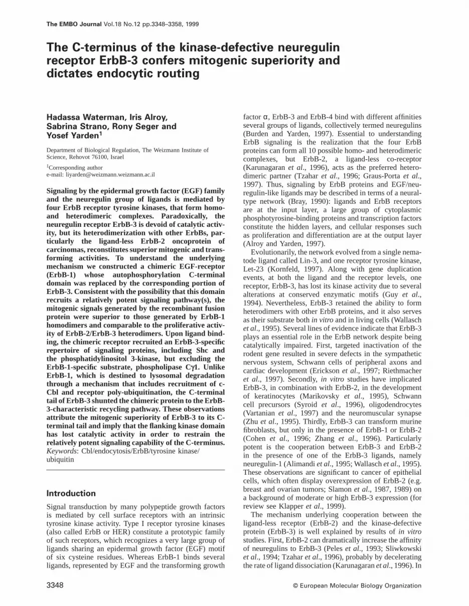
The chimeric ErbB-1T3 receptor recruits Shc andthe regulatory subunit of the phosphoinositide3-kinase, but not PLC-γ1To provide an initial mechanistic basis for the mitogenicsuperiority of the C-terminal tail of ErbB-3, we analyzedits ability to confer recruitment of several signaling pro-teins. First, we tested association of the chimeric receptorwith the Shc adaptor protein. It is noteworthy that in themyeloid 32D cells only p46Shc and p52Shc are expressed,but the larger isoform, p66Shc, is absent (Figure 4A, lowerpanel). Immunoprecipitation with either anti-receptor oranti-phosphotyrosine antibodies followed by immunoblot-ting detected the p52 isoform, which underwent increasedphysical association with the stimulating receptor follow-ing ligand binding (Figure 4A). The other isoform, p46Shc,was found primarily in constitutive complexes with the

Mitogenic superiority of ErbB-3
Fig. 3. Ligand-induced proliferative responses of ErbB-expressing 32D cells. (A) Sublines of 32D cells that express either ErbB-1 alone (D1 cells;squares), a combination of ErbB-2 and ErbB-3 (D23 cells; circles), or the chimeric ErbB-1T3 receptor (D1T3 cells; triangles) were tested for ligand-induced cell proliferation by using the MTT assay. Cells were deprived of IL-3 and plated at a density of 53105 cells/ml in media containing serialdilutions of either EGF (D1 and D1T3 cells) or NDF (D23 cells). The MTT assay was performed 24 h later. The results obtained are presented asfold induction over a control culture maintained for 24 h in the absence of added growth factors. (B) D1 (black line) and D1T3 (gray line) cells(106/ml) were cultured at 37°C for 72 h in the absence (Cont.) or presence of EGF (100 ng/ml). No IL-3 was included in the culture medium. Cellswere harvested 72 h later and analyzed for their DNA content by flow cytometry. The sub-G1 phase of the cell cycle, which represents primarilycells undergoing apoptosis, is indicated. (C) The indicated sublines of 32D cells (53105 cells/ml) were incubated for various time intervals with thefollowing ligands (each at 100 ng/ml): EGF (squares; D1 and D1T3 cells) or NDF (squares; D23 cells). Control cultures were incubated in theabsence (open triangles) or presence of IL-3 (closed triangles). Cell growth was determined daily by using the colorimetric MTT assay, andcompared with the MTT signal obtained with a fresh culture at time zero. The data presented are the mean6 SD of four determinations. Eachexperiment was repeated at least twice.
receptors. The ability to elicit p52 recruitment was sharedby ErbB-1 homodimers (in D1 cells), the ErbB-2/ErbB-3heterodimer (after stimulation with NDF, D23 cells), aswell as by homodimers of the ErbB-1T3 chimera (afterEGF stimulation). In contrast with Shc, ErbB-1 but notErbB-3 can interact with the phosphatidylinositol-specificphospholipase, PLC-γ1 (Fediet al., 1994). This differentialcoupling was confirmed, by co-immunoprecipitation ana-lysis, in the transfected 32D cells we used (compare D1and D23 lanes in Figure 4B). However, examination ofErbB-1T3-expressing cells revealed that the chimericreceptor lost significant interaction with PLC-γ1; thisenzyme was hardly detectable in immunoprecipitates ofan EGF-stimulated ErbB-1T3 (Figure 4B). From the resultsof other experiments not presented here, we learnedthat ErbB-1T3 lost the ability to interact with anotherErbB-1-specific effector, the Ras-GTPase activating pro-tein (Ras-GAP) (Fediet al., 1994), although this effectorunderwent physical association with the ligand-activatedErbB-1 in D1 cells. Unlike Ras-GAP and PLC-γ1, thephosphoinositide 3-kinase can interact efficiently withErbB-3, but in most cell types the interaction between thelipid kinase and ErbB-1 is almost undetectable (Soltoffet al., 1994). In accordance with previous reports, wedetected only weak precipitation of the regulatory subunit,p85, by anti-phosphotyrosine antibodies in response to
3351
ErbB-1 activation (Figure 4C, D1 lanes). In contrast,relatively large quantities of p85 could be co-immuno-precipitated from extracts of ligand-stimulated D23 orD1T3 cells by using anti-phosphotyrosine or anti-receptorantibodies (Figure 4C). Thus, p85 phosphorylation ontyrosine residues, or its association with a tyrosine-phos-phorylated complex, displayed an increase following NDFbinding to an ErbB-2/ErbB-3 heterodimer or EGF bindingto ErbB-1T3 homodimers. However, ErbB-1 homodimerswere unable to recruit the lipid kinase with high efficiency.A similar conclusion was reached on the basis ofdetermination of the lipid kinase activity recovered byanti-phosphotyrosine antibodies (data not shown). Whencombined with the observed ability of ErbB-1T3 to recruitShc but not PLC-γ1, these results indicate that theC-terminal portion of ErbB-3 can confer to ErbB-1 anability to recruit, at least in part, an ErbB-3-specificrepertoire of signaling proteins.
Because receptor coupling to various phosphotyrosine-binding proteins is ultimately followed by activation oflinear cascades of protein kinases (for review see Segerand Krebs, 1995), we analyzed the capacity of ErbB-1T3to funnel signals into two such cascades, the mitogen-activated protein kinase (MAPK or Erk) and the Akt/protein kinase-B pathway. Antibodies specific to the activephosphorylated forms of the two kinases enabled sensitive
H.Waterman et al.
Fig. 4. Downstream signaling by the chimeric ErbB-1T3 receptor andits parental ErbB-1 and ErbB-3 molecules. The indicated sublines of32D cells were incubated for 10 min at 37°C in the absence (–) orpresence of EGF or NDF (each at 100 ng/ml). Whole-cell lysates werethen prepared and subjected to immunoprecipitation (I.P.) with either amAb to phosphotyrosine (P-TYR) or anti-receptor antibodies; eitheranti-ErbB-1 (D1 and D1T3 cells) or anti-ErbB-3 (D23 cells). The anti-receptor immunocomplexes were resolved by SDS–PAGE andsubjected to immunoblot analysis (I.B.) with antibodies specific to oneof the following signaling molecules: (A) Shc, (B) phospholipase Cγ1(PLC-γ1) or (C) the α subunit of the phosphoinositide 3-kinase (p85).Aliquots of whole-cell lysates were directly resolved by SDS–PAGE,followed by immunoblotting to test for equal loading. Arrowheadsindicate the locations of the corresponding signaling proteins.
determination of ligand-induced activation. AlthoughErbB-1T3, like ErbB-1 and the ErbB-2/ErbB-3 combina-tion, rapidly activated the Erk pathway, signaling by thisreceptor was more sustained than that initiated by ErbB-1(Figure 5A); whereas the ErbB-1-initiated signal almostdisappeared after 10 min, Erk activation through ErbB-1T3was still detectable after 60 min, and the kinetics exhibitedby D23 cells was slightly shorter. Control experimentsexcluded the possibility that Erk underwent degradationin ligand-stimulated cells (data not shown). It is worthwhilenoting that differences in kinetics were not reflected inthe time course of receptor phosphorylation (Figure 5A),
3352
Fig. 5. Comparative kinetic analyses of receptor phosphorylation andactivation of the linear kinase cascades Erk and Akt. (A) The indicatedErbB-expressing derivatives of 32D cells were incubated at 37°C forvarious time intervals (indicated in minutes) with the appropriateligand, either EGF (D1 and D1T3 cells) or NDF (D23 cells), each at100 ng/ml. At the end of the incubation period, whole-cell lysateswere prepared, resolved by gel electrophoresis and subjected toimmunoblotting (I.B.) with either an antibody to phosphotyrosine, orwith an antibody specific to the active doubly phosphorylated form ofErk (P-Erk). A separate immunoblot assay indicated that Erkexpression level was similar in D1 and in D1T3 cells and it underwentno significant change following ligand stimulation. (B) Whole-celllysates were prepared as described above and resolved by SDS–PAGEfollowed by immunoblotting (I.B.) with an antibody directed to theactive Ser-473 phosphorylated form of Akt (P-Akt). Signal detectionwas performed by using a chemiluminescence kit.
implying a post-receptor mechanism. Similar to Erk activa-tion, all three receptors we examined were able to stimulateAkt phosphorylation (Figure 5B). However, the differencesin kinetics of Akt activation were moderate comparedwith those exhibited by Erk. Although Akt is considereda down-stream effector of the phosphoinositide 3-kinase,its activation by ErbB-1 may be explained by recentstudies, which uncovered wortmannin-independent path-ways of Akt activation, including a route involving thecalcium/calmodulin-dependent protein kinase kinase(Yano et al., 1998). We conclude that despite differencesin coupling to the phosphoinositide 3-kinase and PLC-γ1,both ErbB-1 and ErbB-3 can recruit the MAPK and theAkt pathways, albeit with disparate kinetics.
The C-terminal domain of ErbB-3 impairsendocytic sorting of the EGF receptorThe superior mitogenic activity mediated by ErbB-1T3,as well as its prolonged signaling, raised the possibilitythat this receptor is less susceptible than ErbB-1 toinactivation mechanisms. To examine this scenario wefollowed the major mechanism of signal inactivation,

Mitogenic superiority of ErbB-3
Fig. 6. Ligand-dependent receptor downregulation and degradation.(A) D1 cells (squares) or D1T3 cells (circles) were incubated withEGF (100 ng/ml) at 37°C for the time intervals indicated. Cells werethen placed on ice and their surface-bound ligand removed using anacidic buffer. Radiolabeled EGF was then added and its binding to thecell surface at neutral pH used to quantify the extent of receptordisappearance. The results are expressed as the average fraction oforiginal binding sites that remained on the cell surface after exposureto the unlabeled ligand at 37°C. (B) To follow receptor degradationdirectly, cells were treated with EGF as described above, except thatwhole-cell lysates were prepared at the end of each time interval.Following separation by SDS–PAGE and transfer to nitrocellulosemembrane filters, membranes were immunoblotted (I.B.) withantibodies directed to the respective receptors, as indicated.
namely the decrease in the number of cell-surface receptorsfollowing stimulation with a growth factor, a processtermed ‘downregulation’. Evidently, and in agreementwith previous reports, EGF induced rapid and extensivedownregulation of ErbB-1 in 32D cells (Figure 6A). Incontrast, almost no parallel decrease in the number ofEGF-binding sites was observed with D1T3 cells followingligand binding. This observation implied that the C-terminus of ErbB-3, when replacing the correspondingdomain of ErbB-1, can impair downregulation of the latterreceptor. Immunoblotting of whole-cell lysates with anti-receptor antibodies confirmed this conclusion since rapiddisappearance of ErbB-1, due to intracellular degradation,was observed upon exposure of D1 cells, but not D1T3cells, to EGF (Figure 6B).
It is important to note that the parental moleculesof ErbB-1T3 are differentially destined to intracellulardegradation; whereas ErbB-3 is destined primarily to therecycling pathway (Watermanet al., 1998), endocytictargeting of ErbB-1 to degradation in lysosomes involvesrecruitment of the c-Cbl adaptor protein and concomitantconjugation of ubiquitin molecules to ErbB-1 (Levkowitzet al., 1998). Therefore, our subsequent experimentsanalyzed ligand-induced ubiquitination of the ErbB-1T3
3353
Fig. 7. Ligand-induced ubiquitination of ErbB proteins and theircoupling to the c-Cbl protein. (A) The indicated ErbB-expressingsublines of 32D cells (107 cells/lane) were treated for the timeintervals indicated at 37°C with or without ligands, either EGF (D1and D1T3 cells) or NDF (D23 cells), each at 100 ng/ml. Whole-celllysates were then prepared and analyzed by immunoprecipitation (I.P.)using antibodies directed to the respective receptor [anti-ErbB-1antibodies (D1 and D1T3 cells) or anti-ErbB-3 antibodies (D23 cells)].Immunocomplexes were washed extensively, resolved by SDS–PAGEand analyzed by immunoblotting (I.B.) with either anti-ubiquitin oranti-c-Cbl antibodies. To detect receptor phosphorylation, whole-celllysates were directly resolved by SDS–PAGE (panel marked asNONE), transferred to membrane filters and immunoblotted with anti-phosphotyrosine (P-TYR) antibodies. The locations of molecularweight marker proteins are indicated in kilodaltons. The filledarrowhead in the anti-ubiquitin panel indicates the location of themajor ErbB-1 protein band, whereas the open arrowhead marks theubiquitinated species. (B) 32D sublines were treated for 10 min withthe indicated ligands as described above and whole-cell lysates wereeither directly resolved by SDS–PAGE (NONE) or first subjected toimmunoprecipitation (I.P.) with antibodies to phosphotyrosine(P-TYR). Following transfer to nitrocellulose membranes, filters wereimmunoblotted with anti-c-Cbl antibodies. Antibody detection wasperformed using a chemiluminescence kit.
chimeric receptor. Although rapid EGF-induced modifica-tion of ErbB-1 was readily detectable in D1 cells, inagreement with observations made with other cell types(Galcheva-Gargovaet al., 1995), no ubiquitination ofeither ErbB-1T3 or ErbB-3 was detectable followingstimulation with EGF or NDF, respectively (Figure 7A).Consistent with a requirement for c-Cbl recruitment, thisadaptor became physically associated only with ErbB-1;no c-Cbl protein could be detected in immunoprecipitatesof an activated ErbB-1T3 or the parental ErbB-3 molecule(Figure 7A, middle panel). Control experiments verifiedthat both receptors, as well as ErbB-2, underwent extensivetyrosine phosphorylation upon ligand binding (Figure 7A,lower panel; data not shown). Because recruitment of c-Cbl by ErbB-1, as well as by other receptors, is followedby extensive tyrosine phosphorylation of the adaptormolecule (reviewed in Thien and Langdon, 1998), weexamined the phosphorylation state of c-Cbl in ligand-
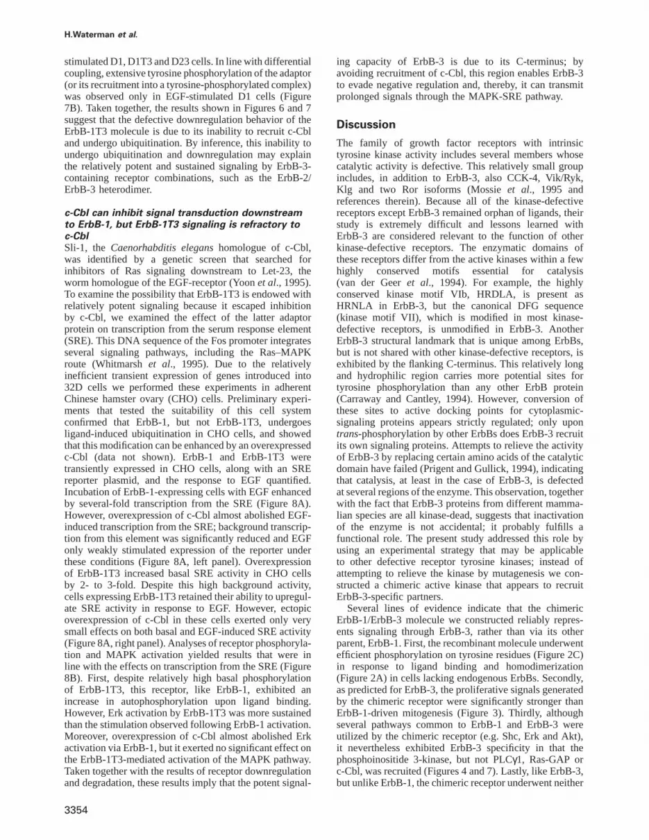
H.Waterman et al.
stimulated D1, D1T3 and D23 cells. In line with differentialcoupling, extensive tyrosine phosphorylation of the adaptor(or its recruitment into a tyrosine-phosphorylated complex)was observed only in EGF-stimulated D1 cells (Figure7B). Taken together, the results shown in Figures 6 and 7suggest that the defective downregulation behavior of theErbB-1T3 molecule is due to its inability to recruit c-Cbland undergo ubiquitination. By inference, this inability toundergo ubiquitination and downregulation may explainthe relatively potent and sustained signaling by ErbB-3-containing receptor combinations, such as the ErbB-2/ErbB-3 heterodimer.
c-Cbl can inhibit signal transduction downstreamto ErbB-1, but ErbB-1T3 signaling is refractory toc-CblSli-1, the Caenorhabditis eleganshomologue of c-Cbl,was identified by a genetic screen that searched forinhibitors of Ras signaling downstream to Let-23, theworm homologue of the EGF-receptor (Yoonet al., 1995).To examine the possibility that ErbB-1T3 is endowed withrelatively potent signaling because it escaped inhibitionby c-Cbl, we examined the effect of the latter adaptorprotein on transcription from the serum response element(SRE). This DNA sequence of the Fos promoter integratesseveral signaling pathways, including the Ras–MAPKroute (Whitmarshet al., 1995). Due to the relativelyinefficient transient expression of genes introduced into32D cells we performed these experiments in adherentChinese hamster ovary (CHO) cells. Preliminary experi-ments that tested the suitability of this cell systemconfirmed that ErbB-1, but not ErbB-1T3, undergoesligand-induced ubiquitination in CHO cells, and showedthat this modification can be enhanced by an overexpressedc-Cbl (data not shown). ErbB-1 and ErbB-1T3 weretransiently expressed in CHO cells, along with an SREreporter plasmid, and the response to EGF quantified.Incubation of ErbB-1-expressing cells with EGF enhancedby several-fold transcription from the SRE (Figure 8A).However, overexpression of c-Cbl almost abolished EGF-induced transcription from the SRE; background transcrip-tion from this element was significantly reduced and EGFonly weakly stimulated expression of the reporter underthese conditions (Figure 8A, left panel). Overexpressionof ErbB-1T3 increased basal SRE activity in CHO cellsby 2- to 3-fold. Despite this high background activity,cells expressing ErbB-1T3 retained their ability to upregul-ate SRE activity in response to EGF. However, ectopicoverexpression of c-Cbl in these cells exerted only verysmall effects on both basal and EGF-induced SRE activity(Figure 8A, right panel). Analyses of receptor phosphoryla-tion and MAPK activation yielded results that were inline with the effects on transcription from the SRE (Figure8B). First, despite relatively high basal phosphorylationof ErbB-1T3, this receptor, like ErbB-1, exhibited anincrease in autophosphorylation upon ligand binding.However, Erk activation by ErbB-1T3 was more sustainedthan the stimulation observed following ErbB-1 activation.Moreover, overexpression of c-Cbl almost abolished Erkactivation via ErbB-1, but it exerted no significant effect onthe ErbB-1T3-mediated activation of the MAPK pathway.Taken together with the results of receptor downregulationand degradation, these results imply that the potent signal-
3354
ing capacity of ErbB-3 is due to its C-terminus; byavoiding recruitment of c-Cbl, this region enables ErbB-3to evade negative regulation and, thereby, it can transmitprolonged signals through the MAPK-SRE pathway.
Discussion
The family of growth factor receptors with intrinsictyrosine kinase activity includes several members whosecatalytic activity is defective. This relatively small groupincludes, in addition to ErbB-3, also CCK-4, Vik/Ryk,Klg and two Ror isoforms (Mossieet al., 1995 andreferences therein). Because all of the kinase-defectivereceptors except ErbB-3 remained orphan of ligands, theirstudy is extremely difficult and lessons learned withErbB-3 are considered relevant to the function of otherkinase-defective receptors. The enzymatic domains ofthese receptors differ from the active kinases within a fewhighly conserved motifs essential for catalysis(van der Geeret al., 1994). For example, the highlyconserved kinase motif VIb, HRDLA, is present asHRNLA in ErbB-3, but the canonical DFG sequence(kinase motif VII), which is modified in most kinase-defective receptors, is unmodified in ErbB-3. AnotherErbB-3 structural landmark that is unique among ErbBs,but is not shared with other kinase-defective receptors, isexhibited by the flanking C-terminus. This relatively longand hydrophilic region carries more potential sites fortyrosine phosphorylation than any other ErbB protein(Carraway and Cantley, 1994). However, conversion ofthese sites to active docking points for cytoplasmic-signaling proteins appears strictly regulated; only upontrans-phosphorylation by other ErbBs does ErbB-3 recruitits own signaling proteins. Attempts to relieve the activityof ErbB-3 by replacing certain amino acids of the catalyticdomain have failed (Prigent and Gullick, 1994), indicatingthat catalysis, at least in the case of ErbB-3, is defectedat several regions of the enzyme. This observation, togetherwith the fact that ErbB-3 proteins from different mamma-lian species are all kinase-dead, suggests that inactivationof the enzyme is not accidental; it probably fulfills afunctional role. The present study addressed this role byusing an experimental strategy that may be applicableto other defective receptor tyrosine kinases; instead ofattempting to relieve the kinase by mutagenesis we con-structed a chimeric active kinase that appears to recruitErbB-3-specific partners.
Several lines of evidence indicate that the chimericErbB-1/ErbB-3 molecule we constructed reliably repres-ents signaling through ErbB-3, rather than via its otherparent, ErbB-1. First, the recombinant molecule underwentefficient phosphorylation on tyrosine residues (Figure 2C)in response to ligand binding and homodimerization(Figure 2A) in cells lacking endogenous ErbBs. Secondly,as predicted for ErbB-3, the proliferative signals generatedby the chimeric receptor were significantly stronger thanErbB-1-driven mitogenesis (Figure 3). Thirdly, althoughseveral pathways common to ErbB-1 and ErbB-3 wereutilized by the chimeric receptor (e.g. Shc, Erk and Akt),it nevertheless exhibited ErbB-3 specificity in that thephosphoinositide 3-kinase, but not PLCγ1, Ras-GAP orc-Cbl, was recruited (Figures 4 and 7). Lastly, like ErbB-3,but unlike ErbB-1, the chimeric receptor underwent neither

Mitogenic superiority of ErbB-3
Fig. 8. c-Cbl negatively regulates signaling downstream of ErbB-1, but the ErbB-1T3 chimeric receptor is refractory to c-Cbl. (A) Monolayers ofCHO cells (in triplicate) were co-transfected with a plasmid vector directing expression of the indicated receptor (1µg of DNA, either pcDNA-3/erbB-1 or pcDNA-3/erbB-1T3) together with a c-Cbl plasmid (1µg or an empty control vector). To follow activation of transcription from the SRE,all cell monolayers were also transfected with an SRE-luciferase reporter plasmid. At 36 h post transfection cells were untreated (open bars) ortreated (closed bars) with EGF (20 ng/ml), and 16 h later we performed a luciferase assay on cell extracts. Signals obtained were normalized to theconcentration of proteins in each cell extract and are presented as the relative light units [average6 SD (bars)] of triplicate determinations.(B) Monolayers of CHO cells were co-transfected with a plasmid encoding c-Cbl, along with ErbB-1- or ErbB-1T3-encoding expression vectors (asindicated). Following stimulation with EGF (100 ng/ml) for the indicated time intervals, whole-cell lysates were prepared and analyzed byimmunoblotting with antibodies to phosphotyrosine (P-TYR), the activated form of Erk (P-Erk), or an anti-HA antibody that detects expression of arecombinant c-Cbl. The arrowheads mark the locations of the c-Cbl protein band.
downregulation nor ligand-dependent ubiquitination(Figures 6 and 7). These biochemical characteristics of thechimeric ErbB-1T3 receptor are not necessarily mutuallyindependent. For example, recruitment of c-Cbl, an ErbB-1-specific substrate (Levkowitzet al., 1996) promotesreceptor ubiquitination and downregulation, two land-marks of signaling through the EGF receptor, rather thanthrough the neuregulin receptor (Levkowitzet al., 1998).Likewise, efficient recruitment of the lipid kinase, anErbB-3 landmark (Soltoffet al., 1994), is known to beessential for receptor recycling (Jolyet al., 1994), anotherintrinsic property of ErbB-3 (Watermanet al., 1998).Despite these consistent observations, we cannot excludethe possibility that ErbB-1T3 has lost interactions with
3355
some of the original ErbB-3 effectors. Moreover, becausethe identity of tyrosine autophosphorylated sites withinligand-activated ErbB dimers may depend on the specificpartner (Olayioyeet al., 1998), different sets of signalingproteins may be recruited to ErbB-3 when it heterodimer-izes with each of the three other ErbBs.
It is important to consider potential mechanisms thatmay explain the mitogenic superiority of ErbB-3 relativeto ErbB-1. According to one scenario, superiority isdetermined by the specific repertoire of signaling proteinsthe neuregulin receptor can recruit. For example, thephosphoinositide 3-kinase, which is specifically recruitedby ErbB-3 and the ErbB-1T3 chimeric receptor, is a well-known regulator of cell growth and survival (reviewed in

H.Waterman et al.
Carpenter and Cantley, 1996). Another candidate is Shc,whose relatively strong interaction with ErbB-3 appearscrucial for neuregulin-induced MAPK activation andmitogenic effect (Vijapurkaret al., 1998). However, superi-ority of ErbB-3 is not limited to positive regulators; it ispossible that the specific partners of ErbB-1 are in factnegative regulators. This possibility may imply that PLCγcan diminish the mitogenic potential of the EGF receptor.Consistently, accelerated generation of inositol trisphos-phate and a rise in cytoplasmic calcium concentrations,two effectors of PLCγ, can attenuate mitogenic signalswhen the interaction of this enzyme with ErbB-1 isartificially enhanced (Obermeieret al., 1996). BesidesPLCγ, Ras-GAP (Fediet al., 1994) and Grb-2 (Prigentand Gullick, 1994; Fiddeset al., 1998) interact withErbB-1, but not with ErbB-3. We are currently investiga-ting whether or not these targets can restrict ErbB-1mitogenesis. However, unlike the unclear role of specificphosphotyrosine-binding proteins in determining signalingpotency, causative relationships between receptor downre-gulation and weaker signaling are well established. Thus,an endocytosis-defective mutant of ErbB-1 is endowedwith enhanced mitogenic ability and oncogenic potential(Wells et al., 1990). Similarly, a dominant-negative dyna-min mutant, which inhibits internalization of the EGF-receptor via clathrin-coated areas of the plasma membraneaugments signaling by EGF (Vieiraet al., 1996). On theligand side, TGFα, a ligand that directs ErbB-1 to recycling(Ebner and Derynck, 1991) is more potent than EGF onmany types of cells. Likewise, growth factors encodedby poxviruses are significantly more potent than theirmammalian counterparts because they escape the normaldownregulation process (Tzaharet al., 1998). It is thereforeconceivable that the ErbB-1T3 receptor we studiedacquired high signaling potency by avoiding the c-Cbl-mediated process of receptor downregulation (Figure 6).This possibility is strongly supported by our analysis ofSRE-mediated transcription, which is sensitive to c-Cblonly when stimulated via ErbB-1 (Figure 8A). At present,however, we cannot exclude alternative mechanisms ortheir combinations. Nevertheless, it is clear that the C-terminal region of ErbB-3 is necessary and sufficient forfull expression of the extremely potent mitogenic powerof ErbB-3.
What is the biological rationale behind the evolution andperseverance of kinase-impaired receptors like ErbB-3? Inthe case the neuregulin receptor this question may beapproached from an evolutionary perspective of networksignaling. The gradual increase in the complexity of theErbB network throughout evolution, an outcome of addingligands, receptors and effectors (Tzahar and Yarden, 1998),remarkably increased the diversification potential of thenetwork by means of combinatorial assemblies. Inevitably,added complexity necessitated more stringent control,especially over the more potent combinations. ErbB-3, bymeans of its uniquely active C-terminal domain, is prob-ably the most potent signal generator of the network.Therefore, its separation from an active kinase created atwo-layer regulatory mechanism; unlike homodimers ofactive kinases, which require only a ligand for fullactivation, stimulation of the more potent heterodimerswith ErbB-3 (Pinkas-Kramarskiet al., 1996) strictlydepends on a ligand and a co-receptor. Thus, ErbB-3 may
3356
have lost enzymatic activity in order to restrain its potentcollection of C-terminally-located tyrosine docking sites.The preferred partner of ErbB-3, namely ErbB-2, raisesanother interesting question: what is the significance ofthe likely possibility that ErbB-2 has no direct ligand ofits own? Besides providing an intact kinase thattrans-phosphorylates ErbB-3, ErbB-2 itself carries a C-terminalregion with unique signaling properties. For example, thisregion strongly couples to the MAPK pathway (Ben-Levyet al., 1994) and it exerts an inhibitory effect on ErbB-2internalization (Sorkinet al., 1993). More mechanisticstudies of signaling from the ErbB-2/ErbB-3 heterodimer,as well as from other ErbB combinations, appear crucialfor understanding the normal physiological role of ErbB-3and its contribution to human cancers.
Materials and methods
Materials, buffers and antibodiesA recombinant form of NDF-β1177–246 was prepared by Amgen(Thousand Oaks, CA). Human recombinant EGF was purchased fromSigma (St Louis, MO). Radioactive materials were from Amersham(Aylesbury, UK). IODOGEN and BS3 were from Pierce. Rabbit anti-c-Cbl(C-15) antibodies, anti-ErbB-1, anti-ErbB-3 and a monoclonal antibody(mAb) to phosphotyrosine were from Santa-Cruz Biotechnology (Santa-Cruz, CA). Rabbit anti-Shc antibodies, and murine anti-PLC-γ1 antibod-ies were purchased from Transduction Laboratories (Lexington, KY).An anti-phosphoinositide 3-kinase (p85) was from Upstate Biotechnology(Lake Placid, NY). A mAb to the active doubly phosphorylated form ofErk (Thr-183 and Tyr-185) was from Sigma, and an antibody to theactive phosphorylated form of Akt (Ser-473) was from New EnglandBiolabs. An anti-ubiquitin antibody was kindly provided by Dr S.Yokota(Yamanashi Medical University, Japan). mAbs to the extracellular domainof ErbB-1 and ErbB-3 used for immunoprecipitation were antibodiesSG199 and X252, respectively (Chenet al., 1996). Binding buffercontained RPMI 1640 supplemented with 0.5% bovine serum albuminand 20 mM HEPES. Solubilization buffer contained 50 mM Tris pH7.5, 150 mM NaCl, 10% glycerol, 1% NP-40, 1 mM EGTA, 1 mMphenylmethylsulfonyl fluoride, 1 mM Na3VO4, 10 µg/ml pepstatin A,10 µg/ml aprotinin and 10µg/ml leupeptin. The HNTG buffer contained20 mM HEPES pH 7.5, 150 mM NaCl, 0.1% Triton X-100 and10% glycerol.
Establishment of transfected cell linesTo construct the ErbB-1T3 chimeric receptor we synthesized by PCRthe portion of the human erbB-3 cDNA corresponding to the C-terminusof the protein, using the following primers: 59-CCAATGCATAGAGA-GAGTGGGCCTGG-39 (this primer was fused to anNsiI site at the 59end) and 39-GCTCTAGAGGGAATAGGGAGAAGACGG-59.
The DNA fragment was cloned into anEcoRV-restricted pBluescriptplasmid. The resulting shuttle plasmid was cut withNsiI and KspI tomatch a pBluescript/erbB-1 from which the C-terminus-encoding portionwas removed using the two enzymes. Proper insertion of the fragmentwas verified by nucleotide sequence analysis. The resulting chimericDNA was cut out of pBluescript usingXbaI and cloned into the samesite in the poly-linker of the pCDNA3-mammalian expression vector(Invitrogen). Right orientation of the insert was tested by restrictionanalysis. The establishment of a series of IL-3-dependent 32D myeloidcells expressing ErbB-1 (D1 cells), ErbB-3 (D3 cells) or the combinationof ErbB-2 with ErbB-3 (D23 cells) has been described elsewhere(Pinkas-Kramarskiet al., 1996). Stable expression of ErbB-1T3 in 32Dcells was achieved using essentially the same procedures as thosedescribed above.
Cell proliferation and survival assaysCells were washed free of IL-3, resuspended in RPMI 1640 medium at53105 cells/ml, and treated without or with growth factors or IL-3(1:1000 dilution of medium conditioned by IL-3-producing cells). Cellproliferation was determined by using the MTT assay (Mosman, 1983).MTT (0.1 mg/ml) was incubated for 2 h at37°C with the cells analyzed.Living cells can transform the tetrazolium ring into dark blue formazancrystals, which can be quantified by reading the optical density at 540–

Mitogenic superiority of ErbB-3
630 nm after lysis of the cells with acidic isopropyl alcohol. For cellcycle analysis, cells were seeded at 106 cells/ml with or without growthfactors. After 72 h, the treated cells were washed once with phosphate-buffered saline and fixed in cold methanol for 1 h at 4°C. RNase A(5 µg) and propidium iodide (10µg) were added and incubated with thecells for 10 min. The stained cells were analyzed in a fluorescence-activated cell sorter (FACScan, Becton Dickinson) within 2 h. Thepercentage of cells in the different phases of the cell cycle was determinedby using the Cellquest program.
Lysate preparation, immunoprecipitation and WesternblottingCells were exposed to the indicated stimuli in RPMI 1640 medium.After treatment, cells were pelleted by centrifugation and extracted insolubilization buffer, mixed harshly and lysates cleared by centrifugation.For direct electrophoretic analysis, boiling gel sample buffer was addedto cell lysates. For other experiments, lysates were first subjected toimmunoprecipitation with immobilized antibodies. Rabbit antibodieswere coupled directly to protein A–Sepharose while shaking for 1 h at4°C. Mouse antibodies were first coupled to anti-mouse IgG-agarose bythe same procedure. The proteins in the lysate supernatants wereimmunoprecipitated for 2 h at 4°C. The immunoprecipitates were washedthree times with HNTG, resolved by SDS–PAGE and electrophoreticallytransferred to a nitrocellulose membrane. Membranes were blocked for1 h in TBST buffer containing 1% milk, blotted for 2 h with a primaryantibody (1µg/ml), followed by a secondary antibody (0.5µg/ml) linkedto horseradish peroxidase. Immunoreactive protein bands were detectedwith the enhanced chemiluminescence reagent (Pharmacia-AmershamCorporation).
Ligand radiolabeling and covalent cross-linkingEGF was labeled by using IODOGEN as described (Karunagaranet al.,1996). The specific activity was ~53105 c.p.m./ng. For covalent cross-linking analysis, cells (13106) were incubated on ice for 1.5 h with125I-radiolabeled EGF at 100 ng/ml. The chemical cross-linking reagentBS3 was then added (1 mM), and after 90 min on ice, cells were pelletedand extracted in solubilization buffer.
Receptor downregulation assayTo quantify receptor downregulation, 13106 cells were washed withbinding buffer and incubated with 100 ng/ml EGF at 37°C for varioustime intervals. Subsequently, cells were precipitated by centrifugation(9000g, 1 min), resuspended, and incubated with ice-cold ligand strippingbuffer (100 mM acetic acid, 150 mm NaCl, pH 2.7) for 2 min on ice.Cells were then pelleted and their medium neutralized by two sequentialwashes with binding buffer. To determine the number of receptormolecules on the cell surface, cells were incubated for 2 h at 4°C witha radioactive EGF, precipitated as above, rinsed once in binding bufferand spun through a serum cushion to remove the unbound ligand, priorto determination of radioactivity.
In vitro kinase assayTo phosphorylate the EGF receptorin vitro, cell lysates were subjectedto immunoprecipitation with the indicated antibodies and the washedimmunoprecipitates incubated for 15 min on ice in phosphorylationsolution containing 5 mM MnCl2 and 0.01µCi [γ-32P]ATP in HNTG.The immobilized immunocomplexes were washed three times withHNTG, eluted with gel sample buffer and resolved by SDS–PAGE.
SRE transcription assayCHO cells were transiently transfected by using the Lipofectaminereagent (Gibco-BRL). Cells were seeded 24 h prior to transfection at23105 cells/60 mm dish and then transfected in triplicates with ErbB-and Cbl-expression vectors as specified. All transfection experimentswere carried out with a reporter pSRE-Fluc plasmid (4µg), containingone copy of the serum response element (SRE) cloned upstream to theFos minimal promoter (nucleotides –53/145) and the luciferase gene(Johansen and Prywes, 1994). Cells were left in transfection mediumfor 14–16 h and then washed and refed with growth medium. Ten hourslater, cells were treated with EGF (20 ng/ml) in growth medium, or leftuntreated for 12 h, and then harvested. Whole-cell extracts for luciferaseassays were prepared by resuspending the cells in 30µl 13 lysis buffer(Promega). Following a 10 min incubation at room temperature, thecellular debris were removed and a 10µl aliquot of the resultingsupernatant was mixed with 100µl luciferin buffer (0.1 M Tris–aceticacid, 10 mM Mg-acetate, 1 mM EDTA, pH 8.0, 74 mM luciferin and2.2 µM ATP). Light intensity was measured by using a luminometer,
3357
and the results were normalized to protein concentrations, which weredetermined by using a kit from Bio-Rad Laboratories (Hercules, CA).
Acknowledgements
We thank B.Ratzkin (Amgen, Thousand Oaks, CA) for the recombinantNDF preparation, S.Yokota (Yamanashi Medical University, Japan) andR.Prywes (Columbia University, NY) for the SRE reporter plasmid. Thiswork was supported by the National Cancer Institute (grant CA72981),and by a grant from The Israel Academy of Sciences administered bythe Israel Science Foundation.
References
Alimandi,M., Romano,A., Curia,M.C., Muraro,R., Fedi,P.,Aaronson,S.A., Di Fiore,P.P. and Kraus,M.H. (1995) Cooperativesignaling of ErbB-3 and ErbB-2 in neoplastic transformation of humanmammary carcinoma cells.Oncogene, 15, 1813–1821.
Alimandi,M. et al. (1997) Epidermal growth factor and betacellulinmediate signal transduction through co-expressed ErbB2 and ErbB3receptors.EMBO J., 16, 5608–5617.
Alroy,I. and Yarden,Y. (1997) The ErbB signaling network inembryogenesis and oncogenesis: signal diversification throughcombinatorial ligand-receptor interactions.FEBS Letts., 410, 83–86.
Barbacci,E.G., Guarino,B.C., Stroh,J.G., Singleton,D.H., Rosnack,K.J.,Moyer,J.D. and Andrews,G.C. (1995) The structural basis for thespecificity of epidermal growth factor and heregulin binding.J. Biol.Chem., 270, 9585–9589.
Baulida,J., Kraus,M.H., Alimandi,M., Di Fiore,P.P. and Carpenter,G.(1996) All ErbB receptors other than the epidermal growth factorreceptor are endocytosis impaired.J. Biol. Chem., 271, 5251–5257.
Ben-Levy,R., Paterson,H.F., Marshall,C.J. and Yarden,Y. (1994) A singleautophosphorylation site confers oncogenicity to the Neu/ErbB-2receptor and enables coupling to the MAP-kinase pathway.EMBO J.,13, 3302–3311.
Bray,D. (1990) Intracellular signalling as a parallel distributed process.J. Theor. Biol., 143, 215–31.
Burden,S. and Yarden,Y. (1997) Neuregulins and their receptors: aversatile signaling module in organogenesis and oncogenesis.Neuron,18, 847–855.
Carpenter,C.L. and Cantley,L.C. (1996) Phosphoinositide 3-kinase andthe regulation of cell growth.Biochem. Biophys. Acta, 1288, M11–M16.
Carraway,K.L. and Cantley,L.C. (1994) A neu acquaintance for ErbB3and ErbB4: A role for receptor heterodimerization in growth signaling.Cell, 78, 5–8.
Chen,X. et al. (1996) An immunological approach reveals biologicaldifferences between the two NDF/heregulin receptors, ErbB-3 andErbB-4.J. Biol. Chem., 271, 7620–7629.
Cohen,B.D., Kiener,P.K., Green,J.M., Foy,L., Fell,H.P. and Zhang,K.(1996) The relationship between human epidermal growth-like factorreceptor expression and cellular transformation in NIH-3T3 cells.J. Biol. Chem., 271, 30897–30903.
Ebner,R. and Derynck,R. (1991) Epidermal growth factor andtransforming growth factor-α: differential intracellular routing andprocessing of ligand-receptor complexes.Cell Regul., 2, 599–612.
Erickson,S.L., O’Shea,K.S., Ghaboosi,N., Loverro,L., Frantz,G.,Bauer,M., Lu,L.H. and Moore,M.W. (1997) ErbB3 is required fornormal cerebellar and cardiac development: a comparison with ErbB2-and heregulin-deficient mice.Development, 124, 4999–5011.
Fedi,P., Pierce,J., Di Fiore,P.P. and Kraus,M.H. (1994) Efficient couplingwith phosphatidylinositol 3-kinase, but not phospholipase Cγ orGTPase-activating protein, distinguishes ErbB-3 signaling from thatof other ErbB/EGFR family members.Mol. Cell. Biol., 14, 492–500.
Fiddes,R.J., Campbell,D.H., Janes,P.W., Sivertsen,S.P., Sasaki,H.,Wallasch,C. and Daly,R.J. (1998) Analysis of Grb7 recruitment byheregulin-activated erbB receptors reveals a novel target selectivityfor erbB3.J. Biol. Chem., 273, 7717–7724.
Galcheva-Gargova,Z., Theroux,S.J. and Davis,R.J. (1995) The epidermalgrowth factor receptor is covalently linked to ubiquitin.Oncogene,11, 2649–2655.
Graus-Porta,D., Beerly,R., Daly,J.M. and Hynes,N.E. (1997) ErbB-2, thepreferred heterodimerization partner of all ErbB receptors, is a mediatorof lateral signaling.EMBO J., 16, 1647–1655.

H.Waterman et al.
Guy,P.M., Platko,J.V., Cantley,L.C., Cerione,R.A. and Carraway,K.L.(1994) Insect cell-expressed p180ErbB3 possesses an impaired tyrosinekinase activity.Proc. Natl Acad. Sci. USA, 91, 8132–8136.
Johansen,F.E. and Prywes,R. (1994) Two pathways for serum regulationof the c-fos response element require specific sequence elements anda minimal domain of serum response factor.Mol. Cell. Biol., 14,5920–5928.
Joly,M., Kazlauskas,A., Fay,F.S. and Corvera,S. (1994) Disruption ofPDGF receptor trafficking by mutation of its PI-3 kinase binding sites.Science, 263, 684–687.
Karunagaran,D., Tzahar,E., Beerli,R.R., Chen,X., Graus-Porta,D.,Ratzkin,B.J., Seger,R., Hynes,N.E. and Yarden,Y. (1996) ErbB-2 is acommon auxiliary subunit of NDF and EGF receptors: implicationsfor breast cancer.EMBO J., 15, 254–264.
Kim,H.H., Vijapurkar,U., Hellyer,N.J., Bravo,D. and Koland,J.G. (1998)Signal transduction by epidermal growth factor and heregulin via thekinase-deficient ErbB3 protein.Biochem. J., 334, 189–195.
Klapper,L.N., Kirschbaum,M.H., Sela,M. and Yarden,Y. (1999)Biochemical and clinical implications of the ErbB/HER signalingnetwork of growth factor receptors. In Klein,G. and Woude,V. (eds),Advances in Cancer Research. Academic Press, in press.
Kornfeld,K. (1997) Vulval development inCaenorhabditis elegans.Trends Genet., 13, 55–61.
Levkowitz,G., Klapper,L.N., Tzahar,E., Freywald,A., Sela,M. andYarden,Y. (1996) Coupling of the c-Cbl protooncogene product toErbB-1/EGF-receptor but not to other ErbB proteins.Oncogene, 12,1117–1125.
Levkowitz,G., Waterman,H., Zamir,L., Kam,Z., Oved,S., Langdon,W.Y.,Beguinot,L., Geiger,B. and Yarden,Y. (1998) c-Cbl/Sli-1 regulatesendocytic sorting and ubiquitination of the epidermal growth factorreceptor.Genes Dev., 12, 3663–3674.
Marikovsky,M., Lavi,S., Pinkas-Kramarski,R., Karunagaran,D., Liu,N.,Wen,D. and Yarden,Y. (1995) ErbB-3 mediates differential mitogeniceffects of NDF/heregulin isoforms on mouse keratinocytes.Oncogene,10, 1403–1411.
Mosman,T. (1983) Rapid colorimetric assay for cellular growth andsurvival: application to proliferation and cytotoxicity assays.J.Immunol. Methods, 65, 55–63.
Mossie,K., Jallal,B., Alves,F., Sures,I., Plowman,G.D. and Ullrich,A.(1995) Colon carcinoma kinase-4 defines a new subclass of thereceptor tyrosine kinase family.Oncogene, 11, 2179–2184.
Obermeier,A., Tinhofer,I., Grunicke,H.H. and Ullrich,A. (1996)Transforming potentials of epidermal growth factor and nerve growthfactor receptors inversely correlate with their phospholipase Cγ affinityand signal activation.EMBO J., 15, 73–82.
Olayioye,M.A., Graus-Porta,D., Beerly,R.R., Rohere,J., Gay,B. andHynes,N.E. (1998) ErbB-1 and ErbB-2 acquire distinct signalingproperties dependent upon their dimerization partner.Mol. Cell. Biol.,18, 5042–5051.
Peles,E., Ben-Levy,R., Tzahar,E., Liu,N., Wen,D. and Yarden,Y. (1993)Cell-type specific interaction of Neu differentiation factor (NDF/heregulin) with Neu/HER-2 suggests complex ligand–receptorrelationships.EMBO J., 12, 961–971.
Pinkas-Kramarski,R.et al. (1996) Diversification of Neu differentiationfactor and epidermal growth factor signaling by combinatorial receptorinteractions.EMBO J., 15, 2452–2467.
Pinkas-Kramarski,R.et al. (1998) The oncogenic ErbB-2/ErbB-3heterodimer is a surrogate receptor of the epidermal growth factorand betacellulin.Oncogene, 16, 1249–1258.
Prigent,S.A. and Gullick,W.J. (1994) Identification of c-erbB-3 bindingsites for phosphatidylinositol 39-kinase and SHC using an EGFreceptor/c-erbB-3 chimera.EMBO J., 13, 2831–2841.
Riethmacher,D., Sonnenberg,R.E., Brinkmann,V., Yamaai,T., Lewin,G.R.and Birchmeier,C. (1997) Severe neuropathies in mice with targetedmutations in the ErbB3 receptor.Nature, 389, 725–30.
Seger,R. and Krebs,E.G. (1995) The MAP kinase signaling cascade.FASEB J., 9, 726–735.
Slamon,D.J., Clark,G.M., Wong,S.G., Levin,W.J., Ullrich,A. andMcGuire,W.L. (1987) Human breast cancer: correlation of relapse andsurvival with amplification of the HER-2/neuoncogene.Science, 235,177–182.
Slamon,D.J.et al. (1989) Studies of the HER-2/neuproto-oncogene inhuman breast and ovarian cancer.Science, 244, 707–712.
Sliwkowski,M.X.et al. (1994) Coexpression of erbB2 and erbB3 proteinsreconstitutes a high affinity receptor for heregulin.J. Biol. Chem.,269, 14661–14665.
3358
Soltoff,S.P., Carraway,K.L., Prigent,S.A., Gullick,W.G. and Cantley,L.C.(1994) ErbB3 is involved in activation of phosphatidylinositol 3-kinase by epidermal growth factor.Mol. Cell. Biol., 14, 3550–3558.
Sorkin,A. and Waters,C.M. (1993) Endocytosis of growth factorreceptors.BioEssays, 15, 375–382.
Sorkin,A., Di Fiore,P.P. and Carpenter,G. (1993) The carboxyl terminusof epidermal growth factor receptor/erbB-2 chimera is internalizationimpaired.Oncogene, 8, 3021–3028.
Syroid,D.E., Maycox,P.R., Burrola,G.P., Liu,N., We,D., Lee,K.-F.,Lemke,G. and Kilpatrick,T.J. (1996) Cell death in the Schwann celllineage and its regulation by neuregulin.Proc. Natl Acad. Sci. USA,93, 9229–9234.
Thien,C.B.F. and Langdon,W. (1998) c-Cbl: a regulator of T cell receptor-mediated signaling.Immunol. Cell Biol., 76, 473–482.
Tzahar,E. and Yarden,Y. (1998) The ErbB-2/HER2 oncogenic receptorof adenocarcinomas: from orphanhood to multiple stromal ligands.BBA Rev. Cancer, 1377, M25–M37.
Tzahar,E., Waterman,H., Chen,X., Levkowitz,G., Karunagaran,D.,Lavi,S., Ratzkin,B.J. and Yarden,Y. (1996) A hierarchical network ofinter-receptor interactions determines signal transduction by NDF/neuregulin and EGF.Mol. Cell. Biol., 16, 5276–5287.
Tzahar,E.et al. (1997) Bivalency of EGF-like ligands drives the ErbBsignaling network.EMBO J., 16, 4938–4950.
Tzahar,E.et al. (1998) Pathogenic poxviruses reveal viral strategies toexploit the ErbB signaling network.EMBO J., 17, 5948–5963.
van der Geer,P., Hunter,T. and Lindberg,R.A. (1994) Receptor protein-tyrosine kinases and their signal transduction pathways.Annu. Rev.Cell Biol., 10, 251–337.
Vartanian,T., Goodearl,A., Viehover,A. and Fischbach,G. (1997) Axonalneuregulin signals through activation of HER4 and Schwann cellsthrough HER2 and HER3.J. Cell Biol., 137, 211–220.
Vieira,A.V., Lamaze,C. and Schmid,S.L. (1996) Control of EGF receptorsignaling by clathrin-mediated endocytosis.Science, 274, 2086–2088.
Vijapurkar,U., Cheng,K. and Koland,J.G. (1998) Mutation of a Shcbinding site tyrosine residue in ErbB3/HER3 blocks heregulin-dependent activation of mitogen-activated protein kinase.J. Biol.Chem., 273, 20996–21002.
Wallasch,C., Weiss,F.U., Niederfellner,G., Jallal,B., Issing,W. andUllrich,A. (1995) Heregulin-dependent regulation of HER2/neuoncogenic signaling by heterodimerization with HER3.EMBO J., 14,4267–4275.
Waterman,H., Sabanai,I., Geiger,B. and Yarden,Y. (1998) Alternativeintracellular routing of ErbB receptors may determine signalingpotency.J. Biol. Chem., 273, 13819–13827.
Wells,A., Welsh,J.B., Lazar,C.S., Wiley,H.S., Gill,G.N. andRosenfeld,M.G. (1990) Ligand-induced transformation by a non-internalizing epidermal growth factor receptor.Science, 247, 962–964.
Whitmarsh,A.J., Shore,P., Sharrocks,A. and Davis,R.J. (1995) Integrationof MAP kinase signal transduction pathways at the serum responseelement.Science, 269, 403–407.
Yano,S., Tokumitsu,H. and Soderling,T.R. (1998) Calcium promotes cellsurvival through CaM-K kinase activation of the protein kinase-Bpathway.Nature, 396, 584–587.
Yoon,C.H., Lee,J., Jongeward,G.D. and Sternberg,P.W. (1995) Similarityof sli-1, a regulator of vulval development inC. elegans, to themammalian proto-oncogenec-Cbl. Science, 269, 1102–1105.
Zhang,K., Sun,J., Liu,N., Wen,D., Chang,D., Thomason,A. andYoshinaga,S.K. (1996) Transformation of NIH 3T3 cells by HER3 orHER4 receptors requires the presence of HER1 or HER2.J. Biol.Chem., 271, 3884–3890.
Zhu,X., Lai,C., Thomas,S. and Burden,S.J. (1995) Neuregulin receptors,erbB3 and erbB4, are localized at neuromuscular synapses.EMBO J,14, 5842–5848.
Received February 8, 1999; revised and accepted April 29, 1999
Recommended


