
Transplantation of expanded bone marrow-derived very small
embryonic-like stem cells (VSEL-SCs) improves left ventricular
function and remodelling after myocardial infarction
Ewa K. Zuba-Surma c, Yiru Guo a, Hisham Taher a, Santosh K. Sanganalmath d, Greg Hunt a, Robert J. Vincent d, Magda Kucia b, Ahmed Abdel-Latif a, Xian-Liang Tang a,
Mariusz Z. Ratajczak b, Buddhadeb Dawn d, *, Roberto Bolli a
a Institute of Molecular Cardiology, University of Louisville, Louisville, KY, USAb Stem Cell Institute, University of Louisville, Louisville, KY, USA,
c Department of Medical Biotechnology, Faculty of Biochemistry, Biophysics and Biotechnology, Jagiellonian University, Crakow, Polandd Division of Cardiovascular Diseases and Cardiovascular Research Institute, University of Kansas Medical Center and
the University of Kansas Hospital, Kansas City, KS, USA
Received: February 12, 2010; Accepted: May 19, 2010
Abstract
Adult bone marrow-derived very small embryonic-like stem cells (VSEL-SCs) exhibit a Sca-1�/Lin–/CD45– phenotype and can differen-tiate into various cell types, including cardiomyocytes and endothelial cells. We have previously reported that transplantation of a smallnumber (1 � 106) of freshly isolated, non-expanded VSEL-SCs into infarcted mouse hearts resulted in improved left ventricular (LV)function and anatomy. Clinical translation, however, will require large numbers of cells. Because the frequency of VSEL-SCs in the mar-row is very low, we examined whether VSEL-SCs can be expanded in culture without loss of therapeutic efficacy. Mice underwent a 30 min. coronary occlusion followed by reperfusion and, 48 hrs later, received an intramyocardial injection of vehicle (group I, n � 11),1 � 105 enhanced green fluorescent protein (EGFP)-labelled expanded untreated VSEL-SCs (group II, n � 7), or 1 � 105 EGFP-labelledexpanded VSEL-SCs pre-incubated in a cardiogenic medium (group III, n � 8). At 35 days after myocardial infarction (MI), mice treatedwith pre-incubated VSEL-SCs exhibited better global and regional LV systolic function and less LV hypertrophy compared with vehicle-treated controls. In contrast, transplantation of expanded but untreated VSEL-SCs did not produce appreciable reparative benefits.Scattered EGFP� cells expressing �-sarcomeric actin, platelet endothelial cell adhesion molecule (PECAM)-1, or von Willebrand factorwere present in VSEL-SC-treated mice, but their numbers were very small. No tumour formation was observed. We conclude that VSEL-SCs expanded in culture retain the ability to alleviate LV dysfunction and remodelling after a reperfused MI provided that they areexposed to a combination of cardiomyogenic growth factors and cytokines prior to transplantation. Counter intuitively, the mechanismwhereby such pre-incubation confers therapeutic efficacy does not involve differentiation into new cardiac cells. These results supportthe potential therapeutic utility of VSEL-SCs for cardiac repair.
Keywords: myocardial infarction • myocardial repair • VSEL-SCs • stem cell • bone marrow •left ventricular function • remodelling
J. Cell. Mol. Med. Vol 15, No 6, 2011 pp. 1319-1328
© 2011 The AuthorsJournal of Cellular and Molecular Medicine © 2011 Foundation for Cellular and Molecular Medicine/Blackwell Publishing Ltd
doi:10.1111/j.1582-4934.2010.01126.x
Introduction
Transplantation of adult bone marrow cells (BMCs) is associ-ated with improvement in left ventricular (LV) structure and
function. Following promising results in animal studies, thistherapeutic approach has been quickly translated into clinicaltrials and several different types of BMCs have been used forthis purpose [1]. However, a meta-analysis of the results ofthese small clinical trials indicates that the benefits of BMCtherapy are only modest [1]. A vigorous effort has thereforebeen mounted to identify not only the optimal cell types, butalso possible modifications prior to transplantation that mayimprove the outcome of BMC therapy.
*Correspondence to: Buddhadeb DAWN, Division of Cardiovascular Diseases, 3901 Rainbow Blvd, Rm. 1001, Eaton Hall, Kansas City, KS 66160, USA.Tel.: (913) 588–6015Fax: (913) 588–6010E-mail: [email protected]
1320
Recent studies from our laboratories have characterized apopulation of pluripotent CXC chemokine receptor (CXCR)4�
cells that reside in the adult bone marrow, are non-haematopoi-etic and are able to differentiate into cells of various lineages,including cardiomyocytes and endothelial cells [2–4]. TheseSca-1�/Lin–/CD45– ‘very small embryonic-like’ stem cells(VSEL-SCs) can acquire a cardiomyocytic phenotype in vitro[3, 5], are mobilized after acute myocardial infarction (MI) [6]and promote cardiac repair following transplantation into theinfarct border zone in vivo [7]. In our previous study [7] VSEL-SC transplantation improved LV function and dimensionsbut was associated with minimal or no differentiation of VSEL-SCs into cardiomyocytes at 35 days after MI; because ofthe scarcity of VSEL-SCs in the bone marrow and our priorinability to expand them effectively, in that investigation [7] wetransplanted only 10,000 freshly isolated, non-expanded VSEL-SCs [7]. Clinical translation of this therapy, however, willrequire transplantation of much greater numbers of cells, partic-ularly when intracoronary infusion (instead if intramyocardialinjection) is used. Consequently, before this new promising celltype can find clinical application in patients with ischemic heartdisease, it is important to develop methods to expand VSEL-SCsin culture while maintaining therapeutic efficacy.
Recently, we have been able to expand VSEL-SCs by culturingthem on C2C12 myoblast cells. Thus, in the present study weexamined whether VSEL-SCs expanded with this method exertsalubrious effects on post-MI LV dysfunction and remodelling,analogous to those previously observed with non-expandedVSEL-SCs [7]. Because we have previously observed that incuba-tion in a cardiomyogenic medium (containing transforminggrowth factor-�1 [TGF-�1], vascular endothelial growth factor[VEGF], basic fibroblast growth factor [bFGF] and insulin-likegrowth factor-1 [IGF-1]) promotes differentiation of freshly iso-lated VSEL-SCs towards a cardiac phenotype [3], we compared
VSEL-SCs incubated in such a medium for 5 days with non-incu-bated VSEL-SCs. The results show that expanded VSEL-SCs arestill effective in improving LV function and dimensions and atten-uating cardiomyocyte hypertrophy when they are pre-incubatedin cardiomyogenic medium, but are ineffective in the absence ofpre-incubation.
Materials and methods
The present study was performed in accordance with the guidelines of theAnimal Care and Use Committee of the University of Louisville School ofMedicine and with the Guide for the Care and Use of Laboratory Animals(Department of Health and Human Services, Publication No. [NIH] 85–23,revised 1996).
Experimental protocol
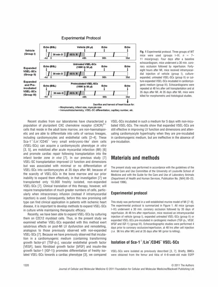
This study was performed in a well-established murine model of MI [7–9].The experimental protocol is summarized in Figure 1. All mice (groupsI–III) underwent a 30 min. coronary occlusion followed by 35 days ofreperfusion. At 48 hrs after reperfusion, mice received an intramyocardialinjection of vehicle (group I), expanded untreated VSEL-SCs (group II) orexpanded VSEL-SCs pre-incubated in cardiogenic medium (TGF-�1, VEGF,bFGF and IGF-1) (group III). Echocardiographic studies were performed 4days prior to coronary occlusion/reperfusion, at 48 hrs after cell injection(i.e. 96 hrs after MI) and at 35 days after MI (prior to killing).
Isolation of Sca-1�/Lin–/CD45– VSEL-SCs
VSEL-SCs were isolated as previously described [3, 7]. Briefly, BMCswere obtained from the femur and tibia of 4–6-week-old male EGFP
© 2011 The AuthorsJournal of Cellular and Molecular Medicine © 2011 Foundation for Cellular and Molecular Medicine/Blackwell Publishing Ltd
Fig. 1 Experimental protocol. Three groups of WTmice were used (groups I–III, n � 7–11 mice/group). Four days after a baselineechocardiogram, mice underwent a 30 min. coro-nary occlusion followed by reperfusion. Forty-eight hours after MI, mice received intramyocar-dial injection of vehicle (group I), culture-expanded, untreated VSEL-SCs (group II) or cul-ture-expanded VSEL-SCs incubated in cardiomyo-genic medium (group III). Echocardiograms wererepeated at 48 hrs after cell transplantation and at35 days after MI. At 35 days after MI, mice werekilled for morphometric and histological studies.

transgenic mice and red blood cells were lysed with a 0.9% solution ofNH4Cl. Freshly isolated BMCs were resuspended in phosphate-bufferedsolution (PBS) containing 1% foetal bovine serum (FBS). The followingprimary antibodies were added simultaneously: biotin-conjugated monoclonal rat antimouse Ly-6A/E (Sca-1) (clone E13-161.7), APC-Cy7-conjugated monoclonal rat antimouse CD45 (clone 30-F11) and phycoery-thrin (PE) conjugated monoclonal rat antimouse lineage markers [anti-CD45R/ B220 (PE; clone RA3-6B2), anti-Gr-1 (PE; clone RB6-8C5),anti-TCR�� (PE; clone H57-597), anti-TCR� (PE; clone GL3), anti-CD11b(PE; clone M1/70), anti-Ter119 (PE; clone TER-119)]. Secondary stainingwas performed with PE-Cy5-conjugated streptavidin. All reagents werepurchased from BD Pharmingen (San Jose, CA, USA). Staining was per-formed at 4C for 20 min., and cells were washed with PBS supplementedwith 1% FBS after staining. Flow cytometric cell sorting was performedwith a MoFlo machine (Dako, Carpinteria, CA, USA) according to thescheme presented in Figure 2. Bulk-sorted cells were collected into 2 mlDMEM with 10% FBS. The purity was assessed by reanalysing isolatedcells immediately following sorting. The viability of sorted cells alwaysexceeded 90%. Sorted cells were pelleted via centrifugation at 1000 � gfor 10 min. and resuspended in DMEM with 10% FBS in a smaller volumeproportional to cell number.
In vitro expansion and pre-differentiation of VSEL-SCs in cardiomyogenic medium
Following isolation by FACS, EGFP-labelled VSEL-SCs were plated over afeeder layer of unlabelled C2C12 cells in DMEM with low concentration ofFBS. The expansion of VSEL-SCs was continued for 9 days with change ofmedium every 3–4 days. Following expansion, cells were trypsinized andEGFP-labelled expanded VSEL-SCs were isolated from C2C12 cells by flowcytometry. In some studies, VSEL-SCs were subsequently plated inmedium containing TGF-�1 (10 ng/ml), VEGF (10 ng/ml), bFGF (10 ng/ml)and IGF-1 (10 ng/ml) for 5 days [3]. Following this treatment, VSEL-SCswere harvested, thoroughly washed in DMEM to remove the cardiogenicingredients, and aliquoted (100,000 cells in 50 �l volume for each mouse)for intramyocardial injection in group III. The same number of expandedVSEL-SCs, cultured for 5 days in identical medium but without cardiogenicgrowth factors (i.e. without pre-incubation) was injected in group II.Because we were unable to obtain a large number of freshly isolated VSEL-
SCs (the primary reason for expansion), and in order to keep the numberof injected cells similar among groups, we did not include a treatmentgroup with freshly isolated VSEL-SCs.
Myocardial infarction and cell transplantation
Three groups of wild-type (WT) mice (C57/BL6 strain, body wt. 20–25 g,age 10–12 weeks; Jackson Laboratories, Bar Harbor, ME, USA) were used.The experimental preparation has been described in detail [7–9]. Briefly,mice were anesthetized with pentobarbital sodium (50 mg/kg i.p.), intu-bated and ventilated using a small rodent ventilator. Body temperature,heart rate and arterial pH were carefully maintained within the physiologi-cal range throughout the experiments [8]. Using a sterile technique, thechest was opened through a midline sternotomy. An 8–0 nylon suture waspassed with a tapered needle under the left anterior descending coronaryartery 2 mm from the tip of the left auricle, and a non-traumatic balloonoccluder was applied on the artery. Coronary occlusion was induced byinflating the balloon occluder. Successful performance of coronary occlu-sion and reperfusion was verified by visual inspection (i.e. by noting thedevelopment of a pale colour in the distal myocardium upon inflation of theballoon and the return of a bright red colour due to hyperaemia after defla-tion) and by observing ST segment elevation and widening of the QRScomplex on the ECG during ischemia and their resolution after reperfusion[8]. The chest was then closed in layers, and a small catheter was left inthe thorax for 10–20 min. to evacuate air and fluids. The mice wereremoved from the ventilator, kept warm with heat lamps, given fluids(1.0–1.5 ml of 5% dextrose in water intraperitoneally) and allowed 100%oxygen via nasal cone. Forty-eight hours later, mice were re-anesthetizedand ventilated and the chest reopened via aseptic technique. Vehicle (50 �l, group I), expanded untreated VSEL-SCs (100,000 cells in 50 �l,group II), or expanded pre-incubated VSEL-SCs (100,000 cells in 50 �l,group III) were injected intramyocardially using a 30 gauge needle. A totalof five injections were made to deliver a total of 100,000 cells per heart inthe peri-infarct region in a circular pattern, at the border between infarctedand surviving myocardium. Because in our previous study [5] we foundthat the expression of chemoattractants in the infarcted myocardium wasmaximal at 48 hrs after reperfusion, this time-point was chosen for VSEL-SC transplantation to ensure maximal retention of injected cells. The chestwas closed in layers and the mice allowed to recover as described above.
J. Cell. Mol. Med. Vol 15, No 6, 2011
1321© 2011 The AuthorsJournal of Cellular and Molecular Medicine © 2011 Foundation for Cellular and Molecular Medicine/Blackwell Publishing Ltd
Fig. 2 Isolation (flow cytometry) and morpho-logic characterization (ImageStream analysis) of BM-derived Sca-1�/Lin–/CD45– VSEL-SCs. (A) Representative dot-plots show the scheme forisolating small cells from the lymphoid gate basedon expression of Sca-1 (PE-Cy5) and lineagemarkers (PE), and further separation based onCD45 (APC-Cy7) expression. Region 3 (R3)contains Sca-1�/Lin–/CD45– VSEL-SCs. (B)Visualization of VSEL-SCs by ImageStream analy-sis demonstrates the very small diameter, positiv-ity for Sca-1 (fluorescein isothiocyanate, green)and negativity for lineage markers (PE) and CD45(PE-Cy5). The nucleus is identified in red (7-amino-actinomycin D). FSC, forward scattercharacteristics; SSC, side scatter characteristics.

1322
Echocardiographic studies
Echocardiograms were obtained using an HDI 5000 SonoCT echocardiog-raphy machine (Philips Medical Systems, Bothell, WA, USA) equipped witha 15–7 MHz linear broadband and a 12–5 MHz phased array transducers[9]. The mice were anesthetized with pentobarbital (25 mg/kg i.p.). Theanterior chest was shaved and the mice were placed in the left lateral decu-bitus position. Using a rectal temperature probe, body temperature wascarefully maintained close to 37.0C with a heating pad throughout thestudy. Modified parasternal long-axis and parasternal short-axis viewswere used to obtain two-dimensional, M-mode and spectral Dopplerimages [9]. Systolic and diastolic anatomic parameters were obtained fromM-mode tracings at the mid-papillary level. LV volume was estimated bythe Teichholz formula. LV mass was estimated by the area-length method.Images were analysed off-line using the Prosolv data analysis software(version 2.5, Problem Solving Concepts, Inc., Indianapolis, IN, USA) by aninvestigator who was blind to the treatment allocation.
Morphometric analysis
At the end of the study, the thorax was opened, the abdominal aorta can-nulated, and the heart arrested in diastole with KCl and CdCl2, excised andperfused retrogradely through the aorta with 10% neutral-buffered forma-lin. The right atrium was cut to allow drainage. The perfusion pressure wasadjusted to match the mean arterial pressure. The LV chamber was filledwith fixative from a pressure reservoir set at a height equivalent to the in vivo measured LV end-diastolic pressure [9–11]. The LV was sectionedserially into four rings perpendicular to its longitudinal axis, processed andembedded in paraffin. The infarct area fraction was calculated by comput-erized planimetry (Image-Pro Plus, Media-Cybernetics, Carlsbad, CA, USA)of digital images of three Masson’s trichrome-stained serial LV sectionstaken at 0.5–1.0 mm intervals along the longitudinal axis [9, 10]. The mid-section was used to measure LV diameter. The thickness of the infarct wall,septal wall and posterior wall was calculated in serial sections and aver-aged [9, 10]. An average sarcomere length of 2.1 �m was utilized in allcases to correct the raw measurements of LV anatomical parameters [11].
For the assessment of cardiomyocyte cross-sectional area, digitalimages were acquired from trichrome-stained myocardial sections.Cardiomyocyte cross-sectional area was measured in transversely sec-tioned myocytes with a circular profile and a central nucleus [7, 12]. Onaverage, a total of 100 myocytes were measured in each heart. All morpho-metric analyses were performed by investigators who were blind to thetreatment allocation.
Immunohistochemistry
Immunohistochemistry was performed in formalin-fixed 4-�m-thick histo-logical sections. Cardiomyogenic differentiation was recognized by thepresence of �-sarcomeric actin (Sigma-Aldrich, St. Louis, MO, USA) andtroponin T (Santa Cruz Biotechnology, Inc., Santa Cruz, CA, USA); endothe-lial differentiation by PECAM-1 (Santa Cruz Biotechnology, Inc.) and vonWillebrand factor (Sigma); and smooth muscle cell differentiation by �-smooth muscle actin (Sigma) [9, 13]. Colocalization of cell-specific mark-ers with EGFP was used to identify cells that originated from BMCs [9, 14].Nuclei were identified with 4�-6-diamidino-2-phenylindole (DAPI).
For the assessment of capillary density [12, 15], sections were stainedwith an anti-CD31 (Santa Cruz Biotechnology, Inc.) primary antibody
followed by the addition of a tetramethylrhodamine isothiocyanate(TRITC)-conjugated secondary antibody [7, 13]. CD31� capillary profileswere counted at 100� magnification in contiguous fields in the infarctzone, border zone and non-ischemic zone. On average, a total of 40–50fields were counted in each heart. Myocardial sections were immunos-tained with directly fluorochrome-conjugated hairpin-1 probe and anti-Ki67 antibody to quantitatively determine the number of apoptotic andcycling cardiomyocytes, respectively [7, 9].
Statistical analysis
Data are reported as means � S.E.M. Morphometric, histological andechocardiographic data were analysed with a one-way ANOVA followed byStudent’s t-tests with the Bonferroni correction as appropriate [16]. Fordata with unequal variance, Kruskal-iWallis one-way ANOVA on ranks andpairwise multiple comparison (Dunn’s method) were performed. All statis-tical analyses were performed with the SPSS software (version 8, SPSS,Inc., Chicago, IL, USA).
Results
Exclusions
A total of 197 mice (55 WT and 142 EGFP transgenic) were used.Forty-nine WT mice were assigned to the MI studies (groupsI–III), 142 EGFP transgenic mice were used as BM donors for cellisolation, and 6 mice were used for the determination of myocytearea. Five mice died in the early post-infarction period and 10 micedied within 72 hrs after intramyocardial injection. Eight mice wereexcluded from the study due to technical reasons, leaving a totalof 11, 7 and 8 mice in groups I–III, respectively.
Myocardial infarct size
The average infarct area fraction did not differ significantly amongthe three groups (15.0 � 1.4%, 14.2 � 3.9% and 14.8 � 2.8% ingroups I, II and III, respectively, Fig. 3). The infarct area fractionmeasures the average area of scarred tissue, expressed as a percentof the LV area in three LV sections 0.5–1.0 mm apart [7, 9, 10].
Transplantation of pre-incubated VSEL-SCsattenuates LV systolic dysfunction
Before coronary occlusion (baseline), all parameters of LV function,measured by echocardiography, were similar in groups I, II and III(Fig. 4). At 48 hrs after cell transplantation (96 hrs after reperfu-sion), the degree of LV systolic functional impairment was alsosimilar among the groups (Fig. 4), indicating that both the injurysustained during ischemia/reperfusion and that associated withintramyocardial injection were comparable. In mice that received
© 2011 The AuthorsJournal of Cellular and Molecular Medicine © 2011 Foundation for Cellular and Molecular Medicine/Blackwell Publishing Ltd

J. Cell. Mol. Med. Vol 15, No 6, 2011
1323
vehicle (group I) or expanded but untreated VSEL-SCs (group II),there was further functional deterioration between 96 hrs and 35 days after reperfusion (Fig. 4). In contrast, LV systolic functionwas better preserved in mice treated with expanded and pre-incubated VSEL-SCs (group III), resulting in significantly greater LVejection fraction (Fig. 4D) in group III compared with groups I andII at 35 days after MI. In group III there was also enhanced regionalmyocardial function in the infarct region, as evidenced by a 98% (P 0.05) and 47% greater systolic wall thickening fractioncompared with groups I and II, respectively (Fig. 4E).
Transplantation of pre-incubated VSEL-SCsattenuates LV expansion
The extent of LV remodelling at 35 days after infarction wasassessed both morphometrically and echocardiographically (Fig. 5). By morphometry, the differences among groups did notreach statistical significance (Fig. S1). By echocardiography, LV end-diastolic volume at 35 days in group III (63 � 11 �l) was 26%smaller compared with group I (86 � 8 �l), albeit without statisticalsignificance (Fig. 5D). These data suggest a possible trend towards
© 2011 The AuthorsJournal of Cellular and Molecular Medicine © 2011 Foundation for Cellular and Molecular Medicine/Blackwell Publishing Ltd
Fig. 3 Myocardial infarct size. Myocardial infarct area fraction [(infarctarea/LV area) � 100] assessed from Masson’s trichrome-stained hearts ingroups I, II and III, which were treated with vehicle, expanded untreatedVSEL-SCs and expanded pre-incubated VSEL-SCs, respectively. Data aremeans � S.E.M. n � 7–11 mice/group.
Fig. 4 Impact of cell therapy on LV function.(A)–(C) Representative M-mode images at 35days after coronary occlusion/reperfusion frommice that were given vehicle (group I) (A),expanded and untreated VSEL-SCs (group II) (B)and expanded pre-incubated VSEL-SCs (groupIII) (C). Compared with the hearts in groups Iand II, the heart in group III exhibited a smallerLV cavity, a thicker infarct wall and improvedmotion of the infarct wall. (D–E) Quantitativeechocardiographic analysis revealed improve-ment in LV functional parameters followingtransplantation of pre-incubated VSEL-SCs at 35 days after MI. Data are means � S.E.M. n � 7–11 mice/group. *P 0.05 versus group Iat 35 days.

1324
improvement in post-MI LV expansion in mice that were given pre-incubated VSEL-SCs as compared with vehicle-treated mice.
Transplantation of pre-incubated VSEL-SCsattenuates LV hypertrophy
Because post-infarct LV remodelling is associated with myocytehypertrophy and increased LV mass, we investigated the effects ofVSEL-SC therapy on these parameters. To this end, we comparedthe three infarcted groups (groups I–III) with a separate controlgroup (n � 6) of non-infarcted mice that were of similar age(10–12 weeks) and did not undergo surgery. Although the differ-ences did not reach statistical significance, in infarcted mice treatedwith vehicle (group I) or expanded but not pre-incubated VSEL-SCs(group II), the cross-sectional myocyte area at 35 days after MI(219 � 21 �m2 and 188 � 31 �m2 in groups I and II, respectively)was 48% and 27% higher compared with non-infarcted controlmice (148 � 16 �m2). However, in mice given expanded and pre-incubated VSEL-SCs (group III), the myocyte area (147 � 17 �m2)was virtually identical to that in non-infarcted mice and 30%smaller than that in group I (Fig. 6). These results were corrobo-rated by the echocardiographic estimates of LV mass, which weresignificantly increased from baseline to 35 days in groups I and IIbut not in group III; in the latter group, the LV mass was 24%smaller than in group I (P 0.05) (Fig. 6E). Together, these dataindicate that transplantation of expanded and pre-incubated VSEL-SCs is associated with attenuation of myocyte hypertrophy.
Myocyte regeneration
Cardiomyogenic specification of transplanted VSEL-SCs wasinferred by concomitant positivity for �-sarcomeric actin and
EGFP [9, 17]. Although scattered EGFP�/�-sarcomeric actin�
cells were identified in the peri-infarct zone in both groups II and III (Fig. 7), the number of such cells was very small in bothgroups and the morphology did not resemble that of adultmyocytes, indicating that the functional and structural benefits ofVSEL-SCs could not be accounted for by their differentiation intonew cardiac myocytes.
Impact of cell therapy on capillary density,myocyte apoptosis and myocyte cycling
Either in the infarct border zone or in the non-ischemic zone, therewas no significant difference among the three groups with respectto capillary density (Fig. S2), Ki67 immunoreactivity (for identifi-cation of cycling cells) (Fig. S3) and immunoreactivity for thehairpin-1 probe (for detection of apoptosis) (data not shown).
Discussion
Although basic as well as clinical studies indicate that transplanta-tion of adult BMCs induces cardiac repair and improves LV func-tion after MI [1], the improvement in LV ejection fraction and thereduction in infarct scar size observed in clinical trials have beenrelatively modest [1], prompting the search for specific cell typesthat may confer more consistent benefits. We have previouslyshown that VSEL-SCs, a recently discovered population ofpluripotent stem cells found in adult bone narrow [3, 4, 6], can beeffectively used to alleviate post-MI cardiac dysfunction andremodelling in mice [7]. However, if this new form of cell therapyis to be used in humans, methods need to be developed that will
© 2011 The AuthorsJournal of Cellular and Molecular Medicine © 2011 Foundation for Cellular and Molecular Medicine/Blackwell Publishing Ltd
Fig. 5 Impact of cell therapy on LV anatomy.(A)–(C) Representative Masson’s trichrome-stained myocardial sections from mice that weregiven vehicle (group I) (A), expanded anduntreated VSEL-SCs (group II) (B) and expandedpre-incubated VSEL-SCs (group III) (C). (D)Echocardiographic analysis of LV end-diastolicvolume at 35 days after MI. Data are means �S.E.M. n � 7–11 mice/group.

J. Cell. Mol. Med. Vol 15, No 6, 2011
1325
enable expansion of VSEL-SCs prior to transplantation, becausetheir numbers in the bone marrow are very low [3]. In this studywe explored the feasibility of expanding VSEL-SCs on a C2C12 cellfeeder layer prior to injection into infarcted hearts.
Our salient findings are the following: (i) in a murine model ofreperfused MI, intramyocardial injection of 105 VSEL-SCsexpanded and then pre-incubated in cardiomyogenic mediumresulted in improvement in LV function and attenuation of LVhypertrophy; (ii) in contrast, injection of the same number ofexpanded but not pre-incubated VSEL-SCs failed to impart anybeneficial effect and (iii) at 35 days after MI, very few cells derivedfrom the transplanted VSEL-SCs were found that expressed mark-ers of cardiomyocytic or endothelial differentiation, implying thatthe salubrious effects of pre-incubated VSEL-SCs are mediated byparacrine mechanisms. Together, these results demonstrate thatVSEL-SCs expanded in vitro can be used to alleviate cardiac dys-function and remodelling after MI, but for this strategy to be effec-tive, the cells need to be exposed to a combination of cardiogenicgrowth factors and cytokines (TGF-�1, VEGF, bFGF and IGF-1)prior to transplantation.
Because the number of cells transplanted may be a majordeterminant of their efficacy [18, 19], and because many cells arelost following myocardial transplantation as a result of washoutand death [20, 21], injecting large numbers of cells is important,at least in principle. VSEL-SCs are very rare in the adult bone mar-row (~0.01% of all nucleated BMCs); consequently, in our previ-ous study [7] we were unable to inject more than 10,000 freshlyisolated VSEL-SCs in each mouse, a number that may have been
© 2011 The AuthorsJournal of Cellular and Molecular Medicine © 2011 Foundation for Cellular and Molecular Medicine/Blackwell Publishing Ltd
Fig. 6 Impact of cell therapy on cardiomyocytehypertrophy and LV mass. (A)–(C)Representative images of cardiomyocytes in theviable myocardium from Masson’s trichrome-stained sections of hearts of mice that weregiven vehicle (group I) (A), expanded anduntreated VSEL-SCs (group II) (B) and expandedpre-incubated VSEL-SCs (group III) (C). (D)Quantitative assessment of myocyte cross-sec-tional area in groups I–III compared with non-infarcted control hearts. (E) Compared withgroup I, echocardiographically estimated LVmass was significantly less in group III. Data aremeans � S.E.M. n � 7–11 mice/group. (E)*P 0.05 versus vehicle (final); #P 0.05 ver-sus respective baseline values.
Fig. 7 Colocalization of EGFP and cardiac proteins after transplantationof pre-incubated VSEL-SCs. VSEL-SC-derived cells are identified byEGFP immunoreactivity [(B)–(D), green]. Cells with possible cardio-genic commitment are identified by �-sarcomeric actin immunoreac-tivity [(C), (D), red]. (D) shows the merged image. Two cells are shownthat are positive for both EGFP and �-sarcomeric actin (arrowheads, D, yellow).

1326
too low to produce an optimal therapeutic effect. This problem islikely to be more severe when repair is attempted in large heartsand when cells are given by intracoronary infusion (the most clin-ically applicable route) rather than intramyocardial injection. Toovercome this limitation, which will likely impede clinical transla-tion, in the present study we expanded VSEL-SCs on a layer ofC2C12 myoblast cells in culture and isolated the expanded EGFP-labelled VSEL-SCs by flow cytometry. Interestingly, although inthis study we injected a 10-fold greater number of VSEL-SCs thanin our previous study of freshly isolated VSEL-SCs [7], transplan-tation of expanded VSEL-SCs without prior exposure to cardio-genic factors (group II) failed to produce statistically significantimprovements in LV function and remodelling. In contrast, pre-incubation of the same number of expanded VSEL-SCs in car-diomyogenic medium (group III) resulted in significant improve-ments in LV function and dimensions. Because transplantation ofonly 10,000 freshly isolated VSEL-SCs is sufficient to effect func-tional and structural improvements in this murine model [7], thepresent findings suggest that the process of expansion itselfsomehow causes the cells to lose their ability to alleviate post-MIdysfunction and remodelling (although expansion in cultureenables the use of greater numbers of VSEL-SCs), and that incu-bation with cardiomyogenic medium can obviate this problem.Importantly, following expansion and isolation by flow cytometry,expanded VSEL-SCs were repeatedly washed to remove the vari-ous components of the expansion medium; therefore, theobserved beneficial effects cannot be ascribed to actions exertedby the medium. Thus, both the mechanism whereby expansioncauses loss of reparative ability and the mechanism wherebyexposure to TGF-�1, VEGF, bFGF and IGF-1 restores this propertyremain unknown and represent important areas for future investi-gations. Furthermore, whether pre-incubation would improve thebenefits observed with freshly isolated VSEL-SCs, and whethertransplantation of larger numbers of expanded untreated (orexpanded pre-incubated) VSEL-SCs would prove more effectivecompared with small number of non-expanded, freshly isolatedVSEL-SCs also remain to be examined in future studies.
The mechanism that underlies the improvement in LV functionobserved with pre-incubated VSEL-SCs is unclear. We have previ-ously reported that VSEL-SCs isolated from the adult bone mar-row express markers indicative of commitment to several unre-lated lineages, including neuronal, skeletal muscle, endothelial andcardiac lineages [2, 3, 5]. As mentioned above, in our previousstudy [7] transplantation of freshly isolated VSEL-SCs improvedLV function and dimensions, but only a small number of VSEL-SC-derived cells expressing cardiac markers were observed after 35days. We reasoned that enhancing the cardiomyogenic specifica-tion of the transplanted cells would improve efficacy and thatexposure of VSEL-SCs to cardiomyogenic factors would achievethis goal (i.e. it would increase the number of VSEL-SC-derivedcardiomyocytes and thereby further improve LV function andgeometry). Several components of our cardiomyogenic medium,including TGF-�1 [22], bFGF [23] and IGF-1 [24], have been impli-cated in cardiogenesis in embryonic developmental studies. Thesefactors have also been used successfully to induce cardiac differ-
entiation of various embryonic and adult cells in vitro [25, 26],suggesting that priming of cells with these factors is likely to augment cardiomyocytic differentiation following transplantation in vivo. Indeed, pre-treatment of bone marrow derived CD117�
cells with TGF-�1 followed by intramyocardial injection has beenreported to enhance cardiomyocyte regeneration and improve LVfunction after acute MI [27]. Similarly, pre-treatment of mes-enchymal stem cells with FGF, IGF-1 and BMP-2 has been shownto induce the expression of cardiac-specific transcription factorsin vitro and improve cardiomyocytic differentiation and infarctrepair in vivo [28]. However, in the present study we detected onlya small number of VSEL-SC-derived cells expressing cardiac-spe-cific proteins, regardless of whether the VSEL-SCs had been incu-bated with (group III) or without (group II) cardiogenic medium.Therefore, generation of new cardiomyocytes was not an impor-tant mechanism whereby pre-incubation with TGF-�1, VEGF, bFGFand IGF-1 improved the outcome.
A similar pattern has been observed in several other studies[7, 29, 30]. Indeed, whether differentiation of transplanted cellsinto cells of cardiac and vascular lineages serves as the primarymechanism of infarct repair with cell therapy remains controver-sial [31–33]. For this reason, paracrine actions of beneficialgrowth factors/cytokines secreted by transplanted cells havebeen proposed to be a major mechanism of cell therapy-inducedcardiac repair [30]. It is plausible that exposure to cardiomyo-genic factors altered the secretome of expanded VSEL-SCs in afavourable manner. Such priming may also enhance the ability ofVSEL-SCs to inhibit myocyte apoptosis and/or activate endoge-nous cardiac stem cells [34, 35], resulting in preservation of car-diac mass and/or new myocyte formation. Although at 35 daysafter MI the number of cardiomyocytes positive for hairpin-1 andKi67 did not differ significantly among the three groups, itremains possible that transplantation of expanded pre-treatedVSEL-SCs was associated with reduction of apoptosis and/orincreased cardiomyocyte proliferation at earlier time-points. It isalso possible that the primary beneficial effect of enhancedsecretion of growth factors by pre-incubated VSEL-SCs wasinhibition of LV hypertrophy, which in turn positively impactedLV function. This hypothesis is supported by the attenuation ofcardiomyocyte hypertrophy in hearts treated with pre-incubatedVSEL-SCs (Fig. 6). Further studies will be necessary to fullycharacterize the genetic properties of expanded VSEL-SCs [36]and to identify the molecular mechanisms via which exposure tocardiomyogenic medium influenced the reparative properties ofexpanded VSEL-SCs.
In conclusion, VSEL-SCs expanded in culture retain their abil-ity to alleviate LV dysfunction and remodelling after a reperfusedMI, provided that prior to transplantation they are exposed to car-diomyogenic growth factors and cytokines (TGF-�1, VEGF, bFGFand IGF-1). The mechanism whereby such pre-incubation conferstherapeutic efficacy remains unclear but, counter intuitively, doesnot involve differentiation of VSEL-SCs into new cardiac cells. Thepresent results support the potential utility of VSEL-SCs as cellu-lar substrates for cardiac repair after MI and should facilitate clin-ical translation of VSEL-SC transplantation. Future investigations
© 2011 The AuthorsJournal of Cellular and Molecular Medicine © 2011 Foundation for Cellular and Molecular Medicine/Blackwell Publishing Ltd

J. Cell. Mol. Med. Vol 15, No 6, 2011
1327
will be necessary to identify the specific molecular mechanismswhereby pre-incubation with TGF-�1, VEGF, bFGF and IGF-1enhances the cardiac reparative benefits of VSEL-SC therapy.
Acknowledgements
This study was supported in part by NIH grants R01 HL-72410, HL-55757,HL-70897, HL-76794, HL-78825, HL-89939, CA-106281, DK-074720 andR21 HL-89737, and the Stella & Henry Hoenig endowment, and the‘Homing’ grant from the Foundation for Polish Science (HOM/2008/15).
Conflict of interest
The authors confirm that there are no conflicts of interest.
Supporting Information
Additional Supporting Information may be found in the online ver-sion of this article:
Fig. S1. Morphometric assessment of LV anatomy. The figureillustrates LV chamber diameter measured postmortem in heartsarrested in diastole. The differences in chamber diameter amongthe three groups did not reach statistical significance. Data aremeans � SEM. n � 7–11 mice/group.
Fig. S2. Quantitative assessment of myocardial capillary density.(A, B) Myocardial capillary density in the infarct border zone (A)and in the nonischemic zone (B) revealed no significant differenceamong mice that were given vehicle, expanded untreated VSEL-SCs, and expanded preincubated VSEL-SCs. Data are means �
SEM. n � 7–11 mice/group.
Fig. S3. Quantitative assessment of cardiomyocyte cell cycleactivity. (A, B) The percentage of myocyte nuclei expressingKi67 in the infarct border zone (A) and in the nonischemic zone(B) revealed no significant difference among mice that weregiven vehicle, expanded untreated VSEL-SCs, and expandedpreincubated VSEL-SCs. Data are means � SEM. n � 7–11mice/group.
Please note: Wiley-Blackwell are not responsible for the content orfunctionality of any supporting materials supplied by the authors.Any queries (other than missing material) should be directed tothe corresponding author for the article.
References
1. Abdel-latif A, Bolli R, Tleyjeh IM, et al.Adult bone marrow-derived cells forcardiac repair: a systematic review andmeta-analysis. Arch Int Med. 2007; 167:989–97.
2. Ratajczak MZ, Kucia M, Reca R, et al.Stem cell plasticity revisited: CXCR4-posi-tive cells expressing mRNA for early mus-cle, liver and neural cells ‘hide out’ in thebone marrow. Leukemia. 2004; 18: 29–40.
3. Kucia M, Reca R, Campbell FR, et al. Apopulation of very small embryonic-like(VSEL) CXCR4(�)SSEA-1(�)Oct-4�
stem cells identified in adult bone marrow.Leukemia. 2006; 20: 857–69.
4. Zuba-Surma EK, Kucia M, Abdel-Latif A,et al. Morphological characterization ofvery small embryonic-like stem cells(VSELs) by ImageStream system analysis.J Cell Mol Med. 2008; 12: 292–303.
5. Kucia M, Dawn B, Hunt G, et al. Cellsexpressing early cardiac markers reside inthe bone marrow and are mobilized intothe peripheral blood following myocardialinfarction. Circ Res. 2004; 95: 1191–9.
6. Zuba-Surma EK, Kucia M, Dawn B, et al.Bone marrow-derived pluripotent verysmall embryonic-like stem cells (VSELs)
are mobilized after acute myocardialinfarction. J Mol Cell Cardiol. 2008; 44:865–73.
7. Dawn B, Tiwari S, Kucia MJ, et al.Transplantation of bone marrow-derivedvery small embryonic-like stem cells atten-uates left ventricular dysfunction andremodeling after myocardial infarction.Stem Cells. 2008; 26: 1646–55.
8. Guo Y, Wu WJ, Qiu Y, et al.Demonstration of an early and a late phaseof ischemic preconditioning in mice. Am JPhysiol. 1998; 275: H1375–87.
9. Dawn B, Guo Y, Rezazadeh A, et al.Postinfarct cytokine therapy regeneratescardiac tissue and improves left ventricu-lar function. Circ Res. 2006; 98:1098–105.
10. Li Q, Li B, Wang X, et al. Overexpressionof insulin-like growth factor-1 in mice pro-tects from myocyte death after infarction,attenuating ventricular dilation, wall stress,and cardiac hypertrophy. J Clin Invest.1997; 100: 1991–9.
11. Anversa P, Olivetti G. The cardiovascularsystem. In: Page E, Fozzard HA, Solaro RJ,editors. The heart. 1st ed. New York, NY:Oxford University Press; 2002. pp. 75–144.
12. Anversa P, Beghi C, Kikkawa Y, et al.Myocardial infarction in rats. Infarct size,myocyte hypertrophy, and capillarygrowth. Circ Res. 1986; 58: 26–37.
13. Dawn B, Stein AB, Urbanek K, et al.Cardiac stem cells delivered intravascu-larly traverse the vessel barrier, regenerateinfarcted myocardium, and improve car-diac function. Proc Natl Acad Sci USA.2005; 102: 3766–71.
14. Dawn B, Zuba-Surma EK, Abdel-Latif A, et al. Cardiac stem cell therapy formyocardial re-generation. A clinical perspective. Minerva Cardioangiol. 2005;53: 549–64.
15. Anversa P, Melissari M, Beghi C, et al. Structural compensatory mecha-nisms in rat heart in early spontaneoushypertension. Am J Physiol. 1984; 246:H739–46.
16. Wallenstein S, Zucker CL, Fleiss JL.Some statistical methods useful in circula-tion research. Circ Res. 1980; 47: 1–9.
17. Orlic D, Kajstura J, Chimenti S, et al.Bone marrow cells regenerate infarctedmyocardium. Nature. 2001; 410: 701–5.
18. Schuster MD, Kocher AA, Seki T, et al.Myocardial neovascularization by bone
© 2011 The AuthorsJournal of Cellular and Molecular Medicine © 2011 Foundation for Cellular and Molecular Medicine/Blackwell Publishing Ltd

1328
marrow angioblasts results in cardiomy-ocyte regeneration. Am J Physiol HeartCirc Physiol. 2004; 287: H525–32.
19. Iwasaki H, Kawamoto A, Ishikawa M, et al. Dose-dependent contribution ofCD34-positive cell transplantation to con-current vasculogenesis and cardiomyoge-nesis for functional regenerative recoveryafter myocardial infarction. Circulation.2006; 113: 1311–25.
20. Dow J, Simkhovich BZ, Kedes L, et al.Washout of transplanted cells from theheart: a potential new hurdle for cell trans-plantation therapy. Cardiovasc Res. 2005;67: 301–7.
21. Hou D, Youssef EA, Brinton TJ, et al.Radiolabeled cell distribution afterintramyocardial, intracoronary, and inter-stitial retrograde coronary venous delivery:implications for current clinical trials.Circulation. 2005; 112: I150–6.
22. Muslin AJ, Williams LT. Well-definedgrowth factors promote cardiac develop-ment in axolotl mesodermal explants.Development. 1991; 112: 1095–101.
23. Spirito P, Fu YM, Yu ZX, et al.Immunohistochemical localization of basicand acidic fibroblast growth factors in thedeveloping rat heart. Circulation. 1991; 84:322–32.
24. Bassas L, Lesniak MA, Serrano J, et al.Developmental regulation of insulin and
type I insulin-like growth factor receptorsand absence of type II receptors in chickenembryo tissues. Diabetes. 1988; 37:637–44.
25. Abdel-Latif A, Zuba-Surma EK, Case J, et al. TGF-beta1 enhances cardiomyo-genic differentiation of skeletal muscle-derived adult primitive cells. Basic ResCardiol. 2008; 103: 514–24.
26. Heng BC, Haider H, Sim EK, et al.Strategies for directing the differentiationof stem cells into the cardiomyogenic line-age in vitro. Cardiovasc Res. 2004; 62:34–42.
27. Li TS, Hayashi M, Ito H, et al.Regeneration of infarcted myocardium byintramyocardial implantation of ex vivotransforming growth factor-beta-prepro-grammed bone marrow stem cells.Circulation. 2005; 111: 2438–45.
28. Bartunek J, Croissant JD, Wijns W, et al. Pretreatment of adult bone marrowmesenchymal stem cells with cardiomyo-genic growth factors and repair of thechronically infarcted myocardium. Am JPhysiol Heart Circ Physiol. 2007; 292:H1095–104.
29. Uemura R, Xu M, Ahmad N, et al. Bonemarrow stem cells prevent left ventricularremodeling of ischemic heart throughparacrine signaling. Circ Res. 2006; 98:1414–21.
30. Gnecchi M, Zhang Z, Ni A, et al.Paracrine mechanisms in adult stem cellsignaling and therapy. Circ Res. 2008; 103:1204–19.
31. Dawn B, Bolli R. Adult bone marrow-derived cells: regenerative potential, plas-ticity, and tissue commitment. Basic ResCardiol. 2005; 100: 494–503.
32. Fukuda K, Yuasa S. Stem cells as a sourceof regenerative cardiomyocytes. Circ Res.2006; 98: 1002–13.
33. Guan K, Hasenfuss G. Do stem cells in theheart truly differentiate into cardiomy-ocytes? J Mol Cell Cardiol. 2007; 43:377–87.
34. Beltrami AP, Barlucchi L, Torella D, et al.Adult cardiac stem cells are multipotentand support myocardial regeneration. Cell.2003; 114: 763–76.
35. Urbanek K, Rota M, Cascapera S, et al.Cardiac stem cells possess growth factor-receptor systems that after activationregenerate the infarcted myocardium,improving ventricular function and long-term survival. Circ Res. 2005; 97:663–73.
36. Shin DM, Zuba-Surma EK, Wu W, et al.Novel epigenetic mechanisms that controlpluripotency and quiescence of adult bonemarrow-derived Oct4(�) very smallembryonic-like stem cells. Leukemia.2009; 23: 2042–51.
© 2011 The AuthorsJournal of Cellular and Molecular Medicine © 2011 Foundation for Cellular and Molecular Medicine/Blackwell Publishing Ltd
Recommended


