Validation of human periodontalligament-derived cells as a reliablesource for cytotherapeutic use
Iwata T, Yamato M, Zhang Z, Mukobata S, Washio K, Ando T, Feijen J, Okano T,Ishikawa I. Validation of human periodontal ligament-derived cells as a reliablesource for cytotherapeutic use. J Clin Periodontol 2010; 37: 1088–1099. doi: 10.1111/j.1600-051X.2010.01597.x.
AbstractAim: Periodontal ligament (PDL) is a reliable cell source for periodontalregeneration. In this study, an optimal protocol for the extraction, expansion, andcharacterization of human PDL (hPDL) cells was examined for clinical trials.
Materials and Methods: hPDL tissues were obtained from 41 surgically extractedteeth and digested with enzymes. Human adipose-derived stem cells (hADSCs), bonemarrow-derived mesenchymal stem cells (hBMMSCs), and gingival fibroblasts (hGFs)were used for comparison. For each sample, the proliferative capacity, colony-formingability, alkaline phosphatase activity, differentiation ability, the cell surface antigens,gene expression, and regenerative potential were examined.
Results: hPDL cells were more successfully extracted with collagenase/dispase [29/30(96.7%)] than with trypsin/EDTA [8/11 (72.7%)], and exhibited osteogenic potential both invitro and in vivo. The proliferation of hPDL cells was rapid at a low cell density. hPDL cellsfrequently differentiated into cementoblastic/osteoblastic lineage (� 60%). In contrast, theiradipogenic and chondrogenic potentials were lower than those of hADSCs and hBMMSCs.Some genes (NCAM1, S100A4, and periostin) were preferentially expressed in hPDL cellscompared with those of hBMMSCs and hGFs. Immunohistochemical studies revealed theexpressions of S100A4 and periostin in hPDL tissue.
Conclusion: A protocol for the successful cultivation and validation of hPDL cells isproposed for clinical settings.
Key words: cytotherapy; gene expressionprofile; marker gene; periodontal ligamentcells; regenerative medicine; transplantation;validation
Accepted for publication 27 May 2010
Periodontal regeneration, i.e., the for-mation of new bone and new cementumwith supportive periodontal ligament
(PDL), has been a challenge in perio-dontics, and numerous studies haveattempted to induce true periodontalregeneration. The key source of perio-dontal regeneration is the ‘‘PDL tissue’’(Karring et al. 1985). Once PDL tissuesare removed from a dental root, anky-losis and root resorption occur (Nymanet al. 1980, Andreasen & Kristerson1981). Therefore, researchers considerPDL tissue as a responsible source forperiodontal regeneration. Based on thisconcept, selective proliferation of PDL-derived cells has been clinically per-formed using barrier membranes(Nyman et al. 1982) or enamel matrixproteins (Hammarstrom et al. 1997). In
addition, either alone or a combinationuse of synthesized and biological mate-rials was utilized to enhance periodontalwound healing during surgical proce-dures. In most cases, however, onlylimited histological evidence of trueregeneration has been demonstrated(Wang et al. 2005, Sculean et al. 2008).
To overcome the limitations of tradi-tional procedures, transplantation ofpostnatal stem cells has the potentialto significantly alter tissue engineering(Bianco & Robey 2001). Several studieshave reported that PDL-derived cells pos-sess stem cell-like properties (Seo et al.2004, Trubiani et al. 2005, Nagatomoet al. 2006, Gay et al. 2007, Coura et al.
Takanori Iwata1,2, Masayuki Yamato1,Zheng Zhang3, Shigeki Mukobata4,Kaoru Washio1, Tomohiro Ando2,Jan Feijen3, Teruo Okano1 andIsao Ishikawa1,2
1Institute of Advanced Biomedical
Engineering and Science; 2Department of
Oral and Maxillofacial Surgery, Tokyo
Women’s Medical University, Tokyo, Japan;3Department of Polymer Chemistry and
Biomaterials, Institute for Biomedical
Technology (BMTI), Faculty of Science and
Technology, University of Twente, AE
Enschede, The Netherlands; 4CellSeed Inc.,
Tokyo, Japan
Conflict of interest and source offunding statement
The authors declare that they have noconflict of interests.This research was supported by ‘‘Forma-tion of Innovation Center for Fusion ofAdvanced Technologies in the SpecialCoordination Funds for PromotingScience and Technology’’ and a Grant-in-Aid for Young Scientists (B)(20791479) from the Ministry of Educa-tion, Culture, Sports, Science, and Tech-nology (MEXT) of Japan.
J Clin Periodontol 2010; 37: 1088–1099 doi: 10.1111/j.1600-051X.2010.01597.x
1088 r 2010 John Wiley & Sons A/S

2008, Lindroos et al. 2008, Zhou et al.2008, Xu et al. 2009). In addition, be-cause PDL tissues have one of the highestmetabolic turnover rates in the body(McCulloch & Bordin 1991, Ramakrish-nan et al. 1995, Coura et al. 2008),researchers have hypothesized that ahighly regenerative population of cellsexists in PDL tissues and that they couldbe a source of cells for the regeneration ofvarious tissues (Coura et al. 2008). Somestudies developed PDL cell transplanta-tion in combination with scaffolds andreported successful periodontal regenera-tion in animal models (Nakahara et al.2004, Seo et al. 2004, Sonoyama et al.2006, Liu et al. 2008, Iwata et al. 2009).
To realize the potential of stem celltherapy for periodontal regeneration,effective methods to extract and expand‘‘human’’ PDL (hPDL) cells have to beestablished. Somerman et al. (1988) firstreported on a method to culture PDLcells, and the method has been widelyused because of its clear concept, ‘‘Extractcells from the mid-third of teeth’’. How-ever, the proliferation of the cells wasslow, and it took a great deal of time toexpand the PDL cells required for trans-plantation. Therefore, recent studies havecombined the use of enzymatic digestionand this concept to obtain a higher yield ofcells (Seo et al. 2004, Sonoyama et al.2006, Liu et al. 2008). Still, the character-istics of PDL cells remain to be eluci-dated, although some PDL-specific geneswere proposed (Duarte 1998, Horiuchiet al. 1999, Yamada et al. 2001, Lallieret al. 2005, Nakamura et al. 2005, Nishidaet al. 2007, Pi et al. 2007).
In this study, 41 teeth were collectedfrom human patients, and the culturecondition of hPDL cells was optimized.Moreover, distinguishable hPDL mar-kers were confirmed for the identifica-tion of hPDL cells, and the potential ofhPDL cells in tissue regeneration wasevaluated both in vitro and in vivo.
Materials and Methods
This study was conducted according tothe principles expressed in the Declara-tion of Helsinki. The study wasapproved by the Institutional ReviewBoard of Tokyo Women’s Medical Uni-versity Human Subjects Research. Allpatients or guardians were fully in-formed and gave written consent forthe donation of their teeth and theirsubsequent use in this research project.
All animal procedures were approvedby the Institutional Animal Care andUse Committee of Tokyo Women’sMedical University.
Cell culture
Normal human teeth were extracted fororthodontic or impaction reasons from 41patients between 14 and 57 years of age atTokyo Women’s Medical University Hos-pital. The age, sex, and position of theextracted tooth from the donors are shownin Table 1. First, each tooth was rinsed fivetimes with antibiotics (Unasyn) (Pfizer
Japan, Tokyo, Japan). Then, the PDLtissue was gently separated from the sur-face of the mid-third of root and subse-quently digested with trypsin/EDTA(0.25% trypsin and 1 mM EDTA) (Invi-trogen, Carlsbad, CA, USA) or a solutionof collagenase type I (varying concentra-tions) (SERVA Electrophoresis, Heidel-berg, Germany) with 1200 PU/ml dispase(Godo Syusei, Tokyo, Japan) in a-MEMglutamax (Invitrogen) for 60 min. at 371Cwith vigorous shaking. Single-cell suspen-sions were obtained by passing the cellsthrough a 70mm strainer (Falcon, FranklinLakes, NJ, USA). After being strained, the
Table 1. Isolation and ALP activity of hPDL cells
# Age (years) Sex Position Enzyme Cell expansion ALP induction
1 19 F 18 T 1 1
2 41 M 28 T 1 1
3 34 F 38 T 1 �4 34 F 28 T 1 �5 14 M 22 T 1 1
6 14 M 34 T �7 24 F 38 T 1 1
8 26 M 38 T 1 1
9 26 M 18 T 1 1
10 30 F 42 T �11 24 F 24 C/D 1 1
12 24 F 14 C/D 1 1
13 26 F 38 T �14 25 F 38 C/D 1 1
15 19 F 38 C/D 1 1
16 32 F 48 C/D 1 1
17 26 F 48 C/D 1 1
18 30 M 28 C/D 1 1
19 57 F 18 C/D 1 1
20 25 M 18 C/D 1 1
21 57 F 28 C/D 1 1
22 18 M 28 C/D 1 1
23 30 F 28 C/D 1 1
24 25 F 48 C/D 1 1
25 19 F 48 C/D 1 1
26 57 F 38 C/D 1 1
27 35 M 38 C/D 1 1
28 23 M 38 C/D �29 47 M 48 C/D 1 1
30 31 M 48 C/D 1 1
31 57 F 28 C/D 1 1
32 23 F 38 C/D 1 1
33 45 M 48 C/D 1 1
34 32 M 48 C/D 1 1
35 25 M 48 C/D 1 1
36 30 F 38 C/D 1 1
37 31 F 38 C/D 1 1
38 34 F 48 C/D 1 1
39 27 F 38 C/D 1 1
40 30 F 48 C/D 1 1
41 25 M 28 C/D 1 1
hPDL tissues from 41 patients were digested with trypsin–EDTA (T) or collagenase/dispase (C/D)
and cultured. Tooth position was indicated by means of Zsigmondy and Palmer system (Peck &
Peck, 1993). Cell expansion was judged from the microscopic view on Day 7 after the spreading.
Expanded cells at Passages 3–5 were cultured with or without osteoinductive supplements (50 mg/ml
ascorbic acid, 10 mM b-glycerophosphate, and 10 nM dexamethasone), then ALP activity was
measured and enhanced cells were considered as ALP induction positive (1).
ALP, alkaline phosphatase; hPDL, human periodontal ligament; F, female; M, male.
Validation of human periodontal ligament cells 1089
r 2010 John Wiley & Sons A/S
cells were plated onto a T25 Primariatculture flask (Falcon) (Passage 0). Thecells were then cultured in complete cul-ture medium [a-MEM glutamax contain-ing 10% foetal bovine serum (FBS)(Moregate Biotech, Bulimba, Queensland,Australia), and 1% penicillin/streptomycin(Sigma-Aldrich, St Louis, MO, USA)].After 48 h, unattached cells and debriswere washed out, and new medium wasadded. The cells were subcultured usingtrypsin/EDTA on Day 5, and spread outon a T75 Primariat culture flask (Passage1). Subculture was performed every 3–4days until Passage 3. Thereafter, cellswere plated at a density of 50 cells/cm2
on 60 cm2 culture dishes every 14 daysuntil their proliferation potential was lost.
Human gingival fibroblasts (hGFs)from three different donors were pur-chased from ScienCell (Carlsbad, CA,USA). These hGFs were cryopreservedat Passage 1 and delivered frozen. Experi-ments were carried out with cells fromthe third to fifth passages. Three differentlots of human bone marrow-derivedmesenchymal stem cells (hBMMSCs)and human adipose-derived stem cells(hADSCs) were purchased from CellularEngineering Technologies (Coralville,IA, USA). Similarly and unless otherwisenoted, experiments were carried out withcells from the third to fifth passages. Allcells were cultured in complete culturemedium as described above.
Colony-forming assay
The cells were plated at a density of100 cells/60 cm2 dish, and cultured incomplete medium for 14 days. The cellswere stained with 0.5% crystal violet inmethanol for 5 min. as previouslydescribed (Nimura et al. 2008). Thecells were washed twice with distilledwater, and the number of colonies wascounted. Colonies o2 mm in diameterand/or faintly stained were ignored.
Alkaline phosphatase (ALP) activity
The cells were plated on 96-well platesat a density of 1 � 104 cells/well andcultured in complete medium for 48 h.Then, the medium was changed to com-plete medium with or without variousconcentrations of osteoinductive supple-ments, ascorbic acid (AA) (Wako PureChemical, Tokyo, Japan), b-glycero-phosphate (bGP) (Sigma-Aldrich), and/or dexamethasone (DEX) (Fuji Pharma,Tokyo, Japan). After five additionaldays of culture, the cells were washed
once with normal saline solution, andALP activity was evaluated afterincubation with 10 mM p-nitrophenyl-phosphate as a substrate in 100 mM 2-amino-2-methyl-1, 3-propanediol–HClbuffer (pH 10.0) containing 5 mMMgCl2 for 5 min. at 371C. The additionof NaOH quenched the reaction, and theabsorbance at 405 nm was measuredusing a plate reader (Bio-Rad Model450, Bio-Rad, Hercules, CA, USA).
Differentiation assay
For osteogenesis studies, 50 cells wereplated in 60 cm2 dishes and cultured for14 days as described previously (Nimuraet al. 2008) with slight modifications. Themedium was then switched to a calcifica-tion medium consisting of complete med-ium supplemented with 50mg/ml AA,10 mM bGP, and 10 nM DEX (osteoin-ductive medium) for an additional 21days. These dishes were stained with1% alizarin red solution, and alizarinred-positive colonies were counted. Thesame calcification cultures were subse-quently stained with crystal violet, andthe total cell colonies were counted.Colonies that were o2 mm in diameteror appeared yellowish were ignored.
For adipogenesis experiments, 50 cellswere plated in 60 cm2 dishes and culturedin complete medium for 14 days. Themedium was then switched to adipogenicmedium, which consisted of completemedium supplemented with 100 nMDEX, 0.5 mM isobutyl-1-methyl xanthine(Sigma-Aldrich), and 50mM indometha-cin (Wako Pure Chemical) for an addi-tional 21 days. The adipogenic cultureswere fixed with 4% paraformaldehydeand stained with fresh oil red O solution,and the numbers of the oil red O-positivecolonies were counted. Colonies that wereo2 mm in diameter or ones that appearedfaint were ignored. The cultures were thenstained with crystal violet, and the totalnumber of cell colonies was counted.
For chondrogenesis studies, 250,000cells were placed in a 15 ml polypropy-lene tube (Becton Dickinson, MountainView, CA, USA) and centrifugedfor 10 min. The pellet was cultured inchondrogenesis medium [high-glucoseDulbecco’s modified Eagle’s medium(Invitrogen) supplemented with 500 ng/mlbone morphogenetic protein 2 (R&D Sys-tems, Minneapolis, MN, USA), 10 ng/mltransforming growth factor b3 (R&DSystems), 10 nM DEX, 50mg/ml AA,40mg/ml proline, 100mg/ml pyruvate,and 50 mg/ml ITS1Premix (Becton Dick-
inson)]. The medium was replaced every3–4 days for 21 days.
For immunohistochemical staining,the pellets were embedded in OCTcompound, and the specimen blockwas cut into 5mm frozen tissue sections.After being dried for 1 h at room tem-perature, the tissue sections were treatedwith chondroitinase ABC (0.25 U/ml)(Seikagaku Biobusiness, Tokyo, Japan)and hyaluronidase (type I-S) (Sigma-Aldrich) for 30 min. at 371C. The sec-tions were washed with Tris-bufferedsaline (TBS) (Takara Bio, Shiga, Japan),and incubated with TBS containing 5%donkey serum and 0.3% Triton X for 1 hto block non-specific reactions. The sec-tions were then incubated with a mousemonoclonal antibody against human typeII collagen (1:100 dilution) (DaiichiFine Chemical, Toyama, Japan) for 12 hat 41C. The slides were again washedthree times with TBS and incubatedwith a horseradish peroxidase-conjugatedsecondary antibody (1:1000 dilution)(Jackson ImmunoResearch Laboratories,West Grove, PA, USA) for 1 h at roomtemperature. Immunostaining was detect-ed by 3, 30-diaminobenzidine (DAB), andthe counterstaining was performed withMayer’s haematoxylin.
Isolation of RNA and polymerase chain
reaction (PCR)
Total RNA was isolated with a QIAshredder and RNeasy Mini kit (Qiagen,Valencia, CA, USA) according to themanufacturer’s instructions. Thereafter,cDNA was synthesized from 500 ng ofthe total RNA using the SuperscriptVILO cDNA Synthesis Kit (Invitrogen).b-Actin was utilized as the internalcontrol gene from the results observedon the TaqMan Human EndogenousControl Plate (Applied Biosystems, Fos-ter City, CA, USA). The mRNA expres-sion levels of periodontal marker genesand osteogenic genes were quantita-tively analysed by real-time PCR (ABIPrism 7300 Sequence detection system,Applied Biosystems) using sequence-specific primers. The primers used wereas follows: asporin (Hs00214395_m1),b-actin (4326315E), bone sialoprotein(BSP; Hs00173720_m1), cementumprotein 1 (CEMP1; Hs03004478_s1),cyclin J (Hs00908190_g1), FDC-SP(Hs00395131_m1), milk fat globule-EGF factor 8 protein (MFGE8;Hs00170712_m1), MSX1 (Hs00427183_m1), MSX2 (Hs00741177_m1), neuralcell adhesion molecule 1 (NCAM1;
1090 Iwata et al.
r 2010 John Wiley & Sons A/S

Hs00941821_m1), osteocalcin (OCN;Hs01587813_g1), osteopontin (OPN;Hs00167093_m1), periostin (Hs00170815_m1), S100A4 (Hs00243202_m1), andtype II collagen (Col2A1; Hs01060345_m1). The samples were analysed intriplicate. The mRNA expression levelsrelative to b-actin were determined, and,in some cases, the fold changes werecalculated using the values obtained bymeans of the 2�DDCt method at eachtime point (Livak & Schmittgen 2001).
PCR array
Human osteogenesis RT2 Profilert PCRArray and Human Cell Surface MarkersRT2 Profilert PCR Array (SuperArrayBioscience, Bethesda, MD, USA) wereused to screen for the expression of multi-ple genes in hPDL cells, hBMMSCs, andhGFs according to the manufacturer’sprotocol, and the levels of gene expres-sion were determined with the compara-tive Ct method.
Flow cytometric assay
One million cells were suspended in100ml Dulbecco’s phosphate-bufferedsaline (PBS) containing 10mg/ml ofeach specific antibody. For determiningthe surface markers, fluorescein isothio-cyanate (FITC)- or phycoerythrin (PE)-coupled antibodies against CD29,CD34, CD44, CD90, CD106, andCD146 (Becton Dickinson), ALP (R&Dsystems), STRO-1 (Santa Cruz Biotech-nology, Santa Cruz, CA, USA), CD14and CD45 (Biolegend, San Diego, CA,USA), and CD105 (Ancell, Bayport,MN, USA) were used. As the isotypecontrol, FITC- or PE-coupled non-speci-fic mouse IgG (Becton Dickinson), andPE-coupled non-specific mouse IgM(Antigenix America, Huntington Station,NY, USA) were substituted for theprimary antibodies. After being incu-bated for 30 min. at 41C, the cells werewashed with PBS and then suspendedin 1 ml of PBS for further analysis. Cellfluorescence was determined using aflow cytometer (Epics-XL; BeckmanCoulter, Fullerton, CA, USA).
Immunohistochemistry
A human tooth surrounded by alveolarbone was subjected to immunohisto-chemistry. The tissue was fixed with4% paraformaldehyde and decalcifiedwith neutralized 10% EDTA (pH 7.4)at 41C for 2 weeks before dehydration
and paraffin embedding. Sections thatwere 5 mm thick were collected on glassslides. To enhance antigen retrieval, theslides were immersed in 1% antigenunmasking solution (Vector Laboratories,Burlingame, CA, USA) and blocked for30 min. at room temperature with non-immune rabbit serum, followed by a 16-hincubation with the specific primaryantibodies. The primary antibodies anddilutions used were anti-human S100A4(1:100) (ab27957; Abcam, Cambridge,UK) and anti-human periostin (1:100)(ab14041; Abcam). The slides werethen rinsed, followed by incubation for30 min. at room temperature with bio-tinylated secondary antibody (1:200)(Vectastain ABC kit AK-5002; VectorLaboratories). The slides were washedwith PBS, and the avidin–biotin complexwas added for 30 min. at room tempera-ture. The slides were then rinsed wellwith PBS and developed with DAB.
Transplantation
hPDL cells at Passage 5 were seeded ontemperature-responsive culture dishes(35 mm in diameter, UpCells, Cell-Seed, Tokyo, Japan) at a cell densityof 3 � 104 cells/dish and cultured incomplete medium for 2 days. Then,the culture medium was changed to anosteoinductive medium, and the cellswere cultured for an additional 14days. For harvesting cell sheets, thetemperature was reduced to room tem-perature. Then, the culture medium wasaspirated, and a wet sheet of wovenpolyglycolic acid (PGA) (Neoveils,PGA Felt-Sheet Type, 0.15 mm in thick-ness; Gunze, Tokyo, Japan) was used formulti-layering, as described previously(Iwata et al. 2009). Because the PGAsheets stuck to the cell sheets withinseveral seconds, PGA sheets holdingcell sheets were harvested by peelingthem from the dishes with forceps. Thisprocedure was repeated, and eventuallyfour, six, eight, and 10 layers of hPDLcell sheets were obtained. F344 athymicrats (6-week-old male; Charles RiverJapan, Tokyo, Japan) were anaesthe-tized with 2% inhaled isoflurane andventilated using a rodent mechanicalventilator. Incisions were made subcuta-neously on the dorsa of the rats. Multi-layered hPDL cell sheets–PGA constructswere placed at the muscle surfaces, andthe incisions were closed with 5-0 silksutures (Nesco-suture; Alfresa, Osaka,Japan). After 3 days, 10 days, or 4weeks, the transplanted grafts were
resected for histological analysis. Threeanimals were used for each time point.The specimens were fixed with 4% para-formaldehyde and routinely processedinto 7-mm-thick paraffin-embedded sec-tions. The paraffin sections were stainedwith either haematoxylin–eosin or ali-zarin red.
Data analysis
All values are expressed as means � SD.All samples were analysed by Student’st-tests. Values of po0.05 were consid-ered statistically significant.
Results
Extraction and characterization of hPDL
cells
A total of 41 teeth (36 wisdom teeth andfive other teeth) (Table 1) were obtainedand digested with either collagenase/dispase or trypsin/EDTA, both of whichwere conventionally used for tissuedissociation. The success rate of cellexpansion with collagenase/dispase[29/30 (96.7%)] was higher than withtrypsin/EDTA [8/11 (72.7%)]. In sometrypsin/EDTA-digested samples, noALP induction of hPDL cells wasobserved (2/8). Because of the smallquantity of hPDL tissue, the initial celldensity was extremely low (usuallyo50 cells/cm2) even when the smallculture flask (T25) was used. hPDL cellsproliferated clonally, and the populationdoubling time was o24 h for 50 days(Fig. 1a). The colony-forming efficiencyof hPDL cells was approximately 30%at Passage 1 and increased with everypassage (Fig. 1b). The proliferation ratesof hPDL cells were higher when theywere spread at lower densities (Fig. 1c).AA, bGP, or DEX treatment enhancedthe ALP activity of hPDL cells in adose-dependent manner, and their usein combination maximized the increasein ALP activity (Fig. 1d). In contrast, noinduction of ALP activity was observedin hGFs (data not shown).
The osteogenic, adipogenic, and chon-drogenic potentials of hPDL cells wereinvestigated in comparison with that ofhADSCs, hBMMSCs, and hGFs. Similarto hADSCs and hBMMSCs, hPDL cellsshowed high osteogenic potential (�60%of total colonies) (Fig. 2a and b). Incontrast, no alizarin red-positive colonywas observed in hGFs. In the adipogenicstudies, hADSCs and hBMMSCs showedsignificant potential (� 85%) compared
Validation of human periodontal ligament cells 1091
r 2010 John Wiley & Sons A/S

with that of hPDL cells and hGFs(�30%) (Fig. 2c and d). In the chondro-genic studies, hPDL cells differentiatedinto chondrocytes, which were positivefor type II collagen antibody (Fig. 2e).Real-time PCR analyses showed that typeII collagen expression was detectable inall types of cells (Fig. 2f) and hBMMSCshad a higher expression of type IIcollagen than other cells. hGFs appearedto have a higher expression of type IIcollagen than hPDL cells. However, therewas no significant difference in type II
collagen expression among hPDL cells,hADSCs, and hGFs.
Next, we investigated the time courseexpression of osteogenic genes, OCN,OPN, and BSP, in hPDL cells. WhenhPDL cells were cultured in completemedium supplemented with AA, bGP,and DEX, the expression levels of OCN,OPN, and BSP were maximized on Day14 (Fig. 3a), and an alizarin red-positivearea was clearly observed at the sametime (Fig. 3b). However, the expressionlevels of all three genes were decreased
on Day 21. When hPDL cells werecultured with AA and bGP, but withoutDEX, the gene expression levels ofOCN, OPN, and BSP were increasedup to Day 21.
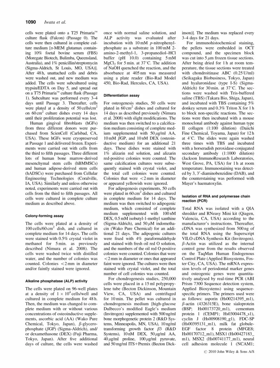
Surface epitopes of hPDL cells
The expression of putative mesenchy-mal stem cell markers (CD29, CD44,CD90, and CD105 positive, and CD14,CD34, and CD45 negative) wasobserved in all types of cells (Fig. 4).CD146, CD106, and ALP were morefrequently positive in hPDL cells com-pared with hGFs. STRO-1, an epitopeoriginally suggested as a marker forMSCs, was expressed in o20% of cellsfor all types of cells.
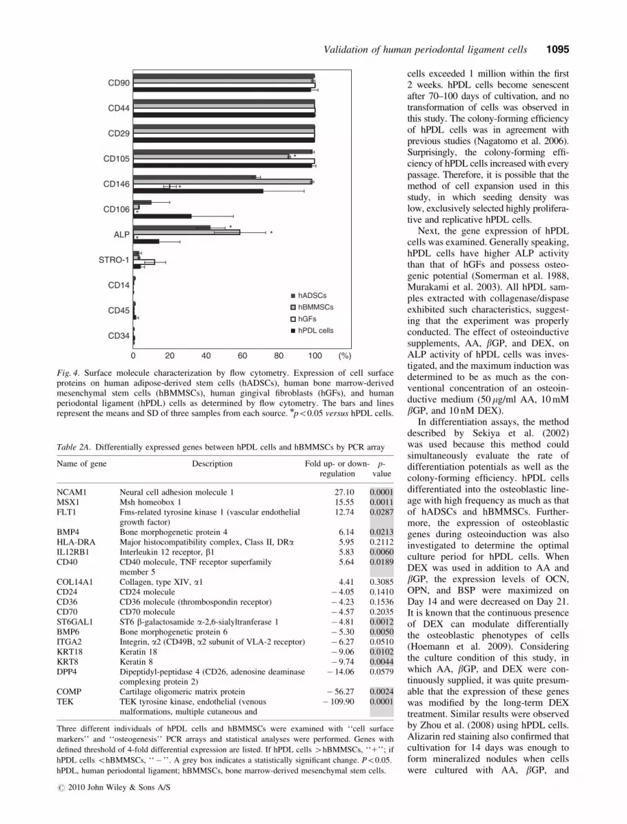
Gene expression profile
Previous studies demonstrated thathPDL cells have osteogenic potential.Thus, we performed both osteogenesisand cell surface markers PCR arrayon hPDL cells and compared it withhBMMSCs (Table 2A) or hGFs (Table2B). Several genes were preferentiallyexpressed in hPDL cells. Neural celladhesion molecule 1 (NCAM1) wasstrongly expressed in hPDL cellscompared with hBMMSCs and hGFs.This difference was statistically signi-ficant. In addition to these PCR arrays,each of the known PDL marker geneswas also verified with a TaqMans geneexpression assay. b-Actin was chosenas the endogenous control from theresults of the TaqMans Human Endo-genous Control Plate because of thestability of the signal between the celltypes and its level of expression (datanot shown). The expression levels ofMSX1, NCAM1, and S100A4 in hPDLcells were higher than in hBMMSCs(Fig. 5a). Moreover, the expressionlevels of NCAM1 and periostin inhPDL cells were higher than in hGFs(Fig. 5a). The immunohistochemicalanalyses confirmed the positive signalsof both S100A4 and periostin in hPDLtissues between the alveolar bone anddentin (Fig. 5b–e).
Transplantation
To verify the osteogenic potential invivo, multi-layered hPDL cell sheetswere transplanted into the backs ofathymic rats. After being cultured withan osteoinductive medium for 14 days,hPDL cells were multi-layered (four,
0
20
40
60
80
100
120
P1 P5P3
CF
U-F
(%
)
1E+16
1E+14
1E+12
1E+10
1E+8
1E+6
1E+4
1E+2
1E+18
10 10080604020
#34 (32M)
#29 (47M)#30 (31M)#31 (57F)#32 (23F)#33 (45M)
Days in Culture
Acc
umul
ated
cel
l num
ber
0
0.2
0.4
0.6
0.8
1
1.2
1.4
1.6
1.8
2
25 50 100200100100100100100100100100
2.5 5 10 20 10 10 10 10
6 12 25 50
ALP
act
ivity
(O
.D. 4
05)
AA
βGP
DEX
#31 (57F)
#33 (45M)
#32 (23F)
**
50 cells /cm2
5000 cells /cm2
500 cells /cm2
Gro
wth
inde
x
0
100
200
300 ****
**
**
*
a b
c d
Fig. 1. Proliferation, colony-forming unit-fibroblast (CFU-F), and alkaline phosphatase(ALP) activity of human periodontal ligament (hPDL) cells. (a) The growth and proliferativelife span of hPDL cells from six different donors. Cells were seeded and subcultured at50 cells/cm2 every 2 weeks after Passage 3. (b) CFU-F of hPDL cells. hPDL cells from sixdonors were examined for colony-forming assay at Passages 1, 3, and 5 in triplicate. The barsand lines represent the means and SD of six samples. Statistically significant difference(npo0.05; nnpo0.01). (c) Effect of the seeding density on the proliferation of hPDL cells.Cells were plated in triplicate at densities of 50, 500, and 5000 cells/cm2, respectively.Medium was changed 24 h after seeding, and hPDL cells were cultured for an additional 7days. The bars and lines represent the means and SD of three samples at Passage 3.nnStatistically significant difference (po0.01). (d) ALP activity of hPDL cells from threedonors induced by the use of a combination of osteoinductive reagents, ascorbic acid (AA)(mg/ml), b-glycerophosphate (bGP) (mM), and dexamethasone (DEX) (nM). The bars andlines represent the means and SD.
1092 Iwata et al.
r 2010 John Wiley & Sons A/S

0
20
40
60
80
100
120
0
10
20
30
40
50
60
70
80
90
100
0
5
10
15
20
25
30
35
40
a b
dc
fe
Rel
ativ
e m
RN
A le
vels
Col2A1
Ave
rage
frac
tion
of a
lizar
in r
edpo
sitiv
e co
lony
rat
io (
%)
Ave
rage
frac
tion
of o
il re
d-O
posi
tive
colo
ny r
atio
(%
)
**
*
*
Fig. 2. Differentiation potential of human periodontal ligament (hPDL) cells. (a) hPDL cells positively stained with alizarin red. hPDL cellswere cultured for 21 days in osteoinductive medium. (b) The ratio of alizarin red-positive colonies in the total number of colonies. Eachsample was examined in triplicate, and the means of each sample were used for statistical analysis. The bars and lines represent the means andSD of human adipose-derived stem cells (hADSCs) (n 5 3), human bone marrow-derived mesenchymal stem cells (hBMMSCs) (n 5 3),human gingival fibroblasts (hGFs) (n 5 3), and hPDL cells (n 5 7). npo0.05 versus hPDL cells. (c) hPDL cells positively stained with oil redO. hPDL cells were cultured for 21 days in adipogenic medium. (d) The ratio of oil red O-positive colonies in the total number of colonies.Each sample was examined in triplicate, and the means of each sample were used for statistical analysis. The bars and lines represent themeans and SD of hADSCs (n 5 3), hBMMSCs (n 5 3), hGFs (n 5 3), and hPDL cells (n 5 7). npo0.05 versus hPDL cells. (e)Immunohistochemical analysis for type II collagen. hPDL cells were pelleted and cultured for 21 days in the defined medium described inthe ‘‘Materials and Methods’’. A macro picture of cartilage pellets with 100mm scale is shown. (f) Gene expression analysis for type IIcollagen on Day 21 was analysed by real-time PCR. The bars and lines represent the means and SD of hADSCs (n 5 3), hBMMSCs (n 5 3),hGFs (n 5 3), and hPDL cells (n 5 7). The mean fold changes in gene expression relative to b-actin were calculated using the values obtainedfrom hPDL cells as a calibrator by means of the 2�DDCt method (Livak & Schmittgen 2001). npo0.05 versus hPDL cells.
Validation of human periodontal ligament cells 1093
r 2010 John Wiley & Sons A/S

six, eight, or 10 layers) with wovenPGA and transplanted into the backs ofathymic rats. Layering structures wereclearly seen on Day 3 (Fig. 6a and b).
An area that was alizarin red positivewas observed on Day 3, and it grew untilDay 10 in both eight- and 10-layertransplantation.
Discussion
Human teeth are routinely extractedbecause of caries or orthodontic reasons,even though they retain healthy PDL orpulp tissues. Therefore, researchers haveutilized dental stem cells as non-inva-sive cell sources for regenerative medi-cine. In fact, PDL cells (Nakahara et al.2004, Sonoyama et al. 2006, Liu et al.2008, Iwata et al. 2009) and dental pulpcells (Iohara et al. 2004, 2006) havealready been examined for their regen-erative potential in large animal models.Importantly, the clinical outcomes ofthese studies were superior to those oftraditional therapies. Thus, human clin-ical trials using hPDL cells have thepotential to treat various defects, whichhave until now been thought to bedifficult to cure. Although the prospectsare increasing, appropriate methods forextracting and expanding hPDL cellsare still not well established. In thisstudy, we determined the optimal meth-od for the isolation and expansion ofhPDL cells. We then examined theirgene expressions and differentiationpotentials and eventually validated thecommon characteristics of hPDL cellsfrom 41 samples.
First, the method of enzymatic diges-tion for hPDL tissues was optimized.Because hPDL tissue mainly consists ofcollagen, the dose–response effect of col-lagenase (0.075, 0.75, and 1.5 PZ-U/ml)was studied, resulting in 0.75 PZ-U/ml asthe optimal concentration (data notshown). The success rate of cell expan-sion with collagenase/dispase was 96.7%(29/30), which was superior to the origi-nal explant method (76%) (Somermanet al. 1988). Only one sample (#28, a23-year-old male) exhibited bacterialcontamination during the initial days ofculture. Trypsin/EDTA also extractedcells from hPDL tissues (72.7%). How-ever, some of the samples lost theirosteogenic potential (Table 1) and hadlow proliferation (data not shown). Thus,collagenase/dispase was chosen forfurther studies. Primaria culture disheswere used in this study because the initialcell attachment was enhanced comparedwith normal Falcon dishes (data notshown). The cells were cultured in a-MEM glutamax for rapid cell expansionas described previously (Sotiropoulou etal. 2006). hPDL cells exhibited anincreased proliferation compared withother mesenchymal tissue-derived cells(Matsubara et al. 2005, Sakaguchi et al.2005). Usually, the number of hPDL
30
10
20
04 7 14 21
OCN
Rel
ativ
e ge
ne e
xpre
ssio
n
4 7 14 210
200
400
600OPN
Rel
ativ
e ge
ne e
xpre
ssio
n
4 7 14 210
40
20
60
80
100BSP
Rel
ativ
e ge
ne e
xpre
ssio
n
Control
AA+βGP
AA+βGP+DEX
a
b
Day 14
Day 21
C ABDABA
(d)
(d)
(d)
Fig. 3. Osteoblastic/cementoblastic gene expression during osteoinduction in human perio-dontal ligament (hPDL) cells. (a) Effects of osteoinductive supplements on the expression ofosteocalcin (OCN), osteopontin (OPN), and bone sialoprotein (BSP). After hPDL cells weretreated with or without supplements [AA (50mg/ml)1bGP (10 mM) or AA1bGP1DEX(10 nM)], the gene expression at various time points (4–21 days) was analysed by real-timePCR. Two individual PDL cells were examined in triplicate, and the average values areshown. The mean fold changes in gene expression relative to b-actin were calculated usingthe values obtained from hPDL cells without induction on Day 4. (b) Alizarin red staining ofhPDL cells cultured with or without osteoinductive supplements. PDL cells were spread at adensity of 5000 cells/cm2. After 48 h, the culture medium was switched to complete mediumonly (c) or complete medium1AA (50mg/ml) (A); AA1bGP (10 mM) (AB); orAA1bGP1DEX (10 nM) (ABD), and cultured for 14 or 21 days.
1094 Iwata et al.
r 2010 John Wiley & Sons A/S
cells exceeded 1 million within the first2 weeks. hPDL cells become senescentafter 70–100 days of cultivation, and notransformation of cells was observed inthis study. The colony-forming efficiencyof hPDL cells was in agreement withprevious studies (Nagatomo et al. 2006).Surprisingly, the colony-forming effi-ciency of hPDL cells increased with everypassage. Therefore, it is possible that themethod of cell expansion used in thisstudy, in which seeding density waslow, exclusively selected highly prolifera-tive and replicative hPDL cells.
Next, the gene expression of hPDLcells was examined. Generally speaking,hPDL cells have higher ALP activitythan that of hGFs and possess osteo-genic potential (Somerman et al. 1988,Murakami et al. 2003). All hPDL sam-ples extracted with collagenase/dispaseexhibited such characteristics, suggest-ing that the experiment was properlyconducted. The effect of osteoinductivesupplements, AA, bGP, and DEX, onALP activity of hPDL cells was inves-tigated, and the maximum induction wasdetermined to be as much as the con-ventional concentration of an osteoin-ductive medium (50mg/ml AA, 10 mMbGP, and 10 nM DEX).
In differentiation assays, the methoddescribed by Sekiya et al. (2002)was used because this method couldsimultaneously evaluate the rate ofdifferentiation potentials as well as thecolony-forming efficiency. hPDL cellsdifferentiated into the osteoblastic line-age with high frequency as much as thatof hADSCs and hBMMSCs. Further-more, the expression of osteoblasticgenes during osteoinduction was alsoinvestigated to determine the optimalculture period for hPDL cells. WhenDEX was used in addition to AA andbGP, the expression levels of OCN,OPN, and BSP were maximized onDay 14 and were decreased on Day 21.It is known that the continuous presenceof DEX can modulate differentiallythe osteoblastic phenotypes of cells(Hoemann et al. 2009). Consideringthe culture condition of this study, inwhich AA, bGP, and DEX were con-tinuously supplied, it was quite presum-able that the expression of these geneswas modified by the long-term DEXtreatment. Similar results were observedby Zhou et al. (2008) using hPDL cells.Alizarin red staining also confirmed thatcultivation for 14 days was enough toform mineralized nodules when cellswere cultured with AA, bGP, and
(%)
*
*
*
*
**
hADSCs
hPDL cells
hGFs
hBMMSCs
0 20 40 60 80 100
CD34
CD45
CD14
STRO-1
ALP
CD106
CD146
CD105
CD29
CD44
CD90
Fig. 4. Surface molecule characterization by flow cytometry. Expression of cell surfaceproteins on human adipose-derived stem cells (hADSCs), human bone marrow-derivedmesenchymal stem cells (hBMMSCs), human gingival fibroblasts (hGFs), and humanperiodontal ligament (hPDL) cells as determined by flow cytometry. The bars and linesrepresent the means and SD of three samples from each source. npo0.05 versus hPDL cells.
Table 2A. Differentially expressed genes between hPDL cells and hBMMSCs by PCR array
Name of gene Description Fold up- or down-regulation
p-value
NCAM1 Neural cell adhesion molecule 1 27.10 0.0001MSX1 Msh homeobox 1 15.55 0.0011FLT1 Fms-related tyrosine kinase 1 (vascular endothelial
growth factor)12.74 0.0287
BMP4 Bone morphogenetic protein 4 6.14 0.0213HLA-DRA Major histocompatibility complex, Class II, DRa 5.95 0.2112IL12RB1 Interleukin 12 receptor, b1 5.83 0.0060CD40 CD40 molecule, TNF receptor superfamily
member 55.64 0.0189
COL14A1 Collagen, type XIV, a1 4.41 0.3085CD24 CD24 molecule � 4.05 0.1410CD36 CD36 molecule (thrombospondin receptor) � 4.23 0.1536CD70 CD70 molecule � 4.57 0.2035ST6GAL1 ST6 b-galactosamide a-2,6-sialyltranferase 1 � 4.81 0.0012BMP6 Bone morphogenetic protein 6 � 5.30 0.0050ITGA2 Integrin, a2 (CD49B, a2 subunit of VLA-2 receptor) � 6.27 0.0510KRT18 Keratin 18 � 9.06 0.0102KRT8 Keratin 8 � 9.74 0.0044DPP4 Dipeptidyl-peptidase 4 (CD26, adenosine deaminase
complexing protein 2)� 14.06 0.0579
COMP Cartilage oligomeric matrix protein � 56.27 0.0024TEK TEK tyrosine kinase, endothelial (venous
malformations, multiple cutaneous and� 109.90 0.0001
Three different individuals of hPDL cells and hBMMSCs were examined with ‘‘cell surface
markers’’ and ‘‘osteogenesis’’ PCR arrays and statistical analyses were performed. Genes with
defined threshold of 4-fold differential expression are listed. If hPDL cells 4hBMMSCs, ‘‘1’’; if
hPDL cells ohBMMSCs, ‘‘� ’’. A grey box indicates a statistically significant change. Po0.05.
hPDL, human periodontal ligament; hBMMSCs, bone marrow-derived mesenchymal stem cells.
Validation of human periodontal ligament cells 1095
r 2010 John Wiley & Sons A/S

cyclin J
MFGE8
S100A4
MSX2
CEMP1asporin
MSX1
NCAM1 periostin
hBMMSCs hGFs hPDL cells
% β
-act
in
0
25
20
15
10
5
0
3
2
1
8
6
4
2
0
600
400
200
0 0
0.2
0.4
0.6
0.8
1
4
8
12
0
**
* ** *
***
*
3
2
1
0
*
****
600
400
200
0
*
40
30
20
10
0
***
a
b
d
S100A4
Periostin
c
AB D
e
AB D
Fig. 5. Expression of PDL-specific markers. (a) Real-time PCR analysis of known PDL-specific genes. The bars and lines represent the meansand SD of human bone marrow-derived mesenchymal stem cells (hBMMSCs) (n 5 3), human gingival fibroblasts (hGFs) (n 5 3), and humanperiodontal ligament (hPDL) cells (n 5 7). Statistically significant difference (npo0.05; nnpo0.01). The tissue sections of a human tooth withsurrounding bone were immunostained with anti-human S100A4 (b and c) or periostin (d and e) polyclonal antibodies. The positive staining isshown by a reddish-brown colour. All sections were counterstained with haematoxylin (blue). The scale bars, 1 mm (B and D) and 50mm (Cand E). AB, alveolar bone and D, dentin.
Table 2B. Differentially expressed genes between hPDL cells and hGFs by PCR array
Name of gene Description Fold up- or down-regulation p-value
HLA-DRA Major histocompatibility complex, Class II, DRa 30.30 0.0557VCAM1 Vascular cell adhesion molecule 1 23.88 0.0269ALPL Alkaline phosphatase, liver/bone/kidney 23.25 0.0486TPSAB1 Tryptase a/b1 20.98 0.0216NCAM1 Neural cell adhesion molecule 1 19.53 0.0456COL14A1 Collagen, type XIV, a1 9.46 0.1882FGFR2 Fibroblast growth factor receptor 2 7.04 0.0019TGFB3 Transforming growth factor b3 6.77 0.0310MYOCD Myocardin 6.22 0.3872CD4 CD4 molecule 5.79 0.3811ITGAM Integrin, aM (complement component 3 receptor 3 subunit) 5.39 0.0178CD74 CD74 molecule, major histocompatibility complex, Class II invariant 5.34 0.3486BGN Biglycan 4.75 0.0652EGF Epidermal growth factor (b-urogastrone) 4.25 0.0658IGF1 Insulin-like growth factor 1 (somatomedin C) 4.20 0.2378ITGA2 Integrin, a2 (CD49B, a2 subunit of VLA-2 receptor) � 5.10 0.0830COL15A1 Collagen, type XV, a1 � 6.38 0.3418CTSK Cathepsin K � 7.22 0.0301
Three different individuals of hPDL cells and hGFs were examined with ‘‘cell surface markers’’ and ‘‘osteogenesis’’ PCR arrays and statistical analyses
were performed. Genes with defined threshold of four-fold differential expression are listed. If hPDL cells 4hGFs, ‘‘1’’; if hPDL cells ohGFs, ‘‘� ’’.
A grey box indicates a statistically significant change. po0.05.
hPDL, human periodontal ligament; hGFs, human gingival fibroblasts.
1096 Iwata et al.
r 2010 John Wiley & Sons A/S

DEX. Thus, we concluded that osteoin-ductive cultivation with DEX for 14days was sufficient to enhance the osteo-blastic differentiation of hPDL cells.
Next, the adipogenic and chondrogenicpotentials of hPDL cells were examined.Both potentials were lower in hPDL cellsthan in hADSCs or hBMMSCs. Theseresults correlated with a previous reportexamining alveolar bone marrow-derivedcells (Matsubara et al. 2005). It is possiblethat the origin of the cells may affect theirpotential. Both PDL tissue and alveolarbone are neural crest-derived mesenchy-mal stromal cells (Chai et al. 2000, Cho& Garant 2000). A recent study showedthat neural crest-derived osteoblasts ex-hibited stronger bone formation activitythan cells derived from the mesoderm(Leucht et al. 2008). Thus, further inves-tigation is needed to clarify the origin-specific plasticity of hPDL cells.
In the adipogenic differentiationstudy, hGFs differentiated into oil redO-positive cells. It is possible that themethod used in this study, where allcells have colony-forming ability, mayaffect the potential of adipogenic differ-entiation in hGFs. Additional studiesshould be undertaken to determine thedifferentiation properties of hGFs.
FCM analysis showed the similarity ofsurface epitopes among four types of cells.Putative markers for MSC (CD29, CD44,
CD90, and CD90) were expressed in allkinds of cells. The expression of CD146,CD106, and ALP in hPDL cells wassignificantly higher than in hGFs, and theseresults agreed with those of other studies(Seo et al. 2004, Gronthos et al. 2006,Lindroos et al. 2008, Wada et al. 2009). Inthis study, only a small fraction of hPDLcells was STRO-1 positive (4.2 � 2.0%).It is possible that the STRO-1 expressionmay be lost over time, as suggested inother studies (Matsubara et al. 2005, Zhouet al. 2008, Itaya et al. 2009).
Next, we studied the distinguishablemarker genes of hPDL cells. Althoughsimilar approaches have been performedby others (Han & Amar 2002, Lallieret al. 2005, Fujita et al. 2007), the resultsof our PCR array showed NCAM1 as anew marker of hPDL cells. In addition,the proposed PDL markers (asporin,CEMP1, cyclin J, FDC-SP, MFGE8,MSX1, MSX2, periostin, and S100A4)(Ishikawa et al. 2009) were also inves-tigated using commercially availablePCR primer sets. The strong expressionof both S100A4 and periostin wasdetectable in all hPDL cells. However,individual variability in the expressionlevels of asporin, MSX1, and MSX2was observed. In this study, the expres-sion of FDC-SP was rarely observed inall types of cells (data not shown), andno preferential expression of CEMP1,
cyclin J, or MFGE8 was observed inhPDL cells (Fig. 5). It is possible thatthe culture conditions used in this studymay affect the expression of thesegenes. Still, immunohistochemical stu-dies confirmed that both S100A4 andperiostin had specific signals in hPDLspecimens. Thus, these two genes can beuseful to identify hPDL cells.
To assess the in vivo osteogenic poten-tial, multi-layered hPDL cell sheets cul-tured with osteoinductive medium weretransplanted into athymic rats. Because noalizarin red-positive area was observed inthe control sites, the calcification wasspeculated to be induced by transplantedhPDL cell sheets. The alizarin red-positivearea was expanded dramatically on Day10 compared with Day 3, suggesting thathPDL cells promoted calcium depositionaround them. Unfortunately, we couldobserve no calcified tissues in 4-weeksamples. It is possible that immunorejec-tion may occur in this subcutaneous trans-plantation model with athymic rats.
From these results, hPDL cells weresuccessfully extracted, expanded, andexamined, and the characteristics anddistinguishable markers of hPDL cellswere proposed. Because true perio-dontal regeneration was already re-ported in a canine model using similarmethods (Iwata et al. 2009), the methodsdescribed in this study might be helpful
8 layered hPDL cell sheets
Day 10
Day 3
A B
C D
10 layered hPDL cell sheets
Day 10
Day 3
DC
BA
a b
Fig. 6. The in vivo potential of multi-layered human periodontal ligament (hPDL) cell sheets that were subcutaneously transplanted inathymic rats. Eight (a)- or 10 (b)-layered hPDL cell sheets with woven PGA carrier were transplanted, and sacrificed on Day 3 (a and b) or Day10 (c and d). The dissected samples were stained with haematoxylin–eosin (a and c) or alizarin red (b and d). The bars show 100mm. Note thatobvious alizarin red-positive areas are observed on Day 10 in both eight (a)- and 10 (b)-layered transplantation of hPDL cell sheets.
Validation of human periodontal ligament cells 1097
r 2010 John Wiley & Sons A/S

for the cultivation and validation of hPDLcells for human clinical application.
Acknowledgements
We thank Professor Emeritus HidekiOgiuchi for his kind supports, IchiroSekiya for excellent technical support,Hiroaki Sugiyama for expert help withhistology, and Machiko Miyoshi forkind support. The authors are gratefulto Norio Ueno (Tokyo Women’s Med-ical University) for his valuable com-ments and suggestions.
References
Andreasen, J. O. & Kristerson, L. (1981) The effect
of limited drying or removal of the periodontal
ligament. Periodontal healing after replantation
of mature permanent incisors in monkeys. Acta
Odontologica Scandinavica 39, 1–13.
Bianco, P. & Robey, P. G. (2001) Stem cells in tissue
engineering. Nature 414, 118–121.
Chai, Y., Jiang, X., Ito, Y., Bringas, P. Jr., Han, J.,
Rowitch, D. H., Soriano, P., McMahon, A. P. &
Sucov, H. M. (2000) Fate of the mammalian cranial
neural crest during tooth and mandibular morpho-
genesis. Development 127, 1671–1679.
Cho, M. I. L. & Garant, P. R. (2000) Development
and general structure of the periodontium. Perio-
dontology 2000 24, 9–27.
Coura, G. S., Garcez, R. C., de Aguiar, C. B., Alvarez-
Silva, M., Magini, R. S. & Trentin, A. G. (2008)
Human periodontal ligament: a niche of neural crest
stem cells. Journal of Periodontal Research 43,
531–536.
Duarte, W. R. (1998) cDNA cloning of S100 calcium-
binding proteins from bovine periodontal ligament
and their expression in oral tissues. Journal of
Dental Research 77, 1694–1699.
Fujita, T., Iwata, T., Shiba, H., Igarashi, A., Hirata, R.,
Takeda, K., Mizuno, N., Tsuji, K., Kawaguchi, H.,
Kato, Y. & Kurihara, H. (2007) Identification of
marker genes distinguishing human periodontal
ligament cells from human mesenchymal stem cells
and human gingival fibroblasts. Journal of Perio-
dontal Research 42, 283–286.
Gay, I. C., Chen, S. & MacDougall, M. (2007)
Isolation and characterization of multipotent human
periodontal ligament stem cells. Orthodontics and
Craniofacial Research 10, 149–160.
Gronthos, S., Mrozik, K., Shi, S. & Bartold, P. M.
(2006) Ovine periodontal ligament stem cells: iso-
lation, characterization, and differentiation poten-
tial. Calcified Tissue International 79, 310–317.
Hammarstrom, L., Heijl, L. & Gestrelius, S. (1997)
Periodontal regeneration in a buccal dehiscence
model in monkeys after application of enamel
matrix proteins. Journal of Clinical Periodontology
24, 669–677.
Han, X. & Amar, S. (2002) Identification of genes
differentially expressed in cultured human perio-
dontal ligament fibroblasts vs. human gingival
fibroblasts by DNA microarray analysis. Journal
of Dental Research 81, 399–405.
Hoemann, C. D., El-Gabalawy, H. & McKee, M. D.
(2009) In vitro osteogenesis assays: influence of
the primary cell source on alkaline phosphatase
activity and mineralization. Pathologie et Biologie
57, 318–323.
Horiuchi, K., Amizuka, N., Takeshita, S., Takamatsu,
H., Katsuura, M., Ozawa, H., Toyama, Y., Bone-
wald, L. F. & Kudo, A. (1999) Identification and
characterization of a novel protein, periostin, with
restricted expression to periosteum and periodontal
ligament and increased expression by transforming
growth factor beta. Journal of Bone and Mineral
Research 14, 1239–1249.
Iohara, K., Nakashima, M., Ito, M., Ishikawa, M.,
Nakasima, A. & Akamine, A. (2004) Dentin regen-
eration by dental pulp stem cell therapy with
recombinant human bone morphogenetic protein
2. Journal of Dental Research 83, 590–595.
Iohara, K., Zheng, L., Ito, M., Tomokiyo, A., Matsushita,
K. & Nakashima, M. (2006) Side population cells
isolated from porcine dental pulp tissue with self-
renewal and multipotency for dentinogenesis, chon-
drogenesis, adipogenesis, and neurogenesis. Stem
Cells 24, 2493–2503.
Ishikawa, I., Iwata, T., Washio, K., Okano, T., Naga-
sawa, T., Iwasaki, K. & Ando, T. (2009) Cell sheet
engineering and other novel cell-based approaches
to periodontal regeneration. Periodontology 2000
51, 220–238.
Itaya, T., Kagami, H., Okada, K., Yamawaki, A.,
Narita, Y., Inoue, M., Sumita, Y. & Ueda, M.
(2009) Characteristic changes of periodontal liga-
ment-derived cells during passage. Journal of
Periodontal Research 44, 425–433.
Iwata, T., Yamato, M., Tsuchioka, H., Takagi, R.,
Mukobata, S., Washio, K., Okano, T. & Ishikawa, I.
(2009) Periodontal regeneration with multi-layered
periodontal ligament-derived cell sheets in a canine
model. Biomaterials 30, 2716–2723.
Karring, T., Isidor, F., Nyman, S. & Lindhe, J. (1985)
New attachment formation on teeth with a reduced
but healthy periodontal ligament. Journal of Clin-
ical Periodontology 12, 51–60.
Lallier, T. E., Spencer, A. & Fowler, M. M. (2005)
Transcript profiling of periodontal fibroblasts
and osteoblasts. Journal of Periodontology 76,
1044–1055.
Leucht, P., Kim, J. B., Amasha, R., James, A. W.,
Girod, S. & Helms, J. A. (2008) Embryonic origin
and Hox status determine progenitor cell fate
during adult bone regeneration. Development 135,
2845–2854.
Lindroos, B., Maenpaa, K., Ylikomi, T., Oja, H.,
Suuronen, R. & Miettinen, S. (2008) Characterisa-
tion of human dental stem cells and buccal mucosa
fibroblasts. Biochemical and Biophysical Research
Communications 368, 329–335.
Liu, Y., Zheng, Y., Ding, G., Fang, D., Zhang, C.,
Bartold, P. M., Gronthos, S., Shi, S. & Wang, S.
(2008) Periodontal ligament stem cell-mediated
treatment for periodontitis in miniature swine.
Stem Cells 26, 1065–1073.
Livak, K. J. & Schmittgen, T. D. (2001) Analysis of
relative gene expression data using real-time quan-
titative PCR and the 2(T)(�Delta Delta C) method.
Methods 25, 402–408.
Matsubara, T., Suardita, K., Ishii, M., Sugiyama, M.,
Igarashi, A., Oda, R., Nishimura, M., Saito, M.,
Nakagawa, K., Yamanaka, K., Miyazaki, K., Shimizu,
M., Bhawal, U. K., Tsuji, K., Nakamura, K. & Kato,
Y. (2005) Alveolar bone marrow as a cell source for
regenerative medicine: differences between alveolar
and iliac bone marrow stromal cells. Journal of
Bone and Mineral Research 20, 399–409.
McCulloch, C. A. & Bordin, S. (1991) Role of
fibroblast subpopulations in periodontal physiology
and pathology. Journal of Periodontal Research 26,
144–154.
Murakami, Y., Kojima, T., Nagasawa, T., Kobayashi,
H. & Ishikawa, I. (2003) Novel isolation of alkaline
phosphatase-positive subpopulation from perio-
dontal ligament fibroblasts. Journal of Perio-
dontology 74, 780–786.
Nagatomo, K., Komaki, M., Sekiya, I., Sakaguchi, Y.,
Noguchi, K., Oda, S., Muneta, T. & Ishikawa, I.
(2006) Stem cell properties of human periodontal
ligament cells. Journal of Periodontal Research 41,
303–310.
Nakahara, T., Nakamura, T., Kobayashi, E., Kuremo-
to, K., Matsuno, T., Tabata, Y., Eto, K. & Shimizu,
Y. (2004) In situ tissue engineering of periodontal
tissues by seeding with periodontal ligament-
derived cells. Tissue Engineering 10, 537–544.
Nakamura, S., Terashima, T., Yoshida, T., Iseki, S.,
Takano, Y., Ishikawa, I. & Shinomura, T. (2005)
Identification of genes preferentially expressed
in periodontal ligament: specific expression of a
novel secreted protein, FDC-SP. Biochemical
and Biophysical Research Communications 338,
1197–1203.
Nimura, A., Muneta, T., Koga, H., Mochizuki, T.,
Suzuki, K., Makino, H., Umezawa, A. & Sekiya, I.
(2008) Increased proliferation of human synovial
mesenchymal stem cells with autologous human
serum: comparisons with bone marrow mesenchy-
mal stem cells and with fetal bovine serum. Arthri-
tis and Rheumatism 58, 501–510.
Nishida, E., Sasaki, T., Ishikawa, S. K., Kosaka, K.,
Aino, M., Noguchi, T., Teranaka, T., Shimizu, N.
& Saito, M. (2007) Transcriptome database KK-
periome for periodontal ligament development:
expression profiles of the extracellular matrix
genes. Gene 404, 70–79.
Nyman, S., Karring, T., Lindhe, J. & Planten, S. (1980)
Healing following implantation of periodontitis-
affected roots into gingival connective tissue.
Journal of Clinical Periodontology 7, 394–401.
Nyman, S., Lindhe, J., Karring, T. & Rylander, H.
(1982) New attachment following surgical
treatment of human periodontal disease. Journal
of Clinical Periodontology 9, 290–296.
Pi, S. H., Lee, S. K., Hwang, Y. S., Choi, M. G. &
Kim, E. C. (2007) Differential expression of perio-
dontal ligament-specific markers and osteogenic
differentiation in human papilloma virus 16-immor-
talized human gingival fibroblasts and periodontal
ligament cells. Journal of Periodontal Research 42,
104–113.
Peck, S. & Peck, L. (1993) A time for change of tooth
numbering systems. Journal of Dental Education
57, 643–647.
Ramakrishnan, P. R., Lin, W. L., Sodek, J. & Cho, M.
I. (1995) Synthesis of noncollagenous extracellular
matrix proteins during development of mineralized
nodules by rat periodontal ligament cells in vitro.
Calcified Tissue International 57, 52–59.
Sakaguchi, Y., Sekiya, I., Yagishita, K. & Muneta, T.
(2005) Comparison of human stem cells derived
from various mesenchymal tissues: superiority of
synovium as a cell source. Arthritis and Rheuma-
tism 52, 2521–2529.
Sculean, A., Nikolidakis, D. & Schwarz, F. (2008)
Regeneration of periodontal tissues: combinations
of barrier membranes and grafting materials
– biological foundation and preclinical evidence: a
systematic review. Journal of Clinical Perio-
dontology 35, 106–116.
Sekiya, I., Larson, B. L., Smith, J. R., Pochampally, R.,
Cui, J. G. & Prockop, D. J. (2002) Expansion of
human adult stem cells from bone marrow stroma:
conditions that maximize the yields of early pro-
genitors and evaluate their quality. Stem Cells 20,
530–541.
Seo, B. M., Miura, M., Gronthos, S., Bartold, P. M.,
Batouli, S., Brahim, J., Young, M., Robey, P. G.,
Wang, C. Y. & Shi, S. (2004) Investigation of
multipotent postnatal stem cells from human perio-
dontal ligament. Lancet 364, 149–155.
Somerman, M. J., Archer, S. Y., Imm, G. R. & Foster,
R. A. (1988) A comparative study of human
1098 Iwata et al.
r 2010 John Wiley & Sons A/S

periodontal ligament cells and gingival fibroblasts
in vitro. Journal of Dental Research 67, 66–70.
Sonoyama, W., Liu, Y., Fang, D., Yamaza, T., Seo, B.
M., Zhang, C., Liu, H., Gronthos, S., Wang, C. Y.,
Shi, S. & Wang, S. (2006) Mesenchymal stem cell-
mediated functional tooth regeneration in swine.
PLoS One 1, e79.
Sotiropoulou, P. A., Perez, S. A., Salagianni, M.,
Baxevanis, C. N. & Papamichail, M. (2006) Char-
acterization of the optimal culture conditions for
clinical scale production of human mesenchymal
stem cells. Stem Cells 24, 462–471.
Trubiani, O., Di Primio, R., Traini, T., Pizzicannella,
J., Scarano, A., Piattelli, A. & Caputi, S. (2005)
Morphological and cytofluorimetric analysis of
adult mesenchymal stem cells expanded ex vivo
from periodontal ligament. International Journal of
Immunopathology and Pharmacology 18, 213–221.
Wada, N., Menicanin, D., Shi, S., Bartold, P. M. &
Gronthos, S. (2009) Immunomodulatory properties
of human periodontal ligament stem cells. Journal
of Cell Physiology 219, 667–676.
Wang, H. L., Greenwell, H., Fiorellini, J., Giannobile,
W., Offenbacher, S., Salkin, L., Townsend, C.,
Sheridan, P. & Genco, R. J. (2005) Periodontal
regeneration. Journal of Periodontology 76, 1601–
1622.
Xu, J., Wang, W., Kapila, Y., Lotz, J. & Kapila, S.
(2009) Multiple differentiation capacity of STRO-
11/CD1461PDL mesenchymal progenitor cells.
Stem Cells and Development 18, 487–496.
Yamada, S., Murakami, S., Matoba, R., Ozawa, Y.,
Yokokoji, T., Nakahira, Y., Ikezawa, K., Takaya-
ma, S., Matsubara, K. & Okada, H. (2001) Expres-
sion profile of active genes in human periodontal
ligament and isolation of PLAP-1, a novel SLRP
family gene. Gene 275, 279–286.
Zhou, Y., Hutmacher, D. W., Sae-Lim, V., Zhou, Z.,
Woodruff, M. & Lim, T. M. (2008) Osteogenic
and adipogenic induction potential of human perio-
dontal cells. Journal of Periodontology 79,
525–534.
Address:
Teruo Okano
Institute of Advanced Biomedical Engineering
and Science
Tokyo Women’s Medical University
8-1 Kawada-cho
Shinjuku-ku
Tokyo 162-8666
Japan
E-mail: [email protected]
Clinical Relevance
Scientific rationale for the study:Cytotherapy using PDL cells hasproven to be effective. However,there is no standard protocol for theculture of human PDL cells. In thisstudy, we validated the culture con-
ditions and characteristics of humanPDL cells for human clinical trials.Principal findings: Human PDL cellswere successfully extracted with col-lagenase/dispase and exhibited highproliferative capacity, high ALP activ-ity, and multi-potency. Some genes
(NCAM1, S100A4, and periostin)were preferentially expressed in humanPDL cells and can be useful markers forthe identification of human PDL cells.Practical implications: Human PDLcells could be a promising source ofcells for regenerative medicine.
Validation of human periodontal ligament cells 1099
r 2010 John Wiley & Sons A/S
Recommended


