
Osteoarthritis and Cartilage 21 (2013) 1668e1673
Varus thrust and knee frontal plane dynamic motion in persons withknee osteoarthritis
A.H. Chang y*, J.S. Chmiel z, K.C. Moisio y, O. Almagor x, Y. Zhang y, S. Cahue x, L. Sharma xyDepartment of Physical Therapy and Human Movement Sciences, Feinberg School of Medicine, Northwestern University, Chicago, IL, USAzDepartment of Preventive Medicine, Feinberg School of Medicine, Northwestern University, Chicago, IL, USAxDepartment of Medicine, Feinberg School of Medicine, Northwestern University, Chicago, IL, USA
a r t i c l e i n f o
Article history:Received 6 March 2013Accepted 4 August 2013
Keywords:Knee osteoarthritisInstabilityVarus thrustGait analysis
* Address correspondence and reprint requests toPhysical Therapy and Human Movement Sciences, FNorthwestern University, 645 N. Michigan Ave. #11001-312-908-8273; Fax: 1-312-908-0741.
E-mail addresses: [email protected] (J.S. Chmiel), k-moisio@[email protected] (O. Almagor),(Y. Zhang), [email protected] (S. Cahue), l-sharma@north
1063-4584/$ e see front matter � 2013 Osteoarthritihttp://dx.doi.org/10.1016/j.joca.2013.08.007
s u m m a r y
Objective: Varus thrust visualized during walking is associated with a greater medial knee load and anincreased risk of medial knee osteoarthritis (OA) progression. Little is known about how varus thrustpresence determined by visual observation relates to quantitative gait kinematic data. We hypothesizedthat varus thrust presence is associated with greater knee frontal plane dynamic movement during thestance phase of gait.Methods: Participants had knee OA in at least one knee. Trained examiners assessed participants forvarus thrust presence during ambulation. Frontal plane knee motion during ambulation was capturedusing external passive reflective markers and an 8-camera motion analysis system. To examine the cross-sectional relationship between varus thrust and frontal plane knee motion, we used multivariableregression models with the quantitative motion measures as dependent variables and varus thrust(present/absent) as predictor; models were adjusted for age, gender, body mass index (BMI), gait speed,and knee static alignment.Results: 236 persons [mean BMI: 28.5 kg/m2 (standard deviation (SD) 5.5), mean age: 64.9 years (SD10.4), 75.8% women] contributing 440 knees comprised the study sample. 82 knees (18.6%) had definitevarus thrust. Knees with varus thrust had greater peak varus angle and greater peak varus angular ve-locity during stance than knees without varus thrust (mean differences 0.90� and 6.65�/s, respectively).These patterns remained significant after adjusting for age, gender, BMI, gait speed, and knee staticalignment.Conclusion: Visualized varus thrust during walking was associated with a greater peak knee varusangular velocity and a greater peak knee varus angle during stance phase of gait.
� 2013 Osteoarthritis Research Society International. Published by Elsevier Ltd. All rights reserved.
Introduction
Excessive loading during weight-bearing activities plays animportant role in the pathogenesis of knee osteoarthritis (OA)1e4.Defined as the dynamic worsening or abrupt onset of varus (bow-leg) alignment as the limb accepts weight (stance phase), with areturn to less varus and more neutral alignment during lift-off andthe non-weight-bearing (swing) phase of gait, varus thrust is apotent mechanical risk factor for medial knee OA progression5. The
: A.H. Chang, Department ofeinberg School of Medicine,, Chicago, IL 60611, USA. Tel:
(A.H. Chang), [email protected] (K.C. Moisio),[email protected] (L. Sharma).
s Research Society International. P
presence of a varus thrust during gait may represent knee frontalplane dynamic instability and malalignment, possibly resultingfrom insufficient neuromuscular and peri-articular stabilization1.
In theory, reduced knee frontal plane stability and increasedknee varus motion can lead to acute elevation of medial compart-ment load during the stance phase of gait and possibly acceleratestructural damage. This theory is consistent with results of previousstudies that showed knees with a visually observed varus thrusthad a greater external knee adduction moment, a major determi-nant of medial compartment mechanical load during gait, thanthose without a thrust5e7. Varus thrust presence has also beenassociated with a four-fold increase in the odds of medial OAradiographic disease progression5, demonstrating a deleteriouseffect of this abnormal lateral knee movement observed inwalking.Knees with varus thrust have been reported to be at least four timesmore likely to have pain during weight-bearing activities thanthose without a varus thrust8.
ublished by Elsevier Ltd. All rights reserved.

A.H. Chang et al. / Osteoarthritis and Cartilage 21 (2013) 1668e1673 1669
Varus thrust observed during walking is a clinical finding thatmay identify some individuals with greater medial knee load and ahigher risk for disease progression. Observation of gait is simple,quick, and inexpensive compared with measuring knee frontalplane dynamic instability in a laboratory using costly equipmentand quantitative gait analysis. However, whether varus thrustpresence determined by visual observation corresponds to agreater knee varus motion measured in quantitative gait analysishas not been investigated. Using non-invasive 3-dimensional (3-D)motion analysis, knee frontal plane varus motion can be quantifiedby the peak varus angle and peak varus angular velocity during thestance phase. The peak knee varus angle represents the maximumknee varus position in stance. Defined as the change of angularposition over time or as the time derivative of angular position, thepeak knee varus angular velocity characterizes both the kneemovement direction and speed (i.e., how fast the knee is movinginto greater varus) in the frontal plane9, similar towhat is seenwithan observed varus thrust.
Increased knee varus motion during gait may represent anobjective sign of the failure of the knee’s neuromuscular and peri-articular stabilizing mechanisms, manifested as a varus thrust,under dynamic and weight-bearing conditions. We hypothesizedthat varus thrust presence determined by visual observation isassociated with a greater knee varus dynamic movement measuredin quantitative gait analysis during the stance phase of gait.
Methods
Sample
Study participants are enrolled in an ongoing natural historystudy of knee OA, the MAK-3 Study (Mechanical Factors in Arthritisof the Knee e Study 3) and were recruited from the communityusing advertising in periodicals targeting elderly persons, neigh-borhood organizations, letters to members of the registry of theBuehler Center on Aging and Illinois Women’s Health Registry, andvia medical center referrals.
The MAK-3 cohort inclusion criteria were: definite tibiofemoralosteophyte presence, i.e., Kellgren/Lawrence (K/L) radiographicscoring system10 grade �2, in one or both knees; and a Likertcategory of at least “a little difficulty” for two or more items in theWestern Ontario and McMaster Universities Osteoarthritis Index(WOMAC) physical function scale11. Exclusion criteria were: use ofassistive ambulatory devices for more than 50% of the time duringwalking; corticosteroid injection within the previous 3 months;history of avascular necrosis, rheumatoid or other inflammatoryarthritis, periarticular fracture, Paget’s disease, villonodular syno-vitis, joint infection, ochronosis, neuropathic arthropathy, acro-megaly, hemochromatosis, gout, pseudogout, osteopetrosis, ormeniscectomy; or exclusion criteria for magnetic resonance imag-ing (MRI) such as presence of a pacemaker, artificial heart valve,aneurysm clip or shunt, metallic stent, implanted device (e.g., paincontrol/nerve stimulator, defibrillator, insulin/drug pump, earimplant), or any metallic fragment in an eye.
Approval was obtained from the Office for the Protection ofResearch Subjects Institutional Review Boards of NorthwesternUniversity and Evanston Northwestern Healthcare. All participantsgave written, informed consent.
Acquisition and reading of knee radiographs
All participants underwent bilateral, anteroposterior, weight-bearing knee radiographs at baseline in the semi-flexed positionwith fluoroscopic confirmation of superimposition of the anteriorand posterior plateau lines of the medial tibia and centering of the
tibial spines within the femoral notch12. To describe the knees, theK/L radiographic score was used (0 ¼ normal; 1 ¼ possible osteo-phytes; 2 ¼ definite osteophytes without definite joint space nar-rowing; 3 ¼ definite joint space narrowing, some sclerosis, andpossible attrition; and 4 ¼ large osteophytes, marked narrowing,severe sclerosis and definite attrition). Reliability for radiographicgrading for the single X-ray reader was high with a Kappa coeffi-cient of 0.86.
Gait observation for varus and valgus thrust
Gait was observed for varus and valgus thrust presence by one ofthe two trained and experienced examiners (AC and KM) followinga standardized protocol and script. To mirror regular daily walking,the participants wore their own gym shoes. Participants did not useany assistive devices during gait observation. Wearing a pair ofshorts to expose the knees, the participant walked at a comfortablespeed towards the examiner, away, and again towards the examinerin a dedicated 10-m hallway. The examiner determined for eachlimb whether a varus thrust was present or not, whether a valgusthrust was present or not, and the level of confidence in theassessment, using a Likert scale of “very confident”, “somewhatconfident”, “not very confident”, or “not at all confident”. The ex-aminers were blinded to the knee disease status and the kneefrontal plane motion data. For quality control purposes, the twoexaminers met every 3 months to observe three participantstogether. We have previously reported good intra-rater reliabilityfor similarly trained examiners (Kappa 0.81)5.
Quantitative gait analysis
Gait analysis was performed in the Jesse Brown VA MedicalCenter Motion Analysis Research Laboratory (VAMC-MARL). Kine-matic data were collected at 120 Hz, using an 8-camera, EagleDigital Real-Time motion measurement system from MotionAnalysis Corporation (MAC). At a sampling rate of 960 Hz, groundreaction forces and moments were measured with six AMTI(AdvancedMechanical Technology Inc., Watertown, MA, USA) forceplatforms embedded flush with the floor as participants walkedalong a 35 � 4 foot walkway. Following the Helen Hayes full-bodymarker set, a widely used, standardized marker arrangement for 3-D motion analysis13, an experienced examiner placed externalpassive reflective markers bilaterally on the tip of the acromionprocess, lateral humeral epicondyle, centered between the styloidprocesses of the radius and ulna, anterior superior iliac spine, su-perior aspect of the sacrum at the L5/sacral interface, lower thigh,along the flexion/extension axis of rotation at the lateral femoralcondyle, lower leg, along the flexion/extension axis of rotation atthe lateral malleolus, posterior calcaneus, center of the foot be-tween the second and third metatarsals. OrthoTrak gait analysissoftware (MAC) was used to calculate 3-D joint angles and tem-poralespatial parameters of gait. Knee angles were calculated usingEuler (fixed axis) rotations performed in the following sequence e
flexion/extension, abduction/adduction, internal/external rotation.For the walking trial, each participant wore his/her own gym
shoes and walked at a self-selected comfortable speed across thewalkway without using any assistive devices. A minimum of fivetrials having clean foot strikes on the force platforms for the left andright feet were acquired, with rest between trials. The knee angularvelocity was computed by taking the time derivative of knee anglein the frontal plane. For knee frontal plane dynamic motion mea-sures, we identified the peak knee varus angle during the entirestance and during each sub-phase of stance14,15 (early stance: 0e16% of stance; mid stance: 17e50%; terminal stance: 51e83%; pre-swing: 84e100%), and peak knee varus angular velocity during
A.H. Chang et al. / Osteoarthritis and Cartilage 21 (2013) 1668e16731670
stance, using customMatlab programs. The reliability of measuringpeak knee varus and valgus angle during stance was excellent withan intraclass correlation coefficient (ICC) of 0.98e0.99.
Acquisition and measurement of knee mechanical alignment as acovariate
To assess knee static alignment, a single anteroposterior radio-graph of both lower extremities was obtained using a 51 �14-inchgraduated-grid cassette, which was large enough to capture thehipekneeeankle joints of all participants, even the tallest. All ra-diographs were obtained in the same unit by two trained techni-cians. Alignment (i.e., the hipekneeeankle angle) was measured asthe angle formed by the intersection of the line connecting thecenters of the femoral head and intercondylar notch with the lineconnecting the centers of the surface of the ankle talus and tips ofthe tibial spines. Image analysis16 was completed by one of thethree trained readers using a customized program (Surveyor 3;OAISYS Inc, Kingston, Ontario, Canada), blinded to all other data. Ina reliability study of 200 full-limb pairs assessed by these threereaders, the inter- and intra-reader ICCs were 0.95 and 0.9617. In ouranalyses, varus was defined as 178� or less, valgus as 182� or greaterand neutral as 178e182�.
Statistical analysis
Characteristics of study participants are summarized usingmeans and standard deviations (SDs) for continuous variables [ageand body mass index (BMI)], and percentages for dichotomous/categorical variables. For knee level variables, similar summarystatistics are used: disease severity is summarized via K/L gradedistribution, knee mechanical alignment is analyzed as a contin-uous variable and presence of varus thrust is a dichotomousvariable.
Varus thrust presence was defined as a very confident assess-ment of varus thrust recorded by the examiner; the comparisongroup consisted of knees without any thrust or with a valgus thrust,each with a very confident assessment. In the knee-based analysis,we used multivariable regression models to examine the cross-sectional relationships between knee frontal plane dynamic mo-tion measures (dependent variables) obtained in quantitative gaitanalysis and varus thrust presence (dichotomous predictor vari-able). All models were fit using generalized estimating equations(GEE) methodology to account for correlations between kneeswithin each person18. All regression analyses were done first asunadjustedmodels, and then adjusted for age, gender, BMI, and gait
Table ICharacteristics of persons and knees with vs without a definite varus thrust
Person-level characteristics With definite varus thrustn ¼ 58Mean (SD)
Without varus thrustn ¼ 178Mean (SD)
Age (year) 65.6 (8.6) 64.7 (10.9)Gender 38 (female) vs 20 141 (female) vs 37BMI (kg/m2) 30.7 (6.0) 27.8 (5.2)
Knee-level characteristics With definite varus thrustn ¼ 82
Without varus thrustn ¼ 358
K/L grade 0, no. (row %) 4 (18%) 18 (82%)1 6 (8%) 69 (92%)2 24 (12%) 175 (88%)3 15 (26%) 42 (74%)4 33 (46%) 39 (54%)No reading 0 (0%) 15 (100%)Knee alignment (�),
mean (SD)4.7 (4.3) 0.3 (3.7)
speed, and finally further adjusted for knee alignment. Results arereported as the estimated mean differences and associated 95%confidence intervals in the knee frontal plane dynamic motionmeasures between the varus thrust positive and negative groups.
Results
Of the original MAK-3 sample of 250 persons, four who did notundergo gait observation were excluded. Of the remaining 492knees from 246 persons, based upon the definitions of varus thrustpresence and absence, 52 knees were excluded due to a gaitobservation confidence level other than “very confident”, resultingin the analysis sample of 440 knees from 236 persons. The meanBMI and age were 28.5 kg/m2 (SD 5.5) and 64.9 years (SD 10.4),respectively. 75.8% were women. Of the 440 knees, 22 (5.0%) wereK/L grade 0, 75 (17.0%) were K/L 1,199 (45.2%) were K/L 2, 57 (13.0%)were K/L 3, 72 (16.4%) were K/L 4, and 15 (3.4%) did not undergo K/Lreading, primarily due to total knee replacement. The mean kneemechanical alignment was 1.05� (SD 4.14) in the varus direction. 82(18.6%) of the knees had a definite varus thrust, i.e., for each ofthese knees, the examiner was “very confident” that the knee
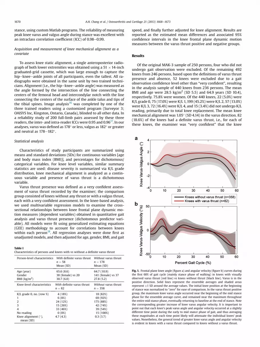
Fig. 1. Frontal plane knee angle (figure a) and angular velocity (figure b) curves duringthe first 60% of gait cycle (mainly stance phase of walking) in knees with visuallyobserved varus thrust (red line) vs knees without thrust (black line). Varus is in thepositive direction. Solid lines represent the ensemble averages and shaded areasrepresent �1 SD around the average values. The initial knee position at the beginningof stance was normalized to “zero” for ease of comparison. In the varus thrust positivegroup, the maximum knee varus angle occurred near the beginning of the mid stancephase for the ensemble average curve, and remained near the maximum throughoutthe entire mid stance phase, eventually returning to baseline at the end of stance. Notethe corresponding greater increase of knee varus angular velocity. It is important topoint out that each knee’s peak varus angle and angular velocity occurred at a slightlydifferent time point during the early to mid stance phase of gait, and thus averagingthese magnitudes at each time point likely will attenuate the individual knees’ peakvalues. Nonetheless, the general trend of greater knee varus angle and angular velocityis evident in knees with a varus thrust compared to knees without a varus thrust.

Table IIMeans (SDs) of knee frontal plane quantitative motion measures during gait byvarus thrust status
Measures of frontal planemotion during gait
All kneesn ¼ 440Mean (SD)
Knees withvarus thrustn ¼ 82Mean (SD)
Knees withoutvarus thrustn ¼ 358Mean (SD)
Peak knee varus angle duringstance (�)
1.77 (1.28) 2.51 (1.55) 1.60 (1.15)
Peak knee varus angle duringearly stance (�)
0.72 (0.98) 1.55 (1.29) 0.53 (0.79)
Peak knee varus angle duringmid-stance (�)
1.32 (1.50) 2.27 (1.64) 1.10 (1.38)
Peak knee varus angle duringterminal stance (�)
1.42 (1.22) 2.03 (1.47) 1.28 (1.11)
Peak knee varus angle duringpre-swing (�)
�0.16 (1.51) 0.66 (1.67) �0.35 (1.40)
Peak knee varus angularvelocity (�/s)
26.81 (15.24) 32.23 (14.23) 25.57 (15.22)
Knee frontal plane quantitative measures are presented in all knees, in knees withvarus thrust only, and in knees without varus thrust.
A.H. Chang et al. / Osteoarthritis and Cartilage 21 (2013) 1668e1673 1671
demonstrated a varus thrust. Twenty-four persons had bilateraland 34 persons had unilateral varus thrust observed during gait.Twelve knees (2.7%) had a definite valgus thrust. Table I showsperson-level and knee-level characteristics of those with andwithout a varus thrust.
Fig. 1 shows the ensemble average curves for the knee varus-valgus angle [Fig. 1(a)] and angular velocity [Fig. 1(b)] during thestance phase of gait cycle for knees with positive varus thrust vsknees without a varus thrust. Table II summarizes the results ofeach knee frontal plane quantitative motion measure overall, andstratified by varus thrust status.
As shown in Table III, knees with a definite varus thrust had astatistically significantly greater peak knee varus angle during theentire stance and each sub-phase as well as greater peak knee varusangular velocity. These patterns remained significant after adjust-ing for age, gender, BMI, and gait speed. Findings persisted afterfurther adjustment for knee alignment, although with somereduction of the mean difference magnitudes in some instances.Notably, the association between varus thrust and greater kneevarus angular velocity was not affected by further adjustment forknee alignment. During ambulation, knees with varus thrust onaverage had 7� per second faster varus angular velocity than didthose without varus thrust.
Among 440 knees in our analysis sample, 82 (18.6%) had varusthrust with a “very confident” rating and 22 knees (5%) had a varusthrust with a “somewhat confident” rating. A sensitivity analysisusing an alternative definition of varus thrust presence duringwalking by including the additional 22 knees yielded similarresults.
Table IIIEstimated differences in means for knee frontal plane motionmeasures based on presence
Measures of frontal plane motion during gait Difference* (95% confidence in
Unadjusted Adjusgende
Peak knee varus angle during stance (�) 0.90 (0.51, 1.30) 0.95 (Peak knee varus angle during early stance (�) 1.02 (0.68, 1.35) 1.03 (Peak knee varus angle during mid-stance (�) 1.16 (0.72, 1.59) 1.29 (Peak knee varus angle during terminal stance (�) 0.74 (0.35, 1.13) 0.81 (Peak knee varus angle during pre-swing (�) 0.99 (0.55, 1.44) 0.92 (Peak knee varus angular velocity (�/s) 6.65 (2.99, 10.31) 6.76 (
95% CI that excludes 0 indicates a statistically significant difference between the groups* Positive difference indicates greater mean value for knees with varus thrust vs those
Discussion
Varus thrust presence visualized during gait observation wasassociated with a greater mean peak knee varus angular velocityduring stance and a greater mean peak knee varus angle duringstance and during most sub-phases of stance measured by 3-Dmotion analysis. Most of the differences remained statisticallysignificant after adjusting for age, gender, BMI, gait speed, and staticknee alignment. For example, knees with a varus thrust had anapproximately 7� per second greater mean varus angular velocitythan knees without a varus thrust, in both unadjusted and adjustedanalyses. Compared to peak knee varus angle, the peak knee varusangular velocity more closely corresponded to a visualized varusthrust and may be a more suitable index for capturing the dynamicnature of a varus thrust.
This is the first study to investigate whether varus thrust pres-ence determined by visual observation corresponds to a greaterknee varus motion measured in quantitative gait analysis. Frontalplane knee angle during ambulation in persons with knee OA hasbeen examined in a limited scope. A few studies explored the cross-sectional relationship between peak knee varus angle and severalneuro-mechanical parameters, including muscle strength, jointlaxity, proprioceptive acuity, knee static alignment, and externalknee adduction moment19e21. In a single case study, Hunt andcolleagues demonstrated that the index knee with an observedvarus thrust had a greater peak knee varus angle than the contra-lateral knee without a thrust; and that the peak knee varus anglecould be reduced by various gait modification strategies6.
In the current study, both peak knee varus angle and angularvelocity were included as indicators of knee frontal plane motion.The knee peak varus angle has been used to quantify frontal planejoint instability6,19,21, however the peak joint varus angle only re-veals the position of the knee at one instant of time and does notfully capture the direction and speed of the dynamic movementseen in a varus thrust. The knee varus angular velocity, on the otherhand, embodies not only direction but also speed of motion. Ouranalysis showed that the mean difference in peak knee varus anglebetween knees with and without a varus thrust was reduced afterfurther adjusting for static knee alignment, but remained signifi-cantly different from zero. This makes sense in that greater varusalignment may contribute to a greater peak varus angle duringwalking. Since varus angular velocity represents speed of move-ment and is less closely related to knee position, it was not sur-prising that the difference in angular velocity between knees withand without varus thrust was not explained by static varus align-ment. Compared with knees without a thrust, having a varus thrustvisualized during gait corresponded to a 7� per second faster meanknee frontal plane movement into greater varus alignment.
The mean peak knee varus angle during stance phase for theentire study sample was 1.77 (SD 1.28) degrees, which falls within
vs absence of varus thrust, from themultiple regression models fit via GEE (n¼ 440)
terval)
ted for age,r, and BMI
Adjusted for age, gender,BMI, and gait speed
Adjusted for age, gender, BMI,gait speed, and alignment
0.55, 1.36) 0.96 (0.56, 1.36) 0.49 (0.13, 0.85)0.69, 1.37) 1.04 (0.70, 1.38) 0.58 (0.29, 0.87)0.83, 1.75) 1.28 (0.82, 1.75) 0.56 (0.19, 0.94)0.40, 1.21) 0.81 (0.41, 1.21) 0.27 (�0.06, 0.61)0.45, 1.38) 0.91 (0.45, 1.38) 0.59 (0.12, 1.05)2.79, 10.73) 7.20 (3.56, 10.84) 6.78 (2.55, 11.01)
, and is shown in bold font.without varus thrust.

A.H. Chang et al. / Osteoarthritis and Cartilage 21 (2013) 1668e16731672
the range of 1.37e4.51� found in other studies19e21. On average, thepeak knee varus angle occurred at 25% time of stance phase, inagreement with the 20e32% time reported in previousresearch6,20,21. It is important to note that the timing of peak varusangle corresponds with the beginning of mid stance phase wherethe limb starts to bear full body weight and thrust is most visible.Our cohort had a mean peak knee varus angular velocity of 26.81(SD 15.24) degrees per second, similar to 21.7 reported in the onlyprevious study measuring the knee frontal plane velocity in 17participants with knee OA20.
Varus thrust during walking was present in 17e36% of threeseparate cohorts of persons with knee OA5,8,22. Biomechanically, anabrupt elevation of medial knee load imparted by a varus thrustwith each step may cause cartilage degradation, bone marrow le-sions, and meniscal damage. This cyclical dynamic impact duringthe most common weight-bearing activity, walking, could be moredetrimental than static varus malalignment assessed in standing.Among varus-aligned knees, thrust increased the likelihood ofmedial disease progression three-fold5, indicating the thrust had anadded influence on medio-lateral compartment load balance thanvarus malalignment alone. In addition, varus thrust had a strongerassociation with greater overall knee pain, particularly duringweight-bearing activities, than did varus malalignment8. Comparedwith full-limb radiographs for capturing knee mechanical align-ment or quantitative gait analysis performed in a lab with costlyequipment for measuring knee frontal plane dynamic instability,thrust observation during gait is a simple clinical tool accessible tomost clinicians. The findings of this study provide evidence thatvarus thrust observed duringwalking is associatedwith an elevatedknee varus angle and angular velocity during the early stance phaseof gait, where the limb accepts the full bodyweight, supporting gaitobservation for varus thrust.
Our hypotheses dealt with the relationship between observedvarus thrust and quantitative knee frontal plane dynamic motionduring gait in both knees of persons with knee OA in one or bothknees. Having radiographic OA in one knee is associated with asubstantially higher risk of having OA in the contralateral knee.There is evidence that many such knees have pre-radiographic OAlesions. To enable generalizing results to persons with OA, weincluded both knees of participants including the full spectrum ofdisease severity.
There are several limitations in this study. Even as performed bya trained and experienced examiner, marker placements in quan-titative gait analysis can be a potential source of variability in thefrontal plane knee motion measures. Despite this potential sourceof variability, we were able to detect significant differences be-tween knees with and without a varus thrust. Gait observation is asubjective assessment. Therefore, as our primary approach, weincluded only the “very confident” rating recorded by the examinerfor thrust presence vs absence. A further sensitivity analysis thatincluded both the “very confident” and “somewhat confident”ratings yielded similar results, suggesting a robust relationshipbetween having a varus thrust and increased knee varus movementquantified by motion analysis. We reported good inter-rater reli-ability of thrust observation, but do not have data on inter-raterreliability. Assignment of examiners to participants was based onschedules and availability of examiners, and we do not anticipateany biases due to this assignment process. Given the expertise ofthe examiners and the scheduled quality control measures, weexpect that the inter-rater reliability would be high. If not, thissource of measurement variability would tend to bias our com-parisons toward the null hypothesis.
In conclusion, varus thrust presence by clinical gait observationwas associated with a greater peak knee varus angular velocity,which captures both the direction and speed of dynamic
movement, and a greater peak knee varus angle during stancerecorded by a 3-D motion analysis system.
Author contributions
Each author participated in drafting the article or revising itcritically for important intellectual content, and all authorsapproved the final version to be published. All authors have accessto the data in the study and can take responsibility for the integrityof the data and the accuracy of the data analysis.
Study conception and design. Chang, Chmiel, SharmaAcquisition of data. Chang, Moisio, Zhang, Cahue, SharmaAnalysis and interpretation of data. Chang, Chmiel, Moisio,
Almagor, Zhang, Cahue, Sharma
Funding sourcesSupport for this project comes from National Institutes of Health(NIH) P60 AR048098 (PIs: Pope/Sharma), NIH R01 AR054806 (PI:Sharma), and NIH R01 AR048748 (PI: Sharma).
Competing interestsThe authors declare no competing interests.
Acknowledgments
The authors would like to thank Mr. Clifton Saurel and Ms.Rebecca Stein for assistance in data collection and all study par-ticipants for their contribution to the study.
References
1. Schipplein OD, Andriacchi TP. Interaction between active andpassive knee stabilizers during level walking. J Orthop Res1991;9(1):113e9.
2. Andriacchi TP, Mündermann A, Smith RL, Alexander EJ,Dyrby CO, Koo S. A framework for the in vivo pathomechanicsof osteoarthritis at the knee. Ann Biomed Eng 2004;32(3):447e57.
3. Miyazaki T, Wada M, Kawahara H, Sato M, Baba H, Shimada S.Dynamic load at baseline can predict radiographic diseaseprogression in medial compartment knee osteoarthritis. AnnRheum Dis 2002;61(7):617e22.
4. Bennell KL, Bowles K-A, Wang Y, Cicuttini F, Davies-Tuck M,Hinman RS. Higher dynamic medial knee load predicts greatercartilage loss over 12 months in medial knee osteoarthritis.Ann Rheum Dis 2011;70(10):1770e4.
5. Chang A, Hayes K, Dunlop D, Hurwitz D, Song J, Cahue S, et al.Thrust during ambulation and the progression of knee osteo-arthritis. Arthritis Rheum 2004;50(12):3897e903.
6. Hunt MA, Schache AG, Hinman RS, Crossley KM. Varus thrustin medial knee osteoarthritis: quantification and effects ofdifferent gait-related interventions using a single case study.Arthritis Care Res (Hoboken) 2011;63(2):293e7.
7. Williams DS, Isom W. Decreased frontal plane hip joint mo-ments in runners with excessive varus excursion at the knee.J Appl Biomech 2012;28(2):120e6.
8. Lo GH, Harvey WF, McAlindon TE. Associations of varus thrustand alignment with pain in knee osteoarthritis. ArthritisRheum 2012;64(7):2252e9.
9. Winter DA. Kinematics. In: Biomechanics and Motor Control ofHuman Movement. 4nd edn. Wiley; 2009:45e81.
10. Kellgren JH, Lawrence JS. Radiological assessment of osteo-arthrosis. Ann Rheum Dis 1957;16(4):494e502.
11. Bellamy N, Buchanan WW, Goldsmith CH, Campbell J, Stitt LW.Validation study of WOMAC: a health status instrument for

A.H. Chang et al. / Osteoarthritis and Cartilage 21 (2013) 1668e1673 1673
measuring clinically important patient relevant outcomes toantirheumatic drug therapy in patients with osteoarthritis ofthe hip or knee. J Rheumatol 1988;15(12):1833e40.
12. Buckland-Wright C. Protocols for precise radio-anatomicalpositioning of the tibiofemoral and patellofemoral compart-ments of the knee. Osteoarthritis Cartilage 1995;3(Suppl A):71e80.
13. Perry J, Burnfield JM. Motion Analysis. In: Gait Analysis:Normal and Pathological Function. 2nd edn. Slack Incorpo-rated; 2010:407e22.
14. Thorp LE, Wimmer MA, Block JA, Moisio KC, Shott S, Goker B,et al. Bone mineral density in the proximal tibia varies as afunction of static alignment and knee adduction angular mo-mentum in individuals with medial knee osteoarthritis. Bone2006;39(5):1116e22.
15. Perry J, Burnfield JM. Phases of Gait. In: Gait Analysis: Normal andPathological Function. 2nd edn. Slack Incorporated; 2010:9e16.
16. Cooke TDV, Sled EA, Scudamore RA. Frontal plane kneealignment: a call for standardized measurement. J Rheumatol2007;34(9):1796e801.
17. Sled EA, Sheehy LM, Felson DT, Costigan PA, Lam M,Cooke TDV. Reliability of lower limb alignment measures using
an established landmark-based method with a customizedcomputer software program. Rheumatol Int 2011;31(1):71e7.
18. Zeger SL, Liang KY, Albert PS. Models for longitudinal data: ageneralized estimating equation approach. Biometrics1988;44(4):1049e60.
19. Van der Esch M, Steultjens M, Harlaar J, Wolterbeek N,Knol DL, Dekker J. Knee varus-valgus motion during gaiteameasure of joint stability in patients with osteoarthritis?Osteoarthritis Cartilage 2008;16(4):522e5.
20. Foroughi N, Smith RM, Lange AK, Baker MK, FiataroneSingh MA, Vanwanseele B. Dynamic alignment and its asso-ciation with knee adduction moment in medial knee osteo-arthritis. Knee 2010;17(3):210e6.
21. Kuroyanagi Y, Nagura T, Kiriyama Y, Matsumoto H, Otani T,Toyama Y, et al. A quantitative assessment of varus thrust inpatients with medial knee osteoarthritis. Knee 2012;19(2):130e4.
22. Chang A, Hochberg M, Song J, Dunlop D, Chmiel JS, Nevitt M,et al. Frequency of varus and valgus thrust and factors asso-ciated with thrust presence in persons with or at higher risk ofdeveloping knee osteoarthritis. Arthritis Rheum 2010;62(5):1403e11.
Recommended


