Embed Size (px)
Citation preview
Hindawi Publishing CorporationCase Reports in DentistryVolume 2013, Article ID 812323, 4 pageshttp://dx.doi.org/10.1155/2013/812323
Case ReportSolitary Angiokeratoma of Oral Mucosa: A Rare Presentation
Shilpa Kandalgaonkar, Suyog Tupsakhare, Ashok Patil, Gaurav Agrawal,Mahesh Gabhane, and Shrikant Sonune
Department Oral Pathology & Microbiology, SMBT Dental College & Hospital, Sangamner,Maharashtra 422608, India
Correspondence should be addressed to Gaurav Agrawal; [email protected]
Received 20 May 2013; Accepted 24 June 2013
Academic Editors: R. S. Brown and A. C. B. Delbem
Copyright © 2013 Shilpa Kandalgaonkar et al. This is an open access article distributed under the Creative Commons AttributionLicense, which permits unrestricted use, distribution, and reproduction in any medium, provided the original work is properlycited.
Solitary angiokeratoma of oral mucosa is rare entity. The term Angiokeratoma is used to refer to several lesions, whose commondenominator is the presence of dilated blood vessels in association with epidermal hyperplasia. Mucosal involvement, includingoral cavity is occasionally found either as a component of the systemic variety, cutaneous involvement or isolated oral involvement.Clinically, the lesion is irregular, whitish to dark brown in color, with female predominance. The etiological factors include injury,trauma, or chronic irritation to the wall of a papillary dermis. Histologically, it is characterized by hyperkeratosis, acanthosis,and dilated vascular spaces with or without organizing thrombi in papillary dermis. The vascular spaces are partly or completelyenclosed by elongated ret-ridges. Along with this reporting a case of solitary angiokeratoma affecting tongue in a 38-year-old malepatient, along with the literature review is presented.
1. Introduction
Angiokeratoma is an acquired vascular lesion which is char-acterized histologically as one or more dilated blood vesselslying directly subepidermally and showing an epidermal pro-liferative reaction especially acanthosis and hyperkeratosiswith dilated capillaries in the papillary dermis [1].
Several clinical types have been described dependingon the multiplicity and location of the lesions. They canbe divided into localized and systemic types [2]. Mucosalinvolvement, including the oral cavity, has been describedboth as localized and systemic types, as a component ofFabry’s disease, or as a component of fucosidosis [2–5].To classify isolated oral mucosal angiokeratomas, otherclassification systems have been proposed by Ranjan andMahajan [6].
However, solitary angiokeratomas of the oral mucosaseem to be a rather infrequent occurrence, and very few caseshave been reported in the literature. According to the best ofour knowledge, since 1997 till date, only 16 cases involvingoral cavity have been reported.
2. Case Report
A 38-year-old male patient reported to the Department ofOral Pathology with chief complaint of growth on tip oftongue since last 10 years. The patient was apparently allright 10 years ago when he noticed small painless growth,and then the growth steadily increased in size up to presentsize involving right side of the tip of the tongue. Sometimesthe Patient also experienced bleeding in that area which wasassociated with trauma duringmastication. Bleeding subside,on its own, the patient had never taken any treatment for thatgrowth and not even for bleeding episodes. No abnormalitywas reveled in his medical, personal histories and generalexamination. The patient had a habit of tobacco chewing forabout 10–15 years.
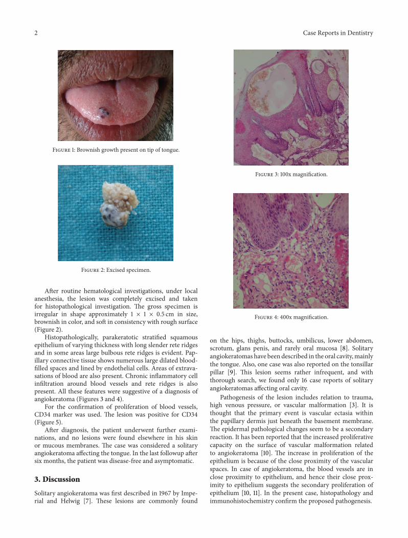
On clinical examination, it was observed that well-cir-cumscribed sessile growth is present on the dorsal surfaceof tip of the tongue, and the growth is of approximately1 × 1 cm in dimension, ovoid in shape. Growth was darkbrownish in color with a granular surface texture (Figure 1).On palpation, growth was non tender and rough. No otherintra- or extraoral lesions are present.
2 Case Reports in Dentistry
Figure 1: Brownish growth present on tip of tongue.
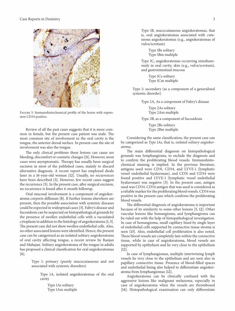
Figure 2: Excised specimen.
After routine hematological investigations, under localanesthesia, the lesion was completely excised and takenfor histopathological investigation. The gross specimen isirregular in shape approximately 1 × 1 × 0.5 cm in size,brownish in color, and soft in consistency with rough surface(Figure 2).
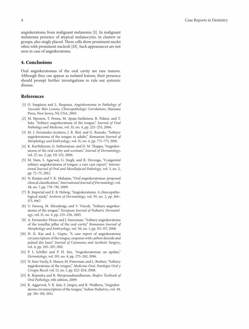
Histopathologically, parakeratotic stratified squamousepithelium of varying thickness with long slender rete ridgesand in some areas large bulbous rete ridges is evident. Pap-illary connective tissue shows numerous large dilated blood-filled spaces and lined by endothelial cells. Areas of extrava-sations of blood are also present. Chronic inflammatory cellinfiltration around blood vessels and rete ridges is alsopresent. All these features were suggestive of a diagnosis ofangiokeratoma (Figures 3 and 4).
For the confirmation of proliferation of blood vessels,CD34 marker was used. The lesion was positive for CD34(Figure 5).
After diagnosis, the patient underwent further exami-nations, and no lesions were found elsewhere in his skinor mucous membranes. The case was considered a solitaryangiokeratoma affecting the tongue. In the last followup aftersix months, the patient was disease-free and asymptomatic.
3. Discussion
Solitary angiokeratoma was first described in 1967 by Impe-rial and Helwig [7]. These lesions are commonly found
Figure 3: 100x magnification.
Figure 4: 400x magnification.
on the hips, thighs, buttocks, umbilicus, lower abdomen,scrotum, glans penis, and rarely oral mucosa [8]. Solitaryangiokeratomas have been described in the oral cavity,mainlythe tongue. Also, one case was also reported on the tonsillarpillar [9]. This lesion seems rather infrequent, and withthorough search, we found only 16 case reports of solitaryangiokeratomas affecting oral cavity.
Pathogenesis of the lesion includes relation to trauma,high venous pressure, or vascular malformation [3]. It isthought that the primary event is vascular ectasia withinthe papillary dermis just beneath the basement membrane.The epidermal pathological changes seem to be a secondaryreaction. It has been reported that the increased proliferativecapacity on the surface of vascular malformation relatedto angiokeratoma [10]. The increase in proliferation of theepithelium is because of the close proximity of the vascularspaces. In case of angiokeratoma, the blood vessels are inclose proximity to epithelium, and hence their close prox-imity to epithelium suggests the secondary proliferation ofepithelium [10, 11]. In the present case, histopathology andimmunohistochemistry confirm the proposed pathogenesis.
Case Reports in Dentistry 3
Figure 5: Immunohistochemical profile of the lesion with expres-sion CD34 positive.
Review of all the past cases suggests that it is more com-mon in female, but the present case patient was male. Themost common site of involvement in the oral cavity is thetongue, the anterior dorsal surface. In present case the site ofinvolvement was also the tongue.
The only clinical problems these lesions can cause arebleeding, discomfort or cosmetic changes [11]. However,mostcases were asymptomatic. Therapy has usually been surgicalexcision in most of the published cases, mainly to discardalternative diagnosis. A recent report has employed diodelaser in a 16-year-old woman [12]. Usually, no recurrenceshave been described [3]. However, few recent cases suggestthe recurrence [5]. In the present case, after surgical excision,no recurrence is found after 6-month followup.
Oral mucosal involvement is a component of angioker-atoma corporis diffusum [8]. If further lesions elsewhere arepresent, then the possible association with systemic diseasescould be expected inwidespread cases [3]. Fabry’s disease andfucosidosis can be suspected on histopathological grounds bythe presence of swollen endothelial cells with a vacuolatedcytoplasm in addition to the histology of angiokeratoma [1, 3].The present case did not show swollen endothelial cells. Also,no other associated lesionswere identified.Hence, the presentcase can be categorized as an isolated solitary angiokeratomaof oral cavity affecting tongue, a recent review by Ranjanand Mahajan. Solitary angiokeratoma of the tongue in adultshas proposed a clinical classification for oral angiokeratomas[6].
Type 1: primary (purely mucocutaneous and notassociated with systemic disorders)
Type 1A, isolated angiokeratomas of the oralcavity
Type 1As solitaryType 1Am multiple
Type 1B, mucocutaneous angiokeratomas, thatis, oral angiokeratomas associated with cuta-neous angiokeratomas (e.g., angiokeratomas ofvulva/scrotum)
Type 1Bs solitaryType 1Bm multiple
Type 1C, angiokeratomas occurring simultane-ously in oral cavity, skin (e.g., vulva/scrotum),and gastrointestinal mucosa
Type 1Cs solitaryType 1Cm multiple
Type 2: secondary (as a component of a generalizedsystemic disorder)
Type 2A, As a component of Fabry’s diseaseType 2As solitaryType 2Ammultiple
Type 2B, as a component of fucosidosisType 2Bs solitaryType 2Bm multiple
Considering the same classification, the present case canbe categorized as Type 1As, that is, isolated solitary angioker-atoma.
The main differential diagnosis on histopathologicalgrounds was lymphangioma, to exclude the diagnosis andto confirm the proliferating blood vessels. Immunohisto-chemical staining is implied. In the previous literature,antigens used were CD31, CD34, and LYVE-1 (lymphaticvessel endothelial hyaluronan), and CD31 and CD34 werefound positive and LYVE-1 (lymphatic vessel endothelialhyaluronan) was negative [3]. In the present case, antigenused was CD34. CD34 antigen that was used is considered asa reliablemarker for the proliferating blood vessels. CD34waspositive in the present case which confirms the proliferatingblood vessels.
The differential diagnosis of angiokeratoma is importantbecause of its similarity to some other lesions [5, 12]. Othervascular lesions like hemangioma, and lymphangioma canbe ruled out with the help of histopathological investigation.In case of hemangioma, small capillary lined by single layerof endothelial cells supported by connective tissue stroma isseen [13]. Also, endothelial cell proliferation is also noted.These blood vessels are completely lain within the connectivetissue, while in case of angiokeratoma, blood vessels aresupported by epithelium and lie very close to the epithelium[12].
In case of lymphangiomas, multiple intertwining lymphvessels lie very close to the epithelium and are seen also inpapillary connective tissue. Presence of blood-filled spacesand endothelial lining also helped to differentiate angioker-atoma from lymphangiomas [12].
Angiokeratoma can be clinically confused with theaggressive lesions like malignant melanoma, especially incase of angiokeratoma when the vessels are thrombosed[14]. Histopathological examination can only differentiate
4 Case Reports in Dentistry
angiokeratoma from malignant melanoma [1]. In malignantmelanoma presence of atypical melanocytes, in clusters orgroups, also singly placed.These cells show prominent nucleioften with prominent nucleoli [13]. Such appearances are notseen in case of angiokeratoma.
4. Conclusions
Oral angiokeratomas of the oral cavity are rare tumors.Although they can appear as isolated lesions, their presenceshould prompt further investigations to rule out systemicdisease.
References
[1] O. Sangueza and L. Requena, Angiokeratoma in Pathology ofVascular Skin Lesions, Clinicopathologic Correlations, HumanaPress, New Jersey, NJ, USA, 2003.
[2] M. Siponen, T. Penna, M. Apaja-Sarkkinen, R. Palatsi, and T.Salo, “Solitary angiokeratoma of the tongue,” Journal of OralPathology and Medicine, vol. 35, no. 4, pp. 252–253, 2006.
[3] M. J. Fernandez-Acenero, J. R. Biel, and G. Renedo, “Solitaryangiokeratoma of the tongue in adults,” Romanian Journal ofMorphology and Embryology, vol. 51, no. 4, pp. 771–773, 2010.
[4] K. Karthikeyan, G. Sethuraman, and D. M.Thappa, “Angioker-atoma of the oral cavity and scrotum,” Journal of Dermatology,vol. 27, no. 2, pp. 131–132, 2000.
[5] M. Nain, S. Agarwal, G. Singh, and R. Devenga, “Congenitalsolitary angiokeratoma of tongue: a rare case report,” Interna-tional Journal of Oral and Maxillofacial Pathology, vol. 3, no. 2,pp. 72–75, 2012.
[6] N. Ranjan and V. K. Mahajan, “Oral angiokeratomas: proposedclinical classification,” International Journal of Dermatology, vol.48, no. 7, pp. 778–781, 2009.
[7] R. Imperial and E. B. Helwig, “Angiokeratoma. A clinicopatho-logical study,” Archives of Dermatology, vol. 95, no. 2, pp. 166–175, 1967.
[8] U. Farooq, M. Mirzabeigi, and V. Vincek, “Solitary angioker-atoma of the tongue,” European Journal of Pediatric Dermatol-ogy, vol. 15, no. 4, pp. 233–236, 2005.
[9] A. Fernandez-Flores and J. Sanroman, “Solitary angiokeratomaof the tonsillar pillar of the oral cavity,” Romanian Journal ofMorphology and Embryology, vol. 50, no. 1, pp. 115–117, 2008.
[10] H. K. Kar and L. Gupta, “A case report of angiokeratomacircumcriptumof the tongue, responsewith carbon dioxide andpulsed dye laser,” Journal of Cutaneous and Aesthetic Surgery,vol. 4, pp. 205–207, 2011.
[11] P. I. Schiller and P. H. Itin, “Angiokeratomas: an update,”Dermatology, vol. 193, no. 4, pp. 275–282, 1996.
[12] N. Sion-Vardy, E.Manor,M. Puterman, and L. Bodner, “Solitaryangiokeratoma of the tongue,” Medicina Oral, Patologia Oral yCirugia Bucal, vol. 13, no. 1, pp. E12–E14, 2008.
[13] R. Rajendra and B. Shivprasadsundharam, Shafers Textbook ofOral Pathology, 6th edition, 2009.
[14] K. Aggarwal, V. K. Jain, S. Jangra, and R. Wadhera, “Angioker-atoma circumscriptum of the tongue,” Indian Pediatrics, vol. 49,pp. 316–318, 2012.

Submit your manuscripts athttp://www.hindawi.com
Hindawi Publishing Corporationhttp://www.hindawi.com Volume 2013
Oral DiseasesJournal of
DentistryInternational Journal of
Hindawi Publishing Corporationhttp://www.hindawi.com Volume 2013
ISRN Dentistry
Hindawi Publishing Corporationhttp://www.hindawi.com Volume 2013
Hindawi Publishing Corporationhttp://www.hindawi.com Volume 2013
Oral ImplantsJournal of
Hindawi Publishing Corporationhttp://www.hindawi.com Volume 2013
Case Reports in Dentistry
Hindawi Publishing Corporation http://www.hindawi.com Volume 2013Hindawi Publishing Corporation http://www.hindawi.com Volume 2013
The Scientific World Journal
Hindawi Publishing Corporationhttp://www.hindawi.com Volume 2013
Dental SurgeryJournal of
BioMed Research International
Hindawi Publishing Corporationhttp://www.hindawi.com Volume 2013
ScientificaHindawi Publishing Corporationhttp://www.hindawi.com Volume 2013
Hindawi Publishing Corporationhttp://www.hindawi.com Volume 2013
Journal of
Drug Delivery
International Journal of
BiomaterialsHindawi Publishing Corporationhttp://www.hindawi.com Volume 2013
Hindawi Publishing Corporationhttp://www.hindawi.com Volume 2013
Computational and Mathematical Methods in Medicine
Hindawi Publishing Corporationhttp://www.hindawi.com Volume 2013
Oral OncologyJournal of
Hindawi Publishing Corporationhttp://www.hindawi.com Volume 2013
OrthopedicsAdvances in
Hindawi Publishing Corporationhttp://www.hindawi.com Volume 2013
Anesthesiology Researchand Practice
Hindawi Publishing Corporationhttp://www.hindawi.com Volume 2013
Environmental andPublic Health
Journal of
Preventive MedicineAdvances in
Hindawi Publishing Corporation http://www.hindawi.com Volume 2013
International Journal of
EndocrinologyHindawi Publishing Corporationhttp://www.hindawi.com
Volume 2013
Hindawi Publishing Corporationhttp://www.hindawi.com Volume 2013
Radiology Research and Practice


![1 1 1 1 1 1 1 ¢ 1 , ¢ 1 1 1 , 1 1 1 1 ¡ 1 1 1 1 · 1 1 1 1 1 ] ð 1 1 w ï 1 x v w ^ 1 1 x w [ ^ \ w _ [ 1. 1 1 1 1 1 1 1 1 1 1 1 1 1 1 1 1 1 1 1 1 1 1 1 1 1 1 1 ð 1 ] û w ü](https://img.pdfslide.net/doc/110x75/5f40ff1754b8c6159c151d05/1-1-1-1-1-1-1-1-1-1-1-1-1-1-1-1-1-1-1-1-1-1-1-1-1-1-w-1-x-v.jpg)


![Case Report Giant Verrucous Haemangioma with Linear ...angiokeratoma, angioma serpiginosum, lymphangioama and pigmented tumours.[7] Recurrent bleeding and infection along with increase](https://img.pdfslide.net/doc/110x75/5e3b758d6f248601c355512e/case-report-giant-verrucous-haemangioma-with-linear-angiokeratoma-angioma-serpiginosum.jpg)

![1 ¢ Ù 1 £¢ 1 £ £¢ 1 - Narodowy Bank Polski · 1 à 1 1 1 1 \ 1 1 1 1 ¢ 1 1 £ 1 £ £¢ 1 ¢ 1 ¢ Ù 1 à 1 1 1 ¢ à 1 1 £ ï 1 1. £¿ï° 1 ¢ 1 £ 1 1 1 1 ] 1 1 1 1 ¢](https://img.pdfslide.net/doc/110x75/5fc6757af26c7e63a70a621e/1-1-1-1-narodowy-bank-polski-1-1-1-1-1-1-1-1-1-1-1.jpg)


![[XLS]fmism.univ-guelma.dzfmism.univ-guelma.dz/sites/default/files/le fond... · Web view1 1 1 1 1 1 1 1 1 1 1 1 1 1 1 1 1 1 1 1 1 1 1 1 1 1 1 1 1 1 1 1 1 1 1 1 1 1 1 1 1 1 1 1 1 1](https://img.pdfslide.net/doc/110x75/5b9d17e509d3f2194e8d827e/xlsfmismuniv-fond-web-view1-1-1-1-1-1-1-1-1-1-1-1-1-1-1-1-1-1-1-1-1-1.jpg)